1 Vol.:(0123456789) Scientific Reports | (2022) 12:6871 | https://doi.org/10.1038/s41598-022-10799-4 www.nature.com/scientificreports Characterization of Klebsiella pneumoniae isolated from patients suspected of pulmonary or bubonic plague during the Madagascar epidemic in 2017 Andriniaina Rakotondrasoa 1* , Lova Maminirina Andrianonimiadana 1 , Soloandry Rahajandraibe 2 , Solohery Razafimahatratra 1 , Voahangy Andrianaivoarimanana 2 , Soanandrasana Rahelinirina 2 , Tania Crucitti 1 , Sylvain Brisse 3 , Victor Jeannoda 4 , Minoarisoa Rajerison 2,6 & Jean‑Marc Collard 1,5,6 Klebsiella pneumoniae can lead to a wide range of diseases including pneumonia, bloodstream and urinary tract infections. During a short period of a pulmonary plague epidemic in October 2017 in Madagascar, 12 K. pneumoniae isolates were identified in ten sputum and two buboes aspirate samples. These isolates were from 12 patients suspected of plague, without epidemiological relationships, but were negative for Yersinia pestis in culture. Data were collected from the plague national surveillance system. The isolates were characterized by antimicrobial susceptibility testing and whole‑genome sequencing. Real‑time PCR was performed to confirm the presence of K. pneumoniae DNA in buboes. All isolates were identified as K. pneumoniae sensu stricto. Five isolates were extended‑spectrum β‑lactamases producers; eleven different sequence types were identified. Five isolates belonged to known hypervirulent sequence types. Our results demonstrate community‑ acquired pneumonia caused by K. pneumoniae isolates in patients suspected of plague stressing the importance of bed‑side differential diagnosis. Between August 1st and November 26th, 2017, a total of 2414 clinically suspected plague cases were reported to the Central Laboratory for Plague (CLP) at the Institut Pasteur de Madagascar, including 1878 (78%) pulmonary plague (PP), 395 (16%) bubonic plague (BP), one (< 1%) septicaemia and 140 (6%) cases with unspecified clinical form 1 . is predominantly urban plague epidemic was characterised by a large volume of notifications in two major urban areas (Antananarivo and Toamasina) and by an unusually high proportion of pneumonic forms. According to the 2006 WHO standard plague case definitions and using the results of three types of diagnostic tests assessed (rapid F1-antigen diagnostic test, (RDT), molecular amplification method, and culture) 2 , 386/1,878 (21%) were probable and 32/1,878 (2%) were confirmed cases among the notified PP cases. e magnitude of this PP outbreak is likely to have been smaller than suggested by notified suspected cases 1 ; and its severity indicated by the case fatality rate among confirmed plus probable cases (about 9%) was substantially lower than observed in the last previous 18 years (25%) 3 . Over-reporting of PP cases due to limited clinical experience in the two most affected areas, and the difficulty to clinically diagnose PP through respiratory signs was speculated. e clinical diagnosis of PP from polymicrobial sputum associated with other potential causes of pneumonia remains a challenge because the isolation of Yersinia pestis (Y. pestis) is more complicated compared to other bacteria. OPEN 1 Experimental Bacteriology Unit, Institut Pasteur Madagascar, Antananarivo, Madagascar. 2 Plague Unit, Institut Pasteur Madagascar, Antananarivo, Madagascar. 3 Biodiversity and Epidemiology of Bacterial Pathogens, Institut Pasteur, Paris, France. 4 Mention Biodiversité Et Santé, Sciences Faculty, University of Antananarivo, Antananarivo, Madagascar. 5 Present address: Experimental Bacteriology Laboratory, Center for Microbes, Development and Health (CMDH), Institut Pasteur of Shanghai/Chinese Academy of Sciences, Shanghai, People’s Republic of China. 6 These authors contributed equally: Minoarisoa Rajerison and Jean-Marc Collard. * email: [email protected]

Welcome message from author
This document is posted to help you gain knowledge. Please leave a comment to let me know what you think about it! Share it to your friends and learn new things together.
Transcript

1
Vol.:(0123456789)
Scientific Reports | (2022) 12:6871 | https://doi.org/10.1038/s41598-022-10799-4
www.nature.com/scientificreports
Characterization of Klebsiella pneumoniae isolated from patients suspected of pulmonary or bubonic plague during the Madagascar epidemic in 2017Andriniaina Rakotondrasoa1*, Lova Maminirina Andrianonimiadana1, Soloandry Rahajandraibe2, Solohery Razafimahatratra1, Voahangy Andrianaivoarimanana2, Soanandrasana Rahelinirina2, Tania Crucitti1, Sylvain Brisse3, Victor Jeannoda4, Minoarisoa Rajerison2,6 & Jean‑Marc Collard1,5,6
Klebsiella pneumoniae can lead to a wide range of diseases including pneumonia, bloodstream and urinary tract infections. During a short period of a pulmonary plague epidemic in October 2017 in Madagascar, 12 K. pneumoniae isolates were identified in ten sputum and two buboes aspirate samples. These isolates were from 12 patients suspected of plague, without epidemiological relationships, but were negative for Yersinia pestis in culture. Data were collected from the plague national surveillance system. The isolates were characterized by antimicrobial susceptibility testing and whole‑genome sequencing. Real‑time PCR was performed to confirm the presence of K. pneumoniae DNA in buboes. All isolates were identified as K. pneumoniae sensu stricto. Five isolates were extended‑spectrum β‑lactamases producers; eleven different sequence types were identified. Five isolates belonged to known hypervirulent sequence types. Our results demonstrate community‑acquired pneumonia caused by K. pneumoniae isolates in patients suspected of plague stressing the importance of bed‑side differential diagnosis.
Between August 1st and November 26th, 2017, a total of 2414 clinically suspected plague cases were reported to the Central Laboratory for Plague (CLP) at the Institut Pasteur de Madagascar, including 1878 (78%) pulmonary plague (PP), 395 (16%) bubonic plague (BP), one (< 1%) septicaemia and 140 (6%) cases with unspecified clinical form1. This predominantly urban plague epidemic was characterised by a large volume of notifications in two major urban areas (Antananarivo and Toamasina) and by an unusually high proportion of pneumonic forms. According to the 2006 WHO standard plague case definitions and using the results of three types of diagnostic tests assessed (rapid F1-antigen diagnostic test, (RDT), molecular amplification method, and culture)2, 386/1,878 (21%) were probable and 32/1,878 (2%) were confirmed cases among the notified PP cases. The magnitude of this PP outbreak is likely to have been smaller than suggested by notified suspected cases1; and its severity indicated by the case fatality rate among confirmed plus probable cases (about 9%) was substantially lower than observed in the last previous 18 years (25%)3. Over-reporting of PP cases due to limited clinical experience in the two most affected areas, and the difficulty to clinically diagnose PP through respiratory signs was speculated. The clinical diagnosis of PP from polymicrobial sputum associated with other potential causes of pneumonia remains a challenge because the isolation of Yersinia pestis (Y. pestis) is more complicated compared to other bacteria.
OPEN
1Experimental Bacteriology Unit, Institut Pasteur Madagascar, Antananarivo, Madagascar. 2Plague Unit, Institut Pasteur Madagascar, Antananarivo, Madagascar. 3Biodiversity and Epidemiology of Bacterial Pathogens, Institut Pasteur, Paris, France. 4Mention Biodiversité Et Santé, Sciences Faculty, University of Antananarivo, Antananarivo, Madagascar. 5Present address: Experimental Bacteriology Laboratory, Center for Microbes, Development and Health (CMDH), Institut Pasteur of Shanghai/Chinese Academy of Sciences, Shanghai, People’s Republic of China. 6These authors contributed equally: Minoarisoa Rajerison and Jean-Marc Collard. *email: [email protected]
2
Vol:.(1234567890)
Scientific Reports | (2022) 12:6871 | https://doi.org/10.1038/s41598-022-10799-4
www.nature.com/scientificreports/
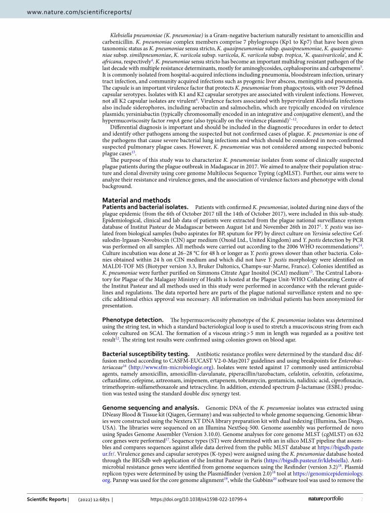
Klebsiella pneumoniae (K. pneumoniae) is a Gram-negative bacterium naturally resistant to amoxicillin and carbenicillin. K. pneumoniae complex members comprise 7 phylogroups (Kp1 to Kp7) that have been given taxonomic status as K. pneumoniae sensu stricto, K. quasipneumoniae subsp. quasipneumoniae, K. quasipneumo-niae subsp. similipneumoniae, K. variicola subsp. variicola, K. variicola subsp. tropica, ‘K. quasivariicola’, and K. africana, respectively4. K. pneumoniae sensu stricto has become an important multidrug resistant pathogen of the last decade with multiple resistance determinants, mostly for aminoglycosides, cephalosporins and carbapenems5. It is commonly isolated from hospital-acquired infections including pneumonia, bloodstream infection, urinary tract infection, and community acquired infections such as pyogenic liver abscess, meningitis and pneumonia. The capsule is an important virulence factor that protects K. pneumoniae from phagocytosis, with over 79 defined capsular serotypes. Isolates with K1 and K2 capsular serotypes are associated with virulent infections. However, not all K2 capsular isolates are virulent6. Virulence factors associated with hypervirulent Klebsiella infections also include siderophores, including aerobactin and salmochelin, which are typically encoded on virulence plasmids; yersiniabactin (typically chromosomally encoded in an integrative and conjugative element), and the hypermucoviscosity factor rmpA gene (also typically on the virulence plasmid)7–12.
Differential diagnosis is important and should be included in the diagnostic procedures in order to detect and identify other pathogens among the suspected but not confirmed cases of plague. K. pneumoniae is one of the pathogens that cause severe bacterial lung infections and which should be considered in non-confirmed suspected pulmonary plague cases. However, K. pneumoniae was not considered among suspected bubonic plague cases13.
The purpose of this study was to characterize K. pneumoniae isolates from some of clinically suspected plague patients during the plague outbreak in Madagascar in 2017. We aimed to analyze their population struc-ture and clonal diversity using core genome Multilocus Sequence Typing (cgMLST). Further, our aims were to analyze their resistance and virulence genes, and the association of virulence factors and phenotype with clonal background.
Material and methodsPatients and bacterial isolates. Patients with confirmed K. pneumoniae, isolated during nine days of the plague epidemic (from the 6th of October 2017 till the 14th of October 2017), were included in this sub-study. Epidemiological, clinical and lab data of patients were extracted from the plague national surveillance system database of Institut Pasteur de Madagascar between August 1st and November 26th in 20171. Y. pestis was iso-lated from biological samples (bubo aspirates for BP, sputum for PP) by direct culture on Yersinia selective Cef-sulodin-Irgasan-Novobiocin (CIN) agar medium (Oxoid Ltd., United Kingdom) and Y. pestis detection by PCR was performed on all samples. All methods were carried out according to the 2006 WHO recommendations14. Culture incubation was done at 26–28 °C for 48 h or longer as Y. pestis grows slower than other bacteria. Colo-nies obtained within 24 h on CIN medium and which did not have Y. pestis morphology were identified on MALDI-TOF MS (Biotyper version 3.3, Bruker Daltonics, Champs-sur-Marne, France). Colonies identified as K. pneumoniae were further purified on Simmons Citrate Agar Inositol (SCAI) medium15. The Central Labora-tory for Plague of the Malagasy Ministry of Health is hosted at the Plague Unit-WHO Collaborating Centre of the Institut Pasteur and all methods used in this study were performed in accordance with the relevant guide-lines and regulations. The data reported here are parts of the plague national surveillance system and no spe-cific additional ethics approval was necessary. All information on individual patients has been anonymized for presentation.
Phenotype detection. The hypermucoviscosity phenotype of the K. pneumoniae isolates was determined using the string test, in which a standard bacteriological loop is used to stretch a mucoviscous string from each colony cultured on SCAI. The formation of a viscous string > 5 mm in length was regarded as a positive test result12. The string test results were confirmed using colonies grown on blood agar.
Bacterial susceptibility testing. Antibiotic resistance profiles were determined by the standard disc dif-fusion method according to CASFM-EUCAST V2-0-May2017 guidelines and using breakpoints for Enterobac-teriaceae16 (http:// www. sfm- micro biolo gie. org). Isolates were tested against 17 commonly used antimicrobial agents, namely amoxicillin, amoxicillin-clavulanate, piperacillin/tazobactam, cefalotin, cefoxitin, cefotaxime, ceftazidime, cefepime, aztreonam, imipenem, ertapenem, tobramycin, gentamicin, nalidixic acid, ciprofloxacin, trimethoprim-sulfamethoxazole and tetracycline. In addition, extended spectrum β-lactamase (ESBL) produc-tion was tested using the standard double disc synergy test.
Genome sequencing and analysis. Genomic DNA of the K. pneumoniae isolates was extracted using DNeasy Blood & Tissue kit (Qiagen, Germany) and was subjected to whole genome sequencing. Genomic librar-ies were constructed using the Nextera XT DNA library preparation kit with dual indexing (Illumina, San Diego, USA). The libraries were sequenced on an Illumina NextSeq-500. Genome assembly was performed de novo using Spades Genome Assembler (Version 3.10.0). Genome analyses for core genome MLST (cgMLST) on 632 core genes were performed17. Sequence types (ST) were determined with an in silico MLST pipeline that assem-bles and compares sequences against allele data derived from the public MLST database at https:// bigsdb. paste ur. fr/. Virulence genes and capsular serotypes (K-types) were assigned using the K. pneumoniae database hosted through the BIGSdb web application of the Institut Pasteur in Paris (https:// bigsdb. paste ur. fr/ klebs iella). Anti-microbial resistance genes were identified from genome sequences using the Resfinder (version 3.2)18. Plasmid replicon types were determined by using the Plasmidfinder (version 2.0)18 tool at https:// genom icepi demio logy. org. Parsnp was used for the core genome alignment19, while the Gubbins20 software tool was used to remove the
3
Vol.:(0123456789)
Scientific Reports | (2022) 12:6871 | https://doi.org/10.1038/s41598-022-10799-4
www.nature.com/scientificreports/
single nucleotide polymorphisms (SNPs) from recombined regions and to create a refined phylogenetic tree; this tool uses RaxML21 to build the maximum likelihood phylogenetic tree on the recombination-free regions. The tree was subsequently annotated with iTOL (http:// itol. embl. de/ itol. cgi)22.
Buboes K. pneumoniae screening by real‑time PCR. We performed a real-time PCR targeting the zur-khe intergenic region (called the ZKIR qPCR assay)23 on the bubo samples in order to confirm the presence of K. pneumoniae DNA in the bubo and to exclude any technical contamination during culture. Bacterial DNA was extracted from the bubo samples using DNeasy Blood & Tissue kit (Qiagen, Germany). The real-time PCR assay was performed as previously described with the difference that we used 10 µl of SsoAdvanced universal SYBR Green Supermix (Bio-Rad, USA)23. Amplifications were performed using the CFX-96 (Bio-Rad, USA) platform. The positive controls consisted of DNA from K. pneumoniae UAA2239 and UAA2016 which are refer-ence strains from the National Reference Center for Antibiotics from the Institut Pasteur in Paris, the negative control was plain molecular grade water.
Ethics statement. The Ethics Committee/IRB authorized the use of the patient samples in this study, as long as they are anonymised/de-identified (reference number 261 MSANP/SG/AMM/CERBM).
No additional data was collected.All patients provided oral consent and voluntarily agreed for sampling for diagnostic purposes.
Nucleotide sequence accession numbers. WGS data have been deposited at the National Center for Biotechnology Information (NCBI) under BioProject PRJNA565154.
ResultsCase presentation and K. pneumoniae antimicrobial susceptibility. Twelve clinical samples (2 bubo aspirates for BP, 10 sputum for PP) out of 496 collected between 06 and 14 October 2017 in Antananarivo (N = 362) and Toamasina (N = 134) screened for Y. pestis presence by culture on CIN medium gave rise to abun-dant colonies (> 103 CFU/ml) with a typical K. pneumoniae morphology (moist, dome-shaped) after 24 h incu-bation. One representative isolate per plate was selected and the twelve isolates were identified as K. pneumoniae by MALDI-TOF MS. Using PCR, Y. pestis DNA was not detected in samples. We did not perform serology for antibody detection in the confirmed and suspected cases. This period was reported as the peak of the plague epidemic curve, with essentially PP cases. Patients had early clinical signs suggesting pulmonary, secondary pulmonary or bubonic plague in an epidemic setting.
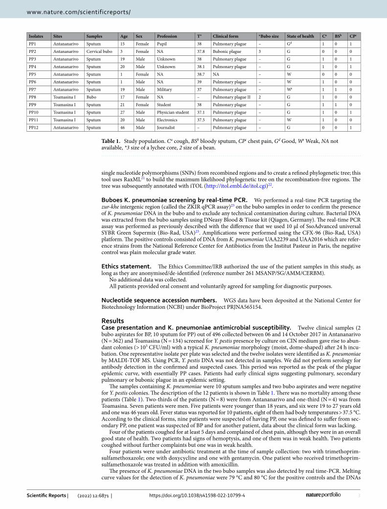
The samples containing K. pneumoniae were 10 sputum samples and two bubo aspirates and were negative for Y. pestis colonies. The description of the 12 patients is shown in Table 1. There was no mortality among these patients (Table 1). Two-thirds of the patients (N = 8) were from Antananarivo and one-third (N = 4) was from Toamasina. Seven patients were men. Five patients were younger than 18 years, and six were 19 to 27 years old and one was 46 years old. Fever status was reported for 10 patients, eight of them had body temperatures > 37.5 °C. According to the clinical forms, nine patients were suspected of having PP, one was defined to suffer from sec-ondary PP, one patient was suspected of BP and for another patient, data about the clinical form was lacking.
Four of the patients coughed for at least 5 days and complained of chest pain, although they were in an overall good state of health. Two patients had signs of hemoptysis, and one of them was in weak health. Two patients coughed without further complaints but one was in weak health.
Four patients were under antibiotic treatment at the time of sample collection: two with trimethoprim-sulfamethoxazole; one with doxycycline and one with gentamycin. One patient who received trimethoprim-sulfamethoxazole was treated in addition with amoxicillin.
The presence of K. pneumoniae DNA in the two bubo samples was also detected by real time-PCR. Melting curve values for the detection of K. pneumoniae were 79 °C and 80 °C for the positive controls and the DNAs
Table 1. Study population. Ca cough, BSb bloody sputum, CPc chest pain, Gd Good, We Weak, NA not available, *3 size of a lychee core, 2 size of a bean.
Isolates Sites Samples Age Sex Profession T° Clinical form *Bubo size State of health Ca BSb CPc
PP1 Antananarivo Sputum 15 Female Pupil 38 Pulmonary plague – Gd 1 0 1
PP2 Antananarivo Cervical bubo 3 Female NA 37.8 Bubonic plague 3 G 0 0 0
PP3 Antananarivo Sputum 19 Male Unknown 38 Pulmonary plague – G 1 0 1
PP4 Antananarivo Sputum 20 Male Unknown 38.1 Pulmonary plague – G 1 0 1
PP5 Antananarivo Sputum 1 Female NA 38.7 NA – W 0 0 0
PP6 Antananarivo Sputum 1 Male NA 39 Pulmonary plague – W 1 0 0
PP7 Antananarivo Sputum 19 Male Military 37 Pulmonary plague – We 1 1 0
PP8 Toamasina I Bubo 17 Female NA – Pulmonary plague II 2 G 1 0 0
PP9 Toamasina I Sputum 21 Female Student 38 Pulmonary plague – G 1 1 0
PP10 Toamasina I Sputum 27 Male Physician student 37.1 Pulmonary plague – G 1 0 1
PP11 Toamasina I Sputum 20 Male Electronics 37.5 Pulmonary plague – W 1 0 0
PP12 Antananarivo Sputum 46 Male Journalist – Pulmonary plague – G 0 0 1

4
Vol:.(1234567890)
Scientific Reports | (2022) 12:6871 | https://doi.org/10.1038/s41598-022-10799-4
www.nature.com/scientificreports/
extracted from buboes, respectively (Fig. 1). Of the 12 isolates, four had a positive string test. Five isolates were ESBL producers. Six isolates were resistant to sulfonamides and trimethoprim. Three and two isolates were resist-ant to gentamycin and tobramycin, respectively. One isolate was resistant to ciprofloxacin (Table 2).
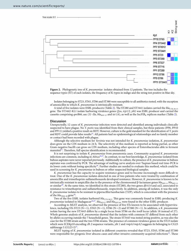
Genome analysis. Whole genome sequencing of the K. pneumoniae isolates allowed us to characterize cgMLST alleles, virulence genes, capsular loci and resistance genes. All K. pneumoniae isolates were K. pneu-moniae sensu stricto (Kp1) (Fig. 2). The 12 isolates had 11 different sequence types (STs): ST23 (N = 1); ST86 (N = 2); ST65 (N = 1); ST280 (N = 1); ST327 (N = 1); ST380 (N = 1); ST716 (N = 1); and ST3012 (N = 1), and three new STs: ST3441 (N = 1), ST3442 (N = 1), ST3443 (N = 1) (Table 2). Comparative genomic analysis of the two Kp ST86 isolates showed that they differed from each other by 123 alleles out of 632 scgMLST gene loci and are therefore unrelated epidemiologically.
The virulence genes identified in most isolates corresponded to the colibactin locus (clb), siderophores (iroB-CDN), iron uptake systems and regulators (kfu, kvgA, respectively), and yersiniabactin (fyuA, irp1/2 and ybt) (Supplementary data). The PP3, PP9, PP11 and PP12 isolates were positive for the string test. These isolates belonged to ST320 (PP3), ST86 (PP9 and PP12), and ST3443 (PP11). All the ST23, ST65, ST86 and ST380 iso-lates had the gene rmpA associated with the hyperproduction of the capsule and also carried the iucABCD genes coding for the synthesis of aerobactin (Supplementary Data).
Figure 1. Melting curve results from ZKIR region detection obtained after 40 cycles using the ZKIR quantitative PCR system. DNA from two positive controls from K. pneumoniae strains (blue: UUA2239, violet: UUA2016). DNA extracted from patients’ buboes is in pink for PP2 and in red for PP8. Orange color corresponds to negative control.
Table 2. MLST profiles, cps K-types, resistance profile, resistance genes and virulence genes. ND not determined.
Isolates Phylogroups String test ST K_type Resistance profile
Resistance genes/
Virulence genesIncompatibility group
PP1 Kp1 280 KL23 AMX, AMC, TZP, CEF, CTX, CAZ, FEP, ATM, TOB, GEN, CIP, SXT, TET
qnrB66, aac(3)-IIa, blaCTX-M-15, blaSHV-27, blaTEM-1B, dfrA14, strA, strB, sul2, tet(A) / IncFIB(K), IncFII(K)
mrk
PP2 Kp1 3441 ND AMX, AMC, CEF, CTX, CAZ, FEP, ATM, ETP, SXT, TET blaCTX-M-15, blaSHV-1 kfuA, mrk
PP3 Kp1 + 380 KL2 AMX, AMC, TET blaSHV-1clb, fyuA, iroBCDN, irp1/2, kfuC, kvgA, mrk, ybt
PP4 Kp1 3442 KL1 AMX, AMC, CEF, CTX, CAZ, FEP, ATM, TOB, GEN, SXT, TET
qnrB66, aac(3)-IIa, blaSHV-27, tet(A) / IncFIB(K) fyuA, irp1/2, mrk, ybt
PP5 Kp1 23 KL1 AMX, AMC, TET blaSHV-108clb, fyuA, kfuA, mrk, allS, fdrA, gcl, glx, hyi, irp1/2, ybt
PP6 Kp1 327 ND AMX, AMC, CEF, CTX, CAZ, FEP, ATM, ETP, TOB, GEN, NAL, SXT aac(3)-IIa, blaSHV-101 kfu, mrk
PP7 Kp1 3012 ND AMX, AMC, SXT,TET blaSHV-11 mrk
PP8 Kp1 716 ND AMX, AMC, CEF, CTX, CAZ, FEP, ATM, ETP, CIP, SXT, TET blaSHV-27 mrk
PP9 Kp1 + 86 KL2 AMX, AMC blaSHV-1 / IncHI1B iroBCDN, iucABCD, kvgA, mrk
PP10 Kp1 65 KL2 AMX, AMC, TET blaSHV-11 / IncHI1B clb, mrk, iucABCD
PP11 Kp1 + 3443 KL2 AMX, AMC blaSHV-1 / IncHI1B iucABCD, kvgS, mrk
PP12 KP1 + 86 KL2 AMX, AMC blaSHV-1 mrk
5
Vol.:(0123456789)
Scientific Reports | (2022) 12:6871 | https://doi.org/10.1038/s41598-022-10799-4
www.nature.com/scientificreports/
Isolates belonging to ST23, ST65, ST86 and ST380 were susceptible to all antibiotics tested, with the exception of amoxicillin to which K. pneumoniae is intrinsically resistant.
A total of five isolates were ESBL producers (Table 2). The ST280 and ST3441 isolates carried the blaCTX-M-15 gene. The ST3442-KL1 isolate harboring virulence genes (fyu, irp1/2, ybt) was ESBL producer and carried the cassette comprising qnrB66, aac (3) -IIa, blaSHV-27 and tet (A), as well as the IncFIBK replicon marker (Table 2).
DiscussionUnexpectedly, 12 cases of K. pneumoniae infection were detected and identified among individuals clinically suspected to have plague. No Y. pestis was identified from their clinical samples, but three patients (PP8, PP10 and PP11) yielded a positive result on RDT. However, culture is the gold standard for the identification of Y. pestis and RDT could provide false results24. All patients had no epidemiological relationships and no family member or contact had been recorded with plague.
Although the selective medium for Yersinia was not intended for K. pneumoniae isolation, K. pneumoniae does grow on the CIN medium in 24 h. The selectivity of this medium is reported as being partial, as other Gram-negative bacilli can grow on CIN medium, including other species of Enterobacterales able to ferment mannitol25. Therefore, full species identification is recommended.
It is not surprising to isolate K. pneumoniae from pneumonia cases. Community-acquired K. pneumoniae infections are common, including in Africa26,27. In contrast, to our best knowledge, K. pneumoniae isolated from buboes aspirates were never reported previously. Additionally to culture, the presence of K. pneumoniae in buboes aspirates was confirmed by PCR. The advantage of melting curve analysis over Taqman based real time-PCR is its lower costs without losing specificity28. Further studies are needed to evaluate this method, which could be used in screening for K. pneumoniae in buboes or other suspected biological samples.
K. pneumoniae has the capacity to acquire resistance genes and to become increasingly more difficult to treat. One of the K. pneumoniae isolates detected in one of two patients who were treated by combination of amoxicillin and trimethoprim-sulfamethoxazole developed resistance. However, K. pneumoniae is known to be intrinsically resistant to ampicillin due to the presence of the chromosomal β-lactamase genes blaSHV-1, blaSHV-11 or similar29. At the same time, we identified in this strain (ST280), the two genes dfrA14 and sul2, associated to resistance to trimethoprim and sulfamethoxazole, respectively. In addition, among all isolates, it was the only K. pneumoniae isolates to be resistant to piperacillin/tazobactam (TZP), which is concordant with the presence of the blaTEM-1B gene30.
Two of the five ESBL isolates harbored blaCTX-M15. This gene was commonly found in ESBL-producing K. pneumoniae isolated in Madagascar26,27. blaSHV27 and blaSHV101 were found in the other ESBL-producer.
According to MLST analysis, we observed the presence of five STs known to be associated with hyperviru-lence, including STs ST23 (N = 1), ST65 (N = 1), ST86 (N = 2) and ST380 (N = 1). In addition, a K. pneumoniae isolate having the new ST3443 differs by a single locus from ST86 on the tonB locus (allele 18 instead of 27). Whole genome analysis of K. pneumoniae showed that the isolates with common ST differed from each other by alleles occurring outside the 7 household genes. The strain ST3443 was tested string positive, as was also the case for the ST380 strain and the two ST86 strains. The presence of common virulence genes in the ST23 isolate which were ICEKp10 encoding clb 2 sequence variants, ybt 1 and rmpA/rmpA2 suggests its belonging to CG23 sublineage I (CG23-I)31.
MLST typing of K. pneumoniae isolated in different countries revealed that ST23, ST65, ST86 and ST380 were responsible for pyogenic liver abscess cases and other invasive community-acquired infections32. These
Figure 2. Phylogenetic tree of K. pneumoniae isolates obtained from 12 patients. The tree includes the sequence types (ST) of each isolates, the frequency of K-types in indigo and the string test positive in blue sky.

6
Vol:.(1234567890)
Scientific Reports | (2022) 12:6871 | https://doi.org/10.1038/s41598-022-10799-4
www.nature.com/scientificreports/
isolates have been reported particularly in Asia, but their diffusion outside Asia has been described31. Among virulence factors, rmpA and aerobactin are the most important ones33. The presence of genes responsible for the hypermucoviscosity phenotype, notably rmpA, plays an important role in the virulence of K. pneumoniae isolates. This gene is often associated with serotype K1 and K2. Expression of rmpA allows the bacteria to escape the host’s defense system and colonize the mucous membranes. Epidemiological studies have shown that the majority of ST23 are related to K1 capsular serotypes and liver abscesses31,34, while K2 is the second capsular serotype resulting in community-acquired pneumonia33. Yersiniabactin, a virulence gene (Ybt), detected in the three K. pneumoniae isolates serotyped KL1 has been reported as the iron absorption system in highly virulent Y. pestis35, and was later shown to have evolved ancestrally within the Klebsiella genus25. Several studies have shown isolates belonging to these STs (23, 65, 86 and 380), with the same combination of virulence factors, to be virulent in mouse models12,31.
During an epidemic, knowledge of the etiology is essential in order to provide the most adapted treatment to patients. Microbiological diagnosis can improve the effectiveness of treatments, avoid long-term complications for the infected patient, and in addition avoid widespread overuse and misuse of antibiotics. Early diagnosis can help to prevent or stop an outbreak too. One of the reasons for a possible treatment failure could arise during inaccurate diagnoses and inappropriate treatments. Similar symptoms can lead to routine treatments based on syndromic approaches which are often applied in developing countries, hence the importance of including differential diagnosis in laboratory diagnostic procedures in order to identify the etiology. As the physician is rarely able to make an etiological diagnosis on clinical grounds alone, treatment should ideally be based on the result of bacteriological examination. In this case, bacteriological diagnosis could be complicated by the fact that the respiratory tract could be infected by K. pneumoniae36. Although the population we included in our study is young, the clinical signs of a few patients warned us of possible serious infections due to K. pneumoniae such as bloody sputum and a chest pain which were among typical signs of pestis pneumonia. However, Y. pestis was not found in culture. Typically, the plague is better known by its three clinical forms: bubonic, septicemic and pulmonary plague while hypervirulent K. pneumoniae strains are known to cause pneumonia, sepsis, liver abscesses and meningitis37–39.
We acknowledge the following limitations of our study. First, we studied a limited number of samples in a short duration of the epidemic, which is far from being representative of all the negative samples for Y. pestis. Second, detailed data about clinical characteristics and outcomes were lacking due to the outbreak emergency context. Finally, we did not confirm the virulence of the K. pneumoniae strains using mouse models.
ConclusionsAlthough few samples were studied, within a short duration of inclusion (9 days from 06/10/17 to 14/10/17), our results show that some plague-suspected patients in fact acquired a pneumonia caused by K. pneumoniae. Bacterial identification proved useful for determining the etiology. WGS and AST results showed that among the 12 K. pneumoniae isolates, there were ESBL producers and virulent strains. This study reports on the genomic characterization of K. pneumoniae isolates isolated from patients during an epidemic of plague. These results could serve to warn clinicians regarding the most adequate treatment. Likewise, we reported the importance of bacteriological diagnosis for improving patient management.
Received: 22 April 2021; Accepted: 15 March 2022
References 1. Randremanana, R. et al. Epidemiological characteristics of an urban plague epidemic in Madagascar, August–November, 2017:
An outbreak report. Lancet Infect. Dis. 19, 537–545 (2019). 2. WHO. International meeting on preventing and controlling plague: the old calamity still has a future. Wkly. Epidemiol. Rec. 81,
278–284 (2006). 3. Andrianaivoarimanana, V. et al. Trends of human plague, Madagascar, 1998–2016. Emerg. Infect. Dis. 25(2), 220–228 (2019). 4. Rodrigues, C. et al. Description of Klebsiella africanensis sp. nov., Klebsiella variicola subsp. tropicalensis subsp. nov. and Klebsiella
variicola subsp. variicola subsp. nov. Res. Microbiol. 170, 165–170 (2019). 5. Wyres, K. L. & Holt, K. E. Klebsiella pneumoniae as a key trafficker of drug resistance genes from environmental to clinically
important bacteria. Curr. Opin. Microbiol. 45, 131–139 (2018). 6. Mizuta, K. et al. Virulence for mice of Klebsiella strains belonging to the O1 group: Relationship to their capsular (K) types. Infect.
Immun. 40(1), 56–61 (1983). 7. Cortés, G. et al. Molecular analysis of the contribution of the capsular polysaccharide and the lipopolysaccharide O side chain to
the virulence of Klebsiella pneumoniae in a murine model of pneumonia. Infect. Immun. 70(5), 2583–2590 (2002). 8. Tomás, J. M., Camprubi, S., Merino, S., Davey, M. R. & Williams, P. Surface exposure of O1 serotype lipopolysaccharide in Klebsiella
pneumoniae strains expressing different K antigens. Infect. Immun. 59(6), 2006–2011 (1991). 9. Di Martino, P., Cafferini, N., Joly, B. & Darfeuille-Michaud, A. Klebsiella pneumoniae type 3 pili facilitate adherence and biofilm
formation on abiotic surfaces. Res. Microbiol. 154(1), 9–16 (2003). 10. Turton, J. F., Baklan, H., Siu, L. K., Kaufmann, M. E. & Pitt, T. L. Evaluation of a multiplex PCR for detection of serotypes K1, K2
and K5 in Klebsiella sp. and comparison of isolates within these serotypes. FEMS Microbiol. Lett. 284, 247–252 (2008). 11. Lin, W. H. et al. Clinical and microbiological characteristics of Klebsiella pneumoniae isolates causing community-acquired urinary
tract infections. Infection 38, 459–464 (2010). 12. Shon, A. S., Bajwa, R. P. & Russo, T. A. Hypervirulent (hypermucoviscous) Klebsiella pneumoniae: A new and dangerous breed.
Virulence. 4(2), 107–118 (2013). 13. EMERGE. Plague diagnostic recommendations (2017). URL http:// www. emerge. rki. eu/ Emerge/ Share dDocs/ Downl oads/ EMERGE-
Plague- recom menda tions/ 14. WHO. Interregional meeting on prevention and control of plague (World Health Organization, 2008).
7
Vol.:(0123456789)
Scientific Reports | (2022) 12:6871 | https://doi.org/10.1038/s41598-022-10799-4
www.nature.com/scientificreports/
15. Passet, V. & Brisse, S. Association of tellurite resistance with hypervirulent clonal groups of Klebsiella pneumoniae. J. Clin. Micro-biol. 53, 1380–1382 (2015).
16. Société Française de Microbiologie. In CASFM/EUCAST: Société Française de microbiologie 2.0: 34-42 (2017). 17. Bialek-Davenet, S. et al. Genomic definition of hypervirulent and multidrug-resistant Klebsiella pneumoniae clonal groups. Emerg.
Infect. Dis. 20, 1812–1820 (2014). 18. Kaas, R. S., Leekitcharoenphon, P., Aarestrup, F. M. & Lund, O. Solving the problem of comparing whole bacterial genomes across
different sequencing platforms. PLoS ONE 9(8), e104984 (2014). 19. Stamatakis, A. RAxML version 8: A tool for phylogenetic analysis and post-analysis of large phylogenies. Bioinformatics 30,
1312–1313 (2014). 20. Treangen, T. J., Ondov, B. D., Koren, S. & Phillippy, A. M. The Harvest suite for rapid core-genome alignment and visualization of
thousands of intraspecific microbial genomes. Genome Biol. 15, 524 (2014). 21. Croucher, N. J. et al. Rapid phylogenetic analysis of large samples of recombinant bacterial whole genome sequences using Gub-
bins. Nucleic Acids Res. 43, e15 (2015). 22. Letunic, I. & Bork, P. Interactive tree of life (iTOL) v3: An online tool for the display and annotation of phylogenetic and other
trees. Nucleic Acids Res. 44, W242–W245 (2016). 23. Barbier, E. et al. The ZKIR assay, a real-time PCR method for the detection of Klebsiella pneumoniae and closely related species in
environmental samples. Appl. Environ. Microbiol. 86(7), e02711-e2719 (2020). 24. Rajerison, M. et al. Performance of plague rapid diagnostic test compared to bacteriology; a retrospective analysis of the data
collected in Madagascar. BMC Infect. Dis. 20, 90 (2020). 25. Weagant, S. D. A new chromogenic agar medium for detection of potentially virulent Yersinia enterocolitica. J. Microbiol. Methods.
72(2), 185–190. https:// doi. org/ 10. 1016/j. mimet. 2007. 11. 019 (2008). 26. Huynh, B. T. et al. Klebsiella pneumoniae carriage in low-income countries: Antimicrobial resistance, genomic diversity and risk
factors. Gut Microbes. 11(5), 1287–1299 (2020). 27. Rakotondrasoa, A. et al. Characterization of Klebsiella pneumoniae isolates from a mother-child cohort in Madagascar. J. Antimi-
crob. Chemother. 75(7), 1736–1746 (2020). 28. Tajadini, M., Panjehpour, M. & Javanmard, S. H. Comparison of SYBR Green and TaqMan methods in quantitative real-time
polymerase chain reaction analysis of four adenosine receptor subtypes. Adv. Biomed. Res. 28(3), 85. https:// doi. org/ 10. 4103/ 2277- 9175. 127998 (2014).
29. Fu, Y. et al. Differential expression of bla(SHV) related to susceptibility to ampicillin in Klebsiella pneumoniae. Int. J. Antimicrob. Agents. 29(3), 344–347 (2007).
30. Hubbard, A. T. M. et al. Piperacillin/tazobactam resistance in a clinical isolate of Escherichia coli due to IS26-mediated amplifica-tion of blaTEM-1B. Nat. Commun. 11(1), 4915 (2020).
31. Lam, M. M. C. et al. Population genomics of hypervirulent Klebsiella pneumoniae clonal-group 23 reveals early emergence and rapid global dissemination. Nat. Commun. 9(1), 2703 (2018).
32. Diancourt, L., Passet, V., Verhoef, J., Grimont, P. A. D. & Brisse, S. Multilocus sequence typing of Klebsiella pneumoniae nosocomial isolates multilocus sequence typing of Klebsiella pneumoniae nosocomial isolates. J. Clin. Microbiol. 43, 4178–4182 (2005).
33. Holt, K. E. et al. Genomic analysis of diversity, population structure, virulence, and antimicrobial resistance in Klebsiella pneu-moniae, an urgent threat to public health. Proc. Natl. Acad. Sci. 112(27), E3574–E3581 (2015).
34. Liao, C. H., Huang, Y. T., Chang, C. Y., Hsu, H. S. & Hsueh, P. R. Capsular serotypes and multilocus sequence types of bacteremic Klebsiella pneumoniae isolates associated with different types of infections. Eur. J. Clin. Microbiol. Infect. Dis. 33(3), 365–369 (2014).
35. Rakin, A., Schneider, L. & Podladchikova, O. Hunger for iron: the alternative siderophore iron scavenging systems in highly virulent Yersinia. Front. Cell Infect. Microbiol. 2, 151 (2012).
36. Sasaki, E. et al. Peracute Bacterial Meningitis due to Infection with Klebsiella pneumoniae in Captive-bred Ruffed Lemurs (Varecia variegate). J. Comp. Pathol. 156(2–3), 281–285 (2017).
37. Galy, A., Loubet, P., Peiffer-Smadja, N. & Yazdanpanah, Y. The plague: An overview and hot topics. Rev. Med. Intern. 39(11), 863–868 (2018).
38. Fung, C. et al. A global emerging disease of Klebsiella pneumoniae liver abscess: Is serotype K1 an important factor for complicated endophthalmitis. Gut 50, 420–424 (2002).
39. Chung, D. R. et al. Fecal carriage of serotype K1 Klebsiella pneumoniae ST23 strains closely related to liver abscess isolates in Koreans living in Korea. Eur. J. Clin. Microbiol. Infect. Dis. 31, 481–486 (2012).
AcknowledgementsWe are deeply grateful to the curators of the Institut Pasteur MLST system (Paris, France) for importing novel alleles, profiles isolates at https:// bigsdb. paste ur. fr.
Author contributionsA.R., T.C., V.J., S.B., M.R. and J.C. wrote the manuscript. A.R., L.M.A., S.A.R., S.H.R., V.A., S.R. performed lab experiments. S.A.R., V.A., S.R., M.R completed data collection. A.R., S.B., V.J., M.R. and J.C. conceived the study. A.R., T.C., S.B., M.R. and J.C. analyzed and interpreted data. All authors read and approved the final manuscript.
FundingInternal funding.
Competing interests The authors declare no competing interests.
Additional informationSupplementary Information The online version contains supplementary material available at https:// doi. org/ 10. 1038/ s41598- 022- 10799-4.
Correspondence and requests for materials should be addressed to A.R.
Reprints and permissions information is available at www.nature.com/reprints.
Publisher’s note Springer Nature remains neutral with regard to jurisdictional claims in published maps and institutional affiliations.

8
Vol:.(1234567890)
Scientific Reports | (2022) 12:6871 | https://doi.org/10.1038/s41598-022-10799-4
www.nature.com/scientificreports/
Open Access This article is licensed under a Creative Commons Attribution 4.0 International License, which permits use, sharing, adaptation, distribution and reproduction in any medium or
format, as long as you give appropriate credit to the original author(s) and the source, provide a link to the Creative Commons licence, and indicate if changes were made. The images or other third party material in this article are included in the article’s Creative Commons licence, unless indicated otherwise in a credit line to the material. If material is not included in the article’s Creative Commons licence and your intended use is not permitted by statutory regulation or exceeds the permitted use, you will need to obtain permission directly from the copyright holder. To view a copy of this licence, visit http:// creat iveco mmons. org/ licen ses/ by/4. 0/.
© The Author(s) 2022
Related Documents