JOURNAL OF VIROLOGY, July 2002, p. 6987–6999 Vol. 76, No. 14 0022-538X/02/$04.000 DOI: 10.1128/JVI.76.14.6987–6999.2002 Copyright © 2002, American Society for Microbiology. All Rights Reserved. Characterization of Human Immunodeficiency Virus Type 1 (HIV-1) Gag- and Gag Peptide-Specific CD4 T-Cell Clones from an HIV-1-Seronegative Donor following In Vitro Immunization Sara Venturini, Donald E. Mosier, Dennis R. Burton,* and Pascal Poignard* Department of Immunology, The Scripps Research Institute, La Jolla, California Received 19 October 2001/Accepted 10 April 2002 Substantial evidence argues that human immunodeficiency virus type 1 (HIV-1)-specific CD4 T cells play an important role in the control of HIV-1 replication in infected individuals. Moreover, it is increasingly clear that an HIV vaccine should elicit potent cytotoxic lymphocyte and antibody responses that will likely require an efficient CD4 T-cell response. Therefore, understanding and characterizing HIV-specific CD4 T-cell responses is an important aim. Here we describe the generation of HIV-1 Gag- and Gag peptide-specific CD4 T-cell clones from an HIV-1-seronegative donor by in vitro immunization with HIV-1 Gag peptides. The Gag peptides were able to induce a strong CD4 T-cell immune response in peripheral blood mononuclear cells from the HIV-1-seronegative donor. Six Gag peptide-specific CD4 T-cell clones were isolated and their epitopes were mapped. The region of p24 between amino acids 201 and 300 of Gag was defined as the immunodominant region of Gag. A new T helper epitope in the p6 protein of Gag was identified. Two clones were shown to recognize Gag peptides and processed Gag protein, while the other four clones reacted only to Gag peptides under the experimental conditions used. Functional analysis of the clones indicated that both Th1 and Th2 types of CD4 T cells were obtained. One clone showed direct antigen-specific cytotoxic activity. These clones represent a valuable tool for understanding the cellular immune response to HIV-1, and the study provides new insights into the HIV-1-specific CD4 T-cell response and the induction of an anti-Gag and -Gag peptide cellular primary immune response in vitro. Substantial evidence argues for the importance of CD4 T helper cells in the immune response to human immunodefi- ciency virus type 1 (HIV-1) infection (47, 55, 56). As in murine models of chronic viral infections, CD4 T cells may be re- quired for the maintenance of functional HIV-specific CD8 cytotoxic T-lymphocyte (CTL) responses (38, 46, 56, 70). The lack of CD4 T-cell responses observed in most HIV-infected individuals could then explain the decline of CTL responses seen over time and the progression of the disease (8, 24). This hypothesis has been strengthened by recent studies that have shown that in long-term nonprogressors and in patients treated with highly active antiretroviral therapy early during primary infection (47, 50, 51, 54, 55), enhanced CD4 T helper cell responses to HIV-1 are associated with higher levels of HIV- 1-specific CTL and with lower viral loads. Helper T cells could also play a role in viral replication during HIV-1 infection via the regulation and control of the B-cell response and the modulation of neutralizing antibody expression (10, 48), although our data with the hu-PBL-SCID mouse model suggest that neutralizing antibodies are unlikely to be involved in controlling viral replication in established HIV-1 infection (49). Finally, a direct role for CD4 T cells in the control of viral replication may be possible, via secretion of cytokines and chemokines or cytotoxic activity (45). The role of CD4 T cells in the prevention of HIV-1 infec- tion is unclear but likely to be beneficial. Studies have shown that vaccines that stimulate a strong cellular response are largely unable to provide complete protection (18, 59, 69) but can lower viral load and slow disease progression, suggesting that CD8 and CD4 T cells can have an impact on infection but cannot provide sterilizing immunity. In contrast, it has been demonstrated that passively administered antibodies can fully protect when high plasma concentrations, unlikely to be achieved through vaccination, are attained (5, 15, 36, 37, 60). Lower levels of antibody have been shown to offer benefit in terms of a delayed and decreased viremia, and it may be that a combination of cellular and antibody responses induced by a vaccine could provide protection. Certainly it seems that an optimal HIV vaccine will elicit strong CTL and antibody re- sponses that will benefit from an efficient CD4 T-cell re- sponse. Some support for this view comes from experiments in the Friend virus model that showed that only a combination of specific CD4 , CD8 , and B cells is able to transfer protection against Friend virus, and any combination of two cell types is insufficient (13). Understanding and characterizing the HIV-specific CD4 T-cell response is therefore important for the design of immu- notherapies and vaccines for HIV-1 infection. We have previ- ously investigated the impact of CTL and antibodies on estab- lished HIV-1 infection with the hu-PBL-SCID mouse model (39, 49). To be able to include CD4 T cells in these studies, we have begun to generate and characterize HIV-1-specific CD4 clones. These clones should allow, in future experiments * Corresponding author. Mailing address: Pascal Poignard, Depart- ment of Immunology, The Scripps Research Institute, 10550 N. Torrey Pines Rd., La Jolla, CA 92037. Phone: (858) 784-9116. Fax: (858) 784-9190. E-mail: [email protected]. Dennis Burton, Department of Immunology, The Scripps Research Institute, 10550 N. Torrey Pines Rd., La Jolla, CA 92037. Phone: (858) 784-9298. Fax: (858) 784-8360. E-mail: [email protected]. 6987

Welcome message from author
This document is posted to help you gain knowledge. Please leave a comment to let me know what you think about it! Share it to your friends and learn new things together.
Transcript

JOURNAL OF VIROLOGY, July 2002, p. 6987–6999 Vol. 76, No. 140022-538X/02/$04.00�0 DOI: 10.1128/JVI.76.14.6987–6999.2002Copyright © 2002, American Society for Microbiology. All Rights Reserved.
Characterization of Human Immunodeficiency Virus Type 1 (HIV-1)Gag- and Gag Peptide-Specific CD4� T-Cell Clones from anHIV-1-Seronegative Donor following In Vitro Immunization
Sara Venturini, Donald E. Mosier, Dennis R. Burton,* and Pascal Poignard*Department of Immunology, The Scripps Research Institute, La Jolla, California
Received 19 October 2001/Accepted 10 April 2002
Substantial evidence argues that human immunodeficiency virus type 1 (HIV-1)-specific CD4� T cells playan important role in the control of HIV-1 replication in infected individuals. Moreover, it is increasingly clearthat an HIV vaccine should elicit potent cytotoxic lymphocyte and antibody responses that will likely requirean efficient CD4� T-cell response. Therefore, understanding and characterizing HIV-specific CD4� T-cellresponses is an important aim. Here we describe the generation of HIV-1 Gag- and Gag peptide-specific CD4�
T-cell clones from an HIV-1-seronegative donor by in vitro immunization with HIV-1 Gag peptides. The Gagpeptides were able to induce a strong CD4� T-cell immune response in peripheral blood mononuclear cellsfrom the HIV-1-seronegative donor. Six Gag peptide-specific CD4� T-cell clones were isolated and theirepitopes were mapped. The region of p24 between amino acids 201 and 300 of Gag was defined as theimmunodominant region of Gag. A new T helper epitope in the p6 protein of Gag was identified. Two cloneswere shown to recognize Gag peptides and processed Gag protein, while the other four clones reacted only toGag peptides under the experimental conditions used. Functional analysis of the clones indicated that both Th1and Th2 types of CD4� T cells were obtained. One clone showed direct antigen-specific cytotoxic activity. Theseclones represent a valuable tool for understanding the cellular immune response to HIV-1, and the studyprovides new insights into the HIV-1-specific CD4� T-cell response and the induction of an anti-Gag and -Gagpeptide cellular primary immune response in vitro.
Substantial evidence argues for the importance of CD4� Thelper cells in the immune response to human immunodefi-ciency virus type 1 (HIV-1) infection (47, 55, 56). As in murinemodels of chronic viral infections, CD4� T cells may be re-quired for the maintenance of functional HIV-specific CD8�
cytotoxic T-lymphocyte (CTL) responses (38, 46, 56, 70). Thelack of CD4� T-cell responses observed in most HIV-infectedindividuals could then explain the decline of CTL responsesseen over time and the progression of the disease (8, 24). Thishypothesis has been strengthened by recent studies that haveshown that in long-term nonprogressors and in patients treatedwith highly active antiretroviral therapy early during primaryinfection (47, 50, 51, 54, 55), enhanced CD4� T helper cellresponses to HIV-1 are associated with higher levels of HIV-1-specific CTL and with lower viral loads.
Helper T cells could also play a role in viral replicationduring HIV-1 infection via the regulation and control of theB-cell response and the modulation of neutralizing antibodyexpression (10, 48), although our data with the hu-PBL-SCIDmouse model suggest that neutralizing antibodies are unlikelyto be involved in controlling viral replication in establishedHIV-1 infection (49). Finally, a direct role for CD4� T cells in
the control of viral replication may be possible, via secretion ofcytokines and chemokines or cytotoxic activity (45).
The role of CD4� T cells in the prevention of HIV-1 infec-tion is unclear but likely to be beneficial. Studies have shownthat vaccines that stimulate a strong cellular response arelargely unable to provide complete protection (18, 59, 69) butcan lower viral load and slow disease progression, suggestingthat CD8� and CD4� T cells can have an impact on infectionbut cannot provide sterilizing immunity. In contrast, it hasbeen demonstrated that passively administered antibodies canfully protect when high plasma concentrations, unlikely to beachieved through vaccination, are attained (5, 15, 36, 37, 60).Lower levels of antibody have been shown to offer benefit interms of a delayed and decreased viremia, and it may be thata combination of cellular and antibody responses induced by avaccine could provide protection. Certainly it seems that anoptimal HIV vaccine will elicit strong CTL and antibody re-sponses that will benefit from an efficient CD4� T-cell re-sponse. Some support for this view comes from experiments inthe Friend virus model that showed that only a combination ofspecific CD4�, CD8�, and B cells is able to transfer protectionagainst Friend virus, and any combination of two cell types isinsufficient (13).
Understanding and characterizing the HIV-specific CD4�
T-cell response is therefore important for the design of immu-notherapies and vaccines for HIV-1 infection. We have previ-ously investigated the impact of CTL and antibodies on estab-lished HIV-1 infection with the hu-PBL-SCID mouse model(39, 49). To be able to include CD4� T cells in these studies,we have begun to generate and characterize HIV-1-specificCD4� clones. These clones should allow, in future experiments
* Corresponding author. Mailing address: Pascal Poignard, Depart-ment of Immunology, The Scripps Research Institute, 10550 N. TorreyPines Rd., La Jolla, CA 92037. Phone: (858) 784-9116. Fax: (858)784-9190. E-mail: [email protected]. Dennis Burton, Departmentof Immunology, The Scripps Research Institute, 10550 N. Torrey PinesRd., La Jolla, CA 92037. Phone: (858) 784-9298. Fax: (858) 784-8360.E-mail: [email protected].
6987
in vitro and in the mouse model, the analysis of the interplay ofCD4� T cells, CD8� T cells, and antibodies in HIV-1 infec-tion.
Very few reports to date have described the characterizationof HIV-1-specific CD4� T-cell clones (11, 28, 44, 52, 68). Thecloning of CD4� T cells is made difficult by the scarcity of thesecells in HIV-1-infected persons and by their typical unrespon-siveness to HIV-1 antigens (46). Previous studies have sug-gested that a cellular immune response to HIV-1 proteins canbe obtained following in vitro stimulation of cells from HIV-1-negative individuals (6, 29, 31, 32, 40, 61, 65). Furthermore,it has been reported for proteins from other pathogens that Thelper epitopes identified following in vitro immunization cor-respond to epitopes that are immunogenic in vivo (23, 26). Thisindicates that this type of study can also provide valuableinformation about the determinants of HIV-1 proteins able toinduce primary CD4� T-cell immune responses in vivo. There-fore we decided to induce an HIV-1-specific CD4� T-cell re-sponse in vitro in peripheral blood mononuclear cells (PBMC)from a normal donor in order to isolate HIV-1-specific CD4�
T-cell clones. We chose the HIV-1 Gag protein as an antigenbecause a number of recent studies have highlighted the im-portance of the CD4� T-cell response to Gag in HIV-1-in-fected individuals (47, 50, 51, 55).
Here we report the in vitro induction in human PBMC froma seronegative donor of a primary immune response to a mix-ture of partially overlapping peptides covering the sequence ofthe HIV-1 IIIB Gag protein. Individual clones with strongreactivity against the Gag peptides were isolated and studied.For each clone, we characterized the peptides recognized, re-activity to processed Gag protein, sensitivity to activation, T-cell receptor (TCR) V� usage, major histocompatibility com-plex (MHC) restriction, and the profile of cytokines secreted.We also determined if cytotoxic activity was present. Theseresults provide new insights into several aspects of the HIV-1Gag-specific CD4� T-cell response and the induction of ananti-Gag and -Gag peptide cellular primary immune responsein vitro.
MATERIALS AND METHODS
Peptides and reagents. The HIV-1 Gag p55 overlapping 20-mer peptides wereobtained from the AIDS Research and Reference Reagent Program (AIDSRRRP), Division of AIDS, National Institute for Allergy and Infectious Disease,National Institutes of Health. The peptides were given an arbitrary number from1 to 49 following the position in the Gag sequence; full sequences are reportedin the AIDS RRRP catalog. The lyophilized peptides were resuspended asindicated by the manufacturers at 1 mg/ml and then stored at �80°C. Twopeptides, peptides 35 (P341-360) and 36 (P350-370), were not included in thisstudy because they were not available. To prepare the peptide mixture, the 47peptides were mixed in equal parts to a final concentration of 1 mg/ml (21 �g/mlfor each individual peptide).
Tetanus toxoid (TT) was purchased from Connaught Laboratories Limited,Willowdale, Canada. Anti-CD3 antibody was purchased from Zymed Laborato-ries, San Francisco, Calif. Human interleukin-2 (IL-2) was obtained from theAIDS RRRP. The recombinant p24 protein was from Austral Biologicals, SanRamon, Calif.
Study subjects and cells. The cells used for the generation of Gag peptide-specific CD4� T-cell clones were derived from the healthy brother of a pair ofmonozygotic twins discordant for HIV-1 infection. PBMC were obtained follow-ing leukopheresis of the uninfected twin and purification on a Ficoll-Hypaquegradient (Sigma, St. Louis, Mo.). The CD8� CTL clone specific for HIV-1 Gag(P11-30) was generated from the infected twin as described previously (53) andwas a generous gift of S. R. Riddell and P. D. Greenberg. The twins’ HLA classII type was DR4, DR7, DQ2, and DQ3, as previously determined by Riddell and
colleagues by low-resolution DNA and sequence-specific oligonucleotide probeHLA typing methods (S. Riddell, personal communication). The B lymphoblas-toid cell line (B-LCL) was obtained by Epstein-Barr virus transformation of thePBMC from the seronegative twin. As feeder cells for the T-cell cloning, PBMCfrom healthy volunteers were used after purification by Ficoll-Hypaque densitygradient and 4,000-rad �-irradiation. Class II-negative murine L-cell fibroblastsstably transfected with HLA DR1, DR7, DR4W10, DR4W14, or DR4W15 wereobtained from R. W. Karr (25).
Generation of antigen-specific T-cell lines. Gag peptide-specific T-cell lineswere generated by culturing PBMC from the uninfected twin with 40 �g of theHIV-1 IIIB Gag peptide mixture (the final concentration of individual peptidewas 0.85 �g/ml) per ml in a Costar 24-well plate (Corning, Garden Grove, Calif.)at 3.5 � 106 cells/well. The complete cell culture medium consisted of RPMI1640 (BioWhittaker, Walkersville, Md.) supplemented with 2 mM L-glutamine,100 U of penicillin per ml, 100 �g of streptomycin per ml (all from Life Tech-nologies, Rockville, Md.), and 10% heat-inactivated AB human serum (R10-HS). Cells were incubated at 37°C and 5% CO2. The control cultures werecarried out under the same conditions but with TT at 5 �g/ml as a positivecontrol antigen or without antigen. After 4 days of incubation, the medium wasremoved and replaced with R10-HS containing 40 U of IL-2 per ml. The cellswere grown for an additional 8 to 12 days, with fresh R10-HS/IL-2 mediumadded every 2 to 3 days. At the end of the in vitro stimulation, the cells weretested for peptide-specific activity.
Generation of T-cell clones. The T cells from the Gag peptide-specific linewere cloned by limiting-dilution. The cells were plated at 1, 5, 10, or 25 cells/wellin 96-well U-bottomed plates (Corning, Garden Grove, Calif.), and 105 heterol-ogous �-irradiated PBMC were added per well as feeder cells in the presence ofanti-CD3 antibody (50 ng/ml) and IL-2 (40 U/ml) in complete medium contain-ing 10% fetal calf serum (FCS) (R10). Four days later the medium was removedand replaced with R10/IL-2 medium. The plates were left untouched for anadditional 12 to 14 days. The percentage of wells in which cell growth wasdetectable was 0, 13, and 26% for plates seeded with 1, 5, and 25 cells/well,respectively.
In order to ensure a high probability of monoclonality, cells were expandedonly from the plates that had the lowest percentage of wells in which cell growthwas detectable. The clones were transferred into 24-well plates and reactivated inthe presence of 2 � 106 irradiated heterologous PBMC as described above. Theclones were expanded and then analyzed by fluorescence-activated cell sorting(FACS), screened for antigen-specific proliferative activity, and cryopreserved inaliquots. When needed, the cells were thawed and restimulated as describedbefore and used after 2 to 3 weeks of stimulation.
Proliferation assay. The cells to be analyzed were washed twice in order toremove all IL-2-containing medium and then resuspended in R10-HS as indi-cated. Cells were seeded in 96-well U-bottomed plates at 105 cells/well andincubated with an equal amount of antigen-presenting cells (APC) in a totalvolume of 200 �l. As APC we used autologous PBMC that were incubated withor without antigen for 2 h and then �-irradiated (4,000 rad). The antigen was leftwith the APC in order to give a final concentration in the well of 40 �g/ml for thepeptide mixture, 5 �g/ml for the TT, 2.5 �g/ml for the recombinant p24, 0.5�g/ml for the single peptides, or otherwise as indicated. Each condition wasassayed in duplicate.
In some experiments, proliferation was also determined following nonspecificactivation with 50 ng of anti-CD3 antibody and 40 U of IL-2 per ml. Three dayslater, the cells were pulsed with [3H]thymidine (ICN Biomedicals, Irvine, Calif.)at 1 �Ci/well, and uptake was measured in a �-scintillation counter after 18 h.The incubation time was extended to 5 days before the addition of [3H]thymidinefor assays involving freshly isolated PMBC. In some experiments, the results arereported as stimulation index. The stimulation index is the ratio of the level ofproliferation, in counts per minute, obtained in the presence of antigen and thelevel of proliferation obtained without antigen.
TCR V� analysis. Following lysis of T-cell clones and PBMC in Trizol solution(Gibco-BRL, Carlsbad, Calif.), total RNA was prepared by chloroform extrac-tion and isopropanol precipitation. The RNA was reverse transcribed to cDNAwith Moloney murine leukemia virus reverse transcriptase (Gibco-BRL, Carls-bad, Calif.) and poly(T) primers (Roche, Basel, Switzerland). Analysis of TCRV� usage was performed with the TCR typing amplimer kit (Clontech, Palo Alto,Calif.) according to the manufacturer’s instructions. The cDNA from each clonewas tested with a panel of 25 sense primers, each specific for one V� sequence,and an antisense primer located in the C� region. Primers specific for the TCR� chain constant region (C�) were used as a positive control. The PCR productswere analyzed on a 1.8% agarose gel.
For clones 6 and 74, analysis of the TCR V� usage was further carried out bycloning and sequencing the complete variable region of the TCR � chain. The
6988 VENTURINI ET AL. J. VIROL.

TCR V� region was reverse transcribed and PCR amplified with the Smart RacecDNA amplification kit from Clontech as specified by the manufacturer’s in-structions and the C� antisense primer provided with the Amplimer kit (Clon-tech). PCR products were cloned with the Topo TA cloning kit (Invitrogen,Carlsbad, Calif.). Double-stranded plasmid DNA was isolated from 13 and 6colonies for clones 6 and 74, respectively. Sequences were obtained by auto-mated sequencing (ABI, Perkin-Elmer, Foster City, Calif.) with the M13 reverseprimer.
Two primers specific for the V�6s5A1N1 and V�17Vs1A1T sequences (5�-AATGAAGCTCAACTAGAA-3� and 5�-CAGATAGTAAATGACTTT-3�, re-spectively) were designed and used for PCR amplification with the cDNA ofclone 6 as a template.
Flow cytometry analysis and intracellular cytokine staining. Cells werestained with fluorochrome-conjugated antibodies to CD4 (clone S3.5) and toCD8 (clone 3B5) from Caltag Laboratories (Burlingame, Calif.) in phosphate-buffered saline (PBS) containing 1% FCS and 0.02% NaN3 (Sigma, St. Louis,Mo.). Stained cells were analyzed on a FACScalibur flow cytometer (BectonDickinson, San Diego, Calif.) with CellQuest software (Becton Dickinson).
For intracellular cytokine staining, cells at days 14 to 18 of activation wereplated in a 96-well flat-bottomed plate at 106 cells/well in 200 �l of R10/IL-2 andincubated at 37°C in the presence of different antigens or without antigen. Singlepeptides were used at 0.5 �g/ml, anti-CD3 antibody at 50 ng/ml, and vacciniavirus-infected cells at 0.5 � 106 cells/well. GolgiPlug (Pharmingen, San Diego,Calif.), a reagent that contains brefeldin A, was added to the cultures to inhibitprotein transport and allow the detection of the cytokines. In some cases, theantibodies anti-HLA DR L243 (clone G46-6) from Pharmingen or anti-HLA DQ(clone SPVL3) from Coulter (Coulter, Westbrook, Maine) were added to thecultures at 0.5 �g/ml. After 6 h, cell surface staining was performed as describedabove, followed by intracellular cytokine staining with a Cytofix/Cytoperm kit(Pharmingen, San Diego, Calif.) in accordance with the manufacturer’s recom-mendations. The following antibodies were used: anti-gamma interferon (IFN-�)-phycoerythrin (PE) conjugate (clone 4S.B3), anti-IL-2–allophycocyanin(clone MQ1-17H12), anti-IL-4–fluorescein isothiocyanate (FITC) conjugate(clone MP4-25D2), and anti-IL-5–PE conjugate (clone JES1-39D10) (Pharmin-gen, San Diego, Calif.).
To characterize the HLA DR subtype that presents peptide 271-290 to clone6, intracellular cytokine staining was performed following activation of clone 6 byHLA DR-transfected fibroblasts pulsed with the specific peptide. HLA DR1-,DR7-, DR4W10-, DR4W14-, and DR4W15-transfected fibroblasts were platedin a 24-well plate at 106 cells/well and incubated for 2 h with peptide 271-290 ata concentration of 4 �g/ml. The cells were then washed seven times with R10 toremove all unbound peptide, and 106 clone 6 T cells were added per well in thepresence of brefeldin A. After 6 h, the cells in suspension were analyzed asdescribed above.
Vaccinia virus infection. The following reagents were obtained through theAIDS RRRP: vP1170, wild-type vaccinia virus, contributed by Daniel R. Kuritz-kes; and vDK1, recombinant vaccinia virus containing the coding region forHIV-1 Gag, contributed by Virogenetics Corp. Vaccinia virus stocks were diluted1:2 in trypsin-EDTA solution for cell culture (Life Technologies, Rockville, Md.)and incubated for 30 min at 37°C, vortexing every 10 min. The virus was addedto B-LCL at a multiplicity of infection (MOI) of 0.3 in a total volume of 2.5 mlof R10 in a six-well plate, and the cells were incubated for 18 h at 37°C. Vacciniavirus-infected cells were used as targets in a cytotoxicity assay and to evaluateT-cell activation induced by Gag-expressing cells.
Cytotoxicity assay. Vaccinia virus-infected and peptide-pulsed autologous B-LCL were used as target cells in a standard chromium release assay. Target cellswere labeled with 200 �Ci of Na2
51CrO4 (Amersham Pharmacia Biotech, Pis-cataway, N.J.) for 2 h at 37°C and washed three times with R10. The cells werethen seeded in U-bottomed 96-well microtiter plates at 2 � 104 cells/well. Forpeptide-pulsed target cells, pulsing was performed overnight with the peptidemixture at 10 �g/ml, and the peptides were added again at a final concentrationof 2.5 �g/ml following chromium labeling.
Effector cells were added, in triplicate, in a final volume of 200 �l. Single T-cellclones at days 15 to 18 of activation were used as effector cells at an effector-to-target cell (E:T) ratio ranging from 0.5 to 20. The plates were incubated at37°C for 6 h. The supernatants were then harvested (100 �l), and radioactivitywas counted in a gamma-counter. Maximal release of radioactivity was deter-mined by lysis of target cells in 1% Empigen BB (Calbiochem, San Diego, Calif.).Spontaneous release was measured in the absence of effector cells. The percentspecific lysis was determined from the formula [(experimental release � spon-taneous release)/(maximum release � spontaneous release)] � 100. Spontane-ous release was less than 30%.
RESULTS
Isolation of Gag peptide-specific limiting-dilution CD4� T-cell clones from a seronegative donor. The in vitro isolation ofantigen-specific T cells from nonimmune individuals is difficultbecause the frequency of antigen-specific cells is low. However,a few reports have shown that it is sometimes possible toinduce in vitro an immune response to certain antigens and toisolate the responsive T cells (23, 26, 30, 31, 61). Here wereport the induction of a CD4� T-cell response to HIV-1 Gagpeptides in PBMC from an HIV-1-seronegative individual andthe subsequent isolation of Gag- and Gag peptide-specificCD4� T-cell clones from the stimulated PBMC.
PBMC of an HIV-1-seronegative donor were stimulated invitro with a mixture of 47 partially overlapping 20-mer peptidescovering the whole sequence of the HIV-1 Gag protein, withthe exception of 10 amino acids at the C-terminal end of p24.Four days later, the peptides were removed, and IL-2-contain-ing medium was added. The cells were then grown for 2 weeksand tested for the presence of a specific proliferative response.Irradiated autologous PBMC were used as APC and added tothe in vitro-stimulated T cells in the presence of the HIV-1Gag peptide mixture or of TT. After a 3-day incubation withthe antigen, incorporation of [3H]thymidine was measured.Based on previous experiments of in vitro immunization, aproliferative response higher than twice the proliferative re-sponse obtained in the absence of antigen was consideredsignificant.
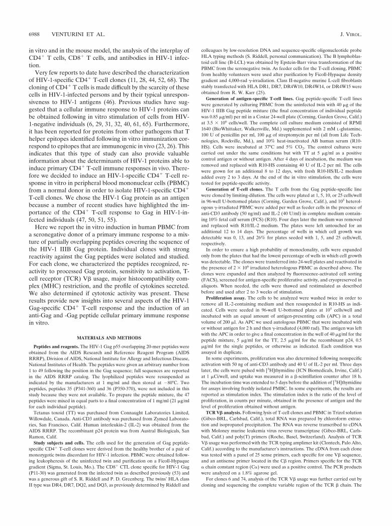
As shown in Fig. 1A, the cells did not proliferate in theabsence of antigen or in response to TT but proliferated sig-nificantly in response to the HIV-1 Gag peptide mixture. Themagnitude of peptide-specific proliferation increased when theconcentration of the peptide mixture was doubled from 3 to 6�g/ml but decreased slightly if the concentration was furtherincreased to 12 �g/ml. As shown in Fig. 1B, the same prolif-eration assay performed on the PBMC before the in vitrostimulation did not reveal any measurable proliferation in re-sponse to the HIV-1 Gag peptide mixture. However, the cellsdid proliferate in response to the recall antigen TT. This resultindicates that the in vitro immune activation performed on thePBMC successfully triggered the expansion of a population ofpreviously undetectable HIV-1 Gag peptide-specific T cells.
In order to isolate individual specific T-cell clones, the Gagpeptide-specific line was stimulated nonspecifically with an an-ti-CD3 antibody and plated in limiting-dilutions. Limiting-di-lution T-cell clones were isolated so as to ensure a high prob-ability of monoclonality, as explained in Materials andMethods, and their CD4� or CD8� phenotype was analyzed byFACS. Sixteen limiting-dilution clones were determined to beCD4� CD8� T cells and 16 were CD8� CD4� T cells. TheCD4� T-cell clones were individually tested in a proliferationassay for Gag peptide-specific reactivity as described above(data not shown). Of the 16 CD4� T-cell clones tested, 6showed an HIV-1 Gag peptide-specific proliferation and wereexpanded for further characterization.
We also investigated whether any of the limiting-dilutionCD8� T-cell clones were specific for the HIV-1 Gag protein orpeptides. A standard 51Cr release assay was performed, asdescribed below, to determine the existence of Gag-specificlysis of target cells. None of the CD8� T-cell clones showed
VOL. 76, 2002 HIV-1 Gag-SPECIFIC CD4� T-CELL CLONES 6989
any detectable Gag- or Gag peptide-specific cytotoxic activity(data not shown).
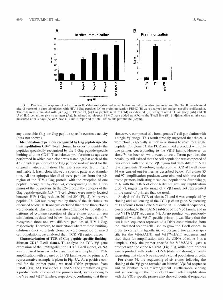
Identification of peptides recognized by Gag peptide-specificlimiting-dilution CD4� T-cell clones. In order to identify thepeptides specifically recognized by the 6 Gag peptide-specificlimiting-dilution CD4� T-cell clones, proliferation assays wereperformed in which each clone was tested against each of the47 individual peptides of the Gag peptide mixture used for theoriginal in vitro stimulation. The results are reported in Fig. 2and Table 1. Each clone showed a specific pattern of stimula-tion. All the epitopes identified were peptides from the p24region of the HIV-1 Gag protein, with the exception of onepeptide, recognized by clone 74, corresponding to the C ter-minus of the p6 protein. In the p24 protein the epitopes of theGag peptide-specific CD4� T-cell clones were mostly locatedbetween HIV-1 Gag residues 201 and 300 (Fig. 2). Moreover,peptide 271-290 was recognized by three of the six clones. Asdiscussed below, TCR analysis excluded that these three cloneswere identical. This result was also confirmed by the differentpatterns of cytokine secretion of these clones upon antigenstimulation, as described below. Interestingly, clones 6 and 74recognized three and two different peptides of HIV-1 Gag,respectively. Therefore, to understand whether these limiting-dilution clones were truly clonal or were composed of mixedcell populations, we analyzed their TCR V� region usage.
Characterization of TCR of Gag peptide-specific limiting-dilution CD4� T-cell clones. To analyze the TCR V� geneexpression of the limiting-dilution CD4� T-cell clones, cDNAwas prepared from each clone and used as a template for PCRamplification with a panel of 25 V� family-specific primers. Arepresentative example is given in Fig. 3A. As a positive con-trol for the primer panel, we used cDNA prepared fromPBMC (Fig. 3A). For clones 37 and 50, the amplification gavea product with only one of the primers used, corresponding tothe V�3 and V�17 families, respectively, confirming that these
clones were composed of a homogenous T-cell population witha single V� usage. This result strongly suggested that the cellswere clonal, especially as they were shown to react to a singlepeptide. For clone 74, the PCR amplified a product with onlyone primer, corresponding to the V�13 family. However, asclone 74 has been shown to react to two different peptides, thepossibility still existed that the cell population was composed oftwo clones with the same V� region but with different VDJrearrangements. Therefore, analysis of the TCR of T-cell clone74 was carried out further, as described below. For clones 85and 97, amplification products were obtained with two of thetested primers, indicating mixed cell populations. Surprisingly,PCR with the cDNA of clone 6 did not give any amplificationproduct, suggesting the usage of a V� family not representedin the panel of primers that we used.
Analysis of the TCR of clones 74 and 6 was completed bycloning and sequencing of the TCR �-chain gene. Sequencingof 13 colonies from clone 6 resulted in 11 identical sequences,corresponding to the s5A1N1 subtype of the V�6 family, and intwo V�17s1A1T sequences (4). As no product was previouslyamplified with the V�17-specific primer, it was likely that thetwo latter sequences represented a DNA contamination fromthe irradiated feeder cells used to grow the T-cell clones. Inorder to verify this hypothesis, we designed two primers spe-cific for the V�6s5A1N1 and V�17Vs1A1T sequences andused them for amplification with the cDNA of clone 6 as atemplate. Only the primer specific for V�6s5A1N1 gave aproduct with the clone 6 cDNA (Fig. 3B), while both primersgave a product with control cDNA (data not shown), stronglysuggesting that clone 6 was indeed a clonal population of cells.
For clone 74, the sequencing of six clones following thecloning of the V� gene revealed an identical V�13s1 sequenceand an identical VDJ rearrangement. Furthermore, cloningand sequencing of the product obtained after amplificationwith the V�13-specific primer also showed identical sequences
FIG. 1. Proliferative response of cells from an HIV-1-seronegative individual before and after in vitro immunization. The T-cell line obtainedafter 2 weeks of in vitro stimulation with HIV-1 Gag peptides (A) or preimmunization PBMC (B) were analyzed for antigen-specific proliferation.The cells were stimulated with (i) 5 �g of TT per ml, (ii) Gag peptide mixture (PM) as indicated, (iii) 50 ng of anti-CD3 antibody (Ab) and 50U of IL-2 per ml, or (iv) no antigen (Ag). Irradiated autologous PBMC were added as APC to the T-cell line (B). [3H]thymidine uptake wasmeasured after 3 days (A) or 5 days (B) and is reported as total 103 counts per minute (kcpm).
6990 VENTURINI ET AL. J. VIROL.
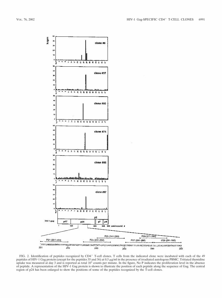
FIG. 2. Identification of peptides recognized by CD4� T-cell clones. T cells from the indicated clone were incubated with each of the 49peptides of HIV-1 Gag protein (except for the peptides 35 and 36) at 0.5 �g/ml in the presence of irradiated autologous PBMC. Tritiated thymidineuptake was measured at day 3 and is reported as total 103 counts per minute. In the figure, No P indicates the proliferation level in the absenceof peptide. A representation of the HIV-1 Gag protein is shown to illustrate the position of each peptide along the sequence of Gag. The centralregion of p24 has been enlarged to show the positions of some of the peptides recognized by the T-cell clones.
VOL. 76, 2002 HIV-1 Gag-SPECIFIC CD4� T-CELL CLONES 6991

in four clones, further confirming the true clonality of limiting-dilution T-cell clone 74. In conclusion, our results suggest thatlimiting-dilution T-cell clones 6, 37, 50, and 74 are true clonalcell populations, while 85 and 97 consist of mixed cell popu-lations. Nevertheless, since lines 85 and 97 exhibit functionalclonal behavior, we shall henceforth refer to these two linesand the four true clones collectively as clones, according to theconvention adopted by Norris et al. (44).
Determination of sensitivity to peptide activation of Gagpeptide-specific T-cell clones. In order to understand whetherthe Gag peptide-specific CD4� clones isolated from the HIV-1-negative individual reacted with physiological concentrationof their specific peptides, we performed proliferation assayswith various concentrations of peptides. Cells were incubatedwith concentrations of peptide from 0.03 ng/ml to 2 �g/ml, andproliferation was measured as described above. The results areshown in Fig. 4. Most clones started to proliferate in responseto concentrations of peptide ranging from 5 to 50 ng/ml (2.5 to25 nM) (Fig. 4A). Interestingly clone 6 showed an extremelyhigh sensitivity to stimulation with peptide 271-290, starting to
proliferate in response to concentrations of this peptide as lowas 0.3 ng/ml (0.15 nM). Clone 6 also proliferated specifically inresponse to peptides 231-250 and 281-300, as expected fromthe epitope mapping experiments, but at higher concentra-tions, suggesting that peptide 271-290 contains the mainepitope of this clone (Fig. 4B). Clone 74 was stimulated by twodistinct peptides but at very different concentrations, 200 ng/ml(100 nM) and 1 �g/ml (0.5 �M) for peptides 480-500 and261-280, respectively (Fig. 4C). The cell line 85 demonstrateda specific reactivity to peptide 201-220 but only at high con-centrations, and even at such concentrations, the proliferationlevel remained low.
Analysis of cytokines secreted by Gag peptide-specific CD4�
T-cell clones upon stimulation. Two subtypes of CD4� Thelper cells, T helper 1 (Th1) and T helper 2 (Th2), have beendescribed, differing in functional role and pattern of cytokinesecretion. Typically, Th1 cells secrete IL-2 and IFN-�, provid-ing support for cellular immune responses, although IL-2 se-cretion in human T cells is not as restricted to the Th1 subsetas in mouse T cells (43). Th2 cells secrete IL-4 and IL-5 among
FIG. 3. TCR characterization of T-cell clones by reverse transcription-PCR. (A) A set of 25 primers, specific for each V� gene family, was usedto perform reverse transcription-PCR in order to determine the V� usage of each T-cell clone. Clone 50 is shown here as a representative example.cDNA from PBMC was used as a control for the primer set. (B) Reverse transcription-PCR on clone 6 with primers specific for two subvariantsof V� 6 and V� 17 (6s5A1N1 and 17s1A1T, respectively) identified by sequencing of the TCR of clone 6. A pair of primers annealing to theconstant region of TCR � chain were included as a positive control (C�).
TABLE 1. Characteristics of HIV-1-specific CD4� T-cell clones
Clone no.Peptide
Th subtype TCR V�No. Positions in Gag Sequence
6 29 281–300 SILDIRQGPKEPFRDYVDRF Th1 6 (6s5A1N1)28 271–290 NKIVRMYSPTSILDIRQGPK24 231–250 PRGSDIAGTTSTLQEQIGWM
37 28 271–290 NKIVRMYSPTSILDIRQGPK Th2 3
50 25 241–260 STLQEQIGWMTNNPPIPVGE Th0 17
74 27 261–280 IYKRWIILGLNKIVRMYSPT Th1 13 (13s1)49 480–500 DKELYPLTSLRSLFGNDPSSQ
85 21 201–220 LKETINEEAAEWDRVHPVHA Mixed 8, 18
97 28 271–290 NKIVRMYSPTSILDIRQGPK Mixed 9, 14
6992 VENTURINI ET AL. J. VIROL.
other cytokines and preferentially support the humoral im-mune response. In order to determine the Th1 or Th2 pheno-type of the six HIV-1 Gag peptide-specific CD4� T-cell clones,we determined the cytokines produced following stimulationby intracellular staining and FACS analysis. The cells were firststimulated with anti-CD3 antibody or with their specific pep-tide in the presence of brefeldin A, a compound that blocksextracellular secretion. After 6 h the cells were fixed and per-meabilized to allow intracellular detection of cytokines by an-tibodies conjugated to fluorochromes. We determined the cy-tokine production profile of each of the HIV-1 Gag peptide-specific CD4� clones in response to activation. Figure 5 showsthe intracellular production of IFN-� and IL-2 (panel A) andIL-4 and IL-5 (panel B) for three representative clones inresponse to anti-CD3 antibody or peptide activation. The Thelper phenotypes of the clones are summarized in Table 1.
All the CD4� clones expressed high levels of IL-2 whenstimulated with their specific peptides, and therefore we basedthe distinction between Th1 and Th2 on the detection ofIFN-�, IL-4, and IL-5. As described by others, IL-4 staining isweak for stimulated CD4� T cells, but it is still sufficient toallow Th1-Th2 discrimination. The two clones 6 and 74 couldbe clearly defined as Th1 because they expressed high levels ofIFN-� and did not express IL-4 and IL-5. Of note, followingactivation of clone 6 with its specific peptide, 100% of the cellsresponded by cytokine production, confirming the clonal na-ture of the cell population. Clone 37 was characterized as Th2because it did not express IFN-� and expressed IL-4. Clone 50expressed IFN-�, IL-4, and low levels of IL-5 and was thusdefined as Th0, a subset of T cells characterized by the secre-tion of both Th1 and Th2 types of cytokines (42, 63). Triplestaining confirmed that the same cells were able to produce
IL-2, IFN-�, and IL-4 (data not shown). The remaining twocell lines, 85 and 97, were confirmed to be composed of anonclonal population of cells, as discussed previously, becausethe IFN-�-positive cells and the IL-4-positive cells constitutedtwo distinct populations. The cytokine production of these twolines was low upon peptide stimulation, suggesting the pres-ence of a relatively small percentage of peptide-specific cells.An alternative explanation is that intracellular cytokine stain-ing was not as sensitive as the proliferation assay in reportingon T-cell activation, at least in these instances.
HLA class II restriction of Gag peptide-specific CD4� T-cellclones. Intracellular cytokine FACS staining was also used toidentify the HLA class II restriction of peptide recognition bythe Gag peptide-specific CD4� T-cell clones. The donor usedfor the cloning was DR4, DR7, DQ2, and DQ3. Each clonewas incubated with the corresponding peptide with or withoutthe addition of anti-DR or anti-DQ antibody, and the assaywas performed as described before. The peptide-mediated ac-tivation of the T-cell clones tested was significantly blocked bythe anti-DR antibody and not affected by the anti-DQ anti-body, as shown for one representative clone in Fig. 6A. Theanti-DR antibody did not inhibit the nonspecific activation ofthe clones mediated by the anti-CD3 antibody (data notshown). This result suggested that peptide recognition wasrestricted by HLA class II DR molecules in all the clones.
Since additional experiments with clone 6 were planned, weextended the HLA restriction analysis for this clone. A panelof HLA DR1-, DR7-, DR4W10-, DR4W14-, and DR4W15-transfected fibroblasts were used as antigen-presenting cells,and activation of clone 6 was detected by intracellular IL-2staining (25). Clone 6 was activated only when its specificpeptide was presented by fibroblasts transfected with the HLA
FIG. 4. Sensitivity of HIV-1 Gag peptide-specific CD4� T-cell clones to activation mediated by individual peptides. Each CD4� T-cell clonewas stimulated with different concentrations of the indicated peptide in the presence of irradiated autologous PBMC. Ctrl P indicates proliferationin the presence of an irrelevant peptide. The lymphoproliferative response was measured after 3 days by [3H]thymidine uptake and is expressedas total 103 counts per minute.
VOL. 76, 2002 HIV-1 Gag-SPECIFIC CD4� T-CELL CLONES 6993

DR4 subtype W14 or W15 (Fig. 6B). No stimulation was de-tected with fibroblasts expressing HLA DR1, DR7, orDR4W10. Of note, upon activation, clone 6 induced a cyto-pathic effect in the adherent layer of fibroblasts expressing
HLA DR4W14 and -W15, leading to detachment from theplate of some cells visible in the FACS analysis as an additionalCD4� IL-2� population of cells.
Analysis of activation of CD4� T-cell clones in response to
FIG. 5. Intracellular cytokine production by Gag peptide-specific CD4� T-cell clones. Cells from the indicated CD4� T-cell clones wereincubated with anti-CD3 antibody (50 ng/ml) or with the indicated peptide (0.5 �g/ml) for 6 h in the presence of brefeldin A and then stainedintracellularly with fluorescently labeled antibodies for FACS analysis. Cells were double-stained with anti-IFN-�–PE and anti-IL-2–Apc (A) orwith anti-IL-4–FITC and anti-IL-5–PE (B).
FIG. 6. HLA class II restriction of CD4� T-cell clone 6 recognition of peptide 271-290. (A) T cells were incubated with 0.5 �g of peptide271-290 per ml and brefeldin A in the presence of 0.5 �g of anti-DR or anti-DQ antibodies per ml or without antibody (Ab) for 6 h. IntracellularFACS staining was performed with anti-IFN-�–PE and anti-IL-2–Apc antibodies. (B) Fibroblasts stably transfected with the indicated HLAmolecules were seeded on plates and pulsed for 2 h with peptide 271-290. The excess peptide was removed, and the cells of clone 6 were addedto the fibroblasts in the presence of brefeldin A. After 6 h, the nonadherent cells were stained extracellularly with anti-CD4–PE and intracellularlywith IL-2–Apc.
6994 VENTURINI ET AL. J. VIROL.
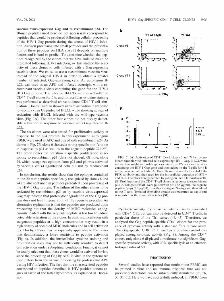
vaccinia virus-expressed Gag and to recombinant p24. The20-mer peptides used here do not necessarily correspond topeptides that would be produced following cellular processingof the HIV-1 Gag protein during the course of HIV-1 infec-tion. Antigen processing into small peptides and the presenta-tion of these peptides on HLA class II depends on multiplefactors and is hard to predict. To determine whether the pep-tides recognized by the clones that we have isolated could bepresented following HIV-1 infection, we first studied the reac-tivity of these clones to cells infected with a Gag-expressingvaccinia virus. We chose to use a recombinant vaccinia virusinstead of the original HIV-1 in order to obtain a greaternumber of infected, Gag-expressing cells. An autologous B-LCL was used as an APC and infected overnight with a re-combinant vaccinia virus containing the gene for the HIV-1IIIB Gag protein. The infected B-LCLs were mixed with theCD4� T-cell clones for 6 h, and intracellular cytokine stainingwas performed as described above to detect CD4� T-cell stim-ulation. Clones 6 and 74 showed signs of activation in responseto vaccinia virus Gag-infected B-LCL while showing no sign ofactivation with B-LCL infected with the wild-type vacciniavirus (Fig. 7A). The other four clones did not display detect-able activation in response to vaccinia virus Gag-infected B-LCL.
The six clones were also tested for proliferative activity inresponse to the p24 protein. In this experiment, autologousPBMC were used as APC and pulsed with recombinant p24. Asshown in Fig. 7B, clone 6 showed a strong specific proliferationin response to p24 as well as to the cognate peptide 271-290.The other clones did not show a specific proliferation in re-sponse to recombinant p24 (data not shown). Of note, clone74, which recognizes epitopes from p24 and p6, was activatedby vaccinia virus-Gag-infected cells but not by recombinantp24.
In conclusion, the results show that the epitopes containedin the 20-mer peptides specifically recognized by clones 6 and74 are also contained in peptides presented upon processing ofthe HIV-1 Gag protein. The failure of the other clones to beactivated by recombinant p24 or by vaccinia virus-expressedGag may indicate that proteolytic degradation of the Gag pro-tein does not lead to generation of the requisite peptides. Analternative explanation is that the peptides are produced uponprocessing but that the density of MHC molecules endog-enously loaded with the requisite peptide is too low to inducedetectable activation of the clones. In contrast, incubation withexogenous peptides at a high concentration may result in ahigh density of occupied MHC molecules and in cell activation(7). This hypothesis may be especially applicable to the clonesthat demonstrated a lower sensitivity to peptide activation(Fig. 4). In addition, the intracellular cytokine staining andproliferation assay may not be sufficiently sensitive to detectcell activation under suboptimal conditions. Finally, it cannotbe totally ruled out that these clones would be activated in vivo,since the processing of Gag by APC in vitro in the systems weused differs from the in vivo processing by professional APCduring HIV infection. The fact that the characterized peptidescorrespond to peptides described in HIV-positive donors ar-gues in favor of the latter hypothesis, as explained in Discus-sion.
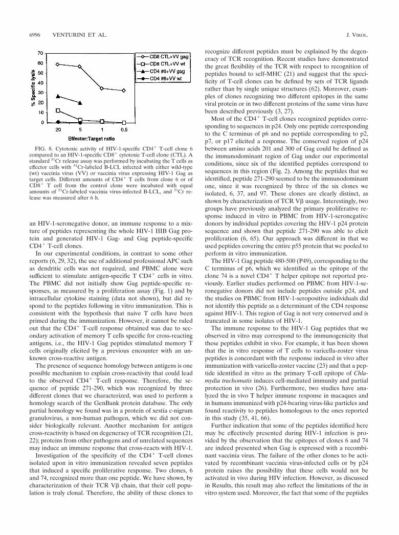
Cytotoxic activity. Cytotoxic activity is usually associatedwith CD8� CTL but can also be detected in CD4� T cells, inparticular those of the Th1 subset (44, 45). Therefore, weanalyzed the Gag peptide-specific CD4� clones for the pres-ence of cytotoxic activity with a standard 51Cr release assay.The Gag-specific CD8� CTL used as a positive control dis-played strong cytotoxic activity (Fig. 8). Among the CD4�
clones, only clone 6 displayed a moderate but significant Gag-specific cytotoxic activity, with 20% specific lysis at an effector-to-target ratio of 5:1.
DISCUSSION
Several studies have reported that nonimmune PBMC canbe primed in vitro and an immune response that was notpreviously detectable can be subsequently stimulated (23, 26,30, 31, 61). Here we have successfully induced, in PBMC from
FIG. 7. (A) Activation of CD4� T-cell clones 6 and 74 by recom-binant vaccinia virus-infected cells expressing HIV-1 Gag. B-LCL wereinfected overnight with wild-type vaccinia virus (VV) or vaccinia viruscontaining the HIV-1 Gag gene and then added to the T cells for 6 hin the presence of brefeldin A. The cells were stained with anti-CD4–FITC antibody and then used for the intracellular detection of IFN-�and IL-2. The plots were generated by gating on the CD4-positive cells.(B) Proliferation of the CD4� T-cell clones in response to recombinantp24. Autologous PBMC were pulsed with p24 (2.5 �g/ml), the cognatepeptide (pep) (2.5 �g/ml), or without antigen (No-Ag) and then addedto the T cells. Tritiated thymidine uptake was measured at day 3 andis reported as the stimulation index (SI).
VOL. 76, 2002 HIV-1 Gag-SPECIFIC CD4� T-CELL CLONES 6995
an HIV-1-seronegative donor, an immune response to a mix-ture of peptides representing the whole HIV-1 IIIB Gag pro-tein and generated HIV-1 Gag- and Gag peptide-specificCD4� T-cell clones.
In our experimental conditions, in contrast to some otherreports (6, 29, 32), the use of additional professional APC suchas dendritic cells was not required, and PBMC alone weresufficient to stimulate antigen-specific T CD4� cells in vitro.The PBMC did not initially show Gag peptide-specific re-sponses, as measured by a proliferation assay (Fig. 1) and byintracellular cytokine staining (data not shown), but did re-spond to the peptides following in vitro immunization. This isconsistent with the hypothesis that naive T cells have beenprimed during the immunization. However, it cannot be ruledout that the CD4� T-cell response obtained was due to sec-ondary activation of memory T cells specific for cross-reactingantigens, i.e., the HIV-1 Gag peptides stimulated memory Tcells originally elicited by a previous encounter with an un-known cross-reactive antigen.
The presence of sequence homology between antigens is onepossible mechanism to explain cross-reactivity that could leadto the observed CD4� T-cell response. Therefore, the se-quence of peptide 271-290, which was recognized by threedifferent clones that we characterized, was used to perform ahomology search of the GenBank protein database. The onlypartial homology we found was in a protein of xestia c-nigrumgranulovirus, a non-human pathogen, which we did not con-sider biologically relevant. Another mechanism for antigencross-reactivity is based on degeneracy of TCR recognition (21,22); proteins from other pathogens and of unrelated sequencesmay induce an immune response that cross-reacts with HIV-1.
Investigation of the specificity of the CD4� T-cell clonesisolated upon in vitro immunization revealed seven peptidesthat induced a specific proliferative response. Two clones, 6and 74, recognized more than one peptide. We have shown, bycharacterization of their TCR V� chain, that their cell popu-lation is truly clonal. Therefore, the ability of these clones to
recognize different peptides must be explained by the degen-eracy of TCR recognition. Recent studies have demonstratedthe great flexibility of the TCR with respect to recognition ofpeptides bound to self-MHC (21) and suggest that the speci-ficity of T-cell clones can be defined by sets of TCR ligandsrather than by single unique structures (62). Moreover, exam-ples of clones recognizing two different epitopes in the sameviral protein or in two different proteins of the same virus havebeen described previously (3, 27).
Most of the CD4� T-cell clones recognized peptides corre-sponding to sequences in p24. Only one peptide correspondingto the C terminus of p6 and no peptide corresponding to p2,p7, or p17 elicited a response. The conserved region of p24between amino acids 201 and 300 of Gag could be defined asthe immunodominant region of Gag under our experimentalconditions, since six of the identified peptides correspond tosequences in this region (Fig. 2). Among the peptides that weidentified, peptide 271-290 seemed to be the immunodominantone, since it was recognized by three of the six clones weisolated, 6, 37, and 97. These clones are clearly distinct, asshown by characterization of TCR V� usage. Interestingly, twogroups have previously analyzed the primary proliferative re-sponse induced in vitro in PBMC from HIV-1-seronegativedonors by individual peptides covering the HIV-1 p24 proteinsequence and shown that peptide 271-290 was able to elicitproliferation (6, 65). Our approach was different in that weused peptides covering the entire p55 protein that we pooled toperform in vitro immunization.
The HIV-1 Gag peptide 480-500 (P49), corresponding to theC terminus of p6, which we identified as the epitope of theclone 74 is a novel CD4� T helper epitope not reported pre-viously. Earlier studies performed on PBMC from HIV-1-se-ronegative donors did not include peptides outside p24, andthe studies on PBMC from HIV-1-seropositive individuals didnot identify this peptide as a determinant of the CD4 responseagainst HIV-1. This region of Gag is not very conserved and istruncated in some isolates of HIV-1.
The immune response to the HIV-1 Gag peptides that weobserved in vitro may correspond to the immunogenicity thatthese peptides exhibit in vivo. For example, it has been shownthat the in vitro response of T cells to varicella-zoster viruspeptides is concordant with the response induced in vivo afterimmunization with varicella-zoster vaccine (23) and that a pep-tide identified in vitro as the primary T-cell epitope of Chla-mydia trachomatis induces cell-mediated immunity and partialprotection in vivo (26). Furthermore, two studies have ana-lyzed the in vivo T helper immune response in macaques andin humans immunized with p24-bearing virus-like particles andfound reactivity to peptides homologous to the ones reportedin this study (35, 41, 66).
Further indication that some of the peptides identified heremay be effectively presented during HIV-1 infection is pro-vided by the observation that the epitopes of clones 6 and 74are indeed presented when Gag is expressed with a recombi-nant vaccinia virus. The failure of the other clones to be acti-vated by recombinant vaccinia virus-infected cells or by p24protein raises the possibility that these cells would not beactivated in vivo during HIV infection. However, as discussedin Results, this result may also reflect the limitations of the invitro system used. Moreover, the fact that some of the peptides
FIG. 8. Cytotoxic activity of HIV-1-specific CD4� T-cell clone 6compared to an HIV-1-specific CD8� cytotoxic T-cell clone (CTL). Astandard 51Cr release assay was performed by incubating the T cells aseffector cells with 51Cr-labeled B-LCL infected with either wild-type(wt) vaccinia virus (VV) or vaccinia virus expressing HIV-1 Gag astarget cells. Different amounts of CD4� T cells from clone 6 or ofCD8� T cell from the control clone were incubated with equalamounts of 51Cr-labeled vaccinia virus-infected B-LCL, and 51Cr re-lease was measured after 6 h.
6996 VENTURINI ET AL. J. VIROL.

we identified as CD4� T-cell epitopes have been shown byothers to be recognized by CD4� T cells from HIV-1-positivepatients suggests that these peptides may be effectively pre-sented through the natural processing of HIV-1 Gag duringinfection.
Peptides 278-292 and 265-286 were found to induce prolif-erative responses in 50 and 33%, respectively, of HIV-1-posi-tive individuals (2, 67). Peptides almost equivalent to our im-munodominant peptide 271-290 and to peptide 241-260 wereidentified by Rosenberg et al. among the determinants of thestrong p24-specific helper cell response observed in two long-term nonprogressor HIV-1-positive patients (55). Further-more, in a recent study, the latter group isolated clones fromHIV-1-infected individuals and characterized epitopes thatoverlapped the epitopes found in our study, in particular in theregion between amino acids 231 and 292 of Gag (44). Theseresults collectively suggest that this region is one of the mostimmunogenic regions of HIV-1 Gag. The region is able toinduce a CD4� T-cell response by immunization followingnatural HIV-1 infection, by vaccination of HIV-1-negative in-dividuals with p24, and following in vitro stimulation of PBMCfrom HIV-1-negative individuals.
Based on their ability to induce an HIV-1-specific immuneresponse, irrespective of whether this is a primary or secondaryresponse, some of the peptides that we identified could becandidates to include in a vaccine against HIV-1. Due to thepolymorphic nature of HLA, only if a peptide could be accom-modated in the groove of several HLA class II moleculeswould it seem a suitable vaccine candidate (14). The HLA classII type of the donor we used was DR4, DR7, DQ2, and DQ3,and we showed that all the peptides that we identified wererecognized by CD4� T cells in the context of HLA DR (Fig. 7).Each of the peptides contained a putative HLA DR4 or DR7binding motif, based on data reported by Marsh et al. andHammer et al. (16, 17, 34). Two possible binding motifs werefound in peptide 271-290. Of note, as only one individual wasused in our study, it is not possible to determine whether thecapacity to mount an in vitro primary immune response to Gagpeptides is related to HLA class II type. In earlier similarstudies, the HLA types of the donors were not specified.
Functional analysis of the in vitro-generated HIV-1 Gag-and Gag peptide-specific CD4� T-cell clones indicates thatmost of the clones proliferate to peptide concentrations thatfall within the range of those reported in the literature fromimmune donors, showing that the clones that we obtained afterin vitro immunization have binding characteristics similar tothose of clones raised in vivo (1, 12, 20, 44, 57, 64). Both Th1and Th2 types of CD4� T cells were obtained, suggesting thatby immunizing with HIV-1 Gag peptides, it may be possible toinduce a CD4� T-helper response of both types in PBMC froma seronegative individual. The presence of CD4� T helper cellsthat could support both the cellular (Th1) and humoral (Th2)responses against HIV-1 could be crucial for protection fromHIV-1 infection. Hasenkrug et al. demonstrated the require-ment for B cells, CD4� T cells, and CD8� T cells in protectionfrom Friend virus infection in a mouse model (19).
Similarly, multiple components of the immune response arelikely to be important in the control of HIV-1 replication andspread. As noted above, the main role of CD4� T cells mayreside in their ability to maintain functional cytotoxic CD8�
T-cell responses. The mechanism by which T helper cells fulfillthis role is poorly understood. A role for IL-2, IL-7, IL-15, andmore recently IL-4 has been evoked, although more for thedevelopment than for the maintenance of the CD8 response(9, 33, 58). Such role in HIV infection remains to be demon-strated, and it is thus unclear whether the CD4� T-cell clonesthat we have characterized could fulfill this role.
As well as their helper role, CD4� T cells can also have adirect antigen-specific cytotoxic activity, as shown here by theactivity of clone 6 against vaccinia virus Gag-infected cells.Interestingly, Gag-specific CD4� T-cell clones isolated by Nor-ris et al. from HIV-1-infected individuals also displayed cyto-toxic activity (44). Even though the function of CD4� T cells askiller cells is usually not considered as important as their roleas helper cells (45), this function may still contribute to theelimination of HIV-1-infected cells and could play a role invivo in the control of HIV-1 infection by CD4� T cells.
In summary, the results presented provide evidence thatHIV-1 Gag peptides are able to induce a strong CD4� T-cellimmune response in PBMC from an HIV-1-seronegative do-nor in vitro. The clones isolated represent a useful tool for theanalysis of interactions between CD4� and CD8� T cells invitro and in the hu-PBL-SCID mouse model of HIV-1 infec-tion. The immunodominant peptides that were identified, inparticular the peptides corresponding to the conserved regionof HIV-1 Gag between amino acids 231 and 292, may repre-sent potential candidates to be included in a vaccine againstHIV-1. Finally, some of the clones that we have identified arein many aspects similar to clones isolated from HIV-1-infectedpatients in a recent study by Norris et al.; they recognize thesame region of the Gag protein, produce IFN-�, and displaycytotoxic activity. This may indicate that some of the clonesthat we have characterized are representative of clones raisedin vivo during the natural infection. However, we also note thatthe opposite argument can be made. The similarity betweenthe CD4� T-cell responses in seropositive and seronegativeindividuals suggests that studies of the CD4� T-cell responsein HIV-infected persons, especially with prolonged in vitroantigenic stimulation, could be reflecting in vitro-primed orcross-reactive responses and, as suggested by Norris et al. intheir discussion, not responses that exist in vivo. Our studycertainly raises concerns about this point and suggests that careshould be taken in the methods used to study the CD4� re-sponse in HIV-infected patients.
ACKNOWLEDGMENTS
We thank Bette Korber and Kersti Rock for help with the proteindatabase search and Louis Picker for manuscript review.
This work was supported by NIH grants AI45357 (P.P.), HL59727(D.R.B.), and AI45376 (D.R.B.). P.P. acknowledges a Scholar Awardfrom the Pediatric AIDS Foundation (77370).
REFERENCES
1. Adams, E., A. Basten, S. Rodda, and W. J. Britton. 1997. Human T-cellclones to the 70-kilodalton heat shock protein of Mycobacterium leprae definemycobacterium-specific epitopes rather than shared epitopes. Infect. Im-mun. 65:1061–1070.
2. Adams, S. L., R. A. Biti, and G. J. Stewart. 1997. T-cell response to HIV innatural infection: optimized culture conditions for detecting responses toGag peptides. J. Acquir. Immune Defic. Syndr. Hum. Retrovirol. 15:257–263.
3. Anderson, R. W., J. R. Bennink, J. W. Yewdell, W. L. Maloy, and J. E.Coligan. 1992. Influenza basic polymerase 2 peptides are recognized by
VOL. 76, 2002 HIV-1 Gag-SPECIFIC CD4� T-CELL CLONES 6997
influenza nucleoprotein-specific cytotoxic T lymphocytes. Mol. Immunol.29:1089–1096.
4. Arden, B., S. P. Clark, D. Kabelitz, and T. W. Mak. 1995. Human T-cellreceptor variable gene segment families. Immunogenetics 42:455–500.
5. Baba, T. W., V. Liska, R. Hofmann-Lehmann, J. Vlasak, W. Xu, S. Ayehunie,L. A. Cavacini, M. R. Posner, H. Katinger, G. Stiegler, B. J. Bernacky, T. A.Rizvi, R. Schmidt, L. R. Hill, M. E. Keeling, Y. Lu, J. E. Wright, T. C. Chou,and R. M. Ruprecht. 2000. Human neutralizing monoclonal antibodies of theIgG1 subtype protect against mucosal simian-human immunodeficiency virusinfection. Nat. Med. 6:200–206.
6. Bedford, P. A., L. B. Clarke, G. Z. Hastings, and S. C. Knight. 1997. Primaryproliferative responses to peptides of HIV Gag p24. J. Acquir. ImmuneDefic. Syndr. Hum. Retrovirol. 14:301–306.
7. Carbone, F. R., M. W. Moore, J. M. Sheil, and M. J. Bevan. 1988. Inductionof cytotoxic T lymphocytes by primary in vitro stimulation with peptides. J.Exp. Med. 167:1767–1779.
8. Carmichael, A., X. Jin, P. Sissons, and L. Borysiewicz. 1993. Quantitativeanalysis of the human immunodeficiency virus type 1 (HIV-1)-specific cyto-toxic T lymphocyte (CTL) response at different stages of HIV-1 infection:differential CTL responses to HIV-1 and Epstein-Barr virus in late disease.J. Exp. Med. 177:249–256.
9. Carvalho, L. H., G. Sano Gi, J. C. Hafalla, A. Morrot, M. A. de Lafaille, andF. Zavala. 2002. IL-4-secreting CD4� T cells are crucial to the developmentof CD8� T-cell responses against malaria liver stages. Nat. Med. 8:166–170.
10. Ciurea, A., L. Hunziker, P. Klenerman, H. Hengartner, and R. Zinkernagel.2001. Impairment of CD4(�) T-cell responses during chronic virus infectionprevents neutralizing antibody responses against virus escape mutants. J.Exp. Med. 193:297–306.
11. Curiel, T. J., J. T. Wong, P. F. Gorczyca, R. T. Schooley, and B. D. Walker.1993. CD4� human immunodeficiency virus type 1 (HIV-1) envelope-spe-cific cytotoxic T lymphocytes derived from the peripheral blood cells of anHIV-1-infected individual. AIDS Res. Hum. Retroviruses 9:61–68.
12. Diepolder, H. M., J. T. Gerlach, R. Zachoval, R. M. Hoffmann, M. C. Jung,E. A. Wierenga, S. Scholz, T. Santantonio, M. Houghton, S. Southwood, A.Sette, and G. R. Pape. 1997. Immunodominant CD4� T-cell epitope withinnonstructural protein 3 in acute hepatitis C virus infection. J. Virol. 71:6011–6019.
13. Dittmer, U., D. M. Brooks, and K. J. Hasenkrug. 1999. Requirement formultiple lymphocyte subsets in protection by a live attenuated vaccineagainst retroviral infection. Nat. Med. 5:189–193.
14. Doolan, D. L., S. Southwood, R. Chesnut, E. Appella, E. Gomez, A. Richards,Y. I. Higashimoto, A. Maewal, J. Sidney, R. A. Gramzinski, C. Mason, D.Koech, S. L. Hoffman, and A. Sette. 2000. HLA-DR-promiscuous T cellepitopes from Plasmodium falciparum pre-erythrocytic-stage antigens re-stricted by multiple HLA class II alleles. J. Immunol. 165:1123–1137.
15. Gauduin, M. C., P. W. Parren, R. Weir, C. F. Barbas, D. R. Burton, and R. A.Koup. 1997. Passive immunization with a human monoclonal antibody pro-tects hu-PBL-SCID mice against challenge by primary isolates of HIV-1.Nat. Med. 3:1389–1393.
16. Hammer, J. 1995. New methods to predict MHC-binding sequences withinprotein antigens. Curr. Opin. Immunol. 7:263–269.
17. Hammer, J., Z. A. Nagy, and F. Sinigaglia. 1994. Rules governing peptide-class II MHC molecule interactions. Behring Inst. Mitt. 94:124–132.
18. Hanke, T., R. V. Samuel, T. J. Blanchard, V. C. Neumann, T. M. Allen, J. E.Boyson, S. A. Sharpe, N. Cook, G. L. Smith, D. I. Watkins, M. P. Cranage,and A. J. McMichael. 1999. Effective induction of simian immunodeficiencyvirus-specific cytotoxic T lymphocytes in macaques by using a multiepitopegene and DNA prime-modified vaccinia virus Ankara boost vaccinationregimen. J. Virol. 73:7524–7532.
19. Hasenkrug, K. J., and U. Dittmer. 2000. The role of CD4 and CD8 T cellsin recovery and protection from retroviral infection: lessons from the Friendvirus model. Virology 272:244–249.
20. Hemmer, B., B. Gran, Y. Zhao, A. Marques, J. Pascal, A. Tzou, T. Kondo, I.Cortese, B. Bielekova, S. E. Straus, H. F. McFarland, R. Houghten, R.Simon, C. Pinilla, and R. Martin. 1999. Identification of candidate T-cellepitopes and molecular mimics in chronic Lyme disease. Nat. Med. 5:1375–1382.
21. Hemmer, B., M. Vergelli, C. Pinilla, R. Houghten, and R. Martin. 1998.Probing degeneracy in T-cell recognition with peptide combinatorial librar-ies. Immunol. Today 19:163–168.
22. Ignatowicz, L., W. Rees, R. Pacholczyk, H. Ignatowicz, E. Kushnir, J. Kap-pler, and P. Marrack. 1997. T cells can be activated by peptides that areunrelated in sequence to their selecting peptide. Immunity 7:179–186.
23. Jenkins, D. E., L. L. Yasukawa, R. Bergen, C. Benike, E. G. Engleman, andA. M. Arvin. 1999. Comparison of primary sensitization of naive human Tcells to varicella-zoster virus peptides by dendritic cells in vitro with re-sponses elicited in vivo by varicella vaccination. J. Immunol. 162:560–567.
24. Klein, M. R., C. A. van Baalen, A. M. Holwerda, S. R. Kerkhof Garde, R. J.Bende, I. P. Keet, J. K. Eeftinck-Schattenkerk, A. D. Osterhaus, H. Schuite-maker, and F. Miedema. 1995. Kinetics of Gag-specific cytotoxic T lympho-cyte responses during the clinical course of HIV-1 infection: a longitudinal
analysis of rapid progressors and long-term asymptomatics. J. Exp. Med.181:1365–1372.
25. Klohe, E. P., R. Watts, M. Bahl, C. Alber, W. Y. Yu, R. Anderson, J. Silver,P. K. Gregersen, and R. W. Karr. 1988. Analysis of the molecular specificitiesof anti-class II monoclonal antibodies by using L cell transfectants expressingHLA class II molecules. J. Immunol. 141:2158–2164.
26. Knight, S. C., S. Iqball, C. Woods, A. Stagg, M. E. Ward, and M. Tuffrey.1995. A peptide of Chlamydia trachomatis shown to be a primary T-cellepitope in vitro induces cell-mediated immunity in vivo. Immunology 85:8–15.
27. Kuwano, K., V. E. Reyes, R. E. Humphreys, and F. A. Ennis. 1991. Recog-nition of disparate HA and NS1 peptides by an H-2Kd-restricted, influenzaspecific CTL clone. Mol. Immunol. 28:1–7.
28. Littaua, R. A., M. B. Oldstone, A. Takeda, and F. A. Ennis. 1992. A CD4�
cytotoxic T-lymphocyte clone to a conserved epitope on human immunode-ficiency virus type 1 p24: cytotoxic activity and secretion of interleukin-2 andinterleukin-6. J. Virol. 66:608–611.
29. Macatonia, S. E., S. Patterson, and S. C. Knight. 1991. Primary proliferativeand cytotoxic T-cell responses to HIV induced in vitro by human dendriticcells. Immunology 74:399–406.
30. Macatonia, S. E., P. M. Taylor, S. C. Knight, and B. A. Askonas. 1989.Primary stimulation by dendritic cells induces antiviral proliferative andcytotoxic T-cell responses in vitro. J. Exp. Med. 169:1255–1264.
31. Manca, F., J. Habeshaw, and A. Dalgleish. 1991. The naive repertoire ofhuman T helper cells specific for gp120, the envelope glycoprotein of HIV.J. Immunol. 146:1964–1971.
32. Manca, F., G. Li Pira, D. Fenoglio, S. P. Fang, A. Habeshaw, S. C. Knight,and A. G. Dalgleish. 1994. Dendritic cells are potent antigen-presenting cellsfor in vitro induction of primary human CD4� T-cell lines specific for HIVgp120. J. Acquir. Immune Defic. Syndr. 7:15–23.
33. Marrack, P., J. Bender, D. Hildeman, M. Jordan, T. Mitchell, M. Murakami,A. Sakamoto, B. C. Schaefer, B. Swanson, and J. Kappler. 2000. Homeosta-sis of alpha beta TCR� T cells. Nat. Immunol. 1:107–111.
34. Marsh, S. G. E., P. Parham, and L. D. Barber. 2000. The HLA FactsBook,1st ed. Academic Press, London, England.
35. Martin, S. J., A. Vyakarnam, R. Cheingsong-Popov, D. Callow, K. L. Jones,J. M. Senior, S. E. Adams, A. J. Kingsman, P. Matear, F. M. Gotch, et al.1993. Immunization of human HIV-seronegative volunteers with recombi-nant p17/p24:Ty virus-like particles elicits HIV-1 p24-specific cellular andhumoral immune responses. AIDS 7:1315–1323.
36. Mascola, J. R., M. G. Lewis, G. Stiegler, D. Harris, T. C. VanCott, D. Hayes,M. K. Louder, C. R. Brown, C. V. Sapan, S. S. Frankel, Y. Lu, M. L. Robb,H. Katinger, and D. L. Birx. 1999. Protection of macaques against patho-genic simian/human immunodeficiency virus 89.6PD by passive transfer ofneutralizing antibodies. J. Virol. 73:4009–4018.
37. Mascola, J. R., G. Stiegler, T. C. VanCott, H. Katinger, C. B. Carpenter,C. E. Hanson, H. Beary, D. Hayes, S. S. Frankel, D. L. Birx, and M. G. Lewis.2000. Protection of macaques against vaginal transmission of a pathogenicHIV-1/SIV chimeric virus by passive infusion of neutralizing antibodies. Nat.Med. 6:207–210.
38. Matloubian, M., R. J. Concepcion, and R. Ahmed. 1994. CD4� T cells arerequired to sustain CD8� cytotoxic T-cell responses during chronic viralinfection. J. Virol. 68:8056–8063.
39. McKinney, D. M., D. A. Lewinsohn, S. R. Riddell, P. D. Greenberg, and D. E.Mosier. 1999. The antiviral activity of HIV-specific CD8� CTL clones islimited by elimination due to encounter with HIV-infected targets. J. Im-munol. 163:861–867.
40. Mehta-Damani, A., S. Markowicz, and E. G. Engleman. 1995. Generation ofantigen-specific CD4� T-cell lines from naive precursors. Eur J. Immunol.25:1206–1211.
41. Mills, K. H., P. A. Kitchin, B. P. Mahon, A. L. Barnard, S. E. Adams, S. M.Kingsman, and A. J. Kingsman. 1990. HIV p24-specific helper T-cell clonesfrom immunized primates recognize highly conserved regions of HIV-1.J. Immunol. 144:1677–1683.
42. Mosmann, T. R., and R. L. Coffman. 1989. Heterogeneity of cytokine secre-tion patterns and functions of helper T cells. Adv. Immunol. 46:111–147.
43. Mosmann, T. R., and S. Sad. 1996. The expanding universe of T-cell subsets:Th1, Th2 and more. Immunol. Today 17:138–146.
44. Norris, P. J., M. Sumaroka, C. Brander, H. F. Moffett, S. L. Boswell, T.Nguyen, Y. Sykulev, B. D. Walker, and E. S. Rosenberg. 2001. Multipleeffector functions mediated by human immunodeficiency virus-specificCD4� T-cell clones. J. Virol. 75:9771–9779.
45. Oxenius, A., R. M. Zinkernagel, and H. Hengartner. 1998. CD4� T-cellinduction and effector functions: a comparison of immunity against solubleantigens and viral infections. Adv. Immunol. 70:313–367.
46. Picker, L. J., and V. C. Maino. 2000. The CD4(�) T-cell response to HIV-1.Curr. Opin. Immunol. 12:381–386.
47. Pitcher, C. J., C. Quittner, D. M. Peterson, M. Connors, R. A. Koup, V. C.Maino, and L. J. Picker. 1999. HIV-1-specific CD4� T cells are detectablein most individuals with active HIV-1 infection, but decline with prolongedviral suppression. Nat. Med. 5:518–525.
48. Planz, O., S. Ehl, E. Furrer, E. Horvath, M. A. Brundler, H. Hengartner, and
6998 VENTURINI ET AL. J. VIROL.
R. M. Zinkernagel. 1997. A critical role for neutralizing-antibody-producingB cells, CD4(�) T cells, and interferons in persistent and acute infections ofmice with lymphocytic choriomeningitis virus: implications for adoptive im-munotherapy of virus carriers. Proc. Natl. Acad. Sci. USA 94:6874–6879.
49. Poignard, P., R. Sabbe, G. R. Picchio, M. Wang, R. J. Gulizia, H. Katinger,P. W. Parren, D. E. Mosier, and D. R. Burton. 1999. Neutralizing antibodieshave limited effects on the control of established HIV-1 infection in vivo.Immunity 10:431–438.
50. Pontesilli, O., P. Carotenuto, S. R. Kerkhof-Garde, M. T. Roos, I. P. Keet,R. A. Coutinho, J. Goudsmit, and F. Miedema. 1999. Lymphoproliferativeresponse to HIV type 1 p24 in long-term survivors of HIV type 1 infectionis predictive of persistent AIDS-free infection. AIDS Res. Hum. Retrovi-ruses 15:973–981.
51. Pontesilli, O., S. Kerkhof-Garde, D. W. Notermans, N. A. Foudraine, M. T.Roos, M. R. Klein, S. A. Danner, J. M. Lange, and F. Miedema. 1999.Functional T cell reconstitution and human immunodeficiency virus-1-spe-cific cell-mediated immunity during highly active antiretroviral therapy. J. In-fect. Dis. 180:76–86.
52. Ratto, S., K. V. Sitz, A. M. Scherer, F. Manca, L. D. Loomis, J. H. Cox, R. R.Redfield, and D. L. Birx. 1995. Establishment and characterization of humanimmunodeficiency virus type 1 (HIV-1) envelope-specific CD4� T lympho-cyte lines from HIV-1-seropositive patients. J. Infect. Dis. 171:1420–1430.
53. Riddell, S. R., M. Elliott, D. A. Lewinsohn, M. J. Gilbert, L. Wilson, S. A.Manley, S. D. Lupton, R. W. Overell, T. C. Reynolds, L. Corey, and P. D.Greenberg. 1996. T-cell mediated rejection of gene-modified HIV-specificcytotoxic T lymphocytes in HIV-infected patients. Nat. Med. 2:216–223.
54. Rosenberg, E. S., M. Altfeld, S. H. Poon, M. N. Phillips, B. M. Wilkes, R. L.Eldridge, G. K. Robbins, R. T. D’Aquila, P. J. Goulder, and B. D. Walker.2000. Immune control of HIV-1 after early treatment of acute infection.Nature 407:523–526.
55. Rosenberg, E. S., J. M. Billingsley, A. M. Caliendo, S. L. Boswell, P. E. Sax,S. A. Kalams, and B. D. Walker. 1997. Vigorous HIV-1-specific CD4� T-cellresponses associated with control of viremia. Science 278:1447–1450.
56. Rosenberg, E. S., and B. D. Walker. 1998. HIV type 1-specific helper T cells:a critical host defense. AIDS Res. Hum. Retroviruses 14(Suppl. 2):S143–S147.
57. Rothbard, J. B., R. I. Lechler, K. Howland, V. Bal, D. D. Eckels, R. Sekaly,E. O. Long, W. R. Taylor, and J. R. Lamb. 1988. Structural model ofHLA-DR1 restricted T cell antigen recognition. Cell 52:515–523.
58. Schluns, K. S., W. C. Kieper, S. C. Jameson, and L. Lefrancois. 2000.Interleukin-7 mediates the homeostasis of naive and memory CD8 T cells invivo. Nat. Immunol. 1:426–432.
59. Schmitz, J. E., M. J. Kuroda, S. Santra, V. G. Sasseville, M. A. Simon, M. A.Lifton, P. Racz, K. Tenner-Racz, M. Dalesandro, B. J. Scallon, J. Ghrayeb,
M. A. Forman, D. C. Montefiori, E. P. Rieber, N. L. Letvin, and K. A.Reimann. 1999. Control of viremia in simian immunodeficiency virus infec-tion by CD8� lymphocytes. Science 283:857–860.
60. Shibata, R., T. Igarashi, N. Haigwood, A. Buckler-White, R. Ogert, W. Ross,R. Willey, M. W. Cho, and M. A. Martin. 1999. Neutralizing antibody di-rected against the HIV-1 envelope glycoprotein can completely block HIV-1/SIV chimeric virus infections of macaque monkeys. Nat. Med. 5:204–210.
61. Siliciano, R. F., T. Lawton, C. Knall, R. W. Karr, P. Berman, T. Gregory, andE. L. Reinherz. 1988. Analysis of host-virus interactions in AIDS with anti-gp120 T-cell clones: effect of HIV sequence variation and a mechanism forCD4� cell depletion. Cell 54:561–575.
62. Sparbier, K., and P. Walden. 1999. T cell receptor specificity and mimotopes.Curr. Opin. Immunol. 11:214–218.
63. Street, N. E., J. H. Schumacher, T. A. Fong, H. Bass, D. F. Fiorentino, J. A.Leverah, and T. R. Mosmann. 1990. Heterogeneity of mouse helper T cells.Evidence from bulk cultures and limiting-dilution cloning for precursors ofTh1 and Th2 cells. J. Immunol. 144:1629–1639.
64. ter Meulen, J., M. Badusche, K. Kuhnt, A. Doetze, J. Satoguina, T. Marti, C.Loeliger, K. Koulemou, L. Koivogui, H. Schmitz, B. Fleischer, and A. Ho-erauf. 2000. Characterization of human CD4� T-cell clones recognizingconserved and variable epitopes of the Lassa virus nucleoprotein. J. Virol.74:2186–2192.
65. Vyakarnam, A., P. M. Matear, C. Cranenburg, C. Michie, P. C. Beverley, B.Wahren, S. K. Gill, and I. Weller. 1991. T-cell responses to peptides coveringthe Gag p24 region of HIV-1 occur in HIV-1 seronegative individuals. Int.Immunol. 3:939–947.
66. Vyakarnam, A., P. M. Matear, S. J. Martin, and M. Wagstaff. 1995. Th1 cellsspecific for HIV-1 Gag p24 are less efficient than Th0 cells in supporting HIVreplication, and inhibit virus replication in Th0 cells. Immunology 86:85–96.
67. Wahren, B., J. Rosen, E. Sandstrom, T. Mathiesen, S. Modrow, and H.Wigzell. 1989. HIV-1 peptides induce a proliferative response in lymphocytesfrom infected persons. J. Acquir. Immune Defic. Syndr. 2:448–456.
68. Walker, C. M., K. S. Steimer, K. L. Rosenthal, and J. A. Levy. 1988. Iden-tification of human immunodeficiency virus (HIV) envelope type-specific Thelper cells in an HIV-infected individual. J. Clin. Investig. 82:2172–2175.
69. Yasutomi, Y., S. Koenig, R. M. Woods, J. Madsen, N. M. Wassef, C. R.Alving, H. J. Klein, T. E. Nolan, L. J. Boots, J. A. Kessler, et al. 1995. Avaccine-elicited, single viral epitope-specific cytotoxic T lymphocyte responsedoes not protect against intravenous, cell-free simian immunodeficiency vi-rus challenge. J. Virol. 69:2279–2284.
70. Zajac, A. J., K. Murali-Krishna, J. N. Blattman, and R. Ahmed. 1998.Therapeutic vaccination against chronic viral infection: the importance ofcooperation between CD4� and CD8� T cells. Curr. Opin. Immunol. 10:444–449.
VOL. 76, 2002 HIV-1 Gag-SPECIFIC CD4� T-CELL CLONES 6999
Related Documents