cancers Article Characterization of HMGB1/2 Interactome in Prostate Cancer by Yeast Two Hybrid Approach: Potential Pathobiological Implications Aida Barreiro-Alonso 1, † , MaríaCámara-Quílez 1, † , Martín Salamini-Montemurri 1 , Mónica Lamas-Maceiras 1 , Ángel Vizoso-Vázquez 1 , Esther Rodríguez-Belmonte 1 , María Quindós-Varela 2 , Olaia Martínez-Iglesias 3 , Angélica Figueroa 3 and María-Esperanza Cerdán 1, * 1 EXPRELA Group, Centro de Investigacións Científicas Avanzadas (CICA), Departamento de Bioloxía, Facultade de Ciencias, INIBIC-Universidade da Coruña, Campus de A Coruña, 15071 A Coruña, Spain; [email protected] (A.B.-A.); [email protected] (M.C.-Q.); [email protected] (M.S.-M.); [email protected] (M.L.-M.); [email protected] (Á.V.-V.); [email protected] (E.R.-B.) 2 Translational Cancer Research Group, Instituto de Investigación Biomédica de A Coruña (INIBIC), Carretera del Pasaje s/n, 15006 A Coruña, Spain; [email protected] 3 Epithelial Plasticity and Metastasis Group, Instituto de Investigación Biomédica de A Coruña (INIBIC), Complexo Hospitalario Universitario de A Coruña (CHUAC), Sergas, 15006 A Coruña, Spain; [email protected] (Q.M.-I.); [email protected] (A.F.) * Correspondence: [email protected] † These authors contributed equally to this work. Received: 25 October 2019; Accepted: 2 November 2019; Published: 5 November 2019 Abstract: High mobility group box B (HMGB) proteins are pivotal in the development of cancer. Although the proteomics of prostate cancer (PCa) cells has been reported, the involvement of HMGB proteins and their interactome in PCa is an unexplored field of considerable interest. We describe herein the results of the first HMGB1/HMGB2 interactome approach to PCa. Libraries constructed from the PCa cell line, PC-3, and from patients’ PCa primary tumor have been screened by the yeast 2-hybrid approach (Y2H) using HMGB1 and HMGB2 baits. Functional significance of this PCa HMGB interactome has been validated through expression and prognosis data available on public databases. Copy number alterations (CNA) affecting these newly described HMGB interactome components are more frequent in the most aggressive forms of PCa: those of neuroendocrine origin or castration-resistant PCa. Concordantly, adenocarcinoma PCa samples showing CNA in these genes are also associated with the worse prognosis. These findings open the way to their potential use as discriminatory biomarkers between high and low risk patients. Gene expression of a selected set of these interactome components has been analyzed by qPCR after HMGB1 and HMGB2 silencing. The data show that HMGB1 and HMGB2 control the expression of several of their interactome partners, which might contribute to the orchestrated action of these proteins in PCa Keywords: two hybrid; interactome; prostate cancer; biomarkers 1. Introduction Human high mobility group box B (HMGB) proteins HMGB1, 2, and 3 are differentially expressed in many different tissues and cell types, whereas HMGB4 expression is restricted to the testis [1]. HMGB2 has 82.3% sequence similarity with HMGB1, and both proteins have common or redundant functions Cancers 2019, 11, 1729; doi:10.3390/cancers11111729 www.mdpi.com/journal/cancers

Welcome message from author
This document is posted to help you gain knowledge. Please leave a comment to let me know what you think about it! Share it to your friends and learn new things together.
Transcript
cancers
Article
Characterization of HMGB1/2 Interactome in ProstateCancer by Yeast Two Hybrid Approach: PotentialPathobiological Implications
Aida Barreiro-Alonso 1,†, María Cámara-Quílez 1,†, Martín Salamini-Montemurri 1,Mónica Lamas-Maceiras 1, Ángel Vizoso-Vázquez 1, Esther Rodríguez-Belmonte 1,María Quindós-Varela 2, Olaia Martínez-Iglesias 3, Angélica Figueroa 3 andMaría-Esperanza Cerdán 1,*
1 EXPRELA Group, Centro de Investigacións Científicas Avanzadas (CICA), Departamento de Bioloxía,Facultade de Ciencias, INIBIC-Universidade da Coruña, Campus de A Coruña, 15071 A Coruña, Spain;[email protected] (A.B.-A.); [email protected] (M.C.-Q.);[email protected] (M.S.-M.); [email protected] (M.L.-M.); [email protected] (Á.V.-V.);[email protected] (E.R.-B.)
2 Translational Cancer Research Group, Instituto de Investigación Biomédica de A Coruña (INIBIC), Carreteradel Pasaje s/n, 15006 A Coruña, Spain; [email protected]
3 Epithelial Plasticity and Metastasis Group, Instituto de Investigación Biomédica de A Coruña (INIBIC),Complexo Hospitalario Universitario de A Coruña (CHUAC), Sergas, 15006 A Coruña, Spain;[email protected] (Q.M.-I.); [email protected] (A.F.)
* Correspondence: [email protected]† These authors contributed equally to this work.
Received: 25 October 2019; Accepted: 2 November 2019; Published: 5 November 2019�����������������
Abstract: High mobility group box B (HMGB) proteins are pivotal in the development of cancer.Although the proteomics of prostate cancer (PCa) cells has been reported, the involvement of HMGBproteins and their interactome in PCa is an unexplored field of considerable interest. We describeherein the results of the first HMGB1/HMGB2 interactome approach to PCa. Libraries constructedfrom the PCa cell line, PC-3, and from patients’ PCa primary tumor have been screened by the yeast2-hybrid approach (Y2H) using HMGB1 and HMGB2 baits. Functional significance of this PCaHMGB interactome has been validated through expression and prognosis data available on publicdatabases. Copy number alterations (CNA) affecting these newly described HMGB interactomecomponents are more frequent in the most aggressive forms of PCa: those of neuroendocrine origin orcastration-resistant PCa. Concordantly, adenocarcinoma PCa samples showing CNA in these genesare also associated with the worse prognosis. These findings open the way to their potential use asdiscriminatory biomarkers between high and low risk patients. Gene expression of a selected setof these interactome components has been analyzed by qPCR after HMGB1 and HMGB2 silencing.The data show that HMGB1 and HMGB2 control the expression of several of their interactomepartners, which might contribute to the orchestrated action of these proteins in PCa
Keywords: two hybrid; interactome; prostate cancer; biomarkers
1. Introduction
Human high mobility group box B (HMGB) proteins HMGB1, 2, and 3 are differentially expressedin many different tissues and cell types, whereas HMGB4 expression is restricted to the testis [1]. HMGB2has 82.3% sequence similarity with HMGB1, and both proteins have common or redundant functions
Cancers 2019, 11, 1729; doi:10.3390/cancers11111729 www.mdpi.com/journal/cancers

Cancers 2019, 11, 1729 2 of 21
in inflammation [2], chromosome remodeling activity [3], V (D) J recombination [4], and embryonicdevelopment [5].
HMGB1 has been related to the onset and progression of cancer, being involved in eventssuch as replenishing telomeric DNA and maintaining cell immortality [6], autophagic increase,evasion of apoptosis [7,8], as well as cell proliferation and invasion [9,10]. HMGB1 is also involvedin dedifferentiation during epithelial to mesenchymal transition (EMT) [11] via the receptor foradvanced glycation endproducts RAGE/ nuclear factor kappaB NF-κB signaling pathways [12] and inangiogenesis [13]. The role of HMGB2 in these processes, although less well studied, has also beenrelated to cell viability and invasion [14], EMT [10], and angiogenesis [15].
The majority of the prostate cancers (PCa) are adenocarcinomas characterized by glandularformation and the expression of androgen receptor (AR) and prostate-specific antigen (PSA). Hormonalinhibition of AR signaling is the therapeutic choice for patients with adenocarcinomas, but unfortunately,the disease usually progresses as it becomes independent of exogenous AR induction, leadingto castration-resistant prostate cancer (CRPC) with a worse prognosis. In prostatic small cellneuroendocrine carcinoma (SCNC), the tumor cells are negative for AR and PSA expression and do notrespond to hormonal therapy [16]. Among the most frequently used PCa cell lines, PC-3 characteristicsare considered closer to a SCCN PCa model and those of DU145 (ATCC® HTB-81™) or LNCaP (lymphnode carcinoma of the prostate) are considered closer to adenocarcinoma models [16]. PC-3 and DU145are AR-independent, and LNCaP is AR-dependent [16,17]. Interestingly, upregulation of HMGB1mRNA and protein have been detected in PCa tumors [12,18] and PCa cell lines (including PC-3and DU145 or LNCaP) compared to the non-transformed immortalized prostate cell line RWPE-1(prostate epithelial transformed by HPV)) [18]. Silencing of HMGB1 in LNCaP cells inhibits cellgrowth [19]. HMGB1 expression is notably high in PCa metastasis [12] and is positively correlatedwith some clinical-pathological parameters, such as Gleason score or preoperative PSA concentration,being associated with a worse prognosis [18].
Proteomic studies in relation to PCa have been reported [20–22], with interactome strategiesbeing outstanding in recent developments [23–25]. The purpose of our study was to analyze proteinsinteracting with HMGB1 and HMGB2 by the yeast 2-hybrid approach (Y2H), using HMGB1 andHMGB2 baits. Results from the screening of libraries constructed from the PC-3 line, as a model ofmetastatic AR-independent PCa, and of libraries obtained from PCa adenocarcinoma primary tumorare presented. Analyses of copy number alterations (CNA) and mRNA levels of detected targets inpublic PCa databases are discussed showing that dysregulation of some HMGB1/2 targets is associatedwith clinical prognosis. Considering that HMGB proteins are known regulators of gene expression,we also tested whether HMGB1 and HMGB2 silencing affects the expression of their Y2H detectedpartners and found that this regulatory mechanism is functional in PC-3 cells.
2. Results
2.1. HMGB1 and HMGB2 Y2H Interactomes in the PCa PC-3 Cell Line and in AdenocarcinomaPrimary Tumor
Human PCa cDNA libraries were constructed using total RNA from PC-3 cells and PCaadenocarcinoma primary tumor. Y2H assays were carried out as described in the Materials andMethods section, using HMGB1 and HMGB2 as baits and triple screening by 3 independent selectionmarkers (Supplementary Figure S1). The panel of proteins interacting with HMGB1 or HMGB2 inthese libraries is summarized in Tables 1–4. The interactions of identified proteins with HMGB1 orHMGB2 have not previously been reported on Biogrid, String, or other public databases, although wehave previously reported that Cytokeratin-7, the human complement subcomponent C1q (C1QPB),and zinc finger p rotein 428 (ZNF428) interact with HMGB1 and that (high density lipoprotein-bindingprotein (HDLBP) and ZNF428 interact with HMGB2 in noncancerous epithelial cells [26].

Cancers 2019, 11, 1729 3 of 21
Table 1. Proteins identified in the high mobility group box B 1 (HMGB1) yeast 2-hybrid (Y2H) approachinteractome in PC-3 cells.
Gene (Aliases) Uniprot Code N A Biological Function Previous References to ProstateCancer (PCa)
DLAT (DLTA) P10515 1 221–420
Dihydrolipoamide- acetyltransferase(DLAT) in pyruvate dehydrogenase
complex control of mitochondrial energeticmetabolism [27].
Enzymatic activity at the basal level issignificantly higher in prostate cancer
cells compared to benign prostatecells [28].
DNAAF2(KTU) Q9NVR5 1 436–621 Cytoskeletal component [29]. Not previously reported
HDLBP (HBP,VGL) Q00341 1 152–374 HDLBP drives cell proliferation [30]. Not previously reported
HOXA10(HOX1H) P31260 1 * Transcriptional control
HoxA10 is highly expressed in PCacells and tissues and is involved in
cancer cell proliferation [31,32].
KRT7 (SCL) P08729 11 109–301 Cytoskeletal componentKRT7 circulating mRNA was
identified in blood samples from acohort of metastatic PCa patients [33].
MNAT1 P51948 1 *
MAT1, encoded by MNAT1, binds to p53and mediates p53 ubiquitin-degradation
through MDM2, increases cell growth, anddecreases cell apoptosis [34].
Not previously reported
SPIN1 Q9Y657 1 130–337 Chromatin reader; promotes the expressionof rRNA [35] Not previously reported
UBE2E3 Q969T4 5 25–111 Control of transcription factor activity [36]. Not previously reported
UBXN1(SAKS1) Q04323 1 57–238 NF-κB can be negatively regulated by
UBXN1 [37]. Not previously reported
UHRF2 (NIRF,RNF107) Q96PU4 4 157–284
UHRF2 encodes a nuclear protein involvedin cell-cycle regulation, and it is an
important mediator of E2F1-induced celldeath [38].
Not previously reported
ZNF428(C19orf37) Q96B54 1 109–188 Unknown Not previously reported
N: redundancy in clone isolation; A: Sequenced region in clones, Aa relative to ATG; * noncoding sequence.
Table 2. Proteins identified in the the high mobility group box B 2 (HMGB2) Y2H interactome inPC-3 cells.
Gene (Aliases) Uniprot Code N A Biological Function Previous References toProstate Cancer (PCa)
C1QBP(GC1QBP,HABP1,SF2P32)
Q07021 3 1–187
Control of mitochondrialenergetic metabolism.
Promotes cell proliferation,migration, and resistance to
cell death. [39].
Highly expressed in prostatecancer and is associated with
shorter prostate-specificantigen relapse time after
radical prostatectomy [40].
SNAPIN(BLOC1S7,
SNAP25BP)O95295 1 54–136
A SNARE-associated proteinwhich binds Snap25
facilitating the vesicularmembrane fusion process [41].
Involved in developingprostate adenocarcinoma in
mice [41].
U2AF1(U2AF35,) Q01081 1 31–104 RNA splicing [42]. Highly expressed in PCa [43].
UHRF2 (NIRF,RNF107) Q96PU4 1 20–169
UHRF2 encodes a nuclearprotein involved in cell-cycleregulation and is an importantmediator of E2F1-induced cell
death [38].
Not previously reported
ZNF428(C19orf37) Q96B54 3 100–188 Unknown Not previously reported
N: redundancy in clone isolation; A: Sequenced region in clones, Aa relative to ATG.
Cancers 2019, 11, 1729 4 of 21
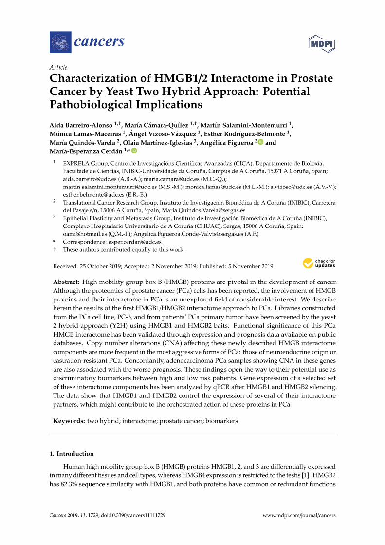
Table 3. Proteins identified in the HMGB1 Y2H interactome in primary tumor adenocarcinoma.
Gene (Aliases) Uniprot Code N A Biological Function Previous References toProstate Cancer (PCa)
c-FOS P01100 2 27–184Transcriptional regulation and
control of cell growth andapoptosis. [44].
Expression is elevated in theprostate upon
castration-mediated androgenwithdrawal [44].
GOLM1 Q8NBJ4 1 236–376 PI3K-AKT-mTORsignaling [45].
Upregulated in PCa hasoncogenic functions [45].
HNRNPU Q00839 1 91–296 DNA and RNA binding [46]. Not previously reported
MAP1B P46821 2 2187–2409 Vesicle formation; it caninteract with p53 [47]. Not previously reported
MAPKAPK5 Q8IW41 1 1–95
Involved in mTOR signaling[48]; MAPKAPK5 has diverse
roles in cell growth,programmed cell death,
senescence, and motility [49].
Not previously reported
MIEN1 Q9BRT3 3 24–204 Regulator of cell migrationand invasion [50].
MIEN1 increases invasivepotential of PCa cells by
NF-κβ-mediated downstreamtarget genes [50].
MT2A P02795 1 8–61 Binding to heavy metals [51].
MT2A is upregulated underhypoxia in PCa cell lines, PCa
tissue, and residual cancercells after androgen ablation
therapy [52].
PSMA7 (PTPT) O14818 1 173–248
PSMA7, a proteasome subunit,enhances AR transactivationin a dose-dependent manner
[53] and inhibits thetransactivation function of
HIF-1A [54].
Proposed biomarker inPCa [55]
PTPN2 P17706 3 1–221
Tyrosine-specific phosphatase(TCPTP) negatively regulatesSTAT3 that is involved in cell
growth and proliferation,differentiation, migration, and
cell death or apoptosis [56].
Not previously reported
RASAL2 Q9UJF2 1 97–334 Tumor suppressor viaRAS [57] Not previously reported
RSF1 Q96T23 1 572–795
Chromatin remodeling factornecessary for p53-dependent
gene expression in response toDNA damage [58].
RSF1 is overexpressed in PCaand contributes to prostate
cancer cell growth andinvasion [59].
SRSF3 P84103 2 1–164 Oncogenic splicing factor [60].SRSF3 expression is induced
by hypoxia in prostatecancerous cells [61].
TAF3 Q5VWG9 5 2–222Transcriptional regulation;
interacts with andinhibits p53 [62].
Not previously reported
TGM3 Q08188 1 480–693
Catalyze the irreversiblecross-linking of
peptide-bound glutamineresidues to lysines or primary
amines; involved inapoptosis [63].
Not previously reported
N: redundancy in clone isolation; A: Sequenced region in clones, Aa relative to ATG.

Cancers 2019, 11, 1729 5 of 21
Table 3. Cont.
Gene (Aliases) Uniprot Code N A Biological Function Previous References toProstate Cancer (PCa)
UBC P0CG48 1 28–181
Unanchored-polyubiquitinhas several roles in activation
of protein kinases,and signaling
Not previously reported
WNK4 Q96J92 4 9–208
Regulates STE20-relatedprotein kinases that function
upstream of the MAPKpathways. [64].
Not previously reported
YY1 P25490 1 27–223 Transcriptionalregulation [65]. Involved in PCa [65–70]
ZNF428 Q96B54 2 89–188 Unknown Not previously reported
N: redundancy in clone isolation; A: Sequenced region in clones, Aa relative to ATG.
Table 4. Proteins identified in the HMGB2 Y2H interactome in primary tumor adenocarcinoma.
Gene (Aliases) Uniprot Code N A Biological Function Previous References toProstate Cancer
C1QBP(GC1QBP,HABP1,SF2P32)
Q07021 10 57–282
Control of mitochondrialenergetic metabolism;
promotes cell proliferation,migration, and resistance to
cell death. [39].
Highly expressed in PCa andassociated with shorter
prostate-specific antigenrelapse time after radical
prostatectomy [40].
COMMD1 Q8N668 1 1–180
Regulates oxidative stress,NF-κB-mediated transcription,DNA damage response, and
oncogenesis [71].
Degradation of COMMD1 andI-kappaB induced by clusterin
enhances NF-κβ activity inprostate cancer cells. [72].
FLNA P21333 5 106–366
A C-terminal fragment ofFLNA co-localizes with the
androgen receptor AR to thenucleus and downregulates
AR function. [73].
FLNA has been clinicallyvalidated for better diagnosis
of PCa [74]; regulated bymiRNA205 [75].
MIEN1 Q9BRT3 4 1–116 Regulates cell migration andapoptosis [50].
Overexpressed in PCa cells.MIEN1 overexpressionfunctionally enhances
migration and invasion oftumor cells via modulating the
activity of AKT [50].
MYL6 P60660 2 1–150
Regulatory light chain ofmyosin II; myosin II,
expressed in non-muscletissues, plays a central role incell adhesion, migration, and
division [76].
Not previously reported
NOP53(GLTSCR2) Q9NZM5 35 163–428
Cell cycle control; NOP53translocates to the
nucleoplasm under ribosomalstress, where it interacts withand stabilizes p53 and inhibits
cell cycle progression [77].
Not previously reported
RPS28 P62857 1 8–52
Ribosome component; itsdecrease blocks pre-18S
ribosomal RNA processing,resulting in a reduction in the
assembly of 40S ribosomalsubunits [78].
Not previously reported
N: redundancy in clone isolation; A: Sequenced region in clones, Aa relative to ATG

Cancers 2019, 11, 1729 6 of 21
Interestingly, 43% of detected HMGB1 interactome targets (10 of a total of 28, referenced inTables 1 and 3) as well as 64% of HMGB2 (7 of a total of 11, referenced in Tables 2 and 4) havepreviously been related to PCa, supporting the functional significance of our Y2H interactome data inPCa research. Furthermore, the detected proteins are remarkably associated with cancer hallmarks.Indeed, the oncogenic capacities of several identified proteins in our Y2H interactome had been alreadyreported in PCa or other cancerous models by wide-ranging functional approaches, which are reviewedin Supplementary Table S1. Figure 1 summarizes the frequency distribution of the identified proteins inrelation to cancer hallmarks (Figure 1a) as well as the number of references of each protein functionallyrelated to cancer progression in diverse models (Figure 1b).
Cancers 2019, 11, x 6 of 21
reduction in the assembly of 40S ribosomal subunits [78].
N: redundancy in clone isolation; A: Sequenced region in clones, Aa relative to ATG
(a) (b)
Figure 1. Relationship between identified proteins and cancer hallmarks: (a) Distribution of HMGB1 and HMGB2 interactome targets according to cancer hallmarks and (b) number of references that associate these proteins with cancer hallmarks according to PubMed (7-31-2019).
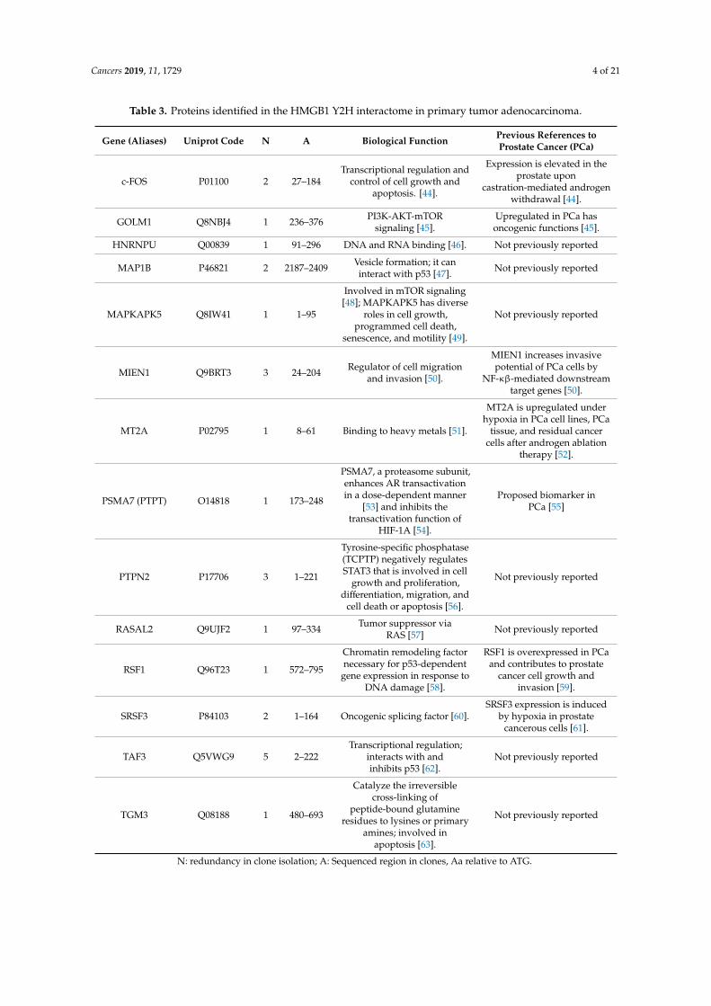
The interaction of HMGB1 with Cytokeratin-7 was validated in PC-3 cells by co-immunoprecipitation and western blot (Figure 2a). Immunodetection of HMGB1 using a green fluorescent antibody (Figure 2b,c) and of Cytokeratin-7 using a red fluorescent antibody (Figure 2c) was also assayed in PC-3 cells. Confocal microscopy showed that co-localization of HMGB1 and Cytokeratin-7 occurred principally in the perinuclear area (Figure 2c), with a Meander's correlation coefficient of 0.87 ± 0.3. Three other interactions were also validated in PC-3 cells by immunoprecipitation and MS identification (Figure 2d).
Figure 1. Relationship between identified proteins and cancer hallmarks: (a) Distribution of HMGB1and HMGB2 interactome targets according to cancer hallmarks and (b) number of references thatassociate these proteins with cancer hallmarks according to PubMed (7-31-2019).
The interaction of HMGB1 with Cytokeratin-7 was validated in PC-3 cells by co-immunoprecipitationand western blot (Figure 2a). Immunodetection of HMGB1 using a green fluorescent antibody(Figure 2b,c) and of Cytokeratin-7 using a red fluorescent antibody (Figure 2c) was also assayed inPC-3 cells. Confocal microscopy showed that co-localization of HMGB1 and Cytokeratin-7 occurredprincipally in the perinuclear area (Figure 2c), with a Meander’s correlation coefficient of 0.87 ± 0.3.Three other interactions were also validated in PC-3 cells by immunoprecipitation and MS identification(Figure 2d).
2.2. Mutations and Copy Number Alterations in HMGB1 and HMGB2 Interactome Targets in PCa
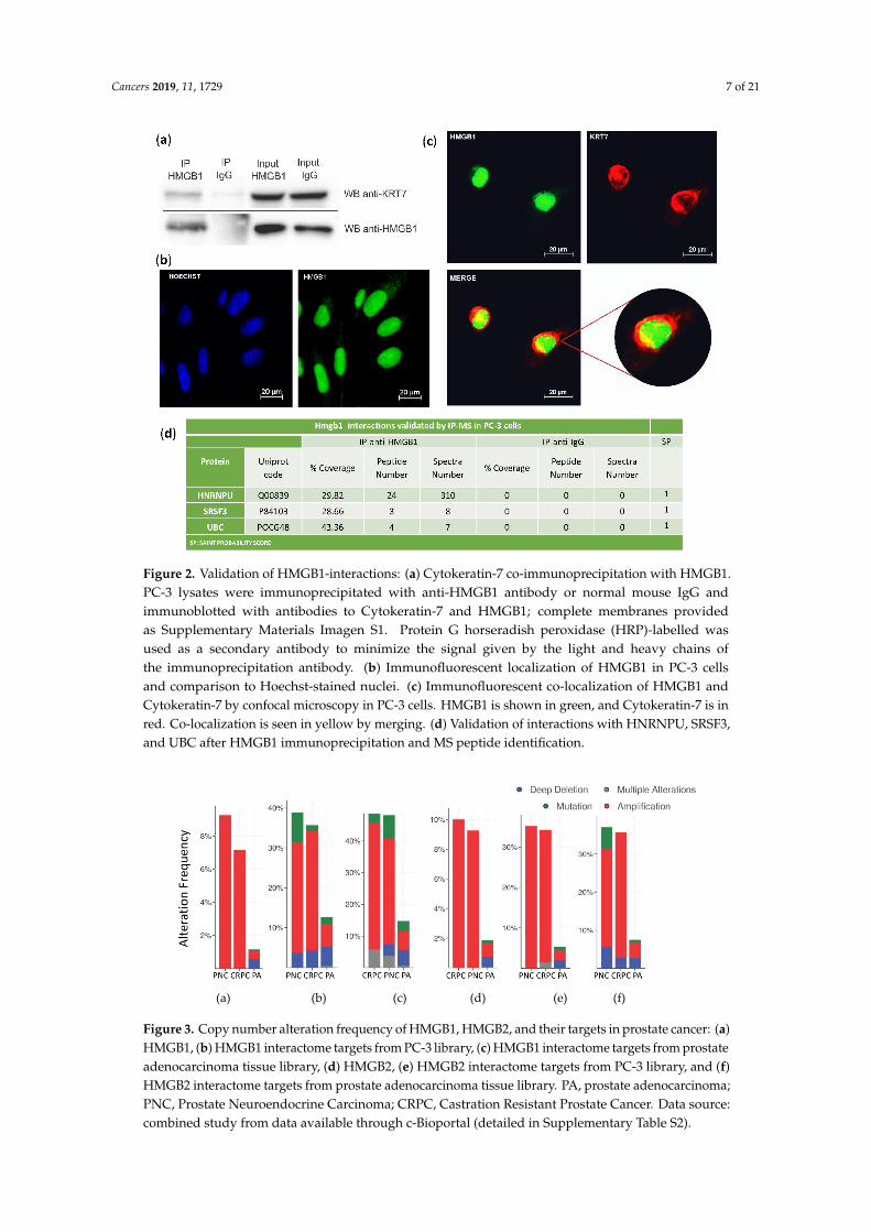
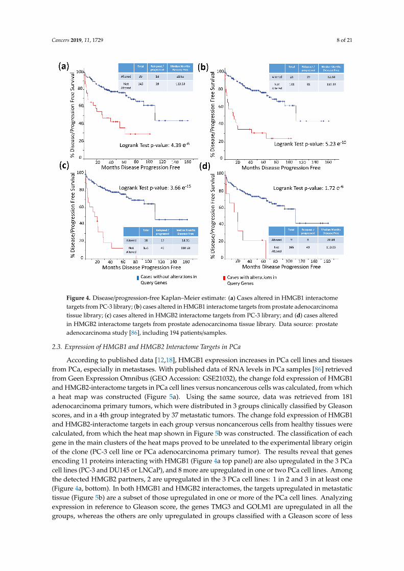
The frequency of mutations and copy number alterations (CNA) in genes encoding HMGB1 andHMGB2 proteins were analyzed as well as in those genes encoding proteins detected in the Y2H searchassociated with PCa, using the open platform for exploring cancer genomics data, c-Bioportal [79,80].We included 14 PCa studies available at cBioPortal (https://www.cbioportal.org/), of which theircharacteristics are summarized in Supplementary Table S2. From these, 10 were adenocarcinomastudies [81–91], with 3218 samples; the other 3 studies corresponded to metastatic PCa [92–94], including655 samples; and finally, one study corresponded to neuroendocrine PCa, which was carried out with114 samples [95]. The data show that mutations and CNA affecting HMGB1, HMGB2, and the proteinsidentified in the corresponding Y2H interactome are more frequently present in neuroendocrine PCaand castration-resistant PCa than in adenocarcinoma (Figure 3). Since neuroendocrine PCa is anaggressive PCa [16], we tested whether CNA of these genes was also related to the poor prognosis inpatients diagnosed with adenocarcinoma. With amplification as the most frequently detected CNAin Figure 3, we compared disease/progression-free Kaplan–Meier estimate rates calculated from thestudy of Taylor et al. [86] among the group of samples having gains or amplifications of these genesand the group integrated by the rest of samples. Figure 4 shows that gain or amplification of HMGB1and HMGB2 interactome targets results in a notorious decrease of the median of months disease-free,with high significant p-values in the Logrank test.
Cancers 2019, 11, 1729 7 of 21
Cancers 2019, 11, x 6 of 21
reduction in the assembly of 40S ribosomal subunits [78].
N: redundancy in clone isolation; A: Sequenced region in clones, Aa relative to ATG
(a) (b)
Figure 1. Relationship between identified proteins and cancer hallmarks: (a) Distribution of HMGB1 and HMGB2 interactome targets according to cancer hallmarks and (b) number of references that associate these proteins with cancer hallmarks according to PubMed (7-31-2019).
The interaction of HMGB1 with Cytokeratin-7 was validated in PC-3 cells by co-immunoprecipitation and western blot (Figure 2a). Immunodetection of HMGB1 using a green fluorescent antibody (Figure 2b,c) and of Cytokeratin-7 using a red fluorescent antibody (Figure 2c) was also assayed in PC-3 cells. Confocal microscopy showed that co-localization of HMGB1 and Cytokeratin-7 occurred principally in the perinuclear area (Figure 2c), with a Meander's correlation coefficient of 0.87 ± 0.3. Three other interactions were also validated in PC-3 cells by immunoprecipitation and MS identification (Figure 2d).
Figure 2. Validation of HMGB1-interactions: (a) Cytokeratin-7 co-immunoprecipitation with HMGB1.PC-3 lysates were immunoprecipitated with anti-HMGB1 antibody or normal mouse IgG andimmunoblotted with antibodies to Cytokeratin-7 and HMGB1; complete membranes providedas Supplementary Materials Imagen S1. Protein G horseradish peroxidase (HRP)-labelled wasused as a secondary antibody to minimize the signal given by the light and heavy chains ofthe immunoprecipitation antibody. (b) Immunofluorescent localization of HMGB1 in PC-3 cellsand comparison to Hoechst-stained nuclei. (c) Immunofluorescent co-localization of HMGB1 andCytokeratin-7 by confocal microscopy in PC-3 cells. HMGB1 is shown in green, and Cytokeratin-7 is inred. Co-localization is seen in yellow by merging. (d) Validation of interactions with HNRNPU, SRSF3,and UBC after HMGB1 immunoprecipitation and MS peptide identification.
Cancers 2019, 11, x 7 of 21
Figure 2. Validation of HMGB1-interactions: (a) Cytokeratin-7 co-immunoprecipitation with HMGB1. PC-3 lysates were immunoprecipitated with anti-HMGB1 antibody or normal mouse IgG and immunoblotted with antibodies to Cytokeratin-7 and HMGB1; complete membranes provided as supplementary materiasl Imagen S1. Protein G horseradish peroxidase (HRP)-labelled was used as a secondary antibody to minimize the signal given by the light and heavy chains of the immunoprecipitation antibody. (b) Immunofluorescent localization of HMGB1 in PC-3 cells and comparison to Hoechst-stained nuclei. (c) Immunofluorescent co-localization of HMGB1 and Cytokeratin-7 by confocal microscopy in PC-3 cells. HMGB1 is shown in green, and Cytokeratin-7 is in red. Co-localization is seen in yellow by merging. (d) Validation of interactions with HNRNPU, SRSF3, and UBC after HMGB1 immunoprecipitation and MS peptide identification.
2.2. Mutations and Copy Number Alterations in HMGB1 and HMGB2 Interactome Targets in PCa
The frequency of mutations and copy number alterations (CNA) in genes encoding HMGB1 and HMGB2 proteins were analyzed as well as in those genes encoding proteins detected in the Y2H search associated with PCa, using the open platform for exploring cancer genomics data, c-Bioportal [79,80]. We included 14 PCa studies available at cBioPortal (https://www.cbioportal.org/), of which their characteristics are summarized in Supplementary Table 2. From these, 10 were adenocarcinoma studies [81–91], with 3218 samples; the other 3 studies corresponded to metastatic PCa [92–94], including 655 samples; and finally, one study corresponded to neuroendocrine PCa, which was carried out with 114 samples [95]. The data show that mutations and CNA affecting HMGB1, HMGB2, and the proteins identified in the corresponding Y2H interactome are more frequently present in neuroendocrine PCa and castration-resistant PCa than in adenocarcinoma (Figure 3). Since neuroendocrine PCa is an aggressive PCa [16], we tested whether CNA of these genes was also related to the poor prognosis in patients diagnosed with adenocarcinoma. With amplification as the most frequently detected CNA in Figure 3, we compared disease/progression-free Kaplan–Meier estimate rates calculated from the study of Taylor et al. [86] among the group of samples having gains or amplifications of these genes and the group integrated by the rest of samples. Figure 4 shows that gain or amplification of HMGB1 and HMGB2 interactome targets results in a notorious decrease of the median of months disease-free, with high significant p-values in the Logrank test.
(a) (b) (c) (d) (e) (f)
Figure 3. Copy number alteration frequency of HMGB1, HMGB2, and their targets in prostate cancer: (a) HMGB1, (b) HMGB1 interactome targets from PC-3 library, (c) HMGB1 interactome targets from prostate adenocarcinoma tissue library, (d) HMGB2, (e) HMGB2 interactome targets from PC-3 library, and (f) HMGB2 interactome targets from prostate adenocarcinoma tissue library. PA, prostate adenocarcinoma; PNC, Prostate Neuroendocrine Carcinoma; CRPC, Castration Resistant Prostate Cancer. Data source: combined study from data available through c-Bioportal (detailed in Supplementary Table S2).
Figure 3. Copy number alteration frequency of HMGB1, HMGB2, and their targets in prostate cancer: (a)HMGB1, (b) HMGB1 interactome targets from PC-3 library, (c) HMGB1 interactome targets from prostateadenocarcinoma tissue library, (d) HMGB2, (e) HMGB2 interactome targets from PC-3 library, and (f)HMGB2 interactome targets from prostate adenocarcinoma tissue library. PA, prostate adenocarcinoma;PNC, Prostate Neuroendocrine Carcinoma; CRPC, Castration Resistant Prostate Cancer. Data source:combined study from data available through c-Bioportal (detailed in Supplementary Table S2).
Cancers 2019, 11, 1729 8 of 21
Cancers 2019, 11, x 8 of 21
Figure 4. Disease/progression-free Kaplan–Meier estimate: (a) Cases altered in HMGB1 interactome targets from PC-3 library; (b) cases altered in HMGB1 interactome targets from prostate adenocarcinoma tissue library; (c) cases altered in HMGB2 interactome targets from PC-3 library; and (d) cases altered in HMGB2 interactome targets from prostate adenocarcinoma tissue library. Data source: prostate adenocarcinoma study [86], including 194 patients/samples.
2.3. Expression of HMGB1 and HMGB2 Interactome Targets in PCa
According to published data [12,18], HMGB1 expression increases in PCa cell lines and tissues from PCa, especially in metastases. With published data of RNA levels in PCa samples [86] retrieved from Geen Expression Omnibus (GEO Accession: GSE21032), the change fold expression of HMGB1 and HMGB2-interactome targets in PCa cell lines versus noncancerous cells was calculated, from which a heat map was constructed (Figure 5a). Using the same source, data was retrieved from 181 adenocarcinoma primary tumors, which were distributed in 3 groups clinically classified by Gleason scores, and in a 4th group integrated by 37 metastatic tumors. The change fold expression of HMGB1 and HMGB2-interactome targets in each group versus noncancerous cells from healthy tissues were calculated, from which the heat map shown in Figure 5b was constructed. The classification of each gene in the main clusters of the heat maps proved to be unrelated to the experimental library origin of the clone (PC-3 cell line or PCa adenocarcinoma primary tumor). The results reveal that genes encoding 11 proteins interacting with HMGB1 (Figure 4a top panel) are also upregulated in the 3 PCa cell lines (PC-3 and DU145 or LNCaP), and 8 more are upregulated in one or two PCa cell lines. Among the detected HMGB2 partners, 2 are upregulated in the 3 PCa cell lines: 1 in 2 and 3 in at least one (Figure 4a, bottom). In both HMGB1 and HMGB2 interactomes, the targets upregulated in metastatic tissue (Figure 5b) are a subset of those upregulated in one or more of the PCa cell lines. Analyzing expression in reference to Gleason score, the genes TMG3 and GOLM1 are upregulated in all the groups, whereas the others are only upregulated in groups classified with a Gleason score of less than or equal to 7 (PTPN2, HDLBP, SRF3, FOS, and WNK4). Regarding a pattern associated with
Figure 4. Disease/progression-free Kaplan–Meier estimate: (a) Cases altered in HMGB1 interactometargets from PC-3 library; (b) cases altered in HMGB1 interactome targets from prostate adenocarcinomatissue library; (c) cases altered in HMGB2 interactome targets from PC-3 library; and (d) cases alteredin HMGB2 interactome targets from prostate adenocarcinoma tissue library. Data source: prostateadenocarcinoma study [86], including 194 patients/samples.
2.3. Expression of HMGB1 and HMGB2 Interactome Targets in PCa
According to published data [12,18], HMGB1 expression increases in PCa cell lines and tissuesfrom PCa, especially in metastases. With published data of RNA levels in PCa samples [86] retrievedfrom Geen Expression Omnibus (GEO Accession: GSE21032), the change fold expression of HMGB1and HMGB2-interactome targets in PCa cell lines versus noncancerous cells was calculated, from whicha heat map was constructed (Figure 5a). Using the same source, data was retrieved from 181adenocarcinoma primary tumors, which were distributed in 3 groups clinically classified by Gleasonscores, and in a 4th group integrated by 37 metastatic tumors. The change fold expression of HMGB1and HMGB2-interactome targets in each group versus noncancerous cells from healthy tissues werecalculated, from which the heat map shown in Figure 5b was constructed. The classification of eachgene in the main clusters of the heat maps proved to be unrelated to the experimental library originof the clone (PC-3 cell line or PCa adenocarcinoma primary tumor). The results reveal that genesencoding 11 proteins interacting with HMGB1 (Figure 4a top panel) are also upregulated in the 3 PCacell lines (PC-3 and DU145 or LNCaP), and 8 more are upregulated in one or two PCa cell lines. Amongthe detected HMGB2 partners, 2 are upregulated in the 3 PCa cell lines: 1 in 2 and 3 in at least one(Figure 4a, bottom). In both HMGB1 and HMGB2 interactomes, the targets upregulated in metastatictissue (Figure 5b) are a subset of those upregulated in one or more of the PCa cell lines. Analyzingexpression in reference to Gleason score, the genes TMG3 and GOLM1 are upregulated in all thegroups, whereas the others are only upregulated in groups classified with a Gleason score of less

Cancers 2019, 11, 1729 9 of 21
than or equal to 7 (PTPN2, HDLBP, SRF3, FOS, and WNK4). Regarding a pattern associated withthe existence of metastasis, 3 genes that are not upregulated in samples from primary tumors areupregulated in metastasis: PSMA7, UBE2E3, and MIEN1 (Figure 5b).
Cancers 2019, 11, x 9 of 21
the existence of metastasis, 3 genes that are not upregulated in samples from primary tumors are upregulated in metastasis: PSMA7, UBE2E3, and MIEN1 (Figure 5b).
(a) (b)
Figure 5. Heat map expression in prostate cancer: (a) Expression of HMGB1 interactome targets (upper panel) and HMGB2 interactome targets (lower panel) in 3 prostate cancer cell lines; (b) expression of HMGB1 interactome targets (upper panel) and HMGB2 interactome targets (lower panel) in prostate adenocarcinoma cases classified by Gleason (G) score groups or metastatic (M) tumors. CL, target detected in the PC-3 library. TJ, target detected in the prostate adenocarcinoma library. Data extracted from Gene Expression Omnibus (GEO ) Accession: GSE21032.
2.4. Silencing of HMGB1 and HMGB2 Reveals Regulation of the Expression of Genes Encoding Their Interactome Targets
To test whether changes in HMGB1/2 protein levels in PCa cells could also be influencing the expression of their interactome targets, HMGB1 and HMGB2 in PC-3 cells were silenced by iRNA (Figure 6a). Levels of mRNA from 14 partners analyzed by qPCR and changes (siHMGB/HMGB) are summarized in the Figure 6b. This analysis also included HMGB1, HMGB2, and well-known PCa biomarkers: PSA (encoded by KLK3); PMEPA1, which is involved in downregulation of the androgen receptor, thus promoting androgen receptor-negative prostate cell proliferation [96]; and RAGE, one of the membrane receptors in the extracellular signaling function of HMGB1 [97]. Silencing of HMGB1 causes overexpression of the larger cluster of the Y2H interactome, whereas siHMGB2 has the opposite effect (Figure 6c). HMGB1 downregulates the expression of the majority of targets analyzed, and conversely, HMGB2 upregulates them. Therefore, the expression level of each regulated target would depend on the relative imbalance of HMGB1 and HMGB2 and on the differential effect of both HMGB proteins on the expression of each partner. PMEPA1 and PSA, well-known PCa biomarkers, are also oppositely regulated by HMGB1 and HMGB2 (Figure 6).
Figure 5. Heat map expression in prostate cancer: (a) Expression of HMGB1 interactome targets (upperpanel) and HMGB2 interactome targets (lower panel) in 3 prostate cancer cell lines; (b) expression ofHMGB1 interactome targets (upper panel) and HMGB2 interactome targets (lower panel) in prostateadenocarcinoma cases classified by Gleason (G) score groups or metastatic (M) tumors. CL, targetdetected in the PC-3 library. TJ, target detected in the prostate adenocarcinoma library. Data extractedfrom Gene Expression Omnibus (GEO ) Accession: GSE21032.
2.4. Silencing of HMGB1 and HMGB2 Reveals Regulation of the Expression of Genes Encoding TheirInteractome Targets
To test whether changes in HMGB1/2 protein levels in PCa cells could also be influencing theexpression of their interactome targets, HMGB1 and HMGB2 in PC-3 cells were silenced by iRNA(Figure 6a). Levels of mRNA from 14 partners analyzed by qPCR and changes (siHMGB/HMGB) aresummarized in the Figure 6b. This analysis also included HMGB1, HMGB2, and well-known PCabiomarkers: PSA (encoded by KLK3); PMEPA1, which is involved in downregulation of the androgenreceptor, thus promoting androgen receptor-negative prostate cell proliferation [96]; and RAGE,one of the membrane receptors in the extracellular signaling function of HMGB1 [97]. Silencing ofHMGB1 causes overexpression of the larger cluster of the Y2H interactome, whereas siHMGB2 has theopposite effect (Figure 6c). HMGB1 downregulates the expression of the majority of targets analyzed,and conversely, HMGB2 upregulates them. Therefore, the expression level of each regulated targetwould depend on the relative imbalance of HMGB1 and HMGB2 and on the differential effect of both

Cancers 2019, 11, 1729 10 of 21
HMGB proteins on the expression of each partner. PMEPA1 and PSA, well-known PCa biomarkers,are also oppositely regulated by HMGB1 and HMGB2 (Figure 6).
Cancers 2019, 11, x 10 of 21
Figure 6. HMGB1 and HMGB2 silencing: (a) Western Blot showing HMGB1 and HMGB2 silencing. Complete membranes provided as supplementary material, Image S2 (b) Heat map comparing the pattern of expression (siHMGB1/HMGB1 and siHMGB2/HMGB2). (c) Summary of regulatory effects of HMGB1 and HMGB2 on the selected genes.
3. Discussion
High mobility group box B (HMGB) proteins are pivotal in the development of cancer [6,8,10], and HMGB1 overexpression has been related to principal cancer hallmarks [7]. Interactome targets of HMGB1 or HMGB2 that have been identified in our Y2H study were previously found to be related to cancer hallmarks (Table S1 and Figure 1), and are also dysregulated in PCa, as confirmed by detection of changes in mRNA or protein levels. DNAAF2 [98], U2AF1 [43], C1QBP [40], Snapin, or HDLBP [99] are upregulated in prostate tumors or PCa cell lines. Others increase their expression after androgen-deprivation therapy, such as KRT7 or NOP53 [100]. Functional studies interfering the expression of several of the proteins revealed by our study also directly associated them to PCa. In this sense, selective knockdown of C1QBP through iRNA decreased cyclin D1, increased p21 expression, led to cell cycle arrest (G1/S transition) in PCa cells, and had no effect on a noncancerous cell line [40]. NOP53 acts as a tumor suppressor, and knockdown of the gene in the PCa LNCaP cell line increased the invasiveness of these cells as measured in a xenograft animal model [101].
Two already known regulatory factors have been found among the HMGB1 interactome targets, YY1 and HOXA10, and both are associated with PCa. YY1 is upregulated in human prostate cancer cell lines and tissues [66]. Inhibition of YY1 reduces expression of genes related to the Krebs cycle and electron transport chain in PCa cell lines [67], and YY1 depletion correlates with delayed progression of PCa [68]. Overexpression of YY1 can promote epithelial-mesenchymal transition by reducing hnRNPM expression [69]. YY1 can also silence tumor suppressor genes, such as XAF1 in PCa [70]. In summary, YY1 is a recognized prostate cancer driver [66] and different complexes in which YY1 takes part can induce activation or repression of gene expression, including also AR-YY1-mediated PSA transcription [102], which we found is also regulated by HMGB1 and HMGB2 silencing. HOXA10 is upregulated in PCa [31], and inverse correlations between HOXA10 expression and Gleason pattern, Gleason score, and pathological stage are found [32], although downregulation of HOXA10 gene
Figure 6. HMGB1 and HMGB2 silencing: (a) Western Blot showing HMGB1 and HMGB2 silencing.Complete membranes provided as Supplementary Material, Image S2 (b) Heat map comparing thepattern of expression (siHMGB1/HMGB1 and siHMGB2/HMGB2). (c) Summary of regulatory effects ofHMGB1 and HMGB2 on the selected genes.
3. Discussion
High mobility group box B (HMGB) proteins are pivotal in the development of cancer [6,8,10],and HMGB1 overexpression has been related to principal cancer hallmarks [7]. Interactome targetsof HMGB1 or HMGB2 that have been identified in our Y2H study were previously found to berelated to cancer hallmarks (Table S1 and Figure 1), and are also dysregulated in PCa, as confirmedby detection of changes in mRNA or protein levels. DNAAF2 [98], U2AF1 [43], C1QBP [40], Snapin,or HDLBP [99] are upregulated in prostate tumors or PCa cell lines. Others increase their expressionafter androgen-deprivation therapy, such as KRT7 or NOP53 [100]. Functional studies interfering theexpression of several of the proteins revealed by our study also directly associated them to PCa. In thissense, selective knockdown of C1QBP through iRNA decreased cyclin D1, increased p21 expression,led to cell cycle arrest (G1/S transition) in PCa cells, and had no effect on a noncancerous cell line [40].NOP53 acts as a tumor suppressor, and knockdown of the gene in the PCa LNCaP cell line increasedthe invasiveness of these cells as measured in a xenograft animal model [101].
Two already known regulatory factors have been found among the HMGB1 interactome targets,YY1 and HOXA10, and both are associated with PCa. YY1 is upregulated in human prostate cancercell lines and tissues [66]. Inhibition of YY1 reduces expression of genes related to the Krebs cycle andelectron transport chain in PCa cell lines [67], and YY1 depletion correlates with delayed progressionof PCa [68]. Overexpression of YY1 can promote epithelial-mesenchymal transition by reducinghnRNPM expression [69]. YY1 can also silence tumor suppressor genes, such as XAF1 in PCa [70].

Cancers 2019, 11, 1729 11 of 21
In summary, YY1 is a recognized prostate cancer driver [66] and different complexes in which YY1takes part can induce activation or repression of gene expression, including also AR-YY1-mediatedPSA transcription [102], which we found is also regulated by HMGB1 and HMGB2 silencing. HOXA10is upregulated in PCa [31], and inverse correlations between HOXA10 expression and Gleason pattern,Gleason score, and pathological stage are found [32], although downregulation of HOXA10 geneexpression may enhance lipogenesis to promote PCa cell growth and tumor progression to thecastration-resistant stage [103]. Silencing of HOXA10 expression in PC-3 cells by iRNA decreasedproliferation rates, whereas HOXA10 overexpression had the opposite effect [31]. Physical interactionbetween these PCa-associated proteins and HMGB proteins has not previously been described, and ourresults therefore show that there is a connection between HMGB1 and HMGB2 functions and those oftheir binding partners in PCa.
Considering that HMGB1, HMGB2, and a subset of their interactome partners are upregulated inPCa, we silenced HMGB1 and HMGB2 and analyzed the mRNA levels of a group of randomly selectedpartners in PC-3 cells (Figure 6). The data show that HMGB1 and HMGB2 control the expression ofthem, which might contribute to the orchestrated action of all these proteins in PCa. HMGB2 activatesmany of the tested targets, but unexpectedly, HMGB1 has the opposite effect. One can propose severalreasons to explain upregulation of targets in these circumstances. Data from the genotype-tissueexpression (GTEx) project [104] indicates that, although both HMGB1 and HMGB2 are upregulated inPCa versus noncancerous cells, the relative increase is higher for HMGB2 (×1.5) than HMGB1 (×1.3);this could explain the increased expression of several of their targets, assuming that positive regulationcaused by HMGB2 predominates over negative regulation caused by HMGB1 during the onset ofPCa. Alternatively, differential interaction of HMGB1 or HMGB2 with their different nuclear partners,the transcript factors detected in our Y2H analysis being among them, might condition their positiveor negative regulatory roles on the expression of specific genes.
Clinically, a high frequency of CNA of the genes encoding the identified proteins is associated withthe most aggressive forms of PCa: small cell neuroendocrine carcinoma (SCNC) or castration-resistantPCa (Figure 3). Their gain or amplification in the genome of the cancerous cells are positively correlatedto a lesser disease-free period for PCa patients (Figure 4). The mRNA levels of a subset of theseproteins are also higher in metastases than primary tumors (Figure 5). In conclusion, the set of proteinsdetected though our HMGB1-HMGB2 Y2H analysis are associated with the most aggressive casesof PCa. Although the PSA-based test is routinely employed for screening of PCa, it has resulted inoverdiagnosis and overtreatment of nonaggressive cancers, thus reducing the quality of life of patients.
Consequently, an improvement is necessary in the initial stages to discriminate between high-riskfrom low risk cancers. Our data on HMGB1 and HMGB2 interactome targets, considering theircorrelation to high aggressiveness and bad prognosis, is a good starting point to develop newserum protein panels for improvement of PCa diagnosis. Indeed, FLNA has already been proposedin a clinical validated PCa biomarker panel in serum [74]. PSMA7 was also proposed as a PCabiomarker [55], and KRT7 is included in a whole blood mRNA 4-gene androgen regulated panel forPCa diagnosis [33]. Considering the relative expression levels of our HMGB1 and HMGB2 interactometargets in noncancerous cells or in blood of health subjects differ quite notably (Figure 7), one mightanticipate that more sensitive analyses could be carried out using as biomarkers those proteins thatare usually lowly expressed in noncancerous cells; thus, their levels are also low in the blood ofhealthy individuals. For instance, FLNA reported as a possible biomarker [74] is one of the 50 proteinsmost strongly expressed in normal prostate, and high levels are also detected in the blood of healthyindividuals, whereas other detected HMGB1 or HMGB2 interactome targets in our study, e.g., DNAAF2,GOLM1, or TGM3, are in the lowest rank of detection in noncancerous samples and their increaseshould become more discriminatory.

Cancers 2019, 11, 1729 12 of 21Cancers 2019, 11, x 12 of 21
Figure 7. Prostate and blood levels of HMGB1 and HMGB2 interactome targets in healthy men: Expression of HMGB1 and HMGB2 interactome targets in prostate tissue and whole blood in healthy men. Data were directly obtained and processed from the GTEx Project through Expression Atlas, an integrated database of gene and protein expression in humans, animals, and plants [105], accessed through https://www.ebi.ac.uk/gxa/ experiments/E-MTAB-5214/Results.
4. Materials and Methods
4.1. Biological Materials
PC-3 is an androgen-independent cell line derived from a bone metastasis [106]. The human PCa PC-3 cell line, regularly validated by DNA typing, was obtained from the American Type Culture Collection ATCC and grown in Roswell Park Memorial Institute RPMI-1640 media, supplemented with 10% heat-inactivated fetal bovine serum and 1% penicillin-streptomycin (Thermo Fisher Scientific, Inc. Waltham, MA, USA). Cells were cultured at 37 °C in 5% CO2 in air in a humidified incubator. RNA from PCa tissue, isolated after radical prostate resection of a 66-year-old man diagnosed with adenocarcinoma (Gleason score 6) and not previously treated with radiotherapy or chemotherapy, was obtained from Biobanco de Andalucía (SPAIN).
4.2. Yeast Two Hybrid Methodology
Sacchacomyces cerevisiae strains were Y187 (MATα, ura3-52, his3-200, ade2-101, trp1-901, leu2-3, 112, gal4Δ, gal80Δ, met-, and URA3::GALuas-GAL1TATA-LacZ MEL1) and Y2HGold (MATa, trp1-901, leu2-3, 112, ura3-52, his3-200, gal4Δ, gal80Δ, LYS2::GAL1uas-GAl1TATA-His3, GAL2uas-Gal2TATA-Ade2 URA3:: MEL1UAS-Mel1TATA, and AUR1-C MEL1).
Total RNA from the PC-3 cell line obtained from the supplier (Sigma-Aldrich, Saint Louis, MO, USA) and RNA from PCa tissue (Biobanco de Andalucía, Spain) were used to construct cDNA libraries. HMGB1 and HMGB2 interacting partners were identified using Matchmaker Gold Yeast 2-Hybrid System (Clontech, Fremont, CA, USA). Library construction, bait construction, and Yeast 2-Hybrid library screening were done according to the Takara Bio USA Matchmaker® Gold Yeast 2-Hybrid System manual. In brief, the baits were cloned as fusions to the GAL4 activation domain in the plasmid pGBKT7-AD and used to transform the yeast haploid strain, Y187. cDNA libraries prepared from RNA extracted from PC-3 cells and PCa cancerous tissue were included as fusions to the GAL4 DNA-binding domain in the plasmid pGBKT7-BD and were used to transform the yeast haploid strain, Y2HGold. RNAs from human samples used to prepare the Y2H libraries were obtained from Biobanco de Andalucía (Spain). RNA was extracted from frozen tissue sections in OCT (Optimal Cutting Temperature) compound, using the Qiacube robot from QIAGEN, based on ion-exchange columns with a silica membrane. RNA was obtained with the miRNeasy mini-kit from QIAGEN that allows recovery of both total RNA and miRNAs. The samples were finally treated with
Figure 7. Prostate and blood levels of HMGB1 and HMGB2 interactome targets in healthy men:Expression of HMGB1 and HMGB2 interactome targets in prostate tissue and whole blood in healthymen. Data were directly obtained and processed from the GTEx Project through Expression Atlas,an integrated database of gene and protein expression in humans, animals, and plants [105], accessedthrough https://www.ebi.ac.uk/gxa/experiments/E-MTAB-5214/Results.
4. Materials and Methods
4.1. Biological Materials
PC-3 is an androgen-independent cell line derived from a bone metastasis [106]. The human PCaPC-3 cell line, regularly validated by DNA typing, was obtained from the American Type CultureCollection ATCC and grown in Roswell Park Memorial Institute RPMI-1640 media, supplementedwith 10% heat-inactivated fetal bovine serum and 1% penicillin-streptomycin (Thermo Fisher Scientific,Inc. Waltham, MA, USA). Cells were cultured at 37 ◦C in 5% CO2 in air in a humidified incubator.RNA from PCa tissue, isolated after radical prostate resection of a 66-year-old man diagnosed withadenocarcinoma (Gleason score 6) and not previously treated with radiotherapy or chemotherapy,was obtained from Biobanco de Andalucía (SPAIN).
4.2. Yeast Two Hybrid Methodology
Sacchacomyces cerevisiae strains were Y187 (MATα, ura3-52, his3-200, ade2-101, trp1-901, leu2-3, 112,gal4∆, gal80∆, met-, and URA3::GALuas-GAL1TATA-LacZ MEL1) and Y2HGold (MATa, trp1-901, leu2-3,112, ura3-52, his3-200, gal4∆, gal80∆, LYS2::GAL1uas-GAl1TATA-His3, GAL2uas-Gal2TATA-Ade2 URA3::MEL1UAS-Mel1TATA, and AUR1-C MEL1).
Total RNA from the PC-3 cell line obtained from the supplier (Sigma-Aldrich, Saint Louis, MO,USA) and RNA from PCa tissue (Biobanco de Andalucía, Spain) were used to construct cDNA libraries.HMGB1 and HMGB2 interacting partners were identified using Matchmaker Gold Yeast 2-HybridSystem (Clontech, Fremont, CA, USA). Library construction, bait construction, and Yeast 2-Hybridlibrary screening were done according to the Takara Bio USA Matchmaker® Gold Yeast 2-HybridSystem manual. In brief, the baits were cloned as fusions to the GAL4 activation domain in theplasmid pGBKT7-AD and used to transform the yeast haploid strain, Y187. cDNA libraries preparedfrom RNA extracted from PC-3 cells and PCa cancerous tissue were included as fusions to the GAL4DNA-binding domain in the plasmid pGBKT7-BD and were used to transform the yeast haploidstrain, Y2HGold. RNAs from human samples used to prepare the Y2H libraries were obtained fromBiobanco de Andalucía (Spain). RNA was extracted from frozen tissue sections in OCT (OptimalCutting Temperature) compound, using the Qiacube robot from QIAGEN, based on ion-exchangecolumns with a silica membrane. RNA was obtained with the miRNeasy mini-kit from QIAGEN that

Cancers 2019, 11, 1729 13 of 21
allows recovery of both total RNA and miRNAs. The samples were finally treated with RNase-freeDNAase from QIAGEN. The RNA was quantified at 260 nm and 280 nm by spectrophotometry usingInfinite F200 equipment of TECAN with a Nanoquant plate. Finally, the integrity of the samples wasevaluated by AGILENT 2200 Tape Station apparatus, with the RIN (RNA Integrity Number) parameterbeing >8. Efficiency in the constructions of libraries was in the range recommended in the kit (alllibraries guaranteed to have >1 × 10⁶ independent clones). As a previous control, we confirmed thatour baits (HMGB1 and HMGB2) do not autonomously activate the reporter genes in Y2HGold inthe absence of a prey protein. Bait and prey fusion proteins are each expressed in different haploidyeast strains that can form diploids. The diploid yeast cell expresses both proteins, and when fusionproteins interact, the transcriptional activator GAL4 is reconstructed and brought into proximity toactivate transcription of the reporter genes. For diploid formation, 1 mL of concentrated bait culturewas combined with 1 mL of library culture and incubated overnight with slow shaking. A drop ofthe culture was checked under a phase-contrast microscope (40×) to confirm the existence of zygotesbefore plating on diploid-selective media. Diploids were tested for expression of the reporter genesin selective media. To reduce the appearance of false positives, a screening based on three differentindependent markers (ADE2, HIS3, and MEL1) was selected. pGBKT7-BD plasmids carrying the preyswere rescued from confirmed positive diploids, and DNA was used to transform E. coli. The insertswere sequenced with primer T7 (5′-TAATACGACTCACTATAGGG-3′). Sequences were used forhomology searches with BlastN and BlastX at the National Center for Biotechnology InformationNCBI (https://blast.ncbi.nlm.nih.gov/) and proteins in the database matching the queries annotatedas positives.
4.3. Expression Analysis by Quantitative Polymerase Chain Reaction (RT-qPCR)
Individual analyses of gene expression were carried out as follows. RNA samples wereretro-transcribed into cDNA and labeled with the KAPPA SYBR FAST universal one-step qRT-PCR kit(Kappa Biosystems, Inc, Woburn, Massachusetts, USA). The primers for qPCR are shown in Table S3.Reaction conditions for thermal cycling were 42 ◦C for 5 min, 95 ◦C for 5 s, 40 cycles of 95 ◦C for 3 s,and finally 60 ◦C for 20 s. ECO Real-Time PCR System was used for the experiments (Illumina, Inc.,San Diego, California, USA), and calculations were made by the 2−∆∆Ct method [107]. Student’s testwas used to check the statistical significance of differences between samples (p < 0.05). The relativemRNA levels of the experimentally selected genes (target genes) were calculated by referring tothe mRNA levels of the housekeeping gene, encoding glyceraldehyde phosphate dehydrogenase(GAPDH), which had been verified as being expressed constitutively under the assay conditions.For valid quantification using the 2−∆∆Ct method, it is crucial that target and housekeeping PCRamplification efficiencies are approximately equal: we therefore verified that the efficiencies of the 2PCR reactions differed by <10%. At least 2 independent biological replicas and 3 technical replicas ofeach of them were made for all the experiments.
4.4. Immunoprecipitation
One hundred µl of Protein G Plus-Agarose immunoprecipitation-reagent (Santa CruzBiotechnology, Dallas, TX, USA) were coupled with 4 µg of anti-HMGB1 antibody (sc-74085; SantaCruz Biotechnology) or anti-mouse antibody (Molecular Probes, A10534) in phosphate buffered saline(PBS) for 1 h at 4 ◦C with rotation. PC-3 cells were lysed in 20 mM Tris/HCl, 150 mM, 1% Triton X-100,1× phenylmethylsulfonyl fluoride (PMSF), and protease inhibitor cocktail (Sigma-Aldrich, Saint Louis,MO, USA) and incubated for 30 min at 4 ◦C with rotation. Total protein (500 µg) was incubated withthe antibody agarose beads overnight and eluted by incubation in 1× lithium dodecyl sulfate LDSloading buffer containing 350 mM β-mercaptoethanol at 95 ◦C for 10 min. Mass spectrometry and dataanalysis were done as previously described [26].

Cancers 2019, 11, 1729 14 of 21
4.5. Western Blot Analysis
Protein samples were run on 10% SDS-PAGE gels at 80 V for 20 min followed by 200 V for45–60 min. Proteins were transferred onto a polyvinylidene fluoride (PVDF) membrane at 0.2 A for1 h. Membranes were blocked by incubating with 5% non-fat dry milk for 1 h at room temperature(RT) and then incubated with primary antibodies, anti-HMGB1 (sc-74085; Santa Cruz Biotechnology)or anti-Cytokeratin 7 (ab181598; Abcam, Cambridge, UK) in phosphate-buffered saline with 0.1%Tween 20® detergent PBST overnight at 4 ◦C. After incubation with the corresponding horseradishperoxidase-conjugated secondary antibody, enhanced chemiluminescence for high sensitivity andlong-lasting signal (ECL) Anti-mouse IgG (NXA931 from GE Healthcare Sciences, Chicago, IL, USA) orECL Anti-rabbit IgG (NA934 from GE Healthcare Sciences, Chicago, IL, USA), protein bands weredetected using LuminataTMCrescendo Western HRP Substrate (Millipore Corporation, Burlington,MA, USA) and a ChemiDocTM imager (Bio-Rad laboratories Hercules, CA, USA).
4.6. Immunofluorescence and Confocal Microscopy
Cells were plated in 6-well plates, each containing 4 sterile 13-mm glass coverslips. When 80%confluent, cells were fixed in 4% paraformaldehyde in PBS for 15 min at RT. Cells were washed 3times with PBS (137 mM NaCl, 2.7 mM KCl, 10 mM Na2HPO4, and 2 mM KH2PO4) and finallytreated with 0.1% Triton/PBS for 15 min at RT. They were then blocked in 1% bovine serum albumin(BSA) in PBS for 1 h at RT. Primary antibodies, anti-HMGB1 (sc-74085; Santa Cruz Biotechnology) oranti-Cytokeratin 7 (ab181598; Abcam, Cambridge, UK) were diluted in 1% BSA in PBS. Cells wereincubated with the corresponding primary antibodies overnight at 4 ◦C, followed by 3 washes withPBS and staining with the secondary antibodies, modified with Alexa Fluor 488 and 568 (Invitrogen,Carlsbad, CA, USA) previously diluted in 1% BSA in PBS for 1 h at RT in the dark. For nuclearstaining, after secondary antibody incubation, wells were washed 3 times and stained with Hoechst(Life Technologies, Carlsbad, CA, USA) for 5 min at RT in the dark. Cells were washed once with PBSand once with sterile distilled water. Each coverslip was mounted on a clean slide using ProLong™Gold Antifade Mountant (Invitrogen). After drying, the slides were stored at 4 ◦C in the dark until theywere examined by confocal microscopy (Nikon A1R). Meander’s correlation coefficient was calculatedusing Nis Elements software from Nikon.
4.7. HMGB1 and HMGB2 Silencing by siRNA
The PC-3 cell line was transfected with small interfering (si)RNA oligonucleotides usingLipofectamine 2000 (Invitrogen). siRNA and Lipofectamine 2000 were each diluted separatelywith Opti-MEM (Gibco), mixed together, and incubated for 5 min at RT. The mixture was added tocells plated in 3 mL RPMI 1610 medium (final concentration of siRNA, 50 nM). Cells were collected at48 h post transfection for further analysis. The following siRNAs (Life Technologies) were used for thesilencing of each gene: s20254 Silencer Select for HMGB1, s6650 for HMGB2, and AS02A5Z3 for thesiRNA negative control.
Total RNA was extracted from different conditions (siHMGB1, siHMGB2, and siCtrl#2) of thePC-3 cell line using GeneJET RNA Purification Kit (#K0731, Thermo Scientific). The remaining DNAwas removed by incubating with DNase I, RNase-free (#EN0521, Thermo Scientific). DNA-free RNAwas finally purified using GeneJET RNA Cleanup and Concentration Micro Kit (#K0842, ThermoScientific). qPCR reactions were run in triplicate in an Eco Real-Time PCR System (Illumina) using1 ng per reaction. PC-3 lysates of each condition were extracted with lysis buffer (50 mM Tris-HCl pH8, 150 mM NaCl, 0.1% NP40, 1 mM ethylenediaminetetraacetic acid disodium salt (EDTA), and 2 mMMgCl2), and protein concentration was quantified using Bradford Reagent (Bio-Rad). Protein samplesof 25–40 µg were loaded for western blotting. PVDF membranes were incubated overnight at 4 ◦C withprimary antibodies, anti-HMGB1 (ab18256, Abcam), anti-HMGB2 (ab67282, Abcam), or anti-α-tubulin(sc53646, Santa Cruz Biotechnology).
Cancers 2019, 11, 1729 15 of 21
4.8. Heat Maps
Heat maps from change-fold ratios (Figures 5 and 6) were drawn with Heatmapper (http://heatmapper.ca/expression/), using complete linkage as clustering method and Euclidean distance asthe measurement method [108].
4.9. Statistical Analysis
Analyses were carried out using GraphPad Prism 6 (GraphPad Software, San Diego, CA, USA).Continuous variables were expressed as mean ± SE. Relative gene expression assays were tested usingindependent t-tests. A 2-tailed p-value test was used with p < 0.05 considered significant.
5. Conclusions
We have carried out the first HMGB1/HMGB2 interactome approach in prostate cancer (PCa)using both the PC-3 cell line and adenocarcinoma tissue. Gene or protein expressions of the majority oftargets are dysregulated in PCa, and functional relationships between these proteins and PCa had alsopreviously been confirmed by different laboratories using different models and technical approaches.We have shown by interference analysis that several HMGB1 and HMGB2 partners are regulatedby HMGB1 and HMGB2 themselves, which might contribute to the coordination of their cellularaction in PCa. Copy number alterations in the detected HMGB1 and HMGB2 partners are associatedwith aggressive forms of PCa and a poor prognosis. These characteristics can potentially be used asdiscriminatory biomarkers between high and low risk patients.
Supplementary Materials: The following are available online at http://www.mdpi.com/2072-6694/11/11/1729/s1,Table S1: Association of proteins that interact with HMGB1 or HMGB2 to cancer hallmarks, Table S2: Prostatecancer studies available through cBioportal, Table S3: List of oligonucleotides used in this work. Figure S1: Y2Htriple screening by 3 independent selection markers. Image S1: Blots corresponding to Figure 2a. Image S2: Blotscorresponding to Figure 6a.
Author Contributions: Conceptualization, M.-E.C. and M.L.-M.; methodology, A.B.-A., M.C.-Q., and M.S.-M.;software, Á.V.-V.; validation, A.B.-A., O.M.-I., Á.V.-V., and A.F.; formal analysis, M.Q.-V. and E.R.-B.; investigation,A.B.-A., M.C.-Q., and M.S.-M.; data curation, A.B.-A. and M.-E.C.; Writing—Original Draft preparation, A.B.-A.,M.L.-M., and M.-E.C.; Writing—Review and Editing, all authors; project administration, M.-E.C.; fundingacquisition, M.-E.C. and M.L.-M.
Funding: This work has been supported by Plan Estatal I + D + I cofunded by the Instituto Carlos III (ISCIII, Spain)under grant agreement AES no. PI14/01031 by Fondo Europeo de Desarrollo Regional-FEDER (The EuropeanRegional Development Fund-ERDF) “A way of Making Europe”, and by Xunta de Galicia (Consolidación GruposReferencia Competitiva contract no. ED431C 2016-012. Aida Barreiro-Alonso was funded by a predoctoralfellowship from Xunta de Galicia-2013 (Spain).
Acknowledgments: We thank the Biobanco of Andalucía (Spain) for the RNA samples from cancerous prostatetissue and adjacent healthy tissue. We also thank technical support for confocal microscopy to the microscopyunit from “Servicio de Apoyo a la Investigación” (SAI) at University of a Coruña (SPAIN). Collaboration of thepostgrad students Ana Feijó and Carlos Coppel in library screening during their laboratory stages is acknowledged.The English text was edited by Biomedes (http://www.biomedes.co.uk/).
Conflicts of Interest: The authors declare no conflict of interest.
References
1. Catena, R.; Escoffier, E.; Caron, C.; Khochbin, S.; Martianov, I.; Davidson, I. HMGB4, a Novel Member of theHMGB Family, is Preferentially Expressed in the Mouse Testis and Localizes to the Basal Pole of ElongatingSpermatids. Biol. Reprod. 2009, 80, 358–366. [CrossRef] [PubMed]
2. Pusterla, T.; de Marchis, F.; Palumbo, R.; Bianchi, M.E. High Mobility Group B2 is Secreted by Myeloid Cellsand has Mitogenic and Chemoattractant Activities Similar to High Mobility Group B1. Autoimmunity 2009,42, 308–310. [CrossRef] [PubMed]
3. Ugrinova, I.; Pashev, I.G.; Pasheva, E.A. Nucleosome Binding Properties and Co-Remodeling Activities ofNative and in Vivo Acetylated HMGB-1 and HMGB-2 Proteins. Biochemistry 2009, 48, 6502–6507. [CrossRef][PubMed]

Cancers 2019, 11, 1729 16 of 21
4. Swanson, P.C. Fine Structure and Activity of Discrete RAG-HMG Complexes on V (D)J RecombinationSignals. Mol. Cell. Biol. 2002, 22, 1340–1351. [CrossRef]
5. Bagherpoor, A.J.; Dolezalova, D.; Barta, T.; Kucirek, M.; Sani, S.A.; Esner, M.; Kunova Bosakova, M.;Vinarsky, V.; Peskova, L.; Hampl, A.; et al. Properties of Human Embryonic Stem Cells and their DifferentiatedDerivatives Depend on Nonhistone DNA-Binding HMGB1 and HMGB2 Proteins. Stem Cells Dev. 2017, 26,328–340. [CrossRef]
6. Ke, S.; Zhou, F.; Yang, H.; Wei, Y.; Gong, J.; Mei, Z.; Wu, L.; Yu, H.; Zhou, Y. Downregulation of High MobilityGroup Box 1 Modulates Telomere Homeostasis and Increases the Radiosensitivity of Human Breast CancerCells. Int. J. Oncol. 2015, 46, 1051–1058. [CrossRef]
7. Tang, D.; Kang, R.; Zeh, H.J., 3rd; Lotze, M.T. High-Mobility Group Box 1 and Cancer. Biochim. Biophys. Acta2010, 1799, 131–140. [CrossRef]
8. Liu, K.; Huang, J.; Xie, M.; Yu, Y.; Zhu, S.; Kang, R.; Cao, L.; Tang, D.; Duan, X. MIR34A Regulates Autophagyand Apoptosis by Targeting HMGB1 in the Retinoblastoma Cell. Autophagy 2014, 10, 442–452. [CrossRef]
9. Chandrasekaran, K.S.; Sathyanarayanan, A.; Karunagaran, D. Downregulation of HMGB1 by miR-34a isSufficient to Suppress Proliferation, Migration and Invasion of Human Cervical and Colorectal Cancer Cells.Tumor Biol. 2016, 37, 13155–13166. [CrossRef]
10. Tang, C.; Yang, Z.; Chen, D.; Xie, Q.; Peng, T.; Wu, J.; Qi, S. Downregulation of miR-130a Promotes CellGrowth and Epithelial to Mesenchymal Transition by Activating HMGB2 in Glioma. Int. J. Biochem. Cell Biol.2017, 93, 25–31. [CrossRef]
11. Liu, P.L.; Liu, W.L.; Chang, J.M.; Chen, Y.H.; Liu, Y.P.; Kuo, H.F.; Hsieh, C.C.; Ding, Y.S.; Chen, W.W.;Chong, I.W. MicroRNA-200c Inhibits Epithelial-Mesenchymal Transition, Invasion, and Migration of LungCancer by Targeting HMGB1. PLoS ONE 2017, 12, e0180844. [CrossRef] [PubMed]
12. Zhang, J.; Shao, S.; Han, D.; Xu, Y.; Jiao, D.; Wu, J.; Yang, F.; Ge, Y.; Shi, S.; Li, Y.; et al. High Mobility Group Box1 Promotes the Epithelial-to-Mesenchymal Transition in Prostate Cancer PC3 Cells Via the RAGE/NF-kappaBSignaling Pathway. Int. J. Oncol. 2018, 53, 659–671. [CrossRef] [PubMed]
13. van Beijnum, J.R.; Nowak-Sliwinska, P.; van den Boezem, E.; Hautvast, P.; Buurman, W.A.; Griffioen, A.W.Tumor Angiogenesis is Enforced by Autocrine Regulation of High-Mobility Group Box 1. Oncogene 2013, 32,363–374. [CrossRef] [PubMed]
14. Wu, Z.B.; Cai, L.; Lin, S.J.; Xiong, Z.K.; Lu, J.L.; Mao, Y.; Yao, Y.; Zhou, L.F. High-Mobility Group Box 2 isAssociated with Prognosis of Glioblastoma by Promoting Cell Viability, Invasion, and ChemotherapeuticResistance. Neuro Oncol. 2013, 15, 1264–1275. [CrossRef] [PubMed]
15. Wang, W.; Jiang, H.; Zhu, H.; Zhang, H.; Gong, J.; Zhang, L.; Ding, Q. Overexpression of High MobilityGroup Box 1 and 2 is Associated with the Progression and Angiogenesis of Human Bladder Carcinoma.Oncol. Lett. 2013, 5, 884–888. [CrossRef]
16. Tai, S.; Sun, Y.; Squires, J.M.; Zhang, H.; Oh, W.K.; Liang, C.Z.; Huang, J. PC3 is a Cell Line Characteristic ofProstatic Small Cell Carcinoma. Prostate 2011, 71, 1668–1679. [CrossRef]
17. Elangovan, I.; Thirugnanam, S.; Chen, A.; Zheng, G.; Bosland, M.C.; Kajdacsy-Balla, A.; Gnanasekar, M.Targeting Receptor for Advanced Glycation End Products (RAGE) Expression Induces Apoptosis and InhibitsProstate Tumor Growth. Biochem. Biophys. Res. Commun. 2012, 417, 1133–1138. [CrossRef]
18. Li, T.; Gui, Y.; Yuan, T.; Liao, G.; Bian, C.; Jiang, Q.; Huang, S.; Liu, B.; Wu, D. Overexpression of High MobilityGroup Box 1 with Poor Prognosis in Patients After Radical Prostatectomy. BJU Int. 2012, 110, E1125–E1130.[CrossRef]
19. Gnanasekar, M.; Thirugnanam, S.; Ramaswamy, K. Short Hairpin RNA (shRNA) Constructs TargetingHigh Mobility Group Box-1 (HMGB1) Expression Leads to Inhibition of Prostate Cancer Cell Survival andApoptosis. Int. J. Oncol. 2009, 34, 425–431. [CrossRef]
20. Flores-Morales, A.; Iglesias-Gato, D. Quantitative Mass Spectrometry-Based Proteomic Profiling for PrecisionMedicine in Prostate Cancer. Front. Oncol. 2017, 7, e267. [CrossRef]
21. Muller, A.K.; Foll, M.; Heckelmann, B.; Kiefer, S.; Werner, M.; Schilling, O.; Biniossek, M.L.; Jilg, C.A.;Drendel, V. Proteomic Characterization of Prostate Cancer to Distinguish Nonmetastasizing and MetastasizingPrimary Tumors and Lymph Node Metastases. Neoplasia 2018, 20, 140–151. [CrossRef] [PubMed]
22. Zhang, Y.; Wang, D.; Li, M.; Wei, X.; Liu, S.; Zhao, M.; Liu, C.; Wang, X.; Jiang, X.; Li, X.; et al. QuantitativeProteomics of TRAMP Mice Combined with Bioinformatics Analysis Reveals that PDGF-B Regulatory

Cancers 2019, 11, 1729 17 of 21
Network Plays a Key Role in Prostate Cancer Progression. J. Proteome Res. 2018, 17, 2401–2411. [CrossRef][PubMed]
23. Stelloo, S.; Nevedomskaya, E.; Kim, Y.; Hoekman, L.; Bleijerveld, O.B.; Mirza, T.; Wessels, L.F.A.; vanWeerden, W.M.; Altelaar, A.F.M.; Bergman, A.M.; et al. Endogenous Androgen Receptor Proteomic ProfilingReveals Genomic Subcomplex Involved in Prostate Tumorigenesis. Oncogene 2018, 37, 313–322. [CrossRef][PubMed]
24. Berger, A.; Brady, N.J.; Bareja, R.; Robinson, B.D.; Conteduca, V.; Augello, M.A.; Puca, L.; Ahmed, A.;Dardenne, E.; Lu, X.; et al. N-Myc-Mediated Epigenetic Reprogramming Drives Lineage Plasticity inAdvanced Prostate Cancer. J. Clin. Investig. 2019, 130, 3924–3940. [CrossRef]
25. Zhang, Z.; Chng, K.R.; Lingadahalli, S.; Chen, Z.; Liu, M.H.; Do, H.H.; Cai, S.; Rinaldi, N.; Poh, H.M.; Li, G.;et al. An AR-ERG Transcriptional Signature Defined by Long-Range Chromatin Interactomes in ProstateCancer Cells. Genome Res. 2019, 29, 223–235. [CrossRef]
26. Barreiro-Alonso, A.; Lamas-Maceiras, M.; García-Díaz, R.; Rodríguez-Belmonte, E.; Yu, L.; Pardo, M.;Choudhary, J.S.; Cerdán, M.E. Delineating the HMGB1 and HMGB2 interactome in prostate and ovaryepithelial cells and its relationship with cancer. Oncotarget 2018, 9, 19050–19064. [CrossRef]
27. Chen, R.; Xiao, M.; Gao, H.; Chen, Y.; Li, Y.; Liu, Y.; Zhang, N. Identification of a Novel MitochondrialInteracting Protein of C1QBP using Subcellular Fractionation Coupled with CoIP-MS. Anal. Bioanal. Chem.2016, 408, 1557–1564. [CrossRef]
28. Li, C.; He, C.; Xu, Y.; Xu, H.; Tang, Y.; Chavan, H.; Duan, S.; Artigues, A.; Forrest, M.L.; Krishnamurthy, P.;et al. Alternol Eliminates Excessive ATP Production by Disturbing Krebs Cycle in Prostate Cancer. Prostate2019, 79, 628–639. [CrossRef]
29. Omran, H.; Kobayashi, D.; Olbrich, H.; Tsukahara, T.; Loges, N.T.; Hagiwara, H.; Zhang, Q.; Leblond, G.;O’Toole, E.; Hara, C.; et al. Ktu/PF13 is Required for Cytoplasmic Pre-Assembly of Axonemal Dyneins.Nature 2008, 456, 611–616. [CrossRef]
30. Massafra, V.; Milona, A.; Vos, H.R.; Burgering, B.M.; van Mil, S.W. Quantitative Liver Proteomics IdentifiesFGF19 Targets that Couple Metabolism and Proliferation. PLoS ONE 2017, 12, e0171185. [CrossRef]
31. Li, B.; Cao, X.; Weng, C.; Wu, Y.; Fang, X.; Zhang, X.; Liu, G. HoxA10 Induces Proliferation in Human ProstateCarcinoma PC-3 Cell Line. Cell Biochem. Biophys. 2014, 70, 1363–1368. [CrossRef] [PubMed]
32. Hatanaka, Y.; de Velasco, M.A.; Oki, T.; Shimizu, N.; Nozawa, M.; Yoshimura, K.; Yoshikawa, K.; Nishio, K.;Uemura, H. HOXA10 Expression Profiling in Prostate Cancer. Prostate 2019, 79, 554–563. [CrossRef][PubMed]
33. Thomas, B.C.; Kay, J.D.; Menon, S.; Vowler, S.L.; Dawson, S.N.; Bucklow, L.J.; Luxton, H.J.; Johnston, T.;Massie, C.E.; Pugh, M.; et al. Whole Blood mRNA in Prostate Cancer Reveals a Four-Gene AndrogenRegulated Panel. Endocr. Relat. Cancer 2016, 23, 797–812. [CrossRef] [PubMed]
34. Zhou, S.; Lu, J.; Li, Y.; Chen, C.; Cai, Y.; Tan, G.; Peng, Z.; Zhang, Z.; Dong, Z.; Kang, T.; et al. MNAT1 isOverexpressed in Colorectal Cancer and Mediates p53 Ubiquitin-Degradation to Promote Colorectal CancerMalignance. J. Exp. Clin. Cancer Res. 2018, 37, e284. [CrossRef]
35. Wang, W.; Chen, Z.; Mao, Z.; Zhang, H.; Ding, X.; Chen, S.; Zhang, X.; Xu, R.; Zhu, B. Nucleolar ProteinSpindlin1 Recognizes H3K4 Methylation and Stimulates the Expression of rRNA Genes. EMBO Rep. 2011,12, 1160–1166. [CrossRef]
36. Plafker, K.S.; Plafker, S.M. The Ubiquitin-Conjugating Enzyme UBE2E3 and its Import Receptor Importin-11Regulate the Localization and Activity of the Antioxidant Transcription Factor NRF2. Mol. Biol. Cell 2015, 26,327–338. [CrossRef]
37. Huang, K.; Yang, C.; Wang, Q.X.; Li, Y.S.; Fang, C.; Tan, Y.L.; Wei, J.W.; Wang, Y.F.; Li, X.; Zhou, J.H.;et al. The CRISPR/Cas9 System Targeting EGFR Exon 17 Abrogates NF-kappaB Activation Via EpigeneticModulation of UBXN1 in EGFRwt/vIII Glioma Cells. Cancer Lett. 2017, 388, 269–280. [CrossRef]
38. Lu, H.; Hallstrom, T.C. The Nuclear Protein UHRF2 is a Direct Target of the Transcription Factor E2F1 in theInduction of Apoptosis. J. Biol. Chem. 2013, 288, 23833–23843. [CrossRef]
39. McGee, A.M.; Douglas, D.L.; Liang, Y.; Hyder, S.M.; Baines, C.P. The Mitochondrial Protein C1qbp PromotesCell Proliferation, Migration and Resistance to Cell Death. Cell Cycle 2011, 10, 4119–4127. [CrossRef]

Cancers 2019, 11, 1729 18 of 21
40. Amamoto, R.; Yagi, M.; Song, Y.; Oda, Y.; Tsuneyoshi, M.; Naito, S.; Yokomizo, A.; Kuroiwa, K.; Tokunaga, S.;Kato, S.; et al. Mitochondrial p32/C1QBP is Highly Expressed in Prostate Cancer and is Associated withShorter Prostate-Specific Antigen Relapse Time After Radical Prostatectomy. Cancer Sci. 2011, 102, 639–647.[CrossRef]
41. Quintero, I.B.; Herrala, A.M.; Araujo, C.L.; Pulkka, A.E.; Hautaniemi, S.; Ovaska, K.; Pryazhnikov, E.;Kulesskiy, E.; Ruuth, M.K.; Soini, Y.; et al. Transmembrane Prostatic Acid Phosphatase (TMPAP) Interactswith Snapin and Deficient Mice Develop Prostate Adenocarcinoma. PLoS ONE 2013, 8, e73072. [CrossRef][PubMed]
42. Fei, D.L.; Motowski, H.; Chatrikhi, R.; Prasad, S.; Yu, J.; Gao, S.; Kielkopf, C.L.; Bradley, R.K.; Varmus, H.Wild-Type U2AF1 Antagonizes the Splicing Program Characteristic of U2AF1-Mutant Tumors and is Requiredfor Cell Survival. PLoS Genet. 2016, 12, e1006384. [CrossRef] [PubMed]
43. Daures, M.; Idrissou, M.; Judes, G.; Rifai, K.; Penault-Llorca, F.; Bignon, Y.J.; Guy, L.; Bernard-Gallon, D.A New Metabolic Gene Signature in Prostate Cancer Regulated by JMJD3 and EZH2. Oncotarget 2018, 9,23413–23425. [CrossRef] [PubMed]
44. Shankar, E.; Song, K.; Corum, S.L.; Bane, K.L.; Wang, H.; Kao, H.Y.; Danielpour, D. A Signaling NetworkControlling Androgenic Repression of c-Fos Protein in Prostate Adenocarcinoma Cells. J. Biol. Chem. 2016,291, 5512–5526. [CrossRef]
45. Yan, G.; Ru, Y.; Wu, K.; Yan, F.; Wang, Q.; Wang, J.; Pan, T.; Zhang, M.; Han, H.; Li, X.; et al. GOLM1 PromotesProstate Cancer Progression through Activating PI3K-AKT-mTOR Signaling. Prostate 2018, 78, 166–177.[CrossRef]
46. Zhang, L.; Song, D.; Zhu, B.; Wang, X. The Role of Nuclear Matrix Protein HNRNPU in Maintaining theArchitecture of 3D Genome. Semin. Cell Dev. Biol. 2019, 90, 161–167. [CrossRef]
47. Lee, S.Y.; Kim, J.W.; Jeong, M.H.; An, J.H.; Jang, S.M.; Song, K.H.; Choi, K.H. Microtubule-Associated Protein1B Light Chain (MAP1B-LC1) Negatively Regulates the Activity of Tumor Suppressor p53 in NeuroblastomaCells. FEBS Lett. 2008, 582, 2826–2832. [CrossRef]
48. Zheng, M.; Wang, Y.H.; Wu, X.N.; Wu, S.Q.; Lu, B.J.; Dong, M.Q.; Zhang, H.; Sun, P.; Lin, S.C.; Guan, K.L.;et al. Inactivation of Rheb by PRAK-Mediated Phosphorylation is Essential for Energy-Depletion-InducedSuppression of mTORC1. Nat. Cell Biol. 2011, 13, 263–272. [CrossRef]
49. Dwyer, S.F.; Gelman, I.H. Cross-Phosphorylation and Interaction between Src/FAK and MAPKAP5/PRAK inEarly Focal Adhesions Controls Cell Motility. J. Cancer Biol. Res. 2014, 2, e1045.
50. Dasgupta, S.; Wasson, L.M.; Rauniyar, N.; Prokai, L.; Borejdo, J.; Vishwanatha, J.K. Novel Gene C17orf37 in17q12 Amplicon Promotes Migration and Invasion of Prostate Cancer Cells. Oncogene 2009, 28, 2860–2872.[CrossRef]
51. Chen, S.H.; Chen, L.; Russell, D.H. Metal-Induced Conformational Changes of Human Metallothionein-2A:A Combined Theoretical and Experimental Study of Metal-Free and Partially Metalated Intermediates. J. Am.Chem. Soc. 2014, 136, 9499–9508. [CrossRef] [PubMed]
52. Yamasaki, M.; Nomura, T.; Sato, F.; Mimata, H. Metallothionein is Up-Regulated Under Hypoxia andPromotes the Survival of Human Prostate Cancer Cells. Oncol. Rep. 2007, 18, 1145–1153. [PubMed]
53. Lin, H.K.; Altuwaijri, S.; Lin, W.J.; Kan, P.Y.; Collins, L.L.; Chang, C. Proteasome Activity is Required forAndrogen Receptor Transcriptional Activity Via Regulation of Androgen Receptor Nuclear Translocationand Interaction with Coregulators in Prostate Cancer Cells. J. Biol. Chem. 2002, 277, 36570–36576. [CrossRef][PubMed]
54. Cho, S.; Choi, Y.J.; Kim, J.M.; Jeong, S.T.; Kim, J.H.; Kim, S.H.; Ryu, S.E. Binding and Regulation of HIF-1alphaby a Subunit of the Proteasome Complex, PSMA7. FEBS Lett. 2001, 498, 62–66. [CrossRef]
55. Romanuik, T.L.; Ueda, T.; Le, N.; Haile, S.; Yong, T.M.; Thomson, T.; Vessella, R.L.; Sadar, M.D. NovelBiomarkers for Prostate Cancer Including Noncoding Transcripts. Am. J. Pathol. 2009, 175, 2264–2276.[CrossRef]
56. Kim, M.; Morales, L.D.; Jang, I.S.; Cho, Y.Y.; Kim, D.J. Protein Tyrosine Phosphatases as Potential Regulatorsof STAT3 Signaling. Int. J. Mol. Sci. 2018, 19, 2708. [CrossRef]
57. Hui, K.; Gao, Y.; Huang, J.; Xu, S.; Wang, B.; Zeng, J.; Fan, J.; Wang, X.; Yue, Y.; Wu, S.; et al. RASAL2, a RASGTPase-Activating Protein, Inhibits Stemness and Epithelial-Mesenchymal Transition Via MAPK/SOX2Pathway in Bladder Cancer. Cell Death Dis. 2017, 8, e2600. [CrossRef] [PubMed]
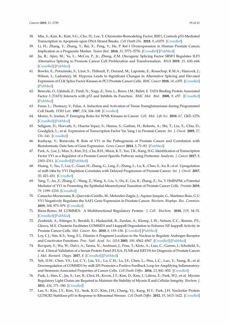
Cancers 2019, 11, 1729 19 of 21
58. Min, S.; Kim, K.; Kim, S.G.; Cho, H.; Lee, Y. Chromatin-Remodeling Factor, RSF1, Controls p53-MediatedTranscription in Apoptosis upon DNA Strand Breaks. Cell Death Dis. 2018, 9, e1079. [CrossRef]
59. Li, H.; Zhang, Y.; Zhang, Y.; Bai, X.; Peng, Y.; He, P. Rsf-1 Overexpression in Human Prostate Cancer,Implication as a Prognostic Marker. Tumor Biol. 2014, 35, 5771–5776. [CrossRef] [PubMed]
60. Jia, R.; Ajiro, M.; Yu, L.; McCoy, P., Jr.; Zheng, Z.M. Oncogenic Splicing Factor SRSF3 Regulates ILF3Alternative Splicing to Promote Cancer Cell Proliferation and Transformation. RNA 2019, 25, 630–644.[CrossRef] [PubMed]
61. Bowler, E.; Porazinski, S.; Uzor, S.; Thibault, P.; Durand, M.; Lapointe, E.; Rouschop, K.M.A.; Hancock, J.;Wilson, I.; Ladomery, M. Hypoxia Leads to Significant Changes in Alternative Splicing and ElevatedExpression of CLK Splice Factor Kinases in PC3 Prostate Cancer Cells. BMC Cancer 2018, 18, e355. [CrossRef][PubMed]
62. Bereczki, O.; Ujfaludi, Z.; Pardi, N.; Nagy, Z.; Tora, L.; Boros, I.M.; Balint, E. TATA Binding Protein AssociatedFactor 3 (TAF3) Interacts with p53 and Inhibits its Function. BMC Mol. Biol. 2008, 9, e57. [CrossRef][PubMed]
63. Fesus, L.; Thomazy, V.; Falus, A. Induction and Activation of Tissue Transglutaminase during ProgrammedCell Death. FEBS Lett. 1987, 224, 104–108. [CrossRef]
64. Moniz, S.; Jordan, P. Emerging Roles for WNK Kinases in Cancer. Cell. Mol. Life Sci. 2010, 67, 1265–1276.[CrossRef] [PubMed]
65. Seligson, D.; Horvath, S.; Huerta-Yepez, S.; Hanna, S.; Garban, H.; Roberts, A.; Shi, T.; Liu, X.; Chia, D.;Goodglick, L.; et al. Expression of Transcription Factor Yin Yang 1 in Prostate Cancer. Int. J. Oncol. 2005, 27,131–141. [CrossRef]
66. Kashyap, V.; Bonavida, B. Role of YY1 in the Pathogenesis of Prostate Cancer and Correlation withBioinformatic Data Sets of Gene Expression. Genes Cancer 2014, 5, 71–83. [PubMed]
67. Park, A.; Lee, J.; Mun, S.; Kim, D.J.; Cha, B.H.; Moon, K.T.; Yoo, T.K.; Kang, H.G. Identification of TranscriptionFactor YY1 as a Regulator of a Prostate Cancer-Specific Pathway using Proteomic Analysis. J. Cancer 2017, 8,2303–2311. [CrossRef] [PubMed]
68. Huang, Y.; Tao, T.; Liu, C.; Guan, H.; Zhang, G.; Ling, Z.; Zhang, L.; Lu, K.; Chen, S.; Xu, B.; et al. Upregulationof miR-146a by YY1 Depletion Correlates with Delayed Progression of Prostate Cancer. Int. J. Oncol. 2017,50, 421–431. [CrossRef]
69. Yang, T.; An, Z.; Zhang, C.; Wang, Z.; Wang, X.; Liu, Y.; Du, E.; Liu, R.; Zhang, Z.; Xu, Y. HnRNPM, a PotentialMediator of YY1 in Promoting the Epithelial-Mesenchymal Transition of Prostate Cancer Cells. Prostate 2019,79, 1199–1210. [CrossRef]
70. Camacho-Moctezuma, B.; Quevedo-Castillo, M.; Melendez-Zajgla, J.; Aquino-Jarquin, G.; Martinez-Ruiz, G.U.YY1 Negatively Regulates the XAF1 Gene Expression in Prostate Cancer. Biochem. Biophys. Res. Commun.2019, 508, 973–979. [CrossRef]
71. Riera-Romo, M. COMMD1: A Multifunctional Regulatory Protein. J. Cell. Biochem. 2018, 119, 34–51.[CrossRef] [PubMed]
72. Zoubeidi, A.; Ettinger, S.; Beraldi, E.; Hadaschik, B.; Zardan, A.; Klomp, L.W.; Nelson, C.C.; Rennie, P.S.;Gleave, M.E. Clusterin Facilitates COMMD1 and I-kappaB Degradation to Enhance NF-kappaB Activity inProstate Cancer Cells. Mol. Cancer. Res. 2010, 8, 119–130. [CrossRef] [PubMed]
73. Loy, C.J.; Sim, K.S.; Yong, E.L. Filamin-A Fragment Localizes to the Nucleus to Regulate Androgen Receptorand Coactivator Functions. Proc. Natl. Acad. Sci. USA 2003, 100, 4562–4567. [CrossRef] [PubMed]
74. Ravipaty, S.; Wu, W.; Dalvi, A.; Tanna, N.; Andreazi, J.; Friss, T.; Klotz, A.; Liao, C.; Garren, J.; Schofield, S.;et al. Clinical Validation of a Serum Protein Panel (FLNA, FLNB and KRT19) for Diagnosis of Prostate Cancer.J. Mol. Biomark. Diagn. 2017, 8. [CrossRef] [PubMed]
75. Yeh, D.W.; Chen, Y.S.; Lai, C.Y.; Liu, Y.L.; Lu, C.H.; Lo, J.F.; Chen, L.; Hsu, L.C.; Luo, Y.; Xiang, R.; et al.Downregulation of COMMD1 by miR-205 Promotes a Positive Feedback Loop for Amplifying Inflammatory-and Stemness-Associated Properties of Cancer Cells. Cell Death Differ. 2016, 23, 841–852. [CrossRef]
76. Park, I.; Han, C.; Jin, S.; Lee, B.; Choi, H.; Kwon, J.T.; Kim, D.; Kim, J.; Lifirsu, E.; Park, W.J.; et al. MyosinRegulatory Light Chains are Required to Maintain the Stability of Myosin II and Cellular Integrity. Biochem. J.2011, 434, 171–180. [CrossRef]
77. Lee, S.; Kim, J.Y.; Kim, Y.J.; Seok, K.O.; Kim, J.H.; Chang, Y.J.; Kang, H.Y.; Park, J.H. Nucleolar ProteinGLTSCR2 Stabilizes p53 in Response to Ribosomal Stresses. Cell Death Differ. 2012, 19, 1613–1622. [CrossRef]

Cancers 2019, 11, 1729 20 of 21
78. Kim, H.K.; Fuchs, G.; Wang, S.; Wei, W.; Zhang, Y.; Park, H.; Roy-Chaudhuri, B.; Li, P.; Xu, J.; Chu, K.; et al.A Transfer-RNA-Derived Small RNA Regulates Ribosome Biogenesis. Nature 2017, 552, 57–62. [CrossRef]
79. Cerami, E.; Gao, J.; Dogrusoz, U.; Gross, B.E.; Sumer, S.O.; Aksoy, B.A.; Jacobsen, A.; Byrne, C.J.; Heuer, M.L.;Larsson, E.; et al. The cBio Cancer Genomics Portal: An Open Platform for Exploring MultidimensionalCancer Genomics Data. Cancer Discov. 2012, 2, 401–404. [CrossRef] [PubMed]
80. Gao, J.; Aksoy, B.A.; Dogrusoz, U.; Dresdner, G.; Gross, B.; Sumer, S.O.; Sun, Y.; Jacobsen, A.; Sinha, R.;Larsson, E.; et al. Integrative Analysis of Complex Cancer Genomics and Clinical Profiles using the cBioPortal.Sci. Signal. 2013, 6, pl1. [CrossRef]
81. Baca, S.C.; Prandi, D.; Lawrence, M.S.; Mosquera, J.M.; Romanel, A.; Drier, Y.; Park, K.; Kitabayashi, N.;MacDonald, T.Y.; Ghandi, M.; et al. Punctuated Evolution of Prostate Cancer Genomes. Cell 2013, 153,666–677. [CrossRef]
82. Barbieri, C.E.; Baca, S.C.; Lawrence, M.S.; Demichelis, F.; Blattner, M.; Theurillat, J.P.; White, T.A.; Stojanov, P.;Van Allen, E.; Stransky, N.; et al. Exome Sequencing Identifies Recurrent SPOP, FOXA1 and MED12 Mutationsin Prostate Cancer. Nat. Genet. 2012, 44, 685–689. [CrossRef] [PubMed]
83. Fraser, M.; Sabelnykova, V.Y.; Yamaguchi, T.N.; Heisler, L.E.; Livingstone, J.; Huang, V.; Shiah, Y.J.; Yousif, F.;Lin, X.; Masella, A.P.; et al. Genomic Hallmarks of Localized, Non-Indolent Prostate Cancer. Nature 2017,541, 359–364. [CrossRef] [PubMed]
84. Leyh-Bannurah, S.R.; Gazdovich, S.; Budaus, L.; Zaffuto, E.; Briganti, A.; Abdollah, F.; Montorsi, F.;Schiffmann, J.; Menon, M.; Shariat, S.F.; et al. Local Therapy Improves Survival in Metastatic Prostate Cancer.Eur. Urol. 2017, 72, 118–124. [CrossRef] [PubMed]
85. Kumar, A.; Coleman, I.; Morrissey, C.; Zhang, X.; True, L.D.; Gulati, R.; Etzioni, R.; Bolouri, H.; Montgomery, B.;White, T.; et al. Substantial Interindividual and Limited Intraindividual Genomic Diversity among Tumorsfrom Men with Metastatic Prostate Cancer. Nat. Med. 2016, 22, 369–378. [CrossRef] [PubMed]
86. Taylor, B.S.; Schultz, N.; Hieronymus, H.; Gopalan, A.; Xiao, Y.; Carver, B.S.; Arora, V.K.; Kaushik, P.;Cerami, E.; Reva, B.; et al. Integrative Genomic Profiling of Human Prostate Cancer. Cancer Cell 2010, 18,11–22. [CrossRef] [PubMed]
87. Armenia, J.; Wankowicz, S.A.M.; Liu, D.; Gao, J.; Kundra, R.; Reznik, E.; Chatila, W.K.; Chakravarty, D.;Han, G.C.; Coleman, I.; et al. The Long Tail of Oncogenic Drivers in Prostate Cancer. Nat. Genet. 2018, 50,645–651. [CrossRef]
88. Rosario, S.R.; Long, M.D.; Affronti, H.C.; Rowsam, A.M.; Eng, K.H.; Smiraglia, D.J. Pan-Cancer Analysisof Transcriptional Metabolic Dysregulation using the Cancer Genome Atlas. Nat. Commun. 2018, 9, e5330.[CrossRef]
89. Hieronymus, H.; Schultz, N.; Gopalan, A.; Carver, B.S.; Chang, M.T.; Xiao, Y.; Heguy, A.; Huberman, K.;Bernstein, M.; Assel, M.; et al. Copy Number Alteration Burden Predicts Prostate Cancer Relapse. Proc. Natl.Acad. Sci. USA 2014, 111, 11139–11144. [CrossRef]
90. Gao, D.; Vela, I.; Sboner, A.; Iaquinta, P.J.; Karthaus, W.R.; Gopalan, A.; Dowling, C.; Wanjala, J.N.;Undvall, E.A.; Arora, V.K.; et al. Organoid Cultures Derived from Patients with Advanced Prostate Cancer.Cell 2014, 159, 176–187. [CrossRef]
91. Abida, W.; Armenia, J.; Gopalan, A.; Brennan, R.; Walsh, M.; Barron, D.; Danila, D.; Rathkopf, D.; Morris, M.;Slovin, S.; et al. Prospective Genomic Profiling of Prostate Cancer Across Disease States Reveals Germlineand Somatic Alterations that may Affect Clinical Decision Making. JCO Precis. Oncol. 2017. [CrossRef][PubMed]
92. Grasso, C.S.; Wu, Y.M.; Robinson, D.R.; Cao, X.; Dhanasekaran, S.M.; Khan, A.P.; Quist, M.J.; Jing, X.;Lonigro, R.J.; Brenner, J.C.; et al. The Mutational Landscape of Lethal Castration-Resistant Prostate Cancer.Nature 2012, 487, 239–243. [CrossRef] [PubMed]
93. Abida, W.; Cyrta, J.; Heller, G.; Prandi, D.; Armenia, J.; Coleman, I.; Cieslik, M.; Benelli, M.; Robinson, D.;Van Allen, E.M.; et al. Genomic Correlates of Clinical Outcome in Advanced Prostate Cancer. Proc. Natl.Acad. Sci. USA 2019, 116, 11428–11436. [CrossRef] [PubMed]
94. Robinson, D.; Van Allen, E.M.; Wu, Y.M.; Schultz, N.; Lonigro, R.J.; Mosquera, J.M.; Montgomery, B.;Taplin, M.E.; Pritchard, C.C.; Attard, G.; et al. Integrative Clinical Genomics of Advanced Prostate Cancer.Cell 2015, 161, 1215–1228. [CrossRef] [PubMed]
Cancers 2019, 11, 1729 21 of 21
95. Beltran, H.; Prandi, D.; Mosquera, J.M.; Benelli, M.; Puca, L.; Cyrta, J.; Marotz, C.; Giannopoulou, E.;Chakravarthi, B.V.; Varambally, S.; et al. Divergent Clonal Evolution of Castration-Resistant NeuroendocrineProstate Cancer. Nat. Med. 2016, 22, 298–305. [CrossRef] [PubMed]
96. Liu, R.; Zhou, Z.; Huang, J.; Chen, C. PMEPA1 Promotes Androgen Receptor-Negative Prostate CellProliferation through Suppressing the Smad3/4-c-Myc-p21 Cip1 Signaling Pathway. J. Pathol. 2011, 223,683–694. [CrossRef]
97. Zhao, C.B.; Bao, J.M.; Lu, Y.J.; Zhao, T.; Zhou, X.H.; Zheng, D.Y.; Zhao, S.C. Co-Expression of RAGEand HMGB1 is Associated with Cancer Progression and Poor Patient Outcome of Prostate Cancer. Am. J.Cancer Res. 2014, 4, 369–377. [CrossRef]
98. Bull, J.H.; Ellison, G.; Patel, A.; Muir, G.; Walker, M.; Underwood, M.; Khan, F.; Paskins, L. Identificationof Potential Diagnostic Markers of Prostate Cancer and Prostatic Intraepithelial Neoplasia using cDNAMicroarray. Br. J. Cancer 2001, 84, 1512–1519. [CrossRef]
99. Iglesias-Gato, D.; Wikstrom, P.; Tyanova, S.; Lavallee, C.; Thysell, E.; Carlsson, J.; Hagglof, C.; Cox, J.;Andren, O.; Stattin, P.; et al. The Proteome of Primary Prostate Cancer. Eur. Urol. 2016, 69, 942–952.[CrossRef]
100. Rajan, P.; Sudbery, I.M.; Villasevil, M.E.; Mui, E.; Fleming, J.; Davis, M.; Ahmad, I.; Edwards, J.; Sansom, O.J.;Sims, D.; et al. Next-Generation Sequencing of Advanced Prostate Cancer Treated with Androgen-DeprivationTherapy. Eur. Urol. 2014, 66, 32–39. [CrossRef]
101. Kim, J.Y.; Cho, Y.E.; Kim, G.Y.; Lee, H.L.; Lee, S.; Park, J.H. Down-Regulation and Aberrant CytoplasmicExpression of GLTSCR2 in Prostatic Adenocarcinomas. Cancer Lett. 2013, 340, 134–140. [CrossRef] [PubMed]
102. Deng, Z.; Cao, P.; Wan, M.M.; Sui, G. Yin Yang 1: A Multifaceted Protein Beyond a Transcription Factor.Transcription 2010, 1, 81–84. [CrossRef] [PubMed]
103. Long, Z.; Li, Y.; Gan, Y.; Zhao, D.; Wang, G.; Xie, N.; Lovnicki, J.M.; Fazli, L.; Cao, Q.; Chen, K.; et al. Roles ofthe HOXA10 Gene during Castrate-Resistant Prostate Cancer Progression. Endocr. Relat. Cancer 2019, 26,279–292. [CrossRef] [PubMed]
104. GTEx Consortium. The Genotype-Tissue Expression (GTEx) Project. Nat. Genet. 2013, 45, 580–585. [CrossRef][PubMed]
105. Petryszak, R.; Keays, M.; Tang, Y.A.; Fonseca, N.A.; Barrera, E.; Burdett, T.; Fullgrabe, A.; Fuentes, A.M.;Jupp, S.; Koskinen, S.; et al. Expression Atlas Update, an Integrated Database of Gene and Protein Expressionin Humans, Animals and Plants. Nucleic Acids Res. 2016, 44, D746–D752. [CrossRef] [PubMed]
106. Kaighn, M.E.; Narayan, K.S.; Ohnuki, Y.; Lechner, J.F.; Jones, L.W. Establishment and Characterization of aHuman Prostatic Carcinoma Cell Line (PC-3). Investig. Urol. 1979, 17, 16–23.
107. Livak, K.J.; Schmittgen, T.D. Analysis of Relative Gene Expression Data using Real-Time Quantitative PCRand the 2(-Delta Delta C (T)) Method. Methods 2001, 25, 402–408. [CrossRef]
108. Babicki, S.; Arndt, D.; Marcu, A.; Liang, Y.; Grant, J.R.; Maciejewski, A.; Wishart, D.S. Heatmapper:Web-Enabled Heat Mapping for all. Nucleic Acids Res. 2016, 44, W147–W153. [CrossRef]
© 2019 by the authors. Licensee MDPI, Basel, Switzerland. This article is an open accessarticle distributed under the terms and conditions of the Creative Commons Attribution(CC BY) license (http://creativecommons.org/licenses/by/4.0/).
Related Documents