Sudan University of Science and Technology College of Graduate Studies Characterization of Foramen Magnum and Occipital Condyles in Adult Sudanese by using Computed Tomography. ييني السودانقم القذالية لدلصيف الثقبة العظمى وال تو الب الغينشعةستخدام ا با المقطعيةA Thesis Submitted for the Requirements of the fulfillment of the Award Ph.D. Degree in Diagnostic Radiological Technology Presented By: ALAMIN MUSA SALIH OMER Supervisor: Associate Prof. Caroline Edward Eyad Khela Aug 2015

Welcome message from author
This document is posted to help you gain knowledge. Please leave a comment to let me know what you think about it! Share it to your friends and learn new things together.
Transcript
Sudan University of Science and Technology
College of Graduate Studies
Characterization of Foramen Magnum and
Occipital Condyles in Adult Sudanese by using
Computed Tomography.
الغينالب توصيف الثقبة العظمى واللقم القذالية لدي السودانيين
المقطعية باستخدام األشعة
A Thesis Submitted for the Requirements of the fulfillment of
the Award Ph.D. Degree in Diagnostic Radiological
Technology
Presented By:
ALAMIN MUSA SALIH OMER
Supervisor:
Associate Prof. Caroline Edward Eyad Khela
Aug 2015

I
اآلية الكريمة
حيم حمن الره الره بسم للاه
ق ل هل يستوى الذين يعلمون والذين ال يعلمون إنما يذكر أولى األلباب
صدق اهلل العظيم
الزمرسورة
(9اآلية )

II
Dedication
To:
My parents for their patience and encouragement,
My Wife and my Sons & daughter “Abdallah, Qusi
& Reem” for their patience, understanding and
encouragement
My brothers and sisters for their help and support,
My friends for their valuable support,
And
To the dearest people in my life.
I dedicated this work.

III
Acknowledgement
My full thanks to my God in every thing.
My great thanks and deep gratitude to my
supervisor Prof. Dr. Caroline Edward Eyad Khela,
for her advices, valuable suggestions and help.
Also very special thank extend to Mr.
Mohamed salman, who helped in performing the
practical part of this research, with his full patience
and cooperation.
My thank to the staff of all Ibn ALhythm
Medical Center who gave me permission to
conduct the practical part of this work at their
center.
IV
ABSTRACT
The foramen magnum (FM) is an important anatomical region of the skull
base and is of significance for anatomy, anthropology and other medical
fields. The transcondylar approach has been used in surgeries to access
lesions in areas close to the foramen magnum (FM) and is performed
directly through the occipital condyle (OC).
This study aims to characterize the foramen magnum shape and contour and
determine of the different anatomical variations through the use of
reconstructed helical CT images. For verify the morphological
characteristics of the (FM) for gender determination in Sudanese individuals
by measuring the saggital, transverse diameter, area, circumference and
characterizing its shape, and to characterize the anatomical variations related
to the (OC) measurements.
A total of 241 Sudanese patients (147 males and 94 females) with mean ages
were 40.96±15.21, 41.02±14.32 years respectively were examined using
reformatted axial CT and three-dimensional CT, between September 2012
and July 2014, referred to the Radiology Department in the Ibn ALhythm
Medical Center, Khartoum, Sudan
The foramen magnum shapes were determined as a round shape in
55(22.9%) of the cases, oval in 115 (47.8%), irregular in 36(14.8%) and
arrow head in 35(14.5%), the mean sagittal and transverse diameters of the
foramen magnum were determined as 34.0±2.98 mm and 29.3±2.44 mm
respectively. Area of the (FM) was 770.0±111.09 and Circumference of the
(FM) was 104.1±11.55, a significant difference between genders were
detected at p-value =0.05.
V
Characteristics of the head, and measures related to the (FM) and right and
left occipital condyles were examined. The results showed no significant
difference between the measurements obtained in the right and in left sides.
The (OC) morphometric parameters had significant relationship with (FM)
antroposterior and transverse diameters. The study revealed a significant
difference between the two genders with no significant relations between
(OC) and head characteristics. The data obtained by three-dimensional CT
images are important in assessing the morphometric variations of (OC) for
Sudanese patients. As the (OC) is the main bony eminences impeded the
anterolateral surface of the brainstem, neurosurgeons should be familiar with
variations of the (OC) and structures surrounding the (FM) in order to
achieve the safest surgical procedure.
knowledge of FM area‘s anatomy is of extreme importance for treating
lesion and help the surgeon regarding selection best surgical approach and
expected changes in the anatomy of these critical structures and resection of
tumors; removal of bony structures such as the occipital condyle (OC) may
result in injury to the vascular structures and lower cranial nerves and result
in craniocervical instability.

VI
البحثملخص
هي المنطقة التشريحية الهامة في قاعدة الجمجمة و ذات أهمية لعلم التشريح وعلم العظمى الثقبة
الوصول لآلفات في المناطق القريبة يتم في العمليات الجراحيةواإلنسان والمجاالت الطبية األخرى.
. مباشرة من خالل اللقمة القذاليه العظمى الثقبة
وذلك االختالفات التشريحية وتهدف هذه الدراسة إلى تحديد خصائص شكل الثقبة العظمى وتحديد
,بواسطة األشعة المقطعية. للتحقق من خصائص شكل ألثقبه الصور بناء إعادة تقنية باستخدام
مساحة ومحيط وتوصيف شكل ألثقبه ,طول ، عرض -العظمى لتحديد الجنس . عن طريق قياس
اشتملت الدراسة وقدالعظمى ، وتوصيف االختالفات التشريحية المتصلة بقياسات اللقمة القذاليه .
.15.21 ± 40.96 األعمار إناث( مع متوسط 94ذكور و 147مريض ) 241علي
لشكل الغير منتظم او(، %47.8( من الحاالت، البيضاوي في )%22الشكل الدائري في ) وقد وجد
طول وعرض الثقبه الثقبة كما يلي (، ومتوسط 14.5( وذو الرأس السهمي في )%14.8في )
770.0 متوسط مساحة الثقبه العظمى ملم على التوالي . وكان 2.44± 29.3ملم و ±2.98 34.0
الجنسين فرق كبير بين ملحظةان هناك، ) تم 11.55 ± 104.1ومتوسط محيطها 111.09 ±
.0.05) القيمة االحتمالية = ,
اللقم القذالي اليمنى واليسرى . من خالل قياسات الثقبه الكبرى واألجزاء التشريحية المتصلة بها مثل
أظهرت النتائج عدم وجود فرق كبير بين القياسات التي تم الحصول عليها في اليمين واليسار في
طول وعرض الثقبه العظمى. وكشفت مع القذاليهلللقمة رةأظهرت الدراسه عالقة كبي كماالجانبين.
اللقمة القذاليه الدراسة أيضا وجود فرق كبير بين الجنسين مع عدم وجود عالقة ذات داللة بين
وخصائص الرأس .
البيانات التي حصلت عليها الصور المقطعية ثالثية األبعاد مهمة في تقييم االختالفات المظهرية من
لسطح لالرئيسي العظمي البروز القذاليه هي اللقمة ان للمرضى السودانيين. باعتبار لقذاليهااللقمة
والثقبه العظمى القذاليهاللقمة ختالفات فياالينبغي معرفة لذالكاألمامي الوحشي من جذع الدماغ ،
.يةمن أجل تحقيق أسلم طريقه إلجراء العمليات الجراح واألجزاء التشريحية ألمحيطه.

VII
المعرفة التشريحية لمنطقة الثقبه العظمى ذو أهمية بالغة لعالج األورام ومساعدة الجراح بشأن
والتغيرات المتوقعة في تشريح األجزاء األساسية واستئصال األورام. ,اختيار أفضل نهج جراحي
األعصاب القحفية وقد يؤدي إلى إصابة األوعية الدموية القذاليهإزالة األجزاء العظمية مثل اللقمة
يؤدي إلى عدم االستقرار في مفصل العنق مع الرأس .مما السفلى

VIII
Contents
Titles Page NO
I اآلية الكريمة
Dedication II
Acknowledgement III
English Abstract V
Arabic Astract VI
Lis of Contents VIII
List of Table XII
List of Figure XIV
List of Abbrivation XVI
Chapter one
1.1 Introduction 1
1.2: Problem of the Study 5
1.3: Objective of the Study 5
1.3.1 General Objective 5
1.32: SpecificObjectives 5
Chapter Two: Litreture Reviews
2.1 An Overview of skull 7
2.2 Embryology Craniocervical Developmental Anatomy 9
2.3 Anatomy and physiology of base of skull 12
2.3.1 Occipital bone 12
2.3.1.1 The Squama (squama occipitalis) 13
2.3.1.2 Lateral Parts (pars lateralis) 15
2.3.1.3 Basilar Part (pars basilaris) 18
2.3.1.3 Foramen Magnum 19
IX
2.3.2 The viatal structures pass through the foramen magnum 22
2.3.2.2 The meninges 23
2.3.2.3 The spinal accessory nerve 24
2.3.2.4 The vertebral arteries (VA) 24
2.3.2.5 The venous structures 25
2.3.3 First Cervical Vertebra 25
2.3.3.1 Stabilizing Cranio cervical junction 28
2.4 Pathology of posterior cranial fossa 28
2.4.1 Achondroplasia 28
2.4.1.1 Radiographic features 29
2.4.1.2 Cranial 29
2.4.1.3 Treatment and prognosis 30
2.4.2 Foramen Magnum Meningiomas 31
2.4.2 .1 Classification of Foramen Magnum Meningiomas 32
2.4.2.2 Imaging Features 35
2.4.2.3 Preoperative considerations 36
2.4.2.3 Surgical Approaches to Foramen Magnum Meningiomas 38
2.4.2.3.1 Suboccipital Craniotomy 39
2.4.2.3.1Transcondylar Approach 40
2.4.2.3.2 Surgical results 42
2.4.3 Chiari malformations 43
2.4.3.1 Types of Chiari malformations 44
2.4.3.1.1Chiari type I 44
2.4.3.1.2 Chiari type II 44
2.4.3.1.1Chiari type III AND IV 44
2.4.3.2 Radiographic features 45
X
2.4.3.3 Indications 47
2.4.3.3 Treatment 48
2.5 Pathology of craniocervial junction 49
2.5 .1 Primary/Congenital Conditions 50
2.5.1.1Congenital Anomalies of Occiput 50
2.5.1.2 Congenital Anomalies of Atlas. 51
2.5.2 Occipital Condyle Fractures. 51
2.5.3 Trauma to Atlas 52
2.5.4 Treatment of occipitocervical 53
2.5.4.1Occipitocervical stabilization 53
2.5.4.2 Surgical stabilization 54
2.8 Computer Tomography posterior cranial fossa 57
2.8 Computer Tomography for head trauma 58
2.8 Computer Tomography: Technique 59
2.9 Previous Studies 59
Chapter Three: Materials and Methods
3.1 Materials 65
3.2 Data Collection & Analysis 66
3.2.1 Data collection 66
3.2.2 Statistical analysis 68
Chapter Four: Result
4.1 Table & Figures 69
Chapter Five
5.1 Discussion 84
5.1.2 Foramen magnum length and width 85
5.1.3 Foramen magnum area and circumference 87
XI
5.1.3 Foramen magnum transverse and saggital diameter of the hesd 88
5.1.4 The occipital condyles measurement 89
5.1.5 The occipital condyles and FM measurement 90
5.2 Conclusion 92
5.3 Recommendation 93
References 94
Appendix 117
Accomplishment 123
XII
List of Tables
Titles Page NO
4.1: Distribution of the study sample according to (Gender) 69
4.2: Distribution of the study sample according to (Age group) 70
4.3: Sudanese foramen magnum morphology, frequency and percentages 71
4.4: Sudanese foramen magnum morphology cross tabulated with Gender 72
4.5: Variables related to gender, mean and standard deviations,
maximum, minimum,
74
4.6: Sudanese foramen magnum morphology area (AFM) and
circumference (CFM) correlated with transverse (TDH) and sagittal
diameter of the head (SDH), Antro posterior (LFM) and transverse
diameter (WFM) of the FM
75
4.7: Statistical measurements of theforamen mafnum variables correlated
with the age classes (mean ± standard deviation, min, max
76
4.8: Descriptive statistics of the studied occipital condyles variables
(Mean± SD, Maximum and Minimum values)
78
4. 9: Morphometric measurements of the Occipital condyles 78
4.10: Morphometric measurements of the Occipital condyles related to
gender (mean ± standard deviation, min, max, p-value).
79
4.11:Statistical measurements of the variables correlated with the age
classes (mean ± standard deviation, min, max,p-value)
80
4.12 Correlation between the foramen magnum (FM) (Length and width)
and the occipital condyles morphometric measurements
82
4.13 Correlation between the head dimensions (transverse and sagittal
diameter) and the occipital condyles morphometric measurements
83
XIII
List of Figures
Titles Page NO
2.1: The bones of skull and their sutures 8
2.2: Occipital bone. Outer surface 13
2.3: Occipital condyles. 17
2.4 Posterior view of the craniocervical junction of an adult skeleton 18
2.5. Inferior view of the cranial base with a circle around the foramen
magnum
21
2.6. First cervical vertebra, or atlas 28
2.7 axial CT show narrowed foramen magnum due to Achondroplasia 30
2.8 :3D VRT CT show narrowed foramen magnum due to
Achondroplasia
31
2.9: Classification of foramen magnum meningiomas 34
2.10: a–c Preoperative MRI. A large lateral foramen magnum
meningioma displaces the neuraxis. d, e Postoperative CT scan
37
2.11. A: Suboccipital craniotomy. B: Tumor growth naturally 38
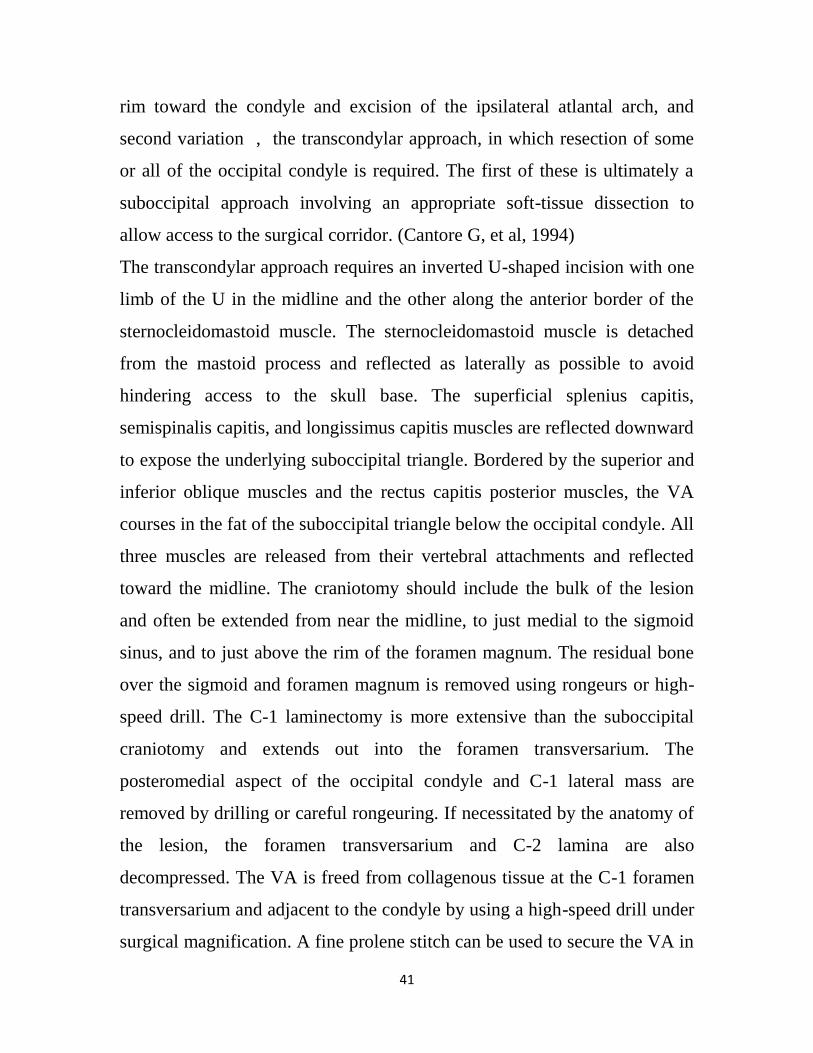
2.12: Sagittal and coronal MRI images of Chiari type I malformation 45
2.13: Resolution of syringomyelia 48
2.14: Intraoperative photograph of Chiari type 1 malformation 49
2.15. Parasagittal computed tomography of adult with congenital atlanto-
occipital assimilation
52
2.16. Sagittal computed tomography of trauma patient 53
2.17. Cadaveric photographs and an illustration showing placement of
occipital condyle screws
55
XIV
2.18. Preoperative reconstructed sagittal (left) and 3D (right) CT of the
cervical spine
56
2.19. Postoperative reconstructed sagittal CT of the cervical spine 56
4.1: Distribution of the study sample according to (Gender) 69
4.2: Distribution of the study sample according to (Age group) 70
4.3: The Sudanese foramen magnum morphology, frequency and
percentages
71
4.4 The Sudanese foramen magnum morphology cross tabulated with
Gender
73
XV
Abbrivations
Area of foramen magnum AFM
Coputed Tomography CT
Circumference of foramen magnum CFM
Cranniocevicle junction CCJ
Chiari malformations CM
Cranial Nerve CN
Cerprospinal Fluid CSF
Foramen Magnum FM
Foramen Magnum Meningiomas FMM
Length of Right occipital Condyle LRC
Length of foramen magnum LFM
Length of left occipitalCondyle LLC
Minimum intercondylar distance MnlD
Maximum medial intercondylar distance MID
Maximum bicondylar distance MBD
Multi Detector Computed Tomography MDCT
Magnetic Resonance Imaging MRI
Occipital Condyle OC
Shape of foramen magnum SFM
Sagittal diameter of the head SDH
XVI
sterno-mastoid SM
Transverse diameter of the head TDH
Width of foramen magnum WFM
Width of Right occipital Condyle WRC
width of left occipital Condyle WLC
vertebral arteries VA
Volume Rendering Technique VRT
1
Chapter One
1.1 Introduction:
The most prominent feature in the floor of the posterior cranial fossa is the
foramen magnum in the occipital bones, this wide communication between
the posterior cranial fossa and the vertebral canal, vertebral arteries and the
spinal accessory nerve. Anteriorly the apical ligament of the dense and
membrane tectoria are in it. Its wider posterior part contains the medulla
oblongata and spinal cord continued. Anterior to its transverse diameter it is
narrowed by the two occipital condyles. (Standring S. et. al, 2008)
Foramen magnum (FM) is the oval shape opening located at the base of the
skull, and bordered by the basilar, squamous, and two lateral parts of the
occipital bone. The FM, as a transition zone between spine and skull, plays a
vital role as a landmark because of its close association to key structures
such as the brain and the spinal cord.( Boni. et. al, 2009).
Since the FM includes specific neuroanatomical structures and their lesions
in that region which require particularly microsurgical intervention,
choosing and establishing the most suitable surgical techniques need a
careful planning mainly based on the FM size to refrain from any
neurological injury. (G. Venkatesh. et. al, 2005).
Moreover, intradural and extradural tumors, common congenital
abnormalities such as FM syndrome produced by atlanto-occipital
assimilation, and cerebellar tissue herniations which invaginated into the FM
may lead to neural compression and even death are commonly met
pathological disorders in this region. (Fatma Hayat Erdil .et al, 2010)
2
The occipital condyles are the prominences of the paired lateral exoccipital
Segments of the occipital bone, which form the foramen magnum together
with the basioccipital segment anteriorly, and the supraoccipital or
squamosal segment posteriorly (Lustrin ES, et al., 1994).
The bone around the foramen magnum constitutes the uppermost border of
an extremely complex three-unit joint with intricate functional relationships
between the occiput, atlas, and axis (ie, the CCJ or occipitoatlantoaxial
complex) (Saldinger P, et al., 1990).
The occipital condyles (OC) of the skull are located with the superior
articular facets of the atlas vertebra and form an important junction between
the cranium and the vertebral column.(Muthukumar N, et al., 2005), ( Naderi
S, et al., 2005) . Each OC is oval in outline and oriented obliquely so that its
anterior end lies nearer the midline. It is markedly convex anteroposteriorly,
slightly convex transversely, and its medial aspect is roughened by
ligamentous attachments, (Schwaber MK, et al., 1990).
Its integrity is of vital importance for the stability of the craniovertebral
junction. The difficulties and high rate of morbidity associated with surgical
approaches (Acikbas SC, et al., 1997), (La Marca F, et al., 2008).
The transcondylar surgical approach has been used to access lesions in areas
close to the (FM) and it is performed directly through the (OC) (George et
al., 1988), therefore the anatomical landmarks of the (FM) should be well
known in order to make a safe occipital condyle resection (Barut et al.,
2009). The surgical errors in this region may result in injury to the vascular
structures and cranial nerves and result in craniocervical instability.
Consequently, neurosurgeons should be more familiar with the anatomy and
variations of this region. Therefore, radiological (Osborn et al., 1978) and
anatomical morphometric studies (Bozbuga et al., 1999; De Oliveira et al.,
3
1985; Prescher, 1997) were performed to contribute to the knowledge of this
area.
Computed tomographic scan is noninvasive modality for the imaging the
skull base. Since this procedure is widely done, this modality was preferred.
(Surwase, et. al., 2013)
Because of the dense bone of the base of skull beam-hardening artifacts are
often seen in images of the posterior fossa, thin slices can help to reduce
these artifacts and produce high spatial rsolution.
Reviewing published literature, identified many study concerning CT
measurements of the foramen magnum in unidentified skulls, and reported
studying 3D CT measurements of the foramen magnum with a resultant sex
discriminant.
The cranial base is such a complex structure that it is only studied
morphometrically. The sites where a number of vital structures have their
entrance or exits are very important for clinical application. Therefore the
assessment of these morphometrics is helpful for surgical approaches for
reaching lesions in the middle and posterior part of cranial base.
(Cicekcibasi AE, et al, 2004)
The study of diameters of foramen magnum is interesting due to the
important relations of the foramen magnum and also its contents.
Dimensions of the foramen magnum have clinical importance because the
vital structures that pass through it may suffer compression. It has also been
noted that longer antero-posterior dimension of foramen magnum permitted
greater contralateral surgical exposure for condylar resection. So, anatomic
and radiologic values of foramen magnum dimensions and their relation to
gender have been the objectives of several studies. (Murshed K.A., et al,
2003).
4
The anatomic and radiologic values have been the objectives of several
studies. (Murshed K.A., et al, 2003). Recent advances in microsurgical
technique and more wide spread use of the operating microscope have now
enabled surgeons to approach previously in operable deep seated lesions of
the skull base. It is therefore necessary that the clinicians should have a
thorough knowledge of anatomy of this region for evaluation of various
disease processes affecting this region (Laine FJ, et al 1990).
FM evaluations are very significant in not only to establish the most suitable
operational procedures, but also to find valuable data for unidentified sex
assessment and determination and individuality in forensic medicine.( Fatma
Hayat Erdil .et al, 2010).
Determining the biological sex of unknown skeletal remains is an important
aspect of medico-legal investigations seeking to establish the identity of a
deceased individual. (R.Gapert, et al 2008).
Anthropologists are often faced with the task of assigning sex to remains
that are incomplete, fragmented or damaged as may result from incidents
such as mass disasters, airplane crashes, fire, explosions or physical violence
(W.R.G.Teixeira,1982). Comprised remains will affect the accuracy of sex
estimation and thus necessitate the development of reliable sexing criteria
based on isolated bony elements (T.D.Holland 1986).
The foramen magnum has attracted considerable interest for the purposes of
sex determination (A.T.Uthman, et al., 2012). The robusticity of the
occipital bone and the relatively protected anatomical position of the
foramen magnum beneath adepth of soft tissue may make it less vulnerable
to fragmentation, or to the effects of inhumation and taphonomic processes
in comparison to other cranial and facial bones (T.D.Holland 1989).
5
1.2 Problem of the Study:
The FM clinically importance since vital structures that pass through it
which may suffer compression; such as in cases of FM achondroplasia
and FM brain herniation as well as in a transcondylar surgical approach to
the FM, when resection of tumors; removal of bony structures such as the
occipital condyle (OC) may result in injury to the vascular structures and
lower cranial nerves and result in craniocervical instability. Hence,
knowledge of FM area‘s anatomy is of extreme importance for treating
lesion and help the surgeon regarding selection best surgical approach and
expected changes in the anatomy of these critical structures.
There is no specific characterization of the morphology and dimensions
of the FM as standard in Sudanese population; so this study is obtain to
study the anatomical variation of the FM for forensics, anthropologic and
surgical purposes.
1.3 Objectives:
1.3.1 General objectives:
To study the Anatomical Variation of foramen magnum and occipital
condyles In Adult Sudanese using computed tomography.
1.3.2 Specific objectives
To characterize the foramen magnum shape and contour.
To measure the foramen magnum size and dimensions.
To determine the correlation between the FM shape and size with
gender.
To determine the impact of the anatomical variation of foramen
magnum in gender estimation.

6
To measure the occipital condyles size and dimensions.
characterize the anatomical variations related to the (OC) with the
relation to the morphometric parameters of the (FM)
To find out an index for the FM for Sudanese compared to the other
populations.
7
Chapter Two
Literature Review
2.1 An Overview of skull:
The skull is supported on the summit of the vertebral column, and is of an
oval shape, wider behind than in front. It is composed of a series of flattened
or irregular bones which, with one exception (the mandible), are immovably
jointed together. It is divisible into two parts :( Gray, H, 1918; Bartleby.com,
2000).
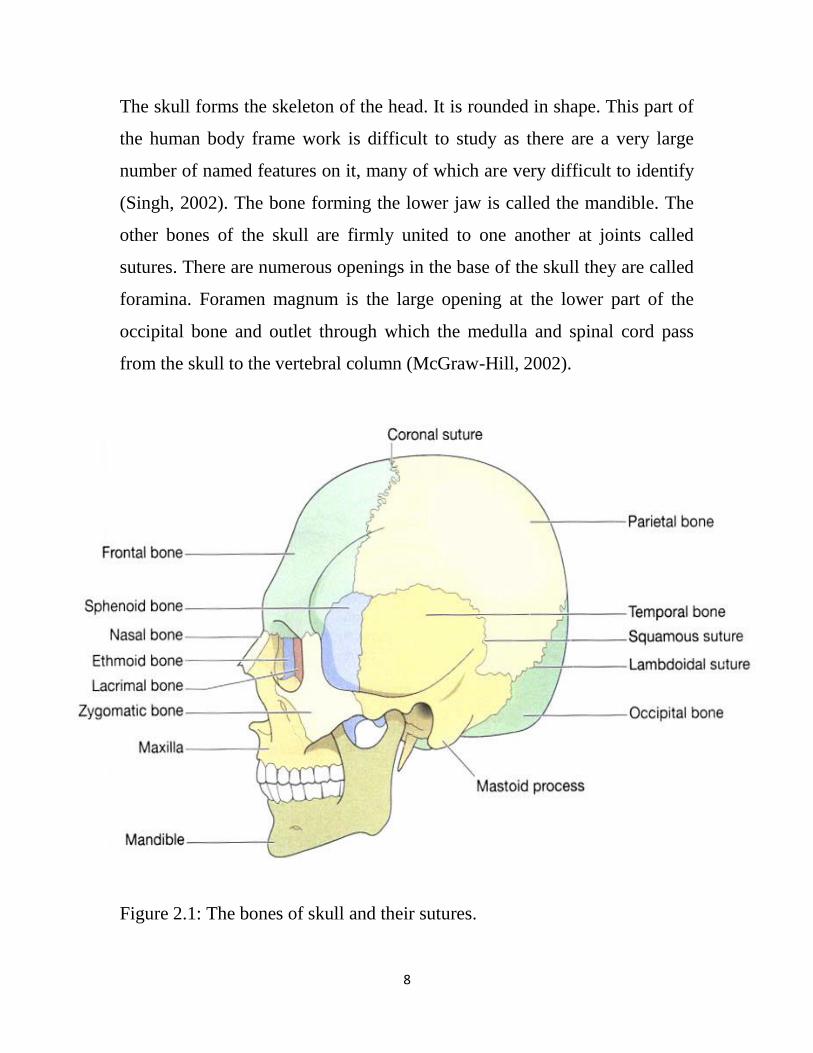
The skul composed of cranium, which lodges and protects the brain, consists
of eight bones, and the skeleton face, of fourteen bones, (Figure 2-1)
The Cranium consist of, 8 bones are, Occipital, Two Parietals, Frontal, Two
Temporals, Sphenoidal and Ethmoidal.
The Face consist of , 14 bones , Two Nasals, Two Maxillæ , Two Lacrimals
, Two Zygomatics,Two Palatines , Two Inferior Nasal Conchæ , Vomer ,and
Mandible.
In the Basle nomenclature, certain bones developed in association with the
nasal capsule, viz., the inferior nasal conchæ, the lacrimals, the nasals, and
the vomer, are grouped as cranial and not as facial bones.
The hyoid bone situated at the root of the tongue and attached to the base of
the skull by ligaments. (Gray, H, 1918; Bartleby.com, 2000).
The occipital bone situated at the back and lower part of the cranium, is
trapezoid in shape and curved on itself. It is pierced by a large oval aperture,
the foramen magnum, through which the cranial cavity communicates with
the vertebral canal.
8
The skull forms the skeleton of the head. It is rounded in shape. This part of
the human body frame work is difficult to study as there are a very large
number of named features on it, many of which are very difficult to identify
(Singh, 2002). The bone forming the lower jaw is called the mandible. The
other bones of the skull are firmly united to one another at joints called
sutures. There are numerous openings in the base of the skull they are called
foramina. Foramen magnum is the large opening at the lower part of the
occipital bone and outlet through which the medulla and spinal cord pass
from the skull to the vertebral column (McGraw-Hill, 2002).
Figure 2.1: The bones of skull and their sutures.
9
Knowledge of normal and variations of skull base is important for
neurosurgeons, and anatomists. (Berlis .A et al, 1992).
In neurosurgery, the assessment of the (FM) dimensions is used for the
access of the brainstem lesions (Furtado SV et al 2010).Studies accounted
that studying the anatomy of the skull base is important for this approach
(Muthukumar N, 2005).
Indexes have been built from the dimensions of the occipital condyles and
the foramen magnum, and various authors have reported its usefulness in
determining the sex, particularly with incomplete skeleton or cranial bones
fractured (Ferreira et al., 1967; Teixeira, 1982; Zadvornov Iu, 1997).
2.2 Embryology Craniocervical Developmental Anatomy
The bony cranial base is developed by a process of enchondral ossification,
in which a cartilaginous framework is first developed and subsequently
resorbed with further deposition of bone, caused by distorting forces, such as
eye development and brain development (Gasser RF., 1976). The clivus and
cranial base elongates by sutural growth at the spheno-occipital and
sphenopetrosal synchondrosis and by further sutural growth along the lateral
portion of the cranial base (Menezes AH., 1998). On the other hand, the
facial bones and the majority of the cranium develop by intramembranous
ossification. This development bypasses the intermediate cartilaginous phase
characteristic of the development of the bony cranial base (Christ B, et al
1992, Dietrich S, 1997, Kessel M, 1991).
During the fourth week of gestation, 42 somites are formed (Ganguly DN, et
al, 1964). There are four occipital somites, eight cervical, 12 thoracic, five
lumbar, five sacral, and 8 to 10 coccygeal pairs (Gasser RF., 1976). Each
10
somite differentiates into an outer dermatome and inner myotome and a
medial sclerotome. The sclerotomes are ventral-medial in their location and
will form the vertebral bodies. These ventral-medial bilateral cells migrate
toward the midline and surround the notochord. Each sclerotome will
develop the fissure of Ebner, which is a central cleft that divides a loose
collection of cells cranially from a dense cellular area caudally. In this
development, the cells from the fissure of Ebner migrate toward and encase
the notochord to become the precursors of the intervertebral disc (Melsen B,
1974). The superior half of one sclerotome unites with the lower half of its
neighbor and, thus, forms the earliest manifestation of the vertebral body.
The first four sclerotomes, however, will not follow this course, and
essentially fuse to form the occipital bone and posterior portions of the
foramen magnum. Simultaneously, vascularization of the occipital bone
begins and differentiation of ganglia and vascular tissue begins. The
hypoglossal and first cervical arteries demarcate the caudal occipital
segment (Gladstone J, et al 1915).
The occipital sclerotomes correspond to the segmental nerves that group
together to form the hypoglossal nerve, with a path through the individual
foramina within the bone (Sensing EC, 1957). The first two occipital
sclerotomes ultimately form the basiocciput. The third sclerotome is
responsible for the exoccipital bone, which forms the jugular tubercles. The
hypocentrum of the fourth occipital sclerotome forms the anterior tubercle of
the clivus. The centrum itself forms the apical cap of the dens and the apical
ligament (Ganguly DN, et al, 1964, Menezes AH, 1995). The neural arch
component of the proatlas divides into a rostral ventral segment and a caudal
segment. The ventral portion forms the anterior margin of the foramen
11
magnum as well as the occipital condyle and the midline third occipital
condyle.( Gladstone RJ, 1915)
The first spinal sclerotome forms the atlas vertebra. It is modified
from the remaining spinal vertebrae, wherein the centrum is
separated to fuse with the axis body and, thus, forms the odontoid
process. The neural arch of this first spinal sclerotome proceeds to
form the posterior and inferior portion of the C1 arch (Menezes AH,
1998). At times, the hypochordal bow, instead of disappearing, may
survive and join with the anterior arch of the atlas to form a variant
with an abnormal articulation, which then exists between the inferior
clivus, the anterior arch of the atlas, and the apical segment of the odontoid
process (Menezes AH, 1995). In our hands, recent computed tomographic
(CT) evaluation of the atlas has shown that several ossification centers are
present in the atlas development (MenezesAH, 1995). However, the lateral
atlantal masses must be present at birth. A complete ring must form by 3
years of age. Abnormal development is observed with the skeletal
dysplasias, such as spondyloepiphyseal dysplasia; achondroplasia;
Goldenhar’s syndrome; and in genetic abnormalities, such as Down’s
syndrome. (Arnold H, et al, 2008)
Posterior fossa expansion occurs because of enchondral resorption, sutural
growth, and bony accretion. Growth of the basal aspect of the clivus
elongates the basiocciput and lowers the frontal margin of the foramen
magnum. Synchondrosial growth occurs until 16 to 18 years of age. The
bony abnormality in hindbrain herniation syndrome has significance here.
Lack of posterior fossa volume results in herniation of the cerebellar tonsils
through the foramen magnum, resulting in tonsillar ectopia (Menezes AH,
12
2003). Significant muscle development takes place dorsal and lateral to the
cervical spine to provide for the top-heavy cranial end of the fetus (Menezes
AH, 1998). The stability of the craniovertebral articulation, with its forward
inclination, is dependent on maintaining the geometry of the articular
surfaces of the craniovertebral junction and the ligamentous attachments
and, more importantly, the heavy musculature. (Arnold H, et al, 2008).
2.3 Anatomy and physiology of base of skull
The cranial base is a complex structure with several different significant
bony landmarks that forensic anthropologists utilize on a regular basis.
(Nevell L et al 2008).
2.3.1 Occipital bone
This bone forms the back of the head and part of the base of the skull. It has
immovable joints with the parietal, temporal and sphenoid bones. Its inner
surface is deeply concave and the concavity is occupied by the occipital
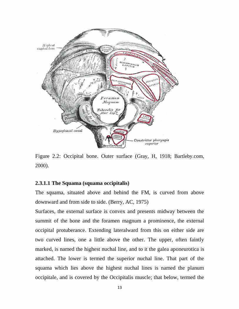
lobes of the cerebrum and by the cerebellum. (Figure 2.2)The occiput has
two articular condyles that form hinge joints with the first bone of the
vertebral column, the atlas. Between the condyles there is the foramen
magnum. (Anne Waugh, et al 2004).
The occipital bone is composed of the curved, expanded plate behind the
foramen magnum is named the squama; the thick, somewhat quadrilateral
piece in front of the foramen is called the basilar part, whilst on either side
of the foramen is the lateral portion. (Gray, H, 2005, Berkovitz B, et al 1994).
13
Figure 2.2: Occipital bone. Outer surface (Gray, H, 1918; Bartleby.com,
2000).
2.3.1.1 The Squama (squama occipitalis)
The squama, situated above and behind the FM, is curved from above
downward and from side to side. (Berry, AC, 1975)
Surfaces, the external surface is convex and presents midway between the
summit of the bone and the foramen magnum a prominence, the external
occipital protuberance. Extending lateralward from this on either side are
two curved lines, one a little above the other. The upper, often faintly
marked, is named the highest nuchal line, and to it the galea aponeurotica is
attached. The lower is termed the superior nuchal line. That part of the
squama which lies above the highest nuchal lines is named the planum
occipitale, and is covered by the Occipitalis muscle; that below, termed the
14
planum nuchale is rough and irregular for the attachment of several muscles.
From the external occipital protuberance a ridge or crest, the median nuchal
line, often faintly marked, descends to the foramen magnum, and affords
attachment to the ligamentum nuchæ; running from the middle of this line
across either half of the nuchal plane is the inferior nuchal line. Several
muscles are attached to the outer surface of the squama, thus: the superior
nuchal line gives origin to the Occipitalis and Trapezius, and insertion to the
Sternocleidomastoideus and Splenius capitis: into the surface between the
superior and inferior nuchal lines the Semispinalis capitis and the Obliquus
capitis superior are inserted, while the inferior nuchal line and the area
below it receive the insertions of the Recti capitis posteriores major and
minor. The posterior atlantoöccipital membrane is attached around the
postero-lateral part of the foramen magnum, just outside the margin of the
foramen. (Berry, AC, 1967)
The internal surface is deeply concave and divided into four fossæ by
a cruciate eminence. The upper two fossæ are triangular and lodge the
occipital lobes of the cerebrum; the lower two are quadrilateral and
accommodate the hemispheres of the cerebellum. At the point of intersection
of the four divisions of the cruciate eminence is the internal occipital
protuberance. From this protuberance the upper division of the cruciate
eminence runs to the superior angle of the bone, and on one side of it
(generally the right) is a deep groove, the sagittal sulcus, which lodges the
hinder part of the superior sagittal sinus; to the margins of this sulcus the
falx cerebri is attached. The lower division of the cruciate eminence is
prominent, and is named the internal occipital crest; it bifurcates near the
foramen magnum and gives attachment to the falx cerebelli; in the attached
margin of this falx is the occipital sinus, which is sometimes duplicated. In
15
the upper part of the internal occipital crest, a small depression is sometimes
distinguishable; it is termed the vermian fossa since it is occupied by part of
the vermis of the cerebellum. Transverse grooves, one on either side, extend
from the internal occipital protuberance to the lateral angles of the bone;
those grooves accommodate the transverse sinuses, and their prominent
margins give attachment to the tentorium cerebelli. The groove on the right
side is usually larger than that on the left, and is continuous with that for the
superior sagittal sinus. Exceptions to this condition are, however, not
infrequent; the left may be larger than the right or the two may be almost
equal in size. The angle of union of the superior sagittal and transverse
sinuses is named the confluence of the sinuses, and its position is indicated
by a depression situated on one or other side of the protuberance. (Bruce V,
et al 1993)
2.3.1.2 Lateral Parts (pars lateralis)
The lateral parts are situated at the sides of the foramen magnum; on their
under surfaces are the condyles for articulation with the superior facets of
the atlas. The condyles are oval or reniform in shape, and their anterior
extremities, directed forward and medialward, are closer together than their
posterior, and encroach on the basilar portion of the bone; the posterior
extremities extend back to the level of the middle of the foramen magnum.
The articular surfaces of the condyles are convex from before backward and
from side to side, and look downward and lateralward. To their margins are
attached the capsules of the atlantoöccipital articulations, and on the medial
side of each is a rough impression or tubercle for the alar ligament. At the
base of either condyle the bone is tunnelled by a short canal, the hypoglossal
canal (anterior condyloid foramen). This begins on the cranial surface of the
bone immediately above the foramen magnum, and is directed lateralward
16
and forward above the condyle. It may be partially or completely divided
into two by a spicule of bone; it gives exit to the hypoglossal or twelfth
cerebral nerve, and entrance to a meningeal branch of the ascending
pharyngeal artery. Behind either condyle is a depression, the condyloid
fossa, which receives the posterior margin of the superior facet of the atlas
when the head is bent backward; the floor of this fossa is sometimes
perforated by the condyloid canal, through which an emissary vein passes
from the transverse sinus. Extending lateralward from the posterior half of
the condyle is a quadrilateral plate of bone, the jugular process, excavated in
front by the jugular notch, which, in the articulated skull, forms the posterior
part of the jugular foramen. The jugular notch may be divided into two by a
bony spicule, the intrajugular process, which projects lateralward above the
hypoglossal canal. The under surface of the jugular process is rough, and
gives attachment to the Rectus capitis lateralis muscle and the lateral
atlantoöccipital ligament; from this surface an eminence, the paramastoid
process, sometimes projects downward, and may be of sufficient length to
reach, and articulate with, the transverse process of the atlas. Laterally the
jugular process presents a rough quadrilateral or triangular area which is
joined to the jugular surface of the temporal bone by a plate of cartilage;
after the age of twenty-five this plate tends to ossify. (Gill GW, et al 2004)
The upper surface of the lateral part presents an oval eminence, the jugular
tubercle, which overlies the hypoglossal canal and is sometimes crossed by
an oblique groove for the glossopharyngeal, vagus, and accessory nerves. On
the upper surface of the jugular process is a deep groove which curves
medialward and forward and is continuous with the jugular notch. This
groove lodges the terminal part of the transverse sinus, and opening into it,
17
close to its medial margin, is the orifice of the condyloid canal. (Ilizarov
GA, et al 1992, Gray, H, 1918; Bartleby.com, 2000).
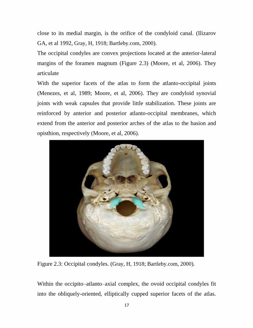
The occipital condyles are convex projections located at the anterior-lateral
margins of the foramen magnum (Figure 2.3) (Moore, et al, 2006). They
articulate
With the superior facets of the atlas to form the atlanto-occipital joints
(Menezes, et al, 1989; Moore, et al, 2006). They are condyloid synovial
joints with weak capsules that provide little stabilization. These joints are
reinforced by anterior and posterior atlanto-occipital membranes, which
extend from the anterior and posterior arches of the atlas to the basion and
opisthion, respectively (Moore, et al, 2006).
Figure 2.3: Occipital condyles. (Gray, H, 1918; Bartleby.com, 2000).
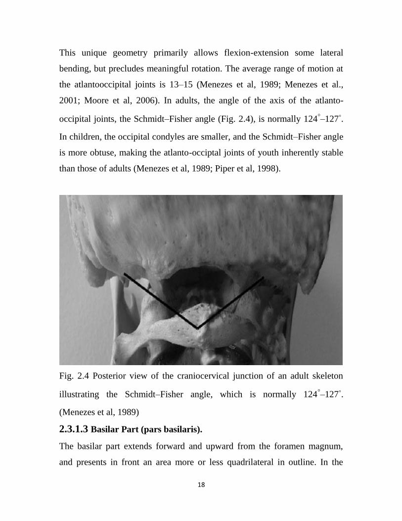
Within the occipito–atlanto–axial complex, the ovoid occipital condyles fit
into the obliquely-oriented, elliptically cupped superior facets of the atlas.
18
This unique geometry primarily allows flexion-extension some lateral
bending, but precludes meaningful rotation. The average range of motion at
the atlantooccipital joints is 13–15 (Menezes et al, 1989; Menezes et al.,
2001; Moore et al, 2006). In adults, the angle of the axis of the atlanto-
occipital joints, the Schmidt–Fisher angle (Fig. 2.4), is normally 124◦–127◦.
In children, the occipital condyles are smaller, and the Schmidt–Fisher angle
is more obtuse, making the atlanto-occiptal joints of youth inherently stable
than those of adults (Menezes et al, 1989; Piper et al, 1998).
Fig. 2.4 Posterior view of the craniocervical junction of an adult skeleton
illustrating the Schmidt–Fisher angle, which is normally 124◦–127◦.
(Menezes et al, 1989)
2.3.1.3 Basilar Part (pars basilaris).
The basilar part extends forward and upward from the foramen magnum,
and presents in front an area more or less quadrilateral in outline. In the
19
young skull this area is rough and uneven, and is joined to the body of the
sphenoid by a plate of cartilage. By the twenty-fifth year this cartilaginous
plate is ossified, and the occipital and sphenoid form a continuous bone.
(Lahr MM 1996).
Surfaces - On its lower surface, about 1cm. in front of the foramen magnum
is the pharyngeal tubercle which gives attachment to the fibrous raphé of the
pharynx. On either side of the middle line the Longus capitis and Rectus
capitis anterior are inserted, and immediately in front of the foramen
magnum the anterior atlantoöccipital membrane is attached. ( Lele S, et al
1991).
The upper surface presents a broad, shallow groove which inclines upward
and forward from the foramen magnum; it supports the medulla oblongata,
and near the margin of the foramen magnum gives attachment to the
membrana tectoria. On the lateral margins of this surface are faint grooves
for the inferior petrosal sinuses. (Lieberman DE, et al 1999).
2.3.1.3 Foramen Magnum.
In anatomy, the foramen magnum (Latin: “great hole”) is a large opening in
the occipital bone of the cranium. The foramen magnum is a large oval
aperture with its long diameter antero-posterior; it is wider behind than in
front where it is encroached upon by the condyles. (Figure 2.5) .It transmits
the medulla oblongata and its membranes, the accessory nerves, the
vertebral arteries, the anterior and posterior spinal arteries, and the
membrana tectoria and alar ligaments. (Lieberman DE, et al 1999).
In humans, the foramen magnum is farther underneath the head than in great
apes. Thus in humans, the neck muscles do not need to be as robust in order
to hold the head upright. Comparisons of the position of the foramen
magnum in early hominid species are useful to determine how comfortable
20
particular species was when walking on two limbs (bipedality) rather than
four. The location of the foramen magnum plays a crucial role in our
understanding of human evolution. Usually, the location of the foramen
magnum is linked to bipedal behavior. Due to the thickness of the cranial
base and its relatively protected anatomical position, this area of skull tends
to withstand both physical insults and inhu- mation somewhat more
successfully than many other areas of the cranium. (Jain D, et al 2014).
The foramen magnum (FM) is an important landmark of the base of skull
and is of particular interest to many fields of medicine. (Gruber P, et al
2009). Variations of the shape of FM have got diagnostic, clinical and
radiological importance.(Murshed K.A.,et al,2003). Also there exists some
correlation between the shape of FM and ancestry of an individual. The
dimensions of FM have clinical importance because the vital structures that
pass through it may suffer compression as in cases of FM achondroplasia.
(Hecht JT et al 1989, Reich JB et al 1993).
Foramen magnum is about 3cm wide by 3.5cm anteroposteriorly.(
Premalatha Gogi et al 2014, Romanes GJ et al 1981). It is located midway
between and on a level with mastoid processes. The foramen magnum is
surrounded by different parts of the occipital bone, squamous part lies
behind and above, basilar part in front and a condylar part on either side
(Oliveira Ed, et al 1985, Rhoton AL.2000). On each side its antero-lateral
margin is encroached by occipital condyles, hence the foramen magnum is
narrow anteriorly. The anterior edge of the foramen magnum is slightly
thickened and lies between the anterior ends of the condyles. The posterior
half of the foramen magnum is thin and semicircular. Upper ends of anterior
and posterior atlanto-occipital membranes are attached to the anterior and
posterior margins of the foramen magnum respectively, and their lower ends
21
are attached to the superior surface of anterior and posterior arches of the
atlas respectively. (Romanes GJ et al 1981) The foramen magnum is a wide
communication between posterior cranial fossa and the vertebral canal. The
narrow anterior part of the foramen magnum has apical ligament of dens,
upper fasciculus of the cruciate ligament and membrana tectoria; both are
attached to the upper surface of basioccipital bone infront of the foramen
magnum. Its wide posterior part contains the medulla oblongata and its
meninges. In subarachnoid space spinal rami of the accessory nerve and
vertebral arteries, with their sympathetic plexus, ascend into the cranium; the
posterior spinal arteries descend posterolateral to the brain stem, where as
anterior spinal artery descends anteromedian to brain stem. The cerebellar
tonsils may project into the foramen magnum. (Bannister, et al1995).
Relations of foramen magnum, anteriorly - basilar part of occipital bone,
Anterolaterally - occipital condyles, hypoglossal canal, jugular foramen
Posteriorly - squamous part of occipital bone with the internal occipital
crest. (Drake. 2010).
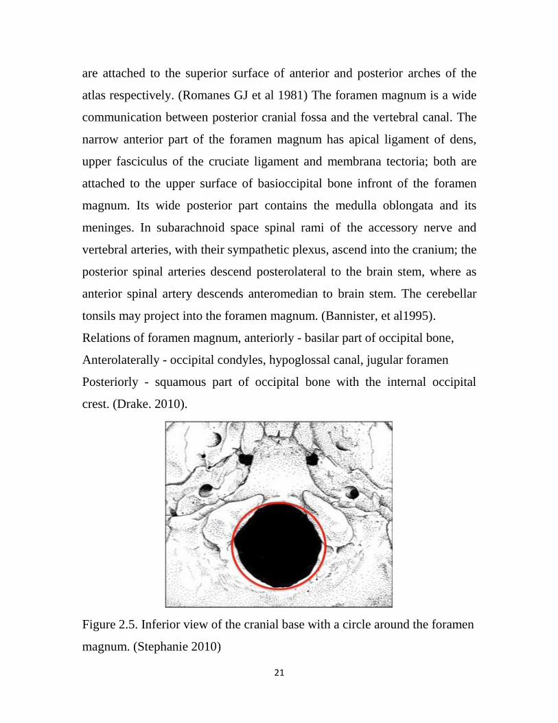
Figure 2.5. Inferior view of the cranial base with a circle around the foramen
magnum. (Stephanie 2010)
22
2.3.2 The viatal structures pass through the foramen magnum:
2.3.2.1 The medulla oblongata (or simply medulla)
Is the most caudal part of the brainstem and sits between the pons inferiorly
and spinal cord superiorly. It is the transition from the spinal cord to the
brain. The medulla contains the vital autonomic cardiovascular and
respiratory centers controlling heart rate, blood pressure, and breathing. It is
composed of grey matter, cranial nerve (CN) nuclei IX-to-XII, and white
matter tracts (DSc SSP.2011, Grossman RI, et al 2003).
The medulla is approximately 3cm in length and 2cm in greatest diameter
(DSc SSP.2011). The caudal border of the medulla is the 1st cervical spinal
nerves. The superior broad part of the medulla joins the pons. (DSc
SSP.2011, Grossman RI, et al 2003). Medulla is separated into two main
parts: ventral (anterior) medulla which contains the olive, pyramidal tracts,
and CN 9-12 rootlets, and tegmentum (dorsal) medulla which contain the
CN nuclei and white matter tracts.
Ventral medulla: Pyramids are paired structures located at the medial aspect
of ventral medulla and flank the anterior median fissure. It contains the the
corticospinal tracts. At the caudal end of pyramids the corticospinal tracts
decussate (DSc SSP.2011, Grossman RI, et al 2003).
Olivary bodies are paired structures located at lateral aspect of ventral
medulla, lateral to the pyramids. They are separated from the pyramids by an
anterolateral sulcus (pre-olivary sulcus). There is also a post-olivary sulcus
lateral to the olivary bodies. Olivary bodies contain the superior and larger
inferior olivary nuclei (DSc SSP.2011).
Medulla tegmentum: The dorsal aspect of the medulla contains the posterior
median sulcus (most dorsal medial sulcus) and more lateral posterolateral
23
sulcus. Between these sulci are the fasciculus gracilis and nuclei forming
gracilis tubercle at the midline and fasciculus cuneatus and nuclei forming
cuneate tubercle more laterally (DSc SSP.2011, Grossman RI, et al 2003).
The superior dorsal aspect of medulla forms the floor of the inferior 4th
ventricle. It is occupied by the inferior cerebellar peduncle situated between
the lower parts of the fourth ventricle. The inferior dorsal and lateral aspect
of the medulla is surrounded by the cisterna magna (posterior
cerebellomedullary cistern), and lateral cerebellomedullary cistern (DSc
SSP.2011, Grossman RI, et al 2003).
The median aperture (foramen of Magendie) and the more superior lateral
apertures (foramina of Luschka) open at the level of the pons, with the
canals projecting to the level of the medulla region and terminating into the
cisterna magna and lateral cerebellomedullary cistern respectively( Rogers
L.2008) .
2.3.2.2 The meninges
Is a collective term for the three membranes that cover the brain and spinal
cord, cerebral meninges surround the brain and is made up of three layers
(from outermost to innermost): dura mater, arachnoid mater and pia mater.
The dura mater can also be known as pachymeninx. The arachnoid mater
and pia mater are collectively known as the leptomeninges. (Strominger NL
et al 2012).
The spinal meninges are similar but have some important differences.
The meninges function to protect the brain but also provides a framework
for blood vessels, nerves, lymphatics and CSF (Ovalle WK,.et al 2013).
24
2.3.2.3 The spinal accessory nerve
Also called accessory nerve, is the eleventh cranial nerve (CN XI) and is
composed of two parts, the cranial part and the spinal part. The cranial part
(accessory portion) is the smaller of the two. Its fibers arise from the cells of
the nucleus ambiguus and emerge as four or five delicate rootlets from the
side of the medulla oblongata, below the roots of the vagus. It runs laterally
to the jugular foramen, where it interchanges fibers with the spinal portion or
becomes united to it for a short distance. (Wilson et al 2002)
The spinal part (spinal portion) is firm in texture, and its fibers arise from the
ventral horn cells in the cord between C1 and C5 of the cervical plexus. The
fibres emerge from the cord laterally between the anterior and posterior
spinal nerve roots to form a single trunk, which ascends into the skull
through the foramen magnum. (Waxman S, et al 2003).
2.3.2.4 The vertebral arteries (VA)
The vertebral artery (VA) arises from the subclavian artery, ascends in the
neck to supply the posterior fossa and occipital lobes as well as provides
segmental vertebral and spinal column blood supply. (Cloud GC, et al 2003)
The origin of the VA is usually from the posterior superior part of the
subclavian arteries bilaterally, although the origin can be variable:
- Brachiocephalic artery (on the right)
- Aortic arch: 6% of cases
The VA is normally 3-5 mm in diameter and the ostium is the most common
site of stenosis.
When the origin is from the arch, then it is common for the artery to enter
the foramen transversarium at a level higher than normal (C5 instead of C6).
(Satti SR, et al 2007).

25
The duramater around the FM is supplied by the anterior and posterior
meningeal branches of the vertebral artery, and the meningeal branches of
the ascending pharyngeal and occipital arteries.
2.3.2.5 The venous structures
The venous structures in the region of the FM are divided into three groups:
-Extraduralveins (extraspinal& intraspinalpart)
-Intradural (neural) veins
-Dural venous sinuses (superior petrosal, marginal & occipital).
The three groups anastomose through bridging and emissary veins.
2.3.3 First Cervical Vertebra
The first cervical vertebra (Figure 2.6) is named the atlas because it supports
the globe of the head. Its chief peculiarity is that it has no body, and this is
due to the fact that the body of the atlas has fused with that of the next
vertebra. Its other peculiarities are that it has no spinous process, is ring-like,
and consists of an anterior and a posterior arch and two lateral masses.
The anterior arch forms about one-fifth of the ring: its anterior surface is
convex, and presents at its center the anterior tubercle for the attachment of
the Longus colli muscles; posteriorly it is concave, and marked by a smooth,
oval or circular facet (fovea dentis), for articulation with the odontoid
process (dens) of the axis. The upper and lower borders respectively give
attachment to the anterior atlantooccipital membrane and the anterior
atlantoaxial ligament; the former connects it with the occipital bone above,
and the latter with the axis below. The posterior arch forms about two-fifths
of the circumference of the ring: it ends behind in the posterior
tubercle, which is the rudiment of a spinous process and gives origin to the
26
Recti capitis posteriores minores. The diminutive size of this process
prevents any interference with the movements between the atlas and the
skull. The posterior part of the arch presents above and behind a rounded
edge for the attachment of the posterior atlantoöccipital membrane, while
immediately behind each superior articular process is a groove (sulcus
arteriæ vertebralis), sometimes converted into a foramen by a delicate bony
spiculum which arches backward from the posterior end of the superior
articular process. This groove represents the superior vertebral notch, and
serves for the transmission of the vertebral artery, which, after ascending
through the foramen in the transverse process, winds around the lateral mass
in a direction backward and medialward; it also transmits the suboccipital
(first spinal) nerve. On the under surface of the posterior arch, behind the
articular facets, are two shallow grooves, the inferior vertebral notches.The
lower border gives attachment to the posterior atlantoaxial ligament, which
connects it with the axis. The lateral masses are the most bulky and solid
parts of the atlas, in order to support the weight of the head. Each carries two
articular facets, a superior and an inferior. The superior facets are of large
size, oval, concave, and approach each other in front, but diverge behind:
they are directed upward, medialward, and a little backward, each forming a
cup for the corresponding condyle of the occipital bone, and are admirably
adapted to the nodding movements of the head. Not infrequently they
are partially subdivided by indentations which encroach upon their margins.
The inferior articular facets are circular in form, flattened or slightly convex
and directed downward and medialward, articulating with the axis, and
permitting the rotatory movements of the head. Just below the medial
margin of each superior facet is a small tubercle, for the attachment of the
transverse atlantal ligament which stretches across the ring of the atlas and
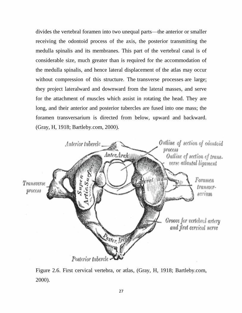
27
divides the vertebral foramen into two unequal parts—the anterior or smaller
receiving the odontoid process of the axis, the posterior transmitting the
medulla spinalis and its membranes. This part of the vertebral canal is of
considerable size, much greater than is required for the accommodation of
the medulla spinalis, and hence lateral displacement of the atlas may occur
without compression of this structure. The transverse processes are large;
they project lateralward and downward from the lateral masses, and serve
for the attachment of muscles which assist in rotating the head. They are
long, and their anterior and posterior tubercles are fused into one mass; the
foramen transversarium is directed from below, upward and backward.
(Gray, H, 1918; Bartleby.com, 2000).
Figure 2.6. First cervical vertebra, or atlas, (Gray, H, 1918; Bartleby.com,
2000).
28
2.3.3.1 Stabilizing Cranio cervical junction
Principal stabilizing ligaments of C1 -
-Transverse atlantalligament
-Alarligaments
Secondary stabilizing ligaments of CVJ are more elastic & weaker than the
primary ligaments.
-Apical ligament
-Anterior & posterior membranes
-Tectorialmembrane
-Ligamentumflavum
-Capsular ligaments
2.4 Pathology of posterior cranial fossa
Congenital and developmental osseous anomalies and abnormalities that
affect the craniovertebral complex can result in neural compression, vascular
compromise, and can manifest with abnormal cerebrospinal fluid dynamics.
2.4.1 Achondroplasia
Achondroplasia is a congenital genetic disorder and the most common
skeletal dysplasia. It has numerous distinctive radiographic features and is
the most common cause of short limb dwarfism. (Kao SC, et al 1989).
It occurs due to sporadic mutations in the majority of cases but can be
inherited as an autosomal dominant condition. Homozygous achondroplasia
is lethal. There is a prevalence of approximately 1 in 25,000-50,000 births
with males affected more frequently than females.( Wynn J, et al 2007)
Achondroplasia is the most common cause of short limb dwarfism. Patients
are of normal intelligence with normal motor function. However, they may
have specific neurologic deficits.
29
The disease results from a mutation in the fibroblast growth factor gene 3
(FGFR3) located on chromosome 4p16.3 which causes abnormal cartilage
formation. All bones that form by enchondral ossification are affected.
Bones that form by membranous ossification are not affected, thus allowing
the skull vault to develop normally. (Cheema JI, et al 2003)
2.4.1.1 Radiographic features
Almost all the bones of the skeleton are affected, and hence all parts of the
body have bony changes with secondary soft tissue changes. Antenatally it is
difficult to diagnose achondroplastic features until the 3rd trimester
(Schramm T, et al 2009).
Antenatal ultrasound :Antenatally detectable sonographic features include:
short femur length measurement: often well below the 5th centile the femur
length (FL) to biparietal diameter (BPD) is taken as a useful measurement
trident hand 11: 2,3 and 4 fingers appearing separated and similar in length
separation of 1st and 2nd, 3rd and 4th fingers protruding forehead: frontal
bossing (Bowerman RA. 1995 ).
2.4.1.2 Cranial
- Relatively large cranial vault with small skull base
- Prominent forehead with depressed nasal bridge
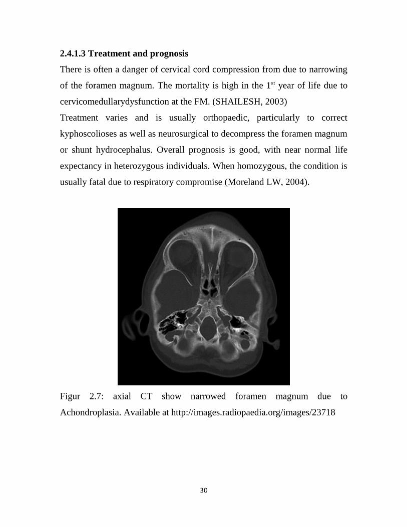
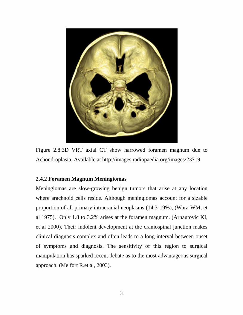
- Narrowed foramen magnum, a small FM with hypertrophicbone & a
posterior duralshelf results in compression of neural structures. (Figure 2.7
& 2.8)
- Cervico medullary kink relative elevation of the brainstem resulting in a
large suprasellar cistern and vertically-oriented straight sinus.
30
2.4.1.3 Treatment and prognosis
There is often a danger of cervical cord compression from due to narrowing
of the foramen magnum. The mortality is high in the 1st year of life due to
cervicomedullarydysfunction at the FM. (SHAILESH, 2003)
Treatment varies and is usually orthopaedic, particularly to correct
kyphoscolioses as well as neurosurgical to decompress the foramen magnum
or shunt hydrocephalus. Overall prognosis is good, with near normal life
expectancy in heterozygous individuals. When homozygous, the condition is
usually fatal due to respiratory compromise (Moreland LW, 2004).
Figur 2.7: axial CT show narrowed foramen magnum due to
Achondroplasia. Available at http://images.radiopaedia.org/images/23718
31
Figure 2.8:3D VRT axial CT show narrowed foramen magnum due to
Achondroplasia. Available at http://images.radiopaedia.org/images/23719
2.4.2 Foramen Magnum Meningiomas
Meningiomas are slow-growing benign tumors that arise at any location
where arachnoid cells reside. Although meningiomas account for a sizable
proportion of all primary intracranial neoplasms (14.3-19%), (Wara WM, et
al 1975). Only 1.8 to 3.2% arises at the foramen magnum. (Arnautovic KI,
et al 2000). Their indolent development at the craniospinal junction makes
clinical diagnosis complex and often leads to a long interval between onset
of symptoms and diagnosis. The sensitivity of this region to surgical
manipulation has sparked recent debate as to the most advantageous surgical
approach. (Melfort R.et al, 2003).
32
Meningiomas are considered to be located in the FM area if their base of
insertion is mainly located within the FM limits. This definition excludes
tumors invading secondarily the FM region (Figure).
The definitive objective of a classification system is to define preoperatively
the surgical strategy based on preoperative imaging characteristics of the
lesion. The surgical strategy in cases of FMMs is the surgical approach but
also the anticipation of modified vital structure position. (Michaël, et al,
2008)
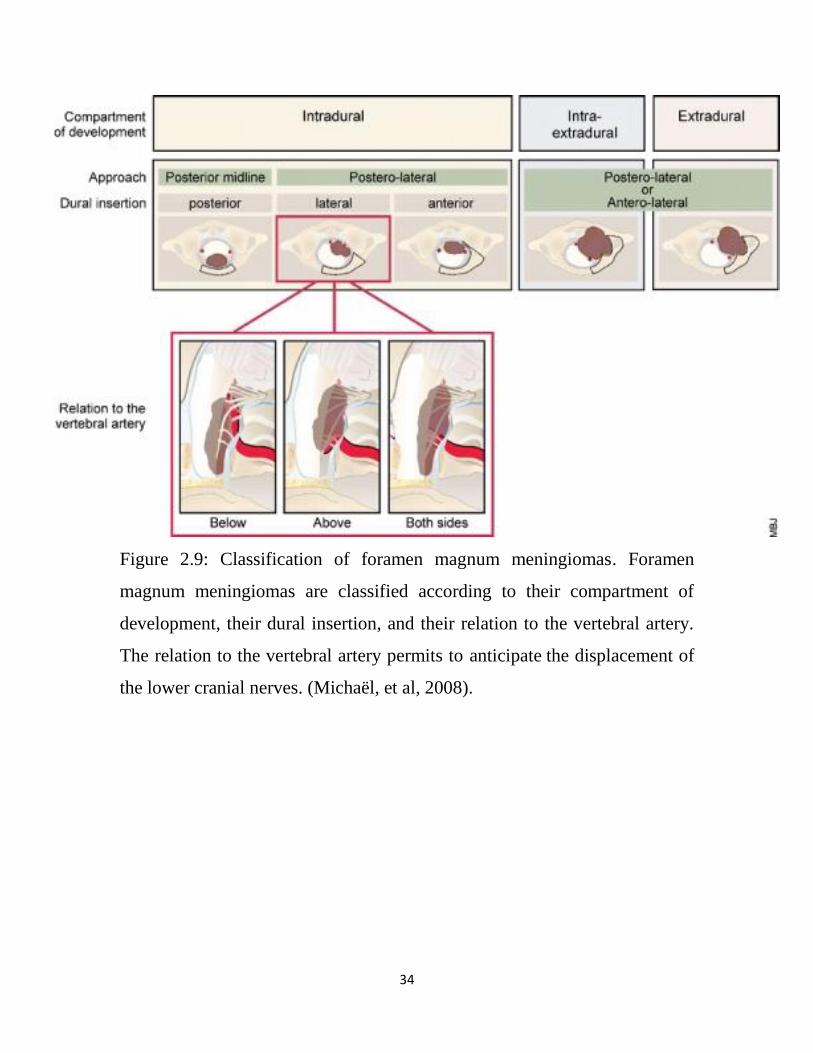
2.4.2 .1 Classification of Foramen Magnum Meningiomas
FMMs can be classified according to their compartment of development,
their dural insertion, and to their relation to the (VA George B, et al 1997).
According to the compartment of development, FMMs can be subdivided in:
(Figure 2.9): Intradural, Extradural and Intra- and extradural.
Intradural meningiomas are the most commonly observed. Extradural
meningiomas like at any other locations are very invasive, into the bone, the
nerves and vessels sheaths, and soft tissues. The VA sheath and even its
adventitia can also be infiltrated. This raises some difficulties and explains
the higher incidence of incomplete removal as compared to intradural
meningiomas (Levy C, et al 1988, Miller E, et al 1987).
According to the insertion on the dura, FMMs can be defined in the antero-
posterior plane as: Anterior, if insertion is on both sides of the anterior
midline, Lateral, if insertion is between the midline and the dentate ligament
and Posterior, if insertion is posterior to the dentate ligament
Anterior meningiomas push the spinal cord posteriorly. Therefore, the
surgical opening between the neuraxis and the FM lateral wall is narrow, and
the drilling must extend to the medial part of the FM lateral wall to improve
the access. In almost every case, no drilling of the lateral mass of the atlas
33
and occipital condyle is necessary. Exceptionally, anterior meningiomas of
small size without anterior compartment enlargement need more bone
resection but never includes more than one fifth of these elements. On the
other hand, lateral meningiomas displace the neuraxis posterolaterally and
widely open the surgical access; therefore, the drilling has never to be
extended into the lateral mass of the atlas or the occipital condyle. (Michaël,
et al, 2008)
Finally, surgical strategies vary according to the relation to the VA, FMM
having the possibility to develop: Above the VA, Below the VA and on
both sides of the VA
Meningiomas are more often located below the VA. In this case, the lower
cranial nerves are always pushed cranially and posteriorly. There is no need
to look for them. They will come into view on reaching the superior tumoral
part. On the other hand, if the lesion grows above the VA, the position of the
lower cranial nerves cannot be anticipated; the nerves may be displaced
separately in any direction. After partial debulking of the tumor, one has to
look for them so as to identify and protect them during the tumor resection.
In case of tumoral development on both sides of the VA, a similar problem
may exist with the position of the lower cranial nerves. Moreover, the dura
around the VA penetration may be infiltrated by the tumor. As previously
mentioned, the dura is normally adherent to the adventitia, and complete
resection of the tumor at this level is hazardous. In this case, which is rarely
observed, it may be safer to leave a cuff of infiltrated dura around the VA
and to coagulate this zone. (Michaël, et al, 2008).
34
Figure 2.9: Classification of foramen magnum meningiomas. Foramen
magnum meningiomas are classified according to their compartment of
development, their dural insertion, and their relation to the vertebral artery.
The relation to the vertebral artery permits to anticipate the displacement of
the lower cranial nerves. (Michaël, et al, 2008).

35
2.4.2.2 Imaging Features
The role of neuroimaging is to confirm the clinical diagnosis and to allow
the planning of a surgical approach. Magnetic resonance imaging is the
modality of choice for defining tumors of the foramen magnum because it
provides high-resolution images of soft-tissue anatomy that is not
susceptible to degradation by surrounding skull base, (Sekhar, et al, 1994).
Although MR imaging provides clearly superior soft-tissue assessment, CT
scanning with osseous algorithms remains the tool of choice for identifying
calcification, hyperostosis, and osseous anatomy. Axial CT scanning allows
planning of the extent of bone resection required to resect tumor safely
because of the sharp contrast between bone and soft tissues. It is sometimes
difficult to outline bone margins on MR images, and this technique may
overestimate the size of the surgical corridor available for extirpation. It is
clearly evident that optimal surgical planning requires both CT and MR
imaging to assess appropriately bone and soft tissues, respectively. (Farb RI,
et al, 2003)
An additional imaging modality that may assist surgery is conventional
angiography with optional embolization of vessels that supply tumor
exclusively. The dural blood supply typically arises as posterior and anterior
meningeal branches from the VAs with the support of meningeal branches
via ascending pharyngeal and occipital arteries. The tumor may derive its
vascular supply from a dominant vessel, which when subjected to contrast
injection, is visualized as a "blush". If vessel is accessible to endovascular
catheterization, one might opt for preoperative embolization to diminish
intraoperative bleeding during tumor debulking[. (Rosen CL, et al .2002).
36
2.4.2.3 Preoperative considerations
Standard preoperative workup includes magnetic resonance imaging (MRI),
computed tomography (CT) scan, and sometimes angiography.
On MRI, gadolinium-enhanced sequences help to precisely delimit the dural
attachment zone, the tumor, and its relation to neural and vascular structures.
On T2-weighted images, the presence of an arachnoid plane between the
tumor and the neuraxis is sometimes visible. (Figure 2.10)
Bone windows CT scan is helpful in case of extradural extension to
investigate bone erosion and to schedule preoperatively the need for fusion.
Conventional angiography is generally useless. There are only two
indications for preoperative angiography:
If a highly vascularized tumor is suspected and embolization is
contemplated.
To perform a balloon occlusion test in case of VA encasement (extradural or
recurrent meningioma and meningioma inserted around the VA). In our
experience, it has never been necessary to occlude the VA. (Michaël, et al,
2008)
Intraoperative neurophysiological monitorings have been used by several
surgeons .(Arnautovic KI, et al 2000, Boulton MR,et al 2003) and includes
somatosensory-evoked potentials, brainstem auditory-evoked potentials, and
electromyographic monitoring of lower cranial nerves, by recordings
through an endotracheal tube (CN X) and with a needle in the sterno-
mastoid (SM) muscle (CN XI) and the tongue (CN XII)
37
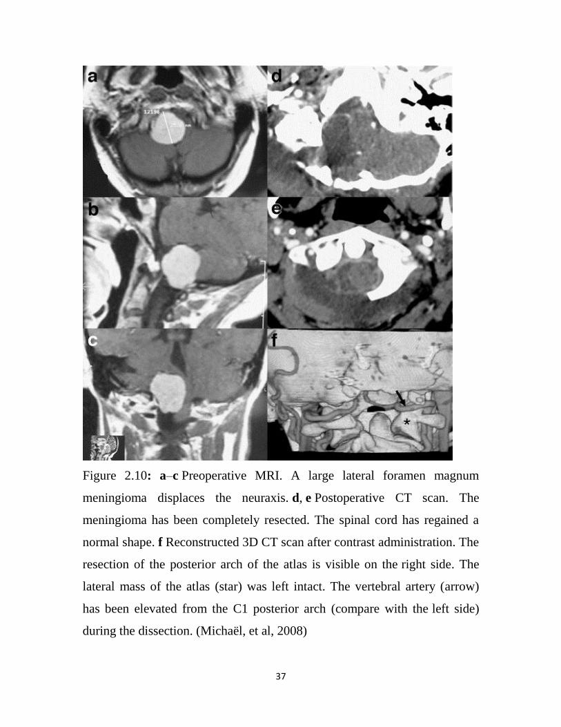
Figure 2.10: a–c Preoperative MRI. A large lateral foramen magnum
meningioma displaces the neuraxis. d, e Postoperative CT scan. The
meningioma has been completely resected. The spinal cord has regained a
normal shape. f Reconstructed 3D CT scan after contrast administration. The
resection of the posterior arch of the atlas is visible on the right side. The
lateral mass of the atlas (star) was left intact. The vertebral artery (arrow)
has been elevated from the C1 posterior arch (compare with the left side)
during the dissection. (Michaël, et al, 2008)
38
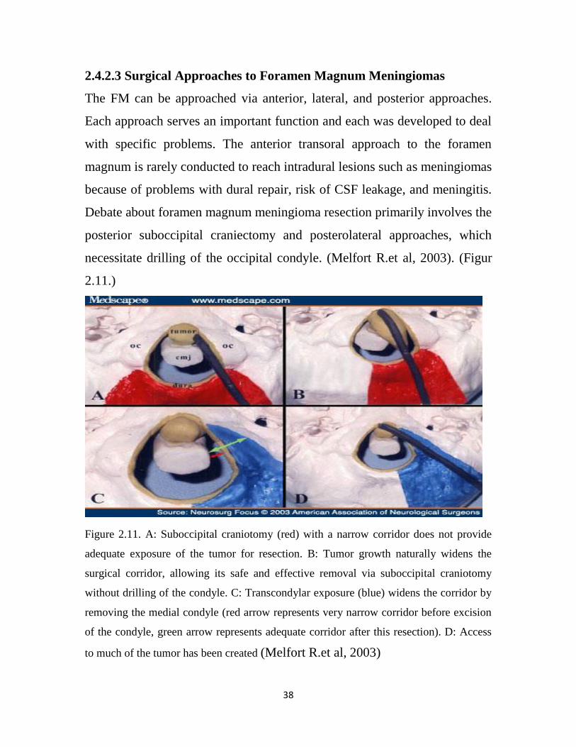
2.4.2.3 Surgical Approaches to Foramen Magnum Meningiomas
The FM can be approached via anterior, lateral, and posterior approaches.
Each approach serves an important function and each was developed to deal
with specific problems. The anterior transoral approach to the foramen
magnum is rarely conducted to reach intradural lesions such as meningiomas
because of problems with dural repair, risk of CSF leakage, and meningitis.
Debate about foramen magnum meningioma resection primarily involves the
posterior suboccipital craniectomy and posterolateral approaches, which
necessitate drilling of the occipital condyle. (Melfort R.et al, 2003). (Figur
2.11.)
Figure 2.11. A: Suboccipital craniotomy (red) with a narrow corridor does not provide
adequate exposure of the tumor for resection. B: Tumor growth naturally widens the
surgical corridor, allowing its safe and effective removal via suboccipital craniotomy
without drilling of the condyle. C: Transcondylar exposure (blue) widens the corridor by
removing the medial condyle (red arrow represents very narrow corridor before excision
of the condyle, green arrow represents adequate corridor after this resection). D: Access
to much of the tumor has been created (Melfort R.et al, 2003)
39
2.4.2.3.1 Suboccipital Craniotomy
Suboccipital craniotomy, or craniectomy, with or without cervical
laminectomy represents the classic approach to the foramen magnum
meningiomas and is familiar to most neurosurgeons. For posteriorly situated
lesions we place the patient prone. For lateral or anterolateral lesions, the
patient is placed in the lateral decubitus position with the vertex of the head
displaced slightly downward to open the space between occiput and the
cervical spine. We also turn the head approximately 20 to 30° toward the
floor, depending on the extent to which the tumor is laterally situated. The
surgical corridor defined on preoperative imaging must be easily within
reach of the surgeon. The corridor should not be hidden under a large bulk of
paracervical muscles deflected laterally. Sufficient soft-tissue dissection to
create access to the corridor is essential. Routine use of computerized
neuronavigation helps to demonstrate subtle variations of anatomical
distortions caused by these sessile meningiomas. (Stein et al 1963)
For midline posterior lesions, we make a midline incision. For posterolateral
lesions that require exposure up to the condyle, we make a "hockey-stick" or
inverted L shaped extension laterally at the superior end of our incision just
beneath the superior nuchal line. Whichever the incision, cutting of the C-2
nerve branches and the 11th cranial nerve distally in the neck should be
avoided.( Strang et al 2001)
The VA is easily identifiable as it curves above the arch of the atlas, in the
depth of the suboccipital triangle, providing proximal vascular control if
required. We use neuronavigation to help determine the extent of the
craniotomy needed. Although some authors prefer to conduct a craniectomy,
we prefer a craniotomy because the incidence of postoperative occipital
pain, we believe, is limited by replacing a firm protective covering over the
40
dura, even if it only covers a fraction of the exposed dura.( Schessel DA, et
al 1993) . If the "surgical corridor" to the tumor cannot be safely accessed as
determined by neuronavigation prior to dura opening, more bone can be
removed laterally toward the condyle. The craniotomy almost always has to
be combined with a laminectomy to the inferior aspect of the tumor. At C-1
the laminectomy should encompass at least the vertebral groove in the lateral
aspect of the C-1 lamina. Care should be taken to not injure the thin-walled
vertebral plexus of veins that surround the thick-walled VA. Of help in this
procedure is bipolar coagulation with constant saline irrigation to avoid
sticking of the tips of the instrument.
The advantage of suboccipital craniotomy includes visualization of the VA,
brainstem, cranial nerves, and tumor in a safe, simple, and rapid manner.
Criticisms of this approach primarily relate to the interposition of brainstem,
cranial nerves, and vessels between an anterior tumor and the surgeon. The
purely anterior midline tumor without an adequate surgical corridor is
completely obscured by these structures. The unmodified suboccipital
craniotomy approach necessitates undue retraction of critical neurological
structures in cases in which the lesion is purely anterior. Fortunately, these
purely ventrally located tumors are the rarest. (Vishteh, et al, 1999)
2.4.2.3.1Transcondylar Approach
In an attempt to offer effective and safer resections particularly in cases of
more anteriorly situated lesions, the transcondylar approach was developed.
(Sen CN, et al 1991). A number of different names exist for the variations in
this approach, and this leads to significant confusion regarding
nomenclature. (Babu RP, et al 1994, Banerji D et al1999,) In the literature
on foramen magnum meningiomas, two major variations have evolved: first
the far-lateral approach, which necessitates removal of the foramen magnum
41
rim toward the condyle and excision of the ipsilateral atlantal arch, and
second variation , the transcondylar approach, in which resection of some
or all of the occipital condyle is required. The first of these is ultimately a
suboccipital approach involving an appropriate soft-tissue dissection to
allow access to the surgical corridor. (Cantore G, et al, 1994)
The transcondylar approach requires an inverted U-shaped incision with one
limb of the U in the midline and the other along the anterior border of the
sternocleidomastoid muscle. The sternocleidomastoid muscle is detached
from the mastoid process and reflected as laterally as possible to avoid
hindering access to the skull base. The superficial splenius capitis,
semispinalis capitis, and longissimus capitis muscles are reflected downward
to expose the underlying suboccipital triangle. Bordered by the superior and
inferior oblique muscles and the rectus capitis posterior muscles, the VA
courses in the fat of the suboccipital triangle below the occipital condyle. All
three muscles are released from their vertebral attachments and reflected
toward the midline. The craniotomy should include the bulk of the lesion
and often be extended from near the midline, to just medial to the sigmoid
sinus, and to just above the rim of the foramen magnum. The residual bone
over the sigmoid and foramen magnum is removed using rongeurs or high-
speed drill. The C-1 laminectomy is more extensive than the suboccipital
craniotomy and extends out into the foramen transversarium. The
posteromedial aspect of the occipital condyle and C-1 lateral mass are
removed by drilling or careful rongeuring. If necessitated by the anatomy of
the lesion, the foramen transversarium and C-2 lamina are also
decompressed. The VA is freed from collagenous tissue at the C-1 foramen
transversarium and adjacent to the condyle by using a high-speed drill under
surgical magnification. A fine prolene stitch can be used to secure the VA in
42
a medial position. Guided by preoperative planning of the surgical corridor
and supplemented intraoperatively, if necessary, by computerized
neuronavgation, the condyle is progressively removed in a mediolateral
direction. Anterior condylar resection can include liberating the hypoglossal
nerve from its canal if necessary to create an adequate surgical corridor.
Because bone removal is extended much more laterally than in the
suboccipital craniotomy, the ipsilateral VA is situated in the center of or
medially in the surgical corridor prior to dural opening.. The dura is opened
by making an incision that parallels the lateral margins of the craniotomy,
with the base of the flap located medially. A ring of dura can be left attached
to the VA where it is pierced. This maneuver allows the artery to be
retracted away from the surgical corridor, thereby providing a clear view of
the anterior portion of the brainstem and ostral cord. Occipitocervical fusion
is recommended in condylar resections of 50% or greater.(Vishteh, et al
1999).
2.4.2.3.2 Surgical results
Morbidity–mortality–prognosis factors in the Yasargyl’s review of the
literature of series published before 1976, the overall mortality rate was
approximately 13% but could be as high as 45% in some series (Love JG,et
al 1954).
Over the last 20 years, the overall mortality is 6.2%. The mortality rate is
comprised between 0 and 25%. Mortality rates higher than 10% were mainly
observed in small series (Kratimenos GP et al 1993, Marin, et al 2002,
Sharma BS et al 1999).
43
In the Yasargil’s review, a good outcome was noted in 69% and a fair and
poor outcome, respectively, in 8 and 10%. In series larger than ten patients
published over the last 20 years, neurological improvement, stability, and
worsening were noted, respectively, in 70–100, 2.5–20, and 7.5–10% of the
cases. The permanent morbidity rate is comprised between 0 and 60%. The
permanent morbidity rate is lower through a far-lateral approach (0–17%),
either transcondylar or retrocondylar, than through an extreme-lateral
transcondylar approach (21%–56%, considering series without recurrent
tumor). Lower cranial nerves dysfunctions are the most frequently
encountered preoperative deficits. These deficits have the propensity to
recover even completely postoperatively [Arnautovic KI, et al 2000
,Bassiouni H,et al 2006], except in cases of en plaque meningiomas or
recurrent tumor (Samii M , et al 1996).
2.4.3 Chiari malformations
The Chiari malformations are a group of hindbrain herniation syndromes
initially, Cleland described the first cases of Chiari malformation in 1883,
the disorder is named after Hans Chiari, an Austrian pathologist, who
classified Chiari malformations into types I through III in 1891. Chiari's
colleague, Julius Arnold, made additional contributions to the definition of
Chiari II malformation. (Koehler PJ, et al 1891,) In his honor, students of
Dr.Arnold later named the type II malformation Arnold-Chiari
malformation. Other investigators later added the type IV malformation.
(Pearce JM, et al 2000).

44
2.4.3.1 Types of Chiari malformations
2.4.3.1.1Chiari type I:
Chiari malformations I, malformation is the most common and the least
severe of the spectrum, often diagnosed in adulthood. Its hallmark is caudal
displacement of peglike cerebellar tonsils below the level of the foramen
magnum, a phenomenon variably referred to as congenital tonsillar
herniation, tonsillar ectopia, or tonsillar descent. The resultant impaction of
the foramen magnum, compression of the cervicomedullary junction by the
ectopic tonsils, and interruption of normal flow of cerebrospinal fluid (CSF)
through the region produce the clinical syndrome. (Figure 2.12) (Koehler PJ,
et al 1891,)
2.4.3.1.2Chiari type II
Chiari malformations II, malformation is less common and more severe,
almost invariably associated with myelomeningocele. Because of its greater
severity, it becomes symptomatic in infancy or early childhood. Its hallmark
is caudal displacement of lower brainstem (medulla, pons, 4th ventricle)
through the foramen magnum. Symptoms arise from dysfunction of
brainstem and lower cranial nerves. (Arnold’s 1991)
2.4.3.1.3 Chiari type III and IV
Chiari type III and IV malformations are exceedingly rare and generally
incompatible with life and are, therefore, of scant clinical significance. The
type III malformation refers to herniation of cerebellum into a high cervical
myelomeningocele, whereas type IV refers to cerebellar agenesis. (Dias M.et
al 1999).
45
Importantly, it is not at all clear that the 4 types of Chiari malformation
represent a disease continuum corresponding to a single disorder. The 4
types (particularly types III and IV) are increasingly believed to have
different pathogenesis and share little in common other than their names.
(Peyman, et al 2014)
Figure: 2.12: Sagittal and coronal MRI images of Chiari type I
malformation. Note descent of cerebellar tonsils (T) below the level of
foramen magnum (white line) down to the level of C1 posterior arch
(asterisk). (Peyman, et al 2014).
2.4.3.2 Radiographic features
Although historically visible on myelography, cross sectional imaging
(especially MRI) is needed to accurately diagnose and assess for Chiari I
malformations. In either case the diagnosis is made by measuring how far
46
the tonsils protrude below the margins of the foramen magnum. The distance
is measured by drawing a line from the inner margins foramen magnum
(basion to opisthion), and measuring the inferior most part of the tonsils.
(Ketonen L, et al 2005).
- Above foramen magnum: normal
- 3 mm: also normal but the term benign tonsillar ectopia can be used
- 3 to 6 mm: indeterminate, and needs to be correlated with symptoms and
presence of syrnix, etc
- 6 mm: Chiari 1 malformation
Some authors advocate a simpler rule (Elster AD et al 1992):
- Above foramen magnum: normal
- 5 mm: also normal but the term benign tonsillar ectopia can be used
- 5 mm: Chiari 1 malformation
To make matters worse the 'normal' position of the cerebellar tonsils varies
with age. In neonates the tonsils are located just below the foramen magnum
and descend further during childhood, reaching their lowest point
somewhere between 5 and 15 years of age. As the individual ages further the
tonsils usually ascend coming to rest at the level of the foramen magnum.
(Kornienko. et al 2008). As such although 5mm descent in an adult should
be viewed with suspicion, in a child it is most likely normal (Kornienko. et
al 2008).
CT With modern volumetric scanning, and high quality sagittal reformats
relatively good views of the foramen magnum and tonsils can be achieved
although the intrinsic lack of contrast (compared to MRI) makes accurate
assessment difficult. More frequently the diagnosis is suspected on axial
images where the medulla is embraced by the tonsils and little if any CSF is
47
present. This is referred to as a crowded foramen magnum. (Chiapparini L,
et al 2011)
MRI is the imaging modality of choice. On sagittal imaging, the best plane
for assessing for the presence of Chiari I malformations, the tonsils are
pointed, rather than rounded and referred to as peg-like. The sulci are
vertically oriented, forming so-called sergeant stripes. Axial images through
the the foramen show crowding of the medulla by the tonsils. A syrinx may
be seen in spinal cord. (Chiapparini L, et al 2011)
2.4.3.3 Indications
In Chiari I, radiographic presence of tonsillar herniation must correlate with
appropriate clinical signs and symptoms before surgical intervention is
undertaken. In frankly symptomatic patients, such as those with lower
cranial nerve dysfunction, myelopathy, syringomyelia, cerebellar symptoms,
or severe post-tussive suboccipital headaches, the decision in favor of
surgery is straightforward. Difficulty arises in minimally symptomatic
patients or those with equivocal symptoms. CSF flow studies across foramen
magnum with phase-contrast cine MRI may help with surgical decision-
making in these cases. Syringomyelia generally improves or resolves after
surgical treatment of Chiari malformation (Figure: 2.13). Rarely is shunting
of the Chiari syrinx necessary. (Hofkes SK, et al 2007)
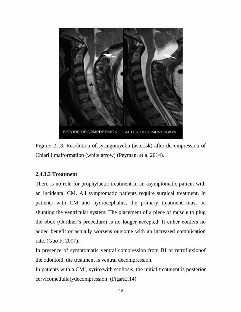
48
Figure: 2.13: Resolution of syringomyelia (asterisk) after decompression of
Chiari I malformation (white arrow) (Peyman, et al 2014).
2.4.3.3 Treatment:
There is no role for prophylactic treatment in an asymptomatic patient with
an incidental CM. All symptomatic patients require surgical treatment. In
patients with CM and hydrocephalus, the primary treatment must be
shunting the ventricular system. The placement of a piece of muscle to plug
the obex (Gardner’s procedure) is no longer accepted. It either confers no
added benefit or actually worsens outcome with an increased complication
rate. (Guo F, 2007).
In presence of symptomatic ventral compression from BI or retroflexionof
the odontoid, the treatment is ventral decompression.
In patients with a CMI, syrinxwith scoliosis, the initial treatment is posterior
cervicomedullarydecompression. (Figure2.14)

49
There is a strong trend toward stabilization or improvement of the curvature
following decompression which is especially true if the curvature is less than
30 degrees. (SHAILESH, 2003).
Figure: 2.14: Intraoperative photograph of Chiari type 1 malformation
showing descent of cerebellar tonsils well below the level of foramen
magnum. (Peyman, et al 2014).
2.5 Pathology of craniocervial junction
Pathologic conditions at the OCJ may compromise normal anatomical
relationships and can lead to superior migration of the dens, atlantoaxial
instability, and neural compression. Several terms arise in discussions of
craniocervical instability, and they should not be used interchangeably.
Basilar invagination differs from basilar impression, which differs from
cranial settling. Basilar invagination refers to superior protrusion of the dens
and loss of skull height secondary to congenital abnormalities. Basilar
impression is secondary to skull-base softening caused by an acquired
condition, such as Paget disease or osteomalacia. Cranial settling refers to
50
vertical subluxation of the dens caused by loss of ligamentous support
structures. Cranial settling can occur with rheumatoid or psoriatic arthritis.(
Michael, et al ,2011)
2.5 .1 Primary/Congenital Conditions
Congenital conditions of the occiput, atlas, and axis can lead to basilar
invagination and brain-stem compression. (Ross JS, 1994, Smoker 2000).
2.5.1.1Congenital Anomalies of Occiput.
Congenital conditions of the occiput may be secondary to failure of
formation (hypoplastic) or failure of segmentation. (VanGilder, et al 1987,
McRae, 1953). Hypoplastic disorders of the occiput include basioccipital
hypoplasia and occipital condyle hypoplasia. (VanGilder, et al 1987, McRae,
1953) .Basioccipital hypoplasia is caused by failure of formation of the 4
occipital sclerotomes—which results in a shortened or hypoplastic clivus
and, often, basilar invagination, best measured with the Chamberlain line.
(Smoker 2000). Occipital condyle hypoplasia leads to short, flat condyles,
which in turn, lead to limited AOJ motion and basilar invagination.
Although the deformity is usually bilateral, unilateral cases have been
reported. (Kunicki, et al 2005, Smoker 2000) Failure of segmentation
between the skull and the first cervical vertebra results in atlanto-occipital
assimilation (Figure 11), which may be complete or partial and invariably
results in basilar invagination. (Smoker 2000). Clinically, patients may
present with a stiff neck or pain after minor trauma. Restricted range of
motion at C0–C1 may lead to instability at C1–C2. Almost 50% of patients
with atlanto-occipital assimilation develop C1–C2 instability and
myelopathy by the third decade of life. (VanGilder, et al 1987, Guille 2002).
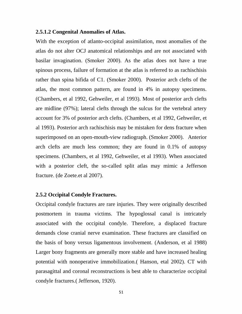
51
2.5.1.2 Congenital Anomalies of Atlas.
With the exception of atlanto-occipital assimilation, most anomalies of the
atlas do not alter OCJ anatomical relationships and are not associated with
basilar invagination. (Smoker 2000). As the atlas does not have a true
spinous process, failure of formation at the atlas is referred to as rachischisis
rather than spina bifida of C1. (Smoker 2000). Posterior arch clefts of the
atlas, the most common pattern, are found in 4% in autopsy specimens.
(Chambers, et al 1992, Gehweiler, et al 1993). Most of posterior arch clefts
are midline (97%); lateral clefts through the sulcus for the vertebral artery
account for 3% of posterior arch clefts. (Chambers, et al 1992, Gehweiler, et
al 1993). Posterior arch rachischisis may be mistaken for dens fracture when
superimposed on an open-mouth-view radiograph. (Smoker 2000). Anterior
arch clefts are much less common; they are found in 0.1% of autopsy
specimens. (Chambers, et al 1992, Gehweiler, et al 1993). When associated
with a posterior cleft, the so-called split atlas may mimic a Jefferson
fracture. (de Zoete.et al 2007).
2.5.2 Occipital Condyle Fractures.
Occipital condyle fractures are rare injuries. They were originally described
postmortem in trauma victims. The hypoglossal canal is intricately
associated with the occipital condyle. Therefore, a displaced fracture
demands close cranial nerve examination. These fractures are classified on
the basis of bony versus ligamentous involvement. (Anderson, et al 1988)
Larger bony fragments are generally more stable and have increased healing
potential with nonoperative immobilization.( Hanson, etal 2002). CT with
parasagittal and coronal reconstructions is best able to characterize occipital
condyle fractures.( Jefferson, 1920).
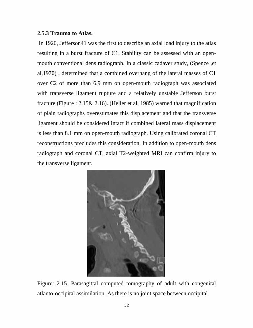
52
2.5.3 Trauma to Atlas.
In 1920, Jefferson41 was the first to describe an axial load injury to the atlas
resulting in a burst fracture of C1. Stability can be assessed with an open-
mouth conventional dens radiograph. In a classic cadaver study, (Spence ,et
al,1970) , determined that a combined overhang of the lateral masses of C1
over C2 of more than 6.9 mm on open-mouth radiograph was associated
with transverse ligament rupture and a relatively unstable Jefferson burst
fracture (Figure : 2.15& 2.16). (Heller et al, 1985) warned that magnification
of plain radiographs overestimates this displacement and that the transverse
ligament should be considered intact if combined lateral mass displacement
is less than 8.1 mm on open-mouth radiograph. Using calibrated coronal CT
reconstructions precludes this consideration. In addition to open-mouth dens
radiograph and coronal CT, axial T2-weighted MRI can confirm injury to
the transverse ligament.
Figure: 2.15. Parasagittal computed tomography of adult with congenital
atlanto-occipital assimilation. As there is no joint space between occipital
53
Condyles and superior articular process of atlas (C1), basilar invagination
often results. (Margali et al 2005)
Figure: 2.16. Sagittal computed tomography of trauma patient reporting neck
pain. Angulated type II dens fracture is evident. (Margali et al 2005)
2.5.4 Treatment of occipitocervical
2.5.4.1 Occipitocervical stabilization
Is performed in both adults and children for a variety of potentially life-
threatening conditions. It has been described as part of the treatment regimen
for congenital instability, (Georgopoulos, etal 1987) degenerative spine
disease,(Grob,2000). Down syndrome, (Menezes et al 1992), traumatic
instability, (Traynelis, et al 1987), tumor involvement of the occipital
condyle, (Deeb, et al 1988) instability following lateral skull–based
approaches, (Bejjani, et al 2000, Margali et al 2005). (Figure 2.17).
54
2.5.4.2 Surgical stabilization
Surgical stabilization of the CVJ in the pediatric population presents a
unique challenge. The pediatric spine is hypermobile as compared with that
of adults because of ligamentous laxity, shallow and angled facets,
underdeveloped spinous processes, flat contouring of the occipital condyles,
and physiological anterior wedging of vertebral bodies. This hypermobility
contributes to high torque and shear forces acting on the C1–2 region.
(Figure: 2.17, 2.18, 2.19, 2.20) (Lustrin, et al 2003) Other factors that make
surgery challenging include incomplete ossification of the odontoid process
and C-1, (Karwacki, et al 2012) a large head compared with body mass, and
weak, underdeveloped cervical musculature. (Lustrin, et al 2003).
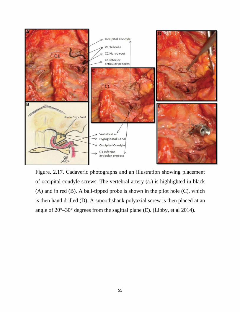
55
Figure. 2.17. Cadaveric photographs and an illustration showing placement
of occipital condyle screws. The vertebral artery (a.) is highlighted in black
(A) and in red (B). A ball-tipped probe is shown in the pilot hole (C), which
is then hand drilled (D). A smoothshank polyaxial screw is then placed at an
angle of 20°–30° degrees from the sagittal plane (E). (Libby, et al 2014).
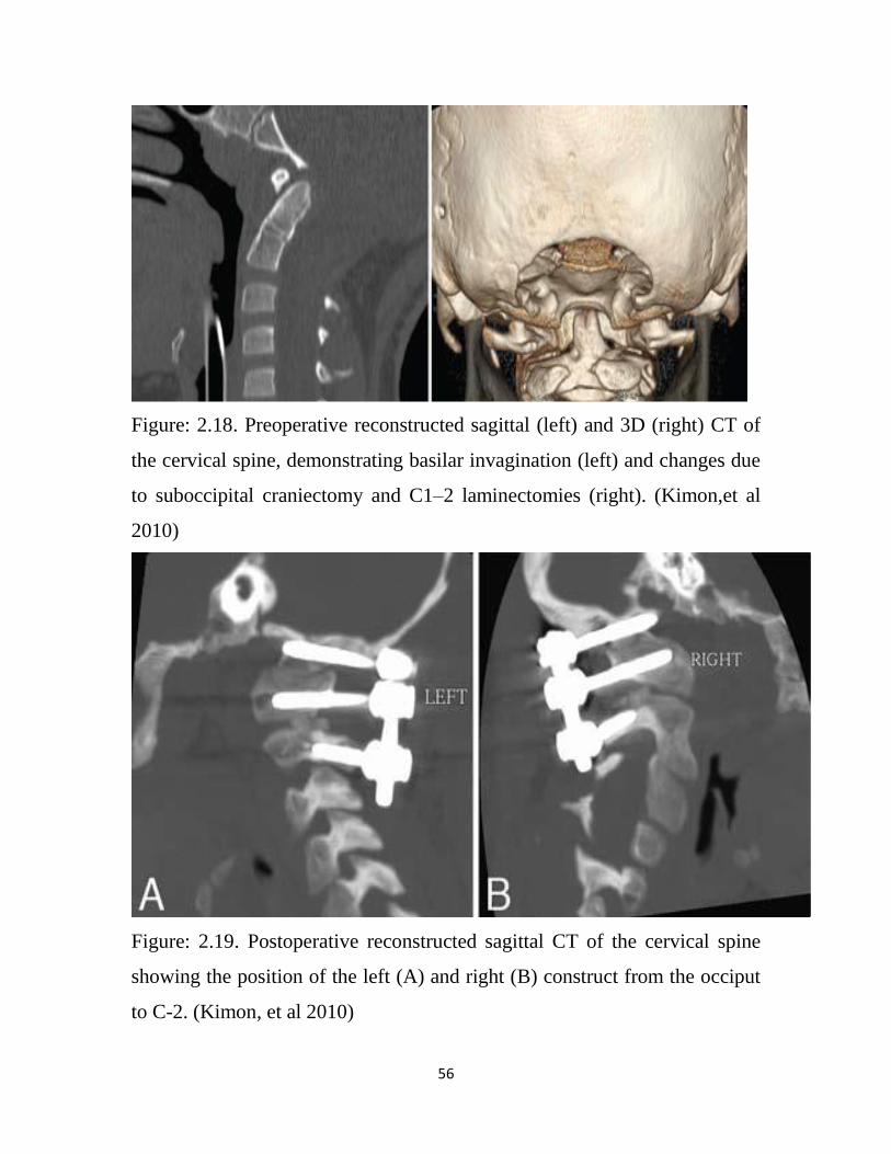
56
Figure: 2.18. Preoperative reconstructed sagittal (left) and 3D (right) CT of
the cervical spine, demonstrating basilar invagination (left) and changes due
to suboccipital craniectomy and C1–2 laminectomies (right). (Kimon,et al
2010)
Figure: 2.19. Postoperative reconstructed sagittal CT of the cervical spine
showing the position of the left (A) and right (B) construct from the occiput
to C-2. (Kimon, et al 2010)
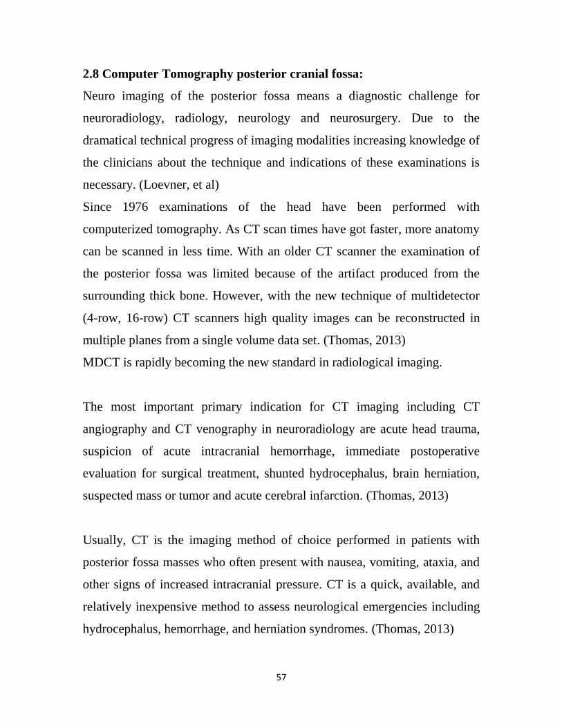
57
2.8 Computer Tomography posterior cranial fossa:
Neuro imaging of the posterior fossa means a diagnostic challenge for
neuroradiology, radiology, neurology and neurosurgery. Due to the
dramatical technical progress of imaging modalities increasing knowledge of
the clinicians about the technique and indications of these examinations is
necessary. (Loevner, et al)
Since 1976 examinations of the head have been performed with
computerized tomography. As CT scan times have got faster, more anatomy
can be scanned in less time. With an older CT scanner the examination of
the posterior fossa was limited because of the artifact produced from the
surrounding thick bone. However, with the new technique of multidetector
(4-row, 16-row) CT scanners high quality images can be reconstructed in
multiple planes from a single volume data set. (Thomas, 2013)
MDCT is rapidly becoming the new standard in radiological imaging.
The most important primary indication for CT imaging including CT
angiography and CT venography in neuroradiology are acute head trauma,
suspicion of acute intracranial hemorrhage, immediate postoperative
evaluation for surgical treatment, shunted hydrocephalus, brain herniation,
suspected mass or tumor and acute cerebral infarction. (Thomas, 2013)
Usually, CT is the imaging method of choice performed in patients with
posterior fossa masses who often present with nausea, vomiting, ataxia, and
other signs of increased intracranial pressure. CT is a quick, available, and
relatively inexpensive method to assess neurological emergencies including
hydrocephalus, hemorrhage, and herniation syndromes. (Thomas, 2013)
58
Searching for an intracerebral aneurysm, the second diagnostic procedure to
be performed immediately after emergency CT is selective cerebral
angiography. Both carotid and vertebral arteries must be injected. External
carotid arteries should also be visualized, particularly if intracranial
angiography is negative, as subarachnoid haemorrhage may sometimes be
due to a rupture not of an aneurysm but of dural arteriovenous
malformations. Angiography should aim at recognizing the aneurysm, its
precise location, size, size of the neck, relationship with the parent vessel
and multiplicity. To achieve this goal the ideal procedure is rotational
angiography with three-dimensional reconstruction. (Provenzale, 2000)
2.8 Computer Tomography for head trauma:
Computed tomography (CT) has become the diagnostic modality of choice
for head trauma due to its accuracy, reliability, safety, and wide availability.
The changes in microcirculation, impaired auto-regulation, cerebral edema,
and axonal injury start as soon as head injury occurs and manifest as clinical,
biochemical, and radiological changes. Proper therapeutic management of
brain injury is based on correct diagnosis and appreciation of the temporal
course of the disease process. (Mukerji, et al 2009)
CT scan detects and precisely localizes the intracranial hematomas, brain
contusions, edema and foreign bodies. Because of the widespread
availability of CT, there is reduction in arteriography, surgical intervention
and skull radiography.
CT scanning is an excellent modality at demonstrating intermediate and late
sequelae of head trauma, such as hydrocephalus, generalized brain atrophy,
encephalomalacia, porencephaly, subdural hygroma leptomeningeal cysts,
and vascular complications. (Mukerji, et al 2009)

59
2.8 Computed Tomography: Technique
Helical and/or multisection CT scanning is helpful in assessing occipital
condylar fractures, but 3-dimensional (3-D) reconstruction is usually not
necessary. However, 3-D reconstructions are valuable when evaluating
facial fractures. The study should always include bone windows to evaluate
for fractures, especially when the skull base or orbits are compromised.
(Ringl et al 2009)
2.9 Previous Studies:
According to Radu et al (1987), C.T. examination of small structures is
made impossible by partial volume effect. Then in 1988 Koster stated that
the increased spatial resolution of C.T. has overcome this difficulty.
However, because of this uncertainty, only a few publications listing C.T.
measurements of these foramina and canals have so far appeared.
Muthukumar et al studied the morphometric analysis of foramen magnum
for the purpose of transcondylar approach on 50 dry skulls. They had given
the anteroposterior length of foramen magnum 33.3 mm and width as 27.9
mm. Also they stated that shape of foramen magnum was ovoid.
Kazikanat and colleagues studied the morphometry of foramen magnum on
59 dry skulls and given anteroposterior and transverse diameter of foramen
magnum as 34.8+2.2 mm and 29.6+2.4 mm and stated that morphometric
analysis of all these components will help in planning of surgical
intervention involving the skull base.
Kosif Rengin studied the midsagittal magnetic resonance images of 194
adults (101females and 93 males) to reveal the relationship between
occipitocervical region and cervical height. They measured the diameter of
60
foramen magnum as 38.19+3.85 mm in males and 36.09+2.79 mm in
females.
Murshed K.A. evaluated morphometrically the foramen magnum and also
studied variations in its shape on C.T. scan of 110 normal subjects with 57
males and 53 females. They had given sagittal diameter as 37.3+3.43 mm
and transverse diameter 31.6+2.99 mm in males and sagittal diameter
34.6+3.16 mm and transverse diameter 29.3+2.19 mm in females.
(Schmeltzer et al. 1971) ,(Wackenheim 1974) and (Þendemir et al. 1994),
studying the FM measurements of 23 subjects through CT images obtained
for the SD and TD a mean of 36.4 mm and 30.0 mm, respectively. These
results are very close to our findings, being 0.5 mm greater than ours in the
SD and 0.4 mm lower than ours in the TD.
Other authors, such as Fischgold and Wackenheim (1965), reported that the
minimum radiographic value for the sagittal diameter is 27 mm, whereas in
ours it is 28 mm. Wackenheim (1974) obtained radiographically mean
values of 35 mm and 30 mm for the sagittal and transverse diameters,
respectively. Compared with our results, without sex distinction, we can see
that these values are slightly lower than ours. In Catalina-HerreraÕs(1987)
study of the FM, diameters were 35.2 mm for the sagittal and 30.3 mm for
the transverse diameter. Similar values were obtained by Testut and Latarjet
(Testut, et al 1977). These results are similar to our findings. Other authors
said that the normal values for the SD measurements of the FM lie between
28.5 mm (Þendemir et al 1994) and 48.0 mm (Adam , 1987), and for the TD
measurements 21.4 mm (Lang , et al1983) and 40.0 mm (Adam , 1987).
61
In a study of 200 skulls, Zaidi and Dayal reported that oval FMs were found
in 128 (64%) skulls. Sindel et al. and Lang et al. reported that this shape was
not found in more than 18.94% and 22.35%, respectively, of their samples.
In a measurement of area of foramen magnum in 219 skeletons (170 males,
39 females) of Turkey, it was observed that mean area of the foramen
magnum was significantly different (909.91±126.02 mm2 in males,
819.01±117, and 24mm 2 in females).
Giles and Elliot have remarked that “next to the pelvis, the skull is the most
easily sexed portion of the skeleton” (Giles E, et al 1963).
There are many studies reported for sex determination by Foramen magnum
dimensions.
Teixeira (1982) reported as study of sex identification utilizing size of FM in
Brazilian individuals. Gunay (2000) confirmed that the area was smaller in
females than in males. Murshed (2003) evaluated CT images of adults that
saggital, transverse diameter and area are grater in males. Uysal (2005)
resulted 81% accuracy rate and Gapert resulted 65.8% accuracy rate in CT
measurements. According to Manoel (2009), linear morphometric method
can be effective to know the sex.
Makaju, for example, worked on 300 samples of CT scan image of head
(Makaju, et al 2013). He studied the antero posterior diameter, transverse
diameter, area, shape of foramen magnum and also the presence of the
accessory hypoglossal canal in the posterior margin of foramen magnum
based on the ethnicity of Nepal. It was concluded that CT scan image of
head can provide valuable measurements of the foramen magnum and could
be used for sexual dimorphism and neurosurgery when other methods are
62
inconclusive. Singh et al also analyzed the foramen magnum of human
skulls (Singh, et al 2013). Fifty adult skulls of known sex were included in
the study. Six standard parameters were measured and analyzed by
discriminant function analysis. The accuracy of sex prediction based on
discriminant function analysis ranged from 66% to 70% and was found a
useful parameter for sex determination.
Uthman et al studied 88 samples for his study (Uthman, et al 2012).
Foramen magnum sagittal diameter, transverse diameter, area and
circumference were measured. Foramen magnum circumference and area
were the best discriminant parameters with an overall accuracy of 67% and
69.3%, respectively. Raghavendra babu et al studied sexual dimorphism of
the antero-posterior diameter, transverse diameter and area of foramen
magnum in a population of coastal Karnataka region using statistical
considerations (Raghavendra Babu, et al, 2012). The predictability of
foramen magnum measurements in sexing of crania was 65.4% for the
antero- posterior diameter.
Catalina-Herrera (Herrera 1987) indicated that the sagittal and transverse
dimensions of the FM were significantly higher in human male than in
human female skulls. Zaidi and Dayal, (Zaidi, et al 1988) classified a
sample of Indian skulls according to the shape and dimensions of the FM,
reporting gender differences which were similar to those reported among
Brazilian skulls.( Manoel et al 2009). (Gu¨nay et al 1997) examined the
usefulness of determining the dimensions of theFMin the diagnosis of sex
and noted that the diameters were of some use while the total area was not a
good indicator. (Yusal et al 2005) reported sexual dimorphism by analysing
63
the dimensions of the FM in three-dimensional (3D) CT with 81% accuracy
in determining the gender.
In 1963, Giles and Elliot (Giles et al 1963) examined sex determination of
the skull by discriminant function analysis using Fisher's method (Fisher,
1936). Their accuracy of Negroid and Caucasoid material range between
82% and 89%.
Hence (Hence, 1977) claimed up to 100% accuracy in predicting sex from
foramen magnum region whilst Holland (Holland, 1989) scored between 71
and 90% in the main sample and 70-80% in the control group. Catallina-
Hercera (Catalina, 1987) indicated that saggital and transverse dimensions of
the foramen magnum were significantly higher in men's skull.
In another study, (Routal, et al 1984) found the dimensions of foramen
magnum in Indian sample to be sexually dimorphic and reported up to 100%
accuracy of correctly identifying sex using simple demarking points.
Gapert, Black and Last also evaluated the foramen magnum of nineteenth
century British adult skulls morphometrically showing significant
differences between men and women classifying 70% of the male skulls and
69.7% of the female skulls by the discriminant function and 76% of men and
70% of women by linear regression. This study confirmed the importance of
this region to identify the gender. (GAPERT, et al 2008).
The amount of partial condylectomy may cause greater atlanto-occipital
instability in shorter OC, especially when more than 2/3 of the OC is
removed (Barut, et al 2009). Therefore, the OCL should be measured
preoperatively, in order to determine the exact area of resection, paying extra
attention when the OC is short. In our study, we found short OC in 7 % of
64
the skulls. These results are in agreement with other investigators’ findings
[Barut, et al 2009, Naderi, et al, 2004], who observed short OC in 8.6 and in
5 %, respectively. In K. Natsis et al study, also observed that women tend to
have shorter OC than men, suggesting that women undertaking partial
condylectomy with extensive condyle resection may be more vulnerable to
atlanto-occipital instability than men, undertaking the same procedure.(
Natsis et al,2013).
65
Chapter Three
Materials and Methods
3.1 Materials
A prospective study of 241 consecutive Sudanese patients (147 were males
and 94 females age range between 20 - 90 years, FM dimensions tend to
stabilize after the second decade of life , were enrolled in the study , between
September 2012 and July 2015.
They were referred to the Radiology Department in the Ibn ALhythm
Medical Center, Khartoum, Sudan, in which the patients subjected to CT
Brian for different clinical indications. The study design was approved by
the Research Council Ethical Committee –College of Medical Radiological
Science. Patients with previous trauma, surgery or pathology in the region of
the FM were excluded, because disorders involving skull base may distort
the normal Anatomy of skull base, and patient who have sound health of
adult age whose skeletal growth is complete were included.
All patients examined on a multislice CT scanner (Asteion, TX-021B
Toshiba scanner). The scan is performed with the patient in the supine
position, It is very important to ensure that there is no rotation or tilt of head
in order to demonstrate any bilateral asymmetry, the protocol used for
routine head scanning from base of skull through apex, with kVp of 120 and
200 mAs, slice thickness is depending on the structure being scanned, thin
data and bone require slice thickness of 5mm and 2mm for reformatted
images. Because of the dense bone of the base of skull beam-hardening
66
artifacts are often seen in images of the posterior fossa, thin slices can help
to reduce these artifacts.
Helical mode is primarily used for studies that require 3D reformations.
All FM measurements were taken from reformatted images (axial, maximum
intensity projection and 3D volume rendering), by the measurement function
available in the CT system (Asteion, TX-021B Toshiba scanner).
3.2 Data Collection & Analysis
3.2.1 Data collection
The Data were collected using the following variables: age, Gender, as well
as the measurements relating to foramen magnum and occipital condyles.
All measurements, of the Foramen Magnum(FM) and occipital condyles
in (mm ) were taken as follows :
Foramen Magnum(FM) measurements :
Length of the foramen magnum (LFM): is the distance taken in a
straight line from the end of the anterior border (basion) through the
center of the foramen magnum until the end of the posterior border toward
the median sagittal plane(Antero posterior diameter) .(appendix A, image
:1)
Width of foramen magnum (WFM) - is the distance in a straight line
from the end of the border right side, with the concavity stronger through
the center of the foramen magnum to the opposite end of the lateral border
of concavity more pronounced, with transverse direction (transverse
diameter). (appendix A, image :1)
67
The of foramen magnum area and circumference, both area and
foramn magnum were measured by tracing the bony border in the 3D
volume rendering and maximum intensity reformatted CT images.
Shape of foramen magnum.
The Occipital Condyles (OC) measurements were taken as follows:
.(appendix A, image :3)
Right occipital condyle maximum width –taken along the articular surface
perpendicular to the right occipital condyle length Image 1: measurement
length (A1- A2, of foramen magnum Width (B1 – B2) and minimum inter
condyle distance (D2- D2 (with maximum bicondyle distance. (C1- C2)
Right occipital condyle length – maximum length taken along the
articular surface perpendicular to the right occipital condyle width.
Left occipital condyle maximum width –taken along the articular
surface perpendicular to the left occipital condyle length.
Left occipital condyle length – maximum length taken along the
articular surface perpendicular to the left occipital condyle width
Maximum.
Distance between occipital condyles – maximum distance between the
lateral edges of the articular surfaces of the occipital condyles
perpendicular to the mid sagittal plane.
Minimum distance between occipital condyles – minimum distance
between the medial edges of the articular surfaces of the occipital
condyles perpendicular to the midsagittal plane.
Maximum internal distance of the occipital condyles – maximum
distance between the medial margins of the occipital condyles,
perpendicular to the mid sagittal plane.

68
Head diameter: including sagittal (SDH) and transverse diameter
(TDH) was obtained from axial reformatted images. .(appendix A, image
:6)
3.2.2 Statistical analysis
The data obtained were analyzed statistically by computing descriptive
statistics like Mean, ± SD values and Percentages, with an independent T-
test, ANOVA test, and by correlation analysis using an IBM SPSS
Statistics software package (Inc., Chicago, Illinois version 16).
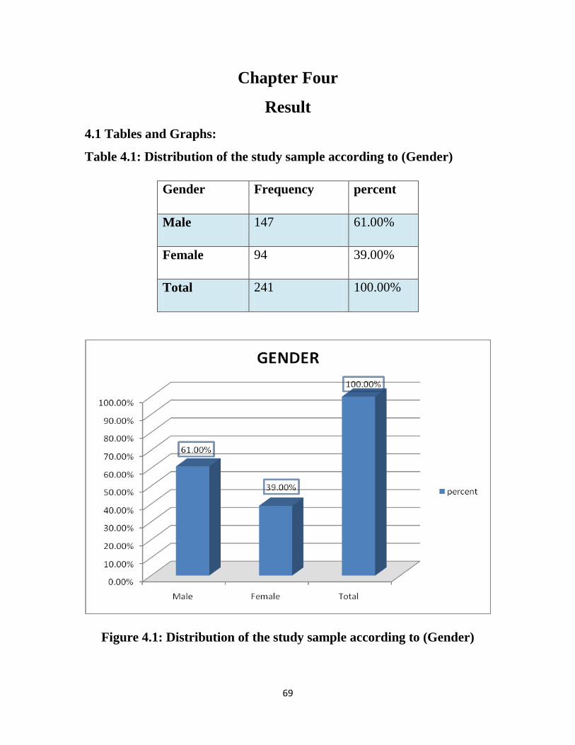
69
Chapter Four
Result
4.1 Tables and Graphs:
Table 4.1: Distribution of the study sample according to (Gender)
Gender Frequency percent
Male 147 61.00%
Female 94 39.00%
Total 241 100.00%
Figure 4.1: Distribution of the study sample according to (Gender)
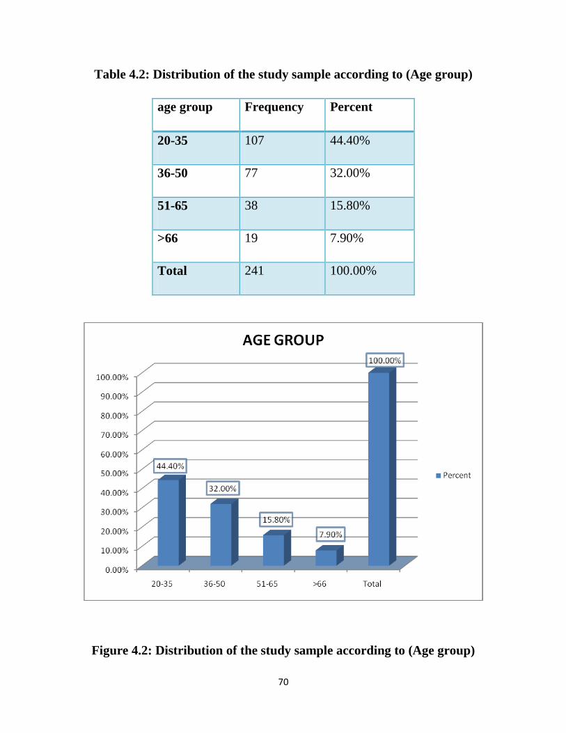
70
Table 4.2: Distribution of the study sample according to (Age group)
age group Frequency Percent
20-35 107 44.40%
36-50 77 32.00%
51-65 38 15.80%
>66 19 7.90%
Total 241 100.00%
Figure 4.2: Distribution of the study sample according to (Age group)
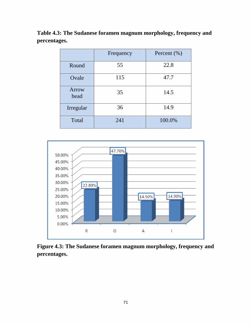
71
Table 4.3: The Sudanese foramen magnum morphology, frequency and
percentages.
Frequency Percent (%)
Round 55 22.8
Ovale 115 47.7
Arrow
head 35 14.5
Irregular 36 14.9
Total 241 100.0%
Figure 4.3: The Sudanese foramen magnum morphology, frequency and
percentages.
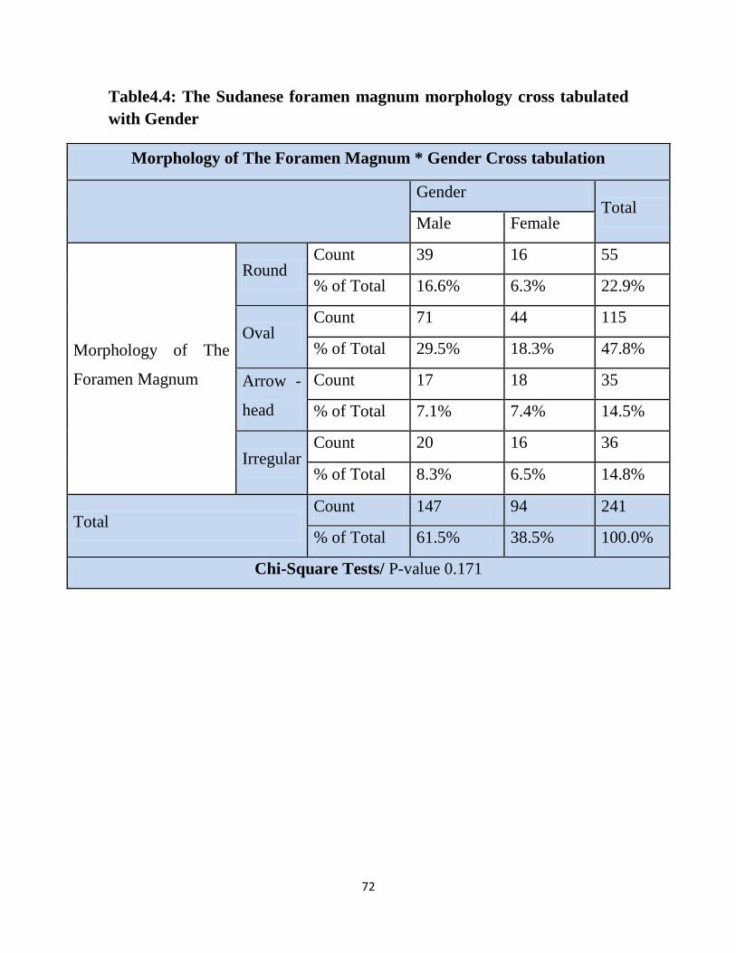
72
Table4.4: The Sudanese foramen magnum morphology cross tabulated
with Gender
Morphology of The Foramen Magnum * Gender Cross tabulation
Gender Total
Male Female
Morphology of The
Foramen Magnum
Round Count 39 16 55
% of Total 16.6% 6.3% 22.9%
Oval Count 71 44 115
% of Total 29.5% 18.3% 47.8%
Arrow -
head
Count 17 18 35
% of Total 7.1% 7.4% 14.5%
Irregular Count 20 16 36
% of Total 8.3% 6.5% 14.8%
Total Count 147 94 241
% of Total 61.5% 38.5% 100.0%
Chi-Square Tests/ P-value 0.171
73
Figure 4.4: Distribution of the foramen magnum morphology according
to (Gender)
74
Table 4.5: Variables related to gender, mean and standard deviations,
maximum, minimum, p-value values.
Variables related Gender Independent Samples
Test T-test for
Equality of Means
Gender N Mean ± S. D Min Max P-value
LFM
Male 147 35.20±2.68 28.70 42.1
<0.001
Female 94 32.17±2.44 25.8 38.3
WFM
Male 147 30.25±2.33 24.6 39.5
<0.001
Female 94 27.89±1.78 23 32.8
AFM
Male 147 819.84±96.53 584.00 1102.00
<0.001
Female 94 692.13±84.64 463.50 980.00
CFM
Male 147 108.84±11.04 87.60 147.30
<0.001
Female 94 96.63±8.84 74.70 125.00
TDH
Male 147 121.45±10.89 104 143.00
<0.001
Female 94 115.29±6.50 101.60 136.00
SDH
Male 147 155.39±8.76 136 175
<0.001
Female 94 147.02±8.86 121 167.1
75
Table 4.6: Sudanese foramen magnum morphology area (AFM) and
circumference (CFM) correlated with transverse (TDH) and sagittal
diameter of the head (SDH), Antro posterior (LFM) and transverse
diameter (WFM) of the FM
Correlations between the variables
TDH SDH LFM WFM
AFM
Pearson Correlation .278** .290** .826** .832**
P-value .000 .001 .000 .000
N 241 241 241 241
CFM
Pearson Correlation .238** .308** .727** .757**
P-value .001 .000 .000 .000
N 241 241 241 241
76
Table 4.7: Statistical measurements of the foramen mafnum variables
correlated with the age classes (mean ± standard deviation, min, max,
and p-value)
ANOVA
N Mean ± S. D Minimu
m Maximum
LFM
20-35 107 33.8019 2.66805 25.80 40.00
.510 36-50 77 34.1260 3.06403 26.20 42.10
51-65 38 34.0553 3.72070 27.30 42.00
>66 19 34.9000 2.71968 29.80 41.20
Total 241 34.0320 2.98196 25.80 42.10
WFM
20-35 107 29.6748 2.71130 23.60 39.50
.244 36-50 77 29.3286 2.31857 23.00 35.00
51-65 38 28.7421 2.20266 24.30 33.20
>66 19 29.3526 1.51451 27.20 32.40
Total 241 29.3917 2.44398 23.00 39.50
TDH
20-35 107 118.9972 12.55660 18.40 140.00
.586
36-50 77 118.8636 7.74179 101.60 143.00
51-65 38 117.9447 6.85769 104.00 128.00
>66 19 121.7842 4.28761 111.30 128.60
Total 241 119.0083 9.90157 18.40 143.00
SDH 20-35 107 152.1449 9.02103 131.00 170.00 .162
36-50 77 151.5234 10.44901 121.00 175.00
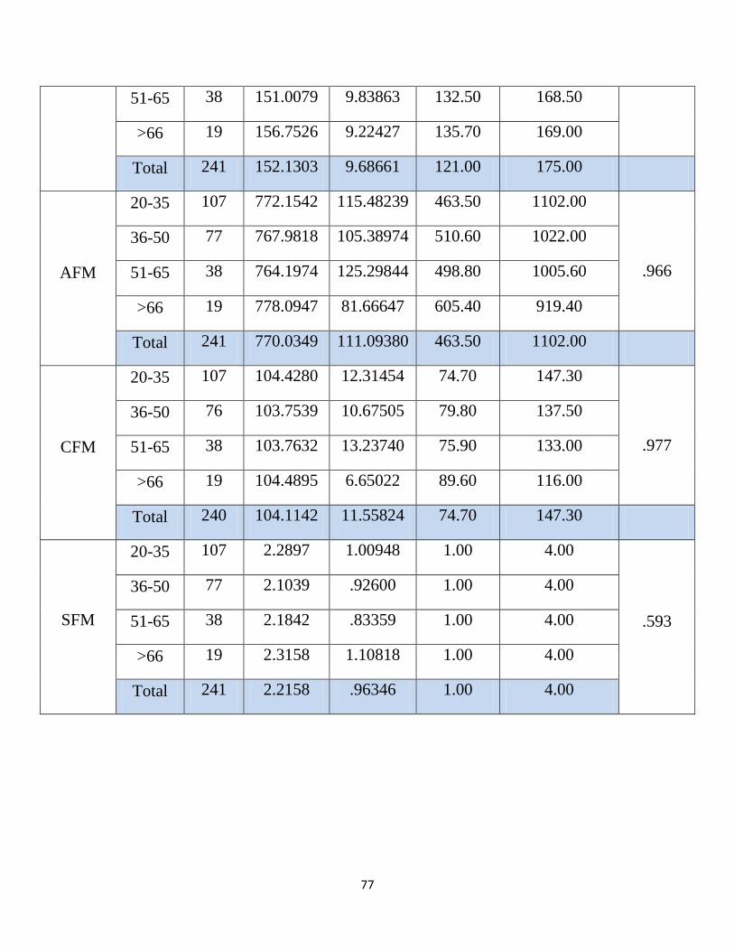
77
51-65 38 151.0079 9.83863 132.50 168.50
>66 19 156.7526 9.22427 135.70 169.00
Total 241 152.1303 9.68661 121.00 175.00
AFM
20-35 107 772.1542 115.48239 463.50 1102.00
.966
36-50 77 767.9818 105.38974 510.60 1022.00
51-65 38 764.1974 125.29844 498.80 1005.60
>66 19 778.0947 81.66647 605.40 919.40
Total 241 770.0349 111.09380 463.50 1102.00
CFM
20-35 107 104.4280 12.31454 74.70 147.30
.977
36-50 76 103.7539 10.67505 79.80 137.50
51-65 38 103.7632 13.23740 75.90 133.00
>66 19 104.4895 6.65022 89.60 116.00
Total 240 104.1142 11.55824 74.70 147.30
SFM
20-35 107 2.2897 1.00948 1.00 4.00
.593
36-50 77 2.1039 .92600 1.00 4.00
51-65 38 2.1842 .83359 1.00 4.00
>66 19 2.3158 1.10818 1.00 4.00
Total 241 2.2158 .96346 1.00 4.00
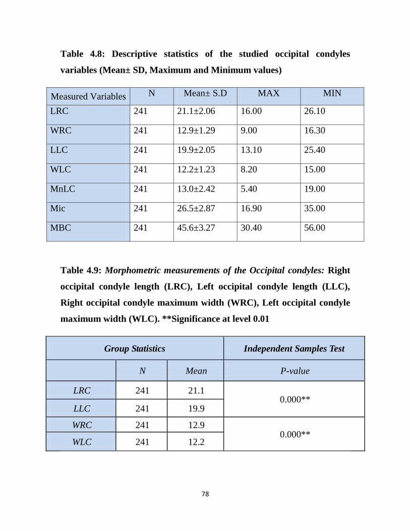
78
Table 4.8: Descriptive statistics of the studied occipital condyles
variables (Mean± SD, Maximum and Minimum values)
Measured Variables N Mean± S.D MAX MIN
LRC 241 21.1±2.06 16.00 26.10
WRC 241 12.9±1.29 9.00 16.30
LLC 241 19.9±2.05 13.10 25.40
WLC 241 12.2±1.23 8.20 15.00
MnLC 241 13.0±2.42 5.40 19.00
Mic 241 26.5±2.87 16.90 35.00
MBC 241 45.6±3.27 30.40 56.00
Table 4.9: Morphometric measurements of the Occipital condyles: Right
occipital condyle length (LRC), Left occipital condyle length (LLC),
Right occipital condyle maximum width (WRC), Left occipital condyle
maximum width (WLC). **Significance at level 0.01
Group Statistics Independent Samples Test
N Mean P-value
LRC 241 21.1 0.000**
LLC 241 19.9
WRC 241 12.9 0.000**
WLC 241 12.2
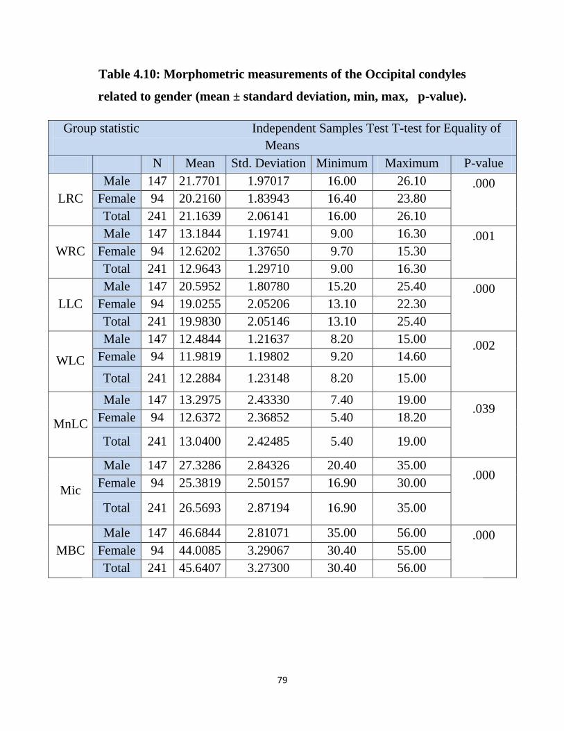
79
Table 4.10: Morphometric measurements of the Occipital condyles
related to gender (mean ± standard deviation, min, max, p-value).
Group statistic Independent Samples Test T-test for Equality of
Means
N Mean Std. Deviation Minimum Maximum P-value
LRC
Male 147 21.7701 1.97017 16.00 26.10 .000
Female 94 20.2160 1.83943 16.40 23.80
Total 241 21.1639 2.06141 16.00 26.10
WRC
Male 147 13.1844 1.19741 9.00 16.30 .001
Female 94 12.6202 1.37650 9.70 15.30
Total 241 12.9643 1.29710 9.00 16.30
LLC
Male 147 20.5952 1.80780 15.20 25.40 .000
Female 94 19.0255 2.05206 13.10 22.30
Total 241 19.9830 2.05146 13.10 25.40
WLC
Male 147 12.4844 1.21637 8.20 15.00 .002
Female 94 11.9819 1.19802 9.20 14.60
Total 241 12.2884 1.23148 8.20 15.00
MnLC
Male 147 13.2975 2.43330 7.40 19.00 .039
Female 94 12.6372 2.36852 5.40 18.20
Total 241 13.0400 2.42485 5.40 19.00
Mic
Male 147 27.3286 2.84326 20.40 35.00 .000
Female 94 25.3819 2.50157 16.90 30.00
Total 241 26.5693 2.87194 16.90 35.00
MBC
Male 147 46.6844 2.81071 35.00 56.00 .000
Female 94 44.0085 3.29067 30.40 55.00
Total 241 45.6407 3.27300 30.40 56.00
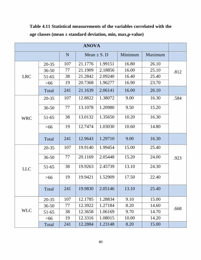
80
Table 4.11 Statistical measurements of the variables correlated with the
age classes (mean ± standard deviation, min, max,p-value)
ANOVA
N Mean ± S. D Minimum Maximum
LRC
20-35 107 21.1776 1.99151 16.80 26.10
.812 36-50 77 21.1909 2.18856 16.00 25.10
51-65 38 21.2842 2.09240 16.40 25.40
>66 19 20.7368 1.96277 16.90 23.70
Total 241 21.1639 2.06141 16.00 26.10
WRC
20-35 107 12.8822 1.38072 9.00 16.30 .584
36-50 77 13.1078 1.20980 9.50 15.20
51-65 38 13.0132 1.35650 10.20 16.30
>66 19 12.7474 1.03030 10.60 14.80
Total 241 12.9643 1.29710 9.00 16.30
LLC
20-35 107 19.9140 1.99454 15.00 25.40
.923
36-50 77 20.1169 2.05448 15.20 24.00
51-65 38 19.9263 2.45739 13.10 24.30
>66 19 19.9421 1.52909 17.50 22.40
Total 241 19.9830 2.05146 13.10 25.40
WLC
20-35 107 12.1785 1.28834 9.10 15.00
.668 36-50 77 12.3922 1.27184 8.20 14.60
51-65 38 12.3658 1.06169 9.70 14.70
>66 19 12.3316 1.08015 10.00 14.20
Total 241 12.2884 1.23148 8.20 15.00
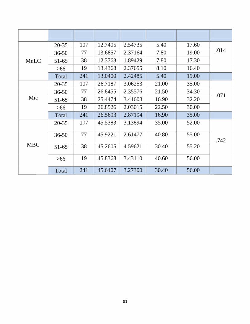
81
MnLC
20-35 107 12.7405 2.54735 5.40 17.60 .014
36-50 77 13.6857 2.37164 7.80 19.00
51-65 38 12.3763 1.89429 7.80 17.30
>66 19 13.4368 2.37655 8.10 16.40
Total 241 13.0400 2.42485 5.40 19.00
Mic
20-35 107 26.7187 3.06253 21.00 35.00
.071 36-50 77 26.8455 2.35576 21.50 34.30
51-65 38 25.4474 3.41608 16.90 32.20
>66 19 26.8526 2.03015 22.50 30.00
Total 241 26.5693 2.87194 16.90 35.00
MBC
20-35 107 45.5383 3.13894 35.00 52.00
.742 36-50 77 45.9221 2.61477 40.80 55.00
51-65 38 45.2605 4.59621 30.40 55.20
>66 19 45.8368 3.43110 40.60 56.00
Total 241 45.6407 3.27300 30.40 56.00
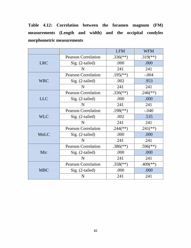
82
Table 4.12: Correlation between the foramen magnum (FM)
measurements (Length and width) and the occipital condyles
morphometric measurements
LFM WFM
LRC
Pearson Correlation .336(**) .319(**)
Sig. (2-tailed) .000 .000
N 241 241
WRC
Pearson Correlation .195(**) -.004
Sig. (2-tailed) .002 .953
N 241 241
LLC
Pearson Correlation .336(**) .246(**)
Sig. (2-tailed) .000 .000
N 241 241
WLC
Pearson Correlation .198(**) -.040
Sig. (2-tailed) .002 .535
N 241 241
MnLC
Pearson Correlation .244(**) .241(**)
Sig. (2-tailed) .000 .000
N 241 241
Mic
Pearson Correlation .386(**) .596(**)
Sig. (2-tailed) .000 .000
N 241 241
MBC
Pearson Correlation .358(**) .409(**)
Sig. (2-tailed) .000 .000
N 241 241
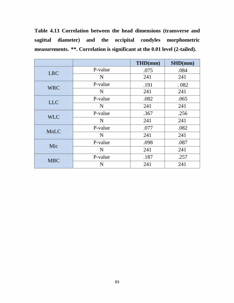
83
Table 4.13 Correlation between the head dimensions (transverse and
sagittal diameter) and the occipital condyles morphometric
measurements. **. Correlation is significant at the 0.01 level (2-tailed).
THD(mm) SHD(mm)
LRC P-value .075 .084
N 241 241
WRC P-value .191 . 082
N 241 241
LLC P-value .082 .065
N 241 241
WLC P-value .367 .256
N 241 241
MnLC P-value .077 .082
N 241 241
Mic P-value .098 .087
N 241 241
MBC P-value .187 .257
N 241 241
84
Chapter Five
Discussion, Conclusion & Recommendation
5.1 Discussion:
5.1.1 Foramen magnum shape:
Since the FM includes specific neuroanatomical structures and their lesions
in that region which require particularly microsurgical intervention,
choosing and establishing the most suitable surgical techniques need a
careful planning mainly based on the FM size to refrain from any
neurological injury (Muthukumar,et al ,2005).
The difference in the FM morphology from various reports indicates racial
variability. (Chet Han et al 2012) but the irregular shape of FM is
highlighted by the developmental cranial anomalies (Furtado SV et al 2010).
The FM is most common described as oval in shape. In a study of 200
skulls, Zaidi and Dayal reported that oval FMs were found in 128 (64%)
skulls (Zaidi et al 1988), Radhakrishna et al, study revealed, 39% with oval
shape, Gupta Chandni et al., reported that oval FMs in 35%, Sindel et al. and
Lang et al. reported that this shape was not found in more than 18.94% and
22.35%, respectively, of their samples (Sindel et al 1989, Lang et al 1983).
In present study a total of four shape categories were identified, Round Oval,
Arrowhead, and Irregular, oval-shaped FM, was the morphological type of
most frequently found in Sudanese; (Zaidi SH et al 1988, Espinoza et al.
2011, Radhakrishna et al. 2012, Avci et al. 2011) reported similar findings.
Table (4.4) shows that there is variation in the morphological types of FM in
85
Sudanese. The variations have been attributed, among some authors due to
different factors such as sexual dimorphism (Ukoha U et al 2011), types of
population (Krıshnamurthy A et al 2012), and ethnic groups (Espinoza E et
al 2011). The oval morphological types of FM substituted 115 (47.8%) of
the sample and the rounded proceeded 55(29.49%) , other types with their
respective frequencies of occurrence have been found as arrow head shape in
36 (14.8%) and the irregular shape achieved 35(14.5%),similar findings
were described by many authors (Chethan P et al 2012 , Natsis K et al
2013).
The Pearson’s chi-square test (P-value 0.171) revealed that there is no
association between foramen magnum shape and gender in the study.
5.1.2 Foramen magnum length and width:
The comparison of the morphometric analysis obtained in this study with the
results of other studies had the following results: the length of the foramen
magnum sudnese (35.2 ± 2.68) was lower than some population studied ,
which was the of Brazilian male skulls (36.5 ± 0.29) , Turkish (37.2 ± 3.43)
Murshed, Cicekcibasi and Tuncer (2003), Spanish (36.2 ± 0.3) Herrera
(1987), and English populations (35.91 ± 2.41) Gapert, Black and Last
(2008), and Indian population (35.5 ± 2.8) Routal, Pal, Bhagwat et al.
(1984). Also is lower measure for the female skulls (32.17 ± 2.44) than
Brazilian population (35.1 ± 0.33), Turkish (34.6 ± 3.16) Murshed,
Cicekcibasi and Tuncer (2003), Spanish (34.30 ± 0) Herrera (1987), Indian
(32.0 ± 2.8) Routal, Pal, Bhagwat et al. (1984), and English populations
(34.71 ± 1.91) Gapert, Black and Last (2008).
Regarding the width of the foramen magnum, the values of the Sudanese
male skull(30.25 ± 2.33) same of Brazilian male skulls (30.3 ± 0.20) and
86
were higher than those of the Indians (29.6 ± 1.9) Routal, Pal, Bhagwat et al.
(1984) only and lower than the Turkish (31.6 ± 2.99) Murshed, Cicekcibasi
and Tuncer (2003), Spanish (31.1 ± 0.3) (HERRERA, 1987), and English
populations (30.51 ± 1.77) Gapert, Black and Last (2008). Also near to be
the same (27.89± 1.78) measure for the female skulls of the Indian (27.1 ±
1.6) Routal, Pal, Bhagwat et al. (1984) and measure lower than and Turkish
populations (29.3 ± 2.19) Murshed, Cicekcibasi and Tuncer (2003) and
lower than Spanish (29.6 ± 0.3) Herrera (1987), English populations (29.36
± 1.96) Gapert, Black and Last (2008), and Brazilian (29.5± 0.23).
In this study, the results were 42.1 and 39.5 mm as the maximum sagittal
and transverse diameter, respectively, in male subjects, and 38.3 and 32.8
mm, respectively for female subjects. The minimum values were 28.70 and
24.6 mm for the sagittal and transverse diameters in male subjects, and 25.8
and 23 mm for the same parameters in female subjects. The mean in males
was 35.20±2.68 mm for the sagittal diameter and 30.25±2.33 mm for the
transverse. In females, the mean was 32.17±2.44 mm and 27.89±1.78 mm,
respectively. Table (4.5)
Obviously, the result of this study was different to the findings of the
studies, in foramen magnum length and width measurement, but slightly
near to (AT Uthman, NH 2011) measurements who was studied Iraqy
population. Lastly in the agreement to present study they stated that there
was significant sex difference in quantified parameters indicating that the
foramen magnum is larger in males.
The (FM) measurements, including length and width and its correlation with
the occipital condyles characteristics, including right occipital condyle
length (LRC), left occipital condyle length (LLC), maximum distance
between occipital condyles (MDC), minimum distance between occipital
87
condyles (MnLC), maximum internal distance of the occipital condyles
(Mic), were found to be significant at p=0.01, and no significant relations
were detected between right and left occipital condyle maximum width
(WRC, WLC) with the length and width of the foramen magnum
(LFM,WFM), these were noticed in ( table 4.12 ).
5.1.3 Foramen magnum area and circumference:
Regarding FMC, the mean values were greater in males than in females
(108.84 mm ±11.04 mm vs 96.63 mm ± 8.84 mm). To our knowledge, the
study of (AT Uthman, NH 2011) was the first that used this measurement
variable and no literature has previously discussed it.
In the present study the area of the foramen magnum of male skulls (819.84
mm2) Table( 4.5 ) was significantly larger than female skulls (692.13 mm2)
the mean area of the foramen magnum of males in present study was similar
to the observations made by (Routal , et al 1984) on Gujarati male skulls
(819.0mm2), However it was lower than the observations made by Catalina
Herrera , on Spain white male skulls (888.4 mm2) and Gunay on Turkey
male skulls (909.9mm2 ), and show greater measurement from that found by
(Muralidhar P Shepur et al ,2014) (748.6mm2) and (Sayee, et al 1987) on
male skulls of Karnataka (769.0mm2).
The mean area of the foramen magnum of female skulls (692.13 mm2) it was
lower in comparison with observations of Catalina Herrera, on Spain white
female skulls (801.0mm2) and Gunay on Turkey female skulls (819.0mm2).
(Sayee, et al 1987) on Karnataka female skulls (746.0 mm2) and (Routal, et
al 1984) on Gujarti female skulls (771.0mm2). Muralidhar P Shepur et al
(711.1 mm2).
88
5.1.3 Foramen magnum transverse and saggital diameter of the hesd
Regarding craniometric measurements, there was a highly significant
statistical difference in head width measurements between genders, in the
study of Deshmukh and Devershi ( Deshmukh et al 2006 ) measured head
width using sliding vernier calipers directly on the crania, which resulted in
mean values of 131mm±0.49 mm for males and 127 mm± 0.49 mm for
females. (AT Uthman,NH 2011) study which is first and only study using
CT in craniometric measurements assessing head width diameter for gender
determination , measured TDH, 143 mm ± 6.49mm for male and 137 mm ±
4.2mm .These values were higher than those recorded in the present
121.45mm ± 10.89mm, for male and 115.29mm ±6.50 mm for female. Table
(4.5)
Current measured 155.39 mm ± 8.76 mm, for male and 147.02 mm ±8.86
mm for female, Table (4.5), for SHD which there is no previous literature in
this case, with statistically significant differences existed between males and
females.
The sex determination is of great value in forensic medicine as well as in
anthropology. Therefore the corroborate that the sagital and transverse
diameters of the FM are of great value for this determination and the
correlation between the area and circumference of the FM is of highly
significance with the transverse and sagital diameter of the head and FM as
shown in table (4.6).
89
5.1.4 The occipital condyles measurement:
The occipital condyles (OC), is an important area in craniovertebral surgery,
but neither its anatomical features, nor the procedures concerning the (OC)
for Sudanese have been studied yet. The morphological analyses of the
structures were made in totally 246sides of the occipital bones of adult
Sudanese craniums by 3DCT images. The length and width of the (OC) were
found to be 21.1±2.06 mm (right), 19.9±2.05 mm (left) and 12.9±1.29 mm
(right), 12.2±1.23 mm (left), respectively.
The maximum distance between occipital condyles, and minimum distance
between inter condyles, and maximum internal distances of the occipital
condyles were measured as 45.6±3.27 and 13.0±2.42 mm and 26.5±2.87,
respectively. These were presented in table (4.9), and these findings were
considered less than the measurements done previously (Mehmet et al.,
2011).
The significance for transcondylar surgical procedure needs information
regarding the morphometric aspects of the OC and surrounded structures
(Muthukumar et al., 2005; Naderi et al., 2005; De Oliveira et al., 1985).
The main issues to be countered in the pre-operative decision-making
process is whether the measurements of the OC may affect surgical
technique, therefore present study was planned to determine the length, and
width of the OC which is important in cases requiring craniovertebral
surgery. When we measured the right and left occipital condyle maximum
length and width, no significant differences were detected between the two
sides at p value =0.000. Similar findings were detected (Mehmat et al.,
2011); this was noticed in table (4.9).
90
The radiological evaluation using 3D CT to the anatomical measurement
strengthens the accepted view that preoperative radiological evaluation is of
greatest importance for attaining successful surgical achievement. The study,
showed that there are significant differences at p value =0.000, between the
males and females regarding the variables.
The Sudanese males were found to have greater measurements than females.
The variables including right occipital condyle length (LRC), left occipital
condyle maximum width (WLC), left occipital condyle length (LLC),
maximum distance between occipital condyles (MDC), maximum internal
distance of the occipital condyles (Mic), transverse head diameter (THD),
sagittal head diameter (SHD),area of the foramen magnum (AFM),
circumference of the foramen magnum (CFM). No significant difference
were detected between right occipital condyle maximum width (WRC) and
minimum distance between occipital condyles (MnLC) with genders this
was observed in table ( 4.10) , as well as the correlations between all of the
variables and different age groups as seen in table (4.11) .
5.1.5 The occipital condyles and FM measurement:
The morphologic variations, including area, circumference, length and width
of the (FM) are important, because the ability of the surgeon to adequately
expose the anterior portion of the (FM) might be difficult.
The (FM) measurements, including length and width and its correlation with
the occipital condyles characteristics, including right occipital condyle
length (LRC), left occipital condyle length (LLC), maximum distance
between occipital condyles (MDC), minimum distance between occipital
condyles (MnLC), maximum internal distance of the occipital condyles
(Mic), were found to be significant at p=0.01, and no significant relations
91
were detected between right and left occipital condyle maximum width
(WRC, WLC) with the length and width of the foramen magnum
(LFM,WFM), these were noticed in table (4.12) .
(FM) measurements, and the length of the (OC) are important factors
because protrusion of the (OC) into (FM) may indicate more extensive bony
removal during surgery and the amount of partial condylectomy may cause
greater occipitocervical instability in short (OC) (Barut et al., 2009). This
also was discussed by (Muthukumar et al., 2005).
The differences between the OC Sudanese measurements, and other
previous studies measurements, (Mehmet et al., 2011) proved that there are
anatomical variations detected regarding the OC.
(Naderi et al., 2005), have done the first study which made a systematic and
detailed classification of the (OC), based on its shape. Shapes have been
described, including two-semicircles, oval type, and rhombus, beanshaped,
prismatic, flattened, convex, flattened-convex, short and broad, flat and long,
small and convex types. (Bozbuga et al., 1999; Muthukumar et al., 2005;
Acikbas et al., 1997) but current study didn’t characterize its shape but did
comprehensive measurements for the (FM),(OC) and surrounding structures,
as well as, the head sagital and transverse diameters. Table (4.13) shows the
correlations between the transverse head diameters (THD) and sagittal head
diameter (SHD) and the occipital condyles measurements for Sudanese, the
presented data showed no significant relationship between the variables and
similar results were found by (Naderi et al., 2005) .
92
5.2 Conclusion
To conclude, the present study regarding morphometry of foramen magnum
was in agreement with earlier studies done so far, hence it shows diameters
of foramen magnum were greater in males than in females.
FM measurements are valuable in studying sexual dimorphism in forensic
investigations with high significant difference was observed between sexes.
The area and circumference of FM varies in relation with gender and
ethnicity group. However, it should be noted that sex differences in the
dimensions of the FM and the variations should be taken into concern during
the performance of clinical and radiological diagnostics and during surgical
approach.
Current study results show the shape of the foramen magnum is not
indicative of a specific ancestral group; and gender; however, the oval shape
is predominated and followed by round one , arrow head and irregular
shape respectively .
Measurements of the Occipital condyles diameter and protrusion contribute
to the shape and size of FM diameter, considering the vital structures that
pass through FM.
The OC the main bony prominences obstructing the anterolateral surface of
the brainstem for the far lateral approaches. The radiological and anatomical
measurements indicated that the radiological assessment greatly helps to
organize the preoperative preparation.
Most of the data obtained in CT corroborate previous studies and are
important parameters in the evaluation of morphometric variations of pre-
surgical preparation in regard to the transcondylar approach, thus helping to
reduce the risk of neurovascular injury during the procedure.
93
Computed tomographic scan is noninvasive modality for the imaging the
skull base. Since this procedure is widely done, this modality was preferred.
CT/3D CT can be accurately used in further investigations to provide basis
for anthropometric and forensic issues
Current study findings may consider as reference for Sudanese, and the
measurements may describe the normal morphological variants of FM for
Sudanese. Since the anatomy of the FM is of interest to many radiological
fields. The radiologists must have knowledge of normal anatomy of skull
base to determine the presence of abnormality and to help in surgical
planning. The information gained from the present study will be of useful to
all of the medical and radiological fields.

94
5.3 Recommendation
The importance of that fact and studying that area comprehensively; is
that the surgeon should not decide about the achievement of condylectomy
based on the head transverse and sagital diameters.
Neurosurgeon has to be familiar with the anatomical structures of the FM
region and the probable variations of the structures in order to achieve the
widest exposure with the best surgical outcome.
The radiological and anatomical measurements indicated that radiological
assessment using CT scan greatly helps to organize the preoperative
preparation, so radiologists must enrich their knowledge to the variations of
this region. FM dimensions tend to stabilize after the second decade of life
and the reconstructed CT images can provide reliable measurement of these
dimensions.
Future research is needed to further confirm the results acquired with this
study for foramen magnum and occipital condyles measurement.
Future research is recommended for foramen magnum and occipital
condyles measurement, in different Sudanese state; since Sudan rich with
ethnic diversity.
95
References:
Adam AM. (1987), Skull radiograph measurements of normals and patients
with basilar impression; use of LanzertÕs angle. Surg Radiol Anat 9: 225-9,.
Acikbas SC, Tuncer R, Demirez I, Rahat O, Kazan S, Sindel M, Saveren M
(1997) The effect of condylectomy on extreme lateral transcondylar
approach to the anterior foramen magnum. Acta Neurochir (Wien)
139(6):546–550.
Anne Waugh, Allison Grant: Ross and Wilson, (2004), Anatomy and
Physiology in Health and Illness. 9th Ed Churchill Livingstone- 403,
Arey LB: Developmental Anatomy. (1965) A Textbook and Laboratory
Manual of Embryology, 7th ed. Philadelphia, Saunders, pp 404–407.
Anderson PA, Montesano PX. (1988) Morphology and treatment of occipital
condyle fractures. Spine. 13(7):731-736.
Arnautovic KI, Al-Mefty O, Husain M. (2000) Ventral foramen magnum
meninigiomas. J Neurosurg (Suppl 1) 92:71-80,
Arnold H, Menezes A. (2008 October) Craniocervical developmental
anatomy and its implications. Childs Nerv Syst. 24(10):1109-22.
Arnold’s (1991 Nov) contributions and some early observations on neural-
tube defects. J Neurosurg. 75(5):823-6
A.T.Uthman, N.H.Al-Rawi,J.F.Al Timimi, (2012) Evaluation of foramen
magnum in Gender determination using helical CT scanning, Dentomaxillo
facial Radiology 41,197–202.

96
Babu RP, Sekhar LN, Wright DC: (1994) Extreme lateral transcondylar
approach: technical improvements and lessons learned. J Neurosurg 81:49-
59,
Banerji D, Behari S, Jain VK, et al: (1999) Extreme lateral transcondylar
approach to the skull base. Neurology India 47:22-30,
Bannister LH, Berry MM, Collins P, Dyson M, et al. (1995) Gray’s anatomy
the anatomical basis of medicine and surgery. 38th ed. Edinburgh: Churchill
Livingstone; .p.567-568.
Barut N, Kale A, Turan Suslu H, Ozturk A, Bozbuga M, Sahinoglu K
(2009). Evaluation of the bony landmarks in transcondylar approach. Br. J.
Neurosurg. 23:276-281
Bassiouni H, Ntoukas V, Asgari S, Sandalcioglu EI, Stolke D, Seifert V
(2006) Foramen magnum meningiomas: clinical outcome after microsurgical
resection via a posterolateral suboccipital retrocondylar approach.
Neurosurgery 59:1177–1187
Berlis A, Putz R, Schumacher M. (1992) Direct and CT measurement of
canals and foramina of the skull base. The British Journal of Radiology; 65;
653-661.
Berkovitz BKB, Moxham BJ (1994) Color Atlas of the Skull. London:
Mosby-Wolfe.
Berry AC, (1975) Factors affecting the incidence of non-metrical skeletal
Variants. J Anat 120: 519–35.

97
Berry AC, Berry RJ (1967) Epigenetic variation in the human cranium. J
Anat: 361–80.
Bejjani GK, Sekhar LN, Riedel CJ: (2000) Occipitocervical fusion following
the extreme lateral transcondylar approach. Surg Neurol 54:109–116,
Bozbuga M, Ozturk A, Bayraktar B, Ari Z, Sahinoglu K, Polat G,Gurel I
(1999). Surgical anatomy and morphometric analysis ofthe occipital
condyles and foramen magnum. Okajimas Folia Anat. Jpn. 75(6):329–334
Boulton MR, Cusimano MD (2003) Foramen magnum meningiomas:
concepts, classifications and nuances. Neurosurg Focus 14(6):e10
Bowerman RA. (1995) Anomalies of the fetal skeleton: sonographic
findings. AJR Am J Roentgenol; 164 (4): 973-9.
Bruce V, Burton AM, Hanna E, Healey P, Mason O, Coombes A, Fright R,
Linney A (1993 ) Sex discrimination: how do we tell the difference between
male and female faces? Perception 22: 131–52.
Cantore G, Ciappetta P, Delfini R: (1994) Choice of neurosurgical approach
in the treatment of cranial base lesions. Neurosurg Rev 17:109-125,
Catalina-Herrera CJ: (1987) Study of the anatomic metric values of the
foramen magnum and its relation to sex. Acta Anat 130:344-347
Chambers AA, Gaskill MF. (1992) Midline anterior atlas clefts: CT findings.
J Comput Assist Tomogr; 16(6):868-870
Cheema JI, Grissom LE, Harcke HT. (2003) Radiographic characteristics of
lower-extremity bowing in children. Radiographics. 23 (4): 871-80
98
Chiapparini L, Saletti V, Solero CL et-al. (2011) Neuroradiological
diagnosis of Chiari malformations. Neurol. Sci.; 32 Suppl 3 (S3): S283-6.
Cicekcibasi AE, Murshed KA, Ziylan T, Seker M, Tuncer I. (2004)
Morphometric evaluation of some important bony landmark on the skull
base related sexes. Turk Journal of Medical Sciences; 34: 37.
Christ B, Wilting J: (1992) from somites to vertebral column. Annals of
Anatomy 174:23–32,
Cloud GC, Markus HS. (2003) Diagnosis and management of vertebral
artery stenosis. QJM; 96 (1): 27-54. QJM (full text) - doi: 10.1093
Chiapparini L, Saletti V, Solero CL et-al. (2011) Neuroradiological
diagnosis of Chiari malformations. Neurol. Sci.; 32 Suppl 3 (S3): S283-6.
Deeb ZL, Rothfus WE, Goldberg AL, and Daffner RH: (1988) Occult
occipital condyle fractures presenting as tumors. J Comput Tomogr 12:261–
263.
De Oliveira E, Rhoton AL Jr, Peace D (1985). Microsurgical anatomy of the
region of the foramen magnum. Surg. Neurol. 24:293-352.
Deshmukh AG, Devershi DB. (2006) Comparison of cranial sex
determination by univariate and multivariate analysis. J Anat Soc India; 55:
1–5.
De Zoete A, Langeveld UA. (2007) A congenital anomaly of the atlas as a
diagnostic dilemma: a case report. J Manipulative Physiol Ther.; 30(1):62-
64.
99
Dietrich S, Kessel M: (1997) The vertebral column, in Thorogood P (Ed):
Embryos, Genes and Birth Defects. Chichester, Wiley, , pp 281–302.
Dias M. Myelomeningocoele. Choux M, Di Rocco C, Hockley A, Walker
M. (1999) Pediatric Neurosurgery. London: Churchill Livingston;. 33-61
Drake. Gray's Basic Anatomy: with STUDENT CONSULT Online Access,
1e (Grays Anatomy for Students). Churchill Livingstone. ISBN:
1455710784
DSc SSP. Gray's Anatomy. Churchill Livingstone. (2011) ISBN:
0443066841.
Elster AD, Chen MY. (1992) Chiari I malformations: clinical and radiologic
reappraisal. Radiology; 183 (2): 347-53.
Farb RI, Scott JN, Willinsky RA, et al (2003) intracranial venous system:
gadolinium-enhanced three-dimensional MR venography with auto-triggered
elliptic centric-ordered sequence-initial experience. Radiology 226:203-209,
Fatma Hayat Erdil, Vedat Sabancıogulları, Mehmet Çimen, Oktay Isık.
(2010). Morphometric Analysis of the Foramen Magnum by Computed
Tomography. Erciyes Medical Journal.32 (3): 167-170.
Ferreira, F. V.; Rosenberg, B. & da Luz, H. P. (1967) The "Foramen
Magnum" index in Brazilians. Rev. Fac. Odontol. Sao Paulo, 5(4):297-302,.
Fischgold H, Wackenheim A. (1965) La radiographie des formations
intrarachidiennes. Masson, Paris.
Fisher RA. (1936)The Use of multiple measurements in Taxonomic
problems. Ann. Eugen.; 7:179-188.
100
Furtado SV, Thakre DJ, Venkatesh PK, Reddy K, Hegde AS:(2010)
Morphometric analysis of foramen magnum dimensions andintracranial
volume in pediatric chiari I malformation. ActaNeurochir (Wien) 152:221-
227
Ganguly DN, Roy KKS (1964) A study on the craniovertebral joint in man.
Anat Anz Bd 114:433–452.
GAPERT, R., BLACK, S. and LAST, J. (2008) Sex determination from the
foramen magnum: discriminant function analysis in an eighteenth and
nineteenth century British sample. International Journal of Legal Medicine,
vol. 123, no. 1, p. 25-33.
Gasser RF: (1976) Early formation of the basicranium in man, in Bosma JF
(ed): Symposium on Development of the Basicranium. Bethesda,
Department of Health Education and Science Publication (NIH), pp 29–43.
George B, Dematons C, Cophignon J (1988). Lateral approach to the
anterior portion of the foramen magnum. Application to surgical removal of
14 benign tumors: technical note. Surg. Neurol. 29(6):484- 490.
George B, Lot G, Boissonnet H (1997) Meningioma of the foramen
magnum: a series of 40 cases. Surg Neurol 47:371–37
Georg opoulos G, Pizzutillo PD, Lee MS: (1987) Occipito-atlantal instability
in children. A report of five cases and review of the literature. J Bone Joint
Surg Am 69:429–436,
Gill GW, Rhine S (2004) Skeletal Attribution of Race: Methods for Forensic
Anthropology. New Mexico: Maxwell Museum of Anthropology.

101
Giles E, Elliot O. (1963) Sex Determination by discriminant functional
analysis of crania. Am J. Phys Anthropol.; 21:53-68.
Gehweiler JA Jr, Daffner RH, Roberts L Jr. (1983) Malformations of the
atlas vertebra simulating the Jefferson fracture. AJR Am J Roentgenol;
140(6):1083- 1086.
Gladstone J, Erickson-Powell W: (1915) Manifestation of occipital vertebra
and fusion of atlas with occipital bone. J Anat & Physiol 49:190–199, 1914–
Gladstone RJ, Wakeley CPG: (1924–1925) Variations of the occipito-
atlantal joint in relation to the metameric structure of the craniovertebral
region. J Anatomy 59:195–216.
Gray, Henry. (2000) Anatomy of the Human Body. Philadelphia: Lea &
Febiger, 1918; Bartleby.com. www.bartleby.com/107/.
Gray, Henry. : (2005) Gray's anatomy (39e). Churchill Livingstone. ISBN:
0443071683.
Grossman RI, Yousem DM. Neuroradiology: (2003) The Requisites, 2e
(Requisites in Radiology). Mosby. ISBN: 032300508X.
Grob D (2000) Posterior occipitocervical fusion in rheumatoid arthritis and
other instabilities. J Orthop Sci 5:82–87,
Gruber P, Henneberg M, Boni T, Ruhli FJ: (2009) Variability of human
foramen magnum size. Anat Rec 292:1713-1719,
Guille JT, Sherk HH. (2002)Congenital osseous anomalies of the upper and
lower cervical spine in children. J Bone Joint Surg Am.; 84(2):277-288.
102
Guo F, Wang M, Long J, Wang H, Sun H, Yang B, et al.( 2007) Surgical
management of Chiari malformation: analysis of 128 cases. Pediatr
Neurosurg. 43(5):375-81.
Gu¨nay Y, Altinko¨k M, C¸ agdir S, Kirangil B.( 1997) Gender
determination with skull measurements (in Turkish). J Forensic Med; 13:
13–19.
Hanson JA, Deliganis AV, Baxter AB, et al. (2002) Radiologic and clinical
spectrum of occipital condyle fractures: retrospective review of 107
consecutive fractures in 95 patients. AJR Am J Roentgenol; 178(5):1261-
1268.
Hecht JT, Horton WA, Reid CS, Pyeritz RE, Chakraborty R: (1989) Growth
of the foramen magnum in achondroplasia. Am J Med Genet 32:528-535,
and FM brain herniation (9)109.
Heller JG, Viroslav S, Hudson T. Jefferson fractures: the role of
magnification artifact.
Hence W. (1977) The method of discriminant function analysis for sexual
determination of the skull. J. Hum Evol. 6:95-100.
http://images.radiopaedia.org/images/23719/bcd119b4b4c67be335e7606bc7
55ec_big_gallery.jpg
http://images.radiopaedia.org/images/23718/d9a30bd2eec8378d039cc168d0
7ae1_big_gallery.jpg
103
Hofkes SK, Iskandar BJ, Turski PA, Gentry LR, McCue JB, Haughton VM.
(2007 Nov) Differentiation between symptomatic Chiari I malformation and
asymptomatic tonsilar ectopia by using cerebrospinal fluid flow imaging:
initial estimate of imaging accuracy. Radiology.245 (2):532-40.
Holland TD. (1986) Sex determination of fragmentary crania by analysis of
crania base. Am J Phys. Anthropol.; 70:203-208.
Jain D, Jasuja OP, Nath S,( 2014) Evaluation of foramen magnum in sex
determination from human crania by using discriminant function analysis
,Elective Medicine Journal, Vol 2, No 2,
Jefferson G. Fractures of the atlas vertebrae. Br J Surg. (1920); 7:407-422.
Junction. In: Youmans JR, editor. Neurological Surgery. 3rd Ed.
Philadelphia: WB Saunders. P 1359–1420.
Ilizarov GA, Ledyaev VI (1992) The replacement of long tubular bone
defects by lengthening distraction osteotomy of one of the fragments. Clin
Orthopaed Relat Res 280: 7–10.
Karwacki GM, Schneider JF: (1882–1887) Normal ossification patterns of
atlas and axis: a CT study. AJNR Am J Neuroradiol 33, 2012.
Kao SC, Waziri MH, Smith WL et-al. (1989) MR imaging of the
craniovertebral junction, cranium, and brain in children with achondroplasia.
AJR Am J Roentgenol; 153 (3): 565-9
Kessel M, Gruss P (1991) Homeotic transformations of murine vertebrae
and concomitant alteration of Hox codes induced by retinoic acid. Cell
67:89–104.
104
Ketonen L, Hiwatashi A, Sidhu R. (2005) Pediatric brain and spine, an atlas
of MRI and spectroscopy. Springer Verlag. ISBN: 3540213406
Kimon Bekelis, Ann-Christine Duhaime, Symeon Missios, Clifford Belden,
and Nathan Simmons, (August 2010) Placement of occipital condyle screws
for occipitocervical fixation in a pediatric patient with occipitocervical
instability after decompression for Chiari malformation J Neurosurg:
Pediatrics / Volume 6 /
Koehler PJ. (1991 Nov) Chiari's description of cerebellar ectopy (1891).
With a summary of Cleland's and Arnold's contributions and some early
observations on neural-tube defects. J Neurosurg. 75(5):823-6.
Kornienko VN, Pronin IN. (2008) Diagnostic Neuroradiology. Springer
Verlag. ISBN: 3540756523
Kratimenos GP, Crockard HA (1993) The far lateral approach for ventrally
placed foramen magnum and upper cervical spine tumours. Br J Neurosurg
7(2):129–140
Kunicki J, Ciszek B. (2005)The clinical anatomy and the occipital condyle
variants [letter]. Clin Anat.; 18(8):646-647.
K. Natsis, M. Piagkou, G. Skotsimara, G. Piagkos, P. Skandalakis (April
2013) A morphometric anatomical and comparative study of the foramen
magnum region in a Greek population Springer-Verlag,: 6.
Laine FJ, Nade L, Braun FI. (1990) CT and MR imaging of the central skull
base Part1 Technique, embryology development and anatomy.
Radiographics; 4: 591.
105
La Marca F, Zubay G, Morrison T, Karahalios D (2008) Cadaveric study for
placement of occipital condyle screws: technique and effects on surrounding
anatomic structures. J Neurosurg Spine 9(4):347–353.
Lateral and anterior views of a human skull (8/26/ 2013) available at: URL:
http://www.britannica.com/science/skull
Lahr MM (1996) The Evolution of Modern Human Diversity: A Study of
Cranial Variation. Cambridge: Cambridge University Press.
Lang VJ, Schafhauser O, Hoffmann S. †ber die postnatale Entwicklung der
transbasalen Shadelpforten: Canalis caroticus, (1983) foramen jugulare,
canalis hypoglossalis, canalis condylaris und foramen magnum. Anat Anz
153: 315-57.
Lele S, Richtsmeier JT (1991) Euclidean distance matrix analysis: a
coordinatefreeapproach for comparing biological shapes using landmark
data. Am J Phys Anthropol 86: 415–27.
Lieberman DE, McCarthy RC (1999) The ontogeny of cranial base
angulation in humans and chimpanzees and its implications for
reconstructing pharyngeal dimensions. J Hum Evol 36: 487–517.
Lieberman DE, Pearson OM, Mowbray KM (2000) Basicranial infl uence on
overall cranial shape. J Hum Evol 38: 291–315.
Levy C, Wassef M, George B, Popot B; Compere A, Tran Ba Huy P (1988)
Meningiomes extra-crâniens. A propos de sept cas. Problemes
pathogeniques, diagnostiques et therapeutiques [in French]. Ann Oto-
laryngol 105:13–2

106
Libby Kosnik-Infinger. Steven S. Glazier, and Bruce M. Frankel, (January
2014) Occipital condyle to cervical spine fixation in the pediatric population,
J Neurosurg: Pediatrics / Volume 13 /
Loevner LA. (1999) Imaging features of posterior fossa neoplasms in
children and adults. Semin Roentgenol.; 34(2):84-101.
Love JG, Thelen EP, Dodge HW (1954) Tumors of the foramen magnum. J
Int Coll Surg 22:1–17
Lustrin ES, Robertson RL, and Tilak S. (1994) Normal anatomy of the skull
base. Neuroimaging Clin N Am; 4:465–478.
Lustrin ES, Karakas SP, Ortiz AO, Cinnamon J, Castillo M, Vaheesan K, et
al (2003) Pediatric cervical spine: normal anatomy, variants, and trauma.
Radiographics 23:539–560.
Makaju S :( 2013) Study of Morphometric Evaluation of Dimorphism and
Shape of the Human Foramen Magnum: A study of Computerized
Tomographic in Nepalese Population. Thesis submitted in the partial
fulfillment of the degree of master of surgery in Anatomy. Kathmandu
University, Nepal.
Manoel C, Prado FB, Caria PHF, Groppo FC.( 2009) Morphometric analysis
of the foramen magnum in human skulls of Brazilian individuals: its relation
to gender. Braz J Morphol Sci; 26: 104–108.
Marin Sanabria EA, Ehara K, Tamaki N (2002) surgical experience with
skull base approaches for foramen magnum meningioma. Neurol Med Chir
(Tokyo) 42:472–480
107
Margalit NS, Lesser JB, Singer M, Sen C: (2005) Lateral approach to
anterolateral tumors at the foramen magnum: factors determining surgical
procedure. Neurosurgery 56 (2 Suppl): 324–336,
Mc Graw Hill Concise Dictionary of Modern Medicine (2002): Mc Graw
HillCompanies Inc
McRae DL. (1953) Bony abnormalities in the region of the foramen
magnum: correlation of the anatomic and neurologic findings. Acta Radiol;
40(2-3):335-354.
McRae DL, Barnum AS. (1953) Occipitalization of the atlas. Am J
Roentgenol Radium Ther Nucl Med.; 70(1):23-46.
Menezes AH: (1998) Embryology, development and classification of
disorders of the craniovertebral junction, in Dickman CA, Sonntag VKH,
Spetzler RF (eds): Surgery of the Craniovertebral Junction. New York:
Thieme Medical Publishers, pp 3–12.
Melsen B: (1974) The cranial base. Acta Odont Scand 32:1–126,.
Menezes AH: (1995) Congenital and acquired abnormalities of the
craniovertebral junction, in Youmans (ed): Neurological Surgery, 4th Ed.
Philadelphia, W.B. Saunders, pp 1035–1089.
Menezes AH: (1995) Primary craniovertebral anomalies and the hindbrain
herniation syndrome (Chiari I): Data base analysis. Pediatr Neurosurg
23:260–269.
Menezes AH: (1998) Embryology, development and classification of
disorders of the craniovertebral junction, in Dickman CA, Sonntag VKH,
Spetzler RF (eds): Surgery of the Craniovertebral Junction. New York:
Thieme Medical Publishers, pp 3–12.
108
Menezes AH: (2003) Developmental abnormalities of the craniovertebral
junction, in Winn HR (ed): Youman’s Neurological Surgery, 5th Ed, Vol 3.
Philadelphia, WB Saunders, pp 3331–3345.
Menezes AH, VanGilder JC. (1989). Anomalies of the craniovertebral.
Menezes AH, Ryken TC, Brockmeyer DL. (2001). Abnormalities of the
craniocervical junction. In: McClone DG, editor. Pediatric Neurosurgery:
Menezes AH, Ryken TC: (1992) Craniovertebral abnormalities in Down’s
syndrome. Pediatr Neurosurg 18:24–33.
Melfort R. Boulton, Michael D. Cusimano, (2003) Neurosurg Focus; 14(6)
Michaël Bruneau and Bernard George: (2008Jan) Foramen magnum
meningiomas: detailed surgical approaches and technical aspects at
Lariboisière Hospital and review of the literature. Neurosurg Rev.; 31(1):
19–33.
Michael Benke, Warren D. Yu, Sean C. Peden, and Joseph R. O’Brien,
(2011) Occipitocervical Junction: Imaging, Pathology, Instrumentation Am J
Orthop; 40(10):E205-E215.
Miller E, Crockard HA (1987) Transoral transclival removal of anteriorly
placed meningiomas at the foramen magnum. Neurosurgery 20:966–968
Moore KL, Dalley AF. (2006). Craniovertebral Joints: Clinically Oriented
Anatomy. 5th Ed. Baltimore: Lippincott Williams and Wilkins.
Moreland LW. (2004) Rheumatology and immunology therapy, A to Z
essentials. Springer Verlag. ISBN: 3540206256.
109
Mukerji N, Cahill J, Paluzzi A, Holliman D, Dambatta S, Kane PJ. (2009
Apr) Emergency head CT scans: can neurosurgical registrars be relied upon
to interpret them? Br J Neurosurg. 23(2):158-61.
Muralidhar P Shepur, Magi M, Nanjundappa B, Pavan P Havaldar,
Premalatha Gogi, Shaik Hussain Saheb.(2014)MORPHOMETRIC
ANALYSIS OF FORAMEN MAGNUM. Int J Anat Res; 2(1):249-55.
Murshed K.A., Cìcekcibasi A.E., and Tuncer I. (2003) “Morphometric
Evaluation of the Foramen Magnum and Variations in its Shape: A Study on
Computerized Tomographic Images of Normal Adults.” Turkish Journal of
Medical Sciences, 33(1):301-306.
Muthukumar N, Swaminathan R, Venkatesh G, Bhanumathy SP (2005) A
morphometric analysis of the foramen magnum region as it relates to the
transcondylar approach. Acta Neurochir (Wien) 147(8):889–895.
N. Muthukumar, R. Swaminathan, G. Venkatesh and S.P. Bhanumathy.
(2005). A morphometric analysis of the foramen magnum region as it relates
to the transcondylar approach. Acta Neuroilogica. 147(8): 889-895.
Naderi S, Korman E, Citak G, Guvenc¸er M, Arman C, Senoglu M, Tetik S,
Arda MN (2005) Morphometric analysis of human occipital condyle. Clin
Neurol Neurosurg 107(3):191–199.
Nevell L, Wood B: (2008) Cranial base evolution within the hominin clade.
J Anat 212:455-468.
Oliveira Ed, Rhoton AL, Peace D. (1985) Microsurgical anatomy of the
region of the foramen magnum. Surg Neurol; 24:293-352.
110
Osborn AG, Brinton R, Smith WH (1978). Radiology of the jugular
tubercles. Am. J. Roentgenol. 131:1037-1040.
Ovalle WK, Nahirney PC. Netter's (2013) Essential Histology: with Student
Consult Access, 2e (Netter Basic Science). Saunders. ISBN: 1455706310.
Pearce JM. (2000)Arnold chiari or "Cruveilhier cleland Chiari"
malformation. J. Neurol. Neurosurg. Psychiatr. 68 (1): 13.
Peyman Pakzaban, Ryszard M Pluta (2014) Chiari Malformation,
http://emedicine.medscape.com/article/1483583
Þendemir E, SavcÝ G, ‚imen A. (1994) Evaluation of the foramen magnum
dimensions. Acta Anat Nippon 69: 50-2,
Philipp Gruber, maciej Henneberg, Thomas Boni and Frank J. Ruhli. (2009).
Variability of Human Foramen Magnum Size. The Anatomical Record.
292:1713–1719.
Piper JG, Traynelis VC. (1998). Congenital malformations of the
craniovertebral junction. In: Dickman CA, Sonntag VK, Spetzler RF,
editors. Surgery of the Craniovertebral Junction. New York: Thieme. p 123–
150.
Prescher A (1997). The craniocervical junction in man, the osseous
variations, their significance and differential diagnosis. Ann. Anat. 179:1-19.
Provenzale JM. (2000) CT and MR Imaging of Nontraumatic Neurologic
Emergencies. AJR; 174:289-299.
111
R. Gapert, S.Black, J.Last, (2008) Sex determination from the foramen
magnum: discriminant function analysis in an eighteenth and nineteenth
century British sample, International Journal of Legal Medicine 123, 25–33.
Radhakrishnan P, Chandni Gupta, Sandeep Kumar, Antony Sylvan D’souza
(2012), A Morphometric Analysis of the Foramen Magnum and Variations
in its Shape: A Computerized Tomographic Study Novel Science
International Journal of Medical Science, 1(9-10): 281-285.
RaghavendraBabu YP, Attiku Y and Kotian MS: (2012), Sex Estimation
from Foramen Magnum Dimensions in an Indian Population. J Forensic
Legal Med., 19:162-167.
Reich JB, Sierra J, Camp W, Zanzonico P, Deck MD, and Plum F: (1993)
Magnetic resonance imaging measurements and clinicalchanges
accompanying transtentorial and foramen magnum brain herniation. Ann
Neurol 33:159-170,
Rhoton AL. (2000) The foramen magnum. Neurosurgery; Suppl,
47(3):S155- S193.
Ringl H, Schernthaner R, Philipp MO, Metz-Schimmerl S, Czerny C, Weber
M, et al. (2009 Oct )Three-dimensional fracture visualisation of
multidetector CT of the skull base in trauma patients: comparison of three
reconstruction algorithms. Eur Radiol. 19(10):2416-24.
Rogers L. (2008) The Foramen of Magendie. J. Anat.; 65 (Pt 4): 457-67
Romanes GJ. (1981) Cunmningham’s text book of anatomy. 12th ed.
Oxford: Oxford University Press; .p.114.
112
Rosen CL, Ammerman JM, Sekhar LN, et al (2002) Outcome analysis of
preoperative embolization in cranial base surgery. Acta Neurochir 144:1157-
1164,
Ross JS. Cervicomedullary and craniovertebral junctions. In: Modic MT,
Masaryk TJ, Ross JS, eds. (1994) Magnetic Resonance Imaging of the
Spine. St. Louis, MO: Mosby-Year Book; 191-215.
Routal RR, Pal GP, Bhagwat SS, Tamankar BP(1984) Metrical studies with
sexual dimorphism in foramen magnum of human crania. J Anat Soc India.;
2(33):85–89.
Saldinger P, Dvorak J, Rahn BA, Perren SM. (1990) Histology of the alar
and transverse ligaments. Spine; 15:257–261.
Samii M, Klekamp J, Carwalho G (1996) Surgical results for meningioma of
the craniocervical junction. Neurosurgery 39:1086–1094
Satti SR, Cerniglia CA, Koenigsberg RA. (2007) cervical vertebral artery
variations: an anatomic study. AJNR Am J Neuroradiol; 28 (5): 976-80.
Sayee R, Jankiram S, Thomas IM. (1987) Foramen magnum measurements
of crania from Karnataka. J Anat Soc India; 36(2):87-89.
Schramm T, Gloning KP, Minderer S et-al. (2009) prenatal sonographic
diagnosis of skeletal dysplasias. Ultrasound Obstet Gynecol.; 34 (2): 160-70.
Schessel DA, Rowed DW, Nedzelski JM, et al: (1993) Postoperative pain
following excision of acoustic neuroma by the suboccipital approach:
observations on possible cause and potential amelioration. Am J Otol
14:491-494.
113
Schwaber MK, Netterville JL, Maciunas R (1990) Microsurgical anatomy of
the lower skullbase—a morphometric analysis. Am J Otol 11(6):401–405
Schmeltzer A, Babin E, Wenger JJ. (1971) Measurement of the foramen
magnum in children and adults. Neuroradiology 213: 162-3.
Sekhar LN, Babu RP, Wright DC: (1994) Surgical resection of cranial base
meningiomas. Neurosurg Clin N Am 5:299-330,
Sen CN, Sekhar LN: (1991) Surgical management of anteriorly placed
lesions at the craniocervical junction an alternative approach. Acta
Neurochir 108:70-77,
Sensing EC: (1957) The development of the occipital and cervical segments
and their associated structures in human embryos. Contrib Embryol 36:152–
161.
SHAILESH JAIN, (2003) CRANIOVERTEBRAL JUNCTION:
ANATOMY & RADIOLOGY, 43 (4): 571-50
Sharma BS, Gupta SK, Khosla VK, Mathuriya SN, Khandelwal N, Pathak
A, Tewari MK, Kak VK (1999) Midline and far lateral approaches to
foramen magnum lesions. Neurol India 47:268–271
Singh G and Talwar I. (2013) Morphometric Analysis of Foramen Magnum
in Human Skull for Sex Determination: Human Biol Rev., 2(1): 29-41.
Singh Inderbir (2002): Essentials of anatomy. 5th edition. Jaypee Brothers
MedicalPublishers (P) ltd. 340:348-350.
Smoker WR. (2000)MR imaging of the craniovertebral junction. Magn
Reson Imaging Clin North Am.; 8(3):635-650.

114
Spence KF Jr, Decker S, Sell KW. (1970)Bursting atlantal fracture
associated with rupture of the transverse ligament. J Bone Joint Surg Am.;
52(3):543-549.
Standring S, Borley N. R., Collins P, Crossman A.R., Gatzoulis M.A.et. al.
(2008) Gray’s Anatomy 40th Ed, London: Elsevier Ltd,.415,420,424,711.
Stephanie Marie Crider (May 2010) ANCESTRAL DETERMINATION
FROM FORAMEN MAGNUM, B.A. University of California Santa Cruz,
Stein BM, Leeds NE, Taveras JM, et al: (1963) Meningiomas of the foramen
magnum. J Neurosurg 20:740-751,
Strang RD, Al-Mefty O (2001) Small skull base meningiomas. Surgical
management. Clin Neurosurg 48:320-339,
Strominger NL, Demarest RJ, Laemle LB. Noback’s (2012) Human Nervous
System, Seventh Edition: Structure and Function (HUMAN NERVOUS
SYSTEM (NOBACK)). Humana Press. ISBN: B00A9YGMYU.
Surwase Ramdas Gopalrao, Prity Solanke, Mahesh Ugale, Smita Balsurkar1,
(2013), Computed tomographic scan study of morphometry of the foramen
magnum, 5, 19:41
Surgery of the Developing Nervous System. 4th Ed. Philadelphia: WB
Saunders. P 400–422.
Teixeira, W. R. (1982) Sex identification utilizing the size of the foramen
magnum. Am. J. Forensic Med. Pathol., 3(3):203-6.
T.D.Holland, (1986) Sexd etermination of fragmentary crania by analysis of
the cranial base, American Journal of Physical Anthropology 70,203–208.
115
T.D. Holland, (1989) Use of the cranial base in the identification of fire
victims, Journal of Forensic Sciences34 (2), 458–460.
Thomas J. Vogl, Marc Harth(2013) Neuro Imaging of the Posterior Fossa,
Department of Radiology University of Frankfurt..
Testut L, Latarjet A. (1977) Tratado de Anatomia humana Salvat, Barcelona.
Traynelis VC, Marano GD, Dunker RO, Kaufman HH (1986) Traumatic
atlanto-occipital dislocation. Case report. J Neurosurg 65:863–870.
Uthman AT, Rawi NA and Timimi JA.( 2012) Evaluation of Foramen
Magnum in Gender Determination using Helical CT Scanning.
Dentomaxillofacial Radiol.; 41: 197-202.
Uysal S, Gokharman D, Kacar M, Tuncbilek I, Kosa U.( 2005) Estimation of
sex by 3D CT measurements of the foramen magnum. J Forensic Sci; 50:
1310–1314.
VanGilder JC, Menezes AH, Dolan KD. (1987)The Craniovertebral Junction
and Its Abnormalities. Mount Kisco, NY: Futura.
Vishteh AG, Crawford NR, Melton MS, et al (1999) Stability of
craniovertebral junction after unilateral occipital condyle resection: a
biomechanical study. J Neurosurg (Spine 1) 90:91-98.
W.R.G.Teixeira, (1982) Sex identification utilizing the size of the foramen
magnum, TheAmericanJournalofForensicMedicineandPathology3 (3), 203–
206.
Wackenheim A. (1974) Roentgen diagnosis of the craniovertebral region.
Springer, New York.
116
Wara WM, Sheline GE, Newman H, et al(1975) Radiation therapy of
meningiomas. Am J Roentgenol Radium Ther Nucl Med 123:453-458,
Waxman S, Waxman SG. (2003) Clinical Neuroanatomy. McGraw-Hill
Medical. ISBN: 0071392386.
Wilson-Pauwels L, Akesson EJ, Stewart PA et-al. (2002) Cranial Nerves.
BC Decker Inc. ISBN: 1550091646.
Wynn J, King TM, Gambello MJ et-al. (2007) Mortality in achondroplasia
study: a 42-year follow-up. Am. J. Med. Genet. A.; 143A (21): 2502-11.
Zadvornov IU, N. (1972) Variations in the shape of the foramen magnum
and the structure of its posterior border. Arkh. Anat. Gistol. Embriol.
63(7):42-50.
Zaidi SH, Dayal SS. (1988) Variations in the shape of foramen magnum in
Indian skulls. AnatAnz Jena.; 167:338-340.
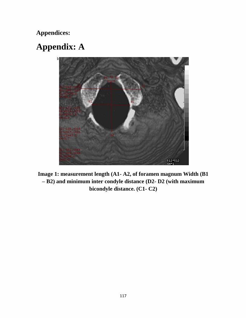
117
Appendices:
Appendix: A
Image 1: measurement length (A1- A2, of foramen magnum Width (B1
– B2) and minimum inter condyle distance (D2- D2 (with maximum
bicondyle distance. (C1- C2)
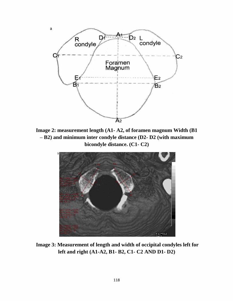
118
Image 2: measurement length (A1- A2, of foramen magnum Width (B1
– B2) and minimum inter condyle distance (D2- D2 (with maximum
bicondyle distance. (C1- C2)
Image 3: Measurement of length and width of occipital condyles left for
left and right (A1-A2, B1- B2, C1- C2 AND D1- D2)
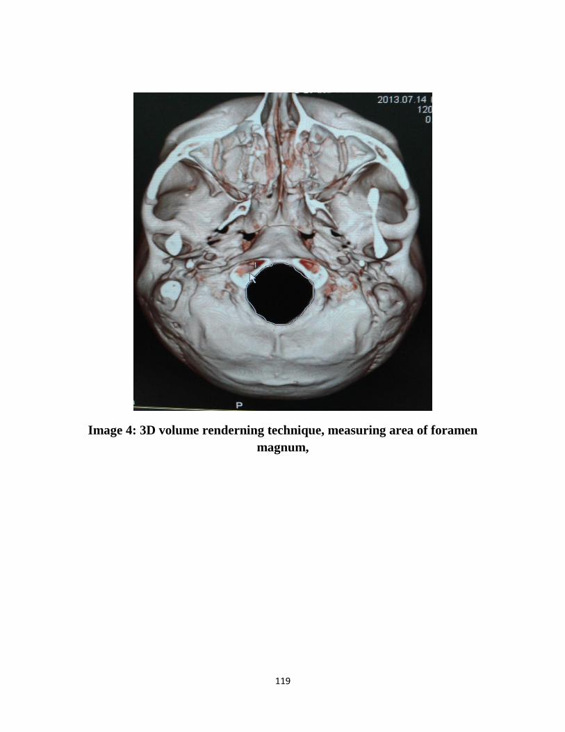
119
Image 4: 3D volume renderning technique, measuring area of foramen
magnum,
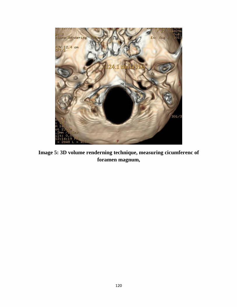
120
Image 5: 3D volume renderning technique, measuring cicumferenc of
foramen magnum,
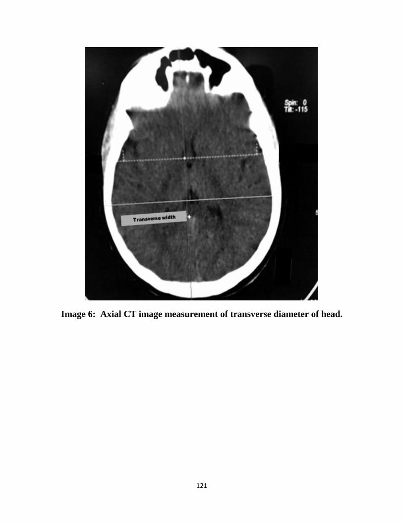
121
Image 6: Axial CT image measurement of transverse diameter of head.
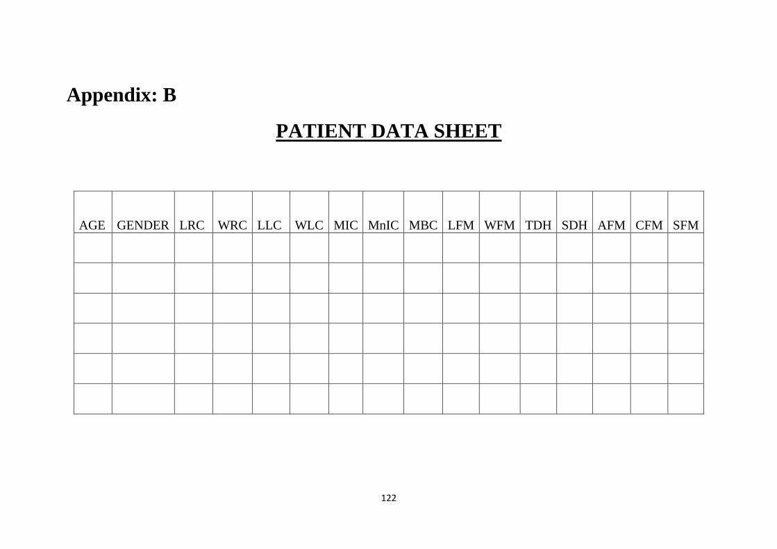
122
Appendix: B
PATIENT DATA SHEET
SFM
CFM AFM SDH TDH WFM LFM MBC MnIC MIC WLC LLC WRC LRC GENDER
AGE
Related Documents