Molecules 2013, 18, 8402-8416; doi:10.3390/molecules18078402 molecules ISSN 1420-3049 www.mdpi.com/journal/molecules Article Characterization of Flavonoids and Phenolic Acids in Myrcia bella Cambess. Using FIA-ESI-IT-MS n and HPLC-PAD-ESI-IT-MS Combined with NMR Luiz L. Saldanha 1 , Wagner Vilegas 2 and Anne L. Dokkedal 3, * 1 Botany Department, Institute of Biosciences, Univ. Estadual Paulista (UNESP), CEP 18618-970, Botucatu, Sao Paulo, Brazil 2 Experimental Campus of the Paulista Coast, Univ. Estadual Paulista (UNESP), CEP 11330-900, Sao Vicente, Sao Paulo, Brazil 3 Biological Science Department, Science Faculty, Univ. Estadual Paulista (UNESP), CEP 17033-360, Bauru, Sao Paulo, Brazil * Author to whom correspondence should be addressed; E-Mail: [email protected]; Tel.: +55-14-3103-6708; Fax: +55-14-3103-6092. Received: 7 June 2013; in revised form: 5 July 2013 / Accepted: 8 July 2013 / Published: 16 July 2013 Abstract: The leaves of Myrcia DC. ex Guill species are used in traditional medicine and are also exploited commercially as herbal drugs for the treatment of diabetes mellitus. The present work aimed to assess the qualitative and quantitative profiles of M. bella hydroalcoholic extract, due to these uses, since the existing legislation in Brazil determines that a standard method must be developed in order to be used for quality control of raw plant materials. The current study identified eleven known flavonoid-O-glycosides and six acylated flavonoid derivatives of myricetin and quercetin, together with two kaempferol glycosides and phenolic acids such as caffeic acid, ethil galate, gallic acid and quinic acid. In total, 24 constituents were characterized, by means of extensive preparative chromatographic analyses, along with MS and NMR techniques. An HPLC-PAD-ESI-IT- MS and FIA-ESI-IT-MS n method were developed for rapid identification of acylated flavonoids, flavonoid-O-glycosides derivatives of myricetin and quercetin and phenolic acids in the hydroalcoholic M. bella leaves extract. The FIA-ESI-IT-MS techinique is a powerful tool for direct and rapid identification of the constituents after isolation and NMR characterization. Thus, it could be used as an initial method for identification of authentic samples concerning quality control of Myrcia spp extracts. OPEN ACCESS

Welcome message from author
This document is posted to help you gain knowledge. Please leave a comment to let me know what you think about it! Share it to your friends and learn new things together.
Transcript
Molecules 2013, 18, 8402-8416; doi:10.3390/molecules18078402
molecules ISSN 1420-3049
www.mdpi.com/journal/molecules
Article
Characterization of Flavonoids and Phenolic Acids in Myrcia bella Cambess. Using FIA-ESI-IT-MSn and HPLC-PAD-ESI-IT-MS Combined with NMR
Luiz L. Saldanha 1, Wagner Vilegas 2 and Anne L. Dokkedal 3,*
1 Botany Department, Institute of Biosciences, Univ. Estadual Paulista (UNESP), CEP 18618-970,
Botucatu, Sao Paulo, Brazil 2 Experimental Campus of the Paulista Coast, Univ. Estadual Paulista (UNESP), CEP 11330-900,
Sao Vicente, Sao Paulo, Brazil 3 Biological Science Department, Science Faculty, Univ. Estadual Paulista (UNESP), CEP 17033-360,
Bauru, Sao Paulo, Brazil
* Author to whom correspondence should be addressed; E-Mail: [email protected];
Tel.: +55-14-3103-6708; Fax: +55-14-3103-6092.
Received: 7 June 2013; in revised form: 5 July 2013 / Accepted: 8 July 2013 /
Published: 16 July 2013
Abstract: The leaves of Myrcia DC. ex Guill species are used in traditional medicine and
are also exploited commercially as herbal drugs for the treatment of diabetes mellitus. The
present work aimed to assess the qualitative and quantitative profiles of M. bella
hydroalcoholic extract, due to these uses, since the existing legislation in Brazil determines
that a standard method must be developed in order to be used for quality control of raw
plant materials. The current study identified eleven known flavonoid-O-glycosides and six
acylated flavonoid derivatives of myricetin and quercetin, together with two kaempferol
glycosides and phenolic acids such as caffeic acid, ethil galate, gallic acid and quinic acid.
In total, 24 constituents were characterized, by means of extensive preparative
chromatographic analyses, along with MS and NMR techniques. An HPLC-PAD-ESI-IT-
MS and FIA-ESI-IT-MSn method were developed for rapid identification of acylated
flavonoids, flavonoid-O-glycosides derivatives of myricetin and quercetin and phenolic
acids in the hydroalcoholic M. bella leaves extract. The FIA-ESI-IT-MS techinique is a
powerful tool for direct and rapid identification of the constituents after isolation and NMR
characterization. Thus, it could be used as an initial method for identification of authentic
samples concerning quality control of Myrcia spp extracts.
OPEN ACCESS

Molecules 2013, 18 8403
Keywords: Brazilian savanna; flavonoid-O-glycosides; medicinal plants; Myrtaceae
1. Introduction
Nowadays, the main concerns about natural medicinal products are effectiveness, safety and the
quality of the herbal drugs [1,2]. Consequently, it is essential to identify and measure all the bioactive
constituents of medicinal plants in order to ensure the biological research reliability and repeatability
as well as to ensure enhancing the quality control over the pharmacological benefits and/or hazardous.
HPLC–MS plays a prominent role as an analytical tool for detecting and identifying pharmacologically
active metabolites and/or reactive metabolites [3,4]. When compared to other detection methods, MS
not just allows determining natural compounds chemical structure with known and unknown
structures, but also offers excellent sensitivity to low amount of samples within relatively short
analysis time as well as plays an important role in screening flavonoids and other phenolics [5–8].
The Brazilian savanna, called Cerrado, is included in the list of global hotspots for conservation due
to its high concentrations of endemic species that had suffered heavy habitat losses [9]. Despite its
relevance, the Brazilian savanna has been continuously destroyed in order to create pastures and crop
fields. Nowadays, in the state of Sao Paulo, southeastern of Brazil, remnants are very reduced and
fragmented [10]. The Myrtaceae is an important family in the Brazilian savanna, with more than
1,000 species countrywide and Myrcia, with 350 species, is one of the richest genus [11]. Myrcia bella
is a common and important species in many savanna fragments, distributed in the state of
Sao Paulo [12,13]. Indigenous and traditional communities in Brazil have used some species of this
genus as astringent, against diabetes, diarrhea, diuretic, to stop bleeding, against hypertension and
ulcers [14,15]. Most phytochemical studies on the Myrcia species are related to essential oils [16–20];
however Yoshikawa et al. [21] and Matsuda et al. [22] described the features of flavanone glucosides,
acylated flavanone glucosides, acetophenones, flavonols and gallic acid in the leaves of
Myrcia multiflora (Lam.) DC.
The present work aimed to assess the qualitative and quantitative profiles of M. bella hydroalcoholic
extract, due to the potential use of the Myrcia species in tradittional medicine and also due to its
comercial exploration as an herbal drug to be used in the treatment of diabetes mellitus [15,23,24] once
the existing legislation in Brazil determines that a standard method must be developed in order to be
used for quality controlling raw plant materials [25].
2. Results and Discussion
2.1. Identification of Constituents by Combination of NMR and FIA-ESI-IT-MS/MSn
In total, 24 constituents (Figure 1) were identified in the 70% EtOH extract. Eighteen of them were
isolated and characterized by UV, MS and NMR spectral data and six were tentatively identified
considering retention time values, co-chromatography with authentic samples, UV and MS spectral
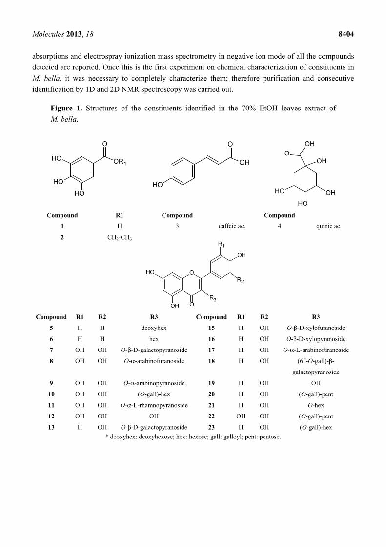
data. In Figure 2, the HPLC-PAD chromatogram of the M. bella 70% EtOH leaves extract is presented.
Data concerning identification of the peaks are shown in Table 1, where the retention time, UV–vis
Molecules 2013, 18 8404
absorptions and electrospray ionization mass spectrometry in negative ion mode of all the compounds
detected are reported. Once this is the first experiment on chemical characterization of constituents in
M. bella, it was necessary to completely characterize them; therefore purification and consecutive
identification by 1D and 2D NMR spectroscopy was carried out.
Figure 1. Structures of the constituents identified in the 70% EtOH leaves extract of
M. bella.
OH
O
OR1OH
OH
O
OH
OH
OH
OH
OH
OHO
OH
Compound R1 Compound Compound
1 H 3 caffeic ac. 4 quinic ac.
2 CH2-CH3
O
OH
O
OH
R3
R1
OH
R2
Compound R1 R2 R3 Compound R1 R2 R3
5 H H deoxyhex 15 H OH O-β-D-xylofuranoside
6 H H hex 16 H OH O-β-D-xylopyranoside
7 OH OH O-β-D-galactopyranoside 17 H OH O-α-L-arabinofuranoside
8 OH OH O-α-arabinofuranoside 18 H OH (6′′-O-gall)-β-
galactopyranoside
9 OH OH O-α-arabinopyranoside 19 H OH OH
10 OH OH (O-gall)-hex 20 H OH (O-gall)-pent
11 OH OH O-α-L-rhamnopyranoside 21 H OH O-hex
12 OH OH OH 22 OH OH (O-gall)-pent
13 H OH O-β-D-galactopyranoside 23 H OH (O-gall)-hex
* deoxyhex: deoxyhexose; hex: hexose; gall: galloyl; pent: pentose.
Molecules 2013, 18 8405
Figure 2. HPLC-PAD analytical chromatogram of 70% EtOH leaves extract of
Myrcia bella with identified peaks. Experimental conditions: eluents A (MeOH + 0.1%
form. ac.) and B (H2O + 0.1% Form. ac.). Gradient system: 10-45% de A em B em
200 min. Column: Phenomenex® Luna C18 (250 × 4.6 mm i.d., 5 μm). Flow rate:
0.8 mL·min−1, λ = 254 nm. Injected volume: 20 μL.
Time (min)
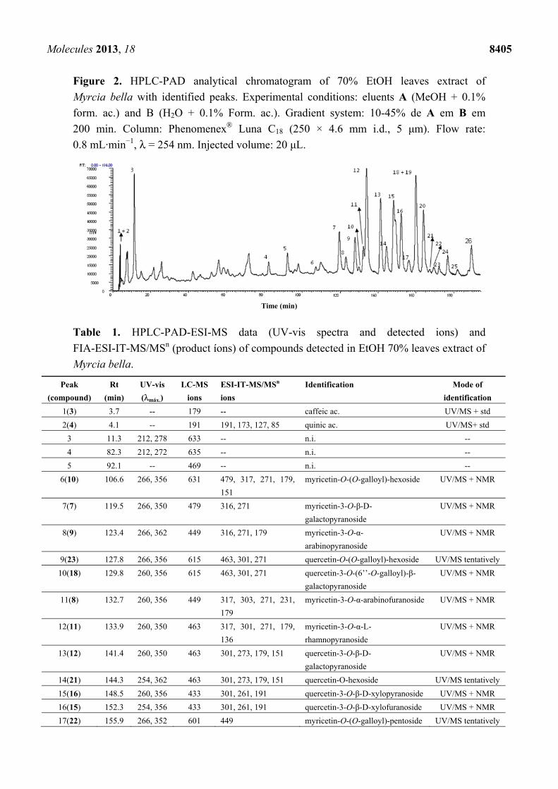
Table 1. HPLC-PAD-ESI-MS data (UV-vis spectra and detected ions) and
FIA-ESI-IT-MS/MSn (product íons) of compounds detected in EtOH 70% leaves extract of
Myrcia bella.
Peak
(compound)
Rt
(min)
UV-vis
(λmáx.)
LC-MS
ions
ESI-IT-MS/MSn
ions
Identification Mode of
identification
1(3) 3.7 -- 179 -- caffeic ac. UV/MS + std
2(4) 4.1 -- 191 191, 173, 127, 85 quinic ac. UV/MS+ std
3 11.3 212, 278 633 -- n.i. --
4 82.3 212, 272 635 -- n.i. --
5 92.1 -- 469 -- n.i. --
6(10) 106.6 266, 356 631 479, 317, 271, 179,
151
myricetin-O-(O-galloyl)-hexoside UV/MS + NMR
7(7) 119.5 266, 350 479 316, 271 myricetin-3-O-β-D-
galactopyranoside
UV/MS + NMR
8(9) 123.4 266, 362 449 316, 271, 179 myricetin-3-O-α-
arabinopyranoside
UV/MS + NMR
9(23) 127.8 266, 356 615 463, 301, 271 quercetin-O-(O-galloyl)-hexoside UV/MS tentatively
10(18) 129.8 260, 356 615 463, 301, 271 quercetin-3-O-(6’’-O-galloyl)-β-
galactopyranoside
UV/MS + NMR
11(8) 132.7 260, 356 449 317, 303, 271, 231,
179
myricetin-3-O-α-arabinofuranoside UV/MS + NMR
12(11) 133.9 260, 350 463 317, 301, 271, 179,
136
myricetin-3-O-α-L-
rhamnopyranoside
UV/MS + NMR
13(12) 141.4 260, 350 463 301, 273, 179, 151 quercetin-3-O-β-D-
galactopyranoside
UV/MS + NMR
14(21) 144.3 254, 362 463 301, 273, 179, 151 quercetin-O-hexoside UV/MS tentatively
15(16) 148.5 260, 356 433 301, 261, 191 quercetin-3-O-β-D-xylopyranoside UV/MS + NMR
16(15) 152.3 254, 356 433 301, 261, 191 quercetin-3-O-β-D-xylofuranoside UV/MS + NMR
17(22) 155.9 266, 352 601 449 myricetin-O-(O-galloyl)-pentoside UV/MS tentatively

Molecules 2013, 18 8406
Table 1. Cont.
Peak
(compound)
Rt
(min)
UV-vis
(λmáx.)
LC-MS
ions
ESI-IT-MS/MSn
ions
Identification Mode of
identification
18 159.7 -- 467 -- n.i. --
19(17) 160.0 266, 356 433 301, 191 quercetin-O-α-L-arabinofuranoside UV/MS + NMR
20(14) 164.0 254, 350 447 301, 271, 255, 179 quercetin-3-O-α-L-
rhamnopyranoside
UV/MS + NMR
21 163.8 -- 483 -- n.i. --
22 169.2 -- 477 -- n.i. --
23(24) 171,0 266, 356 615 463, 317 myricetin-O-(O-galloyl)-
deoxyhexoside
UV/MS tentatively
24(20) 176.8 260, 356 585 433, 301, 179, 151 quercetin- O-(O-galloyl)-pentoside UV/MS + NMR
25 181,0 -- 631 -- n.i. --
26(19) 189.4 254, 374 301 137 quercetin UV/MS + NMR
* n.i.: not identified; std: standard.
Diagnostics mass fragments obtained by FIA-ESI-IT-MS in the negative mode at 285, 301 and 317
characterized aglycones as kaempferol, quercetin and myricetin, respectively. The precursor ion at
169 mu characterized gallic acid. The neutral losses of 132, 162, 146 and 152 mass units allowed the
identification of pentosides (xylose or arabinose), hexosides (glucose or galactose), deoxyhexoside
(rhamnose) and gallic acid respectively. The values of m/z lower than the aglycone (i.e., m/z < 317)
like m/z 179 (1,2A−), 151 (1,3A−) and 137 (1,2B−) are typical of retro Dies-Alder (RDA) reactions of
flavon-3-ols having a dihydroxylated A ring and m/z 137 is a typical fragment of the trihydroxylated B
ring [26].
The HPLC-PAD analysis of the chromatogram peaks with bands at 210–278 nm (0–100 min) were
related to the presence of phenolic acids and bands at 240–285 nm and 350–380 nm (100–180 min)
were related to flavonoids.
The signals in m/z 169 e 197 were diagnostic for compounds 1 and 2 respectively. The 1H-NMR
spectra were consistent with the data obtained by MS. 1H-NMR experiments of compounds 11 and 14
presented diagnostic signals at δ 0.83 and 13C-NMR signal at δ 17.51 for rhamnoses [27,28]. The
anomeric proton signal indicated the configuration α-L-rhamnopyranoside and HMBC experiments of
compound 14 showed the anomeric proton correlation at δ 5.25 with C3 at δ 134.1, confirming the
position of sugar linkage. 1H-NMR data of compounds 15, 16 and 17 were in agreement with typical quercetin chemical
shifts. The anomeric proton of compound 17 at δ 5.18 (J = 3.5 Hz) indicated the presence of
α-L-arabinofuranoside. The anomeric proton at δ 5.32 (J = 7.3 Hz) indicated the presence of
β-D-xylopyranoside in compound 16 and at δ 5.27 (J = 1.5 Hz) the presence of β-D-xylofuranoside in
compound 15. 13C-NMR data of sugar unit were in agreement to the literature [29–32].
The loss of 152 Da in MS spectra of compound 18 suggested the presence of a galloyl unit. 13C-NMR signals were in agreement to MS data and confirm the presence of galloyl unit [29].
In addition to the experiments which led to the isolation and identification of compounds, an
analysis was also carried out to confirm the chemical composition of the 70% EtOH extract employing
the combination of multi-stage analysis of selected ions by ESI-IT-MS/MSn and HPLC-PAD-ESI-MS.
The main objective of this analysis was to evaluate the ability of this technique to produce spectral

Molecules 2013, 18 8407
information on the chemical constitution sample analyses quickly and directly, without the need for
pretreatment steps and/or chromatographic separations. Six constituents (compounds 3–4 and 21–24)
were tentatively identified. Within the exception of the isolated compounds 1, 2, 5, 6 and 12, all other
compounds were simultaneously identified by HPLC-PAD-ESI-MS. The typical precursor ions
spectrum in negative ion mode of 70% EtOH leaves extract of M. bella is presented in Figure 3.
Figure 3. Typical direct flow injection analysis FIA-ESI-IT-MS fingerprint spectra
obtained in negative ion mode of the 70% EtOH from the leaves of M. bella.
(♦) Representative constituents fragmented. For conditions, see Material and Methods part.
The precursor ion at m/z 197 [M – H]− was consistent with the presence of compound 2. The
fragmentation of the precursor ion at m/z 169 [M – H]− produced the ion at m/z 125 [M – 44 – H]−, due
the loss of 44 Da (COO−), thus confirming the presence of compound 1 [33]. The precursor ion at m/z
191 [M – H]− produced fragment ions at m/z 172, m/z 127 and m/z 85. This pattern of fragmentation
led to the identification of quinnic acid (4). Precursor ions at m/z 301 [M – H]− and m/z 317 [M – H]−
in the precursor ion spectrum were in agreement to the presence of compounds 21 and 14.
Figure 4 presents the MSn fragmentation of representative compounds from M. bella. The
nomenclature for the flavonoids was that of Ma et al. [26] and product ions from glycoconjugates were
denoted according to the nomenclature introduced by Domon and Costello [27].
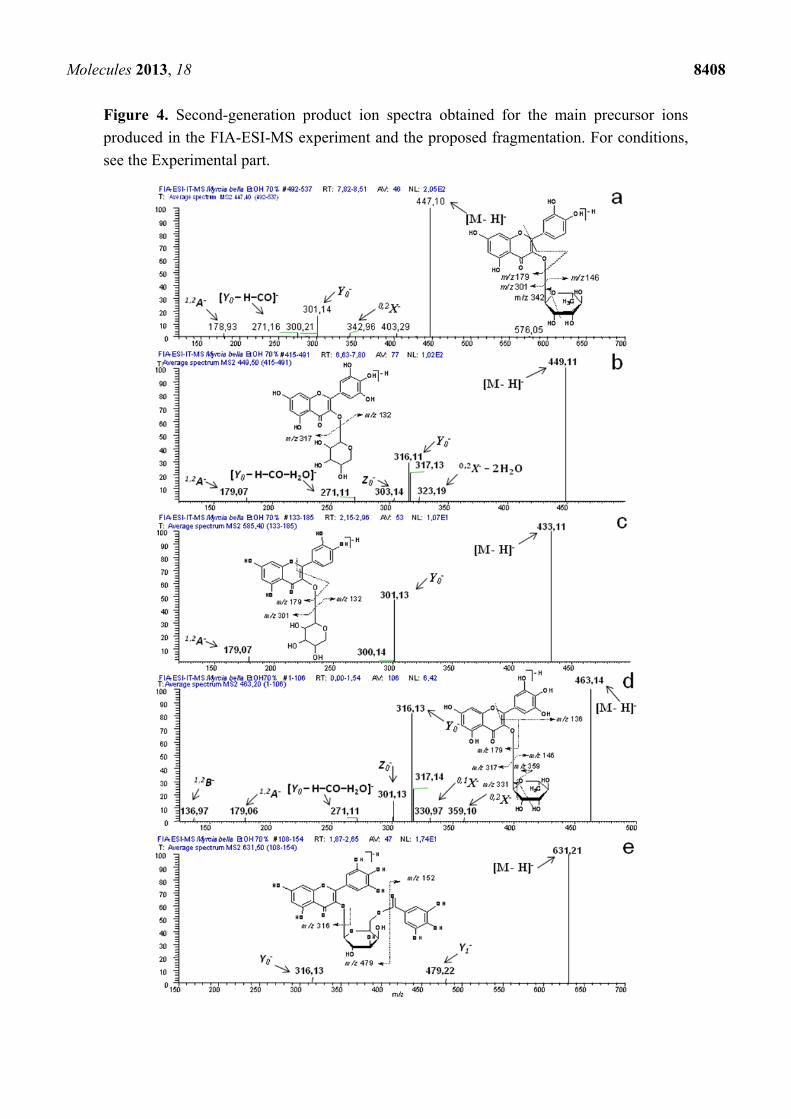
Second-generation product ion spectra of precursor ion at m/z 447 [M – H]− (Figure 4a), produced
the product ion Y0− at m/z 301 [M – 146 – H]− and the diagnostic product ion 0,2X− due to the loss of
104 Da resulting from the cleavage of the sugar unit, typical of deoxyhexoses [28] as well as
[Y0 – H – CO]− at m/z 271 typical of flavon-3-O-monoglycoside [34] and at m/z 179 from the RDA of
ring A. Such fragments confirmed the presence of compound 14.
The second-generation of the precursor ion at m/z 449 [M – H]− (Figure 4b) produced the product
ion Y0− at m/z 316 [M – 132 – 2H]− and 0,2X− – 2H2O at m/z 323 after loss of 126 Da, typical of
pentoses, as well as the product ions 1,2A− at m/z 179, [Y0 – H – CO – H2O]− at m/z 271 and Z0− at m/z
303, resulting from the RDA of ring A, thus confirming the presence of compounds 8 and 9.
Molecules 2013, 18 8408
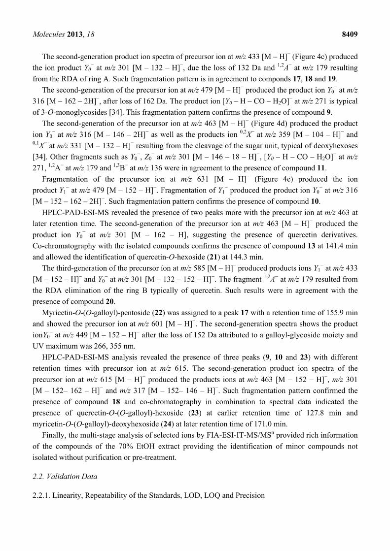
Figure 4. Second-generation product ion spectra obtained for the main precursor ions
produced in the FIA-ESI-MS experiment and the proposed fragmentation. For conditions,
see the Experimental part.
Molecules 2013, 18 8409
The second-generation product ion spectra of precursor ion at m/z 433 [M – H]− (Figure 4c) produced
the ion product Y0− at m/z 301 [M – 132 – H]−, due the loss of 132 Da and 1,2A− at m/z 179 resulting
from the RDA of ring A. Such fragmentation pattern is in agreement to componds 17, 18 and 19.
The second-generation of the precursor íon at m/z 479 [M – H]− produced the product ion Y0− at m/z
316 [M – 162 – 2H]−, after loss of 162 Da. The product ion [Y0 – H – CO – H2O]− at m/z 271 is typical
of 3-O-monoglycosides [34]. This fragmentation pattern confirms the presence of compound 9.
The second-generation of the precursor ion at m/z 463 [M – H]− (Figure 4d) produced the product
ion Y0− at m/z 316 [M – 146 – 2H]− as well as the products ion 0,2X− at m/z 359 [M – 104 – H]− and
0,1X− at m/z 331 [M – 132 – H]− resulting from the cleavage of the sugar unit, typical of deoxyhexoses
[34]. Other fragments such as Y0−, Z0
− at m/z 301 [M – 146 – 18 – H]−, [Y0 – H – CO – H2O]− at m/z
271, 1,2A− at m/z 179 and 1,3B− at m/z 136 were in agreement to the presence of compound 11.
Fragmentation of the precursor ion at m/z 631 [M – H]− (Figure 4e) produced the ion
product Y1− at m/z 479 [M – 152 – H]−. Fragmentation of Y1
− produced the product ion Y0− at m/z 316
[M – 152 – 162 – 2H]−. Such fragmentation pattern confirms the presence of compound 10.
HPLC-PAD-ESI-MS revealed the presence of two peaks more with the precursor ion at m/z 463 at
later retention time. The second-generation of the precursor ion at m/z 463 [M – H]− produced the
product ion Y0− at m/z 301 [M – 162 – H], suggesting the presence of quercetin derivatives.
Co-chromatography with the isolated compounds confirms the presence of compound 13 at 141.4 min
and allowed the identification of quercetin-O-hexoside (21) at 144.3 min.
The third-generation of the precursor íon at m/z 585 [M – H]− produced products ions Y1− at m/z 433
[M – 152 – H]− and Y0− at m/z 301 [M – 132 – 152 – H]−. The fragment 1,2A− at m/z 179 resulted from
the RDA elimination of the ring B typically of quercetin. Such results were in agreement with the
presence of compound 20.
Myricetin-O-(O-galloyl)-pentoside (22) was assigned to a peak 17 with a retention time of 155.9 min
and showed the precursor ion at m/z 601 [M – H]−. The second-generation spectra shows the product
ionY0− at m/z 449 [M – 152 – H]− after the loss of 152 Da attributed to a galloyl-glycoside moiety and
UV maximum was 266, 355 nm.
HPLC-PAD-ESI-MS analysis revealed the presence of three peaks (9, 10 and 23) with different
retention times with precursor ion at m/z 615. The second-generation product ion spectra of the
precursor ion at m/z 615 [M – H]− produced the products ions at m/z 463 [M – 152 – H]−, m/z 301
[M – 152– 162 – H]− and m/z 317 [M – 152– 146 – H]−. Such fragmentation pattern confirmed the
presence of compound 18 and co-chromatography in combination to spectral data indicated the
presence of quercetin-O-(O-galloyl)-hexoside (23) at earlier retention time of 127.8 min and
myricetin-O-(O-galloyl)-deoxyhexoside (24) at later retention time of 171.0 min.
Finally, the multi-stage analysis of selected ions by FIA-ESI-IT-MS/MSn provided rich information
of the compounds of the 70% EtOH extract providing the identification of minor compounds not
isolated without purification or pre-treatment.
2.2. Validation Data
2.2.1. Linearity, Repeatability of the Standards, LOD, LOQ and Precision

Molecules 2013, 18 8410
All compounds showed good linearity. The following r2 values were obtained: gallic acid
r2 = 0.9992 (regression curve: y = 75639x −2.2237) and quercetin r2 = 0.9996 (regression curve:
y = 87488x −3.31288). LOD for gallic acid was 2.27 µg·mL−1 and LOQ was 6.8 µg·mL−1 (10 μL of
injection). For quercetin, LOD was calculated as 1.57 µg·mL−1 and LOQ was 4.75 µg·mL−1.
The repeatability, based on three samples with known concentration, was analyzed by HPLC and
the relative standard deviation (% RSD) of the standards was calculated. RSD values ranged between
0.32 and 3%. The overall intraday time variations of the standards were less than 0.32–2.40% for gallic
acid and 0.39–3.0% for quercetin and interday time variations were less than 0.52–2.32% for gallic
acid and 0.47–1.47% for quercetin. Precision data are displayed in Table 2.
Table 2. Precision data of the two analytes, expressed as RSD (%).
Analytes Concentration
(μg·mL−1)
Precision
Intra-day (mean ± SD) RSD % Inter-day (mean ± SD) RSD %
Gallic ac. 50 41.66 ± 1.0 2.40 35.93 ± 0.85 2.36 (n = 3) 100 77.53 ± 0.25 0.32 74.16 ± 0.49 0.66
200 143.2 ± 0.95 0.66 142.13 ± 0,75 0.52
Quercetin 50 45.23 ± 0.30 0.66 42.63 ± 0.63 1.47 (n = 3) 100 85.2 ± 2.6 3.0 86.43 ± 0.81 0.93
200 163.36 ± 0.64 0.39 169.73 ± 0.8 0.47
Table 3. Estimative of contents of phenolic acids and flavonoids glycosides derivatives in
70% EtOH leaves extract (n = 3) of M. bella expressed by the use of gallic acid and
quercetin linear regression data.
Compound Concentration ± SD (µg.mL−1) Standard
gallic ac. 12.13 ± 2.35 GA n.i. 7.19 ± 0.87 GA n.i. 8.07 ± 0.71 GA n.i. 8.40 ± 0.57 GA Myricetin-3-O-β-D-galactopyranoside 9.92 ± 0.91 Q Quercetin-O-(6’’-O-galloyl)-β-galactopyranoside 5.46 ± 0.65 Q Quercetin-3-O-β-D-galactopyranoside 21.82 ± 2.05 Q Quercetin-O-hexoside 11.09 ± 1.05 Q Quercetin-3-O β-D-xylopyranoside 7.01 ± 0.78 Q Quercetin-3-O-β-D-xylofuranoside 14.06 ± 1.33 Q Quercetin-3-O-α-L-arabinofuranoside 29.99 ± 3.37 Q Quercetin-O-α-L-rhamnopyranoside 15.81 ± 1.49 Q Quercetin 9.83 ± 1.47 Q Phenolic acid estimative 35.80 Flavonoids estimative 129.02
* n.i.: compound not identified; GA = gallic acid, Q = quercetin; Contents (µg·mL−1) corresponding to
10 mg·mL−1 of 70% EtOH leaves extract sample.
2.3. Quantitative Analysis
Molecules 2013, 18 8411
In a first look at the chromatogram it became evident that the main constituents were quercetin
glycosides derivatives containing one sugar unit. In order to make the most pratical method, we used
two reference standards available on the market. Quercetin glycosides derivatives were expressed as
quercetin, whereas phenolic acids derivatives were expressed as gallic acid. The quantitative analysis
results are reported in Table 3.
3. Material and Methods
3.1. Solvents and Chemicals
The solvents used on HPLC analysis were HPLC grade; MeOH and formic acid (85% v/v) for
HPLC were purchased from Merck (Sao Paulo, Brazil). Water was purified by a Milli-Q plus system
from Millipore®. PTFE membrane filter (0.45 mm) was also purchased from Merck. All laboratory
chemicals used in the current study in the isolation protocol were of reagent grade.
3.2. Plant Material
Samples of M. bella leaves were collected in November 2010 at the Botanical Garden of Bauru
(22°20'30" S e 49°00'30" W)—SP, Brazil. Voucher specimens were prepared and identified by A. L.
Dokkedal and stored at the Herbarium of the UNESP—Univ Estadual Paulista “Júlio de Mesquita
Filho”—UNBA (Bauru—SP, Brazil) under code number 5508.
3.3. Standards
The following standards were used for quantitative analysis: gallic acid and quercetin, which were
purchase from Sigma-Aldrich (Sao Paulo, Brazil).
3.4. Isolation of the Characteristic Constituents
Fresh leaves were dried at 40 °C for 48 h. The separated powdered leaves (1.3 kg) were extracted
with EtOH-H2O (7:3) by percolation at room temperature for 2 months [35]. The ethanolic solution
was filtered and concentrated to dryness under reduced pressure at 40 °C furnishing 364 g of the
hydroalcaholic extract (70% EtOH); a portion (15 g) of this extract was redissolved in MeOH-H2O
(1:4) and partitioned with equal volumes of n-hexane, dichloromethane, n-butanol and water (Fr1–Fr4)
to obtain four major fractions. HPLC-PAD analysis showed that fraction Fr3 was richer in flavonoid
glycosides. Fraction Fr3 (3 g) was dissolved in 15 mL of MeOH and subjected to size exclusion
chromatography using a Sephadex LH-20 column (85 × 2.5 cm; H × i.d.) with peristaltic pump and
automatic collector Redifrac using MeOH as mobile phase yielding 366 fractions (10 mL each)
(Mb1–Mb366). Fractions Mb77-Mb366 were combined by similarity and were subjected to purification
by semipreparative HPLC-RI (Knauer® 2300) using a C18 (250 × 10.0 mm i.d.; 5 μm) column and as
mobile phase MeOH-H2O (4:6). Successive chromatographic steps of the above subfractions afforded
9 mg of gallic acid (1), 7 mg of ethyl gallate (2), 3 mg of kaempferol-O-deoxyhexoside (5), 2 mg of
kaempferol-O-hexoside (6), 12 mg of myricetin-3-O-β-D-galactopyranoside (7), 4 mg of myricetin-3-
O-α-arabinofuranoside (8), 26 mg myricetin-3-O-α-arabinopyranoside (9), 3 mg of myricetin-O-(O-
Molecules 2013, 18 8412
galloyl)-hexoside (10), 16 mg of myricetin-3-O-α-L-rhamnopyranoside (11), 18 mg myricetin (12),
19 mg of quercetin-3-O-β-D-galactopyranoside (13), 25 mg of quercetin-3-O-α-L-rhamnopyranoside
(14), 8 mg of quercetin-3-O-β-D-xylofuranoside (15), 5 mg of quercetin-3-O-β-D-xylopyranoside (16),
13 mg of quercetin-3-O-α-L-arabinofuranoside (17), 16 mg of quercetin-3-O-(6’’galloyl)-β-galacto-
pyranoside (18), 15 mg of quercetin (19) and 4 mg of quercetin-O-(O-galloyl)-pentoside (20).
3.5. HPLC-PAD Analysis Instrumentation
The HPLC system consisted of a PU-2089S Plus (Jasco®) pump equipped with a MD-2015 Plus
Photodiode Array Detector (PAD, Jasco®) and AS-2055 automatic injector (Jasco®). The column was
a Luna C18 column (250 × 4.6 mm, i.d.) with a particle size of 5 mm (Phenomenex®) maintained at
35 °C, and managed by the Jasco ChromPass software. The eluents were: A (MeOH + 0.1% formic
acid) and B (H2O + 0.1% formic acid.). The gradient condition was: 5–45% of A in B in 190 min.
Injected volume of the samples was 20 μL solution. The UV–vis spectra were recorded between 200
and 600 nm, and the chromatographic profiles were registered at 254, 280 and 360 nm.
3.6. FIA-ESI-IT-MS/MSn and HPLC-ESI-IT-MS Analysis Instrumentation
The chromatographic profile of the crude extract of M. bella was performed using Accela High
Speed LC (Thermo Scientific®, San Jose, CA, USA), Phenomenex® Luna C18 (250 × 4.6 mm i.d.; 5
μm) column and (Phenomenex®) 4 × 3 mm i.d., column guard, with PAD and coupled to an Accela
(Thermo Scientific®) LCQ Fleet with Ion Trap (IT) 3D and ionization by eletrospray (ESI). Mobile
phase was Water ultra pure (eluent A) and Methanol (eluent B), both containing 0.1% of formic acid.
The ratio was: 10–45 of A in B in 190 min. Injection volume: 20.0 μL; Column temperature: 25 °C;
Flow ratio: 0.8 mL·min−1; the chromatogram was monitored at 254 nm. The effluent from the HPLC
was directed into the ESI probe.
Using this method, we determined the most intense parent ion for each peak in the chromatogram.
A second event, FIA-ESI-IT-MS in the negative ion mode, was performed using the same equipment
described above, equipped with Xcalibur software. The 70% EtOH extract was dissolved in
MeOH-H2O (8:2) and infused in the ESI source by flow injection analysis (FIA) using a syringe pump;
the flow rate was 33 μL·min−1. The capillary voltage was −20 V, the spray voltage was 4 kV, and the
tube lens offset was −55 V. The capillary temperature was 275 °C. Nitrogen was used both as drying
gas at a flow rate of 60 (arbitrary units) and as nebulising gas. The nebulizer temperature was set at
280 °C, and a potential of −4 V was used on the capillary. Negative ion mass spectra were recorded in
the range m/z 100–1,550. Two scan events were prescribed to run in the LCQ mass spectrometry. The
first event was a full-scan spectrum to acquire data on the deprotonated compounds within the scan
range. The second scan event was a MS/MS experiment performed using a datadependent scan on
deprotonated molecule [M − H]−. The collision energy for MS/MS was adjusted to 10–25%.
3.7. Identification of Peaks and Peak Purity
Identification of all constituents was performed by HPLC-PAD and MS analysis by comparing the
retention time, the UV and MS spectra of the peaks in the samples with those of authentic reference
Molecules 2013, 18 8413
samples or isolated compounds. The purity of peaks was checked by a PAD coupled to the HPLC
system, comparing the UV spectra of each peak with those of authentic references samples and/or by
examination of the MS spectra.
3.8. Linearity Limitation of Detection, Limitation of Quantification and Precision
The optimized HPLC-PAD method was validated for the simultaneous analysis of gallic acid and
quercetin in terms of linearity, limit of detection, limit of quantification, precision and accuracy. The
calibration curves were obtained by the external standard method on eight levels of concentration of
standard mixtures, with three injections per level. Chromatogram peak areas at 280 nm for gallic acid
and at 360 nm for quercetin were plotted against the known concentrations of the standard solutions to
establish the calibration equations. A linear regression equation was calculated by the least squares
method. The detection limit (LOD) and limit of quantification (LOQ) were calculated from the
residual standard deviation of the regression (σ) line and the slope (S) as follows: LOD = 3.3σ/S;
LOQ = 10σ/S. Three different concentrations of standard mixtures (0.05; 0.1 and 0.2 mg·mL−1) were
used for intra- and interday precision testing. The areas under curves and retention times of the three
consecutive injections, performed at each concentration on three different days, were used to calculate
% RSD (relative standard deviation) interday precision. Intraday precision data for peak areas and
retention times were calculated from six non-consecutive injections, performed at each concentration
on the same day.
3.9. Quantitative Determination of Constituents
The method of external standard was applied to quantify each compound. Quantification of
individual constituents was performed using a regression curve, each point in triplicate. Measurements
were performed at 360 nm, which is the maximum absorbance for flavonols and 280 nm for
phenolic acids.
3.10. NMR Analysis
The 1H-NMR and 13C-NMR 1D and 1H-NMR 2D-NMR 13C g-HMBC experiments were performed
on a Bruker® 300 MHz (7.0 T) nuclear magnetic resonance spectrometer and/or on a Varian Inova®
500 MHz (11.7 T) nuclear magnetic resonance spectrometer (for sample preparation for the NMR
experiments pyridine-d5 and dimethyl sulfoxide (DMSO-d6, Cambridge Isotope Laboratories, Inc.,
Andover, MA, USA) were used.
4. Conclusions
The qualitative and quantitative profiles of the 70% EtOH extract from the leaves of M. bella were
analyzed. The combined NMR, FIA-ESI-IT-MS/MSn and HPLC-PAD-ESI-MS techniques allowed the
unambiguously identification of 24 compounds. Eighteen constituents were isolated and confirmed by
NMR and/or MS analysis and six were tentatively identified. Main secondary metabolites were
flavonoid glycosides, mainly derivatives of quercetin and myricetin. The HPLC-PAD analysis also
revealed the presence of peaks with typical UV spectra of phenolic acids derivatives in the
chromatogram. The genus is reported to contain flavonol-O-glycosides of myricetin and quercetin as
Molecules 2013, 18 8414
well as flavanones glucosides and acetophenone glucosides in polar extracts [20–22]. The FIA-ESI-IT-MS
techinique is a powerful tool for direct and rapid identification of the constituents after isolation and
NMR characterization. Thus, it could be used as a starting method for identification of authentic
samples for the purposes of quality control of Myrcia spp extracts.
Acknowledgements
The authors would like to thank Valdecir Farias Ximenes for the HPLC-PAD equipment support,
grant #09/52237-9, São Paulo Research Foundation (FAPESP) for financial support and Coordenação
de Aperfeiçoamento de Pessoal de Nível Superior (CAPES) for the fellowship to LLS.
Conflict of Interest
The authors declare no conflict of interest.
References
1. Calixto, J.B. Efficacy, safety, quality control, marketing and regulatory guidelines for herbal
medicines (phytotherapeutic agents). Braz. J. Med. Biol. Res. 2000, 33, 179–189.
2. Mosihuzzaman, M.; Choudhary, M.I. Protocols on safety, efficacy, standardization, and
documentation of herbal medicine (IUPAC Technical Report). Pure Appl. Chem. 2008, 80,
2195–2230.
3. Wolfender, J.L. HPLC in natural product analysis: The detection issue. Planta Med. 2009, 75,
719–734.
4. Xing, J.; Xie, C.F.; Lou, H.X. Recent applications of liquid chromatography-mass spectrometry in
natural products bioanalysis. J. Pharm. Biomed. Anal. 2007, 44, 368–378.
5. Gouveia, S.C.; Castilho, P.C. Characterization of phenolic compounds in Helichrysum melaleucum
by high-performance liquid chromatography with on-line ultraviolet and mass spectrometry
detection. Rapid Commun. Mass Spectrom. 2010, 24, 1851–1868.
6. Huang, X.; Liu, Y.; Song, F.R.; Liu, Z.Q.; Liu, S.Y. Studies on principal components and
antioxidant activity of different Radix astragali samples using high performance liquid
chromatography/electrospray ionization multiple-stage tandem mass spectrometry. Talanta 2009,
78, 1090–1101.
7. Rauter, A.P.; Martins, A.; Lopes, R.; Ferreira, J.; Serralheiro, L.M.; Araújo, M.E.; Borges, C.;
Justino, J.; Silva, F.V.; Goulart, M.; et al. Bioactivity studies and chemical profile of the
antidiabetic plant Genista tenera. J. Ethnopharmacol. 2009, 122, 384–393.
8. Tiberti, L.A.; Yariwake, J.H.; Ndjoko, K.; Hostettmann, K. Identification of flavonols in leaves of
Maytenus ilicifolia and M. aquifolium (Celastraceae) by LC/UV/MS analysis. J. Chromatogr. B
2007, 846, 378–384.
9. Myers, N.; Mittermeier, R.A.; Mittermeier, C.G.; Fonseca, G.A.B.; Kent, J. Biodiversity hotspots
for conservation priorities. Nat. Lond. 2000, 403, 853–858.
Molecules 2013, 18 8415
10. Durigan, G.; Franco, G.A.D.C.; Siqueira, M.F. The Vegetation of Cerrado Remnant in São Paulo
State. In Viability of Conservation of Cerrado Remnants São Paulo State; Bitencourt, M.D.,
Mendonça, R.R., Eds.; Annablume: São Paulo, Brazil, 2004; pp. 29–56.
11. Landrum, L.R.; Kawasaki, M.L. The genera of Myrtaceae in Brazil—An illustrated synoptic
treatment and identification keys. Brittonia 1997, 49, 508–536.
12. Carvalho, M.B.; Ishara, K.L.; Maimoni-Rodella, R.C.S. Vascular Flora of a Cerrado sensu stricto
remnant in Pratânia, state of São Paulo, southeastern Brazil. Check List: J. Species Lists and
Distribution 2010, 6, 350–357.
13. Ishara, K.L.; Déstro, G.F.G.; Maimoni-Rodella, R.C.S.; Yanagizawa, Y.A.N.P. Floristic
composition of sensu stricto cerrado remnants in Botucatu, SP. Brazilian J. Botany 2008, 31,
575–586.
14. Hashimoto, G. Illustrade Cyclopedia of Brazilian Medicinal Plants; Aboc-Sha: Kamakura, Japan,
1996.
15. Russo, E.M.K.; Reichelt, A.A.J.; De-sá, J.R.; Furlanetto, R.P.; Moisés, R.C.S.; Kasamatsu, T.S.;
Chacra, A.R. Clinical trial of Myrcia uniflora and Bauhinia forficata leaf extracts in normal and
diabetic patients. Brazilian J. Med. Biol. Res. 1990, 23, 11–20.
16. Cerqueira, M.D.; Souza-Neta, L.; Passos, M.G.V.M.; Lima, E.O.; Roque, N.F.; Martins, D.;
Guedes, M.L.S.; Cruz, F.G. Seasonal variation and antimicrobial activity of Myrcia myrtifolia
essential oils. J. Braz. Chem. Soc. 2007, 18, 998–1003.
17. Cole, R.A.; Haber, W.A.; Setzer, W.N. The leaf oil composition of Myrcia splendens from
Monteverde, Costa Rica. J. Essent. Oil Bear. Plants 2008, 11, 41–44.
18. Henriques, A.T.; Sobral, M.; Bridi, R.; Vérin, P.; Menut, C.; Lamaty, G.; Bessière, J.M. Essential
oils from five southern brazilian species of Myrcia (Myrtaceae). J. Essent. Oil Res. 1997, 9,
13–18.
19. Limberger, R.P.; Sobral, M.; Henriques, A.T.; Menut, C.; Bessière, J. Óleos voláteis de espécies
de Myrcia nativas do Rio Grande do Sul. Quim. Nova 2004, 27, 916–919.
20. Reynertson, K.A.; Yang, H.; Jiang, B.; Basile, M.J.; Kennelly, E.J. Quantitative analysis of
antiradical phenolic constituents from fourteen edible Myrtaceae fruits. Food Chem. 2008, 109,
883–890.
21. Yoshikawa, M.; Shimada, H.; Nishida, N.; Li, Y.; Toguchida, I.; Yamahara, J.; Matsuda, H.
Antidiabetic principles of natural medicines. II. Aldose reductase and alpha-glucosidase inhibitors
from Brazilian natural medicine, the leaves of Myrcia multiflora DC. (Myrtaceae): Structures of
myrciacitrins I and II and myrciaphenones A and B. Chem. Pharm. Bull. 1998, 46, 113–119.
22. Matsuda, H.; Nishida, N.; Yoshikawa, M. Antidiabetic principles of natural medicines. V.
Aldose reductase inhibitors from Myrcia multiflora DC. (2): Structures of myrciacitrins III, IV,
and V. Chem. Pharm. Bull. 2002, 50, 429–431.
23. Ferreira, A.C.; Neto, J.C.; da Silva, A.C.; Kuster, R.M.; Carvalho, D.P. Inhibition of thyroid
peroxidase by Myrcia uniflora flavonoids. Chem. Res. Toxicol. 2006, 19, 351–355.
24. Pepato, M.T.; Oliveira, J.R.; Kettelhut, I.C.; Migliorini, R.H. Assessment of the antidiabetic
activity of Myrcia uniflora extracts in streptozotocin diabetic rats. Diabetes Res. 1993, 22, 49–57.
Molecules 2013, 18 8416
25. Anvisa 2010. Resolution-RDC n. 14: Registration of herbal drugs. Official Diary of the Union.
Available online: http://www.in.gov.br/visualiza/index.jsp?data=05/04/2010&jornal=1&pagina=
85&totalArquivos=160 (accessed on 18 April 2013).
26. Ma, Y.L.; Li, Q.M.; van den Heuvel, H.; Claeys, M. Characterization of flavone and flavonol
aglycones by collision-induced dissociation tandem mass spectrometry. Rapid Commun. Mass
Spectrom. 1997, 11, 1357–1364.
27. Domon, B.; Costello, C.E. A systematic nomenclature for carbohydrate fragmentations in
FAB-MS/MS spectra of glycoconjugates. Glycoconj. J. 1998, 5, 397–409.
28. Li, Q.M.; Claeys, M. Characterization and differentiation of diglycosyl flavonoids by positive ion
fast atom bombardment and tandem mass spectrometry. Biol. Mass Spectrom. 1994, 23, 406–416.
29. Agrawal, P.K. Carbon 13C- NMR of Flavonoids; Elsevier: New York, NY, USA, 1989.
30. Harborne, J.B. The Flavonoids: Advances in Research since 1986; Chapman & Hall: London,
UK, 1994.
31. Andersen, O.M.; Markhan, K.R. Flavonoides: Chemistry, Biochemistry and Applications;
Taylor & Francis Group: New York, NY, USA, 2006.
32. Lu, Y.; Foo, L.Y. Identification and quantification of major polyphenols in apple pomace.
Food Chem. 1997, 59, 187–194.
33. Chua, L.S.; Latiff, N.A.; Lee, S.Y.; Lee, C.T.; Sarmid, M.R.; Aziz, R.A. Flavonoids and phenolic
acids from Labisia pumila (Kacip Fatimah). Food Chem. 2011, 127, 1186–1192.
34. Ablajan, K.; Abliz, Z.; Shang, X.Y.; He, J.M.; Zhang, R.P.; Shi, J.G. Structural characterization of
flavonol 3,7-di-O-glycosides and determination of the glycosylation position by using negative
ion electrospray ionization tandem mass spectrometry. J. Mass Spectrom. 2006, 41, 352–360.
35. Prista, L.N. Pharmaceutic Tecnology; Calouste Foundation Gulbenkian: Lisboa, Portugal, 1995.
© 2013 by the authors; licensee MDPI, Basel, Switzerland. This article is an open access article
distributed under the terms and conditions of the Creative Commons Attribution license
(http://creativecommons.org/licenses/by/3.0/).
Related Documents