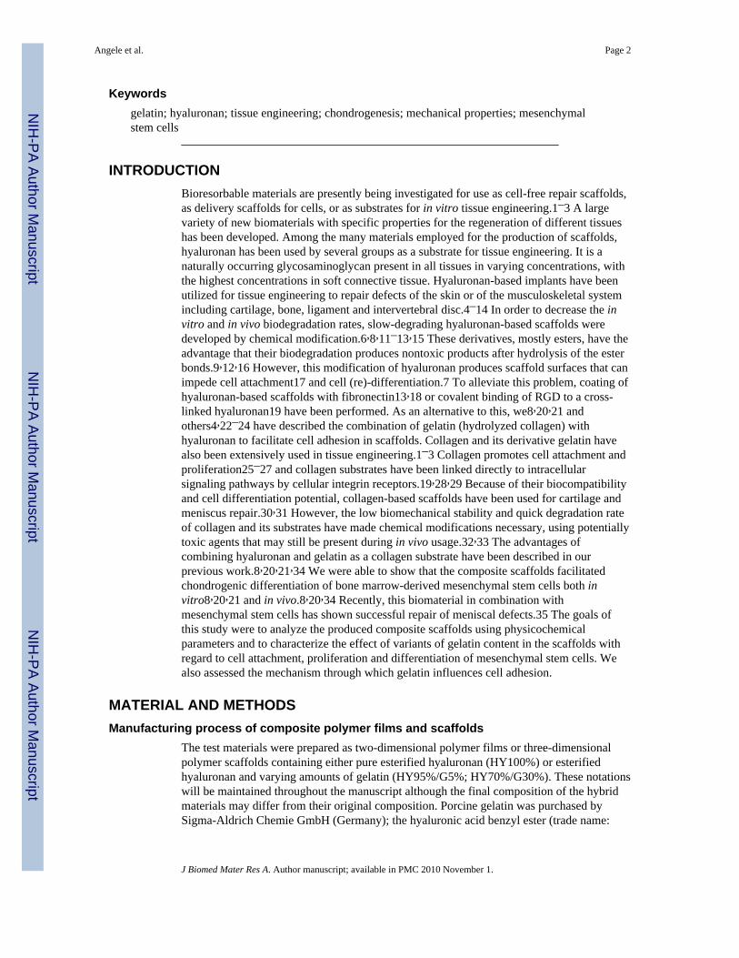
Characterization of esterified hyaluronan-gelatin polymer composites suitable for chondrogenic differentiation of mesenchymal stem cells * Peter Angele 1 , Rainer Müller 2 , Detlef Schumann 1 , Carsten Englert 1 , Johannes Zellner 1 , Brian Johnstone 3 , Jung Yoo 3 , Joachim Hammer 4 , Johann Fierlbeck 4 , Martin K. Angele 5 , Michael Nerlich 1 , and Richard Kujat 1 1 Department of Trauma Surgery, University Hospital of Regensburg, Franz-Josef-Strauss-Allee 11, 93053 Regensburg, Germany 2 Institute of Physical and Theoretical Chemistry, University of Regensburg, Universitätsstr. 31, 93053 Regensburg, Germany 3 Department of Orthopedic Surgery, OHSU, 3181 SW Sam Jackson Park Rd., Portland, OR 97239, USA 4 Department of Biomechanics, University of Applied Sciences, Galgenbergstr. 30, 93053 Regensburg, Germany 5 Department of Surgery, Ludwig Maximilians University, Klinikum Großhadern, Marchioninistr. 15, 81377 München, Germany Abstract Composite scaffolds of homogeneously mixed esterified hyaluronan (HY) and gelatin (G) were manufactured with variable component compositions (HY100%; HY95%/G5%; HY70%/G30%). The goals of this study were to analyze the produced composite scaffolds using physical and chemical methods, e.g., scanning electron microscopy, IR-spectroscopy, water contact angle, protein assay, and tensile testing as well as to assess the effects of adding gelatin to the composite scaffolds on attachment, proliferation and chondrogenic differentiation of human mesenchymal stem cells. Numbers of attached cells were significantly higher on the composite material compared to pure hyaluronan at different time points of two-dimensional or three-dimensional cell culture (p < 0.02). In composite scaffolds, a significantly greater amount of cartilage-specific extracellular matrix components was deposited after 28 days in culture (glycosaminoglycan: p < 0.001; collagen: p < 0.001) as compared with 100% hyaluronan scaffolds. Additionally, gelatin containing composite scaffolds displayed stronger promotion of collagen type II expression than pure hyaluronan scaffolds. The mechanism, by which gelatin influences cell adhesion, was examined. The effect was inhibited by collagenase treatment of the composites or by addition of α5β1-integrin blocking antibodies to the cell suspension. In summary, the results describe the establishment of a class of composite polymer scaffolds, consisting of esterified hyaluronan and gelatin, which are potentially useful for cell-based tissue engineering approaches using mesenchymal stem cells for chondrogenic differentiation. * No benefit of any kind will be received either directly or indirectly by the authors. Correspondence to: P. Angele; e-mail: [email protected]., Tel +49-941-944-6805; Fax +49-941-944-6806. NIH Public Access Author Manuscript J Biomed Mater Res A. Author manuscript; available in PMC 2010 November 1. Published in final edited form as: J Biomed Mater Res A. 2009 November ; 91(2): 416–427. doi:10.1002/jbm.a.32236. NIH-PA Author Manuscript NIH-PA Author Manuscript NIH-PA Author Manuscript

Welcome message from author
This document is posted to help you gain knowledge. Please leave a comment to let me know what you think about it! Share it to your friends and learn new things together.
Transcript
Characterization of esterified hyaluronan-gelatin polymercomposites suitable for chondrogenic differentiation ofmesenchymal stem cells*
Peter Angele1, Rainer Müller2, Detlef Schumann1, Carsten Englert1, Johannes Zellner1,Brian Johnstone3, Jung Yoo3, Joachim Hammer4, Johann Fierlbeck4, Martin K. Angele5,Michael Nerlich1, and Richard Kujat11 Department of Trauma Surgery, University Hospital of Regensburg, Franz-Josef-Strauss-Allee11, 93053 Regensburg, Germany2 Institute of Physical and Theoretical Chemistry, University of Regensburg, Universitätsstr. 31,93053 Regensburg, Germany3 Department of Orthopedic Surgery, OHSU, 3181 SW Sam Jackson Park Rd., Portland, OR97239, USA4 Department of Biomechanics, University of Applied Sciences, Galgenbergstr. 30, 93053Regensburg, Germany5 Department of Surgery, Ludwig Maximilians University, Klinikum Großhadern, Marchioninistr.15, 81377 München, Germany
AbstractComposite scaffolds of homogeneously mixed esterified hyaluronan (HY) and gelatin (G) weremanufactured with variable component compositions (HY100%; HY95%/G5%; HY70%/G30%).The goals of this study were to analyze the produced composite scaffolds using physical andchemical methods, e.g., scanning electron microscopy, IR-spectroscopy, water contact angle,protein assay, and tensile testing as well as to assess the effects of adding gelatin to the compositescaffolds on attachment, proliferation and chondrogenic differentiation of human mesenchymalstem cells. Numbers of attached cells were significantly higher on the composite materialcompared to pure hyaluronan at different time points of two-dimensional or three-dimensional cellculture (p < 0.02). In composite scaffolds, a significantly greater amount of cartilage-specificextracellular matrix components was deposited after 28 days in culture (glycosaminoglycan: p <0.001; collagen: p < 0.001) as compared with 100% hyaluronan scaffolds. Additionally, gelatincontaining composite scaffolds displayed stronger promotion of collagen type II expression thanpure hyaluronan scaffolds. The mechanism, by which gelatin influences cell adhesion, wasexamined. The effect was inhibited by collagenase treatment of the composites or by addition ofα5β1-integrin blocking antibodies to the cell suspension. In summary, the results describe theestablishment of a class of composite polymer scaffolds, consisting of esterified hyaluronan andgelatin, which are potentially useful for cell-based tissue engineering approaches usingmesenchymal stem cells for chondrogenic differentiation.
*No benefit of any kind will be received either directly or indirectly by the authors.Correspondence to: P. Angele; e-mail: [email protected]., Tel +49-941-944-6805; Fax +49-941-944-6806.
NIH Public AccessAuthor ManuscriptJ Biomed Mater Res A. Author manuscript; available in PMC 2010 November 1.
Published in final edited form as:J Biomed Mater Res A. 2009 November ; 91(2): 416–427. doi:10.1002/jbm.a.32236.
NIH
-PA Author Manuscript
NIH
-PA Author Manuscript
NIH
-PA Author Manuscript
Keywordsgelatin; hyaluronan; tissue engineering; chondrogenesis; mechanical properties; mesenchymalstem cells
INTRODUCTIONBioresorbable materials are presently being investigated for use as cell-free repair scaffolds,as delivery scaffolds for cells, or as substrates for in vitro tissue engineering.1–3 A largevariety of new biomaterials with specific properties for the regeneration of different tissueshas been developed. Among the many materials employed for the production of scaffolds,hyaluronan has been used by several groups as a substrate for tissue engineering. It is anaturally occurring glycosaminoglycan present in all tissues in varying concentrations, withthe highest concentrations in soft connective tissue. Hyaluronan-based implants have beenutilized for tissue engineering to repair defects of the skin or of the musculoskeletal systemincluding cartilage, bone, ligament and intervertebral disc.4–14 In order to decrease the invitro and in vivo biodegradation rates, slow-degrading hyaluronan-based scaffolds weredeveloped by chemical modification.6,8,11–13,15 These derivatives, mostly esters, have theadvantage that their biodegradation produces nontoxic products after hydrolysis of the esterbonds.9,12,16 However, this modification of hyaluronan produces scaffold surfaces that canimpede cell attachment17 and cell (re)-differentiation.7 To alleviate this problem, coating ofhyaluronan-based scaffolds with fibronectin13,18 or covalent binding of RGD to a cross-linked hyaluronan19 have been performed. As an alternative to this, we8,20,21 andothers4,22–24 have described the combination of gelatin (hydrolyzed collagen) withhyaluronan to facilitate cell adhesion in scaffolds. Collagen and its derivative gelatin havealso been extensively used in tissue engineering.1–3 Collagen promotes cell attachment andproliferation25–27 and collagen substrates have been linked directly to intracellularsignaling pathways by cellular integrin receptors.19,28,29 Because of their biocompatibilityand cell differentiation potential, collagen-based scaffolds have been used for cartilage andmeniscus repair.30,31 However, the low biomechanical stability and quick degradation rateof collagen and its substrates have made chemical modifications necessary, using potentiallytoxic agents that may still be present during in vivo usage.32,33 The advantages ofcombining hyaluronan and gelatin as a collagen substrate have been described in ourprevious work.8,20,21,34 We were able to show that the composite scaffolds facilitatedchondrogenic differentiation of bone marrow-derived mesenchymal stem cells both invitro8,20,21 and in vivo.8,20,34 Recently, this biomaterial in combination withmesenchymal stem cells has shown successful repair of meniscal defects.35 The goals ofthis study were to analyze the produced composite scaffolds using physicochemicalparameters and to characterize the effect of variants of gelatin content in the scaffolds withregard to cell attachment, proliferation and differentiation of mesenchymal stem cells. Wealso assessed the mechanism through which gelatin influences cell adhesion.
MATERIAL AND METHODSManufacturing process of composite polymer films and scaffolds
The test materials were prepared as two-dimensional polymer films or three-dimensionalpolymer scaffolds containing either pure esterified hyaluronan (HY100%) or esterifiedhyaluronan and varying amounts of gelatin (HY95%/G5%; HY70%/G30%). These notationswill be maintained throughout the manuscript although the final composition of the hybridmaterials may differ from their original composition. Porcine gelatin was purchased bySigma-Aldrich Chemie GmbH (Germany); the hyaluronic acid benzyl ester (trade name:
Angele et al. Page 2
J Biomed Mater Res A. Author manuscript; available in PMC 2010 November 1.
NIH
-PA Author Manuscript
NIH
-PA Author Manuscript
NIH
-PA Author Manuscript
“Jaloskin”) was obtained from Fidia farmaceutici S.p.A. (Italy). The polymer films and thethree dimensional polymer scaffolds were manufactured following a recently developedmanufacturing process8,21 in which the polymers were dissolved and homogeneouslymixed in hexafluoro-2-propanol (HFIP) which was obtained from Fluka GmbH (Germany).For all compositions, the final concentration of dissolved hyaluronan was 4%. Films wereprepared either on smooth polytetrafluoroethylene surfaces (3.5 cm × 3.5 cm), on glasscover slides (1.8 cm × 1.8 cm), or in 24-well plates (1.55 cm diameter, 1.90 cm2 area) by airevaporation of the volatile solvent followed by drying in vacuum. To create the three-dimensional scaffolds, salt crystals with a defined grain size (350–450 μm) were added tothe polymer solutions and served as a porogen for the scaffolds (mass ratio polymer : salt =1 : 60). After compressing the constructs in molds and drying in vacuum, the salt crystalswere removed by leaching in de-ionized water which was controlled by conductimetricmeasurement. Finally, the porous scaffolds were dehydrated in acetone-water mixtures ofincreasing acetone content and dried in vacuum.
Ultrastructure of polymer scaffoldsPure hyaluronan and composite scaffolds were prepared for scanning electron microscopy asdescribed previously.8,36 Briefly, samples were fixed with 1% glutaraldehyde in phosphatebuffer (pH 7.4), osmicated, washed in buffer, dehydrated in increasing concentrations ofethanol and subsequently in acetone. Dehydrated specimens were subjected to critical-pointdrying using liquid carbon dioxide substitution, mounted on aluminum stubs, and gold-coated using a Polaron sputter coater. Samples were examined and photographed with aZeiss DSM 940 microscope.
Physicochemical characterization of polymer compositesPhysicochemical properties of the composite materials were evaluated with respect to thepolymer composition and the effects of the salt-leaching procedure. Air-dried polymer filmsserved as samples for the not salt-leached condition; salt-leached three-dimensionalspecimens were characterized after condensing to a film by the use of a hydraulic press.
For infrared spectroscopy, films were examined in the transmission mode using a Jasco-610FTIR spectrometer. To determine the composition of the films, the peak ratios of the esterand the amide-II signals were evaluated. Estimation of residual organic fluorine containingsolvent was performed by comparison of the peak ratios of the C-F band at 843 cm−1 andthe amide-II signal. Reported values are the average ratios obtained from examination ofthree films per composition.
Contact angle measurements were carried out by the sessile drop technique.37 A waterdroplet with a volume of 2 μl was advanced to the surface of each polymer film by means ofa microsyringe and the contact angle was measured with a G1 goniometer of Erma Co.(Japan). The films were prepared on the surfaces of glass slides by drying the organicsolutions as mentioned before. Reported results are the average contact angles of tendroplets deposited on three individual films. To minimize the effect of film swelling, thevalues were determined exactly 30 sec after deposition of the water droplet.
The gelatin content of the films was further quantified by measuring the amount of freeamino groups which were determined by using 2,4,6-trinitro-benzenesulfonic acid (TNBS,Sigma).38 Light absorption of the diluted solutions was measured at 346 nm against a blanksolution in which the reaction of TNBS was suppressed by adding of hydrochloric acid.Three samples of identical composition were examined and the mean values calculated. UV-VIS spectrometry was carried out on a Lambda-18 spectrometer of Perkin Elmer Corp.(USA).
Angele et al. Page 3
J Biomed Mater Res A. Author manuscript; available in PMC 2010 November 1.
NIH
-PA Author Manuscript
NIH
-PA Author Manuscript
NIH
-PA Author Manuscript
To determine the amount of gelatin that was extracted from the three-dimensional scaffoldsduring the salt leaching procedure, the bicinchoninic acid protein assay (BCA) was used.39The washing solutions were aspirated after extraction times of 15, 30, 60, 120, 180, and 360min. Aliquots (100 μl) of these solutions were incubated with 1ml BCA working solution(Sigma) at 40 °C for 30 min. The light absorption of the resulting solutions was measured at562 nm and compared with the values obtained from solutions of defined gelatinconcentrations (0.01 to 1 mg/ml). Washing solutions of scaffolds made of esterifiedhyaluronan were used as blanks. The reported values represent the means of four scaffoldsof identical composition but of different weight.
Stress-strain characteristics of the scaffoldsThe stress-strain curves of scaffolds were determined by uniaxial measurements using aZwick-1445 mechanical testing device. Cylindrical bars (6 mm length × 5 mm diameter)were cut from the matrix sheets using circular punches. Each sample was glued with definedamounts of low melting dental wax (Miltex Inc., USA) between two standardized aluminumdiscs with a diameter of 10 mm and a thickness of 1 mm. The metal discs allowed aconvenient fixation of the samples between two vertically positioned grips. A highly precisepositioning of one fixing grip in x,y-direction guaranteed a strictly linear alignment of thespecimen axis parallel to the load axis in z-direction. All samples were characterized in thewet state after soaking the specimens in distilled water at room temperature for 10 min(Figure 1). For the tensile experiments, an initial gauge length of 4 mm was used. Allsamples were deformed to rupture at a deformation rate of 1 mm/min. The displacement andload were recorded continuously by a Lab View 5.0 - software. The Young’s modulus wasdirectly determined from the linear slope of the stress-strain curves (n = 6 per group).
Cell cultureThe cellular effects induced by the hyaluronan-gelatin composites were examined in two-dimensional films and three-dimensional scaffolds. The polymer films and scaffolds weresterilized via beta irradiation (e-beam) with a dose of 25 kGy by Beta-Gamma-ServiceGmbH (Germany) according to ISO 11137. Human bone marrow was obtained from bonemarrow aspirates of patients undergoing surgery with bone block harvest of the iliac crest(written consent form signed). After Percoll gradient fractionation, Dulbecco’s modifiedEagle Medium (DMEM) with 10% fetal bovine serum (FBS) was added to the aspirate and10 × 106 nucleated cells/100 cm2 dish were plated and grown at 37 °C with 5% CO2 untilthe cells reached 80% confluency.8,21,40–42
Adhesion and proliferation behavior on polymer filmsFor analysis of adhesion and proliferation rates, human mesenchymal stem cells (hMSC)were trypsinized, counted and then re-suspended in DMEM containing 10% FBS. Cellswere plated (8,000 cells for adhesion analysis and 4,000 cells for proliferation analysis)either in uncoated polystyrene wells (control) of 24-well plates (Sarstedt, Germany) or inwells coated with a film of the polymer composite. The adhesion rate was assessed after 3 hof standard cell culture and the proliferation rate was determined after one, four or sevendays of culture. After removal of non-adherent cells the number of attached cells wasmeasured. The quantification of the cells was performed by their mitochondrialdehydrogenase activity using 3-(4,5-dimethylthiazol-2-yl)-5-(3-carboxymethoxyphenyl)-2-(4-sulfophenyl)-2H-tetrazolium inner salt (MTS) and phenazine ethosulfate (PES) accordingto the protocol provided by the manufacturer (CellTiter96-Kit of Promega, Germany). Eightsamples were examined for each polymer film composition. To assess cell numbers,standard curves were established by plating five dilution steps of cell numbers between1,000 and 10,000 cells per well and performing the MTS assay. Each standard cell numberwas used in quadruplicate.
Angele et al. Page 4
J Biomed Mater Res A. Author manuscript; available in PMC 2010 November 1.
NIH
-PA Author Manuscript
NIH
-PA Author Manuscript
NIH
-PA Author Manuscript

Effect of gelatin on cell adhesionTo further analyze the effect of gelatin on the adhesion and proliferation of mesenchymalstem cells, polymer films were incubated with 0.1% collagenase P (Roche, 1213865) for 4 hprior to cell application. hMSCs (10,000 per polymer film) were incubated for 30 min, 1, 2,12, or 24 h in DMEM containing 10% FBS and then subjected to immunohistochemicalstaining for CD44 (Pharmingen, clone IM7, 553130) and β1-integrin (Chemicon, cloneP4C10, MAB 1987 Z). The cell morphology was examined with fluorescence microscopyafter application of a FITC-conjugated goat anti-mouse secondary polyclonal antibody(Jackson) and compared between the groups. In replicate films the integrin receptor-ligandbinding was blocked by adding a blocking goat anti-agr;5β1 polyclonal antibody(Chemicon, AB1950; diluted 1:250) to the cell suspension. After 24 h the number ofattached cells was measured by cell counting (n = 5 per group).
Chondrogenic differentiation behavior in polymer scaffoldsAdherent hMSC colonies were trypsinized, counted, and 2 × 10⁶ mesenchymal stem cellswere injected into cylindrical polymer scaffolds with defined pore size and cultured at 37 °Cin 5% CO2 in a defined medium, previously shown to induce chondrogenic differentiation ofmesenchymal stem cells.8,21,40–42
DNA contents of the polymer scaffolds were assayed using a fluorimetric assay as describedpreviously.21,42 Scaffolds were individually digested with 150 μg/ml papain overnight atpH 6.0 at 60 °C. Aliquots of the digest (10 μl) were mixed with 200 μl of diluted Hoechst33258 Dye Solution (0.1 μg/ml). The fluorescence emission of the samples was measuredwith Cytofluor at 360 nm excitation, 460 nm emission with a gain of 70. A standard curvewas produced with calf thymus DNA (Sigma) and used to determine the DNA content of theexperimental samples. The glycosaminoglycan content of the scaffolds, used as an indicatorof proteoglycan production, was quantified spectrophotometrically, with a 1,9-dimethylmethylene blue (DMMB) binding assay as described elsewhere.43 Scaffolds weredigested in papain solution at 60 °C overnight. In a 96-well plate, 50 μl aliquots of thesamples were mixed with 200 μl of 50 μmol/l DMMB (Sigma) dissolved in 40 mmol/lglycine-NaCl at pH 3.0 and optical density was read at 540 nm using the ELISA readerEmax from Molecular Devices (Germany). A standard curve was prepared with papain-treated chondroitin sulfate A (Sigma). For the experimental groups, the proteoglycan contentof the negative controls (cell free polymer scaffold) was measured and subtracted to get anestimation of the produced extracellular proteoglycan since the hyaluronan from the scaffoldis detectable in the assay, albeit at a low level.
The hydroxyproline content of the scaffolds was measured with a modified version of thestandard assay.44 Papain digested scaffolds were dried, weighed, and hydrolyzed with 2mol/l sodium hydroxide solution for 4 h in a pressure apparatus. 50 μl, 25 μl, and 10 μltriplicated aliquots of hydrolysate were incubated for 20 min with 450 μl of chloramine Tsolution at room temperature. After addition of 500 μl of Ehrlich′s reagent, the color wasdeveloped for 20 min at 65 °C. Light absorption was read at 560 nm in the ELISA reader.The hydroxyproline content of samples was extrapolated against identically processedhydroxyproline standards between 1 and 20 μg/ml. The collagen content was calculated bymultiplying the total hydroxyproline content of the assay with a conversion factor (×7.5).For the experimental groups the content of the negative controls (cell-free polymer scaffold)was subtracted to get an estimation of the produced extracellular collagen since the gelatinof the scaffolds would influence the measurement. For immunohistochemical analysis ofMSC differentiation, cell-matrix constructs were formaldehyde fixed at day 28 ofchondrogenic cell culture, embedded in Tissue-Tek O.C.T. Compound (Sakura Finetek,Zoeterwoude, Netherlands), and frozen in liquid nitrogen. Expression of collagen type I and
Angele et al. Page 5
J Biomed Mater Res A. Author manuscript; available in PMC 2010 November 1.
NIH
-PA Author Manuscript
NIH
-PA Author Manuscript
NIH
-PA Author Manuscript

collagen type II has been demonstrated on 10 μm cryosections by the immunoperoxidaseABC technique (Vector, Burlingame, CA, USA) after Hsu et al. 45, applying predigestion ofsections (15 min in 0.1% pepsin at pH 3.5), monoclonal primary antibodies mouse anti-collagen type I (clone col-1, Sigma, St. Louis, MO, USA), collagen II (clone II-4C11,Calbiochem – Merck, Schwalbach, Germany), biotin conjugated polyclonal secondary goatanti-mouse IgG (Jackson, West Grove, PA, USA) antibodies, and the Ni and Co enhancedDAB stain visualization.
Statistical AnalysisData are reported as the mean ± SD. One-factor analyses of variance (ANOVAs) withBonferroni-Dunn, Tukey and Fisher’s post hoc tests for multiple comparisons wereperformed using SPSS 12.0G. Significance was accepted at a level of p < 0.05.
RESULTSUltrastructure of polymer scaffolds
Hyaluronan-gelatin composite scaffolds were produced following the protocol of therecently developed manufacturing process.8,20,21 The addition of gelatin (Figure 2A, 2B)produced no significant ultrastructural changes compared to 100% hyaluronan-basedscaffolds (Figure 2C, 2D). Both scaffolds (HY 100%, HY70%/G30%) showed a double-pore structure. In addition to the main pores (mean pore size 350–450 μm) that were formeddirectly by the porogen (salt crystals), a second class of macropores with a smaller pore sizebetween 50 and 100 μm was produced. Figure 2B and 2D display the microstructure of thepore walls which exhibited no significant differences between pure hyaluronan andcomposite scaffolds. The scaffolds revealed thin pore walls of about 3 μm in averagethickness that were essentially solid throughout the scaffolds. A high degree ofinterconnectivity between the primary and secondary pores was noted (Figure 2A, 2C).
Physicochemical characterization of polymer compositesSeveral analytical methods have been used to determine the composition of the polymersand to control unintended changes which may occur during scaffold processing. The infraredspectra of polymer films consisting of esterified hyaluronan and gelatin exhibited signals forthe ester and the amide groups (Figure 3A). The peak ratio of the ester to amide-II signalsdecreased with increased gelatin content of the polymer films (Table 1). Comparable resultshave been obtained by measuring the water contact angle of the film surfaces. The wettingangle decreased with increasing amounts of the hydrophilic gelatin constituent. The aminogroup concentration, which could be quantified by the TNBS assay, increased withincreasing gelatin content of the examined films (Table 1).
Effects of the salt-leaching procedureManufacture of the three-dimensional polymer scaffolds required washing steps with wateror buffer solution in order to remove the pore forming particles consisting of sodiumchloride. Additionally, washing of the scaffolds with water reduced residual organic solventthat withstands vacuum drying. The qualitative effect of rinsing polymer samples of thecomposition 70% derivatized hyaluronan and 30% gelatin with water is shown by the IRspectra of Figure 3A. The spectrum of the vacuum dried film exhibited strong signals in thearea of 1200 cm−1 and between 950 and 840 cm−1 which belong to the vibration of C-Fgroups of the organic solvent HFIP. After leaching the salt crystals from 3-D polymerconstructs with water, the signals disappeared from the IR spectrum.
As a side effect of the washing procedure, the extraction of the gelatin constituent wasqualitatively observed by an increase in the peak ratio of the ester to amide-II signals with
Angele et al. Page 6
J Biomed Mater Res A. Author manuscript; available in PMC 2010 November 1.
NIH
-PA Author Manuscript
NIH
-PA Author Manuscript
NIH
-PA Author Manuscript

IR-spectroscopy (Table 1). For pure hyaluronan-based scaffolds, no differences in infraredband intensities could be detected after the removal of the salt crystals with water. However,for composite scaffolds (HY95%/G5%, HY70%/G30%), a loss of gelatin could be detectedwith this method. These results were confirmed by contact angle measurements of washedpolymer films. The contact angle increased due to dissolution of the more hydrophilicpolymer gelatin from the composites (Table 1).
The amount of dissolved gelatin was quantified with the BCA protein assay (Figure 3B). Aloss of the gelatin content in composite scaffolds could be detected after the removal of theporogen depending on the original gelatin content. The percentage of dissolved gelatinduring washing is higher if a higher amount of gelatin was initially added. Also a timedependency could be detected such that a high percentage of gelatin dissolution was noted atthe beginning of the porogen removal with lower dissolution over time. The remaininggelatin content was estimated combining the results of the different characterizationtechniques including the TNBS assay for primary amino groups (Table 1). For scaffoldswith an original gelatin content of 30%, a remaining content of about 15–20% could beestimated after porogen removal. For composites with an original gelatin content of 5%, afinal gelatin content of about 3–4% was measured. In the following sections of themanuscript the notations HY95%/G5% and HY70%/G30% will be maintained despite theloss of gelatin described in this chapter.
Biomechanical analysis of the composite scaffoldsIndependently of the scaffold composition, no significant swelling or shrinkage of thescaffolds could be detected after cell loading and during culture.
However, the tensile experiments reveal a clear dependency between scaffold compositionand mechanical stability (Table 2). With increasing gelatin content, the rupture strength andthe maximum Young modulus were decreased. Composite scaffolds (HY70%/G30%)displayed a significant reduction in tensile strength (p = 0.03) and Young’s modulus (p =0.003) compared with scaffolds made of 100% hyaluronan. Strain at rupture was slightlyenhanced with scaffolds containing low amounts of gelatin (HY95%/G5%) compared to thehyaluronan scaffold, whereas for scaffolds with high amounts of gelatin (HY70%/G30%) nodifference was detected.
Cell adhesion and proliferation on composite filmsAdhesion of human mesenchymal stem cells on films of 100% esterified hyaluronan weresignificantly lower compared with the polystyrene (PS) reference (p = 0.002). On compositefilms of esterified hyaluronan and gelatin, cell adhesion was significantly greater (p = 0.006)as compared to pure hyaluronan and almost identical to that found with PS (Figure 4A).Significant cell proliferation was only found on PS between day 1 and day 7 (p = 0.002),while on all other surfaces the cell number remained nearly constant. Cell numbers on filmscontaining gelatin were significantly higher as compared to pure hyaluronan at each timepoint of the proliferation experiment (p < 0.001) (Figure 4B).
Role of gelatin in cell adhesionTo analyze the mechanism by which gelatin is mediating the elevated cell adhesion andproliferation further experiments on 100% hyaluronan-based films and composite films(HY70%/G30%) were performed. CD44 expression on the cell surface was moderatelypositive in all groups at all incubation time points (Figure 5). β1-integrin expression couldbe detected on the surface of adherent cells on 100% hyaluronan-based films and compositefilms. After 30 min of incubation, round mesenchymal stem cells were seen adhering to thecomposite polymer films (HY70%/G30%). In this group, the first evidence of cell spreading
Angele et al. Page 7
J Biomed Mater Res A. Author manuscript; available in PMC 2010 November 1.
NIH
-PA Author Manuscript
NIH
-PA Author Manuscript
NIH
-PA Author Manuscript

(spindle-shaped cells) could be detected after 1 h. On 100% hyaluronan-based films orcomposite films treated with collagenase prior to cell seeding, the cells kept their initialrounded morphology. After 2 h, the cells of the untreated composite films were spread out,whereas in the other groups only a small percentage of spindle-shaped cells could bedetected. After 12 and 24 h of cell incubation, cell attachment with a change of cellmorphology to fibroblast-like cells could be seen in all groups (Figure 5).
The composite films facilitated significantly higher cell attachment (p < 0.001) as detectedwith dark field microscopy and cell counting (Figure 6). This effect could be inhibited bycollagenase treatment of the composite films prior to cell application, giving equivalent cellattachment on both polymer films. Blocking of receptor-ligand binding for α5β1-integrin oncomposite films reduced cell adherence to the level seen with 100% hyaluronan-based filmsor composite and hyaluronan films after collagenase treatment, suggesting a α5β1-integrininvolvement in early cell adhesion of mesenchymal stem cells on gelatin-containing surfaces(Figure 6).
Influence of gelatin on cell differentiationThe effect of adding gelatin on chondrogenic differentiation of mesenchymal stem cells wasanalyzed in three-dimensional polymer scaffolds. A significantly higher cell adhesion wasdetected in composite polymer scaffolds (p = 0.02). After chondrogenic culture, the cellnumber between scaffolds with gelatin remained unchanged. However, cells in 100%hyaluronan scaffolds proliferated to reach a number similar to that found in the compositescaffolds (day 14: p = 0.07; day 28: p = 0.7) (Figure 7A). Both the proteoglycan andcollagen contents were higher in composite scaffolds compared with 100% hyaluronanscaffolds. A significant increase in proteoglycan and collagen contents could be observedwith addition of gelatin to the polymers (p < 0.001 for glycosaminoglycan and collagen forboth HY95%/G5% and HY70%/G30%). The inclusion of 30% gelatin led to a significantlyhigher amount of proteoglycan production compared with 5% gelatin (p < 0.001), but nosignificant differences in collagen content (p = 0.09) could be detected (Figure 7B).
Immunohistochemical analysis of cell-matrix constructs disclosed no significant differencesin collagen type I expression within pure hyaluronan scaffolds and hyaluronan-gelatincomposite scaffolds (HY70%/G30%). In both matrices a weakly pronounced reactionagainst collagen type I was observed. On the other hand, expression of collagen type IIstrongly depended on the composition of the scaffold material used. Within compositescaffolds (HY70%/G30%) distinctly higher amounts of expressed collagen type II could bedetected compared to scaffolds made from pure hyaluronan (Figure 7C).
DISCUSSIONWe have previously described the use of composite polymer scaffolds, consisting ofesterified hyaluronan and gelatin, for cartilage tissue engineering with mesenchymal stemcells.8,20,21 Here we analyze this new class of composite polymer scaffolds in terms ofultrastructure, biochemical composition and biological effects compared with 100%hyaluronan-based scaffolds.
The composite polymer scaffolds were manufactured with a newly developed procedureusing the solvent hexafluoro-2-propanol to homogeneously mix esterified hyaluronan andgelatin in the same solution, which resulted in the formation of primary and secondary poresin the scaffolds. The pore walls consisting of the homogeneously mixed polymers were thinand very compact. The ultrastructure of scaffolds created with this protocol differed fromthose made with a previously described manufacturing process, in which only primary poreswith thicker and foam-like pore walls were created.16 The structure of the pore walls in the
Angele et al. Page 8
J Biomed Mater Res A. Author manuscript; available in PMC 2010 November 1.
NIH
-PA Author Manuscript
NIH
-PA Author Manuscript
NIH
-PA Author Manuscript
recently developed manufacturing process was achieved by an air drying step. The dryingsolution of the hyaluronan spreads over the walls of the crystals of the porogen andcondenses to very thin, homogenous, compact layers. By drying, the first solvent evaporatesand its volume gets substituted by air yielding the additional secondary pores. With thepreviously published manufacturing process,16 the esterified hyaluronan does not rigidifyby drying but by flocculation in the second solvent in which it is insoluble. Therefore theester remained voluminous distributed in the pore interstices and resulted in thick pore wallswith foam-like ultrastructure lacking additional secondary pores. The addition of gelatin didnot alter these ultrastructural differences in scaffolds manufactured with our manufacturingprocess.
The rationale behind manufacturing a composite of hyaluronan and collagen was to mimicthe embryonic environment for optimized cell attachment and suitable cell differentiation.9,46 Mixtures of hyaluronan and collagenous components for the development of compositebiomaterials have been described before.4,22,27,47–54 Because of the fast degradation rateof water soluble hyaluronan in vivo, various modifications of hyaluronan (esterification,cross-linking) have been applied.16,24,54–57 These modifications resulted in prolongedscaffold stability and reduced degradation rates, however impaired biological propertiescould be observed. With polymer composites of hydrophobic esterified hyaluronan andhydrophilic gelatin we combined the advantages of esterified hyaluronan scaffolds in termsof scaffold stability, reduced degradation and excellent biocompatibility8,9,11–13,58 withthe advantages of non cross-linked gelatin in terms for optimized cell adhesion anddifferentiation.24–27,51,54 These composites have been shown to facilitate chondrogenicdifferentiation of bone marrow-derived mesenchymal stem cells both in vitro8,21 and invivo.8,20,34
Physicochemical analysis revealed some loss of gelatin during the manufacturing procedure.However, at the end of the manufacturing process, gelatin was still detectable in thescaffolds as seen in Figure 3 and Table 1. The amount of the dissolved gelatin is dependenton the originally added gelatin content. The percentage of dissolved gelatin during washingis higher when a higher percentage of gelatin content was initially added. This can beexplained with the different hydrophilicity of the polymers depending on the amount ofgelatin. Polymers with a higher amount of hydrophilic components like gelatin facilitategreater scaffold swelling, which is accompanied by a higher elution of water-solubleparticles.
The addition of gelatin resulted in a reduction of scaffold stability. This was expected,because gelatin by itself has poor performance in terms of scaffold stability,36,59–61 and soshould decrease the stability of the polymer composite. Earlier studies have shown thatcomposite polymers of 70% esterified hyaluronan and 30% gelatin provide an appropriateenvironment for the chondrogenic differentiation of mesenchymal stem cells.8,20,21,34 Thesame polymer composites also showed good structural stability during in vitro experimentsthat included daily applied compressive loading.21
Modified hyaluronan showed good biocompatibility with different cell types,7,9,10,13,62however, several methods have been used to improve cell attachment and cell proliferationincluding addition of RGD sequences,19,55 protein coating13 or addition of collagen priorto crosslinking.4,22,27,47,48,51–54 In the literature, the attachment of fibroblasts wasshown to significantly improve only after addition of more than 40% cross-linked gelatin tocross-linked hyaluronan.24 In our study, the addition of about 5% gelatin provided asignificant increase in cell adhesion and proliferation in comparison with 100% hyaluronan-based polymers. Why the lower level of gelatin provided such marked improvement in cellattachment in our study compared with those reported by others is unclear, but it could be
Angele et al. Page 9
J Biomed Mater Res A. Author manuscript; available in PMC 2010 November 1.
NIH
-PA Author Manuscript
NIH
-PA Author Manuscript
NIH
-PA Author Manuscript

due to the derivatization of the gelatin component, the cross-linking of the hyaluronancomponent or the different cell types used for the analyses. As well as increased cellattachment to two-dimensional films, the addition of gelatin also facilitated a significantincrease in initial cell uptake in three-dimensional composite scaffolds compared with 100%esterified hyaluronan scaffolds.
After degradation of the collagen component with collagenase, the increased cell adhesion inthe composite films was blocked completely, indicating the specificity of gelatin inpromoting the elevated cell adhesion. Early cell attachment on gelatin-containing surfaces ismediated by early expression of β1-integrin. The higher level of cell adhesion in thecomposite polymer films compared with 100% esterified hyaluronan-based films wasblocked completely with α5β1-integrin blocking antibodies, indicating the involvement ofα5β1-integrins, at least in part, in the attachment of mesenchymal stem cells to thecomposite polymer films. The interaction of cells with their surroundings requires more thana mechanical site for adhesion.63 Receptors for natural extracellular matrix proteins such asmembers of the integrin receptor family have been linked directly with intracellularsignaling events.64–66 Therefore, early expression of α5β1-integrin on the cell surface maysimultaneously induce important signal transduction pathways for chondrogenicdifferentiation. α5β1-integrins seem to be of particular importance in the modulation ofcartilaginous tissue.67
The biological performance of the scaffolds examined in the present study was clearlyimproved after addition of gelatin to the fundamental component hyaluronic acid benzylester. Adhesion as well as chondrogenic differentiation of mesenchymal stem cells in termsof glycosaminoglycan and collagen synthesis and expression of the hyaline-like cartilage-specific collagen type II seem to increase with increasing gelatin. On the other hand,biomechanical properties of the composites decreased with increasing gelatin content.Therefore, composite scaffolds with lower amounts of gelatin (≤5%) could be employed inapplications in which a higher mechanical toughness of the scaffolding material isrecommended, like the engineering of meniscus or vertebral disc structures. Compositeswith higher gelatin content (5%≤ x ≤ 30%) are more favorable for engineering of cartilagestructures which are exposed to a lower degree of mechanical stress, like reconstructiontissues of the ear and nose. The successful use of the new biomaterial in combination withmesenchymal stem cells for the repair of a critical size meniscal defect could be shownrecently.35
CONCLUSIONSA new class of composite polymer scaffolds, consisting of homogeneously mixedhydrophobic esterified hyaluronan and hydrophilic gelatin, was established and thoroughlyanalyzed by physicochemical means. The composites showed appropriate morphologicalstructure with interconnecting pores and initial biomechanical stability without chemicalcross-linking and are, therefore, free of problems usually connected with stabilizationprocedures. The addition of hydrolyzed collagen significantly improved the biologicalproperties of the esterified hyaluronan polymer. The resulting 3-dimensional compositesappear very useful for cell-based tissue engineering approaches with mesenchymal stemcells, especially for their chondrogenic differentiation. Also polylactide- and polyglycolide-based scaffold materials are well-known for their differentiation promoting potential, but incontrast to the composite scaffolds described in the present study these polymers exhibit therisk of generating high amounts of acidic products during biodegradation which could causeadverse effects for the engineered tissue.
Angele et al. Page 10
J Biomed Mater Res A. Author manuscript; available in PMC 2010 November 1.
NIH
-PA Author Manuscript
NIH
-PA Author Manuscript
NIH
-PA Author Manuscript
AcknowledgmentsThe authors thank Tanja Weinfurtner, Daniela Drenkard, Tom Böttner, Dalila Bouakline and Ronny Mai forexcellent technical assistance. This work was supported by grants of the Bavarian State Ministry of Sciences,Research and the Arts within the program “High Tech Offensive Bayern” and NIH-NIAMS (1R01AR48132).
References1. Ibarra C, Koski JA, Warren RF. Tissue engineering meniscus: cells and matrix. Orthop Clin North
Am. 2000; 31:411–418. [PubMed: 10882467]2. Sweigart MA, Athanasiou KA. Toward tissue engineering of the knee meniscus. Tissue Eng. 2001;
7:111–129. [PubMed: 11304448]3. Buma P, Ramrattan NN, van Tienen TG, Veth RP. Tissue engineering of the meniscus.
Biomaterials. 2004; 25:1523–1532. [PubMed: 14697855]4. Chang CH, Liu HC, Lin CC, Chou CH, Lin FH. Gelatin-chondroitin-hyaluronan tri-copolymer
scaffold for cartilage tissue engineering. Biomaterials. 2003; 24:4853–4858. [PubMed: 14530082]5. Crevensten G, Walsh AJ, Ananthakrishnan D, Page P, Wahba GM, Lotz JC, Berven S.
Intervertebral disc cell therapy for regeneration: mesenchymal stem cell implantation in ratintervertebral discs. Ann Biomed Eng. 2004; 32:430–434. [PubMed: 15095817]
6. Cristino S, Grassi F, Toneguzzi S, Piacentini A, Grigolo B, Santi S, Riccio M, Tognana E, FacchiniA, Lisignoli G. Analysis of mesenchymal stem cells grown on a three-dimensional HYAFF 11(R)-based prototype ligament scaffold. J Biomed Mater Res A. 2005; 73:275–283. [PubMed: 15789422]
7. Aigner J, Tegeler J, Hutzler P, Campoccia D, Pavesio A, Hammer C, Kastenbauer E, Naumann A.Cartilage tissue engineering with novel nonwoven structured biomaterial based on hyaluronic acidbenzyl ester. J Biomed Mater Res. 1998; 42:172–181. [PubMed: 9773813]
8. Angele P, Kujat R, Nerlich M, Yoo J, Goldberg V, Johnstone B. Engineering of osteochondraltissue with bone marrow mesenchymal progenitor cells in a derivatized hyaluronan-gelatincomposite sponge. Tissue Eng. 1999; 5:545–554. [PubMed: 10611546]
9. Campoccia D, Doherty P, Radice M, Brun P, Abatangelo G, Williams DF. Semisynthetic resorbablematerials from hyaluronan esterification. Biomaterials. 1998; 19:2101–2127. [PubMed: 9884052]
10. Grigolo B, Roseti L, Fiorini M, Fini M, Giavaresi G, Nicoli Aldini N, Giardino R, Facchini A.Transplantation of chondrocytes seeded on a hyaluronan derivative (hyaff-11) into cartilagedefects in rabbits. Biomaterials. 2001; 22:2417–2424. [PubMed: 11511039]
11. Radice M, Brun P, Cortivo R, Scapinelli R, Battaliard C, Abatangelo G. Hyaluronan-basedbiopolymers as delivery vehicles for bone-marrow- derived mesenchymal progenitors. J BiomedMater Res A. 2000; 50:101–109.
12. Milella E, Brescia E, Massaro C, Ramires PA, Miglietta MR, Fiori V, Aversa P. Physico-chemicalproperties and degradability of non-woven hyaluronan benzylic esters as tissue engineeringscaffolds. Biomaterials. 2002; 23:1053–1063. [PubMed: 11791908]
13. Solchaga LA, Gao J, Dennis JE, Awadallah A, Lundberg M, Caplan AI, Goldberg VM. Treatmentof osteochondral defects with autologous bone marrow in a hyaluronan-based delivery vehicle.Tissue Eng. 2002; 8:333–347. [PubMed: 12031121]
14. Balazs EA, Denlinger JL. Clinical uses of hyaluronan. Ciba Found Symp. 1989; 143:265–275.[PubMed: 2680347]
15. Baier Leach J, Bivens KA, Patrick CW Jr, Schmidt CE. Photocrosslinked hyaluronic acidhydrogels: natural, biodegradable tissue engineering scaffolds. Biotechnol Bioeng. 2003; 82:578–589. [PubMed: 12652481]
16. Valentini, R.; Kim, H. WO-Patent WO 97/45532. 1997. Hyaluronan based biodegradable scaffoldsfor tissue repair.
17. Kito H, Matsuda T. Biocompatible coatings for luminal and outer surfaces of small-caliberartificial grafts. J Biomed Mater Res. 1996; 30:321–330. [PubMed: 8698695]
18. Solchaga LA, Yoo JU, Lundberg M, Dennis JE, Huibregtse BA, Goldberg VM, Caplan AI.Hyaluronan-based polymers in the treatment of osteochondral defects. J Orthop Res. 2000;18:773–780. [PubMed: 11117300]
Angele et al. Page 11
J Biomed Mater Res A. Author manuscript; available in PMC 2010 November 1.
NIH
-PA Author Manuscript
NIH
-PA Author Manuscript
NIH
-PA Author Manuscript

19. Glass JR, Dickerson KT, Stecker K, Polarek JW. Characterization of a hyaluronic acid-Arg-Gly-Asp peptide cell attachment matrix. Biomaterials. 1996; 17:1101–1108. [PubMed: 8718970]
20. Angele P, Kujat R, Faltermeier H, Schumann D, Müller R, Nerlich M. BiodegradableHyaluronsäureester/Gelatine Kompositmatrix zur osteochondralen Differenzierungmesenchymaler Vorläuferzellen. Biomaterialien. 2003; 4:11–18.
21. Angele P, Schumann D, Angele M, Kinner B, Englert C, Hente R, Fuechtmeier B, Nerlich M,Neumann C, Kujat R. Cyclic, mechanical compression enhances chondrogenesis of mesenchymalprogenitor cells in tissue engineering scaffolds. Biorheology. 2004; 41:335–346. [PubMed:15299266]
22. Alini M, Li W, Markovic P, Aebi M, Spiro RC, Roughley PJ. The potential and limitations of acell-seeded collagen/hyaluronan scaffold to engineer an intervertebral disc-like matrix. Spine.2003; 28:446–454. [PubMed: 12616155]
23. Allemann F, Mizuno S, Eid K, Yates KE, Zaleske D, Glowacki J. Effects of hyaluronan onengineered articular cartilage extracellular matrix gene expression in 3-dimensional collagenscaffolds. J Biomed Mater Res A. 2001; 55:13–19.
24. Shu XZ, Liu Y, Palumbo F, Prestwich GD. Disulfide-crosslinked hyaluronan-gelatin hydrogelfilms: a covalent mimic of the extracellular matrix for in vitro cell growth. Biomaterials. 2003;24:3825–3834. [PubMed: 12818555]
25. Elisseeff JH, Lee A, Kleinman HK, Yamada Y. Biological response of chondrocytes to hydrogels.Ann N Y Acad Sci. 2002; 961:118–122. [PubMed: 12081878]
26. Doillon CJ, Silver FH, Olson RM, Kamath CY, Berg RA. Fibroblast and epidermal cell-type Icollagen interactions: cell culture and human studies. Scanning Microsc. 1988; 2:985–992.[PubMed: 3399861]
27. Srivastava S, Gorham SD, Courtney JM. The attachment and growth of an established cell line oncollagen, chemically modified collagen, and collagen composite surfaces. Biomaterials. 1990;11:162–168. [PubMed: 2350552]
28. Enomoto M, Leboy PS, Menko AS, Boettiger D. Beta 1 integrins mediate chondrocyte interactionwith type I collagen, type II collagen, and fibronectin. Exp Cell Res. 1993; 205:276–285.[PubMed: 8387015]
29. Scully SP, Lee JW, Ghert PMA, Qi W. The role of the extracellular matrix in articular chondrocyteregulation. Clin Orthop. 2001; 391(Suppl):S72–89. [PubMed: 11603727]
30. Stone KR, Steadman JR, Rodkey WG, Li ST. Regeneration of meniscal cartilage with use of acollagen scaffold. Analysis of preliminary data. J Bone Joint Surg Am. 1997; 79:1770–1777.[PubMed: 9409790]
31. Wakitani S, Goto T, Pineda SJ, Young RG, Mansour JM, Caplan AI, Goldberg VM. Mesenchymalcell-based repair of large, full-thickness defects of articular cartilage. J Bone Joint Surg Am. 1994;76:579–592. [PubMed: 8150826]
32. van Luyn MJ, van Wachem PB, Damink LO, Dijkstra PJ, Feijen J, Nieuwenhuis P. Relationsbetween in vitro cytotoxicity and crosslinked dermal sheep collagens. J Biomed Mater Res. 1992;26:1091–1110. [PubMed: 1429758]
33. van Wachem PB, van Luyn MJ, Olde Damink LH, Dijkstra PJ, Feijen J, Nieuwenhuis P.Biocompatibility and tissue regenerating capacity of crosslinked dermal sheep collagen. J BiomedMater Res. 1994; 28:353–363. [PubMed: 8077250]
34. Angele P, Johnstone B, Kujat R, Nerlich M, Goldberg V, Yoo J. Meniscus repair withmesenchymal progenitor cells in a biodegradable composite matrix. Trans Orthop Res Soc.2000:605.
35. Angele P, Johnstone B, Kujat R, Zellner J, Nerlich M, Goldberg V, Yoo J. Stem cell based tissue-engineering for meniscus repair. J Biomed Mater Res A. in press. 10.1002/jbm.a.31480
36. Angele P, Abke J, Kujat R, Faltermeier H, Schumann D, Nerlich M, Kinner B, Englert C,Ruszczak Z, Mehrl R, Müller R. Influence of different collagen species on physico-chemicalproperties of crosslinked collagen matrices. Biomaterials. 2004; 25:2831–2841. [PubMed:14962561]
37. Neumann AW, Good GR. Techniques of measuring contact angles. Surf Colloid Sci. 1979; 11:31–91.
Angele et al. Page 12
J Biomed Mater Res A. Author manuscript; available in PMC 2010 November 1.
NIH
-PA Author Manuscript
NIH
-PA Author Manuscript
NIH
-PA Author Manuscript
38. Bubnis WA, Ofner CM 3rd. The determination of epsilon-amino groups in soluble and poorlysoluble proteinaceous materials by a spectrophotometric method using trinitrobenzenesulfonicacid. Anal Biochem. 1992; 207:129–133. [PubMed: 1489085]
39. Smith PK, Krohn RL, Hermanson GT, Mallia AK, Gartner FH, Provenzano MD, Fujimoto EK,Goeke NM, Olson BJ, Klenk DC. Measurement of protein using bicinchoninic acid. AnalBiochem. 1985; 150:72–85.
40. Johnstone B, Hering TM, Caplan AI, Goldberg VM, Yoo JU. In vitro chondrogenesis of bonemarrow-derived mesenchymal progenitor cells. Exp Cell Res. 1998; 238:265–272. [PubMed:9457080]
41. Yoo JU, Barthel TS, Nishimura K, Solchaga L, Caplan AI, Goldberg VM, Johnstone B. Thechondrogenic potential of human bone-marrow-derived mesenchymal progenitor cells. J BoneJoint Surg Am. 1998; 80:1745–1757. [PubMed: 9875932]
42. Angele P, Yoo JU, Smith C, Mansour J, Jepsen KJ, Nerlich M, Johnstone B. Cyclic hydrostaticpressure enhances the chondrogenic phenotype of human mesenchymal progenitor cellsdifferentiated in vitro. J Orthop Res. 2003; 21:451–457. [PubMed: 12706017]
43. Hoemann CD, Sun J, Chrzanowski V, Buschmann MD. A multivalent assay to detectglycosaminoglycan, protein, collagen, RNA, and DNA content in milligram samples of cartilageor hydrogel-based repair cartilage. Anal Biochem. 2002; 300:1–10. [PubMed: 11743684]
44. Stegemann H, Stalder K. Determination of hydroxyproline. Clin Chim Acta. 1967; 18:267–273.[PubMed: 4864804]
45. Hsu SM, Raine L, Fanger H. Use of avidin-biotin-peroxidase complex(ABC) in immunoperoxidasetechniques: a comparison between ABC and unlabeled antibody (PAP) procedures. J HistochemCytochem. 1981; 29:577–580. [PubMed: 6166661]
46. Maleski MPK. Matrix accumulation and retention in embryonic cartilage and in vitrochondrogenesis. Conn Tissue Res. 1996; 34:75–86.
47. Doillon CJ, Silver FH. Collagen-based wound dressing: effects of hyaluronic acid and fibronectinon wound healing. Biomaterials. 1986; 7:3–8. [PubMed: 3955155]
48. Doillon CJ, Wasserman AJ, Berg RA, Silver FH. Behaviour of fibroblasts and epidermal cellscultivated on analogues of extracellular matrix. Biomaterials. 1988; 9:91–96. [PubMed: 3349126]
49. Hong SR, Chong MS, Lee SB, Lee YM, Song KW, Park MH, Hong SH. Biocompatibility andbiodegradation of cross-linked gelatin/hyaluronic acid sponge in rat subcutaneous tissue. JBiomater Sci Polym Ed. 2004; 15:201–214. [PubMed: 15109098]
50. Liu H, Mao J, Yao K, Yang G, Cui L, Cao Y. A study on a chitosan-gelatin-hyaluronic acidscaffold as artificial skin in vitro and its tissue engineering applications. J Biomater Sci Polym Ed.2004; 15:25–40. [PubMed: 15027841]
51. Liu LS, Thompson AY, Heidaran MA, Poser JW, Spiro RC. An osteoconductive collagen/hyaluronate matrix for bone regeneration. Biomaterials. 1999; 20:1097–1108. [PubMed:10382825]
52. Srivastava S, Gorham SD, French DA, Shivas AA, Courtney JM. In vivo evaluation andcomparison of collagen, acetylated collagen and collagen/glycosaminoglycan composite films andsponges as candidate biomaterials. Biomaterials. 1990; 11:155–161. [PubMed: 2161687]
53. Zhang J, Senger B, Vautier D, Picart C, Schaaf P, Voegel JC, Lavalle P. Natural polyelectrolytefilms based on layer-by layer deposition of collagen and hyaluronic acid. Biomaterials. 2005;26:3353–3361. [PubMed: 15603831]
54. Segura T, Anderson BC, Chung PH, Webber RE, Shull KR, Shea LD. Crosslinked hyaluronic acidhydrogels: a strategy to functionalize and pattern. Biomaterials. 2005; 26:359–371. [PubMed:15275810]
55. Taguchi T, Ikoma T, Tanaka J. An improved method to prepare hyaluronic acid and type IIcollagen composite matrices. J Biomed Mater Res A. 2002; 61:330–336.
56. Shu XZ, Ghosh K, Liu Y, Palumbo FS, Luo Y, Clark RA, Prestwich GD. Attachment andspreading of fibroblasts on an RGD peptide-modified injectable hyaluronan hydrogel. J BiomedMater Res A. 2004; 68:365–375. [PubMed: 14704979]
Angele et al. Page 13
J Biomed Mater Res A. Author manuscript; available in PMC 2010 November 1.
NIH
-PA Author Manuscript
NIH
-PA Author Manuscript
NIH
-PA Author Manuscript

57. Park SN, Park JC, Kim HO, Song MJ, Suh H. Characterization of porous collagen/hyaluronic acidscaffold modified by 1-ethyl-3-(3-dimethylaminopropyl)carbodiimide cross-linking. Biomaterials.2002; 23:1205–1212. [PubMed: 11791924]
58. Benedetti L, Cortivo R, Berti T, Berti A, Pea F, Mazzo M, Moras M, Abatangelo G.Biocompatibility and biodegradation of different hyaluronan derivatives (Hyaff) implanted in rats.Biomaterials. 1993; 14:1154–1160. [PubMed: 8130320]
59. Ma L, Gao C, Mao Z, Zhou J, Shen J. Enhanced biological stability of collagen porous scaffolds byusing amino acids as novel cross-linking bridges. Biomaterials. 2004; 25:2997–3004. [PubMed:14967532]
60. Osborne CS, Reid WH, Grant MH. Investigation into the biological stability of collagen/chondroitin-6- sulphate gels and their contraction by fibroblasts and keratinocytes: the effect ofcrosslinking agents and diamines. Biomaterials. 1999; 20:283–290. [PubMed: 10030605]
61. Takahashi K, Nakata Y, Someya K, Hattori M. Improvement of the physical properties of pepsin-solubilized elastin- collagen film by crosslinking. Biosci Biotechnol Biochem. 1999; 63:2144–2149. [PubMed: 10664847]
62. Campoccia D, Hunt JA, Doherty PJ, Zhong SP, O’Regan M, Benedetti L, Williams DF.Quantitative assessment of the tissue response to films of hyaluronan derivatives. Biomaterials.1996; 17:963–975. [PubMed: 8736730]
63. Lin CQ, Bissell MJ. Multi-faceted regulation of cell differentiation by extracellular matrix. FasebJ. 1993; 7:737–743. [PubMed: 8330681]
64. Giancotti FG, Ruoslahti E. Integrin signaling. Science. 1999; 285:1028–1032. [PubMed:10446041]
65. Ruoslahti E. Stretching is good for a cell. Science. 1997; 276:1345–1346. [PubMed: 9190676]66. Ruoslahti E, Pierschbacher MD. New perspectives in cell adhesion: RGD and integrins. Science.
1987; 238:491–497. [PubMed: 2821619]67. Lee JW, Qi WN, Scully SP. The involvement of beta1 integrin in the modulation by collagen of
chondrocyte-response to transforming growth factor-beta1. J Orthop Res. 2002; 20:66–75.[PubMed: 11853092]
Angele et al. Page 14
J Biomed Mater Res A. Author manuscript; available in PMC 2010 November 1.
NIH
-PA Author Manuscript
NIH
-PA Author Manuscript
NIH
-PA Author Manuscript
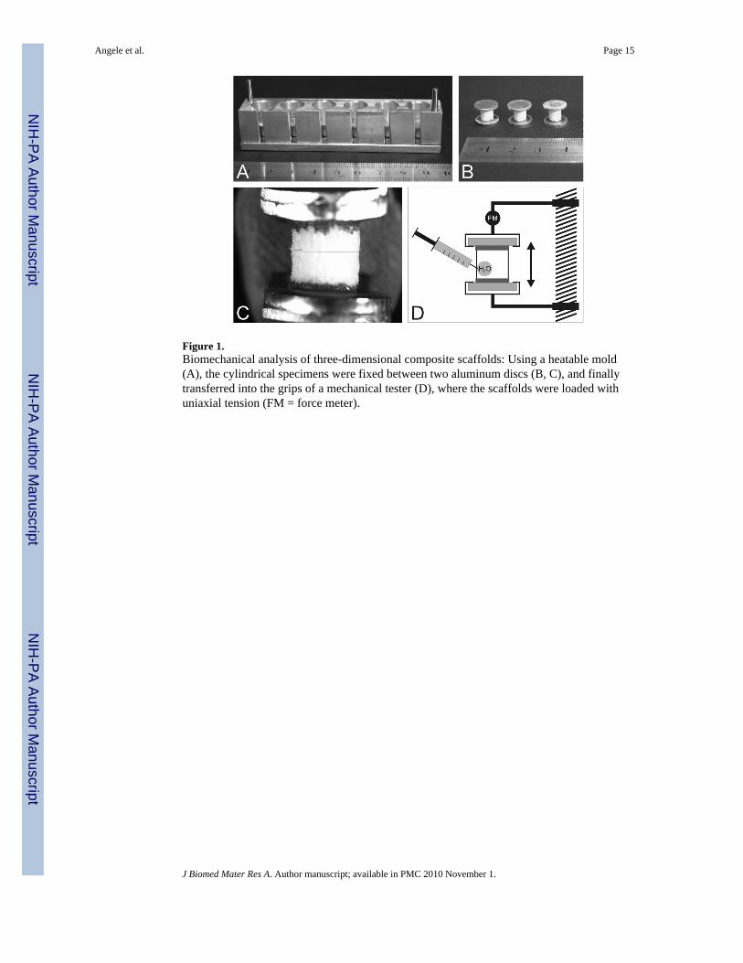
Figure 1.Biomechanical analysis of three-dimensional composite scaffolds: Using a heatable mold(A), the cylindrical specimens were fixed between two aluminum discs (B, C), and finallytransferred into the grips of a mechanical tester (D), where the scaffolds were loaded withuniaxial tension (FM = force meter).
Angele et al. Page 15
J Biomed Mater Res A. Author manuscript; available in PMC 2010 November 1.
NIH
-PA Author Manuscript
NIH
-PA Author Manuscript
NIH
-PA Author Manuscript

Figure 2.Scanning electron micrographs of polymer scaffolds (magnifications: A, C: 200-fold; B, D:10,000-fold). No structural and ultrastructural differences were observed after adding of30% gelatin to hyaluronan scaffolds (A, B) compared to 100% hyaluronan-based scaffolds(C, D). Primary pores are marked with an asterisk (*), regions of secondary pores with acircle (○).
Angele et al. Page 16
J Biomed Mater Res A. Author manuscript; available in PMC 2010 November 1.
NIH
-PA Author Manuscript
NIH
-PA Author Manuscript
NIH
-PA Author Manuscript
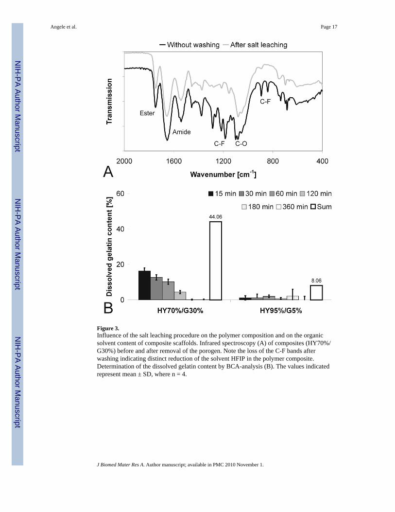
Figure 3.Influence of the salt leaching procedure on the polymer composition and on the organicsolvent content of composite scaffolds. Infrared spectroscopy (A) of composites (HY70%/G30%) before and after removal of the porogen. Note the loss of the C-F bands afterwashing indicating distinct reduction of the solvent HFIP in the polymer composite.Determination of the dissolved gelatin content by BCA-analysis (B). The values indicatedrepresent mean ± SD, where n = 4.
Angele et al. Page 17
J Biomed Mater Res A. Author manuscript; available in PMC 2010 November 1.
NIH
-PA Author Manuscript
NIH
-PA Author Manuscript
NIH
-PA Author Manuscript
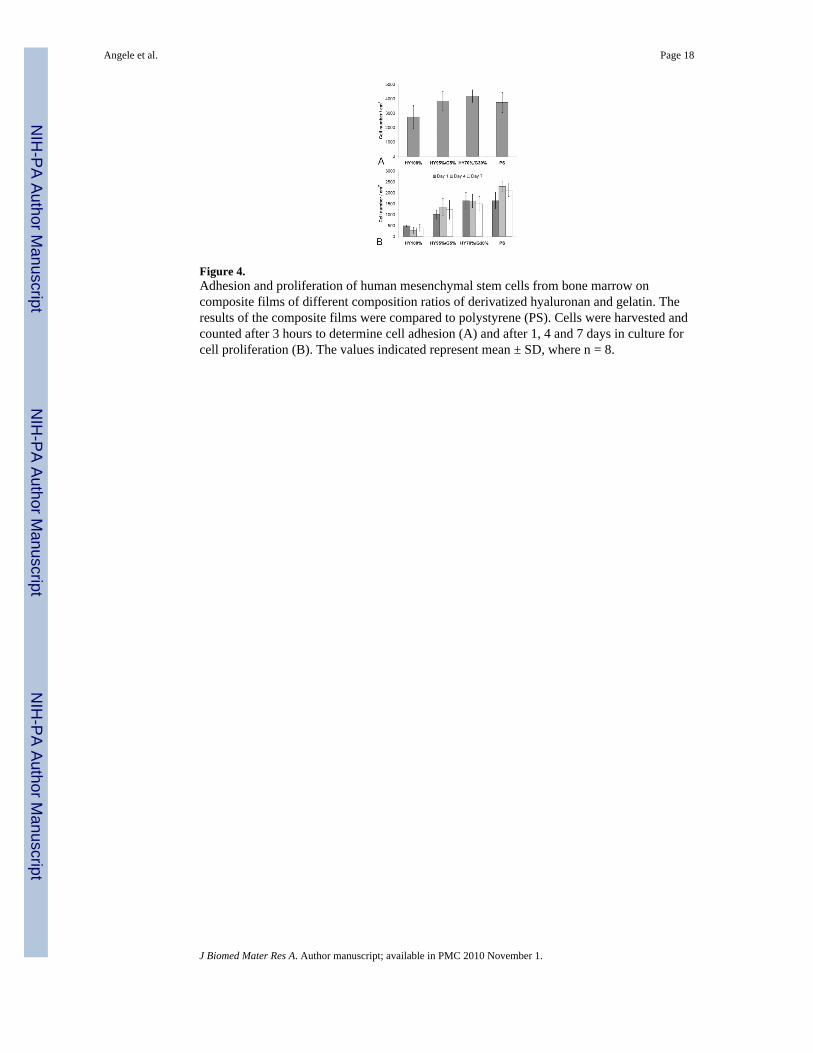
Figure 4.Adhesion and proliferation of human mesenchymal stem cells from bone marrow oncomposite films of different composition ratios of derivatized hyaluronan and gelatin. Theresults of the composite films were compared to polystyrene (PS). Cells were harvested andcounted after 3 hours to determine cell adhesion (A) and after 1, 4 and 7 days in culture forcell proliferation (B). The values indicated represent mean ± SD, where n = 8.
Angele et al. Page 18
J Biomed Mater Res A. Author manuscript; available in PMC 2010 November 1.
NIH
-PA Author Manuscript
NIH
-PA Author Manuscript
NIH
-PA Author Manuscript

Figure 5.Morphology of mesenchymal stem cells attached to films of the composition HY100%,HY95%/G5%, and HY70%/G30% after 1 hour, 2 hours, and 24 hours of 2-dimensional cellculture. In the upper three rows, cells were stained against CD44 expression and the lowerrow shows expression of β1-integrin of stem cells cultured on HY70%/G30% compositefilms. Space bar represents 200 μm.
Angele et al. Page 19
J Biomed Mater Res A. Author manuscript; available in PMC 2010 November 1.
NIH
-PA Author Manuscript
NIH
-PA Author Manuscript
NIH
-PA Author Manuscript

Figure 6.Attachment of mesenchymal stem cells (24 hours incubation) on pure hyaluronan-based(HY100%) and composite polymer films (HY70%/G30%). (A) Dark field microscopy after24 hours of cell culture with (+α5β1-Ab) and without (-α5β1-Ab) addition of α5β1-integrinblocking antibody. (B) Cell count of attached cells (after 24 hours) on pure hyaluronan-based (HY100%) and composite polymer films (HY70%/G30%) without further treatment,after collagenase treatment prior to cell seeding or cell seeding with α5β1-integrin blockingantibodies. The values indicated represent mean ± SD, where n = 5.
Angele et al. Page 20
J Biomed Mater Res A. Author manuscript; available in PMC 2010 November 1.
NIH
-PA Author Manuscript
NIH
-PA Author Manuscript
NIH
-PA Author Manuscript

Figure 7.Chondrogenic differentiation of mesenchymal stem cells. DNA-contents on day 1, 14 and 28(A) and extracellular matrix deposition under chondrogenic culture condition on day 28 (B)of mesenchymal stem cells in hyaluronan-based (HY100%) and composite scaffolds(HY95%/G5%, HY70%/G30%). The values indicated represent mean ± SD, where at least n= 3. Immunohistochemical analysis of collagen type I and collagen type II expression after28 days of chondrogenic cell culture in pure hyaluronan and hyaluronan-gelatin (HY70%/G30%) composite scaffolds (C).
Angele et al. Page 21
J Biomed Mater Res A. Author manuscript; available in PMC 2010 November 1.
NIH
-PA Author Manuscript
NIH
-PA Author Manuscript
NIH
-PA Author Manuscript

NIH
-PA Author Manuscript
NIH
-PA Author Manuscript
NIH
-PA Author Manuscript
Angele et al. Page 22
Table 1
Physicochemical properties of polymer composites depending on composition ratios of derivatized hyaluronanand gelatin (HY100%, HY95%/G5HY70%/G30%) with and without the influence of the salt leachingprocedure. Free amino groups were determined by the TNBS-method. The values indicated represent mean ±SD, where at least n = 3.
IR peak ratio (ester/amide-II) Water contact angle [°] Free amino groups [μmol/g]
Without washing
HY100% 2.01 ± 0.04 51.5 ± 1.5 0
HY95%/G5% 1.78 ± 0.05 37.5 ± 3.0 27.8 ± 5.8
HY70%/G30% 1.21 ± 0.05 24.0 ± 1.5 111.9 ± 12.0
After salt leaching
HY100% 1.96 ± 0.10 48.5 ± 1.5 0
HY95%/G5% 1.92 ± 0.03 39.5 ± 0.5 22.5 ± 8.5
HY70%/G30% 1.29 ± 0.03 35.5 ± 1.0 77.2 ± 5.0
J Biomed Mater Res A. Author manuscript; available in PMC 2010 November 1.

NIH
-PA Author Manuscript
NIH
-PA Author Manuscript
NIH
-PA Author Manuscript
Angele et al. Page 23
Table 2
The mechanical properties of composite scaffolds with varying gelatin contents were obtained in wetconditions. The values indicated represent mean ± SD, with a sample size of n = 6. Statistical analysis wasperformed using SPSS 11.0G One way ANOVA with post hoc Bonferroni test. Significance was acceptedwith a level of p < 0.05 (see text).
Rupture strength [kPa] Max strain at rupture [%] Max Young modulus [kPa]
HY100% 25.3 ± 8.6 36.2 ± 8.4 9.7 ± 2.2
HY95%/G5% 20.7 ± 5.1 43.7 ± 7.4 7.1 ± 1.6
HY70%/G30% 13.8 ± 7.3 38.9 ± 9.8 5.7 ± 2.8
J Biomed Mater Res A. Author manuscript; available in PMC 2010 November 1.
Related Documents