Plant Physiol. (1 995) 107: 1129-1 138 Characterization of the Cell-Wall Polysaccharides of Arabidopsis thaliana Leaves' Earl Zablackis, Jing Huang, Bernd Müllerz, Alan C. Darvill, and Peter Albersheim* Complex Carbohydrate Research Center and Department of Biochemistry and Molecular Biology, University of Georgia, 220 Riverbend Road, Athens, Georgia 30602-471 2 The cell-wall polysaccharides of Arabidopsis fhaliana leaves have been isolated, purified, and characterized. The primary cell walls of all higher plants that have been studied contain cellulose, the three pectic polysaccharides homogalacturonan, rhamnogalacturonan I, and rhamnogalacturonan II, the two hemicelluloses xyloglucan and glucuronoarabinoxylan, and structural glycoproteins. The cell walls of Arabidopsis leaves contain each of these components and no others that we could detect, and these cell walls are remarkable in that they are particularly rich in phosphate buffer-soluble polysac- charides (34% of the wall). The pectic polysaccharides of the purified cell walls consist of rhamnogalacturonan I (1 1 %), rham- nogalacturonan II (8%), and homogalacturonan (23%). Xyloglucan (XC) accounts for 20% of the wall, and the oligosaccharide frag- ments generated from XC by endoglucanase consist of the typical subunits of other higher plant XCs. Clucuronoarabinoxylan (4%), cellulose (14%), and protein (14%) account for the remainder of the wall. Except for the phosphate buffer-soluble pectic polysac- charides, the polysaccharides of Arabidopsis leaf cell walls occur in proportions similar to those of other plants. The structures of the Arabidopsis cell-wall polysaccharides are typical of those of many other plants. The cells of plants are surrounded by a hydrated wall composed of complex carbohydrates, glycoproteins, and phenolics (Darvill et al., 1980a; Fry, 1988).The primary cell walls of a11 plants, those walls that surround growing plant cells, are composed of cellulose, pectins, hemicelluloses, and in most cases glycoproteins (Darvill et al., 1980a). Many primary cell walls of cereals also contain mixed- linked p-glucans. Cellulose, a linear polymer of P-1,4- linked-D-glucopyranosyl residues, is the major load-bear- ing polymer in the wall (Franz and Blaschek, 1990). The pectins are a group of three polysaccharides, each of which contains a high proportion of 1,4-linked a-D-galactosylu- ronic acid residues (York et al., 1985b). The three pectic polysaccharides are homogalacturonan, RG-I, and RG-I1 (ONeill et al., 1990). The hemicellulosic polysaccharides Supported in part by U.S. Department of Energy (DOE)-fund- ed (DE-FG09-93ER20097) Center for Plant and Microbial Complex Carbohydrates and DOE grant DE-FGOY -93ER20115 (to A.G.D.). This is No. XXXIX in the series entitled Structure of Plant Cell Walls. * Present address: SmithKline Beecham Fink GmbH, Benzstrasse 25, 71083 Herrenberg, Germany. * Corresponding author; e-mail palbershQmond1 .ccrc.uga.edu; fax 1-706-542-4412. are functionally defined as those polysaccharides that form strong hydrogen-bonded complexes with cellulose fibers (York et al., 1985b). XG and AX are the hemicelluloses of the primary cell walls of higher plants. Homogalacturonan, which is a chain of partially methyl- esterified 1,4-linked a-D-galactosyluronic acid residues, has a propensity to form gels, a property that may reflect its function in wall structure (Morris, 1986). RG-I is par- tially solubilized from cell walls by treatment with EPG. The backbone of RG-I consists of a linear diglycosyl repeat- ing unit [-a-D-galactopyranosyluronic acid-(1,2)-a-~-rham- nopyranosyl-(1,4)-] (Lau et al., 1985). Approximately one- half of the 2-linked rhamnosyl residues of RG-I are substituted at C-4 with structurally complex oligoglycosyl side chains containing branched and linear arabinosyl and galactosyl residues and to a lesser extent terminal fucosyl and glucosyluronic acid residues (McNeil et al., 1982; Lau et al., 1987; ONeill et al., 1990). RG-I1 is a highly substi- tuted a-1,4-linked homogalacturonan that has a total of approximately 30 glycosyl residues. The 30 glycosyl resi- dues of RG-I1 encompass 11 different sugars, including the following unusual or unusually modified sugars, which are present in cell walls only in RG-11: 2-O-methylxylose, 2-0- methylfucose, Kdo, 3-C-carboxy-5-deoxy-~-xylose (aceric acid), Dha, and apiose (York et al., 1985a; Stevenson et al., 1988a, 1988b). The backbone of RG-I1 consists of at least nine 1,4-1inked a-D-galactosyluronic acid residues. Four different, structurally characterized oligoglycosyl side chains are attached to the RG-I1 backbone. XGs are composed of a branched, 4-linked P-D-glucan backbone. In many plants, approximately 75% of the backbone glucosyl residues are substituted at 0-6 with an a-D-xylosyl residue (Bauer et al., 1973). Selected xylosyl residues are themselves substituted at C-2 with a-L-fuco- pyranosyl-(1,2)-~-~-galactopyranosyl or P-D-galactopyr- anosyl units. The P-D-galactosyl residues are often substi- tuted with one or two O-acetyl groups (York et al., 1988). XGs are thought to be load-bearing structures in the cell wall because of their ability to noncovalently cross-link cellulose microfibrils (Bauer et al., 1973; Hayashi and Mac- lachlan, 1984). The dynamic nature of this cross-linking Abbreviations: AX, arabinoxylan; Dha, 3-deoxy-~-lyxo-2-heptu- losaric acid; EG, endoglucanase; EPG, endopolygalacturonase; GAX, glucuronoarabinoxylan; Kdo, 3-deoxy-~-manno-octu~osonic acid; RG-I, rhamnogalacturonan I; RG-11, rhamnogalacturonan 11; XG, xyloglucan. 1129 www.plant.org on December 24, 2015 - Published by www.plantphysiol.org Downloaded from Copyright © 1995 American Society of Plant Biologists. All rights reserved.

Welcome message from author
This document is posted to help you gain knowledge. Please leave a comment to let me know what you think about it! Share it to your friends and learn new things together.
Transcript

Plant Physiol. (1 995) 107: 11 29-1 138
Characterization of the Cell-Wall Polysaccharides of Arabidopsis thaliana Leaves'
Earl Zablackis, Jing Huang, Bernd Müllerz, Alan C. Darvill, and Peter Albersheim*
Complex Carbohydrate Research Center and Department of Biochemistry and Molecular Biology, University of Georgia, 220 Riverbend Road, Athens, Georgia 30602-471 2
The cell-wall polysaccharides of Arabidopsis fhaliana leaves have been isolated, purified, and characterized. The primary cell walls of all higher plants that have been studied contain cellulose, the three pectic polysaccharides homogalacturonan, rhamnogalacturonan I, and rhamnogalacturonan II, the two hemicelluloses xyloglucan and glucuronoarabinoxylan, and structural glycoproteins. The cell walls of Arabidopsis leaves contain each of these components and no others that we could detect, and these cell walls are remarkable in that they are particularly rich in phosphate buffer-soluble polysac- charides (34% of the wall). The pectic polysaccharides of the purified cell walls consist of rhamnogalacturonan I (1 1 %), rham- nogalacturonan II (8%), and homogalacturonan (23%). Xyloglucan (XC) accounts for 20% of the wall, and the oligosaccharide frag- ments generated from XC by endoglucanase consist of the typical subunits of other higher plant XCs. Clucuronoarabinoxylan (4%), cellulose (14%), and protein (14%) account for the remainder of the wall. Except for the phosphate buffer-soluble pectic polysac- charides, the polysaccharides of Arabidopsis leaf cell walls occur in proportions similar to those of other plants. The structures of the Arabidopsis cell-wall polysaccharides are typical of those of many other plants.
The cells of plants are surrounded by a hydrated wall composed of complex carbohydrates, glycoproteins, and phenolics (Darvill et al., 1980a; Fry, 1988). The primary cell walls of a11 plants, those walls that surround growing plant cells, are composed of cellulose, pectins, hemicelluloses, and in most cases glycoproteins (Darvill et al., 1980a). Many primary cell walls of cereals also contain mixed- linked p-glucans. Cellulose, a linear polymer of P-1,4- linked-D-glucopyranosyl residues, is the major load-bear- ing polymer in the wall (Franz and Blaschek, 1990). The pectins are a group of three polysaccharides, each of which contains a high proportion of 1,4-linked a-D-galactosylu- ronic acid residues (York et al., 1985b). The three pectic polysaccharides are homogalacturonan, RG-I, and RG-I1 (ONeill et al., 1990). The hemicellulosic polysaccharides
Supported in part by U.S. Department of Energy (DOE)-fund- ed (DE-FG09-93ER20097) Center for Plant and Microbial Complex Carbohydrates and DOE grant DE-FGOY -93ER20115 (to A.G.D.). This is No. XXXIX in the series entitled Structure of Plant Cell Walls.
* Present address: SmithKline Beecham Fink GmbH, Benzstrasse 25, 71083 Herrenberg, Germany.
* Corresponding author; e-mail palbershQmond1 .ccrc.uga.edu; fax 1-706-542-4412.
are functionally defined as those polysaccharides that form strong hydrogen-bonded complexes with cellulose fibers (York et al., 1985b). XG and AX are the hemicelluloses of the primary cell walls of higher plants.
Homogalacturonan, which is a chain of partially methyl- esterified 1,4-linked a-D-galactosyluronic acid residues, has a propensity to form gels, a property that may reflect its function in wall structure (Morris, 1986). RG-I is par- tially solubilized from cell walls by treatment with EPG. The backbone of RG-I consists of a linear diglycosyl repeat- ing unit [-a-D-galactopyranosyluronic acid-(1,2)-a-~-rham- nopyranosyl-(1,4)-] (Lau et al., 1985). Approximately one- half of the 2-linked rhamnosyl residues of RG-I are substituted at C-4 with structurally complex oligoglycosyl side chains containing branched and linear arabinosyl and galactosyl residues and to a lesser extent terminal fucosyl and glucosyluronic acid residues (McNeil et al., 1982; Lau et al., 1987; ONeill et al., 1990). RG-I1 is a highly substi- tuted a-1,4-linked homogalacturonan that has a total of approximately 30 glycosyl residues. The 30 glycosyl resi- dues of RG-I1 encompass 11 different sugars, including the following unusual or unusually modified sugars, which are present in cell walls only in RG-11: 2-O-methylxylose, 2-0- methylfucose, Kdo, 3-C-carboxy-5-deoxy-~-xylose (aceric acid), Dha, and apiose (York et al., 1985a; Stevenson et al., 1988a, 1988b). The backbone of RG-I1 consists of at least nine 1,4-1inked a-D-galactosyluronic acid residues. Four different, structurally characterized oligoglycosyl side chains are attached to the RG-I1 backbone.
XGs are composed of a branched, 4-linked P-D-glucan backbone. In many plants, approximately 75% of the backbone glucosyl residues are substituted at 0-6 with an a-D-xylosyl residue (Bauer et al., 1973). Selected xylosyl residues are themselves substituted at C-2 with a-L-fuco- pyranosyl-(1,2)-~-~-galactopyranosyl or P-D-galactopyr- anosyl units. The P-D-galactosyl residues are often substi- tuted with one or two O-acetyl groups (York et al., 1988). XGs are thought to be load-bearing structures in the cell wall because of their ability to noncovalently cross-link cellulose microfibrils (Bauer et al., 1973; Hayashi and Mac- lachlan, 1984). The dynamic nature of this cross-linking
Abbreviations: AX, arabinoxylan; Dha, 3-deoxy-~-lyxo-2-heptu- losaric acid; EG, endoglucanase; EPG, endopolygalacturonase; GAX, glucuronoarabinoxylan; Kdo, 3-deoxy-~-manno-octu~osonic acid; RG-I, rhamnogalacturonan I; RG-11, rhamnogalacturonan 11; XG, xyloglucan.
1129 www.plant.org on December 24, 2015 - Published by www.plantphysiol.orgDownloaded from
Copyright © 1995 American Society of Plant Biologists. All rights reserved.

1130 Zablackis et al. Plant Physiol. Vol. 107, 1995
has been hypothesized to be a major factor in controlling the rate of cell-wall expansion, thereby regulating plant growth. Furthermore, specific XG-derived, Fuc-containing oligosaccharides have been shown to have growth-inhibit- ing and -promoting activity (York et al., 1984; McDougall and Fry, 1991; Aldington and Fry, 1993).
GAX is an acidic hemicellulosic polysaccharide with a backbone consisting of 4-linked-P-~-xylosyl residues. In many dicotyledonous plants, approximately 25% of the backbone xylosyl residues are substituted at 0-2 with ei- ther arabinosyl, glucosyluronic acid, or 4-O-methyl glu- cosyluronic acid residues (Darvill et al., 1980b). GAX and AX are found as the major hemicelluloses of Gramineae cell walls.
Arabidopsis thaliana is used widely in molecular biology. The relative ease with which Arabidopsis mutants can be obtained has led to mutants with altered cell-wall glycosyl compositions (Reiter et al., 1993). Before characterizing the structural alterations in these mutants, we decided to de- termine the compositions and structures of the wall po- lysaccharides of wild-type plants. In this paper, we de- scribe the isolation and characterization of Arabidopsis leaf cell-wall polysaccharides and compare the Arabidopsis po- lysaccharides with those of the walls of suspension-cul- tured sycamore cells.
MATERIALS AND METHODS
Plant Growth and lsolation of Cell Walls
Seeds of wild-type Arabidopsis thaliana (Columbia type, Somerville lab) were obtained from Christopher Somer- ville, Clint Chappel, and Wolf-Dieter Reiter (Michigan State University, East Lansing). Plants were grown under a 12:12 h 1ight:dark regime at 22°C for 4 weeks. The plants were placed in the dark for 24 h prior to harvesting. The leaves were processed and cell walls isolated as illustrated in the scheme shown in Figure 1. The individual wall polysaccharides and/or their oligosaccharide subunits were isolated as shown in Figure 2. The procedures used are described below.
Extraction of Cell-Wall Polysaccharides
Portions of the pectic material were solubilized from the walls by each of the following sequential treatments: phos- phate buffer, EPG, NaOH, EPG, and Na,CO,. Residual starch and pectic material were removed by suspending cell walls (10 g L-') in buffer (0.1 M potassium phosphate, pH 7) containing 0.02% thimerosal and 5000 units of a-amylase (type IIA from Bacillus sp., Sigma) per g of cell wall. The reaction was carried out in polypropylene bottles placed in a shaking water bath at 30°C for 24 h. After 24 h the enzyme reaction mixture was centrifuged at 8000g for 20 min, the supernatant was removed and stored at 4"C, the pellet was suspended in buffer containing 0.02% thimerosal with an additional 5000 units of a-amylase per g of cell wall added, and the digestion was continued for an additional24 h. The sample was then centrifuged as above, and the pellet was washed once with buffer and once with water and recovered by centrifugation. The supernatants
Grow Plants +Harvest h v e s
Grind in liquid N, + Extract with 1.5% SDS + Homogenize + Centrifuge
i I I I I I i I
Extract Pelkt with 0.5% SDS + Centrifuge
Extract Pellet with 0.1 M KP04 -+ Centrifuge
Extract Pellet with 0.5 M KP04+ Centrifuge
Break Cells Using Parr Bomb- Centrifuge
Wash Pellet with 0.5 M D O 4 +Water + Centi:ifuge
Extract Pellet with PhenokAcetic Acid:Water (2A:l) + Centrifuge
Wash Pellet with Water + Dry with Acetone
CELL WALLS
Figure 1. Scheme followed for the preparation of cell walls from leaves of Arabidopsis.
and washes were combined in 1000 mo1 wt cutoi'f dialysis tubing, dialyzed against water at 4"C, concentrated using a Rotovapor, and then lyophilized. The pellet was washed with acetone and air dried. Extraction of the cell walls with potassium phosphate buffer in the absence of tr-amylase extracted the same components from the walls (data not shown). No polygalacturonase activity was detwtable in the a-amylase preparation.
Following treatment with a-amylase, the w 311s were treated with EPG as described by Lerouge et al. (1 993). The walls were then de-esterified by suspending the simples in water, adjusting to pH 12 with cold 0.1 M NzOH, and stirring 4 h at 4°C while maintaining the pH at 12 by addition, as needed, of 0.1 M NaOH. The cell-wa 1 suspen- sion was filtered through glass-fiber filters, and the wall residue was washed extensively with water. The washes and NaOH-solubilized material were combinecl in 1000 mo1 wt cutoff dialysis tubing, dialyzed against water, con- centrated using a Rotovapor, and then 1yophilLzed. The walls were then re-treated with EPG and the extract was recovered as above.
Na,CO, extraction of the walls after the second EPG treatment and purification of the Na,CO,-solubilized ma- terial were carried out as described by Ishii et al. (1989).
RG-I, RG-11, and oligogalacturonides were isolded from the EPG-, NaOH-, and potassium phosphate buff er-solubi- lized material as described by Marfh et al. (1991). RG-I isolated in the various extracts of Arabidopsis leaves had rhamnosy1:galactosyluronic acid ratios between 1 :2 and 1 :5 rather than 1:l as was obtained elsewhere (ONeill et al., 1990), suggesting that the Arabidopsis RG-I contained some 4-linked galactosyluronic acid residues resistant i o EPG. It is known that methyl-esterified galactosyluronic acid resi- dues are resistant to cleavage by EPG (McNeil et al., 1980).
www.plant.org on December 24, 2015 - Published by www.plantphysiol.orgDownloaded from Copyright © 1995 American Society of Plant Biologists. All rights reserved.

Ara bidopsis Ce I I Wa I I s 1131
Cell Walls RG-I-Agarose A-SM-RG-I RG-IiRG-I1 Blo el
NaOH 1 Amylase
RG-uRG-ll Bio Gel P 30 Oligogalact-
uronides Oligogalact-
Solubilized material 4 i uronides
Oligogalact- uronides
kNa2C03-Solubilized material ~ ~ ~ ~ ~ ~ n t RG-IAgarose A-5M
Neutral- Reduction
* ." I
e õ Endoglucanase -Solublized material
u Acidic (RG-I)
Cell Wall Residue
Cell Wall Residue
Figure 2. Scheme followed for the preparation and purification of polysaccharides from leaves of Arabidopsis.
Therefore, the RG-I was re-treated with NaOH and EPG and then treated with exopolygalacturonase as described by Lerouge et al. (1993). The products of the reaction were separated by gel permeation chromatography using a Bio- Gel P-30 column (2.5 X 35 cm) that had been equilibrated with 0.05 M sodium acetate, pH 5.2. The products were eluted from the column with 0.05 M sodium acetate and 5-mL fractions were collected. The RG-I that eluted from the column had a rhamnosy1:galactosyluronic acid ratio of approximately 1:l.
After Na,CO, extraction, the walls were treated sequen- tially with EG, 1 M KOH, and 4 M KOH to solubilize hemicelluloses, xylan, and XG. The residual walls (1 g) remaining after Na,CO, extraction were incubated (24 h, ambient temperature) in buffer (100 mL, 50 mM acetic acid-sodium acetate, 0.02% thimerosal, pH 5.2) containing endo-( 1 +-4)-P-glucanase (60 units) from Trichoderma reesei (Megazyme Australia, Inc., Warriewood, Australia). The wall suspension was then passed through a glass-fiber filter. The filtrate was stored at 4"C, the walls were resus- pended in buffer, another 60 units of the EG was added, and the suspension was incubated at ambient temperature for an additional 24 h. The wall suspension was then filtered as above and washed extensively with water. The EG-solubilized material and washes were combined, neu- tralized, and then separated into neutral and acidic frac- tions by passing the solution over a column of Q-Sepharose (2.5 X 45 cm) that had been equilibrated with 10 mM imidazole buffer, pH 7. The neutral fraction was concen- trated to 10 mL on a Rotovapor, desalted by passing the concentrate over a column of Sephadex G-10 (2.5 X 17 cm), and then fractionated by gel permeation chromatography (see below). The bound acidic fraction was eluted from the column with 2 M imidazole, placed in 1000 mo1 wt cutoff dialysis tubing, dialyzed against water, concentrated using a Rotovapor, and lyophilized.
The KOH extractions were carried out as follows. EG residual walls (1 g) were dispersed in 1 M KOH (200 mL containing 1 mg mL-' NaBH,) and stirred for 2 h at 4°C. The wall suspension was then passed through a glass fiber filter and the walls were washed extensively with water. The 1 M KOH-solubilized material and washes were com- bined, neutralized with acetic acid, dialyzed, and lyophi- lized. The walls were resuspended in 4 M KOH (200 mL containing 1 mg mL-' NaBH,) and stirred for 2 h at ambient temperature. The 4 M KOH-solubilized material was treated as above and the wall residue dried with acetone.
The 1 and 4 M KOH extracts were each separated into neutral and acidic fractions by ion-exchange chromatogra- phy. The extracts (80-100 mg) were separately dissolved in 10 mM imidazole buffer (5-10 mL) and passed over a column of Q-Sepharose (2.5 X 45 cm) that had been equil- ibrated with 10 mM imidazole buffer, pH 7. The neutral fractions were concentrated on a Rotovapor, desalted by passing the concentrate over a column of Sephadex G-10 (2.5 X 17 cm), and lyophilized. The bound acidic fraction was eluted from the column with 2 M imidazole buffer, pH 7, placed in 1000 mo1 wt cutoff dialysis tubing, dialyzed against water, concentrated using a Rotovapor, and lyophilized.
XG oligosaccharide subunits were generated and puri- fied from the neutral fractions of the EG and 4 M KOH extracts as follows. The neutral EG extract was separated into oligosaccharide size classes by Bio-Gel P-6 chromatog- raphy (1.5 X 85 cm column equilibrated with 10 mM imi- dazole buffer, pH 7). Four size-fractions were collected, concentrated to 10 mL, desalted by passing each concen- trate over a column of Sephadex G-10 (2.5 X 17 cm), and lyophilized. The neutral component of the 4 M KOH extract (21 mg) was incubated (48 h, ambient temperature) in buffer (22 mL, 50 mM HOAc-NaOAc, 0.02% thimerosal, pH
www.plant.org on December 24, 2015 - Published by www.plantphysiol.orgDownloaded from Copyright © 1995 American Society of Plant Biologists. All rights reserved.

1132 Zablackis et ai. Plant Physiol. Vol. 107, 1995
5.2) containing endo-(l+)-P-glucanase (1 unit) from T. reesei (Megazyme Australia, Inc.). The pH of the enzyme reaction mixture was adjusted to 7.8 and the solution passed over a Q-Sepharose column (1.5 X 10 cm) previ- ously equilibrated with 10 mM imidazole buffer, pH 7. The neutral fraction was concentrated using a Rotovapor, de- salted by passing the concentrate over a column of Seph- adex G-10 (2.5 x 17 cm), and lyophilized. The neutral fraction of the endo-(l+4)-P-glucanase-re-treated 4 M KOH extract was separated into size classes by high-resolution Bio-Gel P-2 chromatography (York et al., 1990).
XG oligosaccharides from the EG and 4 M KOH extracts were converted to their corresponding oligoglycosyl aldi- tols by reduction with NaBH, (York et al., 1993) and then lyophilized. The reduced XG oligoglycosyl alditols (1-10 mg) were dissolved in water (300 pL) and separated by reversed-phase HPLC using a Spherisorb 5 ODS (1) column (Phenomenex, Torrance, CA). The separation was accom- plished by isocratic elution in 10% methanol (York et al., 1990).
Analytical Methods
Protein content was determined colorimetrically using the method of Bradford (1976). Hyp content and amino acid composition were determined as described by Lam- port and Miller (1971) and Kieliszewski et al. (1992), re- spectively. Cellulose content was determined by the method of Updegraff (1969). Neutral sugar, uronic acid, and Kdo concentrations were determined by the following methods, respectively: anthrone method with Glc as the standard (Dische, 1962), metahydroxydiphenyl method with galacturonic acid as the standard (Blumenkrantz and Asboe-Hansen, 19731, and the modified thiobarbituric acid assay (York et al., 1985a).
Neutral glycosyl-residue compositions were determined by GLC of their alditol acetate derivatives. Combined neu- tral and acidic glycosyl-residue compositions were deter- mined by GLC of their trimethylsilyl methyl-ester methyl- glycoside derivatives (York et al., 198513).
Glycosyl-linkage compositions were determined using a modification of the Hakomori procedure (Hakomori, 1964). Oligo- and polysaccharides were per-O-methylated with butyl-lithium (2 M solution in cyclohexane; Aldrich, Mil- waukee, WI) and iodomethane (Kvernheim, 1987), and the resulting products were isolated using Sep-Pak CI8 car- tridges (An et al., 1994). Uronic acid-containing oligo- and polysaccharides were methylated as above, carboxyl re- duced with Superdeuteride (1 M Li-triethylborodeuteride in tetrahydrofuran, Aldrich) as described by York et al. (1985b), and remethylated as above. The glycosyl-linkage compositions were then determined by GC-MS of the par- tially methylated, partially acetylated alditol acetate deriv- atives (York et al., 1985b).
Oligogalacturonides were separated on an analytical CarboPac-I anion-exchange column installed in a Dionex (Sunnyvale, CA) BioLC system. The oligogalacturonides were detected using pulsed high-performance anion-ex- change chromatography with pulsed amperometric detec- tion as described by ONeill et al. (1990). The components
were com pared to standard oligogalacturonides by their retention times.
XG oligosaccharide subunit structures were determined by 'H-NMR spectroscopy. Hydroxyl protons of the XG oligoglycosyl alditols obtained by treatment of the walls with EG or KOH were exchanged with deuterons and the samples dissolved in distilled water. The 500-1VIHz 'H- NMR spectra were recorded under standard conditions (York et al., 1993).
RESULTS AND DISCUSSION
Arabidopsis Cell Walls Are Composed of Protein m d the Standard Six Wall Polysaccharides
Cell walls were isolated from the leaves of Arubidopsis and analyzed for protein and carbohydrate content. The Arubidopsis leaf cell walls are relatively rich in prc teins that have an amino acid composition similar to those of a variety of other plant tissues (Kieliszewski et al., 3 992). The purified walls contain 136 pg of protein containing only 1.7 pg Hyp per mg of cell wall prior to subjecting th? walls to various extraction procedures. The cellulose-ric I residue that remains after subjecting the walls to a11 of tlie extrac- tion procedures described in this paper contains 20 pg protein and 1.3 pg Hyp per mg. Thus, approxim(ite1y 99% of the total wall protein was removed by the extraction procedures. The intact walls (Table I) contain only 0.6 mol% Hyp. Thus, the Arubidopsis leaf cell walls are not as rich in Hyp as are, for example, the walls of suspension- cultured sycamore cells, which contain 12 mol% H yp (Lam- port and Northcote, 1960).
The carbohydrate components of the Arubidopsis cell walls were isolated, purified, and chemically char,icterized. Only six polysaccharides are present in the walls of Arubi-
.
Table 1. Amino acid compositions of Arabidopsis leaf cell walls and suspension-cultured sycamore ceh
Sycamore" Arabidopsis Amino Residueb Iiitact Acid lntact
mo/ %
HYP 0.6 4.4 12.9 Asx 9.2 7.7 8.5
Ser 5.8 4.3 9.4
Pro 4.8 6.8 4.5 ClY 9.5 16.5 3.2 Ala 9.9 8.6 3.6 Val 6.5 7.1 6.2 CY S n.d.' n.d. n.d. Met 0.8 0.8 1.5
Leu 12.4 10.3 5.7 TY 2.3 1.5 5.1 Phe 3.8 5.2 4.4 LYS 5.4 4.5 13 His 1.3 O 3.4 Arg 4.8 5 3.3
Thr 5.4 3.8 3.8
Clx 11.5 8.2 8.2
Ile 5.4 5.2 3.8
a From Lamport and Northcote, 1960. Cell-wall residue after extraction of polysaccharides. n.d., Not determined.
www.plant.org on December 24, 2015 - Published by www.plantphysiol.orgDownloaded from Copyright © 1995 American Society of Plant Biologists. All rights reserved.
Arabidopsis Cell Walls 1133
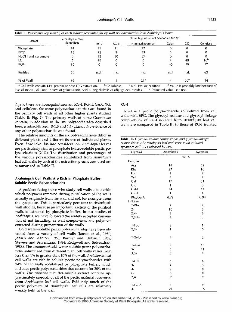
Table II. Percentage (bv weight) of each extract accounted for bv wall polysaccharides from Arabidopsis leaves Percentage of Extract Accounted for by Percentage of Wall Extract
Sol u bi I ized RC-I RC-II Homogalacturonan Xylan XC Cellulose
Phosphate 34 11 11 37 O O O EPGa 18 22 9 39 O O O NaOH and carbonate 8 12 30 37 O O O EG 5 40 O O 4 40 1 6b KOH 10 O O O 40 50 2b
Residue 20 n.d.' n.d. n.d. n.d. n.d. 63
% of Wall 95 11 8 23d 4 2oe 14
a Cell walls contain 14% protein prior to EPG extraction. Cellobiose. n.d., Not determined. Value is probably low because of loss of mono-, di-, and trimers of galacturonic acid during dialysis of oligogalacturonides. e Estimated value, see text.
dopsis; these are homogalacturonan, RG-I, RG-11, GAX, XG, and cellulose, the same polysaccharides that are found in the primary cell walls of a11 other higher plants studied (Table 11; Fig. 2). The primary walls of some Gramineae contain, in addition to the six polysaccharides described here, a mixed-linked (p-19 and 1,4) glucan. No evidence of any other polysaccharide was found.
The relative amounts of the six polysaccharides differ in different plants and different tissues of individual plants. Even if we take this into consideration, Arubidopsis leaves are particularly rich in phosphate buffer-soluble pectic po- lysaccharides (20%). The distribution and percentages of the various polysaccharides solubilized from Arubidopsis leaf cell walls by each of the extraction procedures used are summarized in Table 11.
Arabidopsis Cell Walls Are Rich in Phosphate Buffer- Soluble Pectic Polysaccharides
A problem facing those who study cell walls is to decide which polymers removed during purification of the walls actually originate from the wall and not, for example, from the cytoplasm. This is particularly pertinent to Arubidopsis wall studies, because an important fraction of the purified walls is extracted by phosphate buffer. In our studies of Arubidopsis, we have followed the widely accepted conven- tion of not including, as wall components, any polymers extracted during preparation of the walls.
Cold water-soluble pectic polysaccharides have been ob- tained from a variety of cell walls (Jansen et al., 1960; Jensen and Ashton, 1960; Barbier and Thibault, 1982; Stevens and Selvendran, 1984; Redgwell and Selvendran, 1986). The amount of cold water-soluble pectic polysaccha- rides solubilized from different plant cell walls varies from less than 1% to greater than 10% of the wall. Arubidopsis leaf cell walls are rich in soluble pectic polysaccharides with 34% of the walls solubilized by phosphate buffer, which includes pectic polysaccharides that account for 20% of the walls. The phosphate buffer-soluble extract contains ap- proximately one-half of a11 of the pectic material recovered from Arubidopsis leaf cell walls. Evidently much of the pectic polymers of Arubidopsis leaf cells are relatively weakly held in the wall.
RG-I
RG-I is a pectic polysaccharide solubilized from cell walls with EPG. The glycosyl-residue and glycosyl-linkage compositions of RG-I isolated from Arubidopsis leaf cell walls are compared in Table I11 to those of RG-I isolated
Table 111. Glycosyl-residue compositions and glycosyl-linkage compositions of Arabidopsis leaf and suspension-cultured svcamore cell RG-I released bv EPG
Clycosyl Arabidopsis Sycamore
Residue Ara Rha Fuc
Gal Glc GalA ClcA R h a/G a I A
Linkage T-Rha 2- 2,4- 2,3,4-
XY I
T-FUC 2,3-
T-Xylp
T-Araf 5- 3,5-
T-Cal 3- 4- 6- 2,4
T-CalA 4-
14 27 1 5 17 1
34 1
0.79
2 10 3 4
2 1
mo/ %
32 16 2 2 31 O 17 1
0.94
2 8 8 tr
1 O
4 2
8 6 3
1 21
10 1 1 4
2 15
www.plant.org on December 24, 2015 - Published by www.plantphysiol.orgDownloaded from Copyright © 1995 American Society of Plant Biologists. All rights reserved.

1134 Zablackis et al. Plant Physiol. Vol. 107, 1995
from suspension-cultured sycamore cells. The glycosyl linkages of Arubidopsis RG-I and sycamore RG-I are quali- tatively the same.
The ratio of 2-linked to 2,4-linked rhamnosyl residues in RG-I is indicative of the degree of branching of the mole- cule's linear backbone, because the side chains of RG-I are attached to 0-4 of the 2-linked rhamnosyl residues. Arubi- dopsis RG-I has fewer 2,4-linked rhamnosyl residues than 2-linked rhamnosyl residues in comparison with sycamore RG-I, indicating that Arubidopsis RG-I has fewer branches. Further evidence of less branching in Arubidopsis RG-I is provided by the smaller proportion of galactosyl and ar- abinosyl residues, which are only present in the side chains (Table 111).
RG-I is also present in the EG and Na,CO, extracts of Arubidopsis leaf cell walls. The presence of RG-I in the acidic fraction of the EG extract was verified by glycosyl-linkage composition analysis (Table IV). Although the EG extract accounts for only 5% of the Arubidopsis cell walls, RG-I accounts for 40% of the EG extract. The amount of RG-I solubilized from Arubidopsis leaf cell walls by Na,CO, is similar to that extracted by Na,CO, from sycamore cell walls (Ishii et al., 1989). The glycosyl-residue and glycosyl-
Table IV. Glycosyl-linkage compositions o f the acidic fractions from EG- and KOH-solubilized polvsaccharides from Arabidopsis . , cell walls
Clycosyl Linkage
T-Rha 2- 3- 2,4- T-FUC T-Xylp 4- 2,4- 4-Man T-Araf 2- 3- 5- 2,5- 3,s- T-Arap T-Cal 3- 4- 6- 2,4- T-Glc 4- 6- 4,6- 4-CalA 2,4- T-GICA 2-
4 M EC 1 MKOH KOH
mo/ %
2 trb 12 6 1 1 1 9 tr 7 2 2 4 10 4
36 9 19 2 2
6 2 7 3 1 3
5 tr 7 3 3
9 tr 10 1 7 3 4 7 7 4 1 4
2 4 tr tr 2 2 2
1 4 25 2 10 3 tr
9 tr 3 1
PSa
RG-I, RG-II RG-I, RG-II
RC-I RC-I, RG-II, XG RC-I, RG-II, XG, GAX CAX CAX
RG-I, RG-II, CAX GAX
RG-I
RG-I RC-ll RC-I, RG-II, XC RC-I RG-I RG-I RG-I, RG-II
XG XG XG RC-I, RG-II
GAX RG-II
c
a Polysaccharide(s) that contain the indicated linkage. tr, Trace. Product of undermethylation.
linkage compositions of RG-I solubilized from Arubidopsis leaf cell walls by Na,CO, are similar to thox of RG-I isolated by the other extraction procedures (data not shown), including the ratio of 2-linked to 2,4-linl<ed rham- nosyl residues (1:l). In contrast, RG-I solubilized from suspension-cultured sycamore cells by Na,CO, has a 1:11 ratio of 2-linked to 2,4-linked rhamnosyl residues, indicat- ing a much more highly branched molecule. Thus, Arubi- dopsis cell walls may not have the highly branched compo- nent of RG-I that is present in sycamore cells.
RC-II
RG-I1 is a highly substituted 4-linked homogalacturonan that is distinguished by the presence of severa1 Lmusual or unusually modified glycosyl residues. The gly :osyl-resi- due and glycosyl-linkage compositions of Arubidopsis RG-I1 are similar to those of sycamore RG-I1 (Table U). Indeed, Arubidopsis RG-I1 contains a11 of the unusual sugas unique to RG-11. Our analysis might be taken as indicating that Arubidopsis RG-I1 contains fewer apiosyl and aceryl acid residues than does sycamore RG-11. These difficiilt to ana- lyze, branched glycosyl residues are the attachment points of the RG-I1 side chains to the homogalacturonan back- bone. Since the amount of the other glycosyl-residue com- ponents of the RG-I1 side chains are not lower than their counterparts in sycamore, the apparent low abundance of apiosyl and aceryl acid residues is in a11 likelihood the result of underestimation because of difficulties in quanti- fying these acid-labile residues.
Severa1 glycosyl residues in the Arubidopsis RG-I1 analy- ses, detected by methylation analysis at 3 mol% or less, are not components of sycamore RG-11. Each of these glycosyl residues (2,4- and 3,4-linked rhamnosyl, 5-linked arabino- furanosyl, and 3- and 4-linked galactosyl) are major com- ponents of RG-I. The analysis of Arubidopsis RG-I1 also indicated higher than expected amounts of 2-linked rham- nosyl and 4-linked galactosyluronic acid residues, which are also abundant glycosyl residues of RG-I. Thus, the Arubidopsis RG-I1 preparation was likely contamirlated with approximately 25% RG-I. If we consider the unclerestima- tion of apiosyl and aceryl acid residues and the contami- nation by RG-I, the glycosyl-residue and glycos yl-linkage compositions of Arubidopsis RG-I1 are typical of tk ose of the other plants that have been analyzed.
Homogalacturonan
Homogalacturonan, as evidenced by unsubstituted, 4-linked galactosyluronic acid residues, is presmt in the phosphate buffer, EPG, NaOH, and Na,CO, extracts of Arubidopsis leaf cell walls and accounts for at leelst 23% of the cell wall (Table 11; Fig. 3). Many of the galactcisyluronic acid residues in the homogalacturonan in a11 of the other plants so far studied are methyl esterified. Methyl-esteri- fied galactosyluronic acid residues are resistan t to EPG hydrolysis.
The pectic polysaccharides present in the atfove-men- tioned extracts were treated with NaOH to de-esterify the galactosyluronic acid residues, and then the po1 ysacchar-
www.plant.org on December 24, 2015 - Published by www.plantphysiol.orgDownloaded from Copyright © 1995 American Society of Plant Biologists. All rights reserved.

Arabidopsis Cell Wal 1s 1135
Table V. Glycosyl-residue and glycosyl-linkage compositions of Arabidopsis leaf and svcamore RG-I/
~~
G I ycosyl Arabidopsis Sycamore
Residue Ara Rha Fuc 2Me-Fuc XY I 2Me-Xyl Cal Glc GalA GlcA Api Ace Kdo Dha
Lin kage T-Rha 2-b 3- 2,4-b 3,4-b 2,3,4- T-Fuc 3,4- T-Xylp T-Araf 5-b T-Arap T-Cal 3-b 4-b 2,4- T-GalA 4-b 2,4- 3,4- 2-GlcA
1 7 10 3 1 3 2 9 2
44 2 3 1 3
n.d."
3 7 4 3 3 5 4 4 7 5 3 2 7 3 4 2 7
13 4 3 4
mo/ %
10 12 4 4 2 5 9 2
31 3 6 3 4 4
7 trc 6 O O 5 5 5 5 6 O 5 5 O O 6 10 9 5 7 6
a n.d., Not determined. RG-I contaminants. tr, Trace.
A ArabIdopsis decasaccharide
ides were re-treated with EPG. This procedure generated mono-, di-, and trigalacturonides, providing evidence for the presence of methyl-esterified 4-linked galactosyluronic acid residues in Arabidopsis leaf cell walls.
The oligogalacturonides generated by EPG treatment of the cell walls before they were de-esterified ranged in size from degree of polymerization 1 to 12, similar to the size range of oligogalacturonides isolated by this procedure from suspension-cultured sycamore cells (Marfi et al., 1991). The sizes of the EPG-generated oligogalacturonides were determined by comparison, after de-esterification, of their high-performance anion-exchange chromatography with pulsed amperometric detection retention times with those of oligogalacturonide standards. The isolation from Arabidopsis leaf cell walls of EPG-generated oligosacchar- ides composed of unbranched sequences of 4-linked galac- tosyluronic acid residues establishes the presence of ho- mogalacturonan in these walls.
CAX
GAX accounts for about 5% of the cell walls of dicots (Darvill et al., 1980b; Fry, 1988). This acidic hemicellulose is extracted from cell walls by treatment with base. Arabidop- sis cell walls are no exception. Approximately one-half of the KOH-solubilized material is acidic in nature. Glycosyl- linkage composition analyses of the acidic fractions of the KOH-solubilized material showed that these fractions con- tain glycosyl residues characteristic of GAX, i.e. 4-linked xylosyl, T- and 2-linked arabinosyl, and T-glucosyluronic acid residues (Table IV). The acidic fractions of the KOH- solubilized material account for approximately 4% of the Arabidopsis leaf cell wall. GAX was not further purified from these fractions.
XC
The typical dicot cell wall contains about 20% XG. The EG and KOH extracts of Arabidopsis leaf cell walls contain glycosyl-residue and glycosyl-linkage compositions char- acteristic of XG (Table VI). The recovered XG accounted for only 7% of Arabidopsis leaf cell walls. However, the
Figure 3. The a-anomeric regions of the 'H- NMR spectra of reduced XG decasaccharide (i.e. nonaglycosyl alditol: XLFGol) isolated from Arabidopsis (A) and rapeseed (B). M
B Rapeseed decasaccharide
I - ~ p - . I . . . . , ' . . . l . . . . . . . . . . . . . . I . . . . . . . . 5 .35 5.30 5.25 5.20 Bpils ' 5.110 5.b5 s.'oo 4.lg5
www.plant.org on December 24, 2015 - Published by www.plantphysiol.orgDownloaded from Copyright © 1995 American Society of Plant Biologists. All rights reserved.
1136 Zablackis et al. Plant Physiol. Vol. 07, 1995
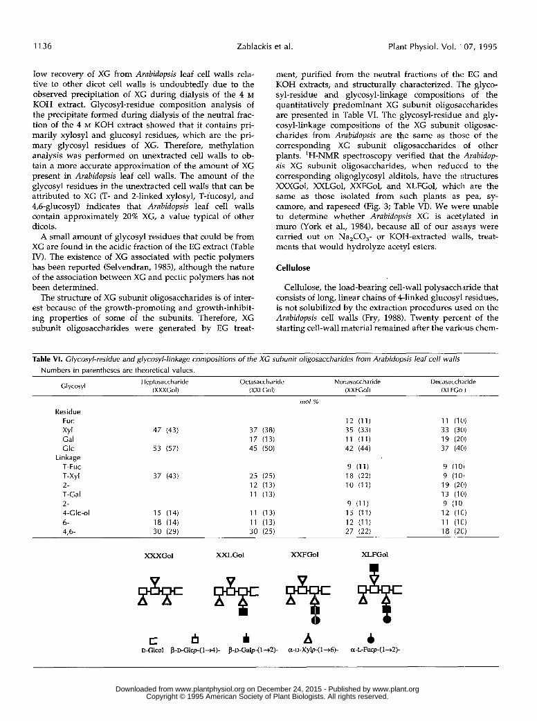
low recovery of XG from Arabidopsis leaf cell walls rela- ment, purified from the neutral fractions of thc EG and tive to other dicot cell walls is undoubtedly due to the observed precipitation of XG during dialysis of the 4 M KOH extract. Glycosyl-residue composition analysis of the precipitate formed during dialysis of the neutral frac- tion of the 4 M KOH extract showed that it contains pri- marily xylosyl and glucosyl residues, which are the pri- mary glycosyl residues of XG. Therefore, methylation analysis was performed on unextracted cell walls to ob- tain a more accurate approximation of the amount of XG present in Arabidopsis leaf cell walls. The amount of the glycosyl residues in the unextracted cell walls that can be attributed to XG (T- and 2-linked xylosyl, T-fucosyl, and 4,6-glucosyl) indicates that Arabidopsis leaf cell walls contain approximately 20% XG, a value typical of other dicots.
KOH extracts, and structurally characterized. The glyco- syl-residue and glycosyl-linkage composition; of the quantitatively predominant XG subunit oligosaccharides are presented in Table VI. The glycosyl-residue and gly- cosyl-linkage compositions of the XG subunit oligosac- charides from Arabidopsis are the same as those of the corresponding XG subunit oligosaccharides of other plants. 'H-NMR spectroscopy verified that the Arabidop- sis XG subunit oligosaccharides, when reduced to the corresponding oligoglycosyl alditols, have the structures XXXGol, XXLGol, XXFGol, and XLFGol, which are the same as those isolated from such plants as pea, sy- camore, and rapeseed (Fig. 3; Table VI). We were unable to determine whether Arabidopsis XG is acetylated in muro (York et al., 1984), because a11 of our assays were
A small amount of glycosyl residues that could be from XG are found in the acidic fraction of the EG extract (Table IV). The existence of XG associated with pectic polymers has been reported (Selvendran, 1985), although the nature of the association between XG and pectic polymers has not been determined.
The structure of XG subunit oligosaccharides is of inter- est because of the growth-promoting and growth-inhibit- ing properties of some of the subunits. Therefore, XG subunit oligosaccharides were generated by EG treat-
carried out on Na,CO,- or KOH-extracted walls, treat- ments that would hydrolyze acetyl esters.
Cellulose
Cellulose, the load-bearing cell-wall polysacchxide that consists of long, linear chains of 4-linked glucosyl residues, is not solubilized by the extraction procedures used on the Arabidopsis cell walls (Fry, 1988). Twenty percent of the starting cell-wall material remained after the varicus chem-
Table VI. Glycosyl-residue and glycosyl-linkage compositions of the XG subunit oligosaccharides from Arabidopsis leaf cell walls Numbers in parentheses are theoretical values.
Heptasaccharide Octasaccharide Nonasaccharide Decasacchwide (XXXCol) (XXLCol) (XXFCol) (XLFCol) Clycosyl
mo/ %
Residue Fuc XY I Cal Glc
Linkage T-FUC T-Xyl 2- T-Cal 2- 4-Clc-01 6- 4,6-
47 (43)
53 (57)
37 (43)
15 (14) 18 (14) 30 (29)
37 (38) 17 (13) 45 (50)
25 (25) 12 (13) 11 (13)
11 (13) 11 (13) 30 (25)
12 (11) 35 (33) 11 (11) 42 (44)
9 (11) 18 (22) 10 (11)
9 (11) 15 (11) 12 (11) 27 (22)
9 (1o:I 9 (10:' 19 (2C9 13 (10) 9 (10: 12 (1C.) 11 (1C) 18 (2C)
XXXGol XXLGol XXFGol XLFGol
V
c I3 I A 4 D-G~co~ p-D-Glcp-( 1-4)- p-D-Galp-( 142)- a-D-Xylp-(l-6)- a-L-Fucp-(l+Z)-
www.plant.org on December 24, 2015 - Published by www.plantphysiol.orgDownloaded from Copyright © 1995 American Society of Plant Biologists. All rights reserved.

Arabidopsis Cel I Walls 1137
ical and enzymatic extractions. Cellulose, as evidenced by 4-linked glucosyl residues, accounts for 63 mol% of the wall residue. Additionally, 1% of the Arubidopsis leaf cell wall was recovered as cellobiose following endo-(l+)-P- glucanase treatment and is assumed to originate from cel- lulose. Thus, cellulose accounts for approximately 14% of the Arubidopsis leaf cell wall.
The Arubidopsis cell-wall residue remaining after a11 of the extraction procedures contained minor amounts of rhamnosyl (3 mol%), mannosyl (11 mol%), arabinosyl (5 mol%), xylosyl (6 mol%), galactosyl (3 mol%), and galac- tosyluronic acid (8 mol%) residues, indicating that the residue contained some of the noncellulosic primary cell- wall polysaccharides that were incompletely extracted by the methods used, a phenomenon observed in studies of sycamore cell walls (Koller et al., 1991). The partia1 extrac- tion of the wall polysaccharides probably reflects hetero- geneity in the bonding of the polysaccharides to other components of the walls.
CONCLUSION
The results presented here show that the leaf cell walls of Arubidopsis contain the same polysaccharides found in the primary cell walls of many other plants. Except for the high proportion of phosphate buffer-soluble pectic polysaccha- rides, Arubidopsis leaf cell walls appear to be typical of other dicots. We are now in a position to characterize the cell walls of Arubidopsis mutants, e.g. the mutant deficient in cell-wall fucosyl residues recently reported by Reiter et al. (1993).
ACKNOWLEDCMENTS
We would like to thank Drs. C. Somerville (Carnegie Institute of Washington at Stanford University), C. Chappel (Purdue Univer- sity), and W-D. Reiter (University of Connecticut) for seeds and leaf material of A. thaliana and Dr. M. Kieliszewski (Complex Carbohydrate Research Center) for the amino acid analyses.
Received July 13,1994; accepted November 21, 1994. Copyright Clearance Center: 0032-0889/95/107/1129/10.
LITERATURE ClTED
Aldington S, Fry SC (1993) Oligosaccharins. Adv Bot Res 19: 1-101 An J, O’Neill MA, Albersheim P, Darvill AG (1994) Isolation and
structural characterization of P-u-glucosyluronic acid and 4-0- methyl P-u-glucosyluronic acid-containing oligosaccharides from the cell-wall pectic polysaccharide, rhamnogalacturonan I. Carbohydr Res 252: 235-243
Barbier M, Thibault J-F (1982) Pectic substances of cherry fruits. Phytochemistry 21: 111-115
Bauer WD, Talmadge KW, Keegstra K, Albersheim P (1973) The structure of plant cell walls. 11. The hemicellulose of the walls of suspension-cultured sycamore cells. Plant Physiol 51: 174-187
Blumenkrantz N, Asboe-Hansen G (1973) New method for quan- titative determination of uronic acids. Anal Biochem 5 4 484489
Bradford MM (1976) A rapid and sensitive method for the quan- titation of microgram quantities of protein utilizing the principle of protein dye binding. Anal Biochem 7 2 248-254
Darvill AG, McNeil M, Albersheim P, Delmer DP (1980a) The primary cell walls of flowering plants. In NE Tolbert, ed, The
Biochemistry of Plants, Vol I. Academic Press, New York, pp
Darvill JE, McNeil M, Darvill AG, Albersheim P (1980b) Struc- ture of plant cell walls. XI. Glucuronoarabinoxylan, a second hemicellulose in the primary cell walls of suspension-cultured sycamore cells. Plant Physiol 66: 1135-1139
Dische Z (1962) Color reactions of carbohydrates. In RL Whistler, ML Wolfrom, eds, Methods in Carbohydrate Chemistry, Vol 1. Academic Press, New York, pp 475-524
Franz G, Blaschek W (1990) Cellulose. In PM Dey, JB Harborne, eds, Methods in Plant Biochemistry, Vol 2. Academic Press, London, pp 291-322
Fry SC (1988) The growing plant cell wall: chemical and metabolic analysis. Longman Scientific & Technical, Essex, UK
Hakomori S (1964) A rapid permethylation of glycolipid and polysaccharide catalyzed by methylsulfinyl carbanion in di- methyl sulfoxide. J Biochem 5 5 205-208
Hayashi T, Maclachlan G (1984) Pea xyloglucan and cellulose. I. Macromolecular organization. Plant Physiol 7 5 596-604
Ishii T, Thomas J, Darvill A, Albersheim P (1989) Structure of plant cell walls. XXVI. The walls of suspension-cultured sy- camore cells contain a family of rhamnogalacturonan-I-like pec- tic polysaccharides. Plant Physiol 89: 421-428
Jansen EF, Jang R, Albersheim P, Bonner J (1960) Pectic metab- olism of growing cell walls. Plant Physiol 35: 87-97
Jensen WA, Ashton M (1960) Composition of developing primary wall in onion root tip cells. I. Quantitative analyses. Plant Physiol 3 5 313-323
Kieliszewski M, de Zacks R, Leykam JF, Lamport DTA (1992) A repetitive proline-rich protein from the gymnosperm Douglas fir is a hydroxyproline-rich glycoprotein. Plant Physiol98: 919-926
Koller A, O’Neill MA, Darvill AG, Albersheim P (1991) A com- parison of the polysaccharides extracted from dried and non- dried walls of suspension-cultured sycamore cells. Phytochem- istry 3 0 3903-3908
Kvemheim AL (1987) Methylation analysis of polysaccharides with butyllithium in dimethyl sulfoxide. Acta Chem Scand Ser B Org Chem Biochem 41: 150-152
Lamport DTA, Miller DH (1971) Hydroxyproline arabinosides in the plant kingdom. Plant Physiol 48: 454-456
Lamport DTA, Northcote DH (1960) Hydroxyproline in primary cell walls of higher plants. Nature 188: 665-666
Lau JM, McNeil M, Darvill AG, Albersheim P (1985) Structure of the backbone of rhamnogalacturonan I, a pectic polysaccharide in the primary cell walls of plants. Carbohydr Res 137: 111-125
Lau JM, McNeil M, Darvill AG, Albersheim P (1987) Treatment of rhamnogalacturonan I with lithium in ethylenediamine. Car- bohydr Res 168: 245-274
Lerouge P, ONeill MA, Darvill AG, Albersheim P (1993) Struc- tural characterization of endo-glycanase-generated oligoglyco- syl side chains of rhamnogalacturonan I. Carbohydr Res 243:
MarfA V, Golin DJ, Eberhard S, Mohnen D, Darvill A, Alber- sheim P (1991) Oligogalacturonides are able to induce flowers to form on tobacco explants. Plant J 1: 217-225
McDougall GJ, Fry SC (1991) Xyloglucan nonasaccharide, a nat- urally-occurring oligosaccharin, arises in vivo by polysaccharide breakdown. J Plant Physiol 137: 332-336
McNeil M, Darvill AG, Albersheim P (1980) Structure of plant cell walls. X. Rhamnogalacturonan I, a structurally complex pectic polysaccharide in the walls of suspension-cultured sy- camore cells. Plant Physiol 66 1128-1134
McNeil M, Darvill AG, Albersheim P (1982) Structure of plant cell walls. XII. Identification of seven differently linked glycosyl residues attached to 0-4 of the 2,4-linked L-rhamnosyl residues of rhamnogalacturonan I. Plant Physiol 70: 1586-1591
Morris RR (1986) Molecular interaction in polysaccharide gelation. Br Polymer J 18: 14-21
ONeill M, Albersheim P, Darvill A (1990) The pectic polysaccha- rides of primary cell walls. In PM Dey, JB Harborne, eds, Meth- ods in Plant Biochemistry, Vol 2. Academic Press, London, pp 415441
91-162
359-371
www.plant.org on December 24, 2015 - Published by www.plantphysiol.orgDownloaded from Copyright © 1995 American Society of Plant Biologists. All rights reserved.

1138 Zablackis et al. Plant Physiol. Vol. '107, 1995
Redgwell RJ, Selvendran RR (1986) Structural features of cell- wall polysaccharides of onion Allium cepa. Carbohydr Res 157:
Reiter W-D, Chapple CCS, Somerville CR (1993) Altered growth and cell walls in a fucose-deficient mutant of Arabidopsis. Science
Selvendran RR (1985) Developments in the chemistry and bio- chemistry of pectic and hemicellulosic polymers. J Cell Sci (Suppl) 2 51-88
Stevens BJH, Selvendran RR (1984) Structural features of cell-wall polysaccharides of the carrot Daucus carota. Carbohydr Res 128:
Stevenson TT, Darvill AG, Albersheim P (1988a) 3-Deoxy-~- lyxo-2-heptulosaric acid, a component of the plant cell-wall polysaccharide rhamnogalacturonan-11. Carbohydr Res 179:
Stevenson TT, Darvill AG, Albersheim P (1988b) Structural fea- tures of the plant cell-wall polysaccharide rhamnogalacturonan- 11. Carbohydr Res 182: 207-226
Updegraff DM (1969) Semimicro determination of cellulose in biological materials. Ana1 Biochem 3 2 420424
York WS, Darvill AG, Albersheim P (1984) Inhibition of 2,4-
183-199
261: 1032-1035
321-333
269-288
dichlorophenoxyacetic acid-stimulated elongation o : pea stem segments by a xyloglucan oligosaccharide. Plant I'hysiol 7 5
York WS, Ilarvill AG, McNeil M, Albersheim P (1985a) 3-Deoxy- ~-manno-2-octulosonic acid (KDO) is a componení of rham- nogalacturonan 11, a pectic polysaccharide in the primary cell walls of plants. Carbohydr Res 138: 109-126
York WS, Darvill AG, McNeil M, Stevenson TT, Albersheim P (198513) Isolation and characterization of plant cell walls and cell wall components. Methods Enzymol 118 3-40
York WS, Harvey LK, Guillen R, Albersheim P, C'arvill AG (1993) Structural analysis of tamarind seed xyloglucan oligosac- charides using P-galactosidase digestion and spwtroscopic methods. Carbohydr Res 248 285-301
York WS, Oates J, Van Halbeek H, Darvill AG, Albersheim P (1988) Location of the O-acetyl substituents on a nonasaccharide repeating unit of sycamore extracellular xyloglucan. Carbohydr Res 173: 113-132
York WS, Van Halbeek H, Darvill AG, Albersheim P (1990) Structural analysis of xyloglucan oligosaccharides bj' 'H-n.m.r. spectroscopy and fast-atom-bombardment mass spcctrometry. Carbohydr Res 200 9-31
295-297
www.plant.org on December 24, 2015 - Published by www.plantphysiol.orgDownloaded from Copyright © 1995 American Society of Plant Biologists. All rights reserved.
Related Documents


