Characterization of Aging-Associated Cardiac Diastolic Dysfunction Wei-Ting Chang 1,2. , Jung-San Chen 3. , Yung-Kung Hung 3 , Wei-Chuan Tsai 1 , Jer-Nan Juang 3 *, Ping-Yen Liu 1,2 * 1 Division of Cardiology, Internal Medicine, National Cheng Kung University Hospital, Tainan, Taiwan, 2 Institute of Clinical Medicine, National Cheng Kung University, Tainan, Taiwan, 3 Department of Engineering Science, National Cheng Kung University, Tainan, Taiwan Abstract Aims: Diastolic dysfunction is common in geriatric heart failure. A reliable parameter to predict myocardium stiffness and relaxation under similar end-diastolic pressure is being developed. We propose a material and mathematical model for calculating myocardium stiffness based on the concept of linear correlation between e=e’ and wedge pressure. Methods and Results: We enrolled 919 patients (male: 52:6%½484=919). Compared with the younger population of controls (mean age: 43:9+11:7 years; n~211; male: 62:1% ½131=211), the elderly (mean age: 76:3+6:2; n~708; male: 52:6% ½484=708) had a greater prevalence of hypertension, diabetes mellitus, and coronary artery disease (all pv0:05). We collected their M-mode and 2-D echocardiographic volumetric parameters, intraventricular filling pressure, and speckle tracking images to establish a mathematical model. The feasibility of this model was validated. The average early diastolic velocity of the mitral annulus assessed using tissue Doppler imaging was significantly attenuated in the elderly (e’: 0:09+0:02 vs. 0:08+0:02; p~0:02) and corresponded to the higher estimated wedge (e=e’) pressure (7:76+2:44 vs. 8:35+2:64; p~0:02) in that cohort. E (Young’s modulus) was calculated to describe the tensile elasticity of the myocardium. With the same intraventricular filling pressure, E was significantly higher in the elderly, especially those with e=e’ values w9. Compared with diastolic dysfunction parameters, E also presented sentinel characteristics more sensitive for detecting early myocardial relaxation impairment, which indicates stiffer myocardium in aging hearts. Conclusion: Our material and geometric mathematical model successfully described the stiffer myocardium in aging hearts with higher intraventricular pressure. Additional studies that compare individual differences, especially in health status, are needed to validate its application for detecting diastolic heart failure. Citation: Chang W-T, Chen J-S, Hung Y-K, Tsai W-C, Juang J-N, et al. (2014) Characterization of Aging-Associated Cardiac Diastolic Dysfunction. PLoS ONE 9(5): e97455. doi:10.1371/journal.pone.0097455 Editor: Sudhiranjan Gupta, Texas A & M, Division of Cardiology, United States of America Received November 6, 2013; Accepted April 20, 2014; Published May 28, 2014 Copyright: ß 2014 Chang et al. This is an open-access article distributed under the terms of the Creative Commons Attribution License, which permits unrestricted use, distribution, and reproduction in any medium, provided the original author and source are credited. Funding: This research was supported in part by the Headquarters of University Advancement at the National Cheng Kung University, which is sponsored by the Ministry of Education, Taiwan, ROC. The research was also granted from Health Promotion Program: Blood Pressure Control from Health Promotion Administration, Ministry of Health and Welfare, Taiwan, ROC and the grant from NSC101-2314-B-006-075-MY2, sponsored by the Ministry of Science and Technology, Taiwan, ROC. The investigator (PY Liu) was granted by ‘‘A Landmark Project to Promote Innovation & Competitiveness of Clinical Trials by the Excellent Clinical Trial and Research Center in National Cheng Kung University Hospital, Ministry of Health and Welfare, Taiwan.’’ The funders had no role in study design, data collection and analysis, decision to publish, or preparation of the manuscript. Competing Interests: The authors have declared that no competing interests exist. * E-mail: [email protected] (JNJ); [email protected] (PYL) . These authors contributed equally to this work. Introduction Heart failure is increasingly prevalent among older adults [1]. Clinically,40{50% of patients with symptomatic heart failure have preserved left heart function, with preserved ventricular ejection fraction (w50%) [2]. Sometimes this is called ‘‘diastolic dysfunction with preserved systolic function’’ or ‘‘diastolic heart failure’’ [3]. People with diastolic heart failure are generally older and female, and tend to have a greater incidence of systemic hypertension than do those with contractile dysfunction (‘‘systolic heart failure’’) [4,5]. Physiologically, diastolic heart failure occurs when the ventricle cannot fill properly because it cannot relax or its wall is too rigid [6]. Histological evidence supports the notion that diastolic dysfunction is related to ventricular hypertrophy, increased interstitial collagen deposition into the myocardium [7]. Similarly, aging hearts continuously lose myocytes, which is compensated for by reactive hypertrophy of the remaining cells; thus, these hearts are filled with fibrotic or adipose tissue [8]. Histological samples for measuring the exact tensile elasticity of the myocardium are possible only in animal studies, not human studies. Some mathematical models have been used to describe the dynamics of remodeling in skeletal muscle, arteries, and even the heart [9,10]. However, a mathematical model to study cardiac aging is still lacking. Establishing a model of the aging heart would provide a tool for us to understand underlying mechanisms of cardiac aging and to define the impaired myocardial relaxation process. It remains uncertain whether current available invasive and noninvasive diagnostic tools can accurately predict myocardium stiffness. The time constant of relaxation (tau, t), which describes PLOS ONE | www.plosone.org 1 May 2014 | Volume 9 | Issue 5 | e97455

Welcome message from author
This document is posted to help you gain knowledge. Please leave a comment to let me know what you think about it! Share it to your friends and learn new things together.
Transcript
Characterization of Aging-Associated Cardiac DiastolicDysfunctionWei-Ting Chang1,2., Jung-San Chen3., Yung-Kung Hung3, Wei-Chuan Tsai1, Jer-Nan Juang3*,
Ping-Yen Liu1,2*
1 Division of Cardiology, Internal Medicine, National Cheng Kung University Hospital, Tainan, Taiwan, 2 Institute of Clinical Medicine, National Cheng Kung University,
Tainan, Taiwan, 3 Department of Engineering Science, National Cheng Kung University, Tainan, Taiwan
Abstract
Aims: Diastolic dysfunction is common in geriatric heart failure. A reliable parameter to predict myocardium stiffness andrelaxation under similar end-diastolic pressure is being developed. We propose a material and mathematical model forcalculating myocardium stiffness based on the concept of linear correlation between e=e’ and wedge pressure.
Methods and Results: We enrolled 919 patients (male: 52:6%½484=919�). Compared with the younger population of controls(mean age: 43:9+11:7 years; n~211; male: 62:1% ½131=211�), the elderly (mean age: 76:3+6:2; n~708; male: 52:6%½484=708�) had a greater prevalence of hypertension, diabetes mellitus, and coronary artery disease (all pv0:05). Wecollected their M-mode and 2-D echocardiographic volumetric parameters, intraventricular filling pressure, and speckletracking images to establish a mathematical model. The feasibility of this model was validated. The average early diastolicvelocity of the mitral annulus assessed using tissue Doppler imaging was significantly attenuated in the elderly (e’:0:09+0:02 vs. 0:08+0:02; p~0:02) and corresponded to the higher estimated wedge (e=e’) pressure (7:76+2:44 vs.8:35+2:64; p~0:02) in that cohort. E (Young’s modulus) was calculated to describe the tensile elasticity of the myocardium.With the same intraventricular filling pressure, E was significantly higher in the elderly, especially those with e=e’ values w9.Compared with diastolic dysfunction parameters, E also presented sentinel characteristics more sensitive for detecting earlymyocardial relaxation impairment, which indicates stiffer myocardium in aging hearts.
Conclusion: Our material and geometric mathematical model successfully described the stiffer myocardium in aging heartswith higher intraventricular pressure. Additional studies that compare individual differences, especially in health status, areneeded to validate its application for detecting diastolic heart failure.
Citation: Chang W-T, Chen J-S, Hung Y-K, Tsai W-C, Juang J-N, et al. (2014) Characterization of Aging-Associated Cardiac Diastolic Dysfunction. PLoS ONE 9(5):e97455. doi:10.1371/journal.pone.0097455
Editor: Sudhiranjan Gupta, Texas A & M, Division of Cardiology, United States of America
Received November 6, 2013; Accepted April 20, 2014; Published May 28, 2014
Copyright: � 2014 Chang et al. This is an open-access article distributed under the terms of the Creative Commons Attribution License, which permitsunrestricted use, distribution, and reproduction in any medium, provided the original author and source are credited.
Funding: This research was supported in part by the Headquarters of University Advancement at the National Cheng Kung University, which is sponsored by theMinistry of Education, Taiwan, ROC. The research was also granted from Health Promotion Program: Blood Pressure Control from Health PromotionAdministration, Ministry of Health and Welfare, Taiwan, ROC and the grant from NSC101-2314-B-006-075-MY2, sponsored by the Ministry of Science andTechnology, Taiwan, ROC. The investigator (PY Liu) was granted by ‘‘A Landmark Project to Promote Innovation & Competitiveness of Clinical Trials by theExcellent Clinical Trial and Research Center in National Cheng Kung University Hospital, Ministry of Health and Welfare, Taiwan.’’ The funders had no role in studydesign, data collection and analysis, decision to publish, or preparation of the manuscript.
Competing Interests: The authors have declared that no competing interests exist.
* E-mail: [email protected] (JNJ); [email protected] (PYL)
. These authors contributed equally to this work.
Introduction
Heart failure is increasingly prevalent among older adults [1].
Clinically,40{50% of patients with symptomatic heart failure
have preserved left heart function, with preserved ventricular
ejection fraction (w50%) [2]. Sometimes this is called ‘‘diastolic
dysfunction with preserved systolic function’’ or ‘‘diastolic heart
failure’’ [3]. People with diastolic heart failure are generally older
and female, and tend to have a greater incidence of systemic
hypertension than do those with contractile dysfunction (‘‘systolic
heart failure’’) [4,5]. Physiologically, diastolic heart failure occurs
when the ventricle cannot fill properly because it cannot relax or
its wall is too rigid [6]. Histological evidence supports the notion
that diastolic dysfunction is related to ventricular hypertrophy,
increased interstitial collagen deposition into the myocardium [7].
Similarly, aging hearts continuously lose myocytes, which is
compensated for by reactive hypertrophy of the remaining cells;
thus, these hearts are filled with fibrotic or adipose tissue [8].
Histological samples for measuring the exact tensile elasticity of
the myocardium are possible only in animal studies, not human
studies. Some mathematical models have been used to describe the
dynamics of remodeling in skeletal muscle, arteries, and even the
heart [9,10]. However, a mathematical model to study cardiac
aging is still lacking. Establishing a model of the aging heart would
provide a tool for us to understand underlying mechanisms of
cardiac aging and to define the impaired myocardial relaxation
process.
It remains uncertain whether current available invasive and
noninvasive diagnostic tools can accurately predict myocardium
stiffness. The time constant of relaxation (tau, t), which describes
PLOS ONE | www.plosone.org 1 May 2014 | Volume 9 | Issue 5 | e97455
the rate of left ventricular (LV) pressure decay during isovolumic
relaxation, is currently the standard parameter for predicting the
relaxation function of myocardium [11,12]. However, several
confounding factors, when echocardiography is used to measure
the deceleration time of mitral inflow, may disturb the equivalence
between echocardiographic and catheterized results. Despite the
ratio of mitral inflow to annulus tissue, Doppler imaging velocity
(e=e’) indicates the intraventricular pressure, which is within the
borderline range of elevated pressure (e=e’~9{14), the discrim-
ination of diastolic dysfunction remains a dilemma [3]. Therefore,
a reliable and noninvasive diagnostic parameter is crucial for
facilitating an accurate diagnosis that indicates whether the
stiffness is myocardial stiffness.
Studies [13–21] on the elastic properties of the contracting left
ventricle have aroused a great deal of interest among scientists and
engineers. Many researchers have developed a series of experi-
mental techniques for determining the elastic properties of the left
ventricle. A simple and practical approach for in vivo determina-
tions of the properties of the canine left ventricle, proposed in 1972
[13], established the relationship between the effective elastic
modulus and the circumferential stress throughout the isovolu-
metric systolic period. After the concept on the elastic properties of
the left ventricle was accepted, some researchers reported that the
effective modulus E measured from experiments could also be
viewed as an additional indication of the left ventricle having
adjusted to the heart disease [14], which showed that normal
values of E during the systole directly indicate that the strength of
the left ventricle contraction is normal. The nomogram, a
clinically usable closed-chest procedure for determining the elastic
modulus, was introduced in 1975 [15]. Using a heart-sound-
frequency analysis followed by a determination of E, the loss of
muscle-medium elasticity can be roughly delineated. In addition to
improving data-acquisition techniques, the modeling of the left
ventricle is becoming a more important factor for determining
myocardial elasticity. These findings showed that the stiffness of
the complete ventricle should be considered a function not only of
myocardial stiffness but also of the cavity shape, dimensions, and
structure of the vessel [16]. A better approximation for ventricular
modeling requires assuming that myocardial stiffness is a function
of geometry and stress. In other words, the geometry of the left
ventricle is essential for simulating it in a model. Thick-walled
models are commonly and widely used to study the dynamics of
the ventricle. Several studies [17–19] on cardiac muscle mechan-
ics, LV pump function, and LV wall thickening view the left
ventricle as a thick-walled cylindrical composite. They report that
the thick-walled cylindrical framework seems to be a good and
practical approximation sufficient for simulating the left ventricle.
One study [20] presented a simple analytical model to describe the
relationship between age-related changes in the structure and
function of mouse cardiac muscle. It suggested that age-related
cardiac sarcopenia likely contributes to depressed LV function in
the absence of overt cardiovascular disease. Recently, an
alternative mathematical model for investigating cardiac aging
characterized by diastolic dysfunction of the left ventricle was
introduced [21]. In contrast to the previous study, a spherical
thick-walled model and stretch-induced tissue-growth postulate
were used to predict LV dimension and wall stiffness changes in
aging mice. The Young’s modulus of the left ventricle was
determined by introducing a smooth monotonic function to fit the
experimental data and a simplified version of the linear mixture
theory of composite material. It was assumed that the pressure
difference and the Young’s modulus of the left ventricle are two
independent factors that affect end-diastolic dimension/diameter
and wall thickness.
The importance of the aging effect on the large vessels and
cardiac structure can be also seen from a study [22] on the effect of
hypertension on the diameter and elastic modulus of the aortic
arch; it showed that the elastic modulus was significantly
correlated with age in patients with, but not without, hypertension.
In addition, both the aortic arch diameter and the elastic modulus
are larger in patients with sustained uncomplicated essential
hypertension. Based on the proven linear correlation between
mitral e velocity, corrected for the influence of relaxation (e=e’ratio) and intraventricular pressure (similar to wedge pressure;
r~0:87; pulmonary capillary wedge pressure (PCWP)~1:24½e=e’�z1:9) [23], we have created a cardiac mathematical model
to simulate the remodeling process under various pressures during
the aging process. Unlike other research groups, we hypothesized
that the pressure, elastic modulus, and LV dimension are mutually
influenced, that the relationship of the elastic modulus to pressure,
wall thickness, and age can be established with the mathematical
model, and that the aging effect on LV wall thickness can also be
determined using the fixed pressure.
Materials and Methods
PatientsWe enrolled 919 patients (male: 52:6% ½484=919�) and divided
them into two cohorts: (1)Echoz: patients given echocardiography
on a physical examination at our university hospital between
February 2012 and June 2013, and (2) TOP (Echo{): the Tianliao
Old People (TOP) study between July 2010 and August 2012
[24,25]. Echocardiographic parameters based on the recommen-
dations of the American Society of Echocardiography [26],
medical records, and clinical questionnaires were collected from
patients in the cohort. Patients with a poor image window, LV
systolic dysfunction, or significant (.moderate severity) valvular
heart disease were excluded. This study was approved by the
Institutional Review Board of National Cheng Kung University
Hospital (IRB no: ER-99-111), and each patient signed an
informed consent form before the physical examination.
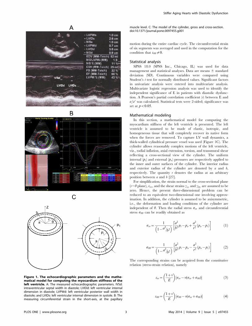
EchocardiographyStandard echocardiography was done (Vivid I; GE Vingmed
Ultrasound AS, Horten, Norway) using a 3.5-MHz multiphase-
array probe. The chamber dimensions and LV mass were
measured using the two-dimensionally guided M-mode method,
and the LV ejection fraction (LVEF) was measured with the two-
dimensional M mode (Figure 1A). Intraventricular septal width
(IVSd), LV internal dimension (LVIDd), LV posterior wall width
(LVPWd), and LV internal dimension (LVIDs) were measured
sequentially to calculate geometry and ejection fraction. Transmi-
tral Doppler flow velocity was obtained from an apical four-
chamber view, and peak early filling velocity (e), peak atrial
velocity (a), and the E=A ratio were recorded. Early diastolic
annular velocity (e’) and atrial annular velocity (a’) were also
measured to estimate the LV end-diastolic pressure (e=e’). The
average of medial and lateral e=e’ was used to represent the
estimated intraventricular pressure.
To measure the circumferential strain, 20 patients were
randomly selected from the Echozcohort to receive speckle-
tracking echocardiography (STE) (Figure 1B). Short-axis views at
the papillary muscle level were recorded in digital loops for a
deformation analysis of the left ventricle. The images were
acquired at 70–90 frame/sec and stored for three cycles. The
images were analyzed offline using computer software (EchoPAC
09; GE-Vingmed Ultrasound AS, Horten, Norway). After tracking
the margin of endocardium, the software detected the myocardial
Stiffer Aging Hearts with Diastolic Dysfunction
PLOS ONE | www.plosone.org 2 May 2014 | Volume 9 | Issue 5 | e97455
motion during the entire cardiac cycle. The circumferential strain
of six segments was averaged and used in the computation for the
condition that ehh=0.
Statistical analysisSPSS 18.0 (SPSS Inc., Chicago, IL) was used for data
management and statistical analyses. Data are means 6 standard
deviation (SD). Continuous variables were compared using
Student’s t test for normally distributed values. Significant factors
in univariate analysis were entered into multivariate analysis.
Multivariate logistic regression analysis was used to identify the
independent significance of E in patients with diastolic dysfunc-
tion. A Pearson’s partial correlation coefficient (r) between E and
e=e’ was calculated. Statistical tests were 2-sided; significance was
set at pv0:05.
Mathematical modelingIn this section, a mathematical model for computing the
myocardium stiffness of the left ventricle is presented. The left
ventricle is assumed to be made of elastic, isotropic, and
homogeneous tissue that will completely recover its native form
when the forces are removed. To capture LV wall dynamics, a
thick-walled cylindrical pressure vessel was used (Figure 1C). The
cylinder allows reasonably complex motions of the left ventricle,
viz., radial inflation, axial extension, torsion, and transmural shear
reflecting a cross-sectional view of the cylinder. The uniform
internal (pi) and external (po) pressures are respectively applied to
the inner and outer surfaces of the cylinder. The interior radius
and exterior radius of the cylinder are denoted by a and b,
respectively. The quantity r denotes the radius at an arbitrary
position between a and b [27].
For simplification, the strain normal to the cross-sectional plane
(r2h plane), ezz, and the shear strains crz and chz are assumed to be
zero. Hence, the present three-dimensional problem can be
reduced to an equivalent two-dimensional one involving approx-
imation. In addition, the cylinder is assumed to be axisymmetric,
i.e., the deformation and loading conditions of the cylinder are
independent of h. Then the radial stress srr and circumferential
stress shh can be readily obtained as
srr~1
1{a2=b2
� � a2
b2pi{poz
a2
r2po{pið Þ
� �ð1Þ
shh~1
1{a2=b2
� � a2
b2pi{po{
a2
r2po{pið Þ
� �ð2Þ
The corresponding strains can be acquired from the constitutive
relation (stress-strain relation), namely
err~1zn
E
� �srr{n(srrzshh)½ � ð3Þ
ehh~1zn
E
� �shh{n(srrzshh)½ � ð4Þ
Figure 1. The echocardiographic parameters and the mathe-matical model for computing the myocardium stiffness of theleft ventricle. A: The measured echocardiographic parameters. IVSd:intraventricular septal width in diastole; LVIDd: left ventricular internaldimension in diastole; LVPWd: left ventricular posterior wall width indiastole; and LVIDs: left ventricular internal dimension in systole. B: Themeasuring circumferential strain in the short-axis, at the papillary
muscle level. C: The model of the cylinder, gross and cross-section.doi:10.1371/journal.pone.0097455.g001
Stiffer Aging Hearts with Diastolic Dysfunction
PLOS ONE | www.plosone.org 3 May 2014 | Volume 9 | Issue 5 | e97455
where err is the radial strain, ehh is the circumferential strain, n is
Poisson’s ratio, and E is the Young’s modulus. Solving Eq. (3) and
Eq. (4) yields
n~shherr{srrehh
srrzshhð Þ err{ehhð Þ ð5Þ
E~srrerr{shhehhz2shherr{2srrehhð Þ err{ehh{shherrzsrrehhð Þ
err srrzshhð Þ err{ehhð Þ2
" #ð6Þ
Substituting Eqs. (1) and (2) into Eq. (6) gives
E~
a2
b2{a2a2
b2 pi{poza2
r2 po{pið Þh in
err{2ehhð Þz a2
b2 pi{po{a2
r2 po{pið Þh i
2err{ehhð Þo
2erra2
b2 pi{po{a2
r2 po{pið Þh i
:ehh
a2
b2 pi{poza2
r2 po{pið Þh i
{erra2
b2 pi{po{a2
r2 po{pið Þh ion
2erra2
b2 pi{po{a2
r2 po{pið Þh i
ð7Þ
If ehh is not considered, Eqs. (5) and (6) can be reduced to
n~shh
srrzshhð8Þ
E~srrz2shhð Þ 1{shhð Þ
err srrzshhð Þ
� �ð9Þ
Then the Young’s modulus for ehh~0 can be readily derived as
E~1
2erra2
b2 pi{po
� � 3a2
b2pi{3po{
a2
r2po{pið Þ
� �
: 1{b2
b2{a2
a2
b2pi{po{
a2
r2po{pið Þ
� �� ð10Þ
The parameters a, b, pi, and po can be acquired from experiments
and shown as follows:
a~LVIDd
2
� �ð11Þ
b~LVIDdz2LVPWd
2
� �ð12Þ
pi~1:9z1:24e=e0
0:0075
� �ð13Þ
Table 1. The clinical and echocardiographic characteristics of the younger and older cohorts.
Variable Younger cohort n = 211 (22.9%) Older cohort n = 708 (77.1%) p-value
Age (years) 43.88611.67 76.2666.2 ,0.001
Male 131 (62.08) 484 (68.82) ,0.001
HTN 21 (9.95) 349 (49.22) ,0.001
DM 11 (5.21) 122 (17.2) ,0.001
CAD 6 (2.84) 71 (10.01) 0.02
ECHOCARDIOGRAPHIC PARAMETERS
IVSd (cm) 0.8160.2 0.7860.2 0.14
LVPWd (cm) 0.7360.1 1.2066.6 0.31
LVIDd (cm) 4.7860.5 5.0360.6 0.01
LVIDs (cm) 2.9060.4 3.0560.5 0.01
LVEF (%) 70.0266.7 68.2068.4 0.09
e (m/s) 0.7460.2 0.6560.3 0.01
e/a 0.8060.8 0.7060.3 0.14
e9 (m/s) 0.0960.02 0.0860.02 0.02
e/e9 7.7662.44 8.3562.64 0.02
IVRT 94.44621.25 107.77619.1 0.09
DT 210.91669.52 179.01694.66 0.67
E (Young’s modulus) 28872.7267710.74 31325.97610275.77 0.001
Data are n %ð Þ or mean or + standard error; HTN = hypertension; DM = diabetes mellitus; CAD = coronary artery disease; IVSd = inter-ventricular septal diameter indiastolic phase; LVPWd = left ventricular posterior wall diameter in diastolic phase; LVIDd = left ventricular internal diastolic dimension; LVIDs = left ventricular internalsystolic dimension; e = early diastolic mitral inflow velocity; e/a = the ratio of early to late diastolic mitral inflow velocity; e9 = the average early diastolic velocity of mitralannulus in tissue Doppler; LVEF = left ventricular ejection fraction.doi:10.1371/journal.pone.0097455.t001
Stiffer Aging Hearts with Diastolic Dysfunction
PLOS ONE | www.plosone.org 4 May 2014 | Volume 9 | Issue 5 | e97455

po~1:9z1:24e=e0
0:0075
� �10 ð14Þ
Also, err is the ratio of the radial elongation to the diastolic radius,
i.e., err~ LVIDd{LVIDsð Þ=LVIDd, and ehh is an average strain
for six segments in the circumferential direction. For computation,
the parameter r in Eqs. (7) and (10) was chosen as azb=2.
Results
The clinical and echocardiographic characteristics of theyounger and older cohorts
The elderly cohort (w65 years old) were significantly older
(mean age~76:26+6:2 vs. 43:88+11:67 years), had a higher
prevalence of hypertension (49:2% vs. 10:1%), diabetes mellitus
(17:2% vs. 5:2%), and coronary artery disease (9:9% vs. 2:74%; all
pv0:05) (Table 1) than did the younger cohort (n~211;
male~62:08%). All of them worked and lived independently.
Based on our previous questionnaire in the TOP study [25], most
lived an active life (11:8+7:61 h of walking/week).
Figure 2. The correlations between e=e’ and E both in theclinical statistics and the mathematical models. A and B: Therelationship between e=e’ and E in clinical statistics, and C: in themathematical model. e=e’= estimated intraventricular pressure by theratio of early diastolic mitral inflow velocity and the averaged earlydiastolic velocity of mitral annulus in tissue Doppler imaging;E~Young0s modulus.doi:10.1371/journal.pone.0097455.g002
Figure 3. The relationship between E and echocardiographicintraventricular pressure in the younger and older cohorts. A:The relationship between e=e’ and E value in various age group. B: Theexponential correlation between age and E in the regression analysis.doi:10.1371/journal.pone.0097455.g003
Stiffer Aging Hearts with Diastolic Dysfunction
PLOS ONE | www.plosone.org 5 May 2014 | Volume 9 | Issue 5 | e97455

To establish a model for predicting the stiffness characteristics of
myocardium, we used the M-mode and 2-D echocardiographic
volumetric parameters, intraventricular filling pressure, and STE
from all clinical patients and the 2 cohorts. The feasibility of this
model was validated. Despite a slight but significant (p~0:01)
difference in intraventricular diameter between the two cohorts
(LVIDd~5:03+0:63 vs. 4:78+0:53), the LV systolic ejection
fraction was within the normal range in all patients
(LVEF~68:20+8:39 vs. 70:02+6:72; p~0:09). The mean early
diastolic velocity of the mitral annulus in tissue Doppler imaging
was significantly attenuated in the elderly cohort (e’~0:09+0:02vs.0:08+0:02; p~0:02), which corresponds to the higher
estimated wedge (e=e’) pressure (7:76+2:44 vs. 8:35+2:64;
p~0:02). E (Young’s modulus) was calculated to describe the
tensile elasticity of the myocardium, which was also significantly
(p~0:001) elevated in the elderly cohort (E~28,872:72+7710:74vs. 31,325:97+10275:77). Instead of a linear relationship, the
regression analysis illustrated a nonlinear association between age
and E (E~10,000e0:0162age; r2~0:61)
After excluding patients with hypertension, diabetes mellitus,
and coronary artery disease, the correlation between E and
diastolic dysfunction remained significant (pv0:05) in the healthy
elderly compared with other echocardiographic parameters (Table
S1).
The relationship between E and echocardiographicintraventricular pressure in the younger and oldercohorts
There was a positive correlation between e=e’ and E
(E~7699:07e=e0z2806:7; r~0:87; pv0:001) both in the clinical
statistics (Figure 2A,B) and in the mathematical models (Figure 2C).
The slope of the trend line shown in Figure 2B is very close to the
one obtained by directly using the mathematical model in
Figure 2C. In the group with an e=e’v5, E was estimated as
18,042(Pa), but once the pressure elevated (e=e’w11), E signifi-
cantly increased (45,030 Pa). Categorizing patients by age groups
(20–45y=o, 45–60y=o, w60 y=o) showed that, although under the
same intraventricular filling pressure, E was significantly higher in
the elderly (w60 y=o), especially with a higher e=e’ (w9)
(E~34,633 in 20–45-year-old group, 33,778 in 45–60-year-old
group, and 42,726 in .60-year-old group) (Figure 3A). However,
the difference was not significant in younger patients or between
genders. Regression analysis showed that an exponential correla-
tion between age and E could be summarized to delineate the
soaring E in older patients (Figure 3B). Multivariate analysis,
compared with other traditional parameters for diagnosing
diastolic dysfunction (deceleration time [DT]; isovolumic relaxa-
tion time [IVRT]), E also showed independent elevation for
detecting early myocardial relaxation impairment in both younger
(odds ratio ½OR�~1:12; 95% confidence interval
½CI �~1:03{1:47; p~0:04) and older patients (OR~1:48; 95%CI~1:22{1:94; p~0:01), which indicates stiffer myocardium in
aging hearts. (Table 2 and 3; Table S2 and S3)
The corresponding trend in various modelsTo simulate the human heart, we created a model to obtain the
circumferential strain. In addition to the radial strain, the
circumferential strain of the actual myocardial contraction was
measured using STE Besides a grossly attenuated E, the model
reflected a similar trend of geometric and pressure change,
regardless of whether the circumferential strain was considered
(Figure 4 A,B). Thus, if the major question is the relationship
between pressure and the tensile elasticity of the myocardium,
rather than the exact values of E, the simpler cylinder model may
replace the complex elliptical model to reduce measurement and
calculation errors.
The relationship between E and echocardiographic wallthickness in the younger and older cohorts
LV hypertrophy was more prevalent in the elderly than in the
younger and middle-aged patients; it led to poor compliance and
to difficulty in shape changing; thus, it impaired diastolic function.
Although the association between a thicker wall and higher
intraventricular pressure has been frequently reported, a suitable
model to illustrate it in patients of various ages is lacking. Our
mathematical model depicted positive associations between IVSd
Table 2. Multivariate regression analyses for the independence of E value in younger patients with diastolic dysfunction.
Variable Odds Ratio 95% Confidence Interval p-value
Age 1.33 0.66–2.68 0.41
Diabetes Mellitus 0.46 0.18–1.13 0.09
IVSd 1.02 0.46–2.23 0.96
IVRT 0.99 0.93–1.02 0.11
E (Young’s modulus) 1.12 1.03–1.47 0.04
Abbreviations: see Table 1.doi:10.1371/journal.pone.0097455.t002
Table 3. Multivariate regression analyses for the independence of E value in older patients with diastolic dysfunction.
Variable Odds Ratio 95% Confidence Interval p-value
Coronary Artery Disease 1.01 0.97–1.03 0.62
e/a 0.46 0.18–1.13 0.90
E (Young’s modulus) 1.48 1.22–1.94 0.01
Abbreviations: see Table 1.doi:10.1371/journal.pone.0097455.t003
Stiffer Aging Hearts with Diastolic Dysfunction
PLOS ONE | www.plosone.org 6 May 2014 | Volume 9 | Issue 5 | e97455

and E, along with the increasing e=e’ (Figure 5A). It also indicated
that in the same LV wall width, a higher intraventricular pressure
correlated to a higher value of E.
In addition, by inputting the representative E (30,000, 35,000,
and 40,000 for the 20–45, 45–60, and w60 y=o groups,
respectively), we discovered that a higher value of E, along with
aging, was associated with a thicker ventricular wall under the
same intraventricular pressure (Figure 5B). This may explain the
deteriorating myocardial relaxation and higher incidence of signs
of heart failure signs in the elderly, who are more vulnerable to the
same pressure.
Discussion
This is the first evaluation of the diastolic dysfunction in the
aging human heart that uses physiologic and echocardiographic
assessments combined with mathematical modeling. Our most
important findings are that: (1) E (Young’s modulus), the tensile
elasticity of the myocardium, can be used clinically to describe
myocardial relaxation and to noninvasively detect early diastolic
dysfunction; (2) aging myocardium becomes thicker, stiffer, and
less expandable, which agrees with previous clinical findings. Even
under the same intraventricular filling pressure, E was significantly
higher in elderly patients (w60 y=o), especially with a higher e=e’(w9); (3) our mathematical model similar trends of geometric and
pressure changes, regardless of whether circumferential strain was
considered.
The criteria for diagnosing diastolic dysfunction and diastolic
heart failure remain imprecise [3,11]. Although there are several
parameters that indicate myocardial relaxation, none directly
describe the tensile property of myocardium. In addition to some
measurement errors, the current diagnostic criteria are nsufficient
when the borderline value of estimated intraventricular pressure
(e=e’~9{14) is considered [12]. Another dilemma is the
coexistence of systolic and diastolic dysfunction [27]. Different
Figure 4. Various models to simulate actual hearts withdifferent conditions. A: The positive association between e=e’ andE, and B: between age and E in the mathematical model, whether or notconsidering circumferential strain. ehh~circumferential strain.doi:10.1371/journal.pone.0097455.g004
Figure 5. The relationship between E and echocardiographicventricular wall thickness in the younger and older cohorts. A:In the mathematical model, the positive association between IVSd andE when the value of e=e’ is low, moderate, and high. B: The positivecorrelation between e=e’ and IVSd with different average values of E(Eave~30,000, 35,000, and 40,000 for the 20–45, 45–60, and w60 y=oage groups, respectively). Because circumferential strain = 0, a (internaldimension), pressure, E = f (LVPWd), or E = f (IVSd) was fixed. IVSd = in-traventricular septal width in diastole; LVPWd = left ventricular posteriorwall width in diastole.doi:10.1371/journal.pone.0097455.g005
Stiffer Aging Hearts with Diastolic Dysfunction
PLOS ONE | www.plosone.org 7 May 2014 | Volume 9 | Issue 5 | e97455
from diastolic dysfunction, which remains preserved in myocardial
structure and contractions, systolic dysfunction leads to fatigue and
a failure of adequate deformation. Because echocardiographic
parameters are too limited to precisely delineate diastolic function
in systolic heart failure, using E offers the physician a new
opportunity to distinguish it from other types of heart failure based
on the change in myocardial stiffness [28].
Researchers have discovered a continuous loss of myocytes
surrounded by the adipose and fibrotic tissue deposited in the
extracellular matrix (ECM) in the aging process [7]. Therefore,
myocardial stiffness is determined by the volume ratio and the
combined mechanical properties of the myocytes and ECM. To
prove that the age-related increase of collagen content shifts
myocardial mechanical properties from a myocyte-based stiffness
to a collagen-based stiffness, researchers have created a number of
mathematical models [13–21]. To describe myocardial stiffness in
various stages, the histological components of myocytes and ECM
were transformed to different input numbers in those models.
Yang et al. [21] created a mathematical model of LV remodeling
in aging mice. Because human myocardial samples are insufficient
to support similar models of the aging human heart, we replaced
the histological parameters with clinical data. Other studies [22–
23] have reported that the e=e’ ratios is highly correlated with
intraventricular pressure, enabled us to illustrate the geometric
and tensile changes under different pressures.
In Yang et al. [21], a spherical thick-walled model and stretch-
induced tissue-growth postulate were used to predict left ventricle
dimensions and wall stiffness changes in aging mice. A generalized
Hook’s Law was considered and used for calculating the strain in
the radial direction. The Young’s modulus of the left ventricle was
determined by introducing a smooth monotonic function to fit the
experimental data and a simplified version of the linear mixture
theory of composite material. It was assumed that the modulus
depends only on the volume fraction of collagen and the Young’s
moduli of collagen and muscle. Also, the pressure difference and
the Young’s modulus of the left ventricle were assumed to be two
independent factors affecting wall thickness and end-diastolic
dimension and diameter. The changes in left ventricular mass and
pressure with time are important throughout the whole study. It
was concluded that senescent mice tend to have a higher modulus
and pressure than do young mice.
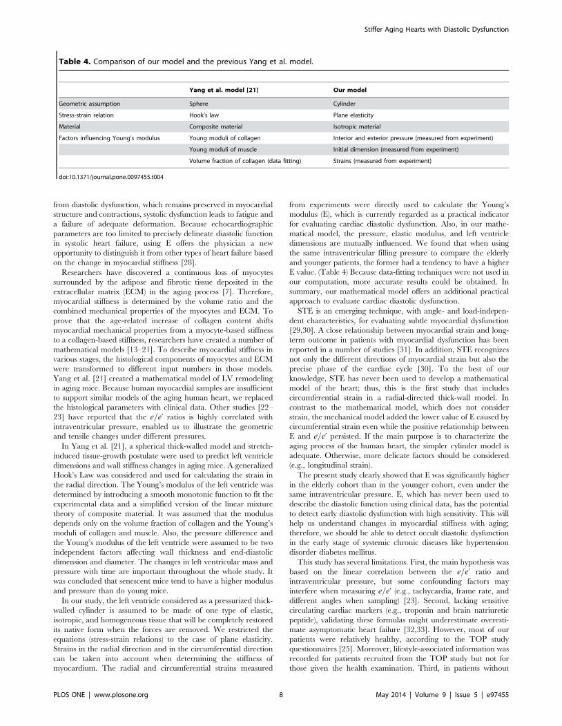
In our study, the left ventricle considered as a pressurized thick-
walled cylinder is assumed to be made of one type of elastic,
isotropic, and homogeneous tissue that will be completely restored
its native form when the forces are removed. We restricted the
equations (stress-strain relations) to the case of plane elasticity.
Strains in the radial direction and in the circumferential direction
can be taken into account when determining the stiffness of
myocardium. The radial and circumferential strains measured
from experiments were directly used to calculate the Young’s
modulus (E), which is currently regarded as a practical indicator
for evaluating cardiac diastolic dysfunction. Also, in our mathe-
matical model, the pressure, elastic modulus, and left ventricle
dimensions are mutually influenced. We found that when using
the same intraventricular filling pressure to compare the elderly
and younger patients, the former had a tendency to have a higher
E value. (Table 4) Because data-fitting techniques were not used in
our computation, more accurate results could be obtained. In
summary, our mathematical model offers an additional practical
approach to evaluate cardiac diastolic dysfunction.
STE is an emerging technique, with angle- and load-indepen-
dent characteristics, for evaluating subtle myocardial dysfunction
[29,30]. A close relationship between myocardial strain and long-
term outcome in patients with myocardial dysfunction has been
reported in a number of studies [31]. In addition, STE recognizes
not only the different directions of myocardial strain but also the
precise phase of the cardiac cycle [30]. To the best of our
knowledge, STE has never been used to develop a mathematical
model of the heart; thus, this is the first study that includes
circumferential strain in a radial-directed thick-wall model. In
contrast to the mathematical model, which does not consider
strain, the mechanical model added the lower value of E caused by
circumferential strain even while the positive relationship between
E and e=e’ persisted. If the main purpose is to characterize the
aging process of the human heart, the simpler cylinder model is
adequate. Otherwise, more delicate factors should be considered
(e.g., longitudinal strain).
The present study clearly showed that E was significantly higher
in the elderly cohort than in the younger cohort, even under the
same intraventricular pressure. E, which has never been used to
describe the diastolic function using clinical data, has the potential
to detect early diastolic dysfunction with high sensitivity. This will
help us understand changes in myocardial stiffness with aging;
therefore, we should be able to detect occult diastolic dysfunction
in the early stage of systemic chronic diseases like hypertension
disorder diabetes mellitus.
This study has several limitations. First, the main hypothesis was
based on the linear correlation between the e=e’ ratio and
intraventricular pressure, but some confounding factors may
interfere when measuring e=e’ (e.g., tachycardia, frame rate, and
different angles when sampling) [23]. Second, lacking sensitive
circulating cardiac markers (e.g., troponin and brain natriuretic
peptide), validating these formulas might underestimate overesti-
mate asymptomatic heart failure [32,33]. However, most of our
patients were relatively healthy, according to the TOP study
questionnaires [25]. Moreover, lifestyle-associated information was
recorded for patients recruited from the TOP study but not for
those given the health examination. Third, in patients without
Table 4. Comparison of our model and the previous Yang et al. model.
Yang et al. model [21] Our model
Geometric assumption Sphere Cylinder
Stress-strain relation Hook’s law Plane elasticity
Material Composite material Isotropic material
Factors influencing Young’s modulus Young moduli of collagen Interior and exterior pressure (measured from experiment)
Young moduli of muscle Initial dimension (measured from experiment)
Volume fraction of collagen (data fitting) Strains (measured from experiment)
doi:10.1371/journal.pone.0097455.t004
Stiffer Aging Hearts with Diastolic Dysfunction
PLOS ONE | www.plosone.org 8 May 2014 | Volume 9 | Issue 5 | e97455
pericardial disease, extracardiac pressure was far lower than
intraventricular pressure. One study [34] on 11 dogs with chronic
heart failure reported that the pressure in the pericardial space was
usually beneath 10 mmHg or 1=3 of the right atrial pressure.
Thus, in most LV models, the extra cardiac pressure has been
neglected. In our study, E in both models, whether or not
extracardiac pressure was considered, showed a trend of changes
similar to that of intraventricular pressure or IVSd. Fourth, we
assumed the LV to be a hollow axisymmetric cylinder with plane
strain, a state of strain in which the strain normal to the x2y plane,
ez, and the shear strain cxz and cyz, are assumed to be zero. The
deformation in the axial direction (z direction) is ignored, and the
strains in the radial and circumferential directions are independent
of h. In reality, the left ventricle does not have a constant cross-
section and does not deform uniformly at different orientations. In
addition, the left ventricle was assumed to be made of elastic,
isotropic, and homogeneous tissue when we determined the
Young’s modulus of the myocardium. Nonetheless, the left
ventricle usually contains at least two basic raw materials, viz.,
muscle and collagen.
Although a mathematical model has limitations, it enables us to
illustrate the quantitative relationships between structural and
functional changes. because diastolic and systolic heart failure
share similar poor outcomes [35] detecting diastolic dysfunction
early helps to allow early interventions, which may lead to a better
prognosis [36]. Furthermore, this model uses echocardiographic
parameters instead of histological data, and it is noninvasive, it
makes early detection more feasible.
In conclusion, this is the first report to document using a
mathematical model to delineate diastolic dysfunction in the aging
human heart. The vulnerability of the elderly to higher pressure
may contribute to their developing earlier signals for heart failure.
E is useful for the early and noninvasive detection of diastolic
dysfunction.
Supporting Information
Table S1 After excluding patients with hypertension, diabetes
mellitus, and coronary artery disease, the correlation between E
and diastolic dysfunction remained significant in the healthy
elders.
(DOC)
Table S2 Multivariate analysis on the relationship between E
and echocardiographic intraventricular pressure in younger
patients.
(DOCX)
Table S3 Multivariate analysis on the relationship between E
and echocardiographic intraventricular pressure in older patients.
(DOCX)
Author Contributions
Conceived and designed the experiments: JNJ PYL. Performed the
experiments: WTC JSC YKH WCT. Analyzed the data: WTC JSC YKH.
Wrote the paper: WTC JSC JNJ PYL.
References
1. Butler J, Kalogeropoulos A, Georgiopoulou V, Belue R, Rodondi N, et al. (2008)Incident heart failure prediction in the elderly: the health ABC heart failure
score. Circ Heart Fail 1: 125–133.
2. Bhatia RS, Tu JV, Lee DS, Austin PC, Fang J, et al. (2006) Outcome of heart
failure with preserved ejection fraction in a population-based study. N Engl J Med355: 260–269.
3. Mor-Avi V, Lang RM, Badano LP, Belohlavek M, Cardim NM, et al. (2011)
Current and evolving echocardiographic techniques for the quantitative
evaluation of cardiac mechanics: ASE/EAE consensus statement on method-ology and indications endorsed by the Japanese Society of Echocardiography.
Eur J Echocardiogr 12: 167–120.
4. Tresch DD, McGough MF (1995) Heart failure with normal systolic function: a
common disorder in older people. J Am Geriatr Soc 43: 1035–1042.
5. Jaarsma T, Halfens R, Abu-Saad HH, Dracup K, Stappers J, et al. (1999)Quality of life in older patients with systolic and diastolic heart failure.
Eur J Heart Fail 1: 151–160.
6. Owan TE, Hodge DO, Herges RM, Jacobsen SJ, Roger VL, et al. (2006) Trends
in prevalence and outcome of heart failure with preserved ejection fraction.N Engl J Med 355: 251–259.
7. Anversa P, Hiler B, Ricci R, Guideri G, Olivetti G (1986) Myocyte cell loss and
myocyte hypertrophy in the aging rat heart. J Am Coll Cardiol 8: 1441–1448.
8. Khan AS, Sane DC, Wannenburg T, Sonntag WE (2002) Growth hormone,
insulin-like growth factor-1 and the aging cardiovascular system. Cardiovasc Res54: 25–35.
9. Fung Y (1990) Biomechanics: motion, flow, stress, and growth. New York:Springer. 569 p.
10. Taber LA (1998) Biomechanical growth laws for muscle tissue. J Theor Biol 193:
201–213.
11. Nishimura RA, Tajik AJ (1997) Evaluation of diastolic filling of left ventricle in
health and disease: Doppler echocardiography is the clinician’s Rosetta Stone.J Am Coll Cardiol 30: 8–18.
12. Myreng Y, Smiseth OA (1990) Assessment of left ventricular relaxation byDoppler echocardiography. Comparison of isovolumic relaxation time and
transmitral flow velocities with time constant of isovolumic relaxation.Circulation 81: 260–266.
13. Gotteiner NL, Han G, Chandran KB, Vonesh MJ, Bresticker M, et al. (1972) Invivo determination of elastic modulus of canine cardiac muscle. J Basic Eng 94:
912–916.
14. Ghista DN, Sandler H, Vayo WH (1975) Elastic modulus of the human intact
left ventricle determination and physiological interpretation. Med Biol Eng 13:151–161.
15. Ghista DN, Advani SH, Rao BN (1975) In vivo elastic modulus of the left
ventricle: its determination by means of a left ventricular vibrational model and
its physiological significance and clinical utility. Med Biol Eng 13: 162–170.
16. Yettram AL, Grewal BS, Gibson DG (1992) Modelling the left ventricle fordetermination of the elasticity of the myocardium. J Eng Med 208: 1–8.
17. Arts T, Renman RS, Veenstra PC (1979) A model of the mechanics of the leftventricle. Ann Biomed Eng 7: 299–318.
18. Dumesnil JG, Shoucri RM, Laurenceau JL, Turcot J (1979) A mathematical
model of the dynamic geometry of the intact left ventricle and its application to
clinical data. Circulation 59: 1024–1034.
19. Ohayon J, Chadwick RS (1982) Mechanics of the left ventricle. Biophys J 39:
279–288.
20. Lin J, Lopez EF, Jin Y, Van Remmen H, Bauch T, et al. (2008) Age-related
cardiac muscle sarcopenia: combining experimental and mathematical modelingto identify mechanisms. Exp Gerontol 43: 296–306.
21. Yang T, Chiao YA, Wang Y, Voorhees A, Han HC, et al. (2012) Mathematical
modeling of left ventricular dimensional changes in mice during aging. BMC
Syst Biol 6: 1–12.
22. Isnard RN, Pannier BM, Laurent S, London GM, Diebold B, et al. (1989)
Pulsatile diameter and elastic modulus of the aortic arch in essentialhypertension: a noninvasive study. J Am Coll Cardiol 13: 399–405.
23. Nagueh SF, Middleton KJ, Kopelen HA, Zoghbi WA, Quinones MA (1997)
Doppler tissue imaging: a noninvasive technique for evaluation of left ventricular
relaxation and estimation of filling pressures. J Am Coll Cardiol 30: 1527–1533.
24. Chang CS, Chang YF, Liu PY, Chen CY, Tsai YS, et al. (2012) Smoking,
habitual tea drinking and metabolic syndrome in elderly men living in ruralcommunity: the Tianliao old people (TOP) study 02. PLOS One 7: e38874.
25. Chang CL, Lee PT, Chang WT, Chang CS, Chen JH, et al. (2013) The
interplay between inflammation, physical activity and metabolic syndrome in a
remote male geriatric community in Southern Taiwan: the Tianliao Old People(TOP) study 03. Diabetol Metab Syndr 5: 60.
26. Lang RM, Bierig M, Devereux RB, Flachskampf FA, Foster E, et al. (2005)Recommendations for chamber quantification: a report from the American
Society of Echocardiography’s Guidelines and Standards Committee and theChamber Quantification Writing Group, developed in conjunction with the
European Association of Echocardiography, a branch of the European Societyof Cardiology. J Am Soc Echocardiogr 18: 1440–1463.
27. Reismann H, Pawlik PS (1980) Elasticity theory and applications. New York:John Wiley & Sons.
28. Tei C, Ling LH, Hodge DO, Bailey KR, Oh JK, et al. (1995) New index ofcombined systolic and diastolic myocardial performance: a simple and
reproducible measure of cardiac function–a study in normals and dilatedcardiomyopathy. J Cardiol 26: 357–366.
29. Liu YW, Su CT, Lin CC, Chen JH (2009) Evidence of left ventricular systolicdysfunction detected by automated functional imaging in patients with heart
failure and preserved left ventricular ejection fraction. J Cardiac Fail 15: 782–
789.
Stiffer Aging Hearts with Diastolic Dysfunction
PLOS ONE | www.plosone.org 9 May 2014 | Volume 9 | Issue 5 | e97455

30. Perk G, Kronzon I (2009) Non-Doppler two dimensional strain imaging for
evaluation of coronary artery disease. Echocardiography 26: 299–306.31. Bertini M, Ng AC, Antoni ML, Nucifora G, Ewe SH, et al. (2012) Global
longitudinal strain predicts long-term survival in patients with chronic ischemic
cardiomyopathy. Circ Cardiovasc Imaging 5: 383–391.32. Lukowicz TV, Fischer M, Hense HW, Doring A, Stritzke J, et al. (2005) BNP as
a marker of diastolic dysfunction in the general population: importance of leftventricular hypertrophy. Eur J Heart Fail 4: 525–531.
33. Huang XP, Du JF (2004) Troponin I, cardiac diastolic dysfunction and
restrictive cardiomyopathy. Acta Pharmacol Sin 12: 1569–1575.34. Horne SG, Belenkie I, Tyberg JV, Smith ER (2000) Pericardial pressure in
experimental chronic heart failure. Can J Cardiol 5: 607–613.
35. Warren SE, Grossman W (1991) Prognosis in heart failure: is systolic or diastolicdysfunction more important? Herz 16 Spec No 1: 324–329.
36. Galderisi M (2005) Diastolic dysfunction and diastolic heart failure: diagnostic,prognostic and therapeutic aspects. Cardiovasc Ultrasound 3: 9.
Stiffer Aging Hearts with Diastolic Dysfunction
PLOS ONE | www.plosone.org 10 May 2014 | Volume 9 | Issue 5 | e97455
Related Documents


![[PPT]DIASTOLIC DYSFUNCTION AND HEART FAILURE ...jacobimed.org/public/Docs/Lecture Power Points/DIASTOLIC... · Web viewDIASTOLIC DYSFUNCTION AND HEART FAILURE PHYSIOLOGY, HISTORICAL](https://static.cupdf.com/doc/110x72/5ab2544e7f8b9ac66c8d51e8/pptdiastolic-dysfunction-and-heart-failure-power-pointsdiastolicweb-viewdiastolic.jpg)