Characterisation of the macular dystrophy in patients with the A3243G mitochondrial DNA point mutation with fundus autofluorescence P P Rath, 1,2 S Jenkins, 1,3 M Michaelides, 1,3 A Smith, 1,4 M G Sweeney, 5 M B Davis, 5 F W Fitzke, 3 A C Bird 1,3 1 Moorfields Eye Hospital, London, UK; 2 Retina Vitreous Consultants, Pittsburgh, PA, USA; 3 Institute of Ophthalmology, London, UK; 4 County Hospital, Hereford, UK; 5 Department of Molecular Neuroscience, Institute of Neurology, London, UK Correspondence to: Dr P P Rath, Retina Vitreous Consultants, 3501 Forbes Avenue, Suite 500, Pittsburgh, PA 15213, USA; prprath@ yahoo.com PR and SJ contributed equally to this study. Accepted 22 January 2008 This paper is freely available online under the BMJ Journals unlocked scheme, see http:// bjo.bmj.com/info/unlocked.dtl ABSTRACT Introduction: The mitochondrial DNA A3243G point mutation is associated with a wide variety of systemic manifestations including a macular dystrophy. The characteristics of fundus autofluorescence (AF) in these patients are distinctive and have not been previously described. Methods: A complete history and ophthalmic examina- tion, including fundus photography and autofluorescence imaging, was performed on twelve probands harbouring the A3243G point mutation. Results: Four patients had diabetes, 10/12 hearing loss, and 7/12 were visually symptomatic. A positive family history was present in 5/12. Fundus findings consisted of two primary phenotypes: discontinuous circumferentially oriented perifoveal atrophy (9/12) or an appearance consistent with pattern dystrophy (3/12). In both phenotypes pale deposits and pigment clumping were seen at the level of the retinal pigment epithelium, with occasional changes also noted outside the arcades and nasal to the optic nerve. Fundus AF imaging revealed decreased autofluorescence in areas of atrophy and increased AF of the pale subretinal deposits. In areas of the retina that appeared normal clinically, variable sized flecks of increased and decreased AF were present. Conclusions: The mitochondrial DNA A3243G point mutation can result in disease with a variable presenta- tion. Fundus autofluorescence reveals a recognisable phenotype in most cases that is different from other macular dystrophies. The single-point mutation of the mitochondrial DNA (mtDNA) at the 3243 position in the tRNA Leu(UUR) gene leads to a wide variety of clinical manifestations. This mutation was originally described in association with Mitochondrial Encephalopathy, Lactic Acidosis and Stroke-like episodes (MELAS), and is now known to produce a variety of other clinical disorders, including mater- nally inherited diabetes and deafness (MIDD), cardiomyopathy, chronic progressive external ophthalmoplegia (CPEO), a pure myopathy, gas- trointestinal dysmotility and renal failure. 1–6 One explanation for this variability is thought to be the load of mutant mtDNA present in an individual, which varies both between individuals and from tissue to tissue within an individual. 7 In general, a higher mutation load is associated with more severe disease. However, this generalisation does not always hold true, and it is possible that environmental factors and/or nuclear genetic influences may modulate disease manifesta- tions. 689 Ocular manifestations of mitochondrial diseases are well recognised, and in the original report by Reardon et al which revealed a new subtype of diabetes caused by a mtDNA mutation, three patients were found to have retinal pigmentary changes. 2 10 However, there were no fundus photo- graphs of the changes described in this initial report. 2 In 1995, Massin et al described the association of a bilateral macular pattern dystro- phy with MIDD and further characterised this in a subsequent publication in 1999. 11 12 Since these reports, there have been several additional publica- tions describing the macular dystrophy of MIDD and MELAS, both clinically and electrophysiologi- cally. 13–18 The use of the confocal scanning laser ophthal- moscope (cSLO) for imaging of macular diseases has been well described. 19–25 In this study, we have characterised in detail the distinct fundus auto- fluorescence (AF) characteristics of the macular dystrophy associated with the A3243G mtDNA mutation. METHODS All patients diagnosed as having a macular dystrophy due to the A3243G mtDNA mutation in the Medical Retina Clinic at Moorfields Eye Hospital between 1995 and 2001 were included in the study. Twelve patients in total were evaluated. Patient demographics, referring diagnosis, presence or absence of visual symptoms, duration of visual symptoms, general medical history including pre- sence or absence of diabetes and deafness, maternal history of diabetes and deafness, and a detailed family history, were obtained for each patient. Best-corrected Snellen visual acuity and slit-lamp biomicroscopy findings were recorded. Colour fundus photographs and fundus autofluorescence imaging were performed. Patients were tested for the A3243G mutation based on the characteristics of their ophthalmic examination, regardless of whether or not there was a personal or family history of diabetes or deafness. The diagnosis of the A3243G mtDNA mutation was established with DNA testing of peripheral blood samples using previously described techniques. 26 Fundus autofluorescence imaging was performed with a confocal scanning laser ophthalmoscope (cSLO, Zeiss, Jena, Germany or HRA, Heidelberg, Germany) using previously published techni- ques. 19 20 27 Autofluorescence images were compared Clinical science Br J Ophthalmol 2008;92:623–629. doi:10.1136/bjo.2007.131177 623

Welcome message from author
This document is posted to help you gain knowledge. Please leave a comment to let me know what you think about it! Share it to your friends and learn new things together.
Transcript
Characterisation of the macular dystrophy in patientswith the A3243G mitochondrial DNA point mutationwith fundus autofluorescence
P P Rath,1,2 S Jenkins,1,3 M Michaelides,1,3 A Smith,1,4 M G Sweeney,5 M B Davis,5
F W Fitzke,3 A C Bird1,3
1 Moorfields Eye Hospital,London, UK; 2 Retina VitreousConsultants, Pittsburgh, PA,USA; 3 Institute ofOphthalmology, London, UK;4 County Hospital, Hereford, UK;5 Department of MolecularNeuroscience, Institute ofNeurology, London, UK
Correspondence to:Dr P P Rath, Retina VitreousConsultants, 3501 ForbesAvenue, Suite 500, Pittsburgh,PA 15213, USA; [email protected]
PR and SJ contributed equally tothis study.
Accepted 22 January 2008
This paper is freely availableonline under the BMJ Journalsunlocked scheme, see http://bjo.bmj.com/info/unlocked.dtl
ABSTRACTIntroduction: The mitochondrial DNA A3243G pointmutation is associated with a wide variety of systemicmanifestations including a macular dystrophy. Thecharacteristics of fundus autofluorescence (AF) in thesepatients are distinctive and have not been previouslydescribed.Methods: A complete history and ophthalmic examina-tion, including fundus photography and autofluorescenceimaging, was performed on twelve probands harbouringthe A3243G point mutation.Results: Four patients had diabetes, 10/12 hearing loss,and 7/12 were visually symptomatic. A positive familyhistory was present in 5/12. Fundus findings consisted oftwo primary phenotypes: discontinuous circumferentiallyoriented perifoveal atrophy (9/12) or an appearanceconsistent with pattern dystrophy (3/12). In bothphenotypes pale deposits and pigment clumping wereseen at the level of the retinal pigment epithelium, withoccasional changes also noted outside the arcades andnasal to the optic nerve. Fundus AF imaging revealeddecreased autofluorescence in areas of atrophy andincreased AF of the pale subretinal deposits. In areas ofthe retina that appeared normal clinically, variable sizedflecks of increased and decreased AF were present.Conclusions: The mitochondrial DNA A3243G pointmutation can result in disease with a variable presenta-tion. Fundus autofluorescence reveals a recognisablephenotype in most cases that is different from othermacular dystrophies.
The single-point mutation of the mitochondrialDNA (mtDNA) at the 3243 position in thetRNALeu(UUR) gene leads to a wide variety of clinicalmanifestations. This mutation was originallydescribed in association with MitochondrialEncephalopathy, Lactic Acidosis and Stroke-likeepisodes (MELAS), and is now known to produce avariety of other clinical disorders, including mater-nally inherited diabetes and deafness (MIDD),cardiomyopathy, chronic progressive externalophthalmoplegia (CPEO), a pure myopathy, gas-trointestinal dysmotility and renal failure.1–6 Oneexplanation for this variability is thought to be theload of mutant mtDNA present in an individual,which varies both between individuals and fromtissue to tissue within an individual.7 In general, ahigher mutation load is associated with moresevere disease. However, this generalisation doesnot always hold true, and it is possible thatenvironmental factors and/or nuclear genetic
influences may modulate disease manifesta-tions.6 8 9
Ocular manifestations of mitochondrial diseasesare well recognised, and in the original report byReardon et al which revealed a new subtype ofdiabetes caused by a mtDNA mutation, threepatients were found to have retinal pigmentarychanges.2 10 However, there were no fundus photo-graphs of the changes described in this initialreport.2 In 1995, Massin et al described theassociation of a bilateral macular pattern dystro-phy with MIDD and further characterised this in asubsequent publication in 1999.11 12 Since thesereports, there have been several additional publica-tions describing the macular dystrophy of MIDDand MELAS, both clinically and electrophysiologi-cally.13–18
The use of the confocal scanning laser ophthal-moscope (cSLO) for imaging of macular diseaseshas been well described.19–25 In this study, we havecharacterised in detail the distinct fundus auto-fluorescence (AF) characteristics of the maculardystrophy associated with the A3243G mtDNAmutation.
METHODSAll patients diagnosed as having a maculardystrophy due to the A3243G mtDNA mutationin the Medical Retina Clinic at Moorfields EyeHospital between 1995 and 2001 were included inthe study. Twelve patients in total were evaluated.Patient demographics, referring diagnosis, presenceor absence of visual symptoms, duration of visualsymptoms, general medical history including pre-sence or absence of diabetes and deafness, maternalhistory of diabetes and deafness, and a detailedfamily history, were obtained for each patient.Best-corrected Snellen visual acuity and slit-lampbiomicroscopy findings were recorded. Colourfundus photographs and fundus autofluorescenceimaging were performed. Patients were tested forthe A3243G mutation based on the characteristicsof their ophthalmic examination, regardless ofwhether or not there was a personal or familyhistory of diabetes or deafness. The diagnosis ofthe A3243G mtDNA mutation was establishedwith DNA testing of peripheral blood samplesusing previously described techniques.26
Fundus autofluorescence imaging was performedwith a confocal scanning laser ophthalmoscope(cSLO, Zeiss, Jena, Germany or HRA, Heidelberg,Germany) using previously published techni-ques.19 20 27 Autofluorescence images were compared
Clinical science
Br J Ophthalmol 2008;92:623–629. doi:10.1136/bjo.2007.131177 623
with colour photographs. The AF images of the patients withthe A3243G mutation were also compared with images ofpatients with other maculopathies, including Stargardt maculardystrophy, R172W peripherin macular dystrophy, bull’s eyemacular dystrophy, geographic atrophy from age-related maculardegeneration and pattern dystrophy.
Patients who did not have a history of diabetes were testedwith fasting blood glucose, where possible, according to therecommendations of the American Diabetic Association.28 Renalfunction tests were also performed when possible due to theassociation of renal dysfunction with the A3243G mutation.29
RESULTSTwelve patients were diagnosed as having the A3243Gmutation between January 1995 and February 2001; in nonehad the diagnosis of a mitochondrial disorder been made prior tobeing seen by the authors. The characteristics of these patientsat presentation are summarised in table 1.
Nine of the patients were female. The average age atpresentation was 47 years (range 36 to 65) with an averageage at diagnosis of 51 (range 36 to 71). In six of 12 patients, thereason for referral to a specialist clinic was an unspecifiedmaculopathy consisting of a combination of retinal pigmentepithelial changes and atrophy. Other diagnoses at referral wereUsher syndrome variant, central areolar choroidal sclerosis,pattern dystrophy, macular degeneration and macular atrophy.Five of the 12 patients did not have visual symptoms atpresentation and were found to have fundus abnormalities onroutine eye examination. Of the patients who were initiallyasymptomatic, two patients became symptomatic duringreview. Three patients are still asymptomatic after follow-up,ranging from 1 to 6 years. Asymptomatic patients tended to beyounger than the patients with symptoms, with a mean age of43 compared with 51 years of age, although the number ofpatients in each group is too small for statistical significance tobe established. Of the seven patients with symptoms, threereported difficulties due to paracentral scotoma, and threereported a general decrease in vision in one eye.
A history of hearing loss was common, with 10 of 12 patientshaving some degree of hearing impairment. Patient 1 had acochlear implant, and four other patients were using hearingaids. Diabetes was present in only four patients, all of whomalso had hearing loss.
A maternal history of diabetes was present in two patientsand a maternal history of deafness in five patients. A maternalhistory of both diabetes and deafness was elicited in only onesubject. There were two additional patients with a maternalhistory of hearing loss diagnosed after the age of 75 years. Thiswas considered to be age-related hearing loss and not includedas a positive history in the context of a mitochondrial disease.Two further patients had a family history of maternal diabetes,which was not diagnosed until the age of 60, and was again notconsidered as a positive finding in the context of the A3243Gmutation.
In all patients, presenting visual acuity was good, each havingat least 6/9 or better vision, and seven patients achieving 6/6 orbetter vision, with each eye. At final follow-up, the vision wasslightly worse, with all patients having 6/12 or better vision inat least one eye. Six out of 12 patients had 6/6 or better visionwith at least one eye at the last documented examination. Ofthe four patients with 6/12 or worse vision, three haddeterioration in vision due to paramacular atrophy encroachingupon fixation, and one had decreased vision bilaterally due toretinal pigment epitheliopathy.
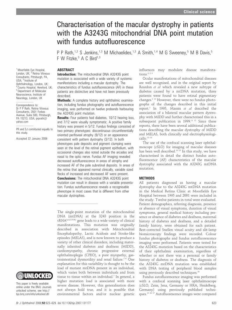
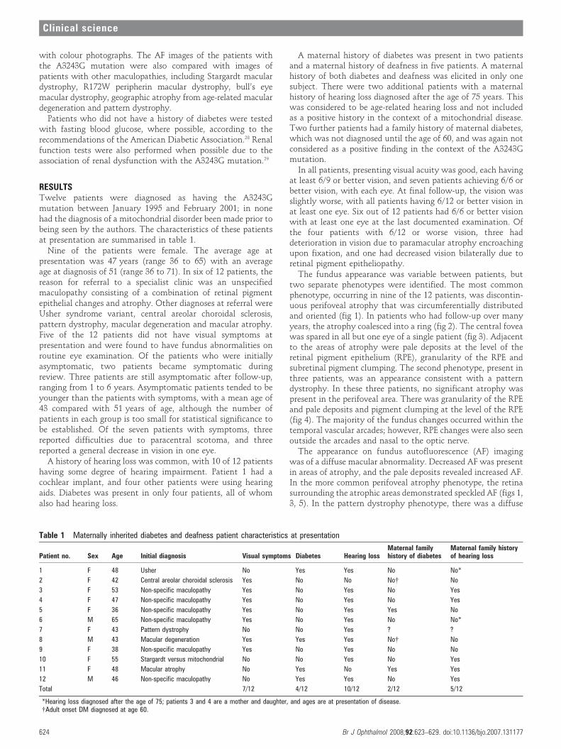
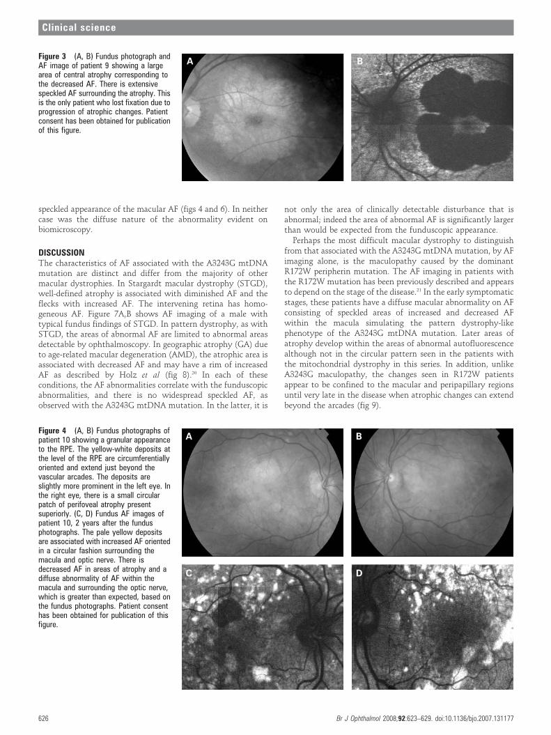
The fundus appearance was variable between patients, buttwo separate phenotypes were identified. The most commonphenotype, occurring in nine of the 12 patients, was discontin-uous perifoveal atrophy that was circumferentially distributedand oriented (fig 1). In patients who had follow-up over manyyears, the atrophy coalesced into a ring (fig 2). The central foveawas spared in all but one eye of a single patient (fig 3). Adjacentto the areas of atrophy were pale deposits at the level of theretinal pigment epithelium (RPE), granularity of the RPE andsubretinal pigment clumping. The second phenotype, present inthree patients, was an appearance consistent with a patterndystrophy. In these three patients, no significant atrophy waspresent in the perifoveal area. There was granularity of the RPEand pale deposits and pigment clumping at the level of the RPE(fig 4). The majority of the fundus changes occurred within thetemporal vascular arcades; however, RPE changes were also seenoutside the arcades and nasal to the optic nerve.
The appearance on fundus autofluorescence (AF) imagingwas of a diffuse macular abnormality. Decreased AF was presentin areas of atrophy, and the pale deposits revealed increased AF.In the more common perifoveal atrophy phenotype, the retinasurrounding the atrophic areas demonstrated speckled AF (figs 1,3, 5). In the pattern dystrophy phenotype, there was a diffuse
Table 1 Maternally inherited diabetes and deafness patient characteristics at presentation
Patient no. Sex Age Initial diagnosis Visual symptoms Diabetes Hearing lossMaternal familyhistory of diabetes
Maternal family historyof hearing loss
1 F 48 Usher No Yes Yes No No*
2 F 42 Central areolar choroidal sclerosis Yes No No No{ No
3 F 53 Non-specific maculopathy Yes No Yes No Yes
4 F 47 Non-specific maculopathy Yes No Yes No Yes
5 F 36 Non-specific maculopathy Yes No Yes Yes No
6 M 65 Non-specific maculopathy Yes No Yes No No*
7 F 43 Pattern dystrophy No No Yes ? ?
8 M 43 Macular degeneration Yes Yes Yes No{ No
9 F 38 Non-specific maculopathy Yes No Yes No No
10 F 55 Stargardt versus mitochondrial No No Yes No Yes
11 F 48 Macular atrophy No Yes No Yes Yes
12 M 46 Non-specific maculopathy No Yes Yes No Yes
Total 7/12 4/12 10/12 2/12 5/12
*Hearing loss diagnosed after the age of 75; patients 3 and 4 are a mother and daughter, and ages are at presentation of disease.{Adult onset DM diagnosed at age 60.
Clinical science
624 Br J Ophthalmol 2008;92:623–629. doi:10.1136/bjo.2007.131177
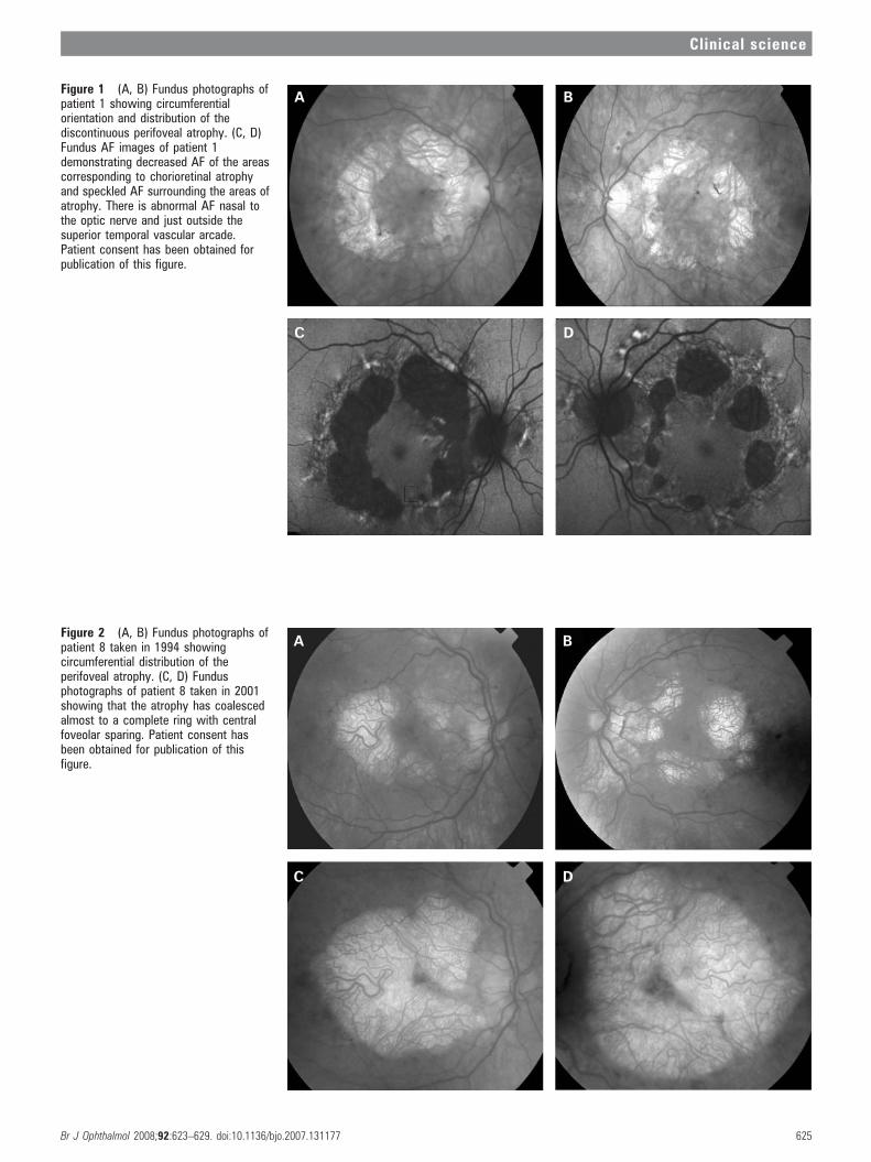
Figure 1 (A, B) Fundus photographs ofpatient 1 showing circumferentialorientation and distribution of thediscontinuous perifoveal atrophy. (C, D)Fundus AF images of patient 1demonstrating decreased AF of the areascorresponding to chorioretinal atrophyand speckled AF surrounding the areas ofatrophy. There is abnormal AF nasal tothe optic nerve and just outside thesuperior temporal vascular arcade.Patient consent has been obtained forpublication of this figure.
Figure 2 (A, B) Fundus photographs ofpatient 8 taken in 1994 showingcircumferential distribution of theperifoveal atrophy. (C, D) Fundusphotographs of patient 8 taken in 2001showing that the atrophy has coalescedalmost to a complete ring with centralfoveolar sparing. Patient consent hasbeen obtained for publication of thisfigure.
Clinical science
Br J Ophthalmol 2008;92:623–629. doi:10.1136/bjo.2007.131177 625
speckled appearance of the macular AF (figs 4 and 6). In neithercase was the diffuse nature of the abnormality evident onbiomicroscopy.
DISCUSSIONThe characteristics of AF associated with the A3243G mtDNAmutation are distinct and differ from the majority of othermacular dystrophies. In Stargardt macular dystrophy (STGD),well-defined atrophy is associated with diminished AF and theflecks with increased AF. The intervening retina has homo-geneous AF. Figure 7A,B shows AF imaging of a male withtypical fundus findings of STGD. In pattern dystrophy, as withSTGD, the areas of abnormal AF are limited to abnormal areasdetectable by ophthalmoscopy. In geographic atrophy (GA) dueto age-related macular degeneration (AMD), the atrophic area isassociated with decreased AF and may have a rim of increasedAF as described by Holz et al (fig 8).20 In each of theseconditions, the AF abnormalities correlate with the funduscopicabnormalities, and there is no widespread speckled AF, asobserved with the A3243G mtDNA mutation. In the latter, it is
not only the area of clinically detectable disturbance that isabnormal; indeed the area of abnormal AF is significantly largerthan would be expected from the funduscopic appearance.
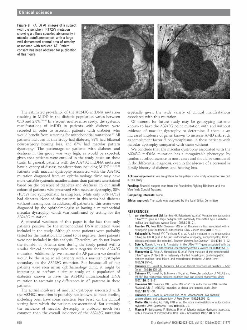
Perhaps the most difficult macular dystrophy to distinguishfrom that associated with the A3243G mtDNA mutation, by AFimaging alone, is the maculopathy caused by the dominantR172W peripherin mutation. The AF imaging in patients withthe R172W mutation has been previously described and appearsto depend on the stage of the disease.21 In the early symptomaticstages, these patients have a diffuse macular abnormality on AFconsisting of speckled areas of increased and decreased AFwithin the macula simulating the pattern dystrophy-likephenotype of the A3243G mtDNA mutation. Later areas ofatrophy develop within the areas of abnormal autofluorescencealthough not in the circular pattern seen in the patients withthe mitochondrial dystrophy in this series. In addition, unlikeA3243G maculopathy, the changes seen in R172W patientsappear to be confined to the macular and peripapillary regionsuntil very late in the disease when atrophic changes can extendbeyond the arcades (fig 9).
Figure 3 (A, B) Fundus photograph andAF image of patient 9 showing a largearea of central atrophy corresponding tothe decreased AF. There is extensivespeckled AF surrounding the atrophy. Thisis the only patient who lost fixation due toprogression of atrophic changes. Patientconsent has been obtained for publicationof this figure.
Figure 4 (A, B) Fundus photographs ofpatient 10 showing a granular appearanceto the RPE. The yellow-white deposits atthe level of the RPE are circumferentiallyoriented and extend just beyond thevascular arcades. The deposits areslightly more prominent in the left eye. Inthe right eye, there is a small circularpatch of perifoveal atrophy presentsuperiorly. (C, D) Fundus AF images ofpatient 10, 2 years after the fundusphotographs. The pale yellow depositsare associated with increased AF orientedin a circular fashion surrounding themacula and optic nerve. There isdecreased AF in areas of atrophy and adiffuse abnormality of AF within themacula and surrounding the optic nerve,which is greater than expected, based onthe fundus photographs. Patient consenthas been obtained for publication of thisfigure.
Clinical science
626 Br J Ophthalmol 2008;92:623–629. doi:10.1136/bjo.2007.131177
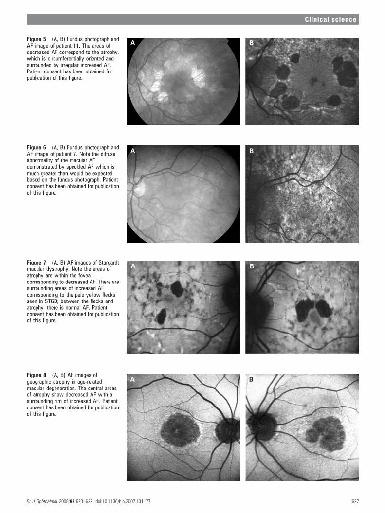
Figure 5 (A, B) Fundus photograph andAF image of patient 11. The areas ofdecreased AF correspond to the atrophy,which is circumferentially oriented andsurrounded by irregular increased AF.Patient consent has been obtained forpublication of this figure.
Figure 6 (A, B) Fundus photograph andAF image of patient 7. Note the diffuseabnormality of the macular AFdemonstrated by speckled AF which ismuch greater than would be expectedbased on the fundus photograph. Patientconsent has been obtained for publicationof this figure.
Figure 7 (A, B) AF images of Stargardtmacular dystrophy. Note the areas ofatrophy are within the foveacorresponding to decreased AF. There aresurrounding areas of increased AFcorresponding to the pale yellow flecksseen in STGD; between the flecks andatrophy, there is normal AF. Patientconsent has been obtained for publicationof this figure.
Figure 8 (A, B) AF images ofgeographic atrophy in age-relatedmacular degeneration. The central areasof atrophy show decreased AF with asurrounding rim of increased AF. Patientconsent has been obtained for publicationof this figure.
Clinical science
Br J Ophthalmol 2008;92:623–629. doi:10.1136/bjo.2007.131177 627
The estimated prevalence of the A3243G mtDNA mutationresulting in MIDD in the diabetic population varies between0.13 and 2.8%.30–38 In a recent multi-centre study, the systemicmanifestations of MIDD in patients with diabetes wererecorded in order to ascertain patients with diabetes whowould benefit from screening for mitochondrial mutations.29 Allpatients included in this study had diabetes, 98% had bilateralneurosensory hearing loss, and 87% had macular patterndystrophy. The percentage of patients with diabetes anddeafness in this group was very high, as would be expected,given that patients were enrolled in the study based on thesetraits. In general, patients with the A3243G mtDNA mutationhave a variety of disease manifestations including MIDD.5 6 39–41
Patients with macular dystrophy associated with the A3243Gmutation diagnosed from an ophthalmology clinic may havemore variable systemic manifestations than patients ascertainedbased on the presence of diabetes and deafness. In our smallcohort of patients who presented with macular dystrophy, 83%(10/12) had symptomatic hearing loss, while only 33% (4/12)had diabetes. None of the patients in this series had diabeteswithout hearing loss. In addition, all patients in this series werediagnosed by the ophthalmologist as having a mitochondrialmacular dystrophy, which was confirmed by testing for theA3243G mutation.
A potential weakness of this paper is the fact that onlypatients positive for the mitochondrial DNA mutation wereincluded in the study. Although some patients were probablytested for the mutation and found to be negative, those patientswere not included in this analysis. Therefore, we do not knowthe number of patients seen during the study period with asimilar clinical phenotype who were negative for the A3243Gmutation. Additionally, we assume the AF pattern we describewould be the same in all patients with a macular dystrophysecondary to the A3243G mutation. But because all of ourpatients were seen in an ophthalmology clinic, it might beinteresting to perform a similar study on a population ofdiabetics known to have the A3243G mitochondrial DNAmutation to ascertain any differences in AF patterns in thesepatients.
The actual incidence of macular dystrophy associated withthe A3243G mutation is probably not known, as most studies,including ours, have some selection bias based on the clinicalsetting from which the patients are ascertained. But certainlythe incidence of macular dystrophy is probably much lesscommon than the overall incidence of the A3243G mutation
especially given the wide variety of clinical manifestationsassociated with this mutation.
Of interest for future study may be genotyping patientsknown to have the A3243G point mutation with and withoutevidence of macular dystrophy to determine if there is anincreased incidence of genes known to increase AMD risk, suchas complement factor H polymorphisms, in those patients withmacular dystrophy compared with those without.
We conclude that the macular dystrophy associated with theA3243G mtDNA mutation has a recognizable phenotype byfundus autofluorescence in most cases and should be consideredin the differential diagnosis, even in the absence of a personal orfamily history of diabetes and hearing loss.
Acknowledgements: We are grateful to the patients who kindly agreed to take partin this study.
Funding: Financial support was from the Foundation Fighting Blindness and theMoorfields Special Trustees.
Competing interests: None.
Ethics approval: The study was approved by the local Ethics Committee.
REFERENCES1. van den Ouweland JM, Lemkes HH, Ruitenbeek W, et al. Mutation in mitochondrial
tRNALeu(UUR) gene in a large pedigree with maternally transmitted type II diabetesmellitus and deafness. Nature Genet 1992;1:368–71.
2. Reardon W, Ross RJM, Sweeney MG, et al. Diabetes mellitus associated with apathogenic point mutation in mitochondrial DNA. Lancet 1992;340:1376–9.
3. Kobayashi Y, Momoi MY, Tominaga K, et al. A point mutation in the mitochondrialtRNA(Leu)(UUR0 gene in MELAS (mitochondrial myopathy, encephalopathy, lacticacidosis and stroke-like episodes). Biochem Biophys Res Commun 1990;173:816–22.
4. Goto Y, Nonaka I, Horai S. A mutation in the tRNALeu(UUR) gene associated with theMELAS subgroup of mitochondrial encephalomyopathies. Nature 1990;348:651–3.
5. Manouvrier S, Rotig A, Hannebique G, et al. Point mutation of the mitochondrialtRNALeu gene (A 3243 G) in maternally inherited hypertrophic cardiomyopathy,diabetes mellitus, renal failure, and sensorineural deafness. J Med Genet1985;32:654–6.
6. Chinnery PF, Howell N, Andrews RM, et al. Clinical mitochondrial genetics. J MedGenet 1999;36:425–36.
7. Chinnery PF, Howell N, Lightowlers RN, et al. Molecular pathology of MELAS andMERRF The relationship between mutation load and clinical phenotypes. Brain1997;120:1713–21.
8. Hammans SR, Sweeney MG, Hanna MG, et al. The mitochondrial DNA transferRNALeu(UUR) ARG(3243) mutation. A clinical and genetic study. Brain1995;118:721–34.
9. Chinnery PF, Howell N, Andrews RM, et al. Mitochondrial DNA analysis:polymorphisms and pathogenicity. J Med Genet 1999;36:505–10.
10. Mullie MA, Harding AE, Petty RKH, et al. The retinal manifestations of mitochondrialmyopathy. Arch Ophthalmol 1985;103:1825–30.
11. Massin P, Guillausseau P, Bialettes B, et al. Macular pattern dystrophy associatedwith a mutation of mitochondrial DNA. Am J Ophthalmol 1995;120:247–8.
Figure 9 (A, B) AF images of a subjectwith the peripherin R172W mutationshowing a diffuse speckled abnormality inmacular autofluorescence, with a largewell-demarcated central area of atrophyassociated with reduced AF. Patientconsent has been obtained for publicationof this figure.
Clinical science
628 Br J Ophthalmol 2008;92:623–629. doi:10.1136/bjo.2007.131177
12. Massin P, Virally-Monod M, Bialettes B, et al. Prevalence of macular patterndystrophy in maternally inherited diabetes and deafness. Ophthalmology1999;106:1821–7.
13. Isashiki Y, Nakagawa M, Ohba N, et al. Acta Ophthalmol Scand 1998;76:6–13.14. Harrison TJ, Boles RG, Johnson DR, et al. Macular pattern retinal dystrophy, adult-
onset diabetes, and deafness: A family study of A3243G mitochondrial heteroplasmy.Am J Ophthalmol 1997;124:217–21.
15. Bonte CA, Matthijs GL, Cassiman JJ, et al. Macular pattern dystrophy in patientswith deafness and diabetes. Retina 1997;17:216–21.
16. Andrews RM, McNeela BJ, Reading P, et al. Mitochondrial DNA diseasemasquerading as age-related macular degeneration. Eye 1999;13:595–6.
17. Latkany P, Ciulla TA, Cucchillo P, et al. Mitochondrial maculopathy: geographicatrophy of the macula in the MELAS associated A to G 3243 mitochondrial DNA pointmutation. Am J Ophthalmol 1999;128:112–14.
18. Smith PR, Bain SC, Good PA, et al. Pigmentary retinal dystrophy and the syndrome ofmaternally inherited diabetes and deafness caused by the mitochondrial DNA 3243tRNALeu A to G mutation. Ophthalmology 1999;106:1101–8.
19. von Ruckmann A, Fitzke FW, Bird AC. In vivo fundus autofluorescence in maculardystrophies. Arch Ophthalmol 1997;115:609–15.
20. Holz FG, Bellmann C, Margaritidis M, et al. Patterns of increased in vivo fundusautofluorescence in the junctional zone of geographic atrophy of the retinal pigmentepithelium associated with age-related macular degeneration. Graefes Arch Clin ExpOphthalmol 1999;237:145–52.
21. Downes SM, Fitzke FW, Holder GE, et al. Clinical features of codon 172 RDSmacular dystrophy. Similar phenotype in 12 families. Arch Ophthalmol1999;117:1373–83.
22. Downes SM, Holder GE, Fitzke FW, et al. Autosomal dominant cone and cone–roddystrophy with mutations in the guanylate cyclase activator 1A gene-encodingguanylate cyclase activating protein-1. Arch Ophthalmol 2001;119:96–105.
23. Lois N, Holder GE, Bunce C, et al. Phenotypic subtypes of Stargardt maculardystrophy-fundus flavimaculatus. Arch Ophthalmol 2001;119:359–69.
24. Bellmann C, Jorzik J, Spital G, et al. Symmetry of bilateral lesions in geographicatrophy in patients with age-related macular degeneration. Arch Ophthalmol2002;120:579–84.
25. Kurz-Levin MM, Halfyard AS, Bunce C, et al. Clinical variations in assessment ofbull’s-eye maculopathy. Arch Ophthalmol 2002;120:567–75.
26. Hammans SR, Sweeney MG, Brockington M, et al. Mitochondrial encephalopathies:molecular genetic diagnosis from blood samples. Lancet 1991;337:1311–13.
27. von Ruckmann A, Fitzke FW, Bird AC. Distribution of fundus autofluorescence witha scanning laser ophthalmoscope. Br J Ophthalmol 1995;79:407–12.
28. Expert Committee on the Diagnosis and Classification of Diabetes Mellitus.Report of the Expert Committee on the Diagnosis and Classification of DiabetesMellitus. Diabetes Care 2003;26:5–20S.
29. Guillausseau P, Massin P, Dubois-LaForgue D, et al. Maternally inherited diabetesand deafness: A multicenter Study. Ann Int Med 2001;134:721–8.
30. Vionnet N, Passa P, Froguel P. Prevalence of mitochondrial gene mutations infamilies with diabetes mellitus. Lancet 1993;342:1429–30.
31. Newkirk JE, Taylor RW, Howell N, et al. Maternally inherited diabetes and deafness:prevalence in a hospital diabetic population. Diabetic Med 1997;14:457–60.
32. Kadowaki T, Kadowaki H, Mori Y, et al. A subtype of diabetes mellitus associatedwith a mutation of mitochondrial DNA. NEJM 1994;330:962–8.
33. Kishimoto M, Hashiramoto M, Araki S, et al. Diabetes mellitus carrying a mutation inthe mitochondrial tRNALeu(UUR) gene. Diabetologia 1995;38:193–200.
34. Saker PJ, Hattersley AT, Barrow B, et al. UKPDS 21: Low prevalence of themitochondrial transfer RNA gene (tRNALeu(UUR)) mutation at position 3243bp in UKcaucasian type 2 diabetic patients. Diabetic Med 1997;14:42–5.
35. t’Hart LM, Lemkes HHPJ, Heine RJ, et al. Prevalence of maternally inherited diabetesand deafness in diabetic populations in the Netherlands. Diabetologia 1994;37:1169–70.
36. Holmes-Walker DJ, Boyages SC. Prevalence of maternally inherited diabetes anddeafness in Australian diabetic subjects. Diabetologia 1999;42:1028–32.
37. Otabe S, Sakura H, Shimokawa K, et al. The high prevalence of the diabetic patientswith a mutation in the mitochondrial gene in Japan. J Clin Endocrinol Metab1994;79:768–71.
38. Lehto M, Wipemo C, Ivarsson S-A, et al. High frequency of mutation in MODY andmitochondrial genes in Scandinavian patients with familial early-onset diabetes.Diabetologia 1999;42:1131–7.
39. Morgan-Hughes JA, Sweeney MG, Cooper JM, et al. Mitochondrial DNA (mtDNA)diseases: correlation of genotype to phenotype. Biochim Biophys Acta1995;1271:135–40.
40. Deschauer M, Wieser T, Neudecker S, et al. Mitochondrial 3243 A G mutation(MELAS mutation) associated with painful muscle stiffness. Neuromusc Disord1999;9:305–7.
41. Chinnery PF, Brown DT, Archibald K, et al. Spinocerebellar ataxia and the A3243Gand A8344G mtDNA mutations. J Med Genet 2002;39:e22.
Stay a step ahead with Online First
We publish all our original articles online before they appear in a print issue. This means that the latestclinical research papers go straight from acceptance to your browser, keeping you at the cutting edgeof medicine. We update the site weekly so that it remains as topical as possible. Follow the Online Firstlink on the home page and read the latest research.
Clinical science
Br J Ophthalmol 2008;92:623–629. doi:10.1136/bjo.2007.131177 629
Related Documents