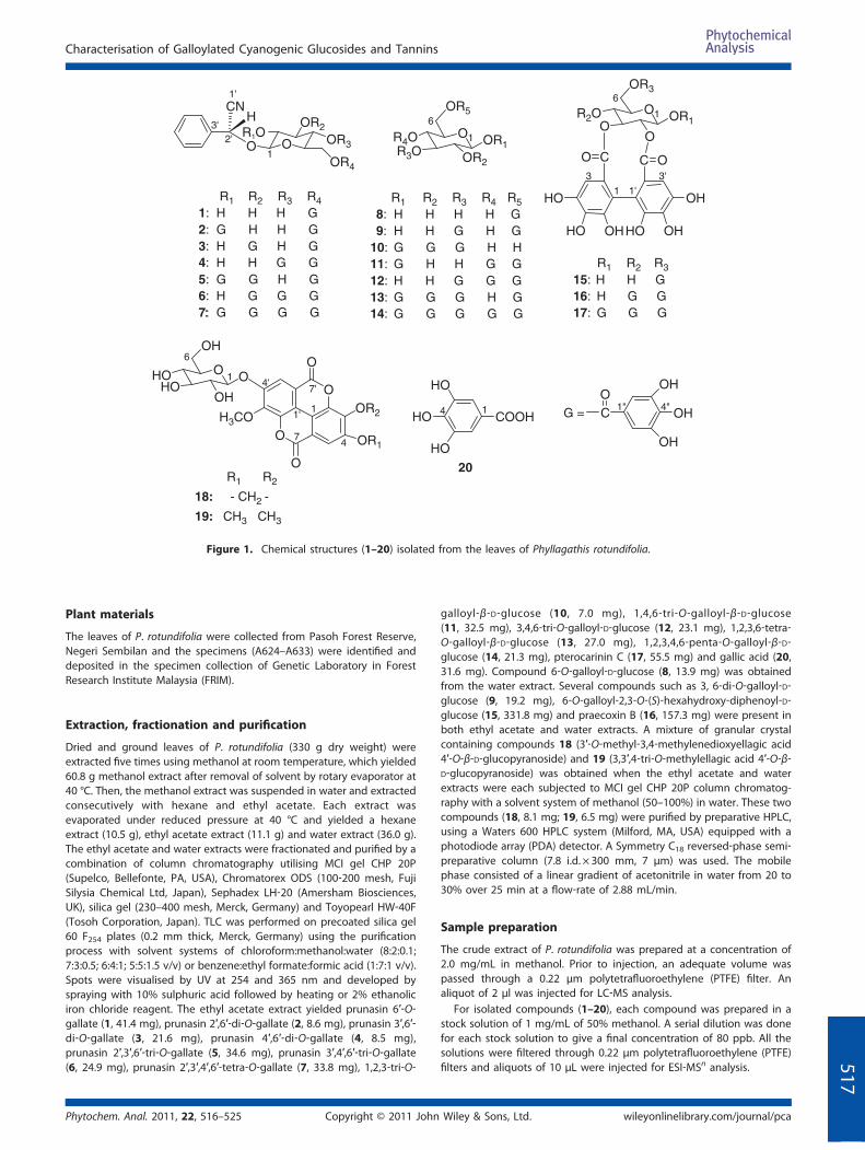
Characterisation of Galloylated Cyanogenic Glucosides and Hydrolysable Tannins from Leaves of Phyllagathis rotundifolia by LC‐ESI‐MS/MS Hooi Poay Tan, a,b * Sui Kiong Ling b and Cheng Hock Chuah a ABSTRACT: Introduction – Phyllagathis rotundifolia (Jack) Bl. (Melastomataceae) is a creeping herb found in Peninsular Malaysia and Sumatra. Traditionally, a decoction of the leaves is used in the treatment of malaria, fever and stomach ache. Objective – To provide ESI‐MS n data which are applicable for chemical fingerprinting of P. rotundifolia to obviate laborious isolation and purification steps. Methodology – The mass spectral data for the compounds isolated from the leaves of P. rotundifolia were obtained by liquid chromatography–electrospray ionisation tandem mass spectrometry. Results – The MS fragmentation patterns were obtained for galloylated cyanogenic glucosides based on prunasin (prunasin 6′‐O‐gallate 1, prunasin 2′,6′‐di‐O‐gallate 2, prunasin 3′,6′‐di‐O‐gallate 3, prunasin 4′,6′‐di‐O‐gallate 4, prunasin 2′,3′,6′‐tri‐O‐ gallate 5, prunasin 3′,4′,6′‐tri‐O‐gallate 6 and prunasin 2′,3′,4′,6′‐tetra‐O‐gallate 7), gallotannins (6‐O‐galloyl‐D‐glucose 8, 3,6‐di‐ O‐galloyl‐D‐glucose 9, 1,2,3‐tri‐O‐galloyl‐β‐D‐glucose 10, 1,4,6‐tri‐O‐galloyl‐β‐D‐glucose 11, 3,4,6‐tri‐O‐galloyl‐D‐glucose 12, 1,2,3,6‐tetra‐O‐galloyl‐β‐D‐glucose 13 and 1,2,3,4,6‐penta‐O‐galloyl‐β‐D‐glucose 14), ellagitannins [6‐O‐galloyl‐2,3‐O‐(S)‐ hexahydroxy‐diphenoyl‐D‐glucose 15, praecoxin B 16 and pterocarinin C 17], ellagic acid derivatives (3′‐O‐methyl‐3,4‐ methylenedioxyellagic acid 4′‐O‐β‐D‐glucopyranoside 18 and 3,3′,4‐tri‐O‐methylellagic acid 4′‐O‐β‐D‐glucopyranoside 19) and gallic acid 20 that were isolated from the leaves of P. rotundifolia. Conclusion – The ESI‐MS n technique facilitates identification of galloylated cyanogenic glucosides, hydrolysable tannins and ellagic acid derivatives that were isolated from the leaves of P. rotundifolia. It yields MS n spectra that are useful for identification of these compounds in complex samples and permit more complete fingerprinting of plant materials. Copyright © 2011 John Wiley & Sons, Ltd. Supporting information can be found in the online version of this article. Keywords: Phyllagathis rotundifolia; Melastomataceae; galloylated cyanogenic glucosides; hydrolysable tannins; ellagic acid derivative; liquid chromatography–mass spectrometry Introduction Phyllagathis rotundifolia (Jack) Bl. from the family of Melastomataceae is a creeping herb found in Peninsular Malaysia and Sumatra. It has heart‐shaped leaves with dark green upper surface and reddish lower surface, short stem and pink or magenta flowers (Henderson, 1954; Ridley, 1922). In traditional medicine, a decoction of the leaves is used for the treatment of malaria, fever and stomachache as well as in parturition and as tonic (Burkill, 1966). Plants in the family of Melastomataceae produce distinct oligomeric tannins (Calderon et al., 2003; Cronquist, 1981; Tan et al., 2010) or ellagic acid and its derivatives such as 3,3′‐ di‐O‐methylellagic acid, 3,3′,4‐tri‐O‐methylellagic acid and 3′‐O‐ methyl‐3,4‐methylenedioxyellagic acid which can be utilised as chemotaxonomic characteristics or definitive systematic markers (Hillis and Yazaki, 1973; Lowry, 1968; Yoshida et al., 2000). In this study, we reported the fragmentation patterns of galloylated cyanogenic glucosides, gallotannins, ellagitannins and ellagic acid derivatives by electrospray ionisation tandem mass spectrometry (ESI‐MS n ) that can be used for fingerprinting of plants collected from different localities as well as in the assessment of chemical changes in response to environmental factors. Experimental Solvents and chemicals All reagent and solvents used were of analytical and HPLC grades. All solvents were purchased from Merck, Germany. * Correspondence to: Hooi Poay Tan, Medicinal Plants Division, Forest Research Institute Malaysia, 52109Kepong, Selangor, Malaysia. E–mail: tanhp@frim. gov.my a Department of Chemistry, University of Malaya, 50603 Kuala Lumpur, Malaysia b Medicinal Plants Division, Forest Research Institute Malaysia, 52109 Kepong, Selangor, Malaysia Phytochem. Anal. 2011, 22, 516–525 Copyright © 2011 John Wiley & Sons, Ltd. Research Article Received: 17 August 2010; Revised: 13 December 2010; Accepted: 14 December 2010 Published online in Wiley Online Library: 14 April 2011 (wileyonlinelibrary.com) DOI 10.1002/pca.1312 516

Welcome message from author
This document is posted to help you gain knowledge. Please leave a comment to let me know what you think about it! Share it to your friends and learn new things together.
Transcript

Research Article
Received: 17 August 2010; Revised: 13 December 2010; Accepted: 14 December 2010 Published online in Wiley Online Library: 14 April 2011
(wileyonlinelibrary.com) DOI 10.1002/pca.1312
516
Characterisation of Galloylated CyanogenicGlucosides and Hydrolysable Tanninsfrom Leaves of Phyllagathis rotundifoliaby LC‐ESI‐MS/MSHooi Poay Tan,a,b* Sui Kiong Lingb and Cheng Hock Chuaha
ABSTRACT:Introduction – Phyllagathis rotundifolia (Jack) Bl. (Melastomataceae) is a creeping herb found in Peninsular Malaysia andSumatra. Traditionally, a decoction of the leaves is used in the treatment of malaria, fever and stomach ache.Objective – To provide ESI‐MSn data which are applicable for chemical fingerprinting of P. rotundifolia to obviate laboriousisolation and purification steps.Methodology – The mass spectral data for the compounds isolated from the leaves of P. rotundifolia were obtained by liquidchromatography–electrospray ionisation tandem mass spectrometry.Results – The MS fragmentation patterns were obtained for galloylated cyanogenic glucosides based on prunasin (prunasin6′‐O‐gallate 1, prunasin 2′,6′‐di‐O‐gallate 2, prunasin 3′,6′‐di‐O‐gallate 3, prunasin 4′,6′‐di‐O‐gallate 4, prunasin 2′,3′,6′‐tri‐O‐gallate 5, prunasin 3′,4′,6′‐tri‐O‐gallate 6 and prunasin 2′,3′,4′,6′‐tetra‐O‐gallate 7), gallotannins (6‐O‐galloyl‐D‐glucose 8, 3,6‐di‐O‐galloyl‐D‐glucose 9, 1,2,3‐tri‐O‐galloyl‐β‐D‐glucose 10, 1,4,6‐tri‐O‐galloyl‐β‐D‐glucose 11, 3,4,6‐tri‐O‐galloyl‐D‐glucose 12,1,2,3,6‐tetra‐O‐galloyl‐β‐D‐glucose 13 and 1,2,3,4,6‐penta‐O‐galloyl‐β‐D‐glucose 14), ellagitannins [6‐O‐galloyl‐2,3‐O‐(S)‐hexahydroxy‐diphenoyl‐D‐glucose 15, praecoxin B 16 and pterocarinin C 17], ellagic acid derivatives (3′‐O‐methyl‐3,4‐methylenedioxyellagic acid 4′‐O‐β‐D‐glucopyranoside 18 and 3,3′,4‐tri‐O‐methylellagic acid 4′‐O‐β‐D‐glucopyranoside 19) andgallic acid 20 that were isolated from the leaves of P. rotundifolia.Conclusion – The ESI‐MSn technique facilitates identification of galloylated cyanogenic glucosides, hydrolysable tanninsand ellagic acid derivatives that were isolated from the leaves of P. rotundifolia. It yields MSn spectra that are useful foridentification of these compounds in complex samples and permit more complete fingerprinting of plant materials.Copyright © 2011 John Wiley & Sons, Ltd.
Supporting information can be found in the online version of this article.
Keywords: Phyllagathis rotundifolia; Melastomataceae; galloylated cyanogenic glucosides; hydrolysable tannins; ellagic acidderivative; liquid chromatography–mass spectrometry
* Correspondence to: Hooi Poay Tan, Medicinal Plants Division, Forest ResearchInstitute Malaysia, 52109Kepong, Selangor, Malaysia. E–mail: [email protected]
a Department of Chemistry, University of Malaya, 50603 Kuala Lumpur, Malaysia
b Medicinal Plants Division, Forest Research Institute Malaysia, 52109 Kepong,Selangor, Malaysia
IntroductionPhyllagathis rotundifolia (Jack) Bl. from the family ofMelastomataceae is a creeping herb found in PeninsularMalaysia and Sumatra. It has heart‐shaped leaves with darkgreen upper surface and reddish lower surface, short stem andpink or magenta flowers (Henderson, 1954; Ridley, 1922). Intraditional medicine, a decoction of the leaves is used for thetreatment of malaria, fever and stomachache as well as inparturition and as tonic (Burkill, 1966).
Plants in the family of Melastomataceae produce distinctoligomeric tannins (Calderon et al., 2003; Cronquist, 1981;Tan et al., 2010) or ellagic acid and its derivatives such as 3,3′‐di‐O‐methylellagic acid, 3,3′,4‐tri‐O‐methylellagic acid and 3′‐O‐methyl‐3,4‐methylenedioxyellagic acid which can be utilised aschemotaxonomic characteristics or definitive systematic markers(Hillis and Yazaki, 1973; Lowry, 1968; Yoshida et al., 2000). In thisstudy, we reported the fragmentation patterns of galloylatedcyanogenic glucosides, gallotannins, ellagitannins and ellagicacid derivatives by electrospray ionisation tandem mass
Copyright © 2011 John
spectrometry (ESI‐MSn) that can be used for fingerprinting ofplants collected from different localities as well as in theassessment of chemical changes in response to environmentalfactors.
Experimental
Solvents and chemicals
All reagent and solvents used were of analytical and HPLC grades. Allsolvents were purchased from Merck, Germany.
Phytochem. Anal. 2011, 22, 516–525Wiley & Sons, Ltd.
R1 R2
R2
R3 R4 R58: H H H H G9: H H G H G
10: G G G H H11: G H H G G12: H H G G G13: G G G H G14: G G G G G
O OR1OH
CNOR2
OR3
OR4
R1 R2 R3 R41: H H H G2: G H H G3: H G H G4: H H G G5: G G H G6: H G G G7:: G G G G
O1 OR1
OR2R3O
R4O
OR5 O1
HO OH
OH
OHHO
HO
OR1OR2O
OR3
O
C OCO
R1 R315: H H G16: H G G17: G G G
R1 R2
18: - CH2 -
19: CH3 CH3
O O
OHHO
HO
OH
O
O
O
O
H3COOR2
OR1
HO
HO
HO
COOH
20
OH
OH
OH
CO
G =
1
1'
2'3' 6
6
1 1'
3'3
1
6
1
47
1'
4'7'
14 1" 4"
Figure 1. Chemical structures (1–20) isolated from the leaves of Phyllagathis rotundifolia.
Characterisation of Galloylated Cyanogenic Glucosides and Tannins
Plant materials
The leaves of P. rotundifolia were collected from Pasoh Forest Reserve,Negeri Sembilan and the specimens (A624–A633) were identified anddeposited in the specimen collection of Genetic Laboratory in ForestResearch Institute Malaysia (FRIM).
51
Extraction, fractionation and purification
Dried and ground leaves of P. rotundifolia (330 g dry weight) wereextracted five times using methanol at room temperature, which yielded60.8 g methanol extract after removal of solvent by rotary evaporator at40 °C. Then, the methanol extract was suspended in water and extractedconsecutively with hexane and ethyl acetate. Each extract wasevaporated under reduced pressure at 40 °C and yielded a hexaneextract (10.5 g), ethyl acetate extract (11.1 g) and water extract (36.0 g).The ethyl acetate and water extracts were fractionated and purified by acombination of column chromatography utilising MCI gel CHP 20P(Supelco, Bellefonte, PA, USA), Chromatorex ODS (100‐200 mesh, FujiSilysia Chemical Ltd, Japan), Sephadex LH‐20 (Amersham Biosciences,UK), silica gel (230–400 mesh, Merck, Germany) and Toyopearl HW‐40F(Tosoh Corporation, Japan). TLC was performed on precoated silica gel60 F254 plates (0.2 mm thick, Merck, Germany) using the purificationprocess with solvent systems of chloroform:methanol:water (8:2:0.1;7:3:0.5; 6:4:1; 5:5:1.5 v/v) or benzene:ethyl formate:formic acid (1:7:1 v/v).Spots were visualised by UV at 254 and 365 nm and developed byspraying with 10% sulphuric acid followed by heating or 2% ethanoliciron chloride reagent. The ethyl acetate extract yielded prunasin 6′‐O‐gallate (1, 41.4 mg), prunasin 2′,6′‐di‐O‐gallate (2, 8.6 mg), prunasin 3′,6′‐di‐O‐gallate (3, 21.6 mg), prunasin 4′,6′‐di‐O‐gallate (4, 8.5 mg),prunasin 2′,3′,6′‐tri‐O‐gallate (5, 34.6 mg), prunasin 3′,4′,6′‐tri‐O‐gallate(6, 24.9 mg), prunasin 2′,3′,4′,6′‐tetra‐O‐gallate (7, 33.8 mg), 1,2,3‐tri‐O‐
Phytochem. Anal. 2011, 22, 516–525 Copyright © 2011 John
galloyl‐β‐D‐glucose (10, 7.0 mg), 1,4,6‐tri‐O‐galloyl‐β‐D‐glucose(11, 32.5 mg), 3,4,6‐tri‐O‐galloyl‐D‐glucose (12, 23.1 mg), 1,2,3,6‐tetra‐O‐galloyl‐β‐D‐glucose (13, 27.0 mg), 1,2,3,4,6‐penta‐O‐galloyl‐β‐D‐glucose (14, 21.3 mg), pterocarinin C (17, 55.5 mg) and gallic acid (20,31.6 mg). Compound 6‐O‐galloyl‐D‐glucose (8, 13.9 mg) was obtainedfrom the water extract. Several compounds such as 3, 6‐di‐O‐galloyl‐D‐glucose (9, 19.2 mg), 6‐O‐galloyl‐2,3‐O‐(S)‐hexahydroxy‐diphenoyl‐D‐glucose (15, 331.8 mg) and praecoxin B (16, 157.3 mg) were present inboth ethyl acetate and water extracts. A mixture of granular crystalcontaining compounds 18 (3′‐O‐methyl‐3,4‐methylenedioxyellagic acid4′‐O‐β‐D‐glucopyranoside) and 19 (3,3′,4‐tri‐O‐methylellagic acid 4′‐O‐β‐D‐glucopyranoside) was obtained when the ethyl acetate and waterextracts were each subjected to MCI gel CHP 20P column chromatog-raphy with a solvent system of methanol (50–100%) in water. These twocompounds (18, 8.1 mg; 19, 6.5 mg) were purified by preparative HPLC,using a Waters 600 HPLC system (Milford, MA, USA) equipped with aphotodiode array (PDA) detector. A Symmetry C18 reversed‐phase semi‐preparative column (7.8 i.d. × 300 mm, 7 μm) was used. The mobilephase consisted of a linear gradient of acetonitrile in water from 20 to30% over 25 min at a flow‐rate of 2.88 mL/min.
Sample preparation
The crude extract of P. rotundifolia was prepared at a concentration of2.0 mg/mL in methanol. Prior to injection, an adequate volume waspassed through a 0.22 μm polytetrafluoroethylene (PTFE) filter. Analiquot of 2 μl was injected for LC‐MS analysis.
For isolated compounds (1–20), each compound was prepared in astock solution of 1 mg/mL of 50% methanol. A serial dilution was donefor each stock solution to give a final concentration of 80 ppb. All thesolutions were filtered through 0.22 μm polytetrafluoroethylene (PTFE)filters and aliquots of 10 μL were injected for ESI‐MSn analysis.
Wiley & Sons, Ltd. wileyonlinelibrary.com/journal/pca
7
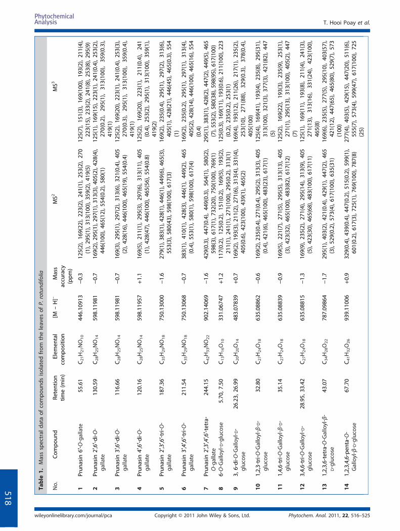
Table
1.Massspectral
data
ofcompo
unds
isolated
from
theleaves
ofP.
rotund
ifolia
No.
Com
poun
dRe
tention
time(m
in)
Elem
ental
compo
sitio
n[M
–H]–
Mass
accuracy
(ppm
)
MS2
MS3
1Prun
asin
6′‐O‐gallate
55.61
C21H21NO10
446.10
913
−0.3
125(2),16
9(22
),22
3(2),24
1(1),25
3(2),27
0(1),29
5(1),3
13(100
),35
9(2),4
19(5)
125(7),15
1(3),16
9(10
0),19
3(2),21
1(4),
223(15
),23
3(2),2
41(8),25
3(8),2
95(9)
2Prun
asin
2′,6′‐d
i‐O‐
gallate
130.59
C28H25NO14
598.11
981
−0.7
169(2),295
(1),29
7(1),313
(3),40
5(2),428
(4),
446(10
0),4
65(12),5
54(0.2),58
0(1)
125(1),16
9(15
),22
3(1),24
1(0.4),25
3(2),
270(0.2),29
5(1),31
3(10
0),35
9(0.3),
419(1)
3Prun
asin
3′,6′‐d
i‐O‐
gallate
116.66
C28H25NO14
598.11
981
−0.7
169(3),29
5(1),29
7(2),31
3(6),32
1(0.4),40
5(2),42
8(16
),44
6(10
0),4
65(19),5
54(0.4)
125(2),16
9(20
),22
3(1),24
1(0.4),25
3(3),
270(0.3),29
5(1),31
3(10
0),35
9(0.4),
419(1)
4Prun
asin
4′,6′‐d
i‐O‐
gallate
120.16
C28H25NO14
598.11
957
+1.1
169(5),21
1(1),29
5(3),29
7(6),31
3(11
),40
5(1),42
8(47
),44
6(10
0),4
65(56),5
54(0.8)
125(2),16
9(20
),22
3(1),21
1(0.4),24
1(0.4),25
3(2),29
5(1),31
3(10
0),35
9(1),
419(2)
5Prun
asin
2′,3′,6′‐tri‐O‐
gallate
187.36
C35H29NO18
750.13
000
−1.6
279(1),383
(1),42
8(1),446
(1),44
9(6),465
(3),
553(3),5
80(43),5
98(100
),61
7(3)
169(2),23
5(0.4),29
5(1),29
7(2),31
3(6),
405(1),42
8(21
),44
6(45
),46
5(0.3),55
4(1)
6Prun
asin
3′,4′,6′‐tri‐O‐
gallate
211.54
C35H29NO18
750.13
068
−0.7
383(1),41
0(1),42
8(3),44
6(1),44
9(2),46
5(0.4),55
3(1),5
80(1),59
8(10
0),6
17(4)
169(2),23
5(0.2),29
5(1),29
7(1),31
3(4),
405(2),428
(14),4
46(100
),46
5(16
),55
4(0.4)
7Prun
asin
2′,3′,4′,6′‐tetra‐
O‐gallate
244.15
C42H33NO22
902.14
069
−1.6
429(0.3),44
7(0.4),44
9(0.3),56
4(1),58
0(2),
598(3),6
17(1),73
2(20
),75
0(10
0),7
69(1)
295(1),383
(1),42
8(2),447
(2),44
9(5),465
(7),55
3(2),580
(38),598
(95),617
(100
)8
6‐O‐Galloyl‐ D‐glucose
5.70
,7.50
C13H16O10
331.06
747
+1.2
117(0.2),12
5(0.2),15
1(0.2),16
9(5),19
3(2),
211(1),2
41(1),27
1(10
0),2
95(0.2),31
3(1)
125(0.3),169
(11),193
(0.6),21
1(10
0),223
(0.2),23
5(0.2),2
53(1)
93,
6‐di‐O‐Galloyl‐ D‐
glucose
26.23,
26.99
C20H20O14
483.07
839
+0.7
169(2),193
(3),21
1(2),271
(6),31
3(4),331
(4),
405(0.6),4
23(100
),43
9(1),4
65(2)
169(4),19
3(12
),21
1(26
),21
7(1),23
5(2),
253(10
),27
1(88
),32
9(0.3),37
8(0.4),
405(10
0)10
1,2,3‐tri‐O
‐Galloyl‐β‐ D‐
glucose
32.80
C27H24O18
635.08
862
−0.6
169(2),235
(0.4),27
1(0.4),295
(2),31
3(3),405
(0.4),42
1(6),4
65(100
),48
3(23
),61
7(1)
125(4),16
9(41
),19
3(4),23
5(8),29
5(31
),31
3(10
0),32
1(3),37
7(3),42
1(82
),44
7(5)
111,4,6‐tri‐O
‐Galloyl‐β‐ D‐
glucose
35.14
C27H24O18
635.08
839
−0.9
169(5),22
1(7),27
1(5),29
5(5),31
3(13
),40
5(3),42
3(32
),46
5(10
0),4
83(82),6
17(12)
125(2),16
9(22
),19
3(3),23
5(9),25
3(1),
271(1),29
5(13
),31
3(10
0),40
5(2),44
7(7)
123,4,6‐tri‐O
‐Galloyl‐ D‐
glucose
28.95,
33.42
C27H24O18
635.08
815
−1.3
169(9),23
5(2),27
1(4),29
5(14
),31
3(9),40
5(5),42
3(30
),46
5(68
),48
3(10
0),6
17(11)
125(1),16
9(11
),19
3(8),21
1(4),24
1(3),
271(13
),31
3(16
),33
1(24
),42
3(10
0),
465(8)
131,2,3,6‐tetra‐O‐Galloyl‐β‐
D‐glucose
43.07
C34H28O22
787.09
864
−1.7
295(1),40
3(2),42
1(0.4),42
9(1),44
7(2),46
5(3),52
9(0.2),5
73(4),61
7(10
0),6
35(31)
169(6),23
5(5),27
7(5),29
5(10
),40
3(57
),42
1(12
),44
7(65
),46
5(80
),52
9(7),57
3(100
)14
1,2,3,4,6‐pe
nta‐O‐
Galloyl‐β‐ D‐glucose
67.70
C41H32O26
939.11
006
+0.9
329(0.4),4
39(0.4),44
7(0.2),5
15(0.2),59
9(1),
601(0.2),6
17(3),72
5(1),7
69(100
),78
7(8)
277(4),40
3(5),42
9(15
),44
7(20
),51
1(6),
555(7),57
3(4),59
9(47
),61
7(10
0),72
5(25)
T. Hooi Poay et al.
Phytochem. Anal. 2011, 22, 516–525Copyright © 2011 John Wiley & Sons, Ltd.wileyonlinelibrary.com/journal/pca
518

Table
1.(Con
tinued)
No.
Com
poun
dRe
tention
time
(min)
Elem
ental
compo
sitio
n[M
–H]–
Mass
accuracy
(ppm
)
MS2
MS3
156‐O‐Galloyl‐2,3‐O‐(S)‐
hexahy
droxy‐
diph
enoy
l‐ D‐glucose
25.48,
27.94
C27H22O18
633.07
252
−1.3
229(1),24
9(1),25
7(1),27
5(19
),30
1(10
0),
319(0.4),4
81(3),5
89(0.3),61
5(1)
171(8),18
5(5),22
9(23
),25
7(10
0),27
3(15),2
84(3)
16PraecoxinB
29.65,
35.82
C34H26O22
785.08
375
−0.7
275(15
),30
1(10
0),31
9(3),40
5(2),42
3(32
),46
5(9),4
83(75),6
33(35),7
41(1),76
7(2)
171(30
),22
9(23
),25
7(10
0),2
73(16)
17Pterocarinin
C56
.65
C41H30O26
937.09
595
+0.7
275(1),3
01(17),3
13(1),46
5(12
),48
3(7),6
17(9),63
5(10
0),7
67(2),78
5(19
),91
9(0.4)
169(1),27
1(0.4),29
5(3),31
3(8),40
5(1),
423(3),46
5(10
0),48
3(64
4),59
1(0.3),
617(0.3)
183′‐O‐M
ethy
l‐3,4‐
methy
lene
dioxyellagic
acid
4′‐O‐β‐ D‐
glucop
yran
oside
76.21
C22H18O13
489.06
738
−0.2
327a,3
12(100
),28
3(0.2),
171(4),2
12(4),24
0(89
),25
6(5),2
84(100
)
193,3′,4‐tri‐O‐M
ethy
lellagic
acid
4′‐O‐β‐ D‐
glucop
yran
oside
91.08
C23H22O13
505.09
872
−0.1
343a,3
28(100
),31
3(0.1)
171(0.2),2
99(1),31
3(10
0)
20Gallic
acid
4.80
C7H6O5
169.01
428
+0.2
125(10
0)‐
a Rep
resented
theintensefrag
men
tiondu
eto
theremov
alof
glucose(m
/z16
2)an
dwas
selected
forfrag
men
tatio
nin
theMS2
Characterisation of Galloylated Cyanogenic Glucosides and Tannins
Phytochem. Anal. 2011, 22, 516–525 Copyright © 2011 John Wiley & Sons, Ltd. wileyonlinelibrary.com/journal/pca
519

OO
O
OO
OHCN
C
OOH
OH
OH
CO
CO
OH
OH
OH
OH
OHHO
C O
OHHO
HO
133
769
152
750
617
732170
A)
7
O
O
CO
HOOH
OH
O
OCO
HO
HO
HO
O
OC
OOH
OH
OHCO
CO
HO
HOOH
OH
OHHO
152
787
170
769
617
152
170599
B)
14
O
O
O
O
O
OH3CO
O
OH
HOHO
OHO
327
162
15
312
28
284
44
18
C)
O
O
O
O
OCH3
OCH3H3CO
O
OH
HOHO
OHO
162
343
15
328
15
313
19
Figure 2. Schematic fragmentation of prunasin (A) 2′,3′,4′,6′‐tetra‐O‐gallate (7), (B) 1,2,3,4,6‐penta‐O‐galloyl‐β‐D‐glucose (14), (C) 3′‐O‐methyl‐3,4‐methylenedioxyellagic acid 4′‐O‐β‐D‐glucopyranoside (18) and 3,3′,4‐tri‐O‐methylellagic acid 4′‐O‐β‐D‐glucopyranoside (19) in negative mode ESI‐MSn.
T. Hooi Poay et al.
520
Liquid chromatography and mass spectrometry
The chromatographic analysis was performed using the Accela™ U‐HPLCsystem (Thermo Scientific, San Jose, CA, USA) equipped with aquaternary pump, a built‐in degasser, a PDA detector and an auto‐sampler. A Hypersil Gold RP C8 column (3 μm, 2.1 mm i.d. × 150 mm)was used. The mobile phase consisted of acetonitrile (A) and waterwith 0.1% formic acid (B) at a flow‐rate of 200 μL/min, with acombination of step‐gradient and isocratic elution, A:B as follows:0 min, 0:100; 12 min, 1:99; 13 min, 15.4:84.6; 15 min, 15.4:84.6; 100 min,16:84; 150 min, 18:82; 205 min, 20:80; 255 min, 28:72; 265 min, 30:70,maintained for 5 min. The effluent from the LC column was directedinto the ESI probe.
The MSn analysis was performed on a LTQ Orbitrap massspectrometer (Thermo Fisher Scientific, Bremen, Germany) equippedwith an electrospray ionisation probe. A negative ion mode wasemployed in the analysis because this mode afforded the best limits ofdetection for the compounds. The MSn data were acquired by OrbitrapFTMS analyser operating at full scan with a target mass resolution of30000 and data‐dependent MSn method with dynamic exclusionenabled and a repeat count of 2. In this method, the most intenseparent ion was isolated and fragmented twice to generate the MS2 data.Then the most intense daughter ion was also isolated and fragmentedtwice to produce the MS3 data. The spectra were recorded in the rangeof m/z 100–1000 and the collision energy for MSn was adjusted to35–40%. The optimised parameters in the negative ion mode were asfollows: ion spray voltage, 3.5 kV; sheath gas (N2), 40 arbitrary units;auxiliary gas (N2), 20 arbitrary units; capillary temperature, 285 °C. Masscalibration was performed according to the manufacturer’s guidelinesusing a manufacturer defined mixture of caffeine, sodium dodecyl
Copyright © 2011 Johnwileyonlinelibrary.com/journal/pca
sulphate, sodium taurocholate, the tetrapeptide MRFA and Ultramark1621. All data were processed using Qual browser (Thermo FisherScientific, San Jose, CA, USA).
Nuclear magnetic resonance spectroscopy
All the compounds except compounds 18 and 19 were dissolvedin Me2CO‐d6 with droplets of D2O to enhance the solubility ofthe samples. Compounds 18 and 19 were dissolved in DMSO‐d6. The1H‐ and 13C‐NMR spectral data were recorded using a Bruker DRX 300NMR spectrometer (300 MHz for 1H‐NMR and 75 MHz for 13C‐NMR).
Results and Discussion
ESI‐MSn analysis of isolated compounds
A total of 20 compounds (Fig. 1) were isolated from the leaves ofP. rotundifolia and characterised using negative mode ofionisation by ESI‐MSn. The experimental m/z values of themolecular ions together with calculated mass accuracy and theirMS2 and MS3 fragment ions are tabulated in Table 1. Fragmentions of galloylated cyanogenic glucosides were found to besimilar for the isomeric compounds such as prunasin–digallate(2–4) and prunasin–trigallate (5–6). The trigalloyl‐glucoseanalogues (compounds 10–12) exhibited similar daughterions in the MS2 spectra except that compound 12 showed adominant fragment ion at m/z 483, which enables it to bedifferentiated from the other two analogues. The ellagitannins
Phytochem. Anal. 2011, 22, 516–525Wiley & Sons, Ltd.
C35H26NO17
m/z 732
C28H22NO13
m/z 580
C21H18NO9
m/z 428
-152 (galloyl)
-152 (galloyl)
-152 (galloyl)
C13H11NO9
m/z 276
-170 (gallate)
-170 (gallate)
-170 (gallate)
-152 (galloyl)
-152 (galloyl)
-152 (galloyl)
Prunasin-tetragallate C42H32NO22
m/z 902
Prunasin-trigallate C35H28NO18
m/z 750
Prunasin-digallate C28H24NO14
m/z 598
Prunasin-monogallate C21H20NO10
m/z 446
-133 (Cyanohydrin)
-133 (Cyanohydrin)
-133 (Cyanohydrin)
-133 (Cyanohydrin)
C34H25O21
m/z 769
C27H21O17
m/z 617
C20H17O13
m/z 465
C13H13O9
m/z 313
C7H5O5
m/z 169
C6H5O3
m/z 125
-152 (galloyl)
-152 (galloyl)
-152 (galloyl)
-170 (gallate)
-170 (gallate)
-170 (gallate)
C27H19O16
m/z 599
C20H15O12
m/z 447
C13H11O8
m/z 295
-152 (galloyl)
-152 (galloyl)
-144 (dehydrated hexosyl)
-18 (H2O)
- -
- -
--
-
-
-
-
--
-
-
-
-
-
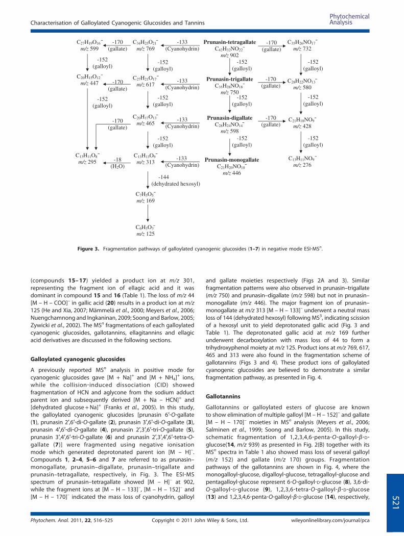
Figure 3. Fragmentation pathways of galloylated cyanogenic glucosides (1–7) in negative mode ESI‐MSn.
Characterisation of Galloylated Cyanogenic Glucosides and Tannins
(compounds 15–17) yielded a product ion at m/z 301,representing the fragment ion of ellagic acid and it wasdominant in compound 15 and 16 (Table 1). The loss of m/z 44[M – H – COO]– in gallic acid (20) results in a product ion at m/z125 (He and Xia, 2007; Mämmelä et al., 2000; Meyers et al., 2006;Nuengchamnong and Ingkaninan, 2009; Soong and Barlow, 2005;Zywicki et al., 2002). The MSn fragmentations of each galloylatedcyanogenic glucosides, gallotannins, ellagitannins and ellagicacid derivatives are discussed in the following sections.
52
Galloylated cyanogenic glucosides
A previously reported MSn analysis in positive mode forcyanogenic glucosides gave [M + Na]+ and [M + NH4]
+ ions,while the collision‐induced dissociation (CID) showedfragmentation of HCN and aglycone from the sodium adductparent ion and subsequently derived [M + Na – HCN]+ and[dehydrated glucose +Na]+ (Franks et al., 2005). In this study,the galloylated cyanogenic glucosides [prunasin 6′‐O‐gallate(1), prunasin 2′,6′‐di‐O‐gallate (2), prunasin 3′,6′‐di‐O‐gallate (3),prunasin 4′,6′‐di‐O‐gallate (4), prunasin 2′,3′,6′‐tri‐O‐gallate (5),prunasin 3′,4′,6′‐tri‐O‐gallate (6) and prunasin 2′,3′,4′,6′‐tetra‐O‐gallate (7)] were fragmented using negative ionisationmode which generated deprotonated parent ion [M – H]–.Compounds 1, 2–4, 5–6 and 7 are referred to as prunasin–monogallate, prunasin–digallate, prunasin–trigallate andprunasin–tetragallate, respectively, in Fig. 3. The ESI‐MSspectrum of prunasin–tetragallate showed [M – H]– at 902,while the fragment ions at [M – H – 133]–, [M – H – 152]– and[M – H – 170]– indicated the mass loss of cyanohydrin, galloyl
Phytochem. Anal. 2011, 22, 516–525 Copyright © 2011 John
and gallate moieties respectively (Figs 2A and 3). Similarfragmentation patterns were also observed in prunasin–trigallate(m/z 750) and prunasin–digallate (m/z 598) but not in prunasin–monogallate (m/z 446). The major fragment ion of prunasin–monogallate at m/z 313 [M – H – 133]– underwent a neutral massloss of 144 (dehydrated hexosyl) following MS3, indicating scissionof a hexosyl unit to yield deprotonated gallic acid (Fig. 3 andTable 1). The deprotonated gallic acid at m/z 169 furtherunderwent decarboxylation with mass loss of 44 to form atrihydroxyphenol moiety atm/z 125. Product ions atm/z 769, 617,465 and 313 were also found in the fragmentation scheme ofgallotannins (Figs 3 and 4). These product ions of galloylatedcyanogenic glucosides are believed to demonstrate a similarfragmentation pathway, as presented in Fig. 4.
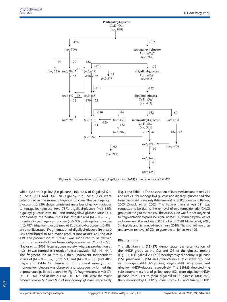
Gallotannins
Gallotannins or galloylated esters of glucose are knownto show elimination of multiple galloyl [M – H – 152]– and gallate[M – H – 170]– moieties in MSn analysis (Meyers et al., 2006;Salminen et al., 1999; Soong and Barlow, 2005). In this study,schematic fragmentation of 1,2,3,4,6‐penta‐O‐galloyl‐β‐D‐glucose(14, m/z 939) as presented in Fig. 2(B) together with itsMSn spectra in Table 1 also showed mass loss of several galloyl(m/z 152) and gallate (m/z 170) groups. Fragmentationpathways of the gallotannins are shown in Fig. 4, where themonogalloyl‐glucose, digalloyl‐glucose, tetragalloyl‐glucose andpentagalloyl‐glucose represent 6‐O‐galloyl‐D‐glucose (8), 3,6‐di‐O‐galloyl‐D‐glucose (9), 1,2,3,6‐tetra‐O‐galloyl‐β‐D‐glucose(13) and 1,2,3,4,6‐penta‐O‐galloyl‐β‐D‐glucose (14), respectively,
Wiley & Sons, Ltd. wileyonlinelibrary.com/journal/pca
1
Pentagalloyl-glucose C41H31O26
(m/z 939)
-071- 152
(m/z 769)
(m/z 725)
tetragalloyl-glucose C34H27O22
(m/z 787)
-44 -170
(m/z 599)
-152
(m/z 617)
-170 -152
trigalloyl-glucose C27H23O18
(m/z 635) -152 -170 -152
(m/z 465)
-170 -152
digalloyl-glucose C20H19O14
(m/z 483)
-44
(m/z 439)
-152
monogalloyl-glucose C20H19O14
(m/z 331)
-60
(m/z 423)
-152
-170
(m/z 313)
-152-170
(m/z 295)
-60
(m/z 271)
-44
(m/z 125)
-60
(m/z 211)
-18
(m/z 405) -102
(m/z 169)
-18 (m/z 447)
-152
-18
-44
(m/z 573)
-126-152
(m/z 287)
-144
(m/z 169)
-162
Figure 4. Fragmentation pathways of gallotannins (8–14) in negative mode ESI‐MSn.
T. Hooi Poay et al.
522
while 1,2,3‐tri‐O‐galloyl‐β‐D‐glucose (10), 1,4,6‐tri‐O‐galloyl‐β‐D‐glucose (11) and 3,4,6‐tri‐O‐galloyl‐D‐glucose (12) werecategorised as the isomeric trigalloyl‐glucose. The pentagalloyl‐glucose (m/z 939) shows consistent mass loss of galloyl moietiesto tetragalloyl‐glucose (m/z 787), trigalloyl‐glucose (m/z 635),digalloyl‐glucose (m/z 483) and monogalloyl‐glucose (m/z 331).Additionally, the neutral mass loss of gallic acid [M – H – 170]–
moieties in pentagalloyl‐glucose (m/z 939), tetragalloyl‐glucose(m/z 787), trigalloyl‐glucose (m/z 635), digalloyl‐glucose (m/z 483)are also illustrated. Fragmentation of digalloyl‐glucose (9) at m/z483 contributed to two major product ions at m/z 423 and m/z439. The product ion at m/z 423 was suggested to be derivedfrom the removal of two formaldehyde moieties [M – H – 60]–
(Taylor et al., 2005) from glucose moiety, whereas product ion atm/z 439 was formed as a result of decarboxylation [M – H – 44]–.The fragment ion at m/z 423 then underwent independentlosses of [M – H – 152]– (m/z 271) and [M – H – 18]– (m/z 405)(Fig.4 and Table 1). Elimination of glucosyl moiety frommonogalloyl‐glucose was observed and subsequently formed thedeprotonatedgallic acid atm/z 169 (Fig. 4). Fragment ions atm/z 271[M – H – 60]– and at m/z 211 [M – H – 60 – 60]– were the majorproduct ions in MS2 and MS3 of monogalloyl‐glucose, respectively
Copyright © 2011 Johnwileyonlinelibrary.com/journal/pca
(Fig. 4 and Table 1). The observation of intermediate ions atm/z 271andm/z 211 formonogalloyl‐glucose and digalloyl‐glucose had alsobeendescribed previously (Mämmelä et al., 2000; Soong andBarlow,2005; Zywicki et al., 2002). The fragment ion at m/z 211 wassuggested to be due to the removal of two formaldehyde (CH2O)groups in the glucosemoiety. Them/z 271 ionwas further subjectedto fragmentation to produce signal atm/z 169, formed by the loss ofa glucosyl unit (He and Xia, 2007; Kool et al., 2010, Mullen et al., 2003;Simirgiotis and Schmeda‐Hirschmann, 2010). The m/z 169 ion thenunderwent removal of CO2 to generate an ion at m/z 125.
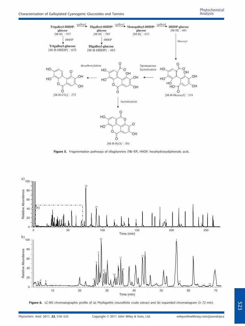
Ellagitannins
The ellagitannins (15–17) demonstrate the esterification ofthe HHDP group at the C‐2 and C‐3 of the glucose moiety(Fig. 1). 6‐O‐galloyl‐2,3‐O‐(S)‐hexahydroxy‐diphenoyl‐D‐glucose(15), praecoxin B (16) and pterocarinin C (17) were groupedas monogalloyl‐HHDP‐glucose, digalloyl‐HHDP‐glucose andtrigalloyl‐HHDP‐glucose respectively. The ESI‐MS depicted thesubsequent mass loss of galloyl (m/z 152), from trigalloyl‐HHDP‐glucose (m/z 937) to yield digalloyl‐HHDP‐glucose (m/z 785),then monogalloyl‐HHDP‐glucose (m/z 633) and finally HHDP‐
Phytochem. Anal. 2011, 22, 516–525Wiley & Sons, Ltd.
-galloyl
Spontaneous lactonization
-Hexosyl
[M-H-Hexosyl] ¯ : 319
-galloyl
[M-H-H2O] ¯ : 301
lactonization
[M-H-CO2] ¯ : 275
-HHDP -HHDP
decarboxylation
Trigalloyl-HHDP-glucose
[M-H] ¯ : 937
Digalloyl-HHDP- glucose
[M-H] ¯ : 785
Monogalloyl-HHDP- glucose
[M-H] ¯ : 633
HHDP-glucose[M-H] ¯ : 481
Trigalloyl-glucose[M-H-HHDP] ¯ : 635
Digalloyl-glucose[M-H-HHDP] ¯ : 483
O
OHOH
OHHO
O
HO
HO O
HO
O
OHOH
OHO
O
HO
HO OOOH
OHO
O
HO
HO
O
OO
OHO
O
HO
HO
-galloyl
Figure 5. Fragmentation pathways of ellagitannins (15–17). HHDP, hexahydroxydiphenolic acid.
0 50 100 150 200 250Time (min)
0
20
40
60
80
100
Rel
ativ
e A
bund
ance
b)
18
19
3
42
65 7
10 20 30 40 50 60 70Time (min)
0
20
40
60
80
100
Rel
ativ
e A
bund
ance
20 8 8
15
9
9
12
11
10 12
16 16
13
1
17 14
b)
a)
15
Figure 6. LC‐MS chromatographic profile of (a) Phyllagathis rotundifolia crude extract and (b) expanded chromatogram (3–72 min).
Characterisation of Galloylated Cyanogenic Glucosides and Tannins
Phytochem. Anal. 2011, 22, 516–525 Copyright © 2011 John Wiley & Sons, Ltd. wileyonlinelibrary.com/journal/pca
523

T. Hooi Poay et al.
524
glucose (m/z 481) as shown in Fig. 5 and Table 1. Theellagitannins were also fragmented to become gallotannins bylosing the mass of 302 (HHDP), consequently resulting infragment ions at m/z 635 (trigalloyl‐glucose) and m/z 483(digalloyl‐glucose). These daughter ions are suggested toundergo similar fragmentation to the gallotannins (Fig. 4).The monogalloyl‐HHDP‐glucose did not show the massof monogalloyl glucose; instead two dominant fragment ionsat m/z 301 and m/z 275 were observed. The ion at m/z 301 (lossof glucose and galloyl groups) was the result of spontaneousdilactonisation of HHDP. The partial lactonisation of HHDP yieldsa product ion at m/z 319 and subsequent decarboxylation yieldsan ion at m/z 275 [M – H – 44]–. In Table 1, the MS3 of m/z 301 incompounds 15 and 16 gave prominent ions at m/z 273, 257 and229, like deprotonated ellagic acid (He and Xia, 2007; Mämmeläet al., 2000; Mullen et al., 2003; Simirgiotis and Schmeda‐Hirschmann, 2010; Soong and Barlow, 2005). These ions weresuggested to correspond to [M – H – CO]–, [M – H – CO2]
– and[M – H– CO2– CO]– respectively.
Ellagic acid derivatives
The ESI‐MSn of compounds 18 and 19 produced the deproton-ated parent ion [M – H]– at m/z 489 and 505, respectively(Table 1). The fragmentation pattern is presented in Fig. 2(C).Both compounds were dominated by intense ion peaks at m/z327 and m/z 343, respectively, due to the removal of glucosemoiety [M – H – 162]–. The major ion peak at m/z 327 in 18 wasfragmented to m/z 312 through the loss of a CH3 group [M – H –15]–. The subsequent fragmentation of this ion yielded productions at m/z 284 and 240, which indicates the mass loss of COand CO2 respectively (Fig. 2C). Similarly, fragmentation of themajor ion in 19 atm/z 343 showed the elimination of first CH3 atm/z 328 in MS2 followed by a second CH3 at m/z 313 in MS3 asdisplayed in Fig. 2(C).
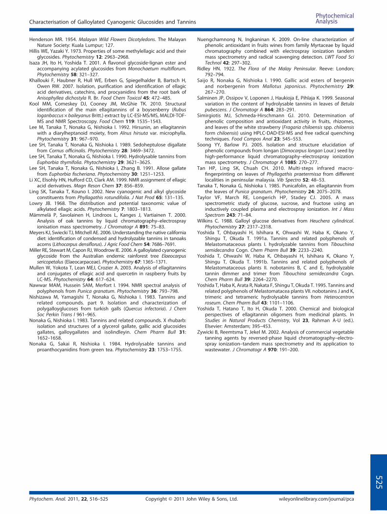
HPLC‐ESI‐MSn analysis of the crude extract ofPhyllagathis rotundifolia
Fig. 6 shows the LC‐MS chromatographic profile of the methanolextract of P. rotundifolia. The peaks were identified bycomparing their mass spectra with those of isolated compoundsas described in the previous sections.
Structural identification of the compounds
The structures of the isolated compounds (1–20) weredetermined by proton and carbon spectroscopic analysis. Thecyanogenic glucosides were identified as prunasin 6′‐O‐gallate,prunasin 2′,6′‐di‐O‐gallate, prunasin 3′,6′‐di‐O‐gallate, prunasin4′,6′‐di‐O‐gallate, prunasin 2′,3′,6′‐tri‐O‐gallate, prunasin 3′,4′,6′‐tri‐O‐gallate and prunasin 2′,3′,4′,6′‐tetra‐O‐gallate (Isaza et al.,2001; Ling et al., 2002; Miller et al., 2006). The seven gallotanninswere identified as 6‐O‐galloyl‐D‐glucose (He et al., 2001; Nonakaand Nishioka, 1983), 3,6‐di‐O‐galloyl‐D‐glucose (He et al., 2001),1,2,3‐tri‐O‐galloyl‐β‐D‐glucose (Lee et al., 1990; Nawwar et al.,1994), 1,4,6‐tri‐O‐galloyl‐β‐D‐glucose (Lee et al., 1992; Nawwaret al., 1994; Nonaka et al., 1984), 3,4,6‐tri‐O‐galloyl‐D‐glucose(He et al., 2001; Lee et al., 1989, 1991; Wilkins, 1988), 1,2,3,6‐tetra‐O‐galloyl‐β‐D‐glucose (Duan et al., 2004; Lee et al., 1991;Nishizawa et al., 1983; Saijo et al., 1990; Yoshida et al., 1991a),and 1,2,3,4,6‐penta‐O‐galloyl‐β‐D‐glucose (Gao et al., 2007;
Copyright © 2011 Johnwileyonlinelibrary.com/journal/pca
Nishizawa et al., 1983; Saijo et al., 1990; Tanaka et al., 1985) bycomparing their spectroscopic data with literature values. Thethree ellagitannins were identified as 6‐O‐galloyl‐2,3‐O‐(S)‐hexahydroxy‐diphenoyl‐D‐glucose (Yoshida et al., 1991a,1991b), praecoxin B (Hatano et al., 1991; Yoshida et al., 1991a)and pterocarinin C (Yoshida et al., 1991a, 1995). The twoellagic acid derivatives and one phenolic acid were identified as3 ′ ‐O‐methyl‐3,4‐methylenedioxyellagic acid 4 ′ ‐O‐β‐D‐glucopyranoside (Khallouki et al., 2007; Li et al., 1999), 3,3′,4‐tri‐O‐methylellagic acid 4′‐O‐β‐D‐glucopyranoside (Li et al., 1999)and gallic acid (Yoshida et al., 1991b; Chanwitheesuk et al.,2007). The 1H‐NMR and 13C‐NMR spectroscopic data forcompounds 1–20 are available in the Supplementary Material.
ConclusionThe fragmentation schemes for galloylated cyanogenic gluco-sides, gallotannins, ellagitannins and ellagic acid derivativesisolated from the leaves of P. rotundifolia provide detailed MSn
information which facilitates rapid monitoring of seasonalvariation, maturation process and quality evaluation of plantmaterial.
Supporting Information
Supporting information can be found in the online version ofthis article.
Acknowledgements
H. P. Tan would like to acknowledge Forest Research InstituteMalaysia (FRIM), Selangor, Malaysia for a scholarship and Mr.Cheah Hun Teong of Alpha Analytical (M) Sdn Bhd for assistancein LC‐MS instrumentation. This study was funded by a grant (02‐03‐10‐SF0066) from the Ministry of Science, Technology andInnovation and an IPPP research grant (PS332/2008C andPS351/2010A) from the University of Malaya.
ReferencesBurkill IH. 1966. A Dictionary of the Economic Products of the Malay
Peninsular, Vol 2. Ministry of Agricultural and Cooperatives: KualaLumpur; 1746–1747.
Calderon AI, Terreaux C, Gupta MP, Hostettmann K. 2003. Occurrence oftaxiphyllin and 3, 3′‐di‐O‐methylellagic acid 4′‐β‐D‐glucoside inHenriettella fascicularis. Biochem Syst Ecol 31: 789–791.
Chanwitheesuk A, Teerawutgulrag A, Kilburn JD, Rakariyatham N. 2007.Antimicrobial gallic acid from Caesalpinia mimosoides Lamk. FoodChem 100: 1044–1048.
Cronquist A. 1981. An Integrated System of Classification of FloweringPlants. Columbia University Press: New York; 649–651.
Duan D, Li Z, Luo H, Zhang W, Chen L, Xu X. 2004. Antiviral compoundsfrom traditional chinese medicines galla chinese as inhibitors of HCVNS3 protease. Bioorg Chem Lett 14: 6041–6044.
Franks TK, Hayasaka Y, Choimes S, Heeswijck RV. 2005. Cyanogenicglucosides in grapevine: polymorphism, identification and develop-mental patterns. Phytochemistry 66: 165–173.
Gao H, Huang YN, Xu PY, Kawabata J. 2007. Inhibitory effect on α‐glucosidase by the fruits of Terminalia chebula Retz. Food Chem 105:628–634.
Hatano T, Yazaki K, Okonogi A, Okuda T. 1991. Tannins of stachyurusspecies II. praecoxin A, B, C and D, four new hydrolysable tanninsfrom Stachyures praecox Leaves. Chem Pharm Bull 39: 1689–1693.
He Q, Shi B, Yao K, Ma Z. 2001. Synthesis of gallotannins. Carbohydr Res335: 245–250.
He Z, Xia W. 2007. Analysis of phenolic compounds in chinese olive(Canarium album L.) fruit by RPHPLC‐DAD‐ESI‐MS. Food Chem 105:1307–1311.
Phytochem. Anal. 2011, 22, 516–525Wiley & Sons, Ltd.
Characterisation of Galloylated Cyanogenic Glucosides and Tannins
Henderson MR. 1954. Malayan Wild Flowers Dicotyledons. The MalayanNature Society: Kuala Lumpur; 127.
Hillis WE, Yazaki Y. 1973. Properties of some methylellagic acid and theirglycosides. Phytochemistry 12: 2963–2968.
Isaza JH, Ito H, Yoshida T. 2001. A flavonol glycoside‐lignan ester andaccompanying acylated glucosides from Monochaetum multiflorum.Phytochemistry 58: 321–327.
Khallouki F, Haubner R, Hull WE, Erben G, Spiegelhalder B, Bartsch H,Owen RW. 2007. Isolation, purification and identification of ellagicacid derivatives, catechins, and procyanidins from the root bark ofAnisophyllea dichostyla R. Br. Food Chem Toxicol 45: 472–485.
Kool MM, Comeskey DJ, Cooney JM, McGhie TK. 2010. Structuralidentification of the main ellagitannins of a boysenberry (Rubusloganbaccus× baileyanus Britt.) extract by LC‐ESI‐MS/MS, MALDI‐TOF‐MS and NMR Spectroscopy. Food Chem 119: 1535–1543.
Lee M, Tanaka T, Nonaka G, Nishioka I. 1992. Hirsunin, an ellagitanninwith a diarylheptanoid moiety, from Alnus hirsuta var. microphylla.Phytochemistry 31: 967–970.
Lee SH, Tanaka T, Nonaka G, Nishioka I. 1989. Sedoheptulose digallatefrom Cornus officinalis. Phytochemistry 28: 3469–3472.
Lee SH, Tanaka T, Nonaka G, Nishioka I. 1990. Hydrolysable tannins fromEuphorbia thymifolia. Phytochemistry 29: 3621–3625.
Lee SH, Tanaka T, Nonaka G, Nishioka I, Zhang B. 1991. Allose gallatefrom Euphorbia fischeriana. Phytochemistry 30: 1251–1253.
Li XC, Elsohly HN, Hufford CD, Clark AM. 1999. NMR assignment of ellagicacid derivatives. Magn Reson Chem 37: 856–859.
Ling SK, Tanaka T, Kouno I. 2002. New cyanogenic and alkyl glycosideconstituents from Phyllagathis rotundifolia. J Nat Prod 65: 131–135.
Lowry JB. 1968. The distribution and potential taxonomic value ofalkylated ellagic acids. Phytochemistry 7: 1803–1813.
Mämmelä P, Savolainen H, Lindroos L, Kanges J, Vartiainen T. 2000.Analysis of oak tannins by liquid chromatography–electrosprayionisation mass spectrometry. J Chromatogr A 891: 75–83.
Meyers KJ, Swiecki TJ, Mitchell AE. 2006. Understanding the native californiadiet: identification of condensed and hydrolyzable tannins in tanoaksacorns (Lithocarpus densiflorus). J Agric Food Chem 54: 7686–7691.
Miller RE, Stewart M, Capon RJ, Woodrow IE. 2006. A galloylated cyanogenicglycoside from the Australian endemic rainforest tree Elaeocarpussericopetalus (Elaeocarpaceae). Phytochemistry 67: 1365–1371.
Mullen W, Yokota T, Lean MEJ, Crozier A. 2003. Analysis of ellagitanninsand conjugates of ellagic acid and quercetin in raspberry fruits byLC‐MS. Phytochemistry 64: 617–624.
Nawwar MAM, Hussein SAM, Merfort I. 1994. NMR spectral analysis ofpolyphenols from Punica granatum. Phytochemistry 36: 793–798.
Nishizawa M, Yamagishi T, Nonaka G, Nishioka I. 1983. Tannins andrelated compounds. part 9. Isolation and characterization ofpolygalloyglucoses from turkish galls (Quercus infectoria). J ChemSoc Perkin Trans I 961–965.
Nonaka G, Nishioka I. 1983. Tannins and related compounds. X rhubarb:isolation and structures of a glycerol gallate, gallic acid glucosidesgallates, galloygallates and isolindleyin. Chem Pharm Bull 31:1652–1658.
Nonaka G, Sakai R, Nishioka I. 1984. Hydrolysable tannins andproanthocyanidins from green tea. Phytochemistry 23: 1753–1755.
Phytochem. Anal. 2011, 22, 516–525 Copyright © 2011 John
Nuengchamnong N, Ingkaninan K. 2009. On‐line characterization ofphenolic antioxidant in fruits wines from family Myrtaceae by liquidchromatography combined with electrospray ionization tandemmass spectrometry and radical scavenging detection. LWT Food SciTechnol 42: 297–302.
Ridley HN. 1922. The Flora of the Malay Peninsular. Reeve: London;792–794.
Saijo R, Nonaka G, Nishioka I. 1990. Gallic acid esters of bergeninand norbergenin from Mallotus japonicus. Phytochemistry 29:267–270.
Salminen JP, Ossipov V, Loponen J, Haukioja E, Pihlaja K. 1999. Seasonalvariation in the content of hydrolysable tannins in leaves of Betulapubescens. J Chromatogr A 864: 283–291.
Simirgiotis MJ, Schmeda‐Hirschmann GJ. 2010. Determination ofphenolic composition and antioxidant activity in fruits, rhizomes,and leaves of the white strawberry (Fragaria chiloensis spp. chiloensisform chiloensis) using HPLC‐DAD‐ESI‐MS and free radical quenchingtechniques. Food Compos Anal 23: 545–553.
Soong YY, Barlow PJ. 2005. Isolation and structure elucidation ofphenolic compounds from longan (Dimocarpus longan Lour.) seed byhigh‐performance liquid chromatography–electrospray ionizationmass spectrometry. J Chromatogr A 1085: 270–277.
Tan HP, Ling SK, Chuah CH. 2010. Multi‐steps infrared macro‐fingerprinting on leaves of Phyllagathis praetermissa from differentlocalities in peninsular malaysia. Vib Spectro 52: 48–53.
Tanaka T, Nonaka G, Nishioka I. 1985. Punicafolin, an ellagitannin fromthe leaves of Punica granatum. Phytochemistry 24: 2075–2078.
Taylor VF, March RE, Longerich HP, Stadey CJ. 2005. A massspectrometric study of glucose, sucrose, and fructose using aninductively coupled plasma and electrospray ionization. Int J MassSpectrom 243: 71–84.
Wilkins C. 1988. Galloyl glucose derivatives from Heuchera cylindrical.Phytochemistry 27: 2317–2318.
Yoshida T, Ohbayashi H, Ishihara K, Ohwashi W, Haba K, Okano Y,Shingu T, Okuda T. 1991a. Tannins and related polyphenols ofMelastomataceous plants I. hydrolyzable tannins from Tibouchinasemidecandra Cogn. Chem Pharm Bull 39: 2233–2240.
Yoshida T, Ohwashi W, Haba K, Ohbayashi H, Ishihara K, Okano Y,Shingu T, Okuda T. 1991b. Tannins and related polyphenols ofMelastomataceous plants II. nobotanins B, C and E, hydrolyzabletannin dimmer and trimer from Tibouchina semidecandra Cogn.Chem Pharm Bull 39: 2264–2270.
Yoshida T, Haba K, Arata R, Nakata F, Shingu T, Okuda T. 1995. Tannins andrelated polyphenols of Melastomatacea plants VII. nobotanins J and K,trimeric and tetrameric hydrolysable tannins from Heterocentronroseum. Chem Pharm Bull 43: 1101–1106.
Yoshida T, Hatano T, Ito H, Okuda T. 2000. Chemical and biologicalperspectives of ellagitannin oligomers from medicinal plants. InStudies in Natural Products Chemistry, Vol 23, Rahman A‐U (ed.).Elsevier: Amsterdam; 395–453.
Zywicki B, Reemtsma T, Jekel M. 2002. Analysis of commercial vegetabletanning agents by reversed‐phase liquid chromatography–electro-spray ionization–tandem mass spectrometry and its application towastewater. J Chromatogr A 970: 191–200.
Wiley & Sons, Ltd. wileyonlinelibrary.com/journal/pca
525
Related Documents