Chapter 3: Gametogenesis and Hormones in Reproduction and Development 7/17/2015 /gmsalas 1 Chapter 3: Gametogenesis and Hormones in Reproduction and Development Gerald M. Salas, MS Department of Sciences, College of Arts and Sciences Pampanga State Agricultural University Gametogenesis Primordial Germ Cells Primordial Germ Cells Spermatocyte Oocyte Primordial Germ Cells ▪ PGCs are formed in the epiblast during the 2 nd week and that move to the wall of the yolk sac. ▪ During the 4 th week, these cells begin to migrate from the yolk sac toward the developing toward the developing gonads, where they arrive by the end of the fifth week ▪ Mitotic divisions increase their number during their migration and also when they arrive in the gonad. In preparation for the fertilization, germ cells undergo gametogenesis, which includes meiosis, to reduce the number of chromosomes and cytodifferentiation to complete their maturation.

chapter_Embryology
Jan 12, 2016
Embryology 102
Welcome message from author
This document is posted to help you gain knowledge. Please leave a comment to let me know what you think about it! Share it to your friends and learn new things together.
Transcript
Chapter 3: Gametogenesis and Hormones in
Reproduction and Development
7/17/2015
/gmsalas 1
Chapter 3:Gametogenesis and Hormones in Reproduction and DevelopmentGerald M. Salas, MSDepartment of Sciences, College of Arts and SciencesPampanga State Agricultural University
Gametogenesis
Primordial Germ Cells
Primordial Germ Cells
Spermatocyte Oocyte
Primordial Germ Cells
▪ PGCs are formed in the epiblast during the 2nd week and that move to the wall of the yolk sac.
▪ During the 4th week, these cells begin to migrate from the yolk sac toward the developing toward the developing gonads, where they arrive by the end of the fifth week
▪ Mitotic divisions increase their number during their migration and also when they arrive in the gonad. In preparation for the fertilization, germ cells undergo gametogenesis, which includes meiosis, to reduce the number of chromosomes and cytodifferentiation to complete their maturation.

Chapter 3: Gametogenesis and Hormones in
Reproduction and Development
7/17/2015
/gmsalas 2
The Chromosome Theory of Inheritance
▪ Humans have approximately 23,000 genes on 46 chromosomes.
▪ Genes on the same chromosome tend to be inherited together and so are known as linked genes.
▪ In somatic cells, chromosomes appear as 23 homologous pairs to form the diploid number of 46. There are 22 pairs of matching chromosomes, the autosomes, and one pair of sex chromosomes.
▪ If the sex pair is XX, the individual is genetically female; if the pair is XY, the individual is genetically male.
Mitosis
▪ Mitosis is the process whereby one cell divides, giving rise to two daughter cells that are genetically identical to the parent cell.
▪ Each daughter cell receives the complete complement of 46 chromosomes.
▪ Before a cell enters mitosis, each chromosome replicates its deoxyribonucleic acid (DNA).
▪ During this replication phase, hromosomes are extremely long, they are spread diffusely through the nucleus, and they cannot be recognized with the light microscope. With the onset of mitosis, the chromosomes begin to coil, contract, and condense; these events mark the beginning of prophase.
Mitosis
▪ Each chromosome now consists of two parallel subunits, chromatids,that are joined at a narrow region common to both called the centromere.
▪ Throughout prophase, the chromosomes continue to condense, shorten, and thicken, but only at prometaphase do the chromatids become distinguishable.
▪ During metaphase, the chromosomes line up in the equatorial plane, and their doubled structure is clearly visible
▪ Each is attached by microtubules extending from the centromere to the centriole, forming the mitotic spindle.
▪ Soon, the centromere of each chromosome divides, marking the beginning of anaphase, followed by migration of chromatids to opposite poles of the spindle.
▪ Finally, during telophase, chromosomes uncoil and lengthen, the nuclear envelope reforms, and the cytoplasm divides.

Chapter 3: Gametogenesis and Hormones in
Reproduction and Development
7/17/2015
/gmsalas 3
Meiosis
▪ Meiosis is the cell division that takes place in the germ cells to generate male and female gametes, sperm and egg cells, respectively.
▪ Meiosis requires two cell divisions, meiosis Iand meiosis II, to reduce the number of chromosomes to the haploid number of 23.
▪ As in mitosis, male and female germ cells (spermatocytes and primary oocytes) at the beginning of meiosis I replicate their DNA so that each of the 46 chromosomes is duplicated into sister chromatids.
▪ In contrast to mitosis, however, homologous chromosomes then align themselves in pairs, a process called synapsis.
Crossover
▪ Crossovers, critical events in meiosis I, are the interchange of chromatid segments between paired homologous chromosomes.
▪ Segments of chromatids break and are exchanged as homologous chromosomes separate.
▪ As separation occurs, points of interchange are temporarily united and form an X-like structure, a chiasma. The approximately 30 to 40 crossovers (one or two per chromosome) with each meiotic I division are most frequent between genes that are far apart on a chromosome.

Chapter 3: Gametogenesis and Hormones in
Reproduction and Development
7/17/2015
/gmsalas 4
Morphological Changes during Maturation
Oogenesis
▪ Process whereby the oogonia differentiate to mature oocytes.
Maturation of Oocytes Before Birth
▪ Once PGCs have arrived in the gonad of a genetic female, they differentiate into oogonia.
▪ The cells undergo a number of mitotic divisions, and by the end in clusters surrounded by a layer of flat epithelial cells.
▪ Whereas all of the oogonia in one cluster are probably derived from a single cell known as follicular cells, originate from the surface epithelium covering the ovary.
Oogenesis
▪ The majority of oogonia continue to divide by mitosis, but some of them arrest their cell division in prophase of meiosis 1 and form primary oocytes.
▪ Oogonia increase rapidly in number over the months and by the fifth month of prenatal development, the total number of germ cells in the ovary reaches its maximum, estimated at 7 M.

Chapter 3: Gametogenesis and Hormones in
Reproduction and Development
7/17/2015
/gmsalas 5
Oogenesis
▪ At this time, cell death begins, and many oogonia as well as primary oocytes degenerate and become atretic.
▪ At 7th month, the majority of oogoniadegenerated except for a few near the surface.
▪ All surviving primary oocytes have entered prophase of Meiosis I, and most of them are individually surrounded by a layer of flat follicular epithelial cells.
▪ A primary oocyte, together with its surrounding flat epithelial cells, is known as a primordial follicle.
Oogenesis
Maturation of Oocytes Continues at Puberty
▪ Near the time of birth, all primary oocytes have started prophase of meiosis I, but instead of proceeding into metaphase, they enter the diplotene stage, a resting stage during prophase that is characterized by a lacy network of chromatin.
▪ Primary oocytes remain arrested in prophase and do not finish their first meiotic division before puberty is reached. This arrested state is produced by oocyte maturation inhibitor (OMI), a small peptide secreted by follicular cells.
▪ The total number of primary oocytes at birth is estimated to vary from 600,000 to 800,000.
▪ During childhood, most oocytes become atretic; only approximately 40,000 are present by the beginning of puberty, and fewer than 500 will be ovulated
Oogenesis
▪ Some oocytes that reach maturity late in life have been dormant in the diplotene stage of the first meiotic division for 40 years or more before ovulation.
▪ Whether the diplotene stage is the most suitable phase to protect the oocyte against environmental influences is unknown. The fact that the risk of having children with chromosomal abnormalities increases with maternal age indicates that primary oocytes are vulnerable to damage as they age.
Oogenesis
▪ At puberty, a pool of growing follicles is established and continuously maintained from the supply of primordial follicles. Each month, 15 to 20 follicles selected from this pool begin to mature. Some of these die, while others begin to accumulate fluid in a space called the antrum, thereby entering the antralorvesicular stage.
▪ Fluid continues to accumulate such that, immediately prior to ovulation, follicles are quite swollen and are called mature vesicular folliclesor Graffian follicles. The antral stage is the longest, whereas the mature vesicular stage encompasses approximately 37 hours prior to ovulation.

Chapter 3: Gametogenesis and Hormones in
Reproduction and Development
7/17/2015
/gmsalas 6
Oogenesis
▪ As primordial follicles begin to grow, surrounding follicular cells change from flat to cuboidal and proliferate to produce a stratified epithelium of granulosa cells, and the unit is called a primary follicle.
▪ Granulosa cells rest on a basement membrane separating them from surrounding ovarian connective tissue (stromal cells) that form the theca folliculi. Also, granulosa cells and the oocyte secrete a layer of glycoproteins on the surface of the oocyte, forming the zona pellucida.
Oogenesis
▪ As follicles continue to grow, cells of the theca folliculi organize into an inner layer of secretory cells, the theca interna, and an outer fibrous capsule, the theca externa. Also, small, finger-like processes of the follicular cells extend across the zona pellucidaand interdigitate with microvilli of the plasma membrane of the oocyte. These processes are important for transport of materials from follicular cells to the oocyte.
▪ As development continues, fluid-filled spaces appear between granulosa cells. Coalescence of these spaces forms the antrum,and the follicle is termed a vesicular or an antralfollicle.Initially, the antrum is crescent-shaped, but with time, it enlarges.
Oogenesis
▪ Granulosa cells surrounding the oocyte remain intact and form the cumulus oophorus. At maturity, the mature vesicular (Graafian) follicle may be 25 mm or more in diameter. It is surrounded by the theca interna, which is composed of cells having characteristics of steroid secretion, rich in blood vessels, and the theca externa, which gradually merges with the ovarian connective tissue
▪ With each ovarian cycle, a number of follicles begin to develop, but usually only one reaches full maturity. The others degenerate and become atretic. When the secondary follicle is mature, a surge in luteinizing hormone (LH)induces the preovulatory growth phase (A).
Chapter 3: Gametogenesis and Hormones in
Reproduction and Development
7/17/2015
/gmsalas 7
Oogenesis
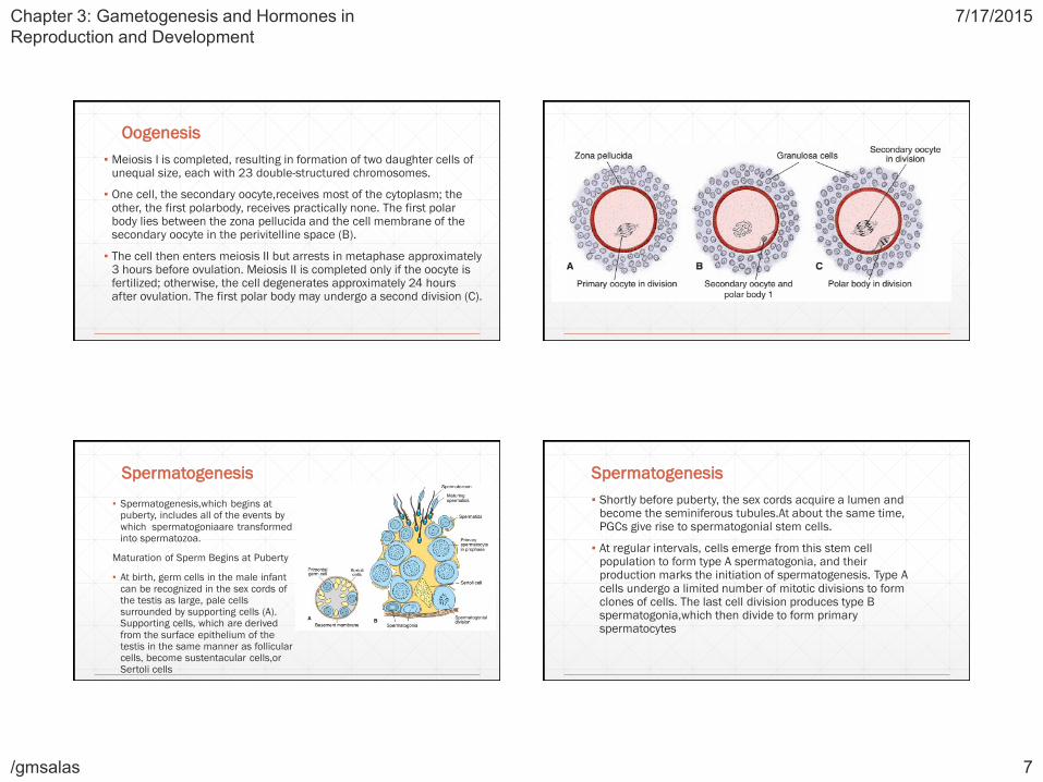
▪ Meiosis I is completed, resulting in formation of two daughter cells of unequal size, each with 23 double-structured chromosomes.
▪ One cell, the secondary oocyte,receives most of the cytoplasm; the other, the first polarbody, receives practically none. The first polar body lies between the zona pellucida and the cell membrane of the secondary oocyte in the perivitelline space (B).
▪ The cell then enters meiosis II but arrests in metaphase approximately 3 hours before ovulation. Meiosis II is completed only if the oocyte is fertilized; otherwise, the cell degenerates approximately 24 hours after ovulation. The first polar body may undergo a second division (C).
Spermatogenesis
▪ Spermatogenesis,which begins at puberty, includes all of the events by which spermatogoniaare transformed into spermatozoa.
Maturation of Sperm Begins at Puberty
▪ At birth, germ cells in the male infant can be recognized in the sex cords of the testis as large, pale cells surrounded by supporting cells (A). Supporting cells, which are derived from the surface epithelium of the testis in the same manner as follicular cells, become sustentacular cells,orSertoli cells
Spermatogenesis
▪ Shortly before puberty, the sex cords acquire a lumen and become the seminiferous tubules.At about the same time, PGCs give rise to spermatogonial stem cells.
▪ At regular intervals, cells emerge from this stem cell population to form type A spermatogonia, and their production marks the initiation of spermatogenesis. Type A cells undergo a limited number of mitotic divisions to form clones of cells. The last cell division produces type B spermatogonia,which then divide to form primary spermatocytes

Chapter 3: Gametogenesis and Hormones in
Reproduction and Development
7/17/2015
/gmsalas 8
Spermatogenesis
▪ Primary spermatocytes then enter a prolonged prophase (22 days) followed by rapid completion of meiosis I and formation of secondary spermatocytes. During the second meiotic division, these cells immediately begin to form haploid spermatids.
▪ Throughout this series of events, from the time type A cells leave the stem cell population to formation of spermatids, cytokinesis is incomplete, so that successive cell generations are joined by cytoplasmic bridges. Thus, the progeny of a single type A spermatogonium form a clone of germ cells that maintain contact throughout differentiation.
Spermatogenesis
▪ Furthermore, spermatogonia and spermatids remain embedded in deep recesses of Sertoli cells throughout their development. In this manner, Sertoli cells support and protect the germ cells, participate in their nutrition, and assist in the release of mature spermatozoa.
▪ Spermatogenesis is regulated by LH production by the pituitary gland. LH binds to receptors on Leydig cells and stimulates testosterone production, which in turn binds to Sertoli cells to promote spermatogenesis.
▪ Follicle-stimulating hormone (FSH)is also essential because its binding to Sertoli cells stimulates testicular fluid production and synthesis of intracellular androgen receptor proteins.

Chapter 3: Gametogenesis and Hormones in
Reproduction and Development
7/17/2015
/gmsalas 9
Spermiogenesis
▪ The series of changes resulting in the transformation of spermatids into spermatozoa is spermiogenesis. These changes include:
▪ (1) formation of the acrosome, which covers half of the nuclear surface and contains enzymes to assist in penetration of the egg and its surrounding layers during fertilization;
▪ (2) condensation of the nucleus;
▪ (3) formation of neck, middle piece, and tail; and
▪ (4) shedding of most of the cytoplasm as residual bodies that are phagocytized by Sertoli cells.
▪ In humans, the time required for a spermatogonium to develop into a mature spermatozoon is approximately 74 days, and approximately 300 million sperm cells are produced daily.
Spermiogenesis
▪ When fully formed, spermatozoa enter the lumen of seminiferous tubules. From there, they are pushed toward the epididymis by contractile elements in the wall of the seminiferous tubules.
▪ Although initially only slightly motile, spermatozoa obtain full motility in the epididymis.
Abnormal Gametes
Chapter 3: Gametogenesis and Hormones in
Reproduction and Development
7/17/2015
/gmsalas 10
Problems
▪ What is the most common cause of abnormal chromosome number? Give an example of a clinical syndrome involving abnormal numbers of chromosomes.
▪ In addition to numerical abnormalities, what types of chromosomal alterations occur?
▪ What is mosaicism, and how does it occur?
Ovarian Cycle
Ovarian Cycle
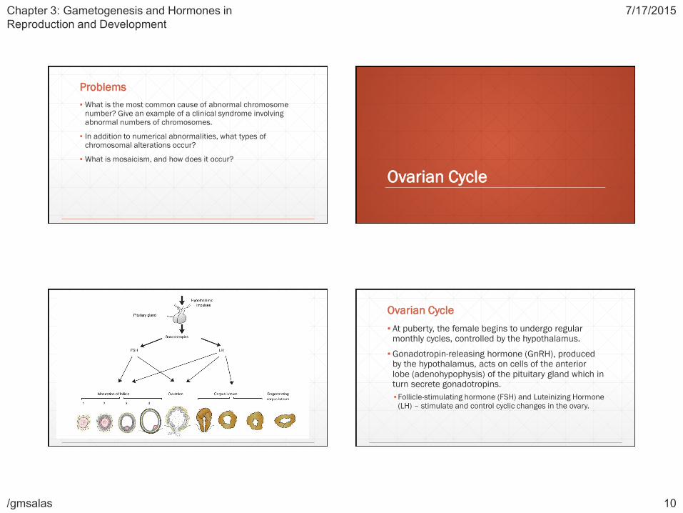
▪ At puberty, the female begins to undergo regular monthly cycles, controlled by the hypothalamus.
▪ Gonadotropin-releasing hormone (GnRH), produced by the hypothalamus, acts on cells of the anterior lobe (adenohypophysis) of the pituitary gland which in turn secrete gonadotropins.
▪ Follicle-stimulating hormone (FSH) and Luteinizing Hormone (LH) – stimulate and control cyclic changes in the ovary.

Chapter 3: Gametogenesis and Hormones in
Reproduction and Development
7/17/2015
/gmsalas 11
Ovarian Cycle
▪ At the beginning of each ovarian cycle, 15 to 20 primary-stage follicles are stimulated to grow under the influence of FSH. Thus, FSH rescues 15-20 of these cells from a pool of continuously forming primary follicles.
▪ Under normal conditions, only one of these follicles reaches full maturity, and only one oocyte discharged; the others degenerate and become atretic.
▪ When a follicle becomes atretic, the oocyte and surrounding follicular cells degenerate and are replaced by connective tissue, forming a corpus atreticum.
Ovarian Cycle
▪ FSH also stimulates maturation of follicular (granulosa) cells surrounding the oocyte. In turn, proliferation of these cells is mediated by growth differentiation factor 9, a member of transforming growth factor-β (TGFβ) family.
▪ In cooperation, theca interna and granulosa cells produce estrogens: theca interna cells produce androstenedione and testosterone, and granular cells convert these hormones to estrone and 17 β-estradiol.
Ovarian Cycle
▪ As a result of this estrogen production,
▪ The uterine endometrium enters the follicular or proliferative phase;
▪ Thinning of the cervical mucus occurs to allow passage of sperm; and
▪ The anterior lobe of the pituitary gland is stimulated to secrete LH
Ovarian Cycle
▪ At midcycle, there is an LH surge that:
▪Elevates concerntrations of maturation-promoting factor, causing oocyctes to complete meiosis I and initiate meiosis II;
▪Stimulates production of progesterone by follicular stromal cells (luteinization); and
▪Causes follicular rupture and ovulation.
Chapter 3: Gametogenesis and Hormones in
Reproduction and Development
7/17/2015
/gmsalas 12
Ovulation
▪ In the days immediately preceding ovulation, under the influence of FSH and LH, the vesicular follicle grows rapidly to a diameter of 25mm to become a mature vesicular (graafian) follicle.
▪ Coincident with final development of the vesicular follicle, there is an abrupt increase in LH that causes the primary oocyte to complete meiosis I and the follicle to enter the preovulatory mature vesicular stage. Meiosis II is also initiated, but the oocyte is arrested in metaphase approximately 3 hours before ovulation. In the meantime, the surface of the ovary begins to bulge locally, and at the apex, an avascular spot, the stigma, appears.
Ovulation
▪ The high concentration of LH increases collagenase activity, resulting in digestion of collagen fibers surrounding the follicle. Prostaglandin levels also increase in response to the LH surge and cause local muscular contractions in the ovarian wall.
▪ Those contractions extrude the oocyte, which together with its surrounding granulosa cells from the region of the cumulus oophorus breaks free (ovulation)and floats out of the ovary. Some of the cumulus oophorus cells then rearrange themselves around the zona pellucida to form the corona radiata.
Corpus Luteum
▪ After ovulation, granulosa cells remaining in the wall of the ruptured follicle, together with cells from the theca interna, are vascularized by surrounding vessels. Under the influence of LH, these cells develop a yellowish pigment and change into lutein cells, which form the corpus luteum and secrete estrogens and progesterone.
▪ Progesterone, together with some estrogen, causes the uterine mucosa to enter the progestationalorsecretory stage in preparation for implantation of the embryo.
Oocyte Transport
▪ Shortly before ovulation, fimbriae of the uterine tube sweep over the surface of the ovary, and the tube itself begins to contract rhythmically. It is thought that the oocyte, surrounded by some granulosa cells, is carried into the tube by these sweeping movements of the fimbriae and by motion of cilia on the epithelial lining.
▪ Once in the tube, cumulus cells withdraw their cytoplasmic processes from the zona pellucida and lose contact with the oocyte.

Chapter 3: Gametogenesis and Hormones in
Reproduction and Development
7/17/2015
/gmsalas 13
Oocyte Transport
▪ Once the oocyte is in the uterine tube, it is propelled by peristaltic muscular contractions of the tube and by cilia in the tubal mucosa with the rate of transport regulated by the endocrine status during and after ovulation.
▪ In humans, the fertilized oocyte reaches the uterine lumen in approximately 3 to 4 days.
Corpus Albicans
▪ If fertilization does not occur, the corpus luteum reaches maximum development approximately 9 days after ovulation. It can easily be recognized as a yellowish projection on the surface of the ovary.
▪ Subsequently, the corpus luteum shrinks because of degeneration of lutean cells (luteolysis)and forms a mass of fibrotic scar tissue, the corpus albicans. Simultaneously, progesterone production decreases, precipitating menstrual bleeding.
Corpus Albicans
▪ If the oocyte is fertilized, degeneration of the corpus luteum is prevented by human chorionic gonadotropin, a hormone secreted by the syncytiotrophoblast of the developing embryo.
▪ The corpus luteum continues to grow and forms the corpus luteum of pregnancy (corpus luteum graviditatis). By the end of the third month, this structure may be one third to one half of the total size of the ovary. Yellowish luteal cells continue to secrete progesterone until the end of the fourth month; thereafter, they regress slowly as secretion of progesterone by the trophoblastic component of the placenta becomes adequate for maintenance of pregnancy.
▪ Removal of the corpus luteum of pregnancy before the fourth month usually leads to abortion.

Chapter 3: Gametogenesis and Hormones in
Reproduction and Development
7/17/2015
/gmsalas 14