105 The Musculoskeletal System After studying this chapter, you will be able to: 5.1 Name the parts of the musculoskeletal system and discuss the func- tion of each part 5.2 Define combining forms used in building words that relate to the musculoskeletal system 5.3 Identify the meaning of related abbreviations 5.4 Name the common diagnoses, laboratory tests, and clinical procedures used in treating disorders of the musculoskeletal system 5.5 List and define the major pathological conditions of the musculoskeletal system 5.6 Define surgical terms related to the musculoskeletal system 5.7 List common pharmacological agents used in treating disorders of the musculoskeletal system Structure and Function The musculoskeletal system forms the framework that holds the body to- gether, enables it to move, and protects and supports all the internal organs. This system includes bones, joints, and muscles. Figure 5-1 shows the mus- culoskeletal system. Bones are made of osseous tissue and include a rich network of blood vessels and nerves. The cells of bone, called osteocytes, are part of a dense network of connective tissue. The cells themselves are surrounded by cal- cium salts. During fetal development, bones are softer and flexible and are composed of cartilage until the hardening process begins. Bone-forming cells are called osteoblasts. As bone tissue develops, some of it dies and is reabsorbed by osteoclasts (also called bone phagocytes). The reabsorption of dead bone cells prevents the bone from becoming overly thick and heavy. Later, if a bone breaks, osteoblasts will add new mineral matter to repair the break and the osteoclasts will remove any bone debris, thereby smoothing over the break. The hardening process and development of the osteocytes is called ossification. This process is largely dependent on calcium, phosphorus, and vitamin D. The skeleton of the body is made up of bones and joints. A mature adult has 206 bones that work together with joints and muscles to move the various parts of the body. The axial portion of the skeleton includes the trunk and head. The appendicular portion of the skeleton includes the limbs. Calcium is important for the formation of bones. It is recommended that you pay attention to your daily calcium intake throughout your life, since lack of calcium is a factor in certain diseases, such as osteoporosis. To find out about the recommended levels, go to the National Osteoporosis Foundation’s Web site (www.nof.org) and click on prevention. 5 5 CHAPTER ORTHOPEDICS, RHEUMATOLOGY

Welcome message from author
This document is posted to help you gain knowledge. Please leave a comment to let me know what you think about it! Share it to your friends and learn new things together.
Transcript

105
The Musculoskeletal System After studying this chapter, you will be able to:
5.1 Name the parts of the musculoskeletal system and discuss the func-tion of each part
5.2 Define combining forms used in building words that relate to the musculoskeletal system
5.3 Identify the meaning of related abbreviations 5.4 Name the common diagnoses, laboratory tests, and clinical
procedures used in treating disorders of the musculoskeletal system 5.5 List and define the major pathological conditions of the
musculoskeletal system 5.6 Define surgical terms related to the musculoskeletal system 5.7 List common pharmacological agents used in treating disorders of
the musculoskeletal system
Structure and Function
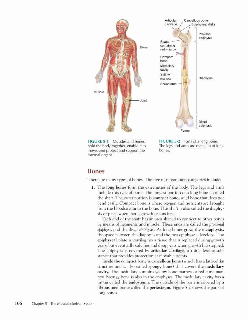
The musculoskeletal system forms the framework that holds the body to-gether, enables it to move, and protects and supports all the internal organs. This system includes bones, joints, and muscles. Figure 5-1 shows the mus-culoskeletal system.
Bones are made of osseous tissue and include a rich network of blood vessels and nerves. The cells of bone, called osteocytes, are part of a dense network of connective tissue. The cells themselves are surrounded by cal-cium salts. During fetal development, bones are softer and flexible and are composed of cartilage until the hardening process begins.
Bone-forming cells are called osteoblasts. As bone tissue develops, some of it dies and is reabsorbed by osteoclasts (also called bone phagocytes ). The reabsorption of dead bone cells prevents the bone from becoming overly thick and heavy. Later, if a bone breaks, osteoblasts will add new mineral matter to repair the break and the osteoclasts will remove any bone debris, thereby smoothing over the break. The hardening process and development of the osteocytes is called ossification. This process is largely dependent on calcium, phosphorus, and vitamin D.
The skeleton of the body is made up of bones and joints. A mature adult has 206 bones that work together with joints and muscles to move the various parts of the body. The axial portion of the skeleton includes the trunk and head. The appendicular portion of the skeleton includes the limbs.
Calcium is important for the formation of bones. It is recommended that you pay attention to your daily calcium intake throughout your life, since lack of calcium is a factor in certain diseases, such as osteoporosis. To find out about the recommended levels, go to the National Osteoporosis Foundation’s Web site ( www.nof.org ) and click on prevention.
Calcium is important for the formation of bones. It is recommended that you pay attention to your daily calcium intake throughout your life, since lack of calcium is a factor in certain diseases, such as osteoporosis. To find out about the recommended levels, go to the National Osteoporosis Foundation’s Web site ( www.nof.org ) and click on prevention.
5 5 CHAPTER
� � ORTHOPEDICS, RHEUMATOLOGY
thi74725_ch05_105-154.indd 105 10/17/08 2:50:25 PM
106 Chapter 5 The Musculoskeletal System
Bones There are many types of bones. The five most common categories include:
1. The long bones form the extremities of the body. The legs and arms include this type of bone. The longest portion of a long bone is called the shaft. The outer portion is compact bone, solid bone that does not bend easily. Compact bone is where oxygen and nutrients are brought from the bloodstream to the bone. This shaft is also called the diaphy-sis or place where bone growth occurs first.
Each end of the shaft has an area shaped to connect to other bones by means of ligaments and muscle. These ends are called the proximal epiphysis and the distal epiphysis. As long bones grow, the metaphysis, the space between the diaphysis and the two epiphyses, develops. The epiphyseal plate is cartilaginous tissue that is replaced during growth years, but eventually calcifies and disappears when growth has stopped. The epiphysis is covered by articular cartilage, a thin, flexible sub-stance that provides protection at movable points.
Inside the compact bone is cancellous bone (which has a latticelike structure and is also called spongy bone ) that covers the medullary cavity. The medullary contains yellow bone marrow or red bone mar-row. Spongy bone is also in the epiphyses. The medullary cavity has a lining called the endosteum. The outside of the bone is covered by a fibrous membrane called the periosteum. Figure 5-2 shows the parts of long bones.
FIGURE 5-1 Muscles and bones hold the body together, enable it to move, and protect and support the internal organs.
Joint
Muscle
Bone
FIGURE 5-2 Parts of a long bone. The legs and arms are made up of long bones.
Compactbone
Medullarycavity
Yellowmarrow
Periosteum
Femur
Spacecontainingred marrow
Cancellous boneArticularcartilage Epiphyseal disks
Proximalepiphysis
Diaphysis
Distalepiphysis
thi74725_ch05_105-154.indd 106 10/17/08 2:50:36 PM
Chapter 5 The Musculoskeletal System 107
2. Short bones are the small, cube-shaped bones of the wrists, ankles, and toes. Short bones consist of an outer layer of compact bone with an inner layer of cancellous bone.
3. Flat bones generally have large, somewhat flat surfaces that cover organs or that provide a surface for large areas of muscle. The shoulder blades, pelvis, and skull include flat bones.
4. Irregular bones are specialized bones with specific shapes. The bones of the ears, vertebrae, and face are irregular bones.
5. Sesamoid bones are bones formed in a tendon near joints. The patella (kneecap) is a sesamoid bone. Sesamoid bones are also found in the hands and feet.
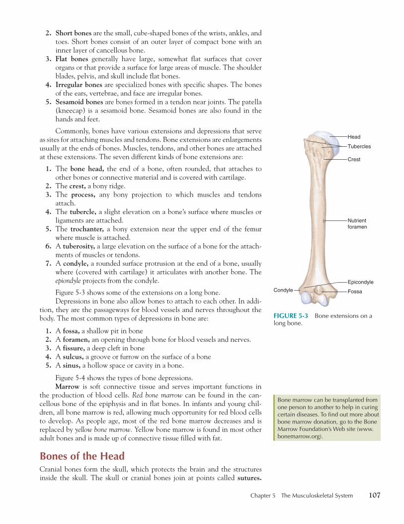
Commonly, bones have various extensions and depressions that serve as sites for attaching muscles and tendons. Bone extensions are enlargements usually at the ends of bones. Muscles, tendons, and other bones are attached at these extensions. The seven different kinds of bone extensions are:
1. The bone head, the end of a bone, often rounded, that attaches to other bones or connective material and is covered with cartilage.
2. The crest, a bony ridge. 3. The process, any bony projection to which muscles and tendons
attach. 4. The tubercle, a slight elevation on a bone’s surface where muscles or
ligaments are attached. 5. The trochanter, a bony extension near the upper end of the femur
where muscle is attached. 6. A tuberosity, a large elevation on the surface of a bone for the attach-
ments of muscles or tendons. 7. A condyle, a rounded surface protrusion at the end of a bone, usually
where (covered with cartilage) it articulates with another bone. The epiondyle projects from the condyle.
Figure 5-3 shows some of the extensions on a long bone. Depressions in bone also allow bones to attach to each other. In addi-
tion, they are the passageways for blood vessels and nerves throughout the body. The most common types of depressions in bone are:
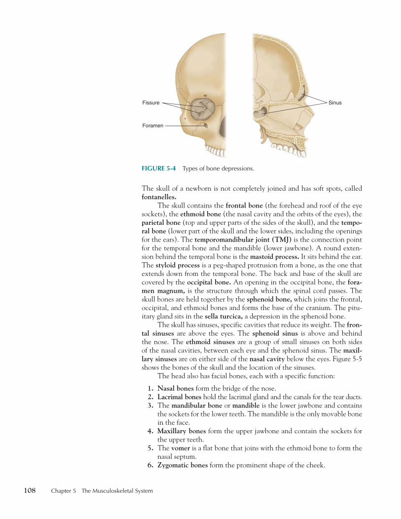
1. A fossa, a shallow pit in bone 2. A foramen, an opening through bone for blood vessels and nerves. 3. A fissure, a deep cleft in bone 4. A sulcus, a groove or furrow on the surface of a bone 5. A sinus, a hollow space or cavity in a bone.
Figure 5-4 shows the types of bone depressions. Marrow is soft connective tissue and serves important functions in
the production of blood cells. Red bone marrow can be found in the can-cellous bone of the epiphysis and in flat bones. In infants and young chil-dren, all bone marrow is red, allowing much opportunity for red blood cells to develop. As people age, most of the red bone marrow decreases and is replaced by yellow bone marrow. Yellow bone marrow is found in most other adult bones and is made up of connective tissue filled with fat.
Bones of the Head Cranial bones form the skull, which protects the brain and the structures inside the skull. The skull or cranial bones join at points called sutures.
Bone marrow can be transplanted from one person to another to help in curing certain diseases. To find out more about bone marrow donation, go to the Bone Marrow Foundation’s Web site ( www.bonemarrow.org ).
Bone marrow can be transplanted from one person to another to help in curing certain diseases. To find out more about bone marrow donation, go to the Bone Marrow Foundation’s Web site ( www.bonemarrow.org ).
FIGURE 5-3 Bone extensions on a long bone.
Tubercles
Head
Crest
Condyle
Nutrientforamen
Fossa
Epicondyle
thi74725_ch05_105-154.indd 107 10/17/08 2:50:41 PM
108 Chapter 5 The Musculoskeletal System
The skull of a newborn is not completely joined and has soft spots, called fontanelles.
The skull contains the frontal bone (the forehead and roof of the eye sockets), the ethmoid bone (the nasal cavity and the orbits of the eyes), the parietal bone (top and upper parts of the sides of the skull), and the tempo-ral bone (lower part of the skull and the lower sides, including the openings for the ears). The temporomandibular joint (TMJ) is the connection point for the temporal bone and the mandible (lower jawbone). A round exten-sion behind the temporal bone is the mastoid process. It sits behind the ear. The styloid process is a peg-shaped protrusion from a bone, as the one that extends down from the temporal bone. The back and base of the skull are covered by the occipital bone. An opening in the occipital bone, the fora-men magnum, is the structure through which the spinal cord passes. The skull bones are held together by the sphenoid bone, which joins the frontal, occipital, and ethmoid bones and forms the base of the cranium. The pitu-itary gland sits in the sella turcica, a depression in the sphenoid bone.
The skull has sinuses, specific cavities that reduce its weight. The fron-tal sinuses are above the eyes. The sphenoid sinus is above and behind the nose. The ethmoid sinuses are a group of small sinuses on both sides of the nasal cavities, between each eye and the sphenoid sinus. The maxil-lary sinuses are on either side of the nasal cavity below the eyes. Figure 5-5 shows the bones of the skull and the location of the sinuses.
The head also has facial bones, each with a specific function:
1. Nasal bones form the bridge of the nose. 2. Lacrimal bones hold the lacrimal gland and the canals for the tear ducts. 3. The mandibular bone or mandible is the lower jawbone and contains
the sockets for the lower teeth. The mandible is the only movable bone in the face.
4. Maxillary bones form the upper jawbone and contain the sockets for the upper teeth.
5. The vomer is a flat bone that joins with the ethmoid bone to form the nasal septum.
6. Zygomatic bones form the prominent shape of the cheek.
FIGURE 5-4 Types of bone depressions.
Fissure Sinus
Foramen
thi74725_ch05_105-154.indd 108 10/17/08 2:50:45 PM

Chapter 5 The Musculoskeletal System 109
7. The palatine bone sits behind the maxillary bones and helps to form the nasal cavity and the hard palate.
Figure 5-6 shows the bones of the face.
Spinal Column The spinal column (also called the vertebral column ) consists of five sets of vertebrae. Each vertebra is a bone segment with a thick, cartilaginous disk (also called intervertebral disk or disk ) that separates the vertebrae. In the middle of the disk is a fibrous mass called the nucleus pulposus. The disks
Sphenoidbone
Frontalsinus
Ethmoidalsinuses
Sphenoidalsinus
Maxillarysinus
Parietalbone
Frontal bone
Coronalsuture
Lacrimalbone
Ethmoidbone
Squamosalsuture
Temporalbone
Mandible
Maxilla
FIGURE 5-5 The bones of the skull and the sinus cavities.
Zygomatic bone
Maxillary bones
Mandible
Palatine bone
Lacrimal bone
Ethmoid bone
Nasal bones
FIGURE 5-6 The bones of the face.
thi74725_ch05_105-154.indd 109 10/17/08 2:50:46 PM
110 Chapter 5 The Musculoskeletal System
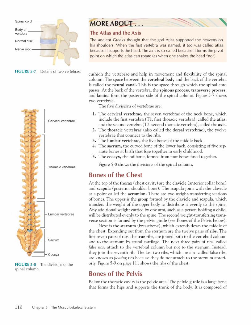
cushion the vertebrae and help in movement and flexibility of the spinal column. The space between the vertebral body and the back of the vertebra is called the neural canal. This is the space through which the spinal cord passes. At the back of the vertebra, the spinous process, transverse process,and lamina form the posterior side of the spinal column. Figure 5-7 shows two vertebrae.
The five divisions of vertebrae are:
1. The cervical vertebrae, the seven vertebrae of the neck bone, which include the first vertebra (T1, first thoracic vertebra), called the atlas,and the second vertebra (T2, second thoracic vertebra), called the axis.
2. The thoracic vertebrae (also called the dorsal vertebrae ), the twelve vertebrae that connect to the ribs.
3. The lumbar vertebrae, the five bones of the middle back. 4. The sacrum, the curved bone of the lower back, consisting of five sep-
arate bones at birth that fuse together in early childhood. 5. The coccyx, the tailbone, formed from four bones fused together.
Figure 5-8 shows the divisions of the spinal column.
Bones of the Chest At the top of the thorax (chest cavity) are the clavicle (anterior collar bone) and scapula (posterior shoulder bone). The scapula joins with the clavicle at a point called the acromion. There are two weight-transferring sections of bones. The upper is the group formed by the clavicle and scapula, which transfers the weight of the upper body to distribute it evenly to the spine. Any additional weight carried by one arm, such as a person holding a child, will be distributed evenly to the spine. The second weight-transferring trans-verse section is formed by the pelvic girdle (see Bones of the Pelvis below).
Next is the sternum (breastbone), which extends down the middle of the chest. Extending out from the sternum are the twelve pairs of ribs. The first seven pairs of ribs, the true ribs, are joined both to the vertebral column and to the sternum by costal cartilage. The next three pairs of ribs, called false ribs, attach to the vertebral column but not to the sternum. Instead, they join the seventh rib. The last two ribs, which are also called false ribs, are known as floating ribs because they do not attach to the sternum anteri-orly. Figure 5-9 on page 111 shows the ribs of the chest.
Bones of the Pelvis Below the thoracic cavity is the pelvic area. The pelvic girdle is a large bone that forms the hips and supports the trunk of the body. It is composed of
MORE ABOUT . . .The Atlas and the AxisThe ancient Greeks thought that the god Atlas supported the heavens on his shoulders. When the first vertebra was named, it too was called atlas because it supports the head. The axis is so-called because it forms the pivot point on which the atlas can rotate (as when one shakes the head “no”).
FIGURE 5-7 Details of two vertebrae.
Normal disk
Nerve root
Body ofvertebra
Spinal cord
Cervical vertebrae
Thoracic vertebrae
Lumbar vertebrae
Sacrum
Coccyx
FIGURE 5-8 The divisions of the spinal column.
thi74725_ch05_105-154.indd 110 10/17/08 2:50:59 PM

Chapter 5 The Musculoskeletal System 111
three fused bones, including the ilium, ischium, and pubes (the anteroin-ferior portion of the hip bone). It is also the point of attachment for the legs. This is the second weight-transferring transverse section of bone. The pelvic girdle easily transfers weight of the body from one leg to the other during running, walking, or any movement.
Inside the pelvic girdle is the pelvic cavity. In the pelvic cavity are located the female reproductive organs, the sigmoid colon, the bladder, and the rectum. The area where the two pubic bones join is called the pubic symphysis. Figure 5-10 shows the bones of the pelvis.
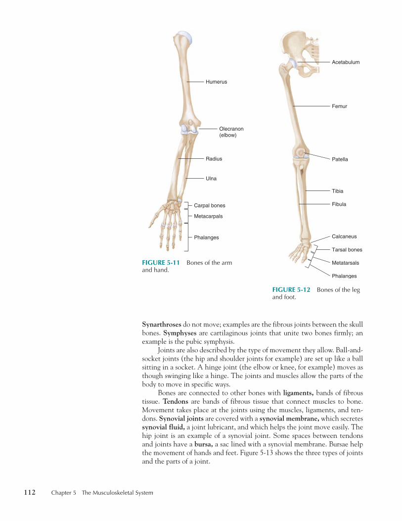
Bones of the Extremities The upper arm bone, the humerus, attaches to the scapula and clavicle. The two lower arm bones are the ulna, which has a bony protrusion called the olecranon (elbow), and the radius, which attaches to the eight carpal bones of the wrist ( carpus ). The metacarpals are the five bones of the palm that radiate out to the finger bones, the phalanges. Each phalanx (except for the thumbs and great toes) has a distal (furthest from the body), middle, and proximal (nearest to the body) segment. Figure 5-11 shows the bones of the arm and hand.
The hip bone has a cup-shaped depression or socket called the acetab-ulum into which the femur (thigh bone) fits. The femur is the longest bone in the body. It meets the two bones of the lower leg, the tibia (also called the shin ) and fibula, at the kneecap or patella. The tibia and fibula have bony protrusions near the foot called the malleoli (singular, malleolus ). The protrusion of the tibia is called the medial malleolus. The protrusion of the fibula is called the lateral malleolus. The malleoli and the tarsal bones (seven small bones of the tarsus or instep) form the ankle. The largest tarsal is the calcaneus (heel). The metatarsals connect to the phalanges of the toes. Figure 5-12 shows the bones of the lower extremities.
Joints Joints are also called articulations, points where bones connect. The move-ment at a particular joint varies depending on the body’s needs. Diarthro-ses are joints that move freely, such as the knee joint. Amphiarthroses are cartilaginous joints that move slightly, such as the joints between vertebrae.
FIGURE 5-9 Ribs of the chest.
True ribs
False ribs
FIGURE 5-10 Bones of the pelvis.
Pelvic girdle
Pelvic cavity
Ilium
Ischium
Pubes
Pubic symphysis
thi74725_ch05_105-154.indd 111 10/17/08 2:51:01 PM
112 Chapter 5 The Musculoskeletal System
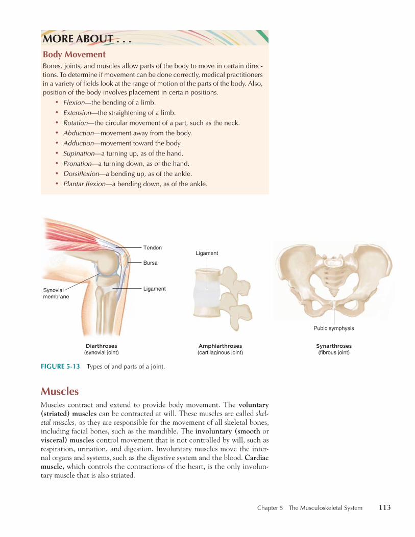
Synarthroses do not move; examples are the fibrous joints between the skull bones. Symphyses are cartilaginous joints that unite two bones firmly; an example is the pubic symphysis.
Joints are also described by the type of movement they allow. Ball-and-socket joints (the hip and shoulder joints for example) are set up like a ball sitting in a socket. A hinge joint (the elbow or knee, for example) moves as though swinging like a hinge. The joints and muscles allow the parts of the body to move in specific ways.
Bones are connected to other bones with ligaments, bands of fibrous tissue. Tendons are bands of fibrous tissue that connect muscles to bone. Movement takes place at the joints using the muscles, ligaments, and ten-dons. Synovial joints are covered with a synovial membrane, which secretes synovial fluid, a joint lubricant, and which helps the joint move easily. The hip joint is an example of a synovial joint. Some spaces between tendons and joints have a bursa, a sac lined with a synovial membrane. Bursae help the movement of hands and feet. Figure 5-13 shows the three types of joints and the parts of a joint.
FIGURE 5-11 Bones of the arm and hand.
Humerus
Olecranon(elbow)
Radius
Carpal bones
Metacarpals
Phalanges
Ulna
FIGURE 5-12 Bones of the leg and foot.
Femur
Tibia
Fibula
Tarsal bones
Calcaneus
Metatarsals
Phalanges
Patella
Acetabulum
thi74725_ch05_105-154.indd 112 10/17/08 2:51:03 PM
Chapter 5 The Musculoskeletal System 113
Muscles Muscles contract and extend to provide body movement. The voluntary (striated) muscles can be contracted at will. These muscles are called skel-etal muscles, as they are responsible for the movement of all skeletal bones, including facial bones, such as the mandible. The involuntary (smooth or visceral) muscles control movement that is not controlled by will, such as respiration, urination, and digestion. Involuntary muscles move the inter-nal organs and systems, such as the digestive system and the blood. Cardiac muscle, which controls the contractions of the heart, is the only involun-tary muscle that is also striated.
MORE ABOUT . . .Body MovementBones, joints, and muscles allow parts of the body to move in certain direc-tions. To determine if movement can be done correctly, medical practitioners in a variety of fields look at the range of motion of the parts of the body. Also, position of the body involves placement in certain positions.
• Flexion—the bending of a limb.• Extension—the straightening of a limb.• Rotation—the circular movement of a part, such as the neck.• Abduction—movement away from the body.• Adduction—movement toward the body.• Supination—a turning up, as of the hand.• Pronation—a turning down, as of the hand.• Dorsiflexion—a bending up, as of the ankle.• Plantar flexion—a bending down, as of the ankle.
Ligament
Pubic symphysis
Ligament
Synovialmembrane
Tendon
Diarthroses(synovial joint)
Amphiarthroses(cartilaginous joint)
Synarthroses(fibrous joint)
Bursa
FIGURE 5-13 Types of and parts of a joint.
thi74725_ch05_105-154.indd 113 10/17/08 2:51:04 PM

114 Chapter 5 The Musculoskeletal System
Most muscles are covered by fascia, a band of connective tissue that supports and covers the muscle. Muscles attach to a stationary bone at a point called the origin. They attach to a movable bone at a point called the insertion. During movement, the muscle contracts and extends and the moveable bone moves in a specific direction. Different muscles have differ-ent functions. For example, the deltoid muscles are used to extend the arms, the biceps of the arm flex the forearms, and the masticatory muscles close and open the jaw for chewing. Figure 5-14 shows the various types of muscle.
MORE ABOUT . . .MusclesNormal muscles contract and extend during routine movement and exer-cise. In unusual circumstances, muscles can atrophy (waste away). This can happen from a number of diseases that affect muscles and movement or from lack of use, as in a sedentary lifestyle. People who are paralyzed and find it difficult to get help moving muscles generally have areas where mus-cle atrophies. On the other hand, overuse of muscles can cause hyperplasia,an abnormal increase in muscle cells.
Building muscle by exercising is generally a healthy thing to do. How-ever, some athletes take dangerous shortcuts to building muscle. They take anabolic steroids or supplements containing products similar to anabolic steroids that build muscle quickly. Unfortunately, these products can have devastating health and emotional consequences, sometimes even fatal ones. Also, athletes who take these illegal substances often have an unfair advan-tage in competition over those who don’t. These substances are outlawed in most competitive sports.
For more information about steroid abuse, go to the National Institute on Drug Abuse’s Web site on steroid abuse (www.steroidabuse.org).
For more information about steroid abuse, go to the National Institute on Drug Abuse’s Web site on steroid abuse (www.steroidabuse.org).
Insertion
Origin
Section ofSmallIntestine
Biceps
Voluntary Muscles(striated)
Involuntary Muscles(smooth or visceral)
InvoluntarymusclesTriceps
Fascia Villi
FIGURE 5-14 Types and parts of muscle.
V OCABULARY R EVIEW In the previous section, you learned terms relating to the musculoskeletal system. Before going on to the exercises, review the terms below and refer to the previous chapters if you have any questions. Pronunciations are provided for
thi74725_ch05_105-154.indd 114 10/17/08 2:51:06 PM
Chapter 5 The Musculoskeletal System 115
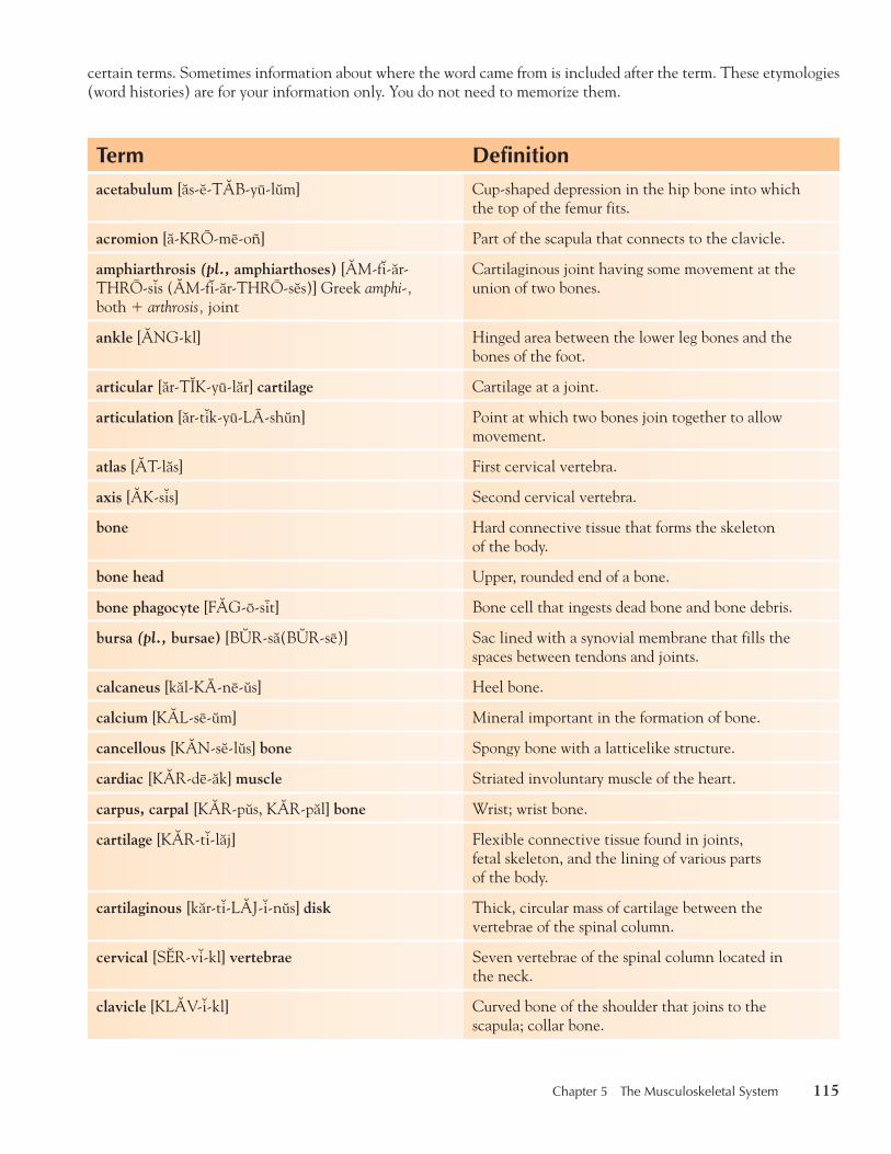
certain terms. Sometimes information about where the word came from is included after the term. These etymologies (word histories) are for your information only. You do not need to memorize them.
Term Definitionacetabulum [ås-6-T0B-yu-l9m] Cup-shaped depression in the hip bone into which
the top of the femur fits.
acromion [å-KRO-me-oñ] Part of the scapula that connects to the clavicle.
amphiarthrosis (pl., amphiarthoses) [0M-f7-år-THRO-s7s (0M-f7-år-THRO-s6s)] Greek amphi-, both � arthrosis, joint
Cartilaginous joint having some movement at the union of two bones.
ankle [0NG-kl] Hinged area between the lower leg bones and the bones of the foot.
articular [år-T2K-yu-lår] cartilage Cartilage at a joint.
articulation [år-t7k-yu-LA-sh9n] Point at which two bones join together to allow movement.
atlas [0T-lås] First cervical vertebra.
axis [0K-s7s] Second cervical vertebra.
bone Hard connective tissue that forms the skeleton of the body.
bone head Upper, rounded end of a bone.
bone phagocyte [F0G-o-sit] Bone cell that ingests dead bone and bone debris.
bursa (pl., bursae) [B4R-så(B4R-se)] Sac lined with a synovial membrane that fills the spaces between tendons and joints.
calcaneus [kål-KA-ne-9s] Heel bone.
calcium [K0L-se-9m] Mineral important in the formation of bone.
cancellous [K0N-s6-l9s] bone Spongy bone with a latticelike structure.
cardiac [K0R-de-åk] muscle Striated involuntary muscle of the heart.
carpus, carpal [K0R-p9s, K0R-pål] bone Wrist; wrist bone.
cartilage [K0R-t7-låj] Flexible connective tissue found in joints, fetal skeleton, and the lining of various parts of the body.
cartilaginous [kår-t7-L0J-7-n9s] disk Thick, circular mass of cartilage between the vertebrae of the spinal column.
cervical [S1R-v7-kl] vertebrae Seven vertebrae of the spinal column located in the neck.
clavicle [KL0V-7-kl] Curved bone of the shoulder that joins to the scapula; collar bone.
thi74725_ch05_105-154.indd 115 10/17/08 2:51:07 PM

116 Chapter 5 The Musculoskeletal System
Term Definitioncoccyx [K3K-s7ks] Small bone consisting of four fused vertebrae at the
end of the spinal column; tailbone.
compact bone Hard bone with a tightly woven structure.
condyle [K3N-dil] Rounded surface at the end of a bone.
crest Bony ridge.
diaphysis [di-0F-7-s7s] Greek, a growing between
Long middle section of a long bone; shaft.
diarthroses (sing., diarthrosis) [di-år-THRO-sez (di-år-THRO-s7s] Greek, articulations
Freely movable joints.
disk [d7sk] Latin discus See cartilaginous disk.
dorsal vertebrae Thoracic vertebrae.
elbow [1L-bo] Joint between the upper arm and the forearm.
endosteum [6n-D3S-te-9m] end(o)-, within � Greek osteon, bone
Lining of the medullary cavity.
epiphyseal [6p-7-F2Z-e-ål] plate Cartilaginous tissue that is replaced during growth years, but eventually calcifies and disappears when growth stops.
ethmoid [1TH-m8yd] bone Irregular bone of the face attached to the sphenoid bone.
ethmoid sinuses Sinuses on both sides of the nasal cavities between each eye and the sphenoid sinus.
fascia (pl., fasciae) [F0SH-e-å (F0SH-e-e)] Sheet of fibrous tissue that encloses muscles.
femur [FE-mur] Long bone of the thigh.
fibula [F2B-yu-lå] Smallest long bone of the lower leg.
fissure [F2SH-9r] Deep furrow or slit.
flat bones Thin, flattened bones that cover certain areas, as of the skull.
fontanelle [F3N-tå-n6l] Soft, membranous section on top of an infant’s skull.
foramen [fo-RA-m6n] Opening or perforation through a bone.
foramen magnum [M0G-n9m] Opening in the occipital bone through which the spinal cord passes.
fossa (pl., fossae) [F3S-å (F3S-e)] Depression, as in a bone.
frontal [FR4N-tål] bone Large bone of the skull that forms the top of the head and forehead.
frontal sinuses Sinuses above the eyes.
thi74725_ch05_105-154.indd 116 10/17/08 2:51:07 PM

Chapter 5 The Musculoskeletal System 117
Term Definitionheel [hel] Back, rounded portion of the foot.
humerus [HYU-m6r-9s] Long bone of the arm connecting to the scapula on top and the radius and ulna at the bottom.
ilium [2L-e-9m] Wide portion of the hip bone.
insertion Point at which a muscle attaches to a movable bone.
intervertebral [7n-t6r-V1R-t6-brål] disk See cartilaginous disk.
involuntary muscle Muscle not movable at will.
irregular bones Any of a group of bones with a special shape to fit into certain areas of the skeleton, such as the skull.
ischium [2S-ke-9m] One of three fused bones that form the pelvic girdle.
joint [j8ynt] Place of joining between two or more bones.
lacrimal [L0K-r7-mål] bone Thin, flat bone of the face.
lamina (pl., laminae) [L0M-7-nå (L0M-7-ne)] Thin, flat part of either side of the arch of a vertebra.
ligament [L2G-å-m6nt] Sheet of fibrous tissue connecting and supporting bones; attaches bone to bone.
long bone Any bone of the extremities with a shaft.
lumbar [L4M-bår] vertebrae Five vertebrae of the lower back.
malleolus (pl., malleoli) [må-LE-o-l9s (må-LE-o-li)] Rounded protrusion of the tibia or fibula on either side of the ankle.
mandible [M0N-d7-bl] U-shaped bone of the lower jaw.
mandibular [mån-D2B-yu-lår] bone Mandible.
marrow [M0R-o] Connective tissue filling the medullary cavity, often rich in nutrients.
mastoid [M0S-t8yd] process Protrusion of the temporal bone that sits behind the ear.
maxillary [M0K-s7-lar-e] bone Bone of the upper jaw.
maxillary sinus Sinus on either side of the nasal cavity below the eyes.
medullary [M1D-u-lar-e] cavity Soft center cavity in bone that often holds marrow.
metacarpal [M1T-å-K0R-pål] meta-, behind � carpal, of the wrist
One of five bones of the hand between the wrist and the fingers.
metaphysis [m6-T0F-7-s7s] meta-, behind � Greek physis, growth
Section of a long bone between the epiphysis and diaphysis.
metatarsal [M1T-å-T0R-sål] bones meta-, behind � tarsus
Bones of the foot between the instep (arch) and the toes.
thi74725_ch05_105-154.indd 117 10/17/08 2:51:07 PM

118 Chapter 5 The Musculoskeletal System
Term Definitionmuscle [M4S-6l] Contractile tissue that plays a major role in body
movement.
musculoskeletal [M4S-kyu-lo-SK1L-6-tål] system musculo-, muscle � skeletal
System of the body including the muscles and skeleton.
nasal bones Bones that form the bridge of the nose.
nasal cavity Cavity on either side of the nasal septum.
neural [NUR-ål] canal Space through which the spinal cord passes.
nucleus pulposus [NU-kle-9s p9l-PO-s9s] Fibrous mass in the center portion of the intervertebral disk.
occipital [8k-S2P-7-tål] bone Bone that forms the lower back portion of the skull.
olecranon [o-L1K-rå-n8n] Curved end of the ulna to which tendons of the arm muscles attach; bony prominence of the elbow.
origin Point at which muscles attach to stationary bone.
osseous [3S-e-9s] tissue Connective tissue into which calcium salts are deposited.
ossification [3S-7-f7-KA-sh9n] Hardening into bone.
osteoblast [3S-te-o-blåst] osteo-, bone � -blast, forming
Cell that forms bone.
osteoclast [3S-te-o-klåst] osteo-, bone � -clast, breaking
Large cell that reabsorbs and removes osseous tissue.
osteocyte [3S-te-o-sit] osteo-, bone � -cyte, cell
Bone cell.
palatine [P0L-å-tin] bone Bone that helps form the hard palate and nasal cavity; located behind the maxillary bones.
parietal [på-RI-6-tål] bone Flat, curved bone on either side of the upper part of the skull.
patella [på-T1L-å] Large, sesamoid bone that forms the kneecap.
pelvic [P1L-v7k] cavity Cup-shaped cavity formed by the large bones of the pelvic girdle; contains female reproductive organs, sigmoid colon, bladder, and rectum.
pelvic girdle Hip bones.
pelvis [P1L-v7s] Cup-shaped ring of bone and ligaments at the base of the trunk.
periosteum [p6r-e-3S-te-9m] Fibrous membrane covering the surface of bone.
phalanges (sing., phalanx) [få-L0N-jez (F0-långks)] Long bones of the fingers and toes.
phosphorus [F3S-for-9s] Mineral important to the formation of bone.
thi74725_ch05_105-154.indd 118 10/17/08 2:51:08 PM

Chapter 5 The Musculoskeletal System 119
Term Definitionprocess [PRO-s6s, PR3S-6s] Bony outgrowth or projection.
pubes [PYU-b7s] Anteroinferior portion of the hip bone.
pubic symphysis [PYU-b7k S2M-få-s7s] Joint between the two public bones.
radius [RA-de-9s] Shorter bone of the forearm.
rib One of twenty-four bones that form the chest wall.
sacrum [SA-kr9m] Next-to-last spinal vertebra made up of five fused bones; vertebra that forms part of the pelvis.
scapula [SK0P-yu-lå] Large flat bone that forms the shoulder blade.
sella turcica [S1L-å T4R-s7-kå] Bony depression in the sphenoid bone where the pituitary gland is located.
sesamoid [S1S-å-m8yd] bone Bone formed in a tendon over a joint.
shin [sh7n] Anterior ridge of the tibia.
short bones Square-shaped bones with approximately equal dimensions on all sides.
sinus [SI-n9s] Hollow cavity, especially either of two cavities on the sides of the nose.
skeleton [SK1L-6-t8n] Bony framework of the body.
smooth muscle Fibrous muscle of internal organs that acts involuntarily.
sphenoid [SFE-n8yd] bone Bone that forms the base of the skull.
sphenoid sinus Sinus above and behind the nose.
spinal column Column of vertebrae at the posterior of the body, from the neck to the coccyx.
spinous [SPI-n9s] process Protrusion from the center of the vertebral arch.
spongy bone Bone with an open latticework filled with connective tissue or marrow.
sternum [ST1R-n9m] Long, flat bone that forms the midline of the anterior of the thorax.
striated [stri-AT-6d] muscle Muscle with a ribbed appearance that is controlled at will.
styloid [STI-l8yd] process Peg-shaped protrusion from a bone.
sulcus (pl., sulci) [S4L-k9s (S4L-si)] Latin, furrow
Groove or furrow in the surface of bone.
suture [SU-chur] Joining of two bone parts with a fibrous membrane.
symphysis [S2M-f7-s7s] Greek, from sym-, together � physis, joint
Type of cartilaginous joint uniting two bones.
thi74725_ch05_105-154.indd 119 10/17/08 2:51:08 PM

120 Chapter 5 The Musculoskeletal System
Term Definitionsynarthrosis [S2N-år-THRO-s7s] Greek, from syn-, together � arthrosis, articulation
Fibrous joint with no movement.
synovial [s7-NO-ve-ål] fluid Fluid that serves to lubricate joints.
synovial joint A joint that moves.
synovial membrane Connective tissue lining the cavity of joints and producing the synovial fluid.
tarsus, tarsal [T0R-s9s, T0R-sål] bones Seven bones of the instep (arch of the foot).
temporal [T1M-po-rål] bone Large bone forming the base and sides of the skull.
temporomandibular [T1M-po-ro-mån-D2B-yu-lår] joint (TMJ)
Joint of the lower jaw between the temporal bone and the mandible.
tendon [T1N-d8n] Fibrous band that connects muscle to bone or other structures.
thoracic [tho-R0S-7k] vertebrae Twelve vertebrae of the chest area.
thorax [THO-råks] Part of the trunk between the neck and the abdomen; chest.
tibia [T2B-e-å] Larger of the two lower leg bones.
transverse process Protrusion on either side of the vertebral arch.
trochanter [tro-K0N-t6r] Bony protrusion at the upper end of the femur.
true ribs Seven upper ribs of the chest that attach to the sternum.
tubercle [TU-b6r-kl] Slight bony elevation to which a ligament or muscle may be attached.
tuberosity [TU-b6r-3S-7-te] Large elevation in the surface of a bone.
ulna [4L-nå] Larger bone of the forearm.
vertebra (pl., vertebrae) [V1R-t6-brå (V1R-t6-bre)] One of the bony segments of the spinal column.
vertebral body Main portion of the vertebra, separate from the arches of the vertebra.
vertebral column Spinal column.
visceral [V2S-6r-ål] muscle Smooth muscle.
vitamin D Vitamin important to the formation of bone.
voluntary muscle Striated muscle.
vomer [VO-m6r] Flat bone forming the nasal septum.
zygomatic [ZI-go-M0T-7k] bone Bone that forms the cheek.
thi74725_ch05_105-154.indd 120 10/17/08 2:51:09 PM
Chapter 5 The Musculoskeletal System 121
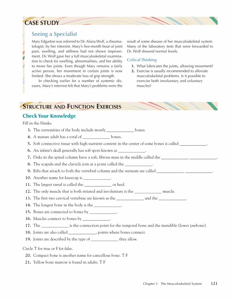
Mary Edgarton was referred to Dr. Alana Wolf, a rheuma-tologist, by her internist. Mary’s five-month bout of joint pain, swelling, and stiffness had not shown improve-ment. Dr. Wolf gave her a full musculoskeletal examina-tion to check for swelling, abnormalities, and her ability to move her joints. Even though Mary remains a fairly active person, her movement in certain joints is now limited. She shows a moderate loss of grip strength.
In checking earlier for a number of systemic dis-eases, Mary’s internist felt that Mary’s problems were the
CASE STUDY
Seeing a Specialistresult of some disease of her musculoskeletal system. Many of the laboratory tests that were forwarded to Dr. Wolf showed normal levels.
Critical Thinking1. What lubricates the joints, allowing movement?2. Exercise is usually recommended to alleviate
musculoskeletal problems. Is it possible to exercise both involuntary and voluntary muscles?
S TRUCTURE AND F UNCTION E XERCISES Check Your Knowledge Fill in the blanks.
3. The extremities of the body include mostly ____________ bones.
4. A mature adult has a total of ____________ bones.
5. Soft connective tissue with high nutrient content in the center of some bones is called ____________ .
6. An infant’s skull generally has soft spots known as ____________ .
7. Disks in the spinal column have a soft, fibrous mass in the middle called the ____________ ____________ .
8. The scapula and the clavicle join at a point called the ____________ .
9. Ribs that attach to both the vertebral column and the sternum are called ____________ ____________ .
10. Another name for kneecap is ____________ .
11. The largest tarsal is called the ____________ or heel.
12. The only muscle that is both striated and involuntary is the ____________ muscle.
13. The first two cervical vertebrae are known as the ____________ and the ____________ .
14. The longest bone in the body is the ____________ .
15. Bones are connected to bones by ____________ .
16. Muscles connect to bones by ____________ .
17. The ____________ is the connection point for the temporal bone and the mandible (lower jawbone).
18. Joints are also called ____________ , points where bones connect.
19. Joints are described by the type of ____________ they allow.
Circle T for true or F for false.
20. Compact bone is another name for cancellous bone. T F
21. Yellow bone marrow is found in adults. T F
thi74725_ch05_105-154.indd 121 10/17/08 2:51:09 PM

122 Chapter 5 The Musculoskeletal System
22. The mandible is the upper jawbone. T F
23. The twelve vertebrae that connect to the ribs are the dorsal vertebrae. T F
Match the Movement Put the letter of the correct movement in the space provided.
24. ____________ extension a. a bending down, as of the ankle
25. ____________ rotation b. movement toward the body
26. ____________ abduction c. the straightening of a limb
27. ____________ adduction d. a bending up, as of the ankle
28. ____________ supination e. the bending of a limb
29. ____________ pronation f. the circular movement of a part, such as the neck
30. ____________ flexion g. movement away from the body
31. ____________ dorsiflexion h. a turning up as of the hand
32. ____________ plantar flexion i. a turning down, as of the hand
Match the Terms Put the letter of the correct definition in the space provided.
33. ____________ articulation a. bony prominence of the elbow
34. ____________ atlas b. point at which muscles attach to stationary bone
35. ____________ axis c. wrist, wrist bone
36. ____________ carpal bone d. tailbone
37. ____________ clavicle e. first cervical vertebrae
38. ____________ coccyx f. second cervical vertebra
39. ____________ olecranon g. collar bone
40. ____________ origin h. bones of the instep (arch) of the foot
41. ____________ insertion i. point at which two bones join together
42. ____________ sternum j. point at which muscle attaches to moveable bone
43. ____________ tarsal bones k. breast bone
Combining Forms and Abbreviations
The lists below include combining forms and abbreviations that relate spe-cifically to the musculoskeletal system. Pronunciations are provided for the examples.
thi74725_ch05_105-154.indd 122 10/17/08 2:51:10 PM

Chapter 5 The Musculoskeletal System 123
COMBINING FORM MEANING EXAMPLE
acetabul(o) acetabulum acetabulectomy [0S-6-tåb-yu-L1K-to-me], excision of the acetabulum
acromi(o) end point of the scapula acromioscapular [å-KRO-me-o-SK0P-yu-lår], relating to the acromion and the body of the scapula
ankyl(o) bent, crooked ankylosis [0NG-k7-LO-s7s], fixation of a joint in a bent position, usually resulting from a disease
arthr(o) joint arthrogram [0R-thro-gråm], x-ray of a joint
brachi(o) arm brachiocephalic [BRA-ke-o-s6-F0L-7k], relating to both the arm and head
burs(o) bursa bursitis [b9r-SI-t7s], inflammation of a bursa
calcane(o) heel calcaneodynia [kål-KA-ne-o-D2N-e-å], heel pain
calci(o) calcium calciokinesis [K0L-se-o-k7-NE-s7s], mobilization of stored calcium in the body
carp(o) wrist carpopedal [K0R-po-P1D-ål], relating to the wrist and foot
cephal(o) head cephalomegaly [S1F-å-lo-M1G-å-le], abnormally large head
cervic(o) neck cervicodynia [S1R-v7-ko-D2N-e-å], neck pain
chondr(o) cartilage chondroplasty [K3N-dro-plås-te], surgical repair of cartilage
condyl(o) knob, knuckle condylectomy [k8n-d7-L1K-to-me], excision of a condyle
cost(o) rib costiform [K3S-t7-f8rm], rib-shaped
crani(o) skull craniotomy [kra-ne-3T-o-me], incision into the skull
dactyl(o) fingers, toes dactylitis [dåk-t7-LI-t7s], inflammation of the finger(s) or toe(s)
fasci(o) fascia fasciotomy [fåsh-e-3T-o-me], incision through a fascia
femor(o) femur femorocele [F1M-o-ro-sel], hernia in the femur
fibr(o) fiber fibroma [fi-BRO-må], benign tumor in fibrous tissue
thi74725_ch05_105-154.indd 123 10/17/08 2:51:10 PM
124 Chapter 5 The Musculoskeletal System
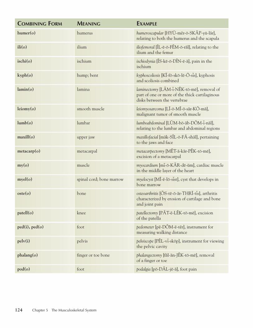
COMBINING FORM MEANING EXAMPLE
humer(o) humerus humeroscapular [HYU-m6r-o-SK0P-yu-lår], relating to both the humerus and the scapula
ili(o) ilium iliofemoral [2L-e-o-F1M-o-rål], relating to the ilium and the femur
ischi(o) ischium ischiodynia [2S-ke-o-D2N-e-å], pain in the ischium
kyph(o) hump; bent kyphoscoliosis [KI-fo-sko-le-O-s7s], kyphosis and scoliosis combined
lamin(o) lamina laminectomy [L0M-7-N1K-to-me], removal of part of one or more of the thick cartilaginous disks between the vertebrae
leiomy(o) smooth muscle leiomyosarcoma [LI-o-MI-o-sår-KO-må], malignant tumor of smooth muscle
lumb(o) lumbar lumboabdominal [L4M-bo-åb-D3M-7-nål], relating to the lumbar and abdominal regions
maxill(o) upper jaw maxillofacial [måk-S2L-o-FA-shål], pertaining to the jaws and face
metacarp(o) metacarpal metacarpectomy [M1T-å-kår-P1K-to-me], excision of a metacarpal
my(o) muscle myocardium [mi-o-K0R-de-9m], cardiac muscle in the middle layer of the heart
myel(o) spinal cord; bone marrow myelocyst [MI-6-lo-s7st], cyst that develops in bone marrow
oste(o) bone osteoarthritis [3S-te-o-år-THRI-t7s], arthritis characterized by erosion of cartilage and bone and joint pain
patell(o) knee patellectomy [P0T-6-L1K-to-me], excision of the patella
ped(i), ped(o) foot pedometer [p6-D3M-6-t6r], instrument for measuring walking distance
pelv(i) pelvis pelviscope [P1L-v7-skop], instrument for viewing the pelvic cavity
phalang(o) finger or toe bone phalangectomy [fål-ån-J1K-to-me], removal of a finger or toe
pod(o) foot podalgia [po-D0L-je-å], foot pain
thi74725_ch05_105-154.indd 124 10/17/08 2:51:11 PM
Chapter 5 The Musculoskeletal System 125
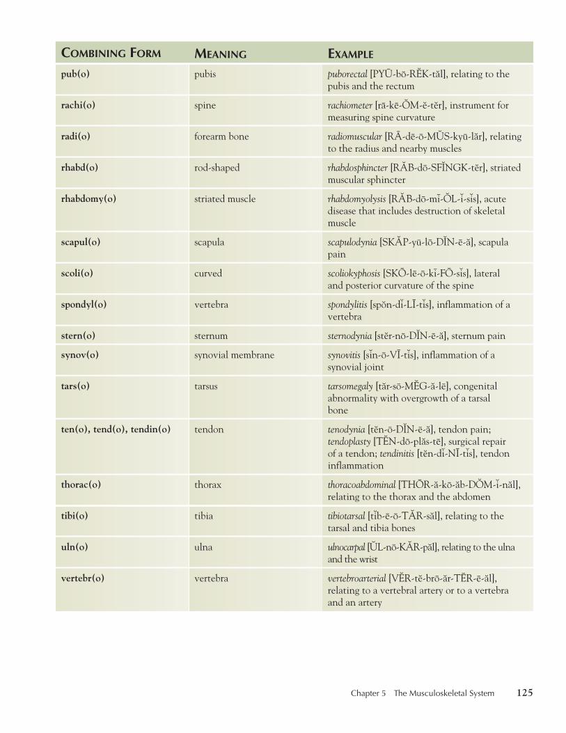
COMBINING FORM MEANING EXAMPLE
pub(o) pubis puborectal [PYU-bo-R1K-tål], relating to the pubis and the rectum
rachi(o) spine rachiometer [ra-ke-3M-6-t6r], instrument for measuring spine curvature
radi(o) forearm bone radiomuscular [RA-de-o-M4S-kyu-lår], relating to the radius and nearby muscles
rhabd(o) rod-shaped rhabdosphincter [R0B-do-SF2NGK-t6r], striated muscular sphincter
rhabdomy(o) striated muscle rhabdomyolysis [R0B-do-mi-3L-7-s7s], acute disease that includes destruction of skeletal muscle
scapul(o) scapula scapulodynia [SK0P-yu-lo-D2N-e-å], scapula pain
scoli(o) curved scoliokyphosis [SKO-le-o-ki-FO-s7s], lateral and posterior curvature of the spine
spondyl(o) vertebra spondylitis [sp8n-d7-LI-t7s], inflammation of a vertebra
stern(o) sternum sternodynia [st6r-no-D2N-e-å], sternum pain
synov(o) synovial membrane synovitis [s7n-o-VI-t7s], inflammation of a synovial joint
tars(o) tarsus tarsomegaly [tår-so-M1G-å-le], congenital abnormality with overgrowth of a tarsal bone
ten(o), tend(o), tendin(o) tendon tenodynia [t6n-o-D2N-e-å], tendon pain; tendoplasty [T1N-do-plås-te], surgical repair of a tendon; tendinitis [t6n-d7-NI-t7s], tendon inflammation
thorac(o) thorax thoracoabdominal [THOR-å-ko-åb-D3M-7-nål], relating to the thorax and the abdomen
tibi(o) tibia tibiotarsal [t7b-e-o-T0R-sål], relating to the tarsal and tibia bones
uln(o) ulna ulnocarpal [4L-no-K0R-pål], relating to the ulna and the wrist
vertebr(o) vertebra vertebroarterial [V1R-t6-bro-år-TER-e-ål], relating to a vertebral artery or to a vertebra and an artery
thi74725_ch05_105-154.indd 125 10/17/08 2:51:11 PM
126 Chapter 5 The Musculoskeletal System
C OMBINING F ORMS AND A BBREVIATIONS E XERCISES Build Your Medical Vocabulary Complete the words using combining forms listed in this chapter.
44. Joint pain: ____________ dynia
45. Plastic surgery of the skull: ____________ plasty
46. Of the upper jaw and its teeth: ____________ dental
47. Relating to the large area of the hip bone and the tibia: ____________ tibial
48. Operation on the instep of the foot: ____________ tomy
49. Relating to the head and chest: cephalo ____________
50. Production of fibrous tissue: ____________ plasia
51. Inflammation of the foot: ____________ itis
52. Instrument for measuring spine curvature: ____________ meter
53. Incision through the sternum: ____________ tomy
ABBREVIATION MEANING
A-K above the knee (amputation)
ASIS anterior superior iliac spine
B bilateral
B-K below the knee (amputation)
C1, C2, etc. first cervical vertebra, second cervical vertebra, etc.
Ca calcium
CTS carpal tunnel syndrome
D1, D2, etc. first dorsal vertebra, second dorsal vertebra, etc. (now referred to as first thoracic vertebra, second thoracic vertebra, etc.)
DJD degenerative joint disease
DTR deep tendon reflex
EMG electromyogram
Fx fracture
IM intramuscularly
ABBREVIATION MEANING
L left
L1, L2, etc. first lumbar vertebra, second lumbar vertebra, etc.
MCP metacarpophalangeal
NSAID nonsteroidal anti-inflammatory drug
OA osteoarthritis
P phosphorus
PIP proximal interphalangeal joints
PSIS posterior superior iliac spine
R right
RA rheumatoid arthritis
ROM range of motion
T1, T2, etc. first thoracic vertebra, second thoracic vertebra, etc.
TMJ temporomandibular joint
thi74725_ch05_105-154.indd 126 10/17/08 2:51:12 PM
Chapter 5 The Musculoskeletal System 127
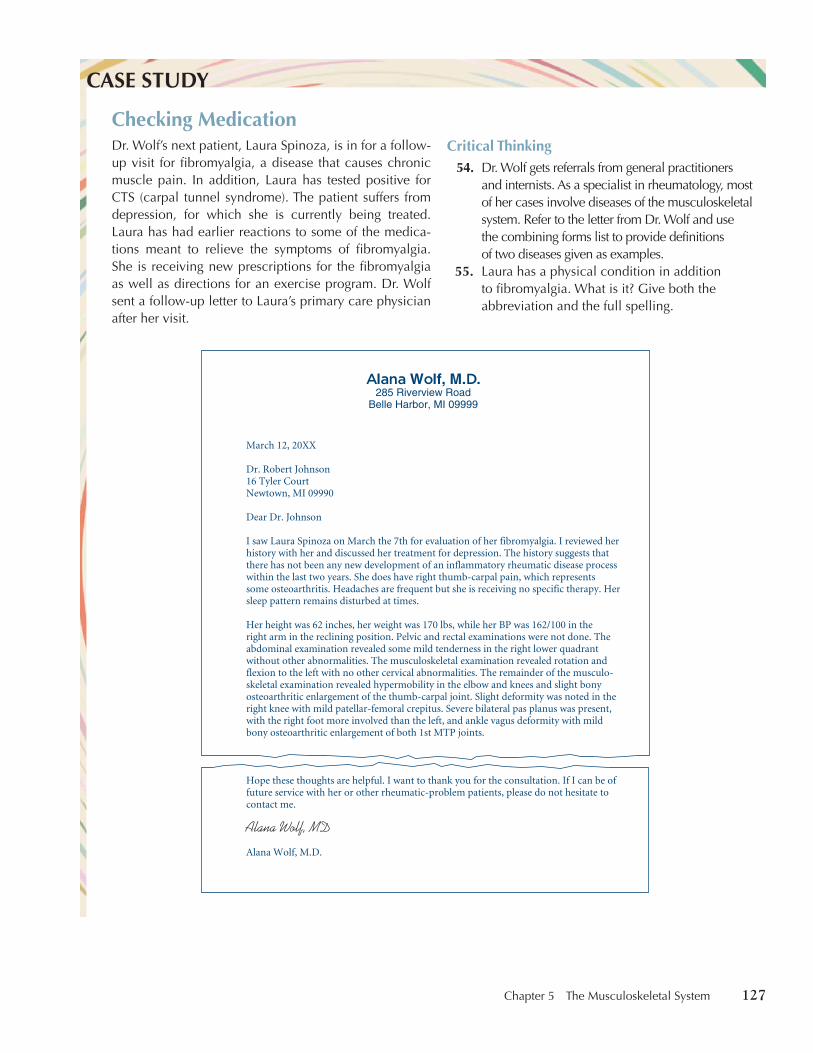
Dr. Wolf’s next patient, Laura Spinoza, is in for a follow-up visit for fibromyalgia, a disease that causes chronic muscle pain. In addition, Laura has tested positive for CTS (carpal tunnel syndrome). The patient suffers from depression, for which she is currently being treated. Laura has had earlier reactions to some of the medica-tions meant to relieve the symptoms of fibromyalgia. She is receiving new prescriptions for the fibromyalgia as well as directions for an exercise program. Dr. Wolf sent a follow-up letter to Laura’s primary care physician after her visit.
CASE STUDY
Checking MedicationCritical Thinking
54. Dr. Wolf gets referrals from general practitioners and internists. As a specialist in rheumatology, most of her cases involve diseases of the musculoskeletal system. Refer to the letter from Dr. Wolf and use the combining forms list to provide definitions of two diseases given as examples.
55. Laura has a physical condition in addition to fibromyalgia. What is it? Give both the abbreviation and the full spelling.
March 12, 20XX
Dr. Robert Johnson16 Tyler CourtNewtown, MI 09990
Dear Dr. Johnson
I saw Laura Spinoza on March the 7th for evaluation of her fibromyalgia. I reviewed her history with her and discussed her treatment for depression. The history suggests that there has not been any new development of an inflammatory rheumatic disease processwithin the last two years. She does have right thumb-carpal pain, which represents some osteoarthritis. Headaches are frequent but she is receiving no specific therapy. Her sleep pattern remains disturbed at times.
Her height was 62 inches, her weight was 170 lbs, while her BP was 162/100 in the right arm in the reclining position. Pelvic and rectal examinations were not done. Theabdominal examination revealed some mild tenderness in the right lower quadrant without other abnormalities. The musculoskeletal examination revealed rotation and flexion to the left with no other cervical abnormalities. The remainder of the musculo-skeletal examination revealed hypermobility in the elbow and knees and slight bony osteoarthritic enlargement of the thumb-carpal joint. Slight deformity was noted in the right knee with mild patellar-femoral crepitus. Severe bilateral pas planus was present, with the right foot more involved than the left, and ankle vagus deformity with mild bony osteoarthritic enlargement of both 1st MTP joints.
Hope these thoughts are helpful. I want to thank you for the consultation. If I can be of future service with her or other rheumatic-problem patients, please do not hesitate to contact me.
Alana Wolf, M.D.
Alana Wolf, M.D.285 Riverview Road
Belle Harbor, MI 09999
Alana Wolf, MD
thi74725_ch05_105-154.indd 127 10/17/08 2:51:12 PM

128 Chapter 5 The Musculoskeletal System
Find the Word Parts Give the term that fits the definition given below. Each term must contain at least one of the combining forms given in the previous section. You may refer to the Appendix of combining forms at the back of the book.
56. Joint pain ____________ .
57. Removal of a bursa ____________ .
58. Inflammation of cartilage ____________ .
59. Removal of a vertebra ____________ .
60. Bone-forming cell ____________ .
61. Abnormal bone hardening ____________ .
62. Plastic surgery on the neck ____________ .
63. Inflammation of the spinal cord ____________ .
64. Foot spasm ____________ .
65. Of the ulna and the carpus ____________ .
Find the misspelled word part. Write the corrected word part in the space with its definition.
66. sinovotomy ____________
67. myellogram ____________
68. arthrodunia ____________
69. ostiomyelitis ____________
70. takiometer ____________
Know the Word Parts Write the meaning of the following word parts in the space provided. As additional practice, use your dictionary to find at least two words for each word part listed below. Learn the meanings of each word you find.
71. arthr(o) _____________________________________________________________________________
72. ankyl(o) ____________________________________________________________________________
73. brachi(o) ___________________________________________________________________________
74. calcane(o) __________________________________________________________________________
75. cephal(o) ___________________________________________________________________________
76. cervic(o) ____________________________________________________________________________
77. chondr(o) ___________________________________________________________________________
78. cost(o) _____________________________________________________________________________
79. crani(o) ____________________________________________________________________________
80. fasci(o) _____________________________________________________________________________
81. kyph(o) _____________________________________________________________________________
82. my(o) ______________________________________________________________________________
83. myel(o) _____________________________________________________________________________
84. oste(o) _____________________________________________________________________________
85. patell(o) ____________________________________________________________________________
86. rachi(o) ____________________________________________________________________________
87. scoli(o) _____________________________________________________________________________
Diagnostic, Procedural, and Laboratory Terms
The musculoskeletal system is often the site of pain caused by conditions in the system itself or by symptoms of other systemic conditions. Specialists in
thi74725_ch05_105-154.indd 128 10/17/08 2:51:16 PM
Chapter 5 The Musculoskeletal System 129
the musculoskeletal system include orthopedists or orthopedic surgeons, physicians who treat disorders of the musculoskeletal system; osteopaths, physicians who combine manipulative procedures with conventional treat-ment; rheumatologists, physicians who treat disorders of the joints, spe-cifically, and of the musculoskeletal system generally; podiatrists, medical specialists who treat disorders of the foot; and chiropractors, health care professionals who manipulate the spine to treat certain ailments.
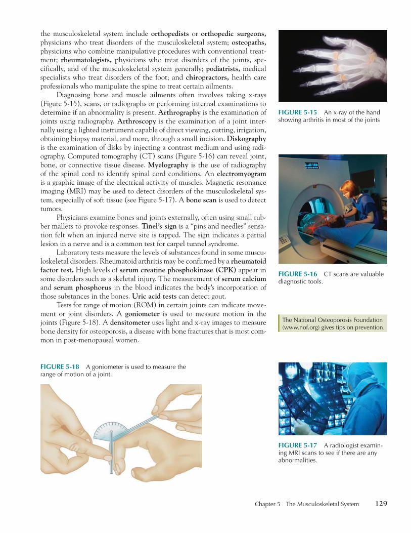
Diagnosing bone and muscle ailments often involves taking x-rays ( Figure 5-15 ), scans, or radiographs or performing internal examinations to determine if an abnormality is present. Arthrography is the examination of joints using radiography. Arthroscopy is the examination of a joint inter-nally using a lighted instrument capable of direct viewing, cutting, irrigation, obtaining biopsy material, and more, through a small incision. Diskography is the examination of disks by injecting a contrast medium and using radi-ography. Computed tomography (CT) scans ( Figure 5-16 ) can reveal joint, bone, or connective tissue disease. Myelography is the use of radiography of the spinal cord to identify spinal cord conditions. An electromyogram is a graphic image of the electrical activity of muscles. Magnetic resonance imaging (MRI) may be used to detect disorders of the musculoskeletal sys-tem, especially of soft tissue (see Figure 5-17 ). A bone scan is used to detect tumors.
Physicians examine bones and joints externally, often using small rub-ber mallets to provoke responses. Tinel’s sign is a “pins and needles” sensa-tion felt when an injured nerve site is tapped. The sign indicates a partial lesion in a nerve and is a common test for carpel tunnel syndrome.
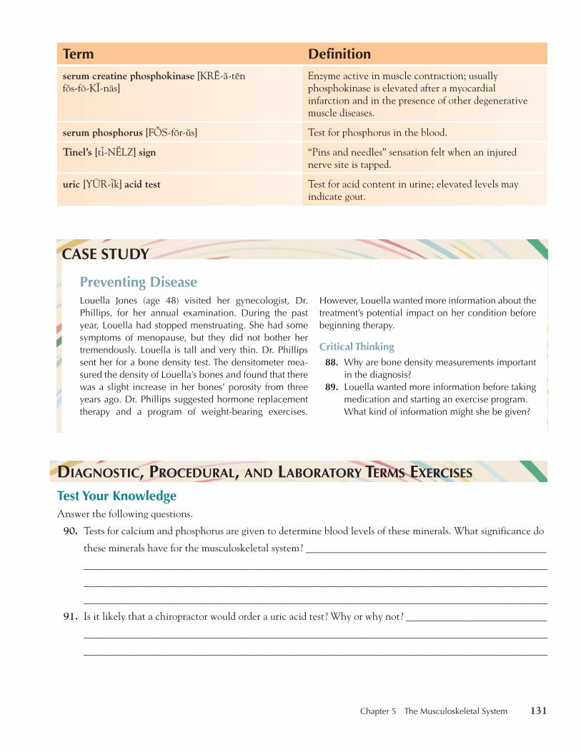
Laboratory tests measure the levels of substances found in some muscu-loskeletal disorders. Rheumatoid arthritis may be confirmed by a rheumatoid factor test. High levels of serum creatine phosphokinase (CPK) appear in some disorders such as a skeletal injury. The measurement of serum calcium and serum phosphorus in the blood indicates the body’s incorporation of those substances in the bones. Uric acid tests can detect gout.
Tests for range of motion (ROM) in certain joints can indicate move-ment or joint disorders. A goniometer is used to measure motion in the joints ( Figure 5-18 ). A densitometer uses light and x-ray images to measure bone density for osteoporosis, a disease with bone fractures that is most com-mon in post-menopausal women.
The National Osteoporosis Foundation ( www.nof.org ) gives tips on prevention. The National Osteoporosis Foundation ( www.nof.org ) gives tips on prevention.
FIGURE 5-18 A goniometer is used to measure the range of motion of a joint.
FIGURE 5-15 An x-ray of the hand showing arthritis in most of the joints
FIGURE 5-16 CT scans are valuable diagnostic tools.
FIGURE 5-17 A radiologist examin-ing MRI scans to see if there are any abnormalities.
thi74725_ch05_105-154.indd 129 10/17/08 2:51:16 PM

130 Chapter 5 The Musculoskeletal System
V OCABULARY R EVIEW In the previous section, you learned terms relating to diagnosis, clinical procedures, and laboratory tests. Before going on to the exercises, review the terms below and refer to the previous section if you have any questions. Pronunciations are provided for certain terms. Sometimes information about where the word came from is included after the term. These etymologies (word histories) are for your information only. You do not need to memorize them.
Term Definitionarthrography [år-THR3G-rå-fe] arthro-, joint � -graphy, process of recording
Radiography of a joint.
arthroscopy [år-THR3S-ko-pe] arthro-, joint � -scopy, a viewing with an instrument
Examination with an instrument that explores the interior of a joint.
bone scan Radiographic or nuclear medicine image of a bone.
chiropractor [ki-ro-PR0K-tor] chiro-, hand � Greek praktikos, efficient
Health care professional who works to align the spinal column so as to treat certain ailments.
densitometer [d6n-s7-T3M-6-t6r] Device that measures bone density using light and x-rays.
diskography [d7s-K3G-rå-fe] Radiographic image of an intervertebral disk by injection of a contrast medium into the center of the disk.
electromyogram [e-l6k-tro-MI-o-gråm] electro-, electrical � myo-, muscle � -gram, recording
A graphic image of muscular action using electrical currents.
goniometer [go-ne-3M-6-t6r] Greek gonia, angle � -meter, measuring device
Instrument that measures angles or range of motion in a joint.
myelography [MI-6-L3G-rå-f6] myelo-, spinal cord � -graphy, process of recording
Radiographic imaging of the spinal cord.
orthopedist [or-tho-PE-d7st], orthopedic [or-tho-PED-7k] surgeon ortho-, straight � Greek pais (paid-), child
Physician who examines, diagnoses, and treats disorders of the musculoskeletal system.
osteopath [3S-te-o-påth] osteo-, bone � -path(y), disease
Physician who combines manipulative treatment with conventional therapeutic measures.
podiatrist [po-DI-å-tr7st] Medical specialist who examines, diagnoses, and treats disorders of the foot.
rheumatoid factor test Test used to detect rheumatoid arthritis.
rheumatologist [ru-må-T3L-o-j7st] Physician who examines, diagnoses, and treats disorders of the joints and musculoskeletal system.
serum calcium [SER-9m K0L-si-9m] Test for calcium in the blood.
thi74725_ch05_105-154.indd 130 10/17/08 2:51:25 PM
Chapter 5 The Musculoskeletal System 131
Term Definitionserum creatine phosphokinase [KRE-å-ten f8s-fo-KI-nas]
Enzyme active in muscle contraction; usually phosphokinase is elevated after a myocardial infarction and in the presence of other degenerative muscle diseases.
serum phosphorus [F3S-for-9s] Test for phosphorus in the blood.
Tinel’s [t7-N1LZ] sign “Pins and needles” sensation felt when an injured nerve site is tapped.
uric [YUR-7k] acid test Test for acid content in urine; elevated levels may indicate gout.
Louella Jones (age 48) visited her gynecologist, Dr. Phillips, for her annual examination. During the past year, Louella had stopped menstruating. She had some symptoms of menopause, but they did not bother her tremendously. Louella is tall and very thin. Dr. Phillips sent her for a bone density test. The densitometer mea-sured the density of Louella’s bones and found that there was a slight increase in her bones’ porosity from three years ago. Dr. Phillips suggested hormone replacement therapy and a program of weight-bearing exercises.
CASE STUDY
Preventing DiseaseHowever, Louella wanted more information about the treatment’s potential impact on her condition before beginning therapy.
Critical Thinking88. Why are bone density measurements important
in the diagnosis?89. Louella wanted more information before taking
medication and starting an exercise program. What kind of information might she be given?
D IAGNOSTIC, P ROCEDURAL, AND L ABORATORY T ERMS E XERCISES Test Your Knowledge Answer the following questions.
90. Tests for calcium and phosphorus are given to determine blood levels of these minerals. What significance do
these minerals have for the musculoskeletal system? __________________________________________
_________________________________________________________________________________________
_________________________________________________________________________________________
_________________________________________________________________________________________
91. Is it likely that a chiropractor would order a uric acid test? Why or why not? ___________________________
_________________________________________________________________________________________
_________________________________________________________________________________________
thi74725_ch05_105-154.indd 131 10/17/08 2:51:26 PM

132 Chapter 5 The Musculoskeletal System
92. Would a bone scan be likely to show bone cancer? ________________________________________________
93. How is an osteopath like a chiropractor? ________________________________________________________
_________________________________________________________________________________________
94. What might a goniometer show about a muscle’s action? ___________________________________________
_________________________________________________________________________________________
True or False For each of the following statements, circle T for true or F for false.
95. A diskography is used to check bone density. T F
96. An electromyogram uses a contrast medium to check for range of motion in a joint. T F
97. A chiropractor can perform surgery. T F
98. A rheumatologist examines, diagnoses, and treats disorders of the joints and musculoskeletal system. T F
99. A podiatrist is a medical specialist who examines, diagnoses, and treats disorders of the foot. T F
Check Your Spelling For each of the following terms, place a C if the spelling is correct. If it is not, write the correct spelling in the space provided.
100. chiropractor _________________
101. densitiometer ________________
102. electromelogram ______________
103. rhuematoid __________________
104. goniometer __________________
105. orthepodist __________________
106. Tenil’s sign __________________
Pathological Terms
Musculoskeletal disorders arise from congenital conditions, injury, degener-ative disease, or other systemic disorders. Birth defects, such as spina bifida, affect the development of the spinal cord. Injuries to the spinal cord may produce paralysis. In some situations, surgery on the fetus while it is in utero can alleviate some of the effects of spina bifida. In such surgery, the abnor-mal spinal cord opening is repaired.
A herniated disk, in which the center of the disk is compressed and presses on nerves in the neural canal, can lead to sciatica, pain radiating down the leg from the lower back. Some diseases, such as rickets, which causes deformities in the legs, may result from a vitamin D deficiency.
Foot deformities may occur in or involve the ankle joint. Talipes calca-neus is a deformity of the heel due to weakened calf muscles; talipes valgus is eversion (a turning outward) of the foot; and talipes varus is inversion (a turning inward) of the foot. A calcar or spur is a bony projection growing out of a bone.
thi74725_ch05_105-154.indd 132 10/17/08 2:51:27 PM
Chapter 5 The Musculoskeletal System 133
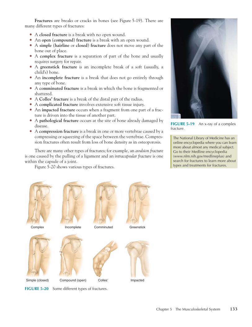
Fractures are breaks or cracks in bones (see Figure 5-19 ). There are many different types of fractures:
� A closed fracture is a break with no open wound. � An open ( compound ) fracture is a break with an open wound. � A simple ( hairline or closed ) fracture does not move any part of the
bone out of place. � A complex fracture is a separation of part of the bone and usually
requires surgery for repair. � A greenstick fracture is an incomplete break of a soft (usually, a
child’s) bone. � An incomplete fracture is a break that does not go entirely through
any type of bone. � A comminuted fracture is a break in which the bone is fragmented or
shattered. � A Colles’ fracture is a break of the distal part of the radius. � A complicated fracture involves extensive soft tissue injury. � An impacted fracture occurs when a fragment from one part of a frac-
ture is driven into the tissue of another part. � A pathological fracture occurs at the site of bone already damaged by
disease. � A compression fracture is a break in one or more vertebrae caused by a
compressing or squeezing of the space between the vertebrae. Compres-sion fractures often result from loss of bone density as in osteoporosis.
There are many other types of fractures; for example, an avulsion fracture is one caused by the pulling of a ligament and an intracapsular fracture is one within the capsule of a joint.
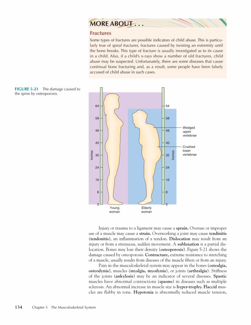
Figure 5-20 shows various types of fractures.
The National Library of Medicine has an online encyclopedia where you can learn more about almost any medical subject. Go to their Medline encyclopedia ( www.nlm.nih.gov/medlineplus ) and search for fractures to learn more about types and treatments for fractures.
The National Library of Medicine has an online encyclopedia where you can learn more about almost any medical subject. Go to their Medline encyclopedia ( www.nlm.nih.gov/medlineplus ) and search for fractures to learn more about types and treatments for fractures.
FIGURE 5-20 Some different types of fractures.
Colles' Impacted
Incomplete ComminutedComplex Greenstick
Simple (closed) Compound (open)
FIGURE 5-19 An x-ray of a complex fracture.
thi74725_ch05_105-154.indd 133 10/17/08 2:51:28 PM
134 Chapter 5 The Musculoskeletal System
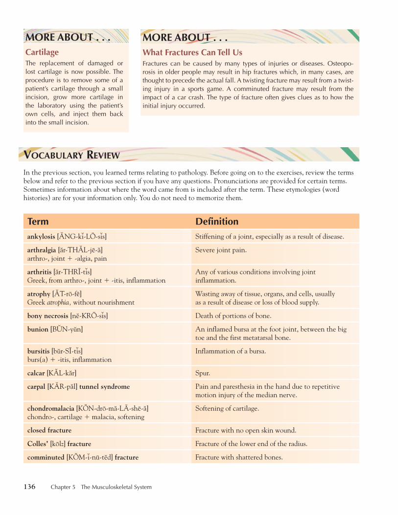
Injury or trauma to a ligament may cause a sprain. Overuse or improper use of a muscle may cause a strain. Overworking a joint may cause tendinitis ( tendonitis ), an inflammation of a tendon. Dislocation may result from an injury or from a strenuous, sudden movement. A subluxation is a partial dis-location. Bones may lose their density ( osteoporosis ). Figure 5-21 shows the damage caused by osteoporosis. Contracture, extreme resistance to stretching of a muscle, usually results from diseases of the muscle fibers or from an injury.
Pain in the musculoskeletal system may appear in the bones ( ostealgia, osteodynia ), muscles ( myalgia, myodynia ), or joints ( arthralgia ). Stiffness of the joints ( ankylosis ) may be an indicator of several diseases. Spasticmuscles have abnormal contractions ( spasms ) in diseases such as multiple sclerosis. An abnormal increase in muscle size is hyper-trophy. Flaccid mus-cles are flabby in tone. Hypotonia is abnormally reduced muscle tension,
MORE ABOUT . . .FracturesSome types of fractures are possible indicators of child abuse. This is parti cu-larly true of spiral fractures, fractures caused by twisting an extremity until the bone breaks. This type of fracture is usually investigated as to its cause in a child. Also, if a child’s x-rays show a number of old fractures, child abuse may be suspected. Unfortunately, there are some diseases that cause continual bone fracturing and, as a result, some people have been falsely accused of child abuse in such cases.
FIGURE 5-21 The damage caused to the spine by osteoporosis.
Youngwoman
Inch
es
0
8
16
24
32
40
48
58
64
Inch
es
0
8
16
24
32
40
48
58
64
Elderlywoman
Wedgeduppervertebrae
Crushedlowervertebrae
thi74725_ch05_105-154.indd 134 10/17/08 2:51:37 PM

Chapter 5 The Musculoskeletal System 135
and rigor (also called rigidity ) is abnormal muscle stiffness as seen in lock-jaw. Dystonia is abnormal tone (tension) in a muscle. A painfully long mus-cle contraction is tetany. Shaking ( tremors ) appears in a number of diseases such as Parkinson’s Disease. Some muscles atrophy (shrink) as a result of dis-use or specific diseases such as muscular dystrophy, a progressive, degenera-tive disorder affecting skeletal muscles. A muscle inflammation is myositis.
Some bone tissue dies ( bony necrosis, sequestrum ), often as a result of loss of blood supply. Abnormal bone growths may be capped with cartilage, as in exostosis. The bursa may become inflamed, causing bursitis. Inflam-mation of the bursa in the big toe causes a bunion. The epiphyses may also become inflamed, causing epiphysitis.
A common inflammation of the joints is arthritis ( Figure 5-22 ). Arthritis is a name for many different joint diseases, such as osteoarthritis or degen-erative arthritis (arthritis characterized by erosion of joint cartilage), rheu-matoid arthritis (a systemic disease affecting connective tissue), and gouty arthritis or gout (a disease characterized by joint pain, as in podagra, pain in the big toe). Certain types of arthritis may cause crepitation (also called crepitus ), noise made when affected surfaces rub together. Infections in the bone may cause osteomyelitis.
Cartilage may soften ( chondromalacia ) or become fragmented, as in a herniated disk. Disks may also slip or become misaligned with other verte-brae ( spondylolisthesis ) or become stiff ( spondylosis ). Various tumors may develop in the muscle, bone, bone marrow, and joints. Myeloma, myoma, leiomyoma, leiomyosarcoma, rhabdomyoma, rhabdomyosarcoma, osteoma, and osteosarcoma are types of musculoskeletal tumors.
Some abnormal posture conditions ( spinal curvature, kyphosis, lor-dosis, and scoliosis ) may cause pain (see Figure 5-23 ). Pain may even be felt in limbs that have been paralyzed or amputated. Phantom limb or phan-tom pain afflicts many who are paralyzed or are missing a limb. Repetitive motion of the hand may cause carpal tunnel syndrome, which is signaled by pain and paresthesia (numbness or tingling) of the hand. Chiropractors treat some spinal conditions by manipulation. Physical therapy is move-ment therapy to restore use of damaged areas of the body.
Go to the Arthritis Foundation’s Web site ( www.arthritis.org ) to learn about arthritis research.
Go to the Arthritis Foundation’s Web site ( www.arthritis.org ) to learn about arthritis research.
Carpal tunnel syndrome usually requires some rest period. For people who work on computers this may be difficult. There are alternative devices, such as the hands-free mouse (it uses head motion) available at www.ctsplace.com.
Carpal tunnel syndrome usually requires some rest period. For people who work on computers this may be difficult. There are alternative devices, such as the hands-free mouse (it uses head motion) available at www.ctsplace.com.
FIGURE 5-23 The three types of spi-nal curvature.
Scoliosis Kyphosis ("hunchback") Lordosis ("swayback")
FIGURE 5-22 An arthritic hand.
thi74725_ch05_105-154.indd 135 10/17/08 2:51:38 PM
136 Chapter 5 The Musculoskeletal System
MORE ABOUT . . .CartilageThe replacement of damaged or lost cartilage is now possible. The procedure is to remove some of a patient’s cartilage through a small incision, grow more cartilage in the laboratory using the patient’s own cells, and inject them back into the small incision.
MORE ABOUT . . .What Fractures Can Tell UsFractures can be caused by many types of injuries or diseases. Osteopo-rosis in older people may result in hip fractures which, in many cases, are thought to precede the actual fall. A twisting fracture may result from a twist-ing injury in a sports game. A comminuted fracture may result from the impact of a car crash. The type of fracture often gives clues as to how the initial injury occurred.
VOCABULARY REVIEW In the previous section, you learned terms relating to pathology. Before going on to the exercises, review the terms below and refer to the previous section if you have any questions. Pronunciations are provided for certain terms. Sometimes information about where the word came from is included after the term. These etymologies (word histories) are for your information only. You do not need to memorize them.
Term Definitionankylosis [0NG-k7-LO-s7s] Stiffening of a joint, especially as a result of disease.
arthralgia [år-TH0L-je-å] arthro-, joint � -algia, pain
Severe joint pain.
arthritis [år-THRI-t7s] Greek, from arthro-, joint � -itis, inflammation
Any of various conditions involving joint inflammation.
atrophy [0T-ro-fe] Greek atrophia, without nourishment
Wasting away of tissue, organs, and cells, usually as a result of disease or loss of blood supply.
bony necrosis [n6-KRO-s7s] Death of portions of bone.
bunion [B4N-y9n] An inflamed bursa at the foot joint, between the big toe and the first metatarsal bone.
bursitis [b9r-SI-t7s] burs(a) � -itis, inflammation
Inflammation of a bursa.
calcar [K0L-kår] Spur.
carpal [K0R-pål] tunnel syndrome Pain and paresthesia in the hand due to repetitive motion injury of the median nerve.
chondromalacia [K3N-dro-må-LA-she-å] chondro-, cartilage � malacia, softening
Softening of cartilage.
closed fracture Fracture with no open skin wound.
Colles’ [kolz] fracture Fracture of the lower end of the radius.
comminuted [K3M-7-nu-t6d] fracture Fracture with shattered bones.
thi74725_ch05_105-154.indd 136 10/17/08 2:51:46 PM
Chapter 5 The Musculoskeletal System 137
Term Definitioncomplex fracture Fracture with part of the bone displaced.
complicated fracture Fracture involving extensive soft tissue injury.
compound fracture Fracture with an open skin wound; open fracture.
compression fracture Fracture of one or more vertebrae caused by compressing of the space between the vertebrae.
contracture [k8n-TR0K-chur] Extreme resistance to the stretching of a muscle.
crepitation, crepitus [kr6p-7-TA-sh9n, KR1P-7-t9s] Noise made by rubbing together of bones.
degenerative arthritis Arthritis with erosion of the cartilage.
dislocation Movement of a joint out of its normal position as a result of an injury or sudden, strenuous movement.
dystonia [d7s-TO-ne-å] Abnormal tone in tissues.
epiphysitis [6-p7f-7-SI-t7s] Inflammation of the epiphysis.
exostosis [6ks-8s-TO-s7s] ex-, out of � ost(eo)-, bone � -osis, condition
Abnormal bone growth capped with cartilage.
flaccid [FL0K-s7d] Without tone; relaxed.
fracture [FR0K-ch9r] A break, especially in a bone.
gouty arthritis, gout [G3WT-e, g8wt] Inflammation of the joints, present in gout; usually caused by uric acid crystals.
greenstick fracture Fracture with twisting or bending of the bone but no breaking; usually occurs in children.
hairline fracture Fracture with no bone separation or fragmentation.
herniated [H1R-ne-a-t6d] disk Protrusion of an intervertebral disk into the neural canal.
hypertrophy [hi-P1R-tro-fe] hyper-, excessive � -trophy, growth
Abnormal increase as in muscle size.
hypotonia [HI-po-TO-ne-å] hypo-, subnormal � Greek tonos, tone
Abnormally reduced muscle tension.
impacted fracture Fracture in which a fragment from one part of the fracture is driven into the tissue of another part.
incomplete fracture Fracture that does not go entirely through a bone.
kyphosis [ki-FO-s7s] Abnormal posterior spine curvature.
leiomyoma [LI-o-mi-O-må] leio-, smooth � my(o)-, muscle � -oma, tumor
Benign tumor of smooth muscle.
leiomyosarcoma [LI-o-MI-o-sår-KO-må] leio-, smooth � myo-, muscle � sarcoma
Malignant tumor of smooth muscle.
thi74725_ch05_105-154.indd 137 10/17/08 2:51:47 PM
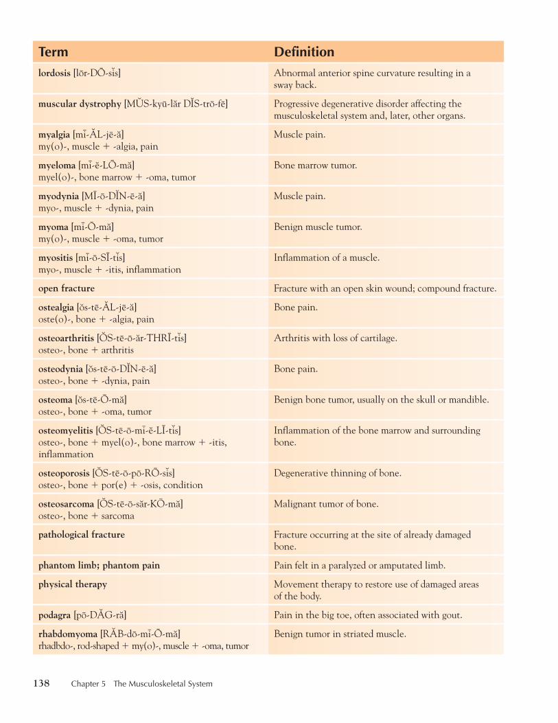
138 Chapter 5 The Musculoskeletal System
Term Definitionlordosis [lor-DO-s7s] Abnormal anterior spine curvature resulting in a
sway back.
muscular dystrophy [M4S-kyu-lår D2S-tro-fe] Progressive degenerative disorder affecting the musculoskeletal system and, later, other organs.
myalgia [mi-0L-je-å] my(o)-, muscle � -algia, pain
Muscle pain.
myeloma [mi-6-LO-må] myel(o)-, bone marrow � -oma, tumor
Bone marrow tumor.
myodynia [MI-o-D2N-e-å] myo-, muscle � -dynia, pain
Muscle pain.
myoma [mi-O-må] my(o)-, muscle � -oma, tumor
Benign muscle tumor.
myositis [mi-o-SI-t7s] myo-, muscle � -itis, inflammation
Inflammation of a muscle.
open fracture Fracture with an open skin wound; compound fracture.
ostealgia [8s-te-0L-je-å] oste(o)-, bone � -algia, pain
Bone pain.
osteoarthritis [3S-te-o-år-THRI-t7s] osteo-, bone � arthritis
Arthritis with loss of cartilage.
osteodynia [8s-te-o-D2N-e-å] osteo-, bone � -dynia, pain
Bone pain.
osteoma [8s-te-O-må] osteo-, bone � -oma, tumor
Benign bone tumor, usually on the skull or mandible.
osteomyelitis [3S-te-o-mi-6-LI-t7s] osteo-, bone � myel(o)-, bone marrow � -itis, inflammation
Inflammation of the bone marrow and surrounding bone.
osteoporosis [3S-te-o-po-RO-s7s] osteo-, bone � por(e) � -osis, condition
Degenerative thinning of bone.
osteosarcoma [3S-te-o-sår-KO-må] osteo-, bone � sarcoma
Malignant tumor of bone.
pathological fracture Fracture occurring at the site of already damaged bone.
phantom limb; phantom pain Pain felt in a paralyzed or amputated limb.
physical therapy Movement therapy to restore use of damaged areas of the body.
podagra [po-D0G-rå] Pain in the big toe, often associated with gout.
rhabdomyoma [R0B-do-mi-O-må] rhadbdo-, rod-shaped � my(o)-, muscle � -oma, tumor
Benign tumor in striated muscle.
thi74725_ch05_105-154.indd 138 10/17/08 2:51:47 PM
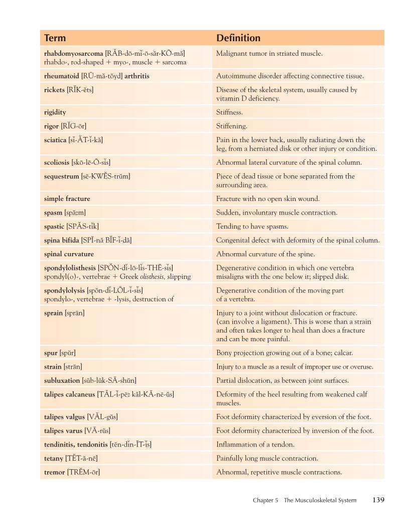
Chapter 5 The Musculoskeletal System 139
Term Definitionrhabdomyosarcoma [R0B-do-mi-o-sår-KO-må] rhabdo-, rod-shaped � myo-, muscle � sarcoma
Malignant tumor in striated muscle.
rheumatoid [RU-må-t8yd] arthritis Autoimmune disorder affecting connective tissue.
rickets [R2K-6ts] Disease of the skeletal system, usually caused by vitamin D deficiency.
rigidity Stiffness.
rigor [R2G-or] Stiffening.
sciatica [si-0T-7-kå] Pain in the lower back, usually radiating down the leg, from a herniated disk or other injury or condition.
scoliosis [sko-le-O-s7s] Abnormal lateral curvature of the spinal column.
sequestrum [se-KW1S-tr9m] Piece of dead tissue or bone separated from the surrounding area.
simple fracture Fracture with no open skin wound.
spasm [spåzm] Sudden, involuntary muscle contraction.
spastic [SP0S-t7k] Tending to have spasms.
spina bifida [SPI-nå B2F-7-då] Congenital defect with deformity of the spinal column.
spinal curvature Abnormal curvature of the spine.
spondylolisthesis [SP3N-d7-lo-l7s-THE-s7s] spondyl(o)-, vertebrae � Greek olisthesis, slipping
Degenerative condition in which one vertebra misaligns with the one below it; slipped disk.
spondylolysis [sp8n-d7-LOL-7-s7s] spondylo-, vertebrae � -lysis, destruction of
Degenerative condition of the moving part of a vertebra.
sprain [spran] Injury to a joint without dislocation or fracture. (can involve a ligament). This is worse than a strain and often takes longer to heal than does a fracture and can be more painful.
spur [sp9r] Bony projection growing out of a bone; calcar.
strain [stran] Injury to a muscle as a result of improper use or overuse.
subluxation [s9b-l9k-SA-sh9n] Partial dislocation, as between joint surfaces.
talipes calcaneus [T0L-7-pez kål-KA-ne-9s] Deformity of the heel resulting from weakened calf muscles.
talipes valgus [V0L-g9s] Foot deformity characterized by eversion of the foot.
talipes varus [VA-r9s] Foot deformity characterized by inversion of the foot.
tendinitis, tendonitis [t6n-d7n-IT-7s] Inflammation of a tendon.
tetany [T1T-å-ne] Painfully long muscle contraction.
tremor [TR1M-or] Abnormal, repetitive muscle contractions.
thi74725_ch05_105-154.indd 139 10/17/08 2:51:47 PM

140 Chapter 5 The Musculoskeletal System
Dr. Millet, a chiropractor, sees many patients for back pain. His treatments consist primarily of spinal manipu-lation, heat, and nutritional and exercise counseling. He currently sees a group of patients, mainly middle-aged men, who complain of sciatica. He has been able to relieve the pain for about 50 percent of them. The oth-ers seem to have more persistent pain. Dr. Millet is not allowed to prescribe medications because he is not a licensed medical doctor. He refers some of his patients
CASE STUDY
Making a Referralto Dr. Wolf, a specialist, who believes that Dr. Millet provides a valuable service.
Critical Thinking107. Chiropractic is one way for some people to
manage pain. Why might spinal manipulation help?
108. If spinal manipulation does not work, why should the patient see a medical specialist?
P ATHOLOGICAL T ERMS E XERCISES Build Your Medical Vocabulary Match the word roots on the left with the proper definition on the right.
109. ____ myo- a. bone
110. ____ myelo- b. hand
111. ____ rhabdo- c. rod-shaped
112. ____ osteo- d. joint
113. ____ arthro- e. bone marrow
114. ____ chiro- f. muscle
Know the Word Parts Match the following terms with the letter that gives the best definition.
115. ____ myeloma a. malignant tumor of smooth muscle
116. ____ myoma b. benign tumor in striated muscle
117. ____ leiomyoma c. benign tumor of smooth muscle
118. ____ leiomyosarcoma d. benign muscle tumor
119. ____ rhabdomyoma e. malignant bone tumor
120. ____ rhabdomyosarcoma f. bone marrow tumor
121. ____ osteoma g. malignant tumor in striated muscle
122. ____ osteosarcoma h. benign tumor, usually on the skull or mandible
Check Your Knowledge Complete the sentences below by filling in the blanks.
123. A patient with painful joints and bulges around the knuckles probably has ___________ .
124. Fractures that are most likely to occur in young children are called ___________ fractures.
thi74725_ch05_105-154.indd 140 10/17/08 2:51:48 PM

Chapter 5 The Musculoskeletal System 141
125. Osteoporosis is usually a disease found in ___________ women.
126. Playing tennis too vigorously may cause ___________ of the elbow.
127. Underworked muscles may become ___________ .
128. A muscle tumor is a(n) ___________ .
129. A slipped disk is called ___________ .
130. A compound fracture is a break accompanied by a(n) ___________ wound.
131. Arthritis is a general term for a number of ___________ diseases.
132. Paralysis may be caused by an injury to the ___________ .
133. A break in soft bone is a(n) ___________ fracture.
134. A strain is a(n) ___________ of the muscle, while a(n) ___________ is a torn or damaged ligament
or damaged muscle due to trauma or injury.
135. An injury or a strenuous, sudden movement of a joint may result in ___________ .
136. A partial dislocation is called a(n) ___________ .
137. Pain in the muscle is called ___________ or ___________ .
138. Pain in the bone is called ___________.
139. Pain in the joints is called ___________.
140. The suffix–desis means fixation or fusion, so the fixing of a joint so it does not move it is
called ___________ desis.
141. Hypertrophy is an increase in muscle ___________, while hypertonia is an increase in
muscle ___________ .
142. Abnormal muscle tone is called ___________ .
143. An infection in the bone is ___________ .
144. Repetitive motion of the hand may cause ___________ .
Know the Fractures Write the letter of the correct fracture description in the space provided.
145. ____ closed fracture a. break with shattered bones
146. ____ open fracture b. break that does not move the bone out of place
147. ____ simple fracture c. break with no open wound
148. ____ greenstick fracture d. break with an open wound
149. ____ comminuted fracture e. incomplete break of a soft bone
150. ____ impacted fracture f. break in a vertebrae caused by compression
151. ____ pathological fracture g. fragment from one part of the bone driven into the tissue of another part
152. ____ compression fracture h. break in bone due to disease (bone may be already diseased in that area)
thi74725_ch05_105-154.indd 141 10/17/08 2:51:49 PM
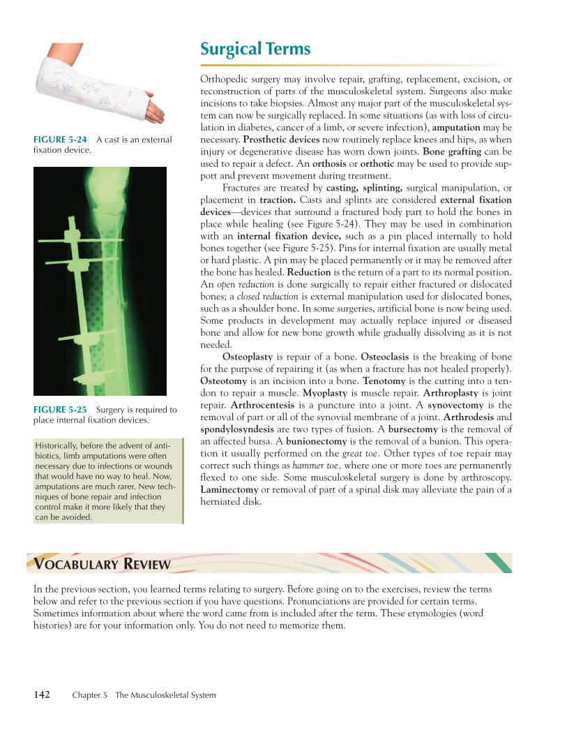
142 Chapter 5 The Musculoskeletal System
Surgical Terms
Orthopedic surgery may involve repair, grafting, replacement, excision, or reconstruction of parts of the musculoskeletal system. Surgeons also make incisions to take biopsies. Almost any major part of the musculoskeletal sys-tem can now be surgically replaced. In some situations (as with loss of circu-lation in diabetes, cancer of a limb, or severe infection), amputation may be necessary. Prosthetic devices now routinely replace knees and hips, as when injury or degenerative disease has worn down joints. Bone grafting can be used to repair a defect. An orthosis or orthotic may be used to provide sup-port and prevent movement during treatment.
Fractures are treated by casting, splinting, surgical manipulation, or placement in traction. Casts and splints are considered external fixation devices —devices that surround a fractured body part to hold the bones in place while healing (see Figure 5-24 ). They may be used in combination with an internal fixation device, such as a pin placed internally to hold bones together (see Figure 5-25 ). Pins for internal fixation are usually metal or hard plastic. A pin may be placed permanently or it may be removed after the bone has healed. Reduction is the return of a part to its normal position. An open reduction is done surgically to repair either fractured or dislocated bones; a closed reduction is external manipulation used for dislocated bones, such as a shoulder bone. In some surgeries, artificial bone is now being used. Some products in development may actually replace injured or diseased bone and allow for new bone growth while gradually dissolving as it is not needed.
Osteoplasty is repair of a bone. Osteoclasis is the breaking of bone for the purpose of repairing it (as when a fracture has not healed properly). Osteotomy is an incision into a bone. Tenotomy is the cutting into a ten-don to repair a muscle. Myoplasty is muscle repair. Arthroplasty is joint repair. Arthrocentesis is a puncture into a joint. A synovectomy is the removal of part or all of the synovial membrane of a joint. Arthrodesis and spondylosyndesis are two types of fusion. A bursectomy is the removal of an affected bursa. A bunionectomy is the removal of a bunion. This opera-tion it usually performed on the great toe. Other types of toe repair may correct such things as hammer toe, where one or more toes are permanently flexed to one side. Some musculoskeletal surgery is done by arthroscopy. Laminectomy or removal of part of a spinal disk may alleviate the pain of a herniated disk.
Historically, before the advent of anti-biotics, limb amputations were often necessary due to infections or wounds that would have no way to heal. Now, amputations are much rarer. New tech-niques of bone repair and infection control make it more likely that they can be avoided.
Historically, before the advent of anti-biotics, limb amputations were often necessary due to infections or wounds that would have no way to heal. Now, amputations are much rarer. New tech-niques of bone repair and infection control make it more likely that they can be avoided.
FIGURE 5-24 A cast is an external fixation device.
FIGURE 5-25 Surgery is required to place internal fixation devices.
V OCABULARY R EVIEW In the previous section, you learned terms relating to surgery. Before going on to the exercises, review the terms below and refer to the previous section if you have questions. Pronunciations are provided for certain terms. Sometimes information about where the word came from is included after the term. These etymologies (word histories) are for your information only. You do not need to memorize them.
thi74725_ch05_105-154.indd 142 10/17/08 2:51:49 PM

Chapter 5 The Musculoskeletal System 143
Term Definitionamputation [0M-pyu-TA-sh9n] Cutting off of a limb or part of a limb.
arthrocentesis [0R-thro-s6n-TE-s7s] arthro-, joint � Greek kentesis, puncture
Removal of fluid from a joint with use of a puncture needle.
arthrodesis [år-thro-DE-s7s] arthro-, � Greek desis, a binding
Surgical fusion of a joint to stiffen it.
arthroplasty [0R-thro-plås-te] arthro- � -plasty, repair Surgical replacement or repair of a joint.
bone grafting Transplantation of bone from one site to another.
bunionectomy [b9n-y9n-1K-to-me] bunion � -ectomy, removal
Removal of a bunion.
bursectomy [b9r-S1K-to-m6] burs(a) � -ectomy, removal
Removal of a bursa.
casting Forming of a cast in a mold; placing of fiberglass or plaster over a body part to prevent its movement.
external fixation device Device applied externally to hold a limb in place.
internal fixation device Device, such as a pin, inserted in bone to hold it in place.
laminectomy [L0M-7-N1K-to-me] lamin(a) � -ectomy, removal
Removal of part of an intervertebral disk.
myoplasty [MI-o-plås-te] myo-, muscle � -plasty, repair
Surgical repair of muscle tissue.
orthosis, orthotic [or-THO-s7s, or-TH3T-7k] External appliance used to immobilize or assist the movement of the spine or limbs.
osteoclasis [3S-te-3K-la-s7s] osteo-, bone � -clasis, breaking
Breaking of a bone in order to repair or reposition it.
osteoplasty [3S-te-o-plås-te] osteo-, bone � -plasty, repair
Surgical replacement or repair of bone.
osteotomy [8s-te-3T-o-me] osteo-, bone � -tomy, cutting
Cutting of bone.
prosthetic [pr8s-TH1T-7k] device Artificial device used as a substitute for a missing or diseased body part.
reduction Return of a part to its normal position.
splinting Applying a splint to immobilize a body part.
spondylosyndesis [SP3N-d7-lo-s7n-DE-s7s] spondylo-, vertebrae � Greek syndesis, a binding together
Fusion of two or more spinal vertebrae.
synovectomy [s7n-o-V1K-to-me] synovi(o)-, synovial fluid � -ectomy, removal
Removal of part or all of a joint’s synovial membrane.
tenotomy [t6-N3T-o-me] teno-, tendon � -tomy, cutting Surgical cutting of a tendon.
traction [TR0K-sh9n] Dragging or pulling or straightening of something, as a limb, by attachment of elastic or other devices.
thi74725_ch05_105-154.indd 143 10/17/08 2:52:00 PM

144 Chapter 5 The Musculoskeletal System
John Positano, a track star at a large university, suffered a knee injury during a meet. The team physician pre-scribed rest and medication first, to be followed by a gradual program of physical therapy. John missed about six weeks of meets and seemed fine until the end of the season, when a particularly strenuous run in which he twisted his knee left him writhing in pain. It was the same knee on which fluid had accumulated during the previous week. X-rays showed no fractures. Later, after examination by a specialist, arthroscopic surgery was recommended. John had to go through another
CASE STUDY
Musculoskeletal Injuryrehabilitative program (rest, medication, and physical therapy) after the surgery.
Critical Thinking153. A program of physical therapy was prescribed
for John. Which one of his tests was most important in determining whether or not he could exercise?
154. Is physical therapy always appropriate for a musculoskeletal injury?
S URGICAL T ERMS E XERCISES Build Your Medical Vocabulary Form two surgical words for each of the following word roots by adding suffixes learned in Chapter 2.
155. osteo- __________
156. arthro-__________
157. myo- ___________
158. spondylo- _______
Find a Match Match the terms in the second column to the terms in the first.
159. ________ amputation a. replacement device
160. ________ prosthesis b. molding
161. ________ orthosis, orthotic c. muscle repair
162. ________ traction d. bone cutting
163. ________ casting e. limb removal
164. ________ splinting f. bone repair
165. ________ myoplasty g. external supporting or immobilizing device
166. ________ osteoplasty h. wrapping to immobilize
167. ________ osteotomy i. pulling to straighten
168. ________ arthroplasty j. joint repair
thi74725_ch05_105-154.indd 144 10/17/08 2:52:00 PM

Chapter 5 The Musculoskeletal System 145
Understanding Surgical Procedures Explain the following surgical terms in simple words.
169. reduction (of a bone) _______________________________________________________________________
170. synovectomy ______________________________________________________________________________
171. arthrodesis ________________________________________________________________________________
172. bunionectomy _____________________________________________________________________________
173. laminectomy ______________________________________________________________________________
174. orthotic ___________________________________________________________________________________
175. arthrocentesis _____________________________________________________________________________
Pharmacological Terms
Most medications for treatment of the musculoskeletal system treat symp-toms, not causes. Pain medications, such as analgesics, narcotics, anti-inflammatories (corticosteroids), muscle relaxants, and nonsteroidal anti-inflammatory drugs (NSAIDs), all relieve or relax the area of pain either by numbing the area or by reducing the inflammation. Table 5-1 shows some common medications.
TABLE 5-1 Some Medications for the Musculoskeletal System
Drug Class Purpose Generic Trade Name
analgesic to relieve pain aspirinacetaminophen (NSAIDs are also analgesics.)
Bayer, Excedrin, and variousTylenol and various
anti-inflammatory (corticosteroids)
to reduce inflammation prednisone (Aspirin and NSAIDs also reduce inflammation.)
Deltasone, Orasone, Cortan
muscle relaxant to relieve stiffness carisoprodolcyclobenzaprinemethocarbamol
SomaFlexerilDelaxin, Robaxin
NSAIDs to reduce inflammation ibuprofen
naproxenketorolac, tromethaminenabutemone
Advil, Motrin, NuprinNaproxynToradol (IV)Relafen
V OCABULARY R EVIEW In the previous section, you learned terms relating to pharmacology. Before going on to the exercises, review the terms below and refer to the previous section if you have questions. Pronunciations are provided for certain terms.
thi74725_ch05_105-154.indd 145 10/17/08 2:52:01 PM
146 Chapter 5 The Musculoskeletal System
Sometimes information about where the word came from is included after the term. These etymologies (word histories) are for your information only. You do not need to memorize them.
Term Definition
analgesic [ån-ål-JE-z7k] Agent that relieves pain.
anti-inflammatory (corticosteroid) Agent that reduces inflammation.
muscle relaxant Agent that relieves muscle stiffness.
narcotic Agent that relieves pain by affecting the body in ways that are similar to opium.
nonsteroidal [n8n-ST1R-8y-dål] anti-inflammatory drug (NSAID)
Agent that reduces inflammation without the use of steroids.
In her follow-up letter on Laura Spinoza’s visit, Dr. Wolf listed a number of medications to treat the symptoms of fibromyalgia. Part of the difficulty in treating musculosk-eletal disorders is that many of the diseases are degen-erative, and damage cannot be reversed. Some of these diseases, such as muscular dystrophy, currently have no cure. Many forms of arthritis are degenerative and, short of replacing joints, cannot be improved significantly. Alleviating the pain is the only available course of treat-ment in many instances.
Critical Thinking176. Narcotics can be addictive. The long-term use of
steroids can cause other health problems. What
CASE STUDY
Treating the Symptomsdoes Dr. Wolf prescribe to avoid these two problems?
177. Many athletes use anabolic steroids illegally for strength and endurance building. (Corticosteroids are not used for this purpose.) Anabolic-steroid use can cause heart damage and many other serious health problems. What are some ways to increase strength and endurance without the use of dangerous drugs?
P HARMACOLOGICAL T ERMS E XERCISES Fill in the Blank Choose one or more of the following terms to fill in each blank. Each term may be used more than once.
analgesic anti-inflammatory antibiotic
178. Treatment for bursitis _______________________________________________________________________ .
179. Treatment for myalgia ______________________________________________________________________ .
180. Treatment for bone infection ________________________________________________________________ .
thi74725_ch05_105-154.indd 146 10/17/08 2:52:02 PM
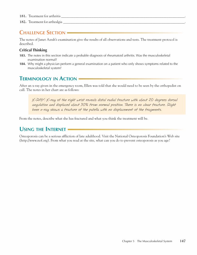
Chapter 5 The Musculoskeletal System 147
181. Treatment for arthritis _____________________________________________________________________ .
182. Treatment for arthralgia ____________________________________________________________________ .
C HALLENGE S ECTION The notes of Janet Azrah’s examination give the results of all observations and tests. The treatment protocol is described.
Critical Thinking 183. The notes in this section indicate a probable diagnosis of rheumatoid arthritis. Was the musculoskeletal
examination normal? 184. Why might a physician perform a general examination on a patient who only shows symptoms related to the
musculoskeletal system?
T ERMINOLOGY IN A CTION After an x-ray given in the emergency room, Ellen was told that she would need to be seen by the orthopedist on call. The notes in her chart are as follows:
X-RAY: X-ray of the right wrist reveals distal radial fracture with about 20 degrees dorsal angulation and displaced about 30% from normal position. There is no ulnar fracture. Right knee x-ray shows a fracture of the patella with no displacement of the fragments.
From the notes, describe what she has fractured and what you think the treatment will be.
U SING THE I NTERNET Osteoporosis can be a serious affliction of late adulthood. Visit the National Osteoporosis Foundation’s Web site ( http://www.nof.org ). From what you read at the site, what can you do to prevent osteoporosis as you age?
thi74725_ch05_105-154.indd 147 10/17/08 2:52:03 PM
148 Chapter 5 The Musculoskeletal System
CHAPTER REVIEW The material that follows is to help you review all the material in this chapter.
Explain the Terms Write out the following sentences in lay terms.
185. The pt had a Fx of L1.
186. The ROM was decreased in the right shoulder due to myalgia.
187. The pt was placed on an NSAID due to OA.
188. The pt has CTS, it is B.
189. The pt has severe RA, which has caused arthrodynia and hypertonic muscles in the R leg.
190. On review of the medical history, the pt has TMJ, CTS, has had a Fx of the R wrist, has some DJD.
191. On examination it was found the DTR of the R leg was decreased. The ROM was also decreased on the R
side of the body. The muscles in the R leg were flaccid and hypotonic.
Know the Medical Terms Rewrite the following sentences to include proper medical terminology and abbreviations.
192. The patient came in today for a test that uses electricity to check muscle activity.
193. The pt will have a below the knee amputation on his right leg due to severe frostbite.
194. The child’s break needs to be set.
True or False For each of the following statements, circle T for true or F for false.
195. The clavicle is the posterior shoulder bone. T F
196. The femur is the upper arm bone. T F
197. The tibia is a flat bone on the front of the leg. T F
198. The sternum is also known as the breastbone. T F
199. The coccyx is also known as the tailbone. T F
200. The cervical vertebrae attach to ribs. T F
201. The false ribs do not attach to the sternum. T F
202. A tight muscle could be considered hypotonic. T F
203. A massage therapist would help someone with subluxations. T F
204. A chiropractor works only on the spine. T F
205. The radius is a bone in the leg. T F
206. The patella is another name for kneecap. T F
207. A fracture is considered a break in the continuity of the bone. T F
208. There are many types of fractures. T F
209. A fracture always goes completely through the bone. T F
thi74725_ch05_105-154.indd 148 10/17/08 2:52:04 PM

Chapter 5 The Musculoskeletal System 149
210. acetabul(o) 211. acetabulum [ås-6-T0B-yu-
l9m] {acetábulo} 212. acromi(o) 213. acromion [å-KRO-me-on]
{acromion} 214. amphiarthrosis [0M-f7-år-
THRO-s7s] {anfiartrosis} 215. amputation [0M-pyu-TA-
sh9n] {amputación} 216. analgesic [ån-ål-JE-z7k] 217. ankle [0NG-kl] {tobillo} 218. ankyl(o) 219. ankylosis [0NG-k7-LO-s7s]
{anquilosis} 220. anti-inflammatory 221. arthr(o) 222. arthralgia [år-THR0L-je-å]
{artralgia} 223. arthritis [år-THR2-t7s] {artritis} 224. arthrocentesis [0R-thro-s6n-
TE-s7s] {artrocentesis} 225. arthrodesis [år-thro-DE-s7s] 226. arthrography
[år-THR3G-rå-fe] 227. arthroplasty [0R-thro-plås-te] 228. arthroscopy
[år-THROS-k8-pe] 229. articular [år-T2K-yu-lår]
cartilage 230. articulation [år-t7k-yu-LA-
sh9n] {articulación} 231. atlas [0T-lås] {atlas} 232. atrophy [0T-ro-fe] {atrofia} 234. axis [0K-s7s] {axis} 235. bone {hueso} 236. bone grafting 237. bone head
238. bone phagocyte [F0G-o-sit] 239. bone scan 240. bony necrosis [n6-KRO-s7s] 241. brachi(o) 242. bunion [B4N-y9n] {bunio} 243. bunionectomy [b9n-y9n-1K-
to-me] {bunionectomía} 244. burs(o) 245. bursa (pl., bursae) [B4R-så
(B4R-se)] {bursa} 246. bursectomy [b9r-S1K-to-me]
{bursectomía} 247. bursitis [b9r-SI-t7s] {bursitis} 248. calcane(o) 249. calcaneus [kål-KA-ne-9s]
{calcáneo} 250. calcar [K0L-kår] {calcar} 251. calci(o) 252. calcium [K0L-se-9m] {calcio} 253. cancellous [K0N-s6-l9s] {can-
celoso} bone 254. cardiac [K0R-de-åk] muscle 255. carp(o) 256. carpal [K0R-pål] tunnel
syndrome 257. carpus [K0R-p9s], carpal bone 258. cartilage [K0R-t7-låj]
{cartílago} 259. cartilaginous [kår-t7-L0J-7-
n9s] disk 260. casting {colado} 261. cephal(o) 262. cervic(o) 263. cervical [S1R-v7-kål]
vertebrae 264. chiropractor [ki-ro-PR0K-t6r]
{quiropráctico} 265. chondr(o)
266. chondromalacia [K3N-dro-må-LA-she-å] {condromalacia}
267. clavicle [KL0V-7-kl] {clavicula}
268. closed fracture 269. coccyx [K3K-s7ks] {cóccix} 270. Colles’ [kolz] fracture 271. comminuted [K3M-7-nu-t6d]
fracture 272. compact bone 273. complex fracture 274. complicated fracture 275. compound fracture 276. compression fracture 277. condyl(o) 278. condyle [K3N-dil] 279. contracture [k8n-TR0K-ch9r] 280. corticosteroid 281. cost(o) 282. crani(o) 283. crepitation [kr6-p7-TA-sh9n],
crepitus [KR1P-7-t9s] 284. crest {cresta} 285. dactyl(o) 286. degenerative arthritis 287. densitometer
[d6n-s7-T3M-6-t6r] 288. diaphysis [di-0F-7-s7s]
{diáfisis} 289. diarthroses [di-år-THRO-sez] 290. disk [d7sk] {disco} 291. diskography [d7s-K3G-rå-fe]
{discografía} 292. dislocation {dislocación} 293. dorsal vertebrae 294. dystonia [d7s-TO-ne-å]
{distonia} 295. elbow [1L-bo] {codo}
WORD
D EFINITIONS Define the following terms and combining forms. Review the chapter before starting. Make sure you know how to pronounce each term as you define it. The blue words in curly brackets are references to the Spanish glossary the student Web site ( www.mhhe.com/medterm3e ).
thi74725_ch05_105-154.indd 149 10/17/08 2:52:04 PM
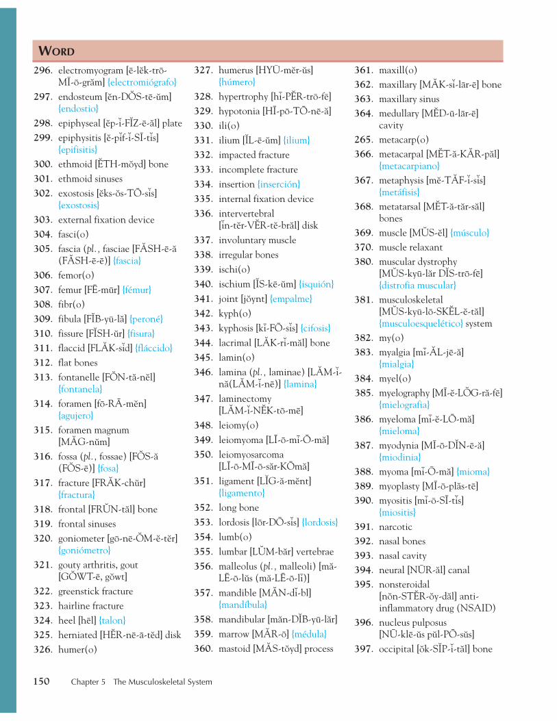
150 Chapter 5 The Musculoskeletal System
296. electromyogram [e-l6k-tro-MI-o-gråm] {electromiógrafo}
297. endosteum [6n-D3S-te-9m] {endostio}
298. epiphyseal [6p-7-F2Z-e-ål] plate 299. epiphysitis [6-p7f-7-SI-t7s]
{epifisitis} 300. ethmoid [1TH-m8yd] bone 301. ethmoid sinuses 302. exostosis [6ks-8s-TO-s7s]
{exostosis} 303. external fixation device 304. fasci(o) 305. fascia (pl., fasciae [F0SH-e-å
(F0SH-e-e)] {fascia} 306. femor(o) 307. femur [FE-mur] {fémur} 308. fibr(o) 309. fibula [F2B-yu-lå] {peroné} 310. fissure [F2SH-9r] {fisura} 311. flaccid [FL0K-s7d] {fláccido} 312. flat bones 313. fontanelle [F3N-tå-n6l]
{fontanela} 314. foramen [fo-RA-m6n]
{agujero} 315. foramen magnum
[M0G-n9m] 316. fossa (pl., fossae) [F3S-å
(F3S-e)] {fosa} 317. fracture [FR0K-ch9r]
{fractura} 318. frontal [FR4N-tål] bone 319. frontal sinuses 320. goniometer [go-ne-3M-6-t6r]
{goniómetro} 321. gouty arthritis, gout
[G3WT-e, g8wt] 322. greenstick fracture 323. hairline fracture 324. heel [hel] {talon} 325. herniated [H1R-ne-a-t6d] disk 326. humer(o)
327. humerus [HYU-m6r-9s] {húmero}
328. hypertrophy [hi-P1R-tro-fe] 329. hypotonia [HI-po-TO-ne-å] 330. ili(o) 331. ilium [2L-e-9m] {ilium} 332. impacted fracture 333. incomplete fracture 334. insertion {inserción} 335. internal fixation device 336. intervertebral
[7n-t6r-V1R-t6-brål] disk 337. involuntary muscle 338. irregular bones 339. ischi(o) 340. ischium [2S-ke-9m] {isquión} 341. joint [j8ynt] {empalme} 342. kyph(o) 343. kyphosis [ki-FO-s7s] {cifosis} 344. lacrimal [L0K-r7-mål] bone 345. lamin(o) 346. lamina (pl., laminae) [L0M-7-
nå(L0M-7-ne)] {lamina} 347. laminectomy
[L0M-7-N1K-to-me] 348. leiomy(o) 349. leiomyoma [LI-o-mi-O-må] 350. leiomyosarcoma
[LI-o-MI-o-sår-KOmå] 351. ligament [L2G-å-m6nt]
{ligamento} 352. long bone 353. lordosis [lor-DO-s7s] {lordosis} 354. lumb(o) 355. lumbar [L4M-bår] vertebrae 356. malleolus (pl., malleoli) [må-
LE-o-l9s (må-LE-o-li)] 357. mandible [M0N-d7-bl]
{mandíbula} 358. mandibular [mån-D2B-yu-lår] 359. marrow [M0R-o] {médula} 360. mastoid [M0S-t8yd] process
361. maxill(o) 362. maxillary [M0K-s7-lar-e] bone 363. maxillary sinus 364. medullary [M1D-u-lar-e]
cavity 265. metacarp(o) 366. metacarpal [M1T-å-K0R-pål]
{metacarpiano} 367. metaphysis [m6-T0F-7-s7s]
{metáfisis} 368. metatarsal [M1T-å-tår-sål]
bones 369. muscle [M4S-6l] {músculo} 370. muscle relaxant 380. muscular dystrophy
[M4S-kyu-lår D2S-tro-fe] {distrofia muscular}
381. musculoskeletal [M4S-kyu-lo-SK1L-6-tål] {musculoesquelético} system
382. my(o) 383. myalgia [mi-0L-je-å]
{mialgia} 384. myel(o) 385. myelography [MI-6-L3G-rå-fe]
{mielografia} 386. myeloma [mi-6-LO-må]
{mieloma} 387. myodynia [MI-o-D2N-e-å]
{miodinia} 388. myoma [mi-O-må] {mioma} 389. myoplasty [MI-o-plås-te] 390. myositis [mi-o-SI-t7s]
{miositis} 391. narcotic 392. nasal bones 393. nasal cavity 394. neural [NUR-ål] canal 395. nonsteroidal
[n8n-ST1R-8y-dål] anti-inflammatory drug (NSAID)
396. nucleus pulposus [NU-kle-9s p9l-PO-s9s]
397. occipital [8k-S2P-7-tål] bone
WORD
thi74725_ch05_105-154.indd 150 10/17/08 2:52:05 PM
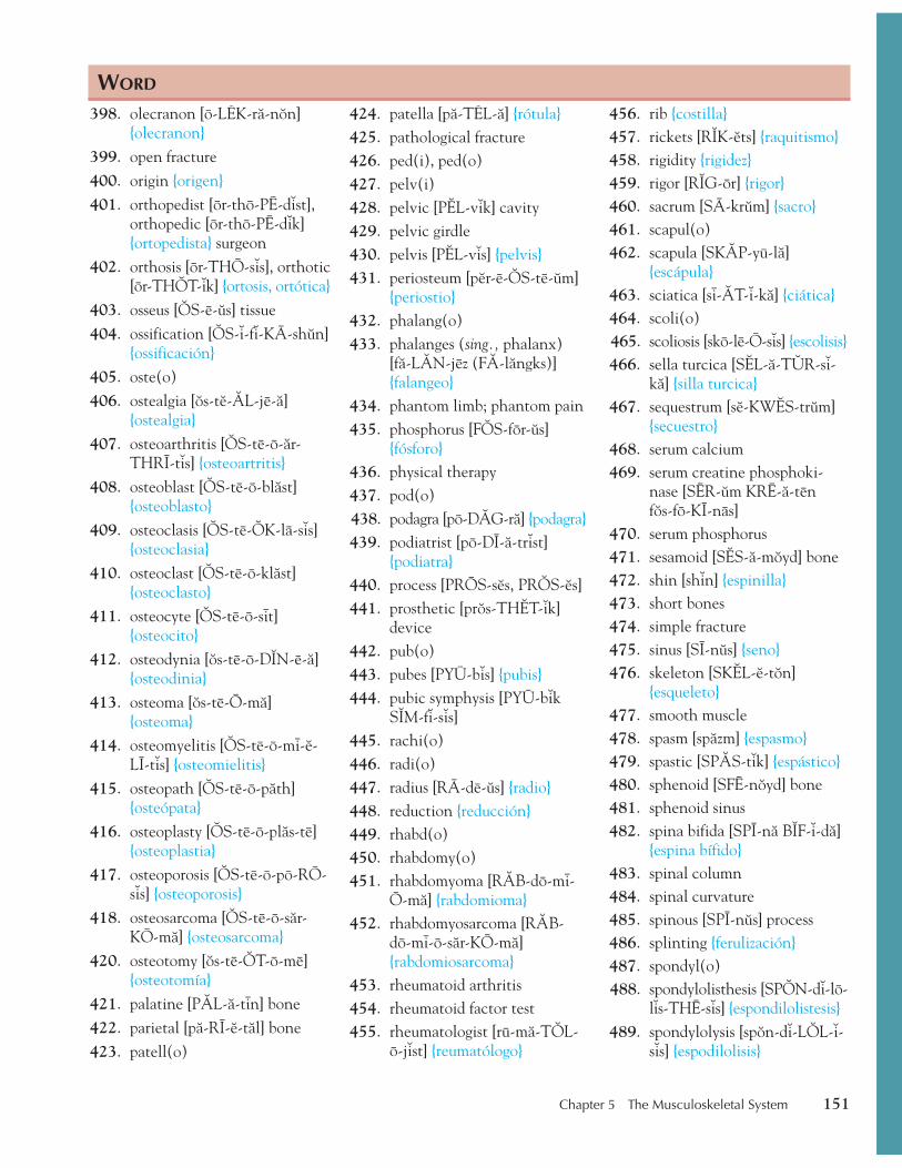
Chapter 5 The Musculoskeletal System 151
398. olecranon [o-L1K-rå-n8n] {olecranon}
399. open fracture 400. origin {origen} 401. orthopedist [or-tho-PE-d7st],
orthopedic [or-tho-PE-d7k] {ortopedista} surgeon
402. orthosis [or-THO-s7s], orthotic [or-TH3T-7k] {ortosis, ortótica}
403. osseus [3S-e-9s] tissue 404. ossification [3S-7-f7-KA-sh9n]
{ossificación} 405. oste(o) 406. ostealgia [8s-t6-0L-je-å]
{ostealgia} 407. osteoarthritis [3S-te-o-år-
THRI-t7s] {osteoartritis} 408. osteoblast [3S-te-o-blåst]
{osteoblasto} 409. osteoclasis [3S-te-3K-la-s7s]
{osteoclasia} 410. osteoclast [3S-te-o-klåst]
{osteoclasto} 411. osteocyte [3S-te-o-sit]
{osteocito} 412. osteodynia [8s-te-o-D2N-e-å]
{osteodinia} 413. osteoma [8s-te-O-må]
{osteoma} 414. osteomyelitis [3S-te-o-mi-6-
LI-t7s] {osteomielitis} 415. osteopath [3S-te-o-påth]
{osteópata} 416. osteoplasty [3S-te-o-plås-te]
{osteoplastia} 417. osteoporosis [3S-te-o-po-RO-
s7s] {osteoporosis} 418. osteosarcoma [3S-te-o-sår-
KO-må] {osteosarcoma} 420. osteotomy [8s-te-3T-o-me]
{osteotomía} 421. palatine [P0L-å-tin] bone 422. parietal [på-RI-6-tål] bone 423. patell(o)
424. patella [på-T1L-å] {rótula} 425. pathological fracture 426. ped(i), ped(o) 427. pelv(i) 428. pelvic [P1L-v7k] cavity 429. pelvic girdle 430. pelvis [P1L-v7s] {pelvis} 431. periosteum [p6r-e-3S-te-9m]
{periostio} 432. phalang(o) 433. phalanges (sing., phalanx)
[få-L0N-jez (F0-långks)] {falangeo}
434. phantom limb; phantom pain 435. phosphorus [F3S-for-9s]
{fósforo} 436. physical therapy 437. pod(o) 438. podagra [po-D0G-rå] {podagra} 439. podiatrist [po-DI-å-tr7st]
{podiatra} 440. process [PROS-s6s, PR3S-6s] 441. prosthetic [pr8s-TH1T-7k]
device 442. pub(o) 443. pubes [PYU-b7s] {pubis} 444. pubic symphysis [PYU-b7k
S2M-f7-s7s] 445. rachi(o) 446. radi(o) 447. radius [RA-de-9s] {radio} 448. reduction {reducción} 449. rhabd(o) 450. rhabdomy(o) 451. rhabdomyoma [R0B-do-mi-
O-må] {rabdomioma} 452. rhabdomyosarcoma [R0B-
do-mi-o-sår-KO-må] {rabdomiosarcoma}
453. rheumatoid arthritis 454. rheumatoid factor test 455. rheumatologist [ru-må-T3L-
o-j7st] {reumatólogo}
456. rib {costilla} 457. rickets [R2K-6ts] {raquitismo} 458. rigidity {rigidez} 459. rigor [R2G-or] {rigor} 460. sacrum [SA-kr9m] {sacro} 461. scapul(o) 462. scapula [SK0P-yu-lå]
{escápula} 463. sciatica [si-0T-7-kå] {ciática} 464. scoli(o) 465. scoliosis [sko-le-O-s7s] {escolisis} 466. sella turcica [S1L-å-T4R-s7-
kå] {silla turcica} 467. sequestrum [s6-KW1S-tr9m]
{secuestro} 468. serum calcium 469. serum creatine phosphoki-
nase [SER-9m KRE-å-ten f8s-fo-KI-nas]
470. serum phosphorus 471. sesamoid [S1S-å-m8yd] bone 472. shin [sh7n] {espinilla} 473. short bones 474. simple fracture 475. sinus [SI-n9s] {seno} 476. skeleton [SK1L-6-t8n]
{esqueleto} 477. smooth muscle 478. spasm [spåzm] {espasmo} 479. spastic [SP0S-t7k] {espástico} 480. sphenoid [SFE-n8yd] bone 481. sphenoid sinus 482. spina bifida [SPI-nå B2F-7-då]
{espina bífido} 483. spinal column 484. spinal curvature 485. spinous [SPI-n9s] process 486. splinting {ferulización} 487. spondyl(o) 488. spondylolisthesis [SP3N-d7-lo-
l7s-THE-s7s] {espondilolistesis} 489. spondylolysis [sp8n-d7-L3L-7-
s7s] {espodilolisis}
WORD
thi74725_ch05_105-154.indd 151 10/17/08 2:52:05 PM

152 Chapter 5 The Musculoskeletal System
546. A-K
547. ASIS
548. B-K
549. C1, C2, etc.
550. Ca
551. CTS
552. D1, D2, etc.
553. DJD
554. DTR
555. EMG
556. Fx
557. IM
558. L
559. L1, L2, etc.
560. MCP
561. NSAID
562. OA
563. P
564. PIP
565. PSISRRA
566. ROM
567. T1, T2, etc.
568. TMJ
ABBREVIATION
Abbreviations Write out the full meaning of each abbreviation.
490. spondylosyndesis [SPON-di-lo-sin-DE-s7s] {espondilosindesis}
491. spongy bone
492. sprain [spran]
493. spur [sp9r]
494. stern(o)
495. sternum [ST1R-n9m] {esternón}
496. strain [stran] {distender}
497. striated [stri-AT-6d] muscle
498. styloid [STI-l8yd] process
499. subluxation [s9b-l9k-SA-sh9n] {subluxación}
500. sulcus (pl., sulci) [S4L-k9s,[S4L-si ] {surco}
501. suture [SU-chur] {sutura}
502. symphysis [S2M-f7-s7s] {sinfisis}
503. synarthrosis [S2N-år-THRO-s7s] {sinartrosis}
504. synov(o)
505. synovectomy [sin-o-V1K-to-me] {sinovectomi}
506. synovial [s7-NO-ve-ål] fluid
507. synovial joint
508. synovial membrane
509. talipes calcaneus [T0L-7-pez kål-KA-ne-9s]
510. talipes valgus [T0L-7-pez V0L-g9s]
511. talipes varus [T0L-7-pez VA-r9s]
512. tars(o)
513. tarsus, tarsal [T0R-s9s, T0R-sål] bones
514. temporal [T1M-po-R0L] bone
515. temporomandibular [T1M-po-ro-mån-D2B-yu-lår] joint
516. ten(o), tend(o), tendin(o)
517. tendinitis, tendonitis {tendonitis}
518. tendon [T1N-d8n] {tendon}
519. tenotomy [t6-N3T-o-me] {tenotomía}
520. tetany [T1T-å-ne] {tetania}
521. thorac(o)
522. thoracic [tho-R0S-7k] vertebrae
523. thorax [THO-råks] {tórax}
524. tibi(o)
525. tibia [T2B-e-å] {tibia}
526. Tinel’s [t7-N1LZ] sign
527. traction [TR0K-sh9n] {tracción}
528. transverse process
529. tremor [TR1M-or] {temblor}
530. trochanter [tro-K0N-t6r] {trocánter}
531. true ribs
532. tubercle [TU-b6r-kl] {tubérculo}
533. tuberosity [TU-b6r-8s-7-te] {tuberosidad}
534. uln(o)
535. ulna [4L-nå] {ulna}
536. uric [YUR-7k] acid test
537. vertebr(o)
538. vertebra (pl., vertebrae) [V1R-t6-brå (V1R-t6-bre)] {vertebra}
539. vertebral [v6r-T1-brål, V1R-t6-brål] body
540. vertebral column
541. visceral [V2S-6r-ål] muscle
542. vitamin D
543. voluntary muscle
544. vomer [VO-m6r] {vómer}
545. zygomatic [ZI-go-M0T-7k] bone
WORD
thi74725_ch05_105-154.indd 152 10/17/08 2:52:06 PM

Name Date
Chapter 5: Word-Building (20 questions—1 pts. each)
Using the following combining forms, complete the word that best fits the definition of each word relating to the musculoskeletal system listed below. Combining forms may be used more than once.
acetabul(o) cost(o) lamin(o) radi(o) brachi(o) dactyl(o) lumb(o) scapul(o) burs(o) femor(o) myel(o) stern(o) calci(o) fibr(o) patell(o) synov(o) cervic(o) kyph(o) ped(i) uln(o)
1. Formation of bone marrow tissue: _______________ poiesis
2. Relating to the arm and head: _______________ cephalic
3. Toward the ulna: _______________ ad
4. Repair of part of the hip: _______________ plasty
5. Condition with insufficient calcium: _______________ penia
6. Inflammation of a lamina: _______________ itis
7. Surgical fixing of the scapula: _______________ pexy
8. Cyst with fibrous tissue: _______________ cyst
9. Patella pain: _______________ algia
10. Of the sternum and pericardium: _______________ pericardial
11. Swelling of the finger: _______________ edema
12. Of the lumbar vertebrae and the ribs: _______________ costal
13. Relating to the neck and arm: _______________ brachial
14. Of the radius and humerus: _______________ humeral
15. Brace used for the spine: _______________ tone
16. Care of the feet: _______________ cure
17. Inflammation of the synovial membrane: _______________ itis
18. Spasm of the fingers: _______________ spasm
19. Of the upper ribs: _______________ superior
20. Neck pain: _______________ dynia
Related Documents