IAEA International Atomic Energy Agency Slide set of 107 slides based on the chapter authored by R. C. Smart of the IAEA publication (ISBN 978–92–0–143810–2): Review of Nuclear Medicine Physics: A Handbook for Teachers and Students Objective: To familiarize the student with the basic physics of the radiopharmacy laboratory. Chapter 9: Physics in the Radiopharmacy Slide set prepared in 2015 by R. Fraxedas (INEF, Havana, Cuba)

Welcome message from author
This document is posted to help you gain knowledge. Please leave a comment to let me know what you think about it! Share it to your friends and learn new things together.
Transcript

IAEA International Atomic Energy Agency
Slide set of 107 slides based on the chapter authored by R. C. Smart of the IAEA publication (ISBN 978–92–0–143810–2): Review of Nuclear Medicine Physics: A Handbook for Teachers and Students
Objective: To familiarize the student with the basic physics of the radiopharmacy laboratory.
Chapter 9: Physics in the Radiopharmacy
Slide set prepared in 2015 by R. Fraxedas (INEF, Havana, Cuba)
IAEA Nuclear Medicine Physics: A Handbook for Teachers and Students – Chapter 9 – Slide 2/107
CHAPTER 9 TABLE OF CONTENTS
9.1 The modern radionuclide calibrator 9.2 Dose calibrator acceptance testing and quality control 9.3 Standards applying to dose calibrators 9.4 National activity intercomparisons 9.5 Dispensing radiopharmaceuticals for individual patients 9.6 Radiation safety in the radiopharmacy 9.7 Product containment enclosures 9.8 Shielding for radionuclides 9.9 Designing a radiopharmacy 9.10 Security of the radiopharmacy 9.11 Record keeping

IAEA Nuclear Medicine Physics: A Handbook for Teachers and Students – Chapter 9 – Slide 3/107
9.1.1 Construction of dose calibrators
9.1 THE MODERN RADIONUCLIDE CALIBRATOR
IAEA Nuclear Medicine Physics: A Handbook for Teachers and Students – Chapter 9 – Slide 4/107
9.1 THE MODERN RADIONUCLIDE CALIBRATOR 9.1.1 Construction of dose calibrators
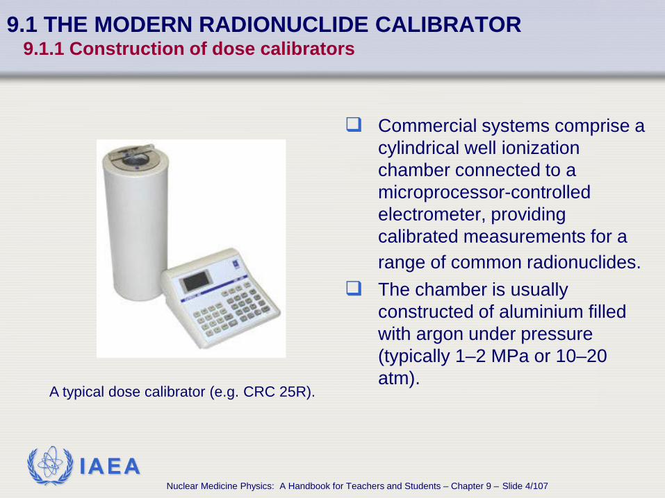
Commercial systems comprise a cylindrical well ionization chamber connected to a microprocessor-controlled electrometer, providing calibrated measurements for a range of common radionuclides.
The chamber is usually constructed of aluminium filled with argon under pressure (typically 1–2 MPa or 10–20 atm). A typical dose calibrator (e.g. CRC 25R).

IAEA Nuclear Medicine Physics: A Handbook for Teachers and Students – Chapter 9 – Slide 5/107
9.1 THE MODERN RADIONUCLIDE CALIBRATOR 9.1.1 Construction of dose calibrators
The chamber is typically shielded by the manufacturer with 6 mm of lead to ensure low background readings.
If additional shielding is used, the dose calibrator should be recalibrated or correction factors determined to ensure that the activity readings remain correct.

IAEA Nuclear Medicine Physics: A Handbook for Teachers and Students – Chapter 9 – Slide 6/107
9.1 THE MODERN RADIONUCLIDE CALIBRATOR 9.1.1 Construction of dose calibrators
SPECIFICATIONS OF TWO COMMERCIAL DOSE CALIBRATORS

IAEA Nuclear Medicine Physics: A Handbook for Teachers and Students – Chapter 9 – Slide 7/107
9.1.2 Calibration of dose calibrators
9.1 THE MODERN RADIONUCLIDE CALIBRATOR
IAEA Nuclear Medicine Physics: A Handbook for Teachers and Students – Chapter 9 – Slide 8/107
9.1 THE MODERN RADIONUCLIDE CALIBRATOR 9.1.2 Calibration of dose calibrators
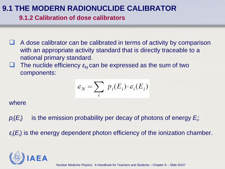
A dose calibrator can be calibrated in terms of activity by comparison with an appropriate activity standard that is directly traceable to a national primary standard.
The nuclide efficiency εN can be expressed as the sum of two components:
where pi(Ei) is the emission probability per decay of photons of energy Ei; εi(Ei) is the energy dependent photon efficiency of the ionization chamber.
IAEA Nuclear Medicine Physics: A Handbook for Teachers and Students – Chapter 9 – Slide 9/107
9.1 THE MODERN RADIONUCLIDE CALIBRATOR 9.1.2 Calibration of dose calibrators
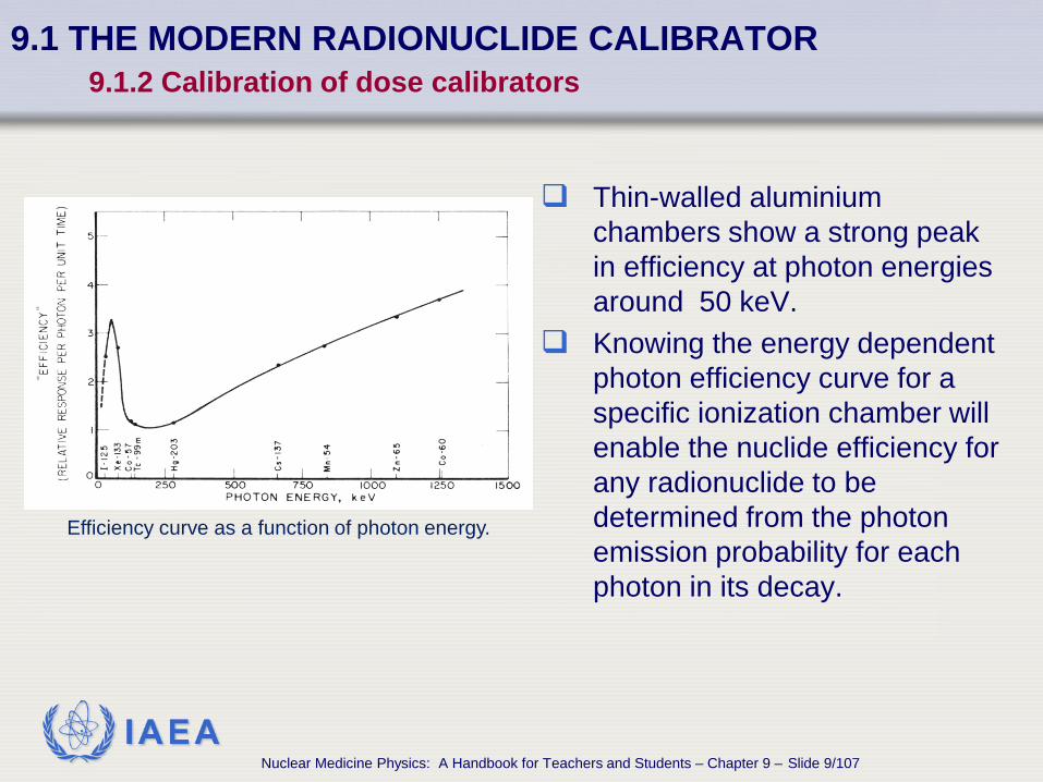
Thin-walled aluminium chambers show a strong peak in efficiency at photon energies around 50 keV.
Knowing the energy dependent photon efficiency curve for a specific ionization chamber will enable the nuclide efficiency for any radionuclide to be determined from the photon emission probability for each photon in its decay.
Efficiency curve as a function of photon energy.

IAEA Nuclear Medicine Physics: A Handbook for Teachers and Students – Chapter 9 – Slide 10/107
9.1.3 Uncertainty of activity measurements
9.1 THE MODERN RADIONUCLIDE CALIBRATOR

IAEA Nuclear Medicine Physics: A Handbook for Teachers and Students – Chapter 9 – Slide 11/107
9.1 THE MODERN RADIONUCLIDE CALIBRATOR 9.1.3 Uncertainty of activity measurements
Major sources of uncertainty in dose calibrator measurements
Calibration factor Electronics Statistical considerations Ion recombination Background radiation Source container and volume effects Source position Source adsorption

IAEA Nuclear Medicine Physics: A Handbook for Teachers and Students – Chapter 9 – Slide 12/107
9.1 THE MODERN RADIONUCLIDE CALIBRATOR 9.1.3 Uncertainty of activity measurements
9.1.3.1 Calibration factor For 99mTc and 131I, the uncertainty
of national standards is typically in the range of 1–3%.
The calibration factor for different
containers and/or a different volume may vary from the established calibration by a significant amount.
IAEA Nuclear Medicine Physics: A Handbook for Teachers and Students – Chapter 9 – Slide 13/107
9.1 THE MODERN RADIONUCLIDE CALIBRATOR 9.1.3 Uncertainty of activity measurements
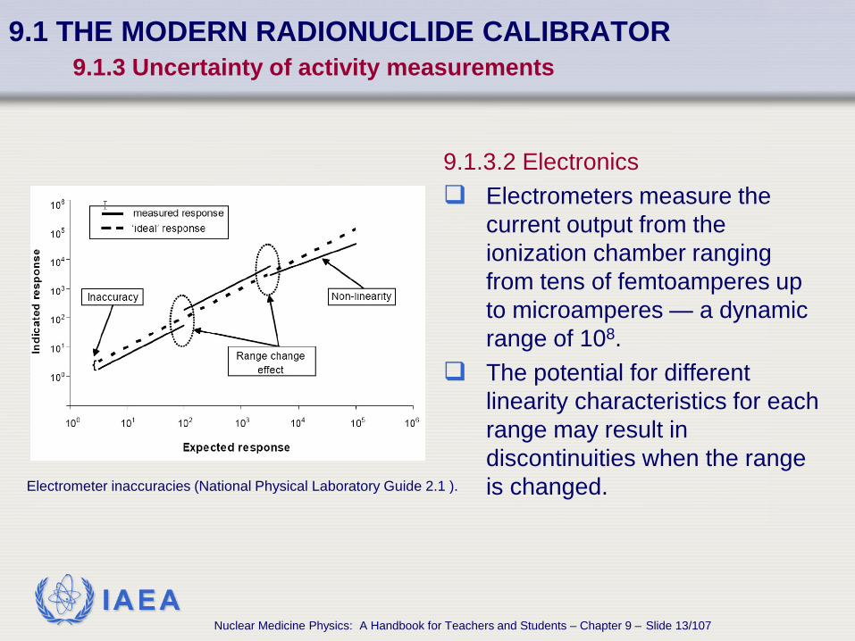
9.1.3.2 Electronics Electrometers measure the
current output from the ionization chamber ranging from tens of femtoamperes up to microamperes — a dynamic range of 108.
The potential for different linearity characteristics for each range may result in discontinuities when the range is changed. Electrometer inaccuracies (National Physical Laboratory Guide 2.1 ).
IAEA Nuclear Medicine Physics: A Handbook for Teachers and Students – Chapter 9 – Slide 14/107
9.1 THE MODERN RADIONUCLIDE CALIBRATOR 9.1.3 Uncertainty of activity measurements
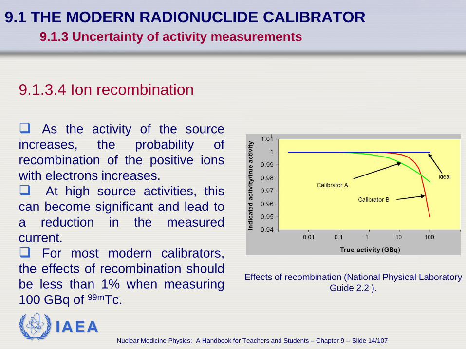
9.1.3.4 Ion recombination As the activity of the source increases, the probability of recombination of the positive ions with electrons increases. At high source activities, this can become significant and lead to a reduction in the measured current. For most modern calibrators, the effects of recombination should be less than 1% when measuring 100 GBq of 99mTc.
Effects of recombination (National Physical Laboratory Guide 2.2 ).

IAEA Nuclear Medicine Physics: A Handbook for Teachers and Students – Chapter 9 – Slide 15/107
9.1 THE MODERN RADIONUCLIDE CALIBRATOR 9.1.3 Uncertainty of activity measurements
9.1.3.6 Source container and volume effects Variations in the composition and thickness of the source container
will give rise to corresponding variations in the measured activity.
These effects will be most noticeable for low energy photon emitters and pure beta emitters.
IAEA Nuclear Medicine Physics: A Handbook for Teachers and Students – Chapter 9 – Slide 16/107
9.1 THE MODERN RADIONUCLIDE CALIBRATOR 9.1.3 Uncertainty of activity measurements
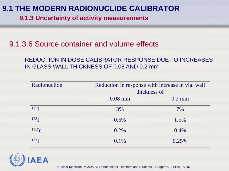
9.1.3.6 Source container and volume effects
Radionuclide Reduction in response with increase in vial wall thickness of
0.08 mm 0.2 mm 125I 3% 7% 123I 0.6% 1.5% 111In 0.2% 0.4% 131I 0.1% 0.25%
REDUCTION IN DOSE CALIBRATOR RESPONSE DUE TO INCREASES IN GLASS WALL THICKNESS OF 0.08 AND 0.2 mm

IAEA Nuclear Medicine Physics: A Handbook for Teachers and Students – Chapter 9 – Slide 17/107
9.1 THE MODERN RADIONUCLIDE CALIBRATOR 9.1.3 Uncertainty of activity measurements
Variations in source geometry When the activity is drawn into a syringe, the source
geometry will be different from that in a vial. Composition of the container, thickness and distribution
will affect the measurement. Self-absorption of the emitted radiation will change as the
source volume changes.

IAEA Nuclear Medicine Physics: A Handbook for Teachers and Students – Chapter 9 – Slide 18/107
9.1 THE MODERN RADIONUCLIDE CALIBRATOR 9.1.3 Uncertainty of activity measurements
Activity measurements variation due to container type and size.

IAEA Nuclear Medicine Physics: A Handbook for Teachers and Students – Chapter 9 – Slide 19/107
9.1 THE MODERN RADIONUCLIDE CALIBRATOR 9.1.3 Uncertainty of activity measurements
9.1.3.7 Source position
The manufacturer’s source holder is designed to keep the source at the area of maximum response on the vertical axis of the well.
Variations in response due to changes in vertical height or horizontal position of a few millimetres are usually insignificant.
IAEA Nuclear Medicine Physics: A Handbook for Teachers and Students – Chapter 9 – Slide 20/107
9.1 THE MODERN RADIONUCLIDE CALIBRATOR 9.1.3 Uncertainty of activity measurements
9.1.3.8 Source adsorption Certain radiopharmaceuticals have been observed to
adsorb to the surface of the container.
Adsorbed activity can be a significant percentage of the total.
The possibility of activity adsorption should be considered whenever the facility uses syringes from a different manufacturer.

IAEA Nuclear Medicine Physics: A Handbook for Teachers and Students – Chapter 9 – Slide 21/107
9.1.4 Measuring pure beta emitters
9.1 THE MODERN RADIONUCLIDE CALIBRATOR

IAEA Nuclear Medicine Physics: A Handbook for Teachers and Students – Chapter 9 – Slide 22/107
9.1 THE MODERN RADIONUCLIDE CALIBRATOR 9.1.4 Measuring pure beta emitters
Characteristics of beta emitters measurement The detection efficiency of ionization chambers for beta
radiation is low.
The dose calibrator response from beta particles will be almost entirely from bremsstrahlung radiation.
IAEA Nuclear Medicine Physics: A Handbook for Teachers and Students – Chapter 9 – Slide 23/107
9.1 THE MODERN RADIONUCLIDE CALIBRATOR 9.1.4 Measuring pure beta emitters
Measured activities of beta emitters In argon-filled ionization chambers, significant activities
are required in order to obtain a precise estimate of the activity.
However, as substantial activities of radionuclides are required to be used therapeutically, reliable measurements are possible using pure beta emitters used clinically such as 90Y, 89Sr and 32P.

IAEA Nuclear Medicine Physics: A Handbook for Teachers and Students – Chapter 9 – Slide 24/107
9.1 THE MODERN RADIONUCLIDE CALIBRATOR 9.1.4 Measuring pure beta emitters
Dose calibrators efficiency The intrinsic efficiencies of dose calibrators can vary
widely. Data from five different manufacturers showed that all
systems had: • a good calibration for 32P. • a reduction in efficiency of approximately 10–20% for
89Sr. • a wide divergence in efficiency for 90Y.

IAEA Nuclear Medicine Physics: A Handbook for Teachers and Students – Chapter 9 – Slide 25/107
9.1 THE MODERN RADIONUCLIDE CALIBRATOR 9.1.4 Measuring pure beta emitters
90Y measurements The results obtained using the calibration factors supplied
by the manufacturers ranged from 64 to 144% of the true value.
This re-emphasizes the need for the calibration to be confirmed within the nuclear medicine department.

IAEA Nuclear Medicine Physics: A Handbook for Teachers and Students – Chapter 9 – Slide 26/107
9.1 THE MODERN RADIONUCLIDE CALIBRATOR 9.1.4 Measuring pure beta emitters
153Sm and 186Re measurements 153Sm (103 keV, 28% abundance) and 186Re (137 keV,
9.5% abundance) are gamma-beta emitting radionuclides. For these radionuclides the ionization chamber efficiency
is primarily determined by the gamma contribution and the manufacturer’s supplied calibrations will usually be accurate to within ±10%.
IAEA Nuclear Medicine Physics: A Handbook for Teachers and Students – Chapter 9 – Slide 27/107
9.1.5 Problems arising from radionuclide contaminants
9.1 THE MODERN RADIONUCLIDE CALIBRATOR

IAEA Nuclear Medicine Physics: A Handbook for Teachers and Students – Chapter 9 – Slide 28/107
9.1 THE MODERN RADIONUCLIDE CALIBRATOR 9.1.5 Problems arising from radionuclide contaminants
Radionuclide purity The proportion of the total radioactivity that is present as a
specific radionuclide is defined as the radionuclide purity.
National and international pharmacopoeia specify the radionuclidic purity of a radiopharmaceutical.
IAEA Nuclear Medicine Physics: A Handbook for Teachers and Students – Chapter 9 – Slide 29/107
9.1 THE MODERN RADIONUCLIDE CALIBRATOR 9.1.5 Problems arising from radionuclide contaminants
Effects of contaminants
The presence of contaminants, even when less than 1% of the total activity, can have a marked effect on the ionization chamber current and, thus, on the measured activity.
The presence of high energy contaminants will have an
adverse effect on image quality due to increased septal penetration and will also lead to an increased radiation dose to the patient.

IAEA Nuclear Medicine Physics: A Handbook for Teachers and Students – Chapter 9 – Slide 30/107
9.2.1 Acceptance tests
9.2 DOSE CALIBRATOR ACCEPTANCE TESTING AND QC

IAEA Nuclear Medicine Physics: A Handbook for Teachers and Students – Chapter 9 – Slide 31/107
9.2 DOSE CALIBRATOR ACCEPTANCE TESTING AND QC 9.2.1 Acceptance tests
Acceptance tests for dose calibrators Accuracy and reproducibility Linearity Geometry response

IAEA Nuclear Medicine Physics: A Handbook for Teachers and Students – Chapter 9 – Slide 32/107
9.2 DOSE CALIBRATOR ACCEPTANCE TESTING AND QC 9.2.1 Acceptance tests
9.2.1.1 Accuracy and reproducibility The accuracy is determined by comparing activity
measurements using a traceable calibrated standard with the supplier’s stated activity, corrected for radioactive decay.
The reproducibility, or constancy, can be assessed by taking repeated measurements of the same source.
IAEA Nuclear Medicine Physics: A Handbook for Teachers and Students – Chapter 9 – Slide 33/107
9.2 DOSE CALIBRATOR ACCEPTANCE TESTING AND QC 9.2.1 Acceptance tests
9.2.1.2 Linearity Methods for assessment of linearity of dose response: Decaying source method
Multiple dilutions method
Graded attenuators method

IAEA Nuclear Medicine Physics: A Handbook for Teachers and Students – Chapter 9 – Slide 34/107
9.2 DOSE CALIBRATOR ACCEPTANCE TESTING AND QC 9.2.1 Acceptance tests
9.2.1.3 Geometry response The measured activity may vary with:
• the position of the source within the ionization chamber • the composition of the vial or syringe • the volume of liquid within the vial or syringe
Correction factors can be determined for the different
volumes or containers used.

IAEA Nuclear Medicine Physics: A Handbook for Teachers and Students – Chapter 9 – Slide 35/107
9.2.2 Quality control
9.2 DOSE CALIBRATOR ACCEPTANCE TESTING AND QC

IAEA Nuclear Medicine Physics: A Handbook for Teachers and Students – Chapter 9 – Slide 36/107
9.2 DOSE CALIBRATOR ACCEPTANCE TESTING AND QC 9.2.2 Quality control
9.2.2.1 Background check
Even if the source holder is empty, the dose calibrator will still record an ‘activity’ due to background radiation.
At a minimum, the background should be determined each
morning before the dose calibrator is used, and recorded. The technologist should also confirm the absence of any
additional background before all activity measurements during the day.
IAEA Nuclear Medicine Physics: A Handbook for Teachers and Students – Chapter 9 – Slide 37/107
9.2 DOSE CALIBRATOR ACCEPTANCE TESTING AND QC 9.2.2 Quality control
9.2.2.2 Check source reproducibility
A long lived check source should be used on a daily basis to confirm the constancy of the response of the dose calibrator.
Sealed radioactive sources of 57Co and 137Cs, shaped to mimic a vial, are available commercially for this purpose.
The check source should be measured on all radionuclide settings that are used clinically.
A reading outside of that expected from previous results may indicate a faulty dose calibrator or a change in calibration factor.
IAEA Nuclear Medicine Physics: A Handbook for Teachers and Students – Chapter 9 – Slide 38/107
9.3 STANDARDS APPLYING TO DOSE CALIBRATORS

IAEA Nuclear Medicine Physics: A Handbook for Teachers and Students – Chapter 9 – Slide 39/107
9.3 STANDARDS APPLYING TO DOSE CALIBRATORS
International and national standards The International Electrotechnical Commission (IEC) has
published two standards and a technical report relating to dose calibrators.
IEC standards are often adopted by national standards organizations.
There should also be national standards covering dose calibrators. The American National Standards Institute publication ANSI N42.13-2004 is often referenced by US manufacturers.
IAEA Nuclear Medicine Physics: A Handbook for Teachers and Students – Chapter 9 – Slide 40/107
9.4 NATIONAL ACTIVITY INTERCOMPARISONS

IAEA Nuclear Medicine Physics: A Handbook for Teachers and Students – Chapter 9 – Slide 41/107
9.4 NATIONAL ACTIVITY INTERCOMPARISONS
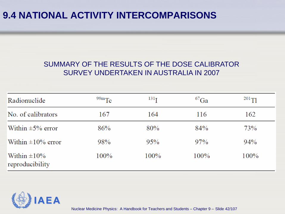
National metrology institutes are responsible for the development and maintenance of standards, including activity standards and have undertaken national comparisons of the accuracy of the dose calibrators used in clinical practice.
Such comparisons have used, where possible, the clinical radionuclides 67Ga, 123I, 131I, 99mTc and 201Tl.
In some countries they are voluntary, while in others it is mandatory.
IAEA Nuclear Medicine Physics: A Handbook for Teachers and Students – Chapter 9 – Slide 42/107
9.4 NATIONAL ACTIVITY INTERCOMPARISONS
SUMMARY OF THE RESULTS OF THE DOSE CALIBRATOR SURVEY UNDERTAKEN IN AUSTRALIA IN 2007

IAEA Nuclear Medicine Physics: A Handbook for Teachers and Students – Chapter 9 – Slide 43/107
9.5.1 Adjusting the activity for differences in patient size and weight
9.5 DISPENSING RADIOPHARMACEUTICALS FOR INDIVIDUAL PATIENTS

IAEA Nuclear Medicine Physics: A Handbook for Teachers and Students – Chapter 9 – Slide 44/107
9.5 DISPENSING RADIOPHARMACEUTICALS FOR INDIVIDUAL PATIENTS 9.5.1 Adjusting the activity for differences in patient size and weight
Protocols Protocols used in nuclear medicine practices should
specify the usual activity of the radiopharmaceutical to be administered to a standard patient.
If a fixed activity is used for all patients, this will lead to an unnecessarily high radiation exposure to an underweight patient and may lead to images of unacceptable quality or very long imaging times in obese patients.

IAEA Nuclear Medicine Physics: A Handbook for Teachers and Students – Chapter 9 – Slide 45/107
9.5 DISPENSING RADIOPHARMACEUTICALS FOR INDIVIDUAL PATIENTS 9.5.1 Adjusting the activity for differences in patient size and weight
Scaling factors Scaling factors for the activity, to give a constant effective
dose, can be derived from the expression (W/70)a
where W represents the weight of the person and the power factor a is specific for the radiopharmaceutical (ICRP 53,80,106).

IAEA Nuclear Medicine Physics: A Handbook for Teachers and Students – Chapter 9 – Slide 46/107
9.5 DISPENSING RADIOPHARMACEUTICALS FOR INDIVIDUAL PATIENTS 9.5.1 Adjusting the activity for differences in patient size and weight
Radiopharmaceutical a value Radiopharmaceutical a value
99mTc-DMSA –0.706 99mTc-IDA –0.840 99mTc-DTPA –0.801 99mTc-tetrafosmin –0.834 99mTc-MAG3 –0.520 99mTc-red cells –0.859 99mTc-HMPAO –0.849 99mTc-white cells –0.869 99mTc-MAA –0.842 18F-FDG –0.782 99mTc-sestamibi –0.871 67Ga-citrate –0.931 99mTc-phosphonates –0.763 123I or 131I iodide –1.11
THE POWER FACTOR a RELATING BODY WEIGHT TO A CONSTANT EFFECTIVE DOSE ACCORDING TO THE EXPRESSION (W/70)a
FOR 14 COMMON RADIOPHARMACEUTICALS

IAEA Nuclear Medicine Physics: A Handbook for Teachers and Students – Chapter 9 – Slide 47/107
9.5.2 Paediatric dosage charts
9.5 DISPENSING RADIOPHARMACEUTICALS FOR INDIVIDUAL PATIENTS
IAEA Nuclear Medicine Physics: A Handbook for Teachers and Students – Chapter 9 – Slide 48/107
9.5 DISPENSING RADIOPHARMACEUTICALS FOR INDIVIDUAL PATIENTS 9.5.2 Paediatric dosage charts
Paediatric dose considerations Children are approximately three times more
radiosensitive than adults, so determining the appropriate activity to be administered for paediatric procedures is essential.
In addition to the scaling factor to be applied to the adult activity, a minimum activity must be specified in order to ensure adequate image quality.
IAEA Nuclear Medicine Physics: A Handbook for Teachers and Students – Chapter 9 – Slide 49/107
9.5 DISPENSING RADIOPHARMACEUTICALS FOR INDIVIDUAL PATIENTS 9.5.2 Paediatric dosage charts
Dose scaling factors In the past, the scaling factors were assessed using weight
alone or body surface area obtained from both height and weight.
Recently, the European Association of Nuclear Medicine (EANM) Dosimetry and Paediatric Committees have prepared a dosage card which recognizes that a single scaling factor is not optimal for all radiopharmaceuticals.
Radiopharmaceuticals could be grouped into three classes (renal, thyroid and others), with different scaling factors for each class.

IAEA Nuclear Medicine Physics: A Handbook for Teachers and Students – Chapter 9 – Slide 50/107
9.5 DISPENSING RADIOPHARMACEUTICALS FOR INDIVIDUAL PATIENTS 9.5.2 Paediatric dosage charts
A dosage card is available on the EANM web site that gives the minimum recommended activity and a weight dependent scaling factor for each radiopharmaceutical.
It was determined to give weight independent effective doses.
An app for iOs and Android devices featuring the chart is now available.
Dosage card can be accessed online: http://www.eanm.org/docs/EANM_Dosage_Card_040214.pdf?PHPSESSID=sf56mg9ehjv5r9t4v50mre3375

IAEA Nuclear Medicine Physics: A Handbook for Teachers and Students – Chapter 9 – Slide 51/107
9.5.3 Diagnostic reference levels in nuclear medicine
9.5 DISPENSING RADIOPHARMACEUTICALS FOR INDIVIDUAL PATIENTS

IAEA Nuclear Medicine Physics: A Handbook for Teachers and Students – Chapter 9 – Slide 52/107
9.5 DISPENSING RADIOPHARMACEUTICALS FOR INDIVIDUAL PATIENTS 9.5.3 Diagnostic reference levels in nuclear medicine
Diagnostic reference levels The ICRP introduced in 1996 the term ‘diagnostic
reference level’ (DRL) for patients. DRLs are investigation levels and are based on an easily
measured quantity, usually the entrance surface dose in the case of diagnostic radiology, or the administered activity in the case of nuclear medicine.
DRLs are referred to by the IAEA as guidance levels in Safety Report Series No. 40.
IAEA Nuclear Medicine Physics: A Handbook for Teachers and Students – Chapter 9 – Slide 53/107
9.6.1 Surface contamination limits
9.6 RADIATION SAFETY IN THE RADIOPHARMACY

IAEA Nuclear Medicine Physics: A Handbook for Teachers and Students – Chapter 9 – Slide 54/107
9.6 RADIATION SAFETY IN THE RADIOPHARMACY 9.6.1 Surface contamination limits
External and internal contamination Surface contamination with radioactivity could lead to:
• contamination of a radiation worker • external irradiation of the skin of the worker
Internal contamination could arise from
• inhalation of the radionuclide • ingestion of the radionuclide
IAEA Nuclear Medicine Physics: A Handbook for Teachers and Students – Chapter 9 – Slide 55/107
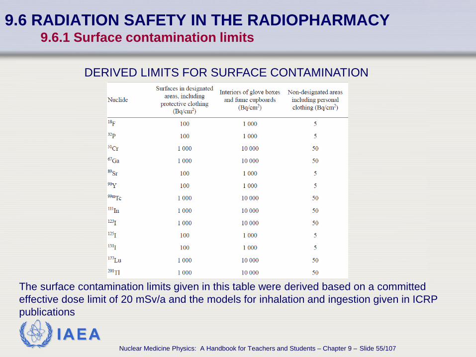
9.6 RADIATION SAFETY IN THE RADIOPHARMACY 9.6.1 Surface contamination limits
The surface contamination limits given in this table were derived based on a committed effective dose limit of 20 mSv/a and the models for inhalation and ingestion given in ICRP publications
DERIVED LIMITS FOR SURFACE CONTAMINATION

IAEA Nuclear Medicine Physics: A Handbook for Teachers and Students – Chapter 9 – Slide 56/107
9.6.2 Wipe tests and daily surveys
9.6 RADIATION SAFETY IN THE RADIOPHARMACY

IAEA Nuclear Medicine Physics: A Handbook for Teachers and Students – Chapter 9 – Slide 57/107
9.6 RADIATION SAFETY IN THE RADIOPHARMACY 9.6.2 Wipe tests and daily surveys
Surveys of the radiopharmacy areas To ensure that contamination limits are not exceeded,
surveys of radiopharmacy areas should be routinely done. Logical sequence of surveys
• Use survey meter to find unexpected exposed sources. • Check surfaces with contamination meter with appropriate probe,
according to the radionuclides used. • Use wipe tests for areas of high background or for low energy beta
emitters.

IAEA Nuclear Medicine Physics: A Handbook for Teachers and Students – Chapter 9 – Slide 58/107
9.6 RADIATION SAFETY IN THE RADIOPHARMACY 9.6.2 Wipe tests and daily surveys
Wipe tests A minimum area of 100 cm2 should be wiped. Activity can be assessed using a pancake probe, or more accurately
in a well counter. For low energy beta emitters such as 3H or 14C, liquid scintillation
counting must be used. When quantifying the surface contamination, it is generally assumed
that a wipe test using a dry wipe will remove one tenth of the contamination.
It is assumed that a wet wipe will remove one fifth of the contamination.
IAEA Nuclear Medicine Physics: A Handbook for Teachers and Students – Chapter 9 – Slide 59/107
9.6.3 Monitoring of staff finger doses during dispensing
9.6 RADIATION SAFETY IN THE RADIOPHARMACY
IAEA Nuclear Medicine Physics: A Handbook for Teachers and Students – Chapter 9 – Slide 60/107
9.6 RADIATION SAFETY IN THE RADIOPHARMACY 9.6.3 Monitoring of staff finger doses during dispensing
Hand and finger doses The most exposed parts of the hands are likely to be the tips of the
index and middle fingers, and the thumb of the dominant hand. Finger doses may approach or exceed the annual dose limit of 500
mSv to the extremities. A practical way to monitor hands is to wear a ring monitor at the base
of the finger. The ICRP recommends that the ring monitor be worn on the middle
finger with the element positioned on the palm side, and that a factor of three should be applied to derive an estimate of the dose to the tip.
The dose to the fingers is critically dependent on the dispensing technique used and the skill of the operator.

IAEA Nuclear Medicine Physics: A Handbook for Teachers and Students – Chapter 9 – Slide 61/107
9.7.1 Fume cupboards
9.7 PRODUCT CONTAINMENT ENCLOSURES
IAEA Nuclear Medicine Physics: A Handbook for Teachers and Students – Chapter 9 – Slide 62/107
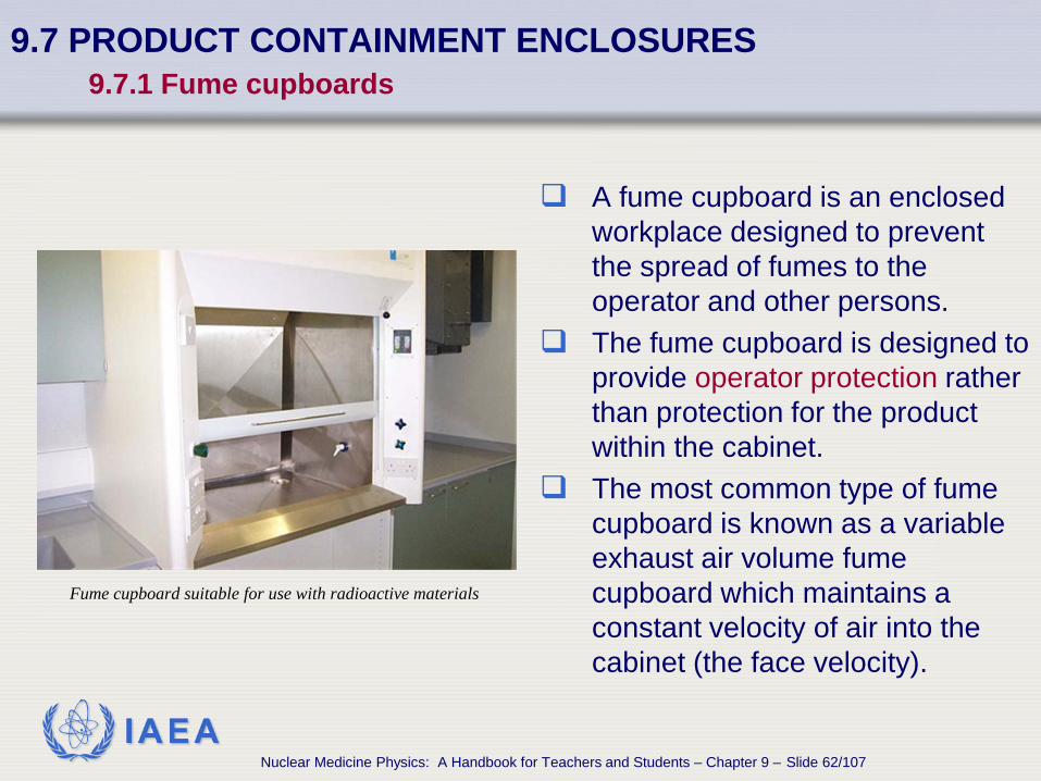
9.7 PRODUCT CONTAINMENT ENCLOSURES 9.7.1 Fume cupboards
A fume cupboard is an enclosed workplace designed to prevent the spread of fumes to the operator and other persons.
The fume cupboard is designed to provide operator protection rather than protection for the product within the cabinet.
The most common type of fume cupboard is known as a variable exhaust air volume fume cupboard which maintains a constant velocity of air into the cabinet (the face velocity).
Fume cupboard suitable for use with radioactive materials
IAEA Nuclear Medicine Physics: A Handbook for Teachers and Students – Chapter 9 – Slide 63/107
9.7 PRODUCT CONTAINMENT ENCLOSURES 9.7.1 Fume cupboards
Cupboard air discharge Air discharge type
• Direct (or through filter) to the atmosphere. • Recirculating, after filtration or absorption (normally not applicable
in radiopharmacies). Air discharged must meet local regulatory requirements. Smoke tests should be performed as part of QC schedule.

IAEA Nuclear Medicine Physics: A Handbook for Teachers and Students – Chapter 9 – Slide 64/107
9.7.2 LAMINAR FLOW CABINETS
9.7 PRODUCT CONTAINMENT ENCLOSURES

IAEA Nuclear Medicine Physics: A Handbook for Teachers and Students – Chapter 9 – Slide 65/107
9.7 PRODUCT CONTAINMENT ENCLOSURES 9.7.2 Laminar flow cabinets
Laminar flow cabinets characteristics Laminar flow cabinets provide a non-turbulent airstream of near
constant velocity, which has a substantially uniform flow cross-section and with a variation in velocity of not more than 20%.
Laminar flow cabinets provide product protection while a fume cupboard is designed to provide operator protection.
The air supplied to the cabinet is usually passed through a high efficiency particulate air filter (99.999%).
Operator protection cannot be ensured if airflow is disturbed during radiopharmaceutical manipulation.

IAEA Nuclear Medicine Physics: A Handbook for Teachers and Students – Chapter 9 – Slide 66/107
9.7.3 Isolator cabinets
9.7 PRODUCT CONTAINMENT ENCLOSURES

IAEA Nuclear Medicine Physics: A Handbook for Teachers and Students – Chapter 9 – Slide 67/107
9.7 PRODUCT CONTAINMENT ENCLOSURES 9.7.3 Isolator cabinets
Isolator cabinets characteristics Isolator cabinets provide both
operator and product protection, used frequently for cell labelling.
The product is manipulated through glove ports so that the interior of the cabinet is maintained totally sterile and full operator protection is provided.
The isolator incorporates timed interlocks on the vacuum door seals to ensure that the product remains sterile.

IAEA Nuclear Medicine Physics: A Handbook for Teachers and Students – Chapter 9 – Slide 68/107
9.8.1 Shielding for gamma, beta and positron emitters
9.8 SHIELDING FOR RADIONUCLIDES

IAEA Nuclear Medicine Physics: A Handbook for Teachers and Students – Chapter 9 – Slide 69/107
9.8 SHIELDING FOR RADIONUCLIDES 9.8.1 Shielding for gamma, beta and positron emitters
Shielding requirements and materials Shielding is required
• in the walls of the radiopharmacy. • in any containment enclosures. • in a body shield to protect the operator at the dispensing station • around individual vials and syringes containing radionuclides.
Shielding materials for different purposes • Lead and concrete in walls. • Lead or tungsten in local shielding for gamma emitting
radionuclides. • Aluminium or Perspex for pure beta emitters (to minimize
bremsstrahlung radiation).
IAEA Nuclear Medicine Physics: A Handbook for Teachers and Students – Chapter 9 – Slide 70/107
9.8 SHIELDING FOR RADIONUCLIDES 9.8.1 Shielding for gamma, beta and positron emitters
Shielding for beta emitters For beta emitters, the thickness of the shielding must be
greater than its range to ensure that all betas are absorbed.
Polymethyl methacrylate (Perspex or lucite) has a density of 1.19 g/cm3, similar to the density of tissue and water, and is highly suitable for absorbing betas.
IAEA Nuclear Medicine Physics: A Handbook for Teachers and Students – Chapter 9 – Slide 71/107
9.8 SHIELDING FOR RADIONUCLIDES 9.8.1 Shielding for gamma, beta and positron emitters
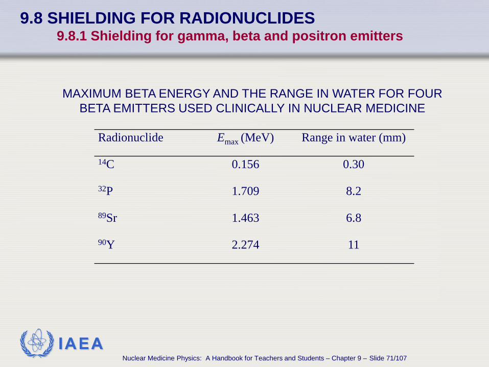
Radionuclide Emax (MeV) Range in water (mm)
14C 0.156 0.30
32P 1.709 8.2
89Sr 1.463 6.8
90Y 2.274 11
MAXIMUM BETA ENERGY AND THE RANGE IN WATER FOR FOUR BETA EMITTERS USED CLINICALLY IN NUCLEAR MEDICINE

IAEA Nuclear Medicine Physics: A Handbook for Teachers and Students – Chapter 9 – Slide 72/107
9.8 SHIELDING FOR RADIONUCLIDES 9.8.1 Shielding for gamma, beta and positron emitters
Doses due to generators The highest surface dose rates
encountered in the radiopharmacy are likely to be from 99Mo/99mTc generators.
It requires several centimetres of lead shielding to reduce the dose rates to an acceptable level.
The generator as supplied will already contain substantial shielding but additional shielding will usually be required.

IAEA Nuclear Medicine Physics: A Handbook for Teachers and Students – Chapter 9 – Slide 73/107
9.8 SHIELDING FOR RADIONUCLIDES 9.8.1 Shielding for gamma, beta and positron emitters
Manipulation of vials Vials of radiopharmaceuticals
must be kept shielded. The shields are usually
constructed so that only the rubber septum of the vial is accessible, thereby protecting the hands of the operator during dispensing.

IAEA Nuclear Medicine Physics: A Handbook for Teachers and Students – Chapter 9 – Slide 74/107
9.8 SHIELDING FOR RADIONUCLIDES 9.8.1 Shielding for gamma, beta and positron emitters
Manipulation of vials Measurements in calibrators
are done with the unshielded vials, increasing the exposure to the operator.
Long forceps should always be used to manipulate radioactive vials.

IAEA Nuclear Medicine Physics: A Handbook for Teachers and Students – Chapter 9 – Slide 75/107
9.8 SHIELDING FOR RADIONUCLIDES 9.8.1 Shielding for gamma, beta and positron emitters
Manipulation of syringes Syringe shields must be
used whenever possible. These must be made of
Perspex for the pure beta emitters and of lead or tungsten for the gamma emitters.
A lead–glass window is necessary to permit observation of the contents of the syringe.

IAEA Nuclear Medicine Physics: A Handbook for Teachers and Students – Chapter 9 – Slide 76/107
9.8.2 Transmission factors for lead and concrete
9.8 SHIELDING FOR RADIONUCLIDES

IAEA Nuclear Medicine Physics: A Handbook for Teachers and Students – Chapter 9 – Slide 77/107
9.8 SHIELDING FOR RADIONUCLIDES 9.8.2 Transmission factors for lead and concrete
Transmission factors characteristics The attenuation of monoenergetic photons through
materials such as lead or concrete will be exponential, characterized by the linear attenuation coefficient or the half-value layer (HVL).
This is only true for narrow beam geometries. Moreover, non-monoenergetic radionuclides emit more
than one gamma photon and their attenuation cannot be expressed as a simple HVL.
Measured broad-beam transmission factors are available for lead and concrete, two of the most common shielding materials.

IAEA Nuclear Medicine Physics: A Handbook for Teachers and Students – Chapter 9 – Slide 78/107
9.8 SHIELDING FOR RADIONUCLIDES 9.8.2 Transmission factors for lead and concrete
MEASURED TRANSMISION FACTORS FOR LEAD

IAEA Nuclear Medicine Physics: A Handbook for Teachers and Students – Chapter 9 – Slide 79/107
9.8 SHIELDING FOR RADIONUCLIDES 9.8.2 Transmission factors for lead and concrete
MEASURED TRANSMISSION FACTORS FOR CONCRETE (DENSITY: 2.35 g/cm3)

IAEA Nuclear Medicine Physics: A Handbook for Teachers and Students – Chapter 9 – Slide 80/107
9.9 DESIGNING A RADIOPHARMACY

IAEA Nuclear Medicine Physics: A Handbook for Teachers and Students – Chapter 9 – Slide 81/107
9.9 DESIGNING A RADIOPHARMACY
Location of a radiopharmacy The radiopharmacy should be located in an area that is not accessible
to members of the public.
There should be easy access from the radiopharmacy to the injection rooms and imaging rooms to minimize the distance that radioactive materials need to be transported.
The radiopharmacy should not be adjacent to areas that require a low and constant radiation background such as a counting room.

IAEA Nuclear Medicine Physics: A Handbook for Teachers and Students – Chapter 9 – Slide 82/107
9.9 DESIGNING A RADIOPHARMACY
Storage needs for the radiopharmacy A refrigerator will be required for the storage of lyophilized
radiopharmaceutical kits. A storage area will be required for reconstituted radiopharmaceuticals,
in shielded containers, together with radiopharmaceuticals purchased ready for dispensing such as 67Ga-citrate and 201Tl-chloride.
The radiopharmacy must contain facilities for radioactive waste disposal.
In addition, there must be shielded containers for ‘sharps’, such as syringes with needles.
A separate shielded storage bin may be required if a large number of bulky items, such as aerosol or Technegas kits, need to be stored.

IAEA Nuclear Medicine Physics: A Handbook for Teachers and Students – Chapter 9 – Slide 83/107
9.9 DESIGNING A RADIOPHARMACY
Areas of the radiopharmacy There should be an area within the radiopharmacy designated as a
non-active area that is used for record keeping and/or computer entry. A dedicated dispensing area with a body shield and lead–glass
viewing window will be required.
If a Mo/Tc generator is used, this should be positioned away from the dispensing area to minimize the dose received by the person dispensing the radiopharmaceuticals.
Labelling areas are dependent of the type of radiopharmaceutical that
will be prepared, generally requiring specialized equipment.

IAEA Nuclear Medicine Physics: A Handbook for Teachers and Students – Chapter 9 – Slide 84/107
9.9 DESIGNING A RADIOPHARMACY
Dedicated equipment for specific labelling techniques
If cell labelling procedures are to be performed, a dedicated area with a laminar flow cabinet or isolator will be required to ensure that the product remains sterile during the labelling procedure.
A fume cupboard, together with an activated charcoal filter on the exhaust, will be required if radio-iodination procedures are to be performed.
Some radiopharmaceuticals require a heating step in their preparation. This is often performed using a temperature controlled heating block. This must be in a dedicated separately shielded area,. Similarly, the radiolabelling of blood samples may require local shielding of mixers and centrifuges.
IAEA Nuclear Medicine Physics: A Handbook for Teachers and Students – Chapter 9 – Slide 85/107
9.9 DESIGNING A RADIOPHARMACY
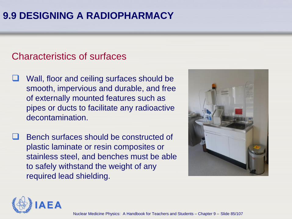
Characteristics of surfaces Wall, floor and ceiling surfaces should be
smooth, impervious and durable, and free of externally mounted features such as pipes or ducts to facilitate any radioactive decontamination.
Bench surfaces should be constructed of
plastic laminate or resin composites or stainless steel, and benches must be able to safely withstand the weight of any required lead shielding.

IAEA Nuclear Medicine Physics: A Handbook for Teachers and Students – Chapter 9 – Slide 86/107
9.9 DESIGNING A RADIOPHARMACY
Contamination monitoring A contamination monitor must be
available in a readily accessible location.
A wall-mounted monitor to check for any hand contamination should be mounted near the exit from the radiopharmacy.
A model which can be removed and used as a general contamination monitor is useful.

IAEA Nuclear Medicine Physics: A Handbook for Teachers and Students – Chapter 9 – Slide 87/107
9.9 DESIGNING A RADIOPHARMACY
Decontamination facilities Hand washing facilities must be available which can be operated
without the use of the operator’s hands to prevent the spread of any contamination.
An eye-wash should also be available.

IAEA Nuclear Medicine Physics: A Handbook for Teachers and Students – Chapter 9 – Slide 88/107
9.10 SECURITY OF THE RADIOPHARMACY

IAEA Nuclear Medicine Physics: A Handbook for Teachers and Students – Chapter 9 – Slide 89/107
9.10 SECURITY OF THE RADIOPHARMACY
Category of radioactive sources The IAEA has categorized radioactive sources on a scale of 1 to 5,
based on activity and nuclide, where category 1 is potentially the most hazardous.
Sources categorized as 1, 2 or 3 are known as security-enhanced sources.
The security measures in place for safety purposes are considered adequate to ensure the physical security of category 4 and 5 sources.
A Mo/Tc generator with an activity of greater than 300 GBq is a category 3 source.

IAEA Nuclear Medicine Physics: A Handbook for Teachers and Students – Chapter 9 – Slide 90/107
9.10 SECURITY OF THE RADIOPHARMACY
Physical security of radioactive sources Radioactive materials are at most risk of being stolen or lost when they
are being transported to and from the facility. It is essential that all consignments of radioactive materials to the
nuclear medicine facility are left in a secure area and not left, for example, on a loading dock.

IAEA Nuclear Medicine Physics: A Handbook for Teachers and Students – Chapter 9 – Slide 91/107
9.10 SECURITY OF THE RADIOPHARMACY
Radiopharmacy access Whether secure access (such as electronic card access) to the
radiopharmacy during working hours is required will depend on local requirements and the layout of the nuclear medicine department.
It is essential that only trained nuclear medicine staff have access to the radiopharmacy.

IAEA Nuclear Medicine Physics: A Handbook for Teachers and Students – Chapter 9 – Slide 92/107
9.11 RECORD KEEPING

IAEA Nuclear Medicine Physics: A Handbook for Teachers and Students – Chapter 9 – Slide 93/107
9.11 RECORD KEEPING
Record generation and keeping Records can be generated as • part of the quality assurance (QA) programme. • for the receipt and subsequent administration of a radiopharmaceutical
to a patient. • for waste disposal.
The local regulations may specify • the form in which these must be kept (paper and/or electronic). • the minimum records that must be kept at the facility. • the time for which the records must be kept.

IAEA Nuclear Medicine Physics: A Handbook for Teachers and Students – Chapter 9 – Slide 94/107
9.11.1 Quality control records 9.11 RECORD KEEPING

IAEA Nuclear Medicine Physics: A Handbook for Teachers and Students – Chapter 9 – Slide 95/107
9.11 RECORD KEEPING 9.11.1 Quality control records
Records should at the very least include details of: Acceptance testing of the dose calibrator
All constancy tests
Radiopharmaceutical testing

IAEA Nuclear Medicine Physics: A Handbook for Teachers and Students – Chapter 9 – Slide 96/107
9.11 RECORD KEEPING 9.11.1 Quality control records
Record of failures and malfunctions Failures identified at acceptance testing.
Failures of constancy testing.
Failures of radiopharmaceutical testing. The actions taken to remedy those failures. All these should be documented and these records kept for the lifetime of the equipment.

IAEA Nuclear Medicine Physics: A Handbook for Teachers and Students – Chapter 9 – Slide 97/107
9.11 RECORD KEEPING 9.11.1 Quality control records
Generator elutions records The following records should be kept for all generator elutions:
Time of elution
Volume of eluate
99mTc activity
99Mo activity
Radionuclidic purity

IAEA Nuclear Medicine Physics: A Handbook for Teachers and Students – Chapter 9 – Slide 98/107
9.11.2 Records of receipt of radioactive materials
9.11 RECORD KEEPING

IAEA Nuclear Medicine Physics: A Handbook for Teachers and Students – Chapter 9 – Slide 99/107
9.11 RECORD KEEPING 9.11.2 Records of receipt of radioactive materials
Radioactive materials records
Complete records should be kept of: • The radionuclide • Activity • Chemical form • Supplier • Supplier’s batch number • Purchase date On arrival, if a package containing radioactive material is suspected of
being damaged, the package should be: • Monitored for leakage with a wipe test; • Checked with a survey meter for unexpectedly high external radiation levels.
If a package is damaged or suspected of being damaged, the supplier should be contacted immediately, and the details recorded

IAEA Nuclear Medicine Physics: A Handbook for Teachers and Students – Chapter 9 – Slide 100/107
9.11.3 Records of radiopharmaceutical preparation and dispensing
9.11 RECORD KEEPING
IAEA Nuclear Medicine Physics: A Handbook for Teachers and Students – Chapter 9 – Slide 101/107
9.11 RECORD KEEPING 9.11.3 Records of radiopharmaceutical preparation and dispensing
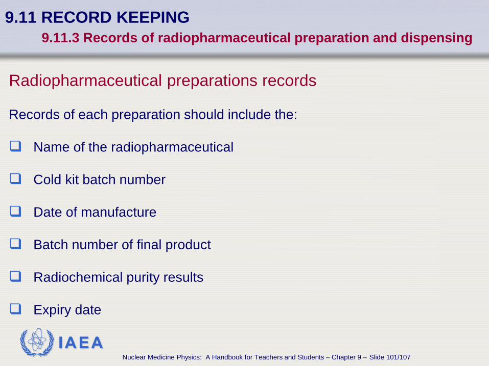
Radiopharmaceutical preparations records Records of each preparation should include the: Name of the radiopharmaceutical
Cold kit batch number
Date of manufacture Batch number of final product
Radiochemical purity results
Expiry date

IAEA Nuclear Medicine Physics: A Handbook for Teachers and Students – Chapter 9 – Slide 102/107
9.11 RECORD KEEPING 9.11.3 Records of radiopharmaceutical preparation and dispensing
Patient dose dispensed records A record for each patient dose dispensed must be kept with the: Name of the patient
Name of the radiopharmaceutical
Measured radioactivity
Time and date of measurement

IAEA Nuclear Medicine Physics: A Handbook for Teachers and Students – Chapter 9 – Slide 103/107
9.11.4 Radioactive waste records 9.11 RECORD KEEPING
IAEA Nuclear Medicine Physics: A Handbook for Teachers and Students – Chapter 9 – Slide 104/107
9.11 RECORD KEEPING 9.11.4 Radioactive waste records
Characteristics of Nuclear Medicine radioactive wastes Radioactive waste generated within a nuclear medicine facility usually
consists of radionuclides with half-lives of less than one month.
This waste will normally be stored on-site, be allowed to decay to background radiation levels.
After decay, it can then be disposed of as normal waste or biologically contaminated waste.
IAEA Nuclear Medicine Physics: A Handbook for Teachers and Students – Chapter 9 – Slide 105/107
9.11 RECORD KEEPING 9.11.4 Radioactive waste records
Radioactive waste packages labelling Each package of waste (bag, sharps container, wheeled bin) must be marked with the: Radionuclide, if known. Maximum dose rate at the surface of the container or at a fixed
distance (e.g. 1 m). Date of storage.

IAEA Nuclear Medicine Physics: A Handbook for Teachers and Students – Chapter 9 – Slide 106/107
9.11 RECORD KEEPING 9.11.4 Radioactive waste records
Records information and update The wastes information should be recorded, together with information
identifying the location of the container within the store, and the likely release date (e.g. ten half-lives of the longest lived radionuclide in the container).
When the package is finally released for disposal, the record should be updated to record the dose rate at that time, which should be at background levels, the date of disposal, and the identification of the person authorizing its disposal.
IAEA Nuclear Medicine Physics: A Handbook for Teachers and Students – Chapter 9 – Slide 107/107
9.11 RECORD KEEPING 9.11.4 Radioactive waste records
Disposal of old sealed sources Old sealed sources, previously used for quality control or transmission scans, such as
137Cs 57Co 153Gd 68Ge
should be kept in a secure store until the activity has decayed to a level permitted for disposal, or the source can be disposed of by a method approved by the regulatory authority.
Related Documents