Chapter 8 The Sensory System Chapter Objectives Upon completion of the chapter the participant will be able to: 1. Describe the structures involved with hearing. 2. Describe the structures involved with seeing. 3. Discuss the physiology of being able to see. 4. Discuss the physiology of being able to hear. 5. Analyze, define, spell and pronounce the medical terms common to the ear. 6. Analyze, define, spell and pronounce the medical terms common to the eye. 7. Successfully complete the review exercises at the end of the chapter. The Eye Our eyes are the receptor organs for the sense of sight and provide the window that lets in the light of the outside world. Light waves are transformed by the eye into nerve impulses that are sent to the occipital lobe of the brain. Here the waves are processed and we experience vision. It is the job of the eye to let light in, focus it, transform it into an impulse and send the impulse to the brain. Light enters the eye through the pupil (pupil/o, core/o) and then passes through the lens (phac/o, phak/o) found behind the pupil. The lens has the ability to adjust its shape in order to adjust to objects that are close at hand and those that are at a distance. As many people age their lens may become cloudy which will affect their ability to see. This condition is referred to as cataracts. The light from the lens then goes to the back of the eyeball where it strikes the retina (retin/o). It is the retina that transforms the image into a nerve impulse. The impulses travel along the optic nerve to the brain. Revised August 2003 -83-

Welcome message from author
This document is posted to help you gain knowledge. Please leave a comment to let me know what you think about it! Share it to your friends and learn new things together.
Transcript

Chapter 8 The Sensory System
Chapter Objectives
Upon completion of the chapter the participant will be able to:
1. Describe the structures involved with hearing.2. Describe the structures involved with seeing.3. Discuss the physiology of being able to see.4. Discuss the physiology of being able to hear.5. Analyze, define, spell and pronounce the medical terms common to the ear.6. Analyze, define, spell and pronounce the medical terms common to the eye.7. Successfully complete the review exercises at the end of the chapter.
The Eye
Our eyes are the receptor organs for the sense of sight and provide the window that lets in the light of the outside world. Light waves are transformed by the eye into nerve impulses that are sent to the occipital lobe of the brain. Here the waves are processed and we experience vision.
It is the job of the eye to let light in, focus it, transform it into an impulse and send the impulse to the brain. Light enters the eye through the pupil (pupil/o, core/o) and then passes through the lens (phac/o, phak/o) found behind the pupil. The lens has the ability to adjust its shape in order to adjust to objects that are close at hand and those that are at a distance. As many people age their lens may become cloudy which will affect their ability to see. This condition is referred to as cataracts. The light from the lens then goes to the back of the eyeball where it strikes the retina (retin/o). It is the retina that transforms the image into a nerve impulse. The impulses travel along the optic nerve to the brain.
The eye consists of two parts: the inner eye and the outer eye.
Inner Eye
Consists of three layers: outer, middle and inner Outer layer consists of the cornea (corne/o, kerat/o) and the sclera (scler/o).
The cornea is the transparent anterior portion of the sclera that allows light into the eye and allows for focusing of the light on the back of the eye. The sclera is known as the “white of the eye” which maintains the shape of the eye and protects the delicate inner structures.
Middle layer is the vascular layer and is referred to as the uvea (uve/o) and consists of the choroid (choroid/o) the ciliary body (cycl/o) and iris (ir/o, irido/, irit/o, ir/i). The choroid is the inner lining of the sclera and has the blood vessels that nourish the eye. The ciliary body consists of muscles that change the shape of the lens so you are able to see things at various distances. This process is
Revised August 2003 -83-

referred to as accommodation. The ciliary body is responsible to produce a liquid that bathes the anterior surface of the eye.
The iris is the circular colored part of the eye. The center of the iris is a circular opening called the pupil (pupill/o, core/o) that regulates the amount of light that can get into the eye. In bright light the pupil will constrict and in darkness it will dilates.
Inner layer consists of the retina. This is nerve tissue made up of rods and cones. They transform light into nerve impulses.
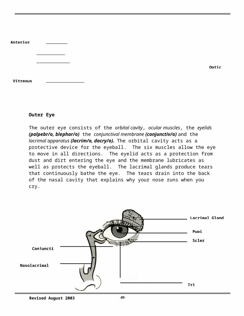
Outer Eye
The outer eye consists of the orbital cavity, ocular muscles, the eyelids (palpebr/o, blephar/o) the conjunctival membrane (conjunctiv/o) and the lacrimal apparatus (lacrim/o, dacry/o). The orbital cavity acts as a protective device for the eyeball. The six muscles allow the eye to move in all directions. The eyelid acts as a protection from dust and dirt entering the eye and the membrane lubricates as well as protects the eyeball. The lacrimal glands produce tears that continuously bathe the eye. The tears drain into the back of the nasal cavity that explains why your nose runs when you cry.
Revised August 2003 -84-
Ciliary Body
Conjunctiva
Cornea
Anterior ChamberPath of Light
Blood Vessels
Optic Nerve
Vitreous
Sclera
Choroid
Iris
Pupil
Lens
Choroid
Retina
Word Parts for the Eye
aque/o water blephar/o, palpebr/o eyelid chrom/o color chori/o, choroid/o choroids conjunctiv/o conjunctiva core/o, pupill/o pupil corne/o, kerat/o cornea cycl/o ciliary body dacry/o, lacrim/o tear/lacrimal duct, tears dipl/o double irid/o, ir/o, ir/i, irit/o iris mi/o contraction, less, smaller mydri/o wide, dilation ocul/o, ophthalm/o, opt/o, optic/o, opt/i eye, vision, sight papill/o optic disc phac/o, phak/o lens phot/o light presby/o old age retin/o retina scler/o sclera ton/o tension uve/o uvea vitre/o glasslike, glassy, gel like -chalasis relaxation -opia, -opsia vision, visual condition -ptosis drooping, sagging -pexy put in place -tropia, -tropion turning
Revised August 2003 -85-
Iris
ScleraPupil
Lacrimal Gland
Conjunctiv
Nasolacrimal

eso- inward exo- outward extra- on the outside intra- on the inside
Term Analysis and Definition (The Eye)
Word Part Term Term Analysis Definition
aque/o aqueous humor
aque = water-ous = pertaining tohumor = body fluid
Pertaining to the watery fluid found in the anterior chamber of the eye.
blephar/o, palpebr/o
blepharoptosis
palpebral
blephar = eyelid-ptosis = drooping
palpebr = eyelid-al = pertaining to
Drooping of the eyelid
Pertaining to the eyelid
chori/o, choroid/o
chorioretinitis
choroiditis
chori = choroid-itis = inflammationretin = retina
Inflammation of the choroid and retina
Inflammation of the choroid
conjunctiv/o conjunctivitis conjunctiv = conjunctiva-itis = inflammation
Inflammation of the conjunctivia(Also known as “pinkeye”)
core/o pupill/o
coreometer
pupillary
core = pupil-meter = instrument to measure
pupil l= pupil-ary = pertaining to
Instrument used to measure the size of the pupil
Pertaining to the pupil
corne/o, kerat/o
corneal
keratoconus
corne = cornea-al = pertaining to
kerat = cornea-conus = cone shaped
Pertaining to the cornea
Abnormal, cone-shaped protrusion of the cornea
cycl/o cycloplegia cycl = ciliary body-plegia = paralysis
Paralysis of the ciliary body
dacry/olacrim/o
dacryostenosis dacry = tear duct-stenosis =
Narrowing of the tear duct
Revised August 2003 -86-

Word Part Term Term Analysis Definitionnasolacrimal narrowing
nas = noselacrim = tear-al = pertaining to
Pertaining to the nose and tear (lacrimal) apparatus
irid/o, ir/o iritis
iridectomy
irid = iris-itis = inflammation
-ectomy = excision
Inflammation of the iris
Excision of the iris
mi/o miosis
miotic
mi = contraction, less, smaller-osis = abnormal condition
-tic = pertaining to
Abnormal contraction of the pupil
Pertaining to a drug that constricts the pupil
mydri/o mydriatic mydri = wide, dilation-tic = pertaining to
Pertaining to a drug that dilates the pupil
ocul/o, opt/o, optic/o, opt/i, ophthalm/
binocular
extraocular
intraocular
exophthalmia
ophthalmologist
ophthalmology
bi = twoocul = eye-ar = pertaining to
extra = outside-ar = pertaining to
intra = inside, within
ex = outwardophthalm = eye-ia = condition
-logist = specialist
-logy = study of
Pertaining to both eyes
Pertaining to the outside of the eye
Pertaining to within the eye
Condition where there is outward protrusion of the eyeball
Specialist in the study and diagnosis of diseases of the eye
Study of diseases of the eye
opt/o optic
optician
opt = vision, sight-ic = pertaining to
-ician = specialist, expert
Pertaining to vision or sight
One who specializes in prescribing glasses for improving sight
Revised August 2003 -87-

Word Part Term Term Analysis Definition
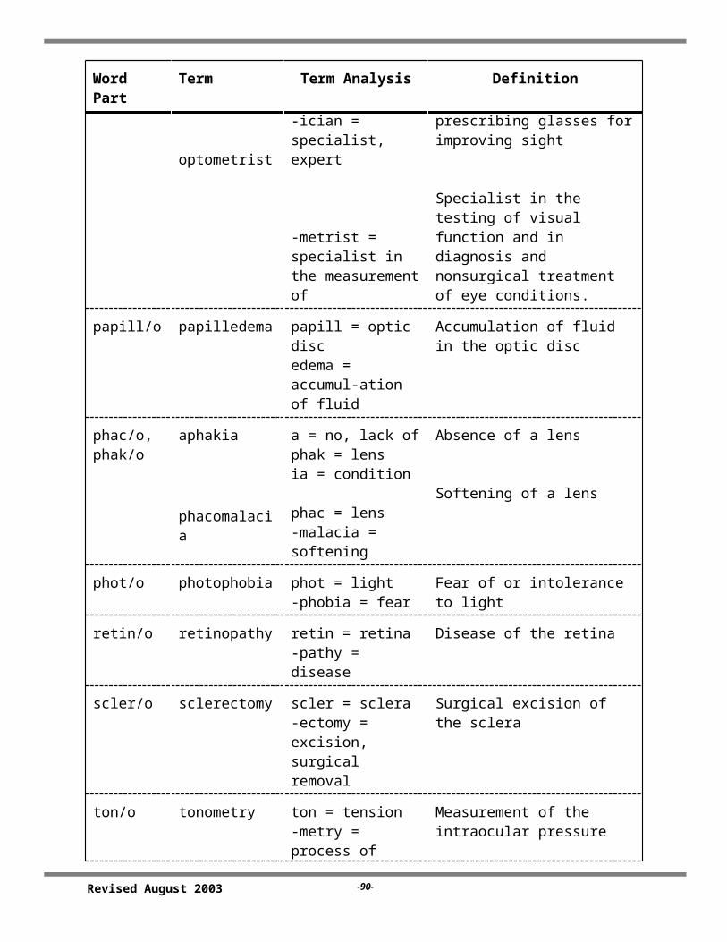
optometrist-metrist = specialist in the measurement of
Specialist in the testing of visual function and in diagnosis and nonsurgical treatment of eye conditions.
papill/o papilledema papill = optic discedema = accumul-ation of fluid
Accumulation of fluid in the optic disc
phac/o, phak/o
aphakia
phacomalacia
a = no, lack ofphak = lensia = condition
phac = lens-malacia = softening
Absence of a lens
Softening of a lens
phot/o photophobia phot = light-phobia = fear
Fear of or intolerance to light
retin/o retinopathy retin = retina-pathy = disease
Disease of the retina
scler/o sclerectomy scler = sclera-ectomy = excision, surgical removal
Surgical excision of the sclera
ton/o tonometry ton = tension-metry = process of measuring
Measurement of the intraocular pressure
uve/o uveitis uve = uvea-itis = inflammation
Inflammation of the uvea of the eye
vitre/o vitreous humor vitre = gel-like-ous = pertaining to
Pertaining to the gel-like substance in the posterior cavity of the eye
-chalasis blepharochalasis
blephar = eyelid-chalasis = relaxation
Relaxation of the eyelid
-tropia esotropia
exotropia
eso = inward-tropia = turning
exo = outward
Turning inward of the eyeball. Also referred to as “cross-eyes”
Turning outward of the eyeball
Revised August 2003 -88-

Word Part Term Term Analysis Definition
-opia, -opsia
amblyopia
diplopia
presbyopia
hyperopia
myopia
ambly = dull, dim-opia = vision
di = double
presby = old age
hyper = above, excessive
my = to shut
Dimness of vision
Double vision
Impaired vision due to aging
Farsightedness ( the light rays go beyond the retina)
(Light rays focus in front of the retina) nearsightedness
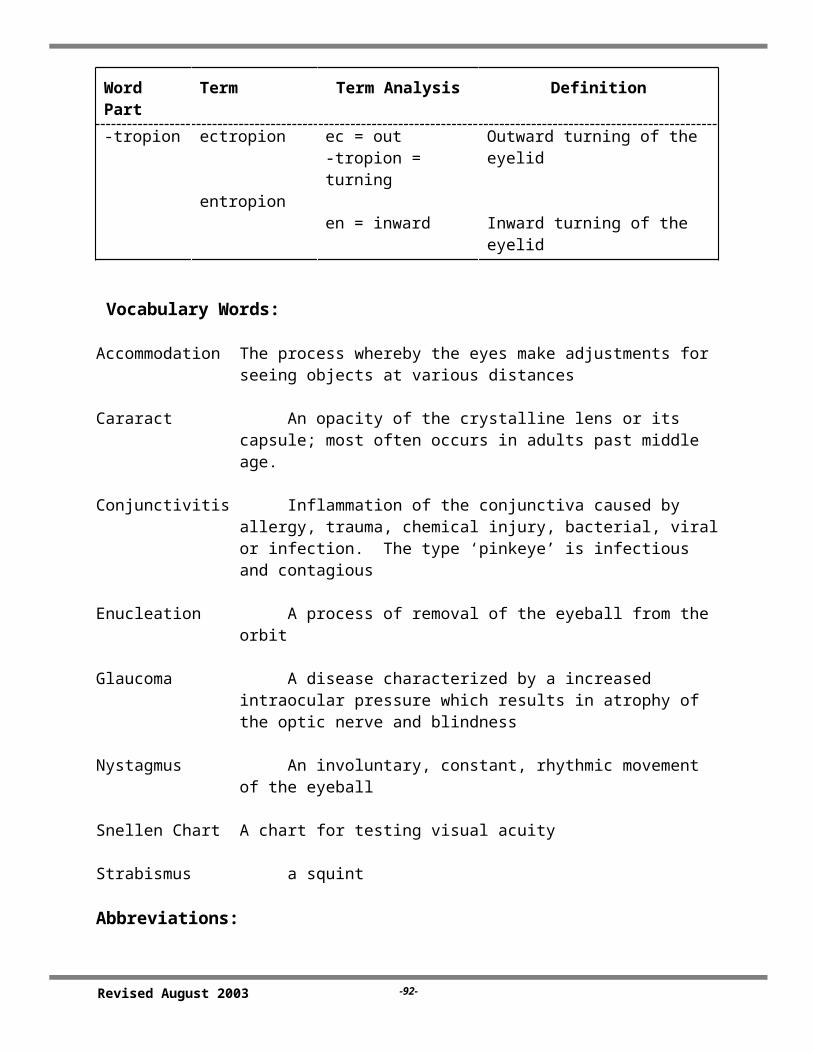
-tropion ectropion
entropion
ec = out-tropion = turning
en = inward
Outward turning of the eyelid
Inward turning of the eyelid
Vocabulary Words:
Accommodation The process whereby the eyes make adjustments for seeing objects at various distances
Cararact An opacity of the crystalline lens or its capsule; most often occurs in adults past middle age.
Conjunctivitis Inflammation of the conjunctiva caused by allergy, trauma, chemical injury, bacterial, viral or infection. The type ‘pinkeye’ is infectious and contagious
Enucleation A process of removal of the eyeball from the orbit
Glaucoma A disease characterized by a increased intraocular pressure which results in atrophy of the optic nerve and blindness
Nystagmus An involuntary, constant, rhythmic movement of the eyeball
Snellen Chart A chart for testing visual acuity
Strabismus a squint
Abbreviations:
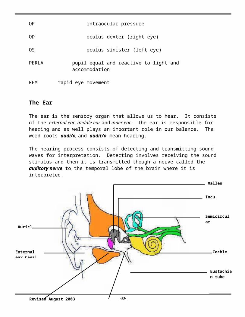
OP intraocular pressure
Revised August 2003 -89-
OD oculus dexter (right eye)
OS oculus sinister (left eye)
PERLA pupil equal and reactive to light and accommodation
REM rapid eye movement
The Ear
The ear is the sensory organ that allows us to hear. It consists of the external ear, middle ear and inner ear. The ear is responsible for hearing and as well plays an important role in our balance. The word roots audi/o, and audit/o mean hearing.
The hearing process consists of detecting and transmitting sound waves for interpretation. Detecting involves receiving the sound stimulus and then it is transmitted though a nerve called the auditory nerve to the temporal lobe of the brain where it is interpreted.
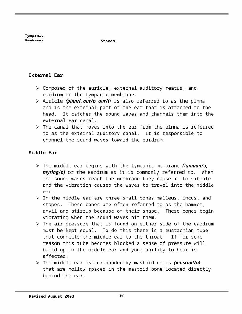
External Ear
Composed of the auricle, external auditory meatus, and eardrum or the tympanic membrane.
Revised August 2003 -90-
Cochlea
Stapes
Incus
Malleus
Auricle
SemicircularCanals
Eustachian tube
Tympanic Membrane
External ear Canal

Auricle (pinn/i, aur/o, aur/i) is also referred to as the pinna and is the external part of the ear that is attached to the head. It catches the sound waves and channels them into the external ear canal.
The canal that moves into the ear from the pinna is referred to as the external auditory canal. It is responsible to channel the sound waves toward the eardrum.
Middle Ear
The middle ear begins with the tympanic membrane (tympan/o, myring/o) or the eardrum as it is commonly referred to. When the sound waves reach the membrane they cause it to vibrate and the vibration causes the waves to travel into the middle ear.
In the middle ear are three small bones malleus, incus, and stapes. These bones are often referred to as the hammer, anvil and stirrup because of their shape. These bones begin vibrating when the sound waves hit them.
The air pressure that is found on either side of the eardrum must be kept equal. To do this there is a eustachian tube that connects the middle ear to the throat. If for some reason this tube becomes blocked a sense of pressure will build up in the middle ear and your ability to hear is affected.
The middle ear is surrounded by mastoid cells (mastoid/o) that are hollow spaces in the mastoid bone located directly behind the ear.
Inner Ear
If you think of a sponge you have a good idea of what the inner ear is like; a twisting series of canals and larger spaces. These canals and spaces are encased in bone that is referred to as the bony labyrinth (labyrinth/o).
This is the part of the ear that has the receptors needed for both hearing and your balance.
The bony labyrinth consists of the vestibule, semicircular canals and the cochlea. The vestibule and semicircular canals are important in maintaining your sense of balance. The cochlea is responsible for hearing.
Sound is transmitted into the cochlea through the oval window. In the cochlea is the organ of Corti that contains fine hair cells that move because of the sound waves. This movement stimulates the nerve cells that create the impulse that is transmitted to the brain.
Word Parts for the Ear
Roots
acoust/o, acous/o hearing, related to hearing audi/o, audit/o hearing, ear aur/o, aur/i,ot/o ear, hearing
Revised August 2003 -91-
cochle/o cochlea (snail, spiral) labyrinth/o labyrinth, inner ear mastoid/o mastoid process myring/o, tympan/o ear drum, tympanic membrane myc/o fungus pinn/i external ear salping/o eustachian tube staped/o stapes tinnit/o ringing, buzzing
Suffixes
-algia, -dynia pain -cusis hearing
Prefixes
bi- two mon- one
Term Analysis and Definition (The Ear)
Word Part Term Term Analysis Definition
audio/o, audit/o
audiogram
audiometry
auditory
audio = hearing-gram = record
-merty = process of measuring
audit = hearing-ory = pertaining to
Record of hearing
Measurement of hearing
Pertaining to hearing
aur/o , ot/o, aur/i
aural
otalgia
otitis media
otorrhea
otoscope
aur = ear-al = pertaining to
ot = ear-algia = pain
-itis = inflammationmedia = middle
-rrhea = discharge
-scope = instrument to visually examine
Pertaining to the ear
Earache
Inflammation in the middle ear
Discharge from the ear
Instrument to visually examine the ear
Revised August 2003 -92-
Word Part Term Term Analysis Definition
otosclerosis
otorrhagia
-sclerosis = hardening
-rrhagia = bleeding
Hardening of the bones of the ear
Bleeding from the ear
cochle/o cochlear cochle - cochlea-ar = pertaining to
Pertaining to the cochlea
labryinth/o labyrinthitis labyrinth = inner ear-itis = inflammation
Inflammation of the inner ear
mastoid/o mastoidectomy mastoid = mastoid bone-ectomy = surgical removal
Surgical removal of the mastoid bone
myring/o, tympan/o
myringotomy
tympanocentesis
myring = ear drum-tomy = surgical incision
-centesis = puncture to remove fluid
Surgical incision into the ear drum
Puncture in the ear drum for removal of fluid
salping/o salpingoscope salping = eustachian tube (ear)scope = instrument used to visually examine
Instrument used to visually examine the eustachian tube
staped/o,stapedi/o
stapedectomy staped = stapes-ectomy = surgical excision
Surgical excision of the stapes
tinnit/o tinnitus tinnit = ringing-us = thing
Ringing in something. Term used to refer to “ringing in the ears”
-cusis presbycusis presby = old age-cusis = hearing
Diminished hearing from old age
Vocabulary Words:
Cerumen earwax
Equilibrium a state of balance
Revised August 2003 -93-
Vertigo a feeling of dizziness
Abbreviations:
AD auris dexter (right ear)
AS auris sinistra (left ear)
ENT ear, nose and throat
EENT eyes, ears, nose throat
Revised August 2003 -94-
Related Documents