Chapter 8: The Muscular System © 2013 John Wiley & Sons, Inc. All rights reserved.

Chapter 8: The Muscular System © 2013 John Wiley & Sons, Inc. All rights reserved.
Dec 14, 2015
Welcome message from author
This document is posted to help you gain knowledge. Please leave a comment to let me know what you think about it! Share it to your friends and learn new things together.
Transcript

Chapter 8: The Muscular System
© 2013 John Wiley & Sons, Inc. All rights reserved.

© 2013 John Wiley & Sons, Inc. All rights reserved.
The Muscular System Overview of muscular tissue Skeletal muscle tissue Contraction and relaxation of skeletal muscle Metabolism of skeletal muscle tissue Control of muscle tension Exercise and skeletal muscle tissue Cardiac muscle tissue Smooth muscle tissue Aging and muscular tissue How skeletal muscles produce movement Principal skeletal muscles

Overview of Muscular Tissue The three types of muscular tissue are skeletal muscle, cardiac muscle, and
smooth muscle.
Skeletal muscle tissue is mostly attached to bones. It is striated and voluntary.
Cardiac muscle tissue forms most of the wall of the heart. It is striated and involuntary.
Smooth muscle tissue is located in viscera. It is nonstriated and involuntary.
Through contraction and relaxation, muscular tissue has five key functions: producing body movements, stabilizing body positions, regulating organ volume, moving substances within the body, and producing heat.
© 2013 John Wiley & Sons, Inc. All rights reserved.

© 2013 John Wiley & Sons, Inc. All rights reserved.
Overview of Muscular Tissue

Skeletal Muscle Tissue Connective tissue coverings associated with skeletal muscle include the
epimysium, covering an entire muscle; perimysium, covering fascicles; and endomysium, covering individual muscle fibers.
Tendons are extensions of connective tissue beyond muscle fibers that attach the muscle to bone.
Skeletal muscles are well supplied with nerves and blood vessels, which provide nutrients and oxygen for contraction.
Skeletal muscle consists of muscle fibers (cells) covered by a sarcolemma that features tunnel-like extensions, the transverse tubules. The fibers contain sarcoplasm, multiple nuclei, many mitochondria, myoglobin, and sarcoplasmic reticulum.
© 2013 John Wiley & Sons, Inc. All rights reserved.

Skeletal Muscle Tissue
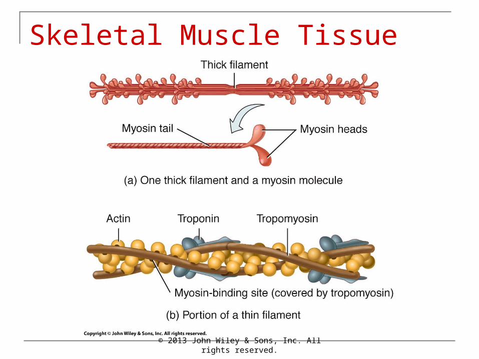
Each fiber also contains myofibrils that contain thin filaments and thick filaments. The filaments are arranged in functional units called sarcomeres.
Thick filaments consist of myosin; thin filaments are composed of actin, tropomyosin, and troponin.
© 2013 John Wiley & Sons, Inc. All rights reserved.

© 2013 John Wiley & Sons, Inc. All rights reserved.
Anatomy Overview:
You must be connected to the internet to run this animation.
• Muscle Tissue

© 2013 John Wiley & Sons, Inc. All rights reserved.
Skeletal Muscle Tissue

© 2013 John Wiley & Sons, Inc. All rights reserved.
SkeletalMuscle Tissue
© 2013 John Wiley & Sons, Inc. All rights reserved.
Skeletal Muscle Tissue

Contraction and Relaxation of Skeletal Muscle Muscle contraction occurs when myosin heads attach to and “walk” along
the thin filaments at both ends of a sarcomere, progressively pulling the thin filaments toward the center of a sarcomere. As the thin filaments slide inward, the Z discs come closer together, and the sarcomere shortens.
The neuromuscular junction (NMJ) is the synapse between a motor neuron and a skeletal muscle fiber. The NMJ includes the axon terminals and synaptic end bulbs of a motor neuron plus the adjacent motor end plate of the muscle fiber sarcolemma.
A motor neuron and all of the muscle fibers it stimulates form a motor unit. A single motor unit may include as few as 10 or as many as 2000 muscle fibers.
© 2013 John Wiley & Sons, Inc. All rights reserved.

Contraction and Relaxation of Skeletal Muscle
When a nerve impulse reaches the synaptic end bulbs of a somatic motor neuron, it triggers the release of acetylcholine (ACh) from synaptic vesicles. ACh diffuses across the synaptic cleft and binds to Ach receptors, initiating a muscle action potential. Acetylcholinesterase then quickly destroys ACh.
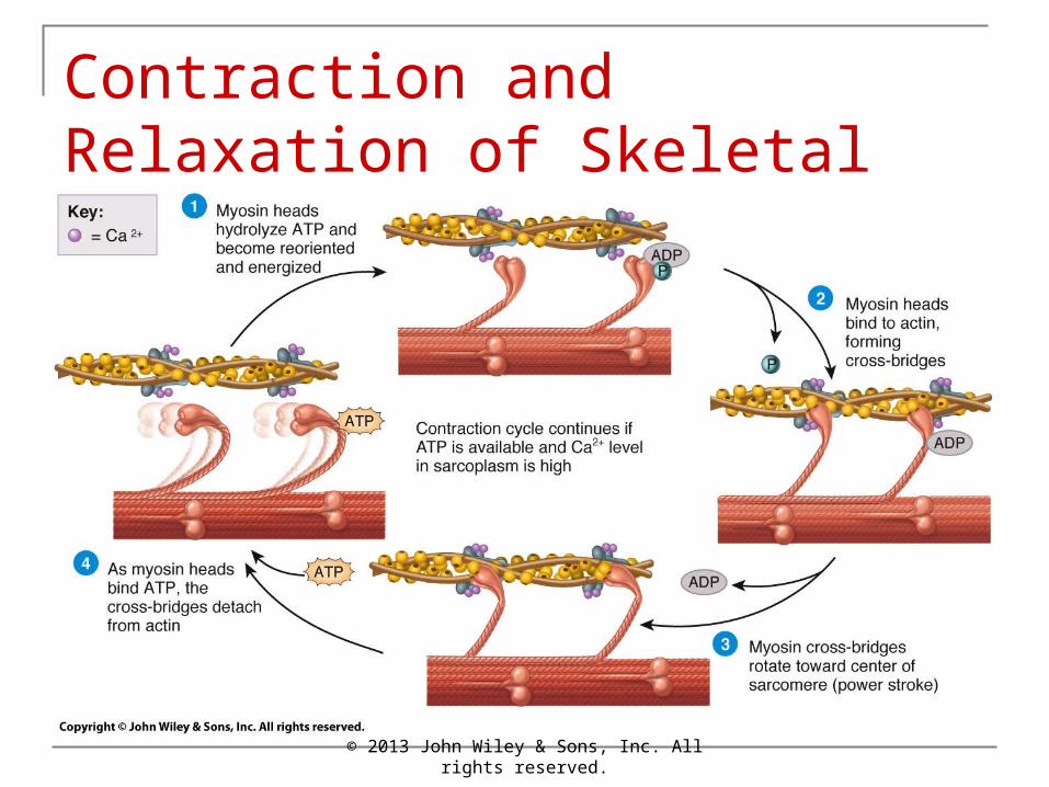
The sliding-filament mechanism of muscle contraction is the sliding of filaments and shortening of sarcomeres that cause the shortening of muscle fibers.
An increase in the level of Ca2+ in the sarcoplasm, caused by the muscle action potential, starts the contraction cycle; a decrease in the level of Ca2+ turns off the contraction cycle.
© 2013 John Wiley & Sons, Inc. All rights reserved.

Contraction and Relaxation of Skeletal Muscle
The contraction cycle is the repeating sequence of events that causes sliding of the filaments: (1) myosin ATPase splits ATP and becomes energized, (2) the myosin head attaches to actin, forming a cross-bridge, (3) the cross-bridge generates force as it swivels or rotates toward the center of the sarcomere (power stroke), and (4) binding of ATP to myosin detaches myosin from actin. The myosin head again splits ATP, returns to its original position, and binds to a new site on actin as the cycle continues.
Ca2+ active transport pumps continually remove Ca2+ from the sarcoplasm into the sarcoplasmic reticulum (SR). When the level of Ca2+ in the sarcoplasm decreases, tropomyosin slides back over and covers the myosin-binding sites, and the muscle fiber relaxes.
Continual involuntary activation of a small number of motor units produces muscle tone, which is essential for maintaining posture.
© 2013 John Wiley & Sons, Inc. All rights reserved.

© 2013 John Wiley & Sons, Inc. All rights reserved.
Contraction and Relaxation of Skeletal Muscle

© 2013 John Wiley & Sons, Inc. All rights reserved.
Animation:
You must be connected to the internet to run this animation.
• Neuromuscular Junctions

© 2013 John Wiley & Sons, Inc. All rights reserved.
Contraction and Relaxation of Skeletal Muscle
© 2013 John Wiley & Sons, Inc. All rights reserved.
Contraction and Relaxation of Skeletal Muscle

© 2013 John Wiley & Sons, Inc. All rights reserved.
Contraction and Relaxation of Skeletal Muscle

© 2013 John Wiley & Sons, Inc. All rights reserved.
Animation:
You must be connected to the internet to run this animation.
• Contraction of Skeletal Muscle Cells

Metabolism of Skeletal Muscle Tissue
Muscle fibers have three sources for ATP production: creatine phosphate, anaerobic cellular respiration, and aerobic cellular respiration.
The transfer of a high-energy phosphate group from creatine phosphate to ADP forms new ATP molecules. Together, creatine phosphate and ATP provide enough energy for muscles to contract maximally for about 15 seconds.
Glucose is converted to pyruvic acid in the reactions of glycolysis, which yield two ATPs without using oxygen. These reactions, referred to as anaerobic cellular respiration, can provide enough ATP for about 30 to 40 seconds of maximal muscle activity.
© 2013 John Wiley & Sons, Inc. All rights reserved.

Metabolism of Skeletal Muscle Tissue
Muscular activity that lasts longer than half a minute depends on aerobic cellular respiration, mitochondrial reactions that require oxygen to produce ATP. Aerobic cellular respiration yields about 36 molecules of ATP from each glucose molecule.
The inability of a muscle to contract forcefully after prolonged activity is muscle fatigue.
Elevated oxygen use after exercise is called recovery oxygen uptake.
© 2013 John Wiley & Sons, Inc. All rights reserved.

© 2013 John Wiley & Sons, Inc. All rights reserved.
Metabolism of Skeletal Muscle Tissue

© 2013 John Wiley & Sons, Inc. All rights reserved.
Metabolism of Skeletal Muscle Tissue

© 2013 John Wiley & Sons, Inc. All rights reserved.
Metabolism of Skeletal Muscle Tissue

© 2013 John Wiley & Sons, Inc. All rights reserved.
Animation:
You must be connected to the internet to run this animation.
• Muscle Metabolism
Control of Muscle Tension
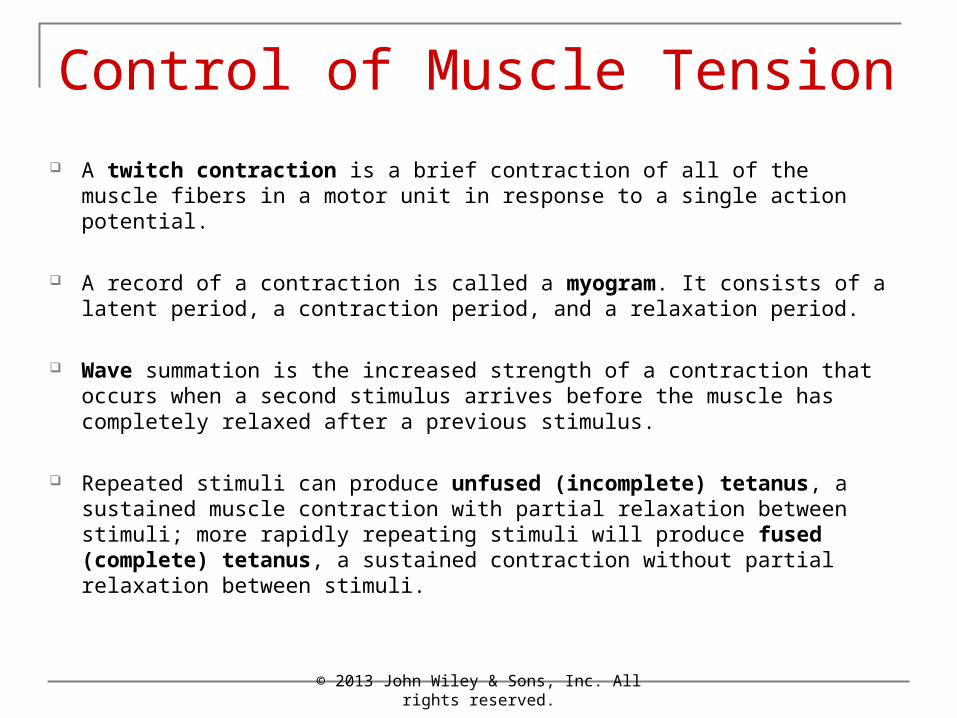
A twitch contraction is a brief contraction of all of the muscle fibers in a motor unit in response to a single action potential.
A record of a contraction is called a myogram. It consists of a latent period, a contraction period, and a relaxation period.
Wave summation is the increased strength of a contraction that occurs when a second stimulus arrives before the muscle has completely relaxed after a previous stimulus.
Repeated stimuli can produce unfused (incomplete) tetanus, a sustained muscle contraction with partial relaxation between stimuli; more rapidly repeating stimuli will produce fused (complete) tetanus, a sustained contraction without partial relaxation between stimuli.
© 2013 John Wiley & Sons, Inc. All rights reserved.

Control of Muscle Tension
Motor unit recruitment is the process of increasing the number of active motor units.
On the basis of their structure and function, skeletal muscle fibers are classified as slow oxidative (SO) fibers, fast oxidative-glycolytic (FOG) fibers, and fast glycolytic (FG) fibers.
Most skeletal muscles contain a mixture of all three fiber types; their proportions vary with the typical action of the muscle.
The motor units of a muscle are recruited in the following order: first SO fibers, then FOG fibers, and finally FG fibers.
© 2013 John Wiley & Sons, Inc. All rights reserved.

© 2013 John Wiley & Sons, Inc. All rights reserved.
Control of Muscle Tension

© 2013 John Wiley & Sons, Inc. All rights reserved.
Control of Muscle Tension

© 2013 John Wiley & Sons, Inc. All rights reserved.
Animation:
You must be connected to the internet to run this animation.
• Control of Muscle Tension

Exercise and Skeletal Muscle Tissue
Various types of exercises can induce changes in the fibers in a skeletal muscle. Endurancetype (aerobic) exercises cause a gradual transformation of some fast glycolytic (FG) fibers into fast oxidative–glycolytic (FOG) fibers.
Exercises that require great strength for short periods produce an increase in the size and strength of fast glycolytic (FG) fibers. The increase in size is due to increased synthesis of thick and thin filaments.
© 2013 John Wiley & Sons, Inc. All rights reserved.

Cardiac Muscle Tissue
Cardiac muscle tissue, which is striated and involuntary, is found only in the heart.
Each cardiac muscle fiber usually contains a single centrally located nucleus and exhibits branching.
Cardiac muscle fibers are connected by means of intercalated discs, which hold the muscle fibers together and allow muscle action potentials to quickly spread from one cardiac muscle fiber to another.
Cardiac muscle tissue contracts when stimulated by its own autorhythmic fibers. Due to its continuous, rhythmic activity (autorhythmicity), cardiac muscle depends greatly on aerobic cellular respiration to generate ATP.
© 2013 John Wiley & Sons, Inc. All rights reserved.

© 2013 John Wiley & Sons, Inc. All rights reserved.
Cardiac Muscle Tissue

Smooth Muscle Tissue
Smooth muscle tissue is nonstriated and involuntary.
In addition to thin and thick filaments, smooth muscle fibers contain intermediate filaments and dense bodies.
Visceral (single-unit) smooth muscle tissue is found in the walls of hollow viscera and of small blood vessels. Many visceral fibers form a network that contracts in unison.
Multiunit smooth muscle tissue is found in large blood vessels, large airways to the lungs, arrector pili muscles, and the eye. The fibers contract independently rather than in unison.
© 2013 John Wiley & Sons, Inc. All rights reserved.

Smooth Muscle Tissue
The duration of contraction and relaxation is longer in smooth muscle than in skeletal muscle. Smooth muscle tone is a state of continuous partial contraction of smooth muscle tissue.
Smooth muscle fibers can be stretched considerably and still retain the ability to contract.
Smooth muscle fibers contract in response to nerve impulses, stretching, hormones, and local factors.
© 2013 John Wiley & Sons, Inc. All rights reserved.

© 2013 John Wiley & Sons, Inc. All rights reserved.
Smooth Muscle Tissue

© 2013 John Wiley & Sons, Inc. All rights reserved.
Smooth Muscle Tissue

Aging and Muscular Tissue
Beginning at about 30 years of age, there is a slow, progressive loss of skeletal muscle, which is replaced by fibrous connective tissue and fat.
Aging also results in a decrease in muscle strength, slower muscle reflexes, and loss of flexibility.
© 2013 John Wiley & Sons, Inc. All rights reserved.
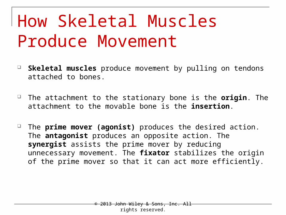
How Skeletal Muscles Produce Movement Skeletal muscles produce movement by pulling on tendons attached to
bones.
The attachment to the stationary bone is the origin. The attachment to the movable bone is the insertion.
The prime mover (agonist) produces the desired action. The antagonist produces an opposite action. The synergist assists the prime mover by reducing unnecessary movement. The fixator stabilizes the origin of the prime mover so that it can act more efficiently.
© 2013 John Wiley & Sons, Inc. All rights reserved.

© 2013 John Wiley & Sons, Inc. All rights reserved.
How Skeletal Muscles Produce Movement
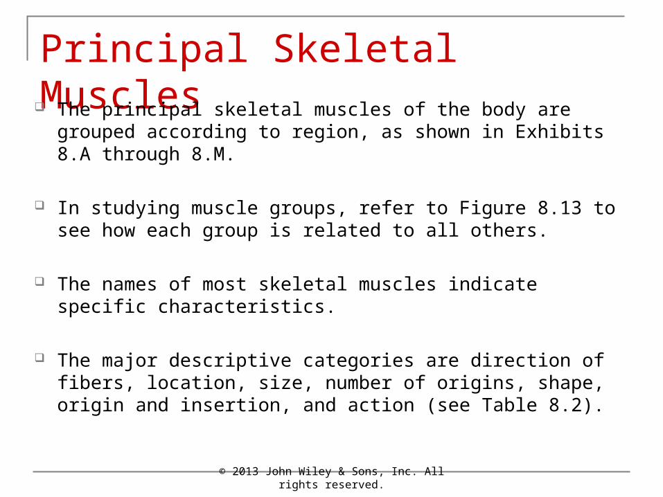
Principal Skeletal Muscles The principal skeletal muscles of the body are grouped
according to region, as shown in Exhibits 8.A through 8.M.
In studying muscle groups, refer to Figure 8.13 to see how each group is related to all others.
The names of most skeletal muscles indicate specific characteristics.
The major descriptive categories are direction of fibers, location, size, number of origins, shape, origin and insertion, and action (see Table 8.2).
© 2013 John Wiley & Sons, Inc. All rights reserved.

© 2013 John Wiley & Sons, Inc. All rights reserved.
Principal Skeletal Muscles

© 2013 John Wiley & Sons, Inc. All rights reserved.
Principal Skeletal Muscles
© 2013 John Wiley & Sons, Inc. All rights reserved.
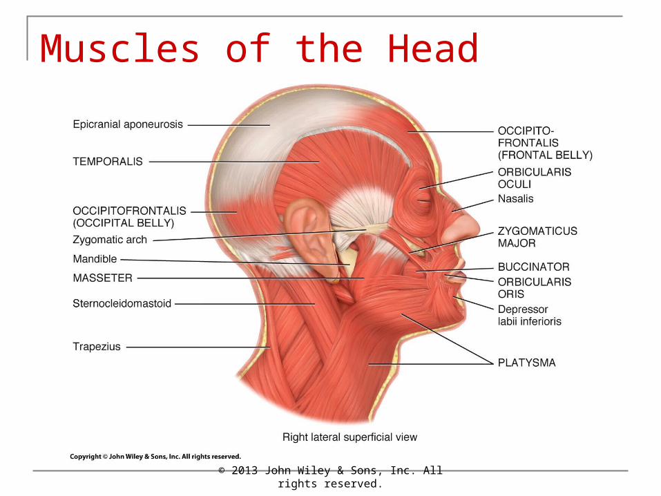
Muscles of the Head

© 2013 John Wiley & Sons, Inc. All rights reserved.
Muscles That Move the Eyeballs

© 2013 John Wiley & Sons, Inc. All rights reserved.
Muscles That Move the Eyeballs

© 2013 John Wiley & Sons, Inc. All rights reserved.
Muscles of the Abdomen

© 2013 John Wiley & Sons, Inc. All rights reserved.
Muscles of the Abdomen

© 2013 John Wiley & Sons, Inc. All rights reserved.
Muscles of the Thorax

© 2013 John Wiley & Sons, Inc. All rights reserved.
Muscles of the Thorax

© 2013 John Wiley & Sons, Inc. All rights reserved.
Muscles of the Thorax

© 2013 John Wiley & Sons, Inc. All rights reserved.
Muscles of the Thorax

© 2013 John Wiley & Sons, Inc. All rights reserved.
Muscles of the Shoulder

© 2013 John Wiley & Sons, Inc. All rights reserved.
Muscles of the Arm
© 2013 John Wiley & Sons, Inc. All rights reserved.
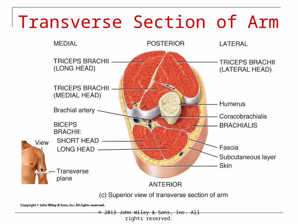
Transverse Section of Arm

© 2013 John Wiley & Sons, Inc. All rights reserved.
Muscles of the Forearm

© 2013 John Wiley & Sons, Inc. All rights reserved.
Transverse Section of Wrist

© 2013 John Wiley & Sons, Inc. All rights reserved.
Muscles that Move the Vertebral Column

© 2013 John Wiley & Sons, Inc. All rights reserved.
Muscles of the Gluteal Region

© 2013 John Wiley & Sons, Inc. All rights reserved.
Muscles of the Gluteal Region

© 2013 John Wiley & Sons, Inc. All rights reserved.
Transverse Section of the Thigh

© 2013 John Wiley & Sons, Inc. All rights reserved.
Muscles of the Lower Leg

© 2013 John Wiley & Sons, Inc. All rights reserved.
Muscles of the Lower Leg

© 2013 John Wiley & Sons, Inc. All rights reserved.
Principal Skeletal Muscles

© 2013 John Wiley & Sons, Inc. All rights reserved.
Principal Skeletal Muscles

End of Chapter 8
Copyright 2013 John Wiley & Sons, Inc. All rights reserved. Reproduction or translation of this work beyond that permitted in section 117 of the 1976 United States Copyright Act without express permission of the copyright owner is unlawful. Request for further information should be addressed to the Permission Department, John Wiley & Sons, Inc. The purchaser may make back-up copies for his/her own use only and not for distribution or resale. The Publishers assumes no responsibility for errors, omissions, or damages caused by the use of these programs or from the use of the information herein.
© 2013 John Wiley & Sons, Inc. All rights reserved.
Related Documents