Chapter 7 CLINICAL AND BIOCHEMICAL EVALUATION CHANGES OVER AGING Angela Abbatecola, B. Gwen Windham, Stefania Bandinelli, Fulvio Lauretani, Giuseppe Paolisso, Luigi Ferrucci Angela Abbatecola, Research Scientist, Department of Geriatric Medicine and Metabolic Diseases, II University of Naples, Naples, Italy. B. Gwen Windham, Staff Clinician, Longitudinal Studies Section, Clinical Research Branch, National Institute on Aging, Baltimore, MD, USA. Stefania Bandinelli, Director of Clinical Rehabilitation, Department of Geriatrics, Italian National Institute of Research and Care on Aging (INRCA), Florence, Italy. Fulvio Laurentani, Research Scientist, INRCA, University of Florence, Department of Geriatrics, Firenze, Italy. Giuseppe Paolisso, Professor of Geriatrics, Department of Geriatric Medicine and Metabolic Diseases, II University of Naples, Naples, Italy. Luigi Ferrucci, Director, Baltimore Longitudinal Study on Aging, Longitudinal Studies Section, Clinical Research Branch, National Institute on Aging, Baltimore, MD, USA. Aging is associated with susceptibility and reduced ability to respond to internal and external stressors. A reduction of functional reserve occurs in many physiological systems and determines increased vulnerability to diseases and high risk of functional dependence. While these modifications can be observed in most persons, particularly in the context of longitudinal studies, they are characterized by extreme variability across individuals and only modest synchronism with chronological aging. As a result, the degree of susceptibility to stressors and exhaustion of functional reserve is dispersed over a wide spectrum in persons of the same age, and the amount of dispersion becomes even greater when we consider older age groups. By convention, geriatricians define “frail” as those individuals that are at the extreme edge of the severity spectrum in this process. Because of this conventional attitude, frailty is often used as an exchangeable term for disability, comorbidity and poor health status, and it is also considered as an

Welcome message from author
This document is posted to help you gain knowledge. Please leave a comment to let me know what you think about it! Share it to your friends and learn new things together.
Transcript
Chapter 7
CLINICAL AND BIOCHEMICALEVALUATION CHANGES OVER AGING
Angela Abbatecola, B. Gwen Windham, Stefania Bandinelli,Fulvio Lauretani, Giuseppe Paolisso, Luigi Ferrucci
Angela Abbatecola, Research Scientist, Department of Geriatric Medicine and MetabolicDiseases, II University of Naples, Naples, Italy. B. Gwen Windham, Staff Clinician,Longitudinal Studies Section, Clinical Research Branch, National Institute on Aging,Baltimore, MD, USA. Stefania Bandinelli, Director of Clinical Rehabilitation, Department ofGeriatrics, Italian National Institute of Research and Care on Aging (INRCA), Florence, Italy.Fulvio Laurentani, Research Scientist, INRCA, University of Florence, Department ofGeriatrics, Firenze, Italy. Giuseppe Paolisso, Professor of Geriatrics, Department of GeriatricMedicine and Metabolic Diseases, II University of Naples, Naples, Italy. Luigi Ferrucci,Director, Baltimore Longitudinal Study on Aging, Longitudinal Studies Section, ClinicalResearch Branch, National Institute on Aging, Baltimore, MD, USA.
Aging is associated with susceptibility and reduced ability torespond to internal and external stressors. A reduction of functional reserveoccurs in many physiological systems and determines increased vulnerabilityto diseases and high risk of functional dependence. While thesemodifications can be observed in most persons, particularly in the context oflongitudinal studies, they are characterized by extreme variability acrossindividuals and only modest synchronism with chronological aging. As aresult, the degree of susceptibility to stressors and exhaustion of functionalreserve is dispersed over a wide spectrum in persons of the same age, and theamount of dispersion becomes even greater when we consider older agegroups.
By convention, geriatricians define “frail” as those individuals thatare at the extreme edge of the severity spectrum in this process. Because ofthis conventional attitude, frailty is often used as an exchangeable term fordisability, comorbidity and poor health status, and it is also considered as an
136 A. ABBATECOLA ET AL
irreversible condition leading to adverse health outcomes. On thecontrary, it is important to conceptualize “frailty” as a continuous, other thana discrete process, where several stages or degrees of severity can be definedas they become useful in research and clinical practice, and that below acertain degree of severity it can be reversed with appropriate interventions.
The aging process is probably associated with the development ofsome unavoidable degree of “frailty” which in the literature is often referredto as “normal aging”. Even the healthiest octogenarian is more sensitive tothe effects of stressors than the healthiest teenager. Diseases and behavioralrisk factors contribute to frailty in ways cannot be completely explained bytraditional biomedical expectations. The effect of environment on frailty iscomplex. An environment that is too challenging causes a rapid exhaustionof functional reserves and leads to an overt instability of the biologicalhomeostasis. On the other hand, an environment that is not at all challenging,leads to a progressive “atrophy” of the homeostatic mechanisms and makesthe individual more susceptible to future stressors.
Clinicians who care for older patients face several times daily thecomplex implications and questions posed by the age-associated frailty. Forexample, it is clearly difficult to prescribe a pharmacological treatment,make decisions about rehabilitation, give advice to patients about therisk/benefits of a specific surgical procedure or establish the prognosis ofdiseases, without having information regarding the degree of functionalreserve and ability to respond to stress. On the other hand, there is still muchdisagreement and discussion of the criteria that should be used for theoperational definition of frailty, and most have no idea as to how to grade itsseverity.
There is some consensus that the basic clinical features of the frailtysyndrome should include the following domains: a) mobility, such as lowerextremity performance and gait abnormalities; b) muscle weakness; c) poorexercise tolerance; d) unstable balance; e) factors related to bodycomposition, such as malnutrition, and sarcopenia (loss of lean body mass),and weight loss. Validity of these factors as critical elements of the frailtysyndrome is provided by studies showing that in older, non-disabled persons,individual components are associated with the classical geriatric syndromes(e.g. falls, symptomatic depression, urinary incontinence and functionalimpairment) and are strong and independent risk factors of disability anddeath.
In 1999, Walston and Fried 1 developed an interpretive frameworkthat combines the elements of the “body composition” and “mobility”domains of the frailty syndrome into a pathophysiologic pathway wheresarcopenia and poor muscle strength, by limiting mobility and physicalactivity, reduce total energy expenditure and nutritional intake, which, inturn, lead to weight loss and further aggravate sarcopenia. Using data from
CLINICAL AND BIOCHEMICAL CHANGES 137
the Cardiovascular Health Study the elements of the pathway were asfollows: 1) unexplained weight loss; 2) poor grip strength; 3) self-reportedexhaustion; 4) slow walking speed; and 5) low physical activity. Afteradjusting for significant confounders, participants with 3 or more of thesecharacteristics were at significantly increased risk of disability,hospitalization and death. The work of Walston and Fried 1 demonstratesthat aggregating measures in the domains of physical function and bodycomposition are an effective initial basis for developing screening criteria foran intrinsic vulnerability that have predictive validity. However, withoutunderstanding the pathophysiologic pathway that leads to frailty as asyndrome that justifies the aggregation of the domains proposed by Waltsonand Fried 1, we lack the critical information to envision any serious attemptsto apply the concept of frailty into clinical practice. In this chapter weexplore some of the biological mechanisms that tend to become dysregulatedwith aging and may contribute to the pathophysiology of the frailtysyndrome. Some of this information concerns biological markers of frailtythat can be already measured. So the possible use of these measures incurrent clinical practice are pointed out in the various sections of thischapter. As our understanding of the pathophysiology, clinical presentation,and consequences of the frailty syndrome improves, many additional uses ofthe measures will emerge, and will help identify new potential targets forintervention.
1. HUMAN AGING
Gradual physiological changes that often parallel the aging processcontribute to the conventional view of “normal” aging. Normal agingimplies a progressive decline of the physiological reserve and the ability tocompensate, but it is compatible with autonomy over the entire life span. Infrail, older persons the decline in functional reserve is accelerated andcompensatory mechanisms start failing with consequent negative healthoutcomes as the functional reserves are depleted.
A better understanding of physiologic changes that proceed andaccompany frailty and, over time, lead to disability is needed if we want tocapture this pathological process in an early stage, and develop targetedinterventions that will delay or postpone the onset of disability.Unfortunately, we have very little information on this topic and, worse yet,what is known is sparse and difficult to reconnect to an overall paradigm.This chapter attempts to address this problem. We will focus on bodycomposition changes, chronic inflammation, oxidative stress and hormonalchanges that often occur in older persons and are accelerated over the agingprocess. Additionally, in the final part of our discussion, we provide our
138 A. ABBATECOLA ET AL
view on how this information can be used in clinical practice to providebetter care to frail older persons.
2. OVERVIEW OF BIOCHEMICAL MARKERS AND AGING (ORFRAILTY)
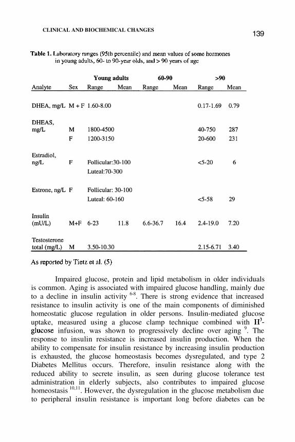
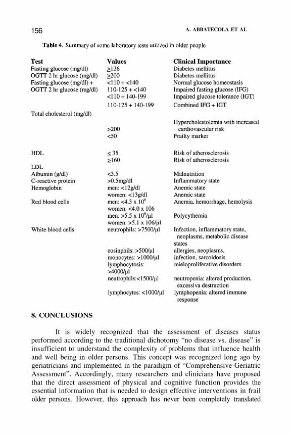
Many efforts have been made to identify biochemical markers ofaging in both normal and frail older individuals 2,3. Traditionally, clinicalchemistry results obtained from laboratory testing are compared with thecorresponding reference values in order to determine whether such valuesfall within the central 95% area under the Gaussian symmetric bell-shapedcurve, or the “normal range”. Reference values calculated using this methodare reported, for example, in the recommendations of the Expert Panel onTheory of Reference Values of the International Federation of ClinicalChemistry and the published guidelines of the National Committee forClinical Laboratory Standards 4. In spite of this generalized trend, severallines of research indicate that a purely statistical approach to theidentification of “normal” values can be misleading, and methods based onpredictive validity in relation to health outcomes should be explored. This isparticularly evident for reference values in geriatric patients. Tietz et al. 5
obtained data from 236 individuals, ages 60 to 90 years, 22 individuals, ages90 to 99 years, and 69, 100 years of age or older. As shown in Table 1 (Tietzet al), plasma levels of dehydroepiandrosterone (DHEA) and DHEA sulphate(DHEAS) were lower in individuals over the age of 90 compared to those ofyoung adults. Other sex hormones, estradiol, estrone and testosterone werefound to be much lower in persons over the age of 90 than those of youngadults. Interestingly, insulin levels tended to increase in adults aging from 60to 90 years of age, while a decline in insulin levels was observed in personsover the age of 90.* (Table 1)
The need for biological markers of pathology in the evaluation ofolder persons is justified by the peculiar relationship that exists betweendiseases and health status in old age. Because aging is associated with anincrement in the global susceptibility to diseases, multiple morbidities arevery common. Analogously, diseases that are not clinically overt are oftenassociated with pathological processes that already affect the health statusbut have not reached the severity threshold that makes them identifiable as“diseases”. Thus, the global burden of comorbidity can be captured onlyindirectly, using functional or biological markers.
* Note that age-adjusted “normal” values for most of these hormones are lacking andthe values prepared in the literature are highly variable from author to author.
CLINICAL AND BIOCHEMICAL CHANGES139
Impaired glucose, protein and lipid metabolism in older individualsis common. Aging is associated with impaired glucose handling, mainly dueto a decline in insulin activity 6-8. There is strong evidence that increasedresistance to insulin activity is one of the main components of diminishedhomeostatic glucose regulation in older persons. Insulin-mediated glucoseuptake, measured using a glucose clamp technique combined with
infusion, was shown to progressively decline over aging 9. Theresponse to insulin resistance is increased insulin production. When theability to compensate for insulin resistance by increasing insulin productionis exhausted, the glucose homeostasis becomes dysregulated, and type 2Diabetes Mellitus occurs. Therefore, insulin resistance along with thereduced ability to secrete insulin, as seen during glucose tolerance testadministration in elderly subjects, also contributes to impaired glucosehomeostasis 10,11. However, the dysregulation in the glucose metabolism dueto peripheral insulin resistance is important long before diabetes can be
140 A. ABBATECOLA ET AL
diagnosed and cannot be overlooked in the clinical evaluation of olderpersons.
Total body protein, lean body mass and the rates of protein synthesisdecline with increasing age. More importantly, such changes are componentsof an impaired homeostatic phenomenon, which is not always balanced withadequate dietary protein intake. Furthermore, an altered state of hepaticprotein synthesis with reduced fibrinogen and other protein carriers, such asthyroxine-binding protein and iron-binding protein may result in an alteredcoagulatory state, reduced thyroxine plasma concentrations and an anemicstate. In addition, reduced plasma concentrations of albumin have beencorrelated with a higher degree of oxidative stress.
Lipid and lipoprotein concentrations vary over an individual’slifespan 12 . In particular, total cholesterol and triglyceride levels tend toincrease up until 50 years of age and then a gradual decline starts to occur.Interestingly, a positive correlation exists between total cholesterol and/ortriglyceride levels with the incidence of cardiovascular disease up to the ageof 50 years. However, the ability of total cholesterol to predict coronary heartdisease in very old individuals remains controversial. Raiha et al 13 reportedthat an elevated level of total cholesterol was not a cardiovascular risk inolder persons, but predicted survival for non-cardiovascular diseasemortality, while Manolio et al 14 did not find any correlation between totalcholesterol and all-cause mortality in older subjects 13. Interestingly, studieshave reported that persistently low cholesterol levels increased the risk ofmortality in males aged 71 to 93 l5. Low total cholesterol levels have alsobeen associated with all-cause mortality in elderly Italian men and women,thus underscoring the potential importance of low levels of cholesterol as awarning sign of rapidly declining health 16.
Such discrepancies can be explained by one of the major differencesbetween middle-aged and older populations, which is the presence of anincreased prevalence of poorer health of older individuals. In fact, older frailpersons with low total cholesterol levels are more likely to have a decreasedsurvival rate than older persons with little or no disease in the presence ofchronically low cholesterol values 17. Interestingly, after adjusting for frailtymarkers in a large sample of older persons, elevated total cholesterol levelspredicted an increased risk for death from CHD, and the risk of death fromCHD decreased as cholesterol levels declined 18. These authors alsoemphasized the finding that frailty markers were consistently associated withlow cholesterol levels, thus confirming similar previous reports. Only furtherinvestigations aimed at evaluating controlled clinical trials with lipid-lowering therapy in non-frail older persons can shed light on the risks or thebenefits of such treatment.
Regarding lipoproteins, high-density lipoprotein cholesterol (HDL-C) is a considered a protective factor for CHD 19. In particular, HDL-C levels

CLINICAL AND BIOCHEMICAL CHANGES141
have been associated with good health status, while reduced HDL-Cvalues are recognized as risk factors for CHD in both middle-aged and olderpersons. Furthermore, it has been shown that reduced HDL-C also predictsnon-CHD/stroke mortality in older persons 20. Thus, low HDL-C may also beconsidered a valid biomarker for chronic disease and poor health status in oldage.
Ueno et al3 have recently described biomarkers of aging in women.In particular, these authors suggest that five variables should be consideredspecific biomarkers for aging in women: forced expiratory volume in 1.0 s
systolic blood pressure (SBP), glucose (GLU, mg/dl), ratio ofalbumin to globulin (A/G) and mean corpuscular hemoglobin (MCH, pg).Such multiple physiological variables reflect the function of diverse vitalfunctions, in particular, pulmonary function, blood pressure, glucosehandling, protein metabolism and hematological functioning. Biological agescores (BAS) were calculated using the parameters mentioned above. Uenoet al 3 concluded that the rate at which women age is relatively slow up until65 years of age. Then after 65 years of age their rates of aging rapidlyincrease. Therefore, the biological processes aimed at maintaining a stablehomeostasis correctly function up to age 65; after 65, false signaling of sucha complex system occurs causing it to lose its effectiveness. This observationis of extreme importance as altered biomarkers are highly correlated withmortality 21 and the frailty syndrome is commonly observed in persons overthe age of 65.
The aggregation of variables in global indices based on theirpredictive role for specific outcomes is very appealing for clinical use, butadds very little to our understanding of the global burden of disease in oldage. Recently, authors have also suggested that the involvement in multiplephysiological systems that is characteristic of older patients with comorbidityshould be interpreted in the context of the “frailty syndrome”. According tocurrent views of frailty, homeostasis is disrupted when the ability ofindividuals to respond to internal and external changes declines below thethreshold of effective compensation. When this occurs, abnormalconcentrations of specific biomarkers of frailty become detectable in thebiological fluids, and structural changes take place in cells and tissues.Unfortunately, serum biomarkers are not currently used to identify frailty,which still remains a clinical diagnosis based on medical history, symptoms,and signs. Clinically, the frailty syndrome is characterized by an excessivereduction in lean body mass, in walking performance and in endurance,associated with a perception of exhaustion and fatigue 22. Several lines ofevidence, however, show that this syndrome is often paralleled by importantchanges in physiological systems accompanied by changes in serum levels ofbiomarkers.
142 A. ABBATECOLA ET AL
3. FRAILTY AND THE NEUROMUSCULAR SYSTEM
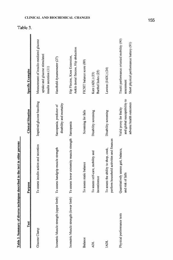
There is growing evidence that the core target of the frailty syndromeis motor organization, specifically the muscular and nervous systems.Disease, disuse and aging trigger a mechanism that impoverishes theredundancy of muscular and nervous backup systems, leading to ameasurable decline of motor performance. Once the process is activated, itsconsequences follow a common pathway leading to a more generalized lossof motor functioning. There is good evidence that measures that are relatedto mobility and motor performance are interpretable as proxy markers offrailty. However, the “diagnosis” of frailty, as a syndrome, hides an array ofdifferent pathologic processes that may involve the integrity andfunctionality of selected physiological subsystems implicated in motorperformance 2. Some of these subsystems include: bone, joints, muscles,peripheral nerves, metabolic efficiency, aerobic capacity and energyproduction. Clinically, the best criteria for screening of frailty are tests ofmobility, gait, balance, manual dexterity, activities of daily living (ADLs) 23,instrumental activities of daily living (IADL) (24) and the Barthel Index 25.However, it is conceivable that specific biomarkers could be measured inorder to identify the involvement of each one of these physiological systemsin the early stages of the disablement process.
Lower extremity performance in non-disabled persons is an excellentpredictor of poor quality of life, deterioration of health status, incidentdisability, health care utilization, nursing home admission and death. Thus,physical performance measures have been considered “vital signs” offunctional decline in older persons 26. In particular, gait speed and the shortperformance battery, developed in the context of the EPESE study, havebeen identified as quantitative estimates of future risk for functional declineand hospitalization 26 Observational studies provide good evidence thatperformance-based measures of mobility are valid proxy measures of frailtyand global susceptibility to adverse health outcomes.
In older persons, poor muscle strength and poor physicalperformance often coexist. Midlife handgrip muscle strength has beenrecognized as an important factor that predicts old age functional ability 27.Observational studies have consistently shown that chronic conditions suchas coronary heart disease, diabetes and pulmonary obstructive disease areassociated with lower muscle strength. These findings suggest that a coremechanism exists that is responsible for changes in body composition anddisease susceptibility in old age and ultimately to the age-associated changesin functional capacity. Possible links between diseases in old age and“frailty” are: nutritional depletion, inflammation, reduced physical activityor inactivity. These mechanisms are, in turn, risk factors for mortality. Thus,
CLINICAL AND BIOCHEMICAL CHANGES143
in persons afflicted with chronic illness, reduced muscle strength couldbe considered an important marker of disease severity. Indeed, handgripmuscle strength has also been associated with overall mortality,independently of poor nutritional status, inflammation and physical inactivity28. These findings suggest that muscle strength has a direct effect onmortality or increases the risk of mortality through a mechanism that is stillunclear.
4. BODY COMPOSITION CHANGES
The two main components of body composition are fat mass andlean (fat-free) mass. Fat-free mass consists of body cell mass, extracellularfluid and the extracellular solids such as collagen and bone mineral 29. Thebody cell mass may be further subdivided into the fat-free portion of cellswithin muscle, viscera and the immune system. The body cell mass isfunctionally the most important compartment in determining energyproduction and expenditure, protein needs, and metabolic response to stress(acute phase response).
There are substantial changes in body composition that accompanythe aging process 30. In particular, the fat mass increases and accumulatespreferentially in the abdominal area, while a parallel decline in muscle massand bone density occurs. Interestingly, the changes in body composition thatbegin to manifest during adulthood may be partially explained by animbalance of energy intake and expenditure. In older adults, however, thesechanges are extremely accelerated compared to younger cohorts, and cannotbe explained simply as an imbalance between energy intake and expenditure.
In most older persons, fat mass constitutes a greater percentage oftotal weight than individuals at younger ages. A population-based study inwhich anthropometric parameters were measured over the entire life span(age range: 20-103 yrs.) demonstrated that the accumulation of abdominal fatwith age occurs primarily during middle age 31 but is different for men andwomen. In particular, the greatest change of waist circumference seems tooccur in men between 20 and 55 years of age, while in women, the waistcircumference tends to increase progressively across the entire life span.
Many studies have shown that increased visceral fat is a risk factorfor age-related diseases such as hypertension, type 2 diabetes, cardiovasculardisease and some types of cancer 20,32. Adipose tissue has also beencorrelated with oxidative stress, reduced glucose uptake, and reduced insulinclearance. Understanding how changes in body composition and, inparticular, fat distribution, affect the risk for many disease states andmortality is one of the most important research questions that should beaddressed in future studies.
144 A. ABBATECOLA ET AL
The simplest clinical indicator of visceral fat is the waistcircumference. A number of studies have shown that waist circumference isan independent risk factor for cardiovascular disease in adults, includingthose 65 years and older. On the contrary, the relationship between bodymass index (BMI), cardiovascular disease and all-cause mortality iscontroversial. The highest mortality rates have been found in older personswith very low BMI, while in middle-aged persons, BMI was positivelyassociated with mortality 33. These data suggest that the relationship betweenbody composition and health-related outcomes in older persons cannot beevaluated simply in conventional terms of body fat, but rather fat distributionand type of fat accumulated, both providing essential information forassessing such risk.
The notion that aging is associated with gradual reduction of leanbody mass is also generic. In fact, selected tissues seem to be more affectedby aging than others. In particular, the decline in non-fat mass is largelyattributed to sarcopenia. Sarcopenia has been increasingly used to describethe age-related decline in both muscle mass and muscle strength. However,despite the term “sarcopenia”, the precise criteria that define such a statehave still not been agreed upon.
Changes in body composition that parallel the aging process arestrongly associated with a decline in physical function and mortality risk.The underlying mechanisms responsible for the excess age-associateddecline of muscle mass and function compared to other sections of lean bodymass are still unknown. Several hypotheses have been proposed, whichinclude: i) intrinsic biochemical and physical changes leading to muscleatrophy 34; ii) reduced neuronal stimulation due to reduction in the number of
or their activity 35; iii) oxidative damage of mitochondrialDNA with accumulations of mutations that reduce the efficiency of themetabolic pathways aimed at energy production 36,37; iv) influence ofexternal factors such as malnutrition, sedentary life-style and diseasetypically observed in older persons (38); v) loss of endogenous hormoneproduction 39,40; and vi) dysregulation of catabolic cytokines 41,42. Over theaging process, both changes in the contractile efficiency of muscle fibers andchanges in tissue quality, such as an increase in connective tissue andpericellular fat infiltration, may also contribute to altered muscle function.
There are many methods that simultaneously measure body fat andfat-free components of body composition. Some important measures are asfollows: 1) skin-fold measurements are obtained using hand-held calipers,which exert a standardized pressure at various body locations. The sum ofthese measurements is used to derive body fat percentage. The calipermethod is based on the idea that the thickness of subcutaneous fat reflects aconstant proportion of the total body fat and that the sites selected formeasurements represent the average thickness of the subcutaneous fat 43,44.
CLINICAL AND BIOCHEMICAL CHANGES145
Also, the mean arm circumference with triceps skin-fold thickness isused to calculate muscle area and thus, derive fat-free mass; 2)hydrodensitometry or underwater weighing is another instrumental methodthat measures whole body density by determining body volume. Thistechnique is based on the two-compartment model (fat and fat-free mass).The densities of bone and muscle are higher than water. Body fat percentageis then calculated from body density using standard equations (sirk orbrozek); 3) bioelectrical impedance analysis (BIA) is measured utilizing asafe electrical signal that passes through the body. Impedance is greatest infat tissue (10-20% water), while fat-free mass (70-75% water) allows thesignal to pass much more easily. The measurement obtained is entered into aformula along with height, weight, and gender to determine lean and fatmass. However, in order to obtain correct evaluations from the BIA it isnecessary that the body is within normal hydration ranges.
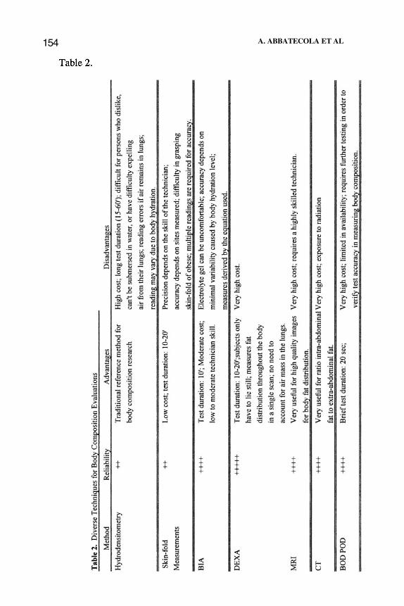
Newer more sophisticated methods for the assessment of bodycomposition include the Dual Energy X-ray Absorptiometer (DEXA),computed tomography (CT), magnetic resonance imaging (MRI) and airdisplacement plethysmography . DEXA is a relatively new method that usesthree compartments (total body mineral, fat-free mass, and fat mass). DEXAconsists of a dual energy beam (two low dose x-ray sources) that scans boneand soft tissue simultaneously. DEXA is currently considered the “goldstandard” measure because of the high degree of precision in a singlemeasurement and the ability to provide the exact location of fat tissuedistribution. CT scanning produces cross-sectional scans of the body. As thebeam rotates data is collected, stored and applied to algorithms to buildimages that describe body composition. MRI utilizes a magnetic field that“excites” water and fat molecules, producing a measurable signal, which isthen measured and analyzed. Whole body air displacement plethysmography(trade name BOD POD) is a new technique that is similar to the underwatermethod, but uses air displacement instead of water. It is based on Boyle’slaw, which states that volume and pressure are inversely related. All themethods described above are summarized in Table 2 (shown on page 154),with information on their reliability, advantages and disadvantages.
It is interesting to speculate on the consequences of sarcopenia thatare not directly related to poor muscle strength. A number of physiologicalfunctions that take place within muscle tissues have a critical effect onhuman metabolism: muscles are a reservoir of body proteins and energy thatcan be utilized in periods of extreme stress or malnutrition; amino-acids canbe mobilized during acute infections and are used as building blocks forantibodies; hormones are produced and catabolized in muscle tissue. Thus,age-related muscle mass reduction may explain the lower metabolicadaptation and immunological response to disease. Indeed, poor musclestrength is a strong predictor of mortality, independent of any other known
146 A. ABBATECOLA ET AL
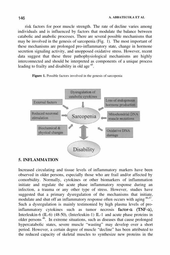
risk factors for poor muscle strength. The rate of decline varies amongindividuals and is influenced by factors that modulate the balance betweencatabolic and anabolic processes. There are several possible mechanisms thatmay be involved in the genesis of sarcopenia (Fig. 1). The most important ofthese mechanisms are prolonged pro-inflammatory state, change in hormonesecretion signaling activity, and unopposed oxidative stress. However, recentdata suggest that these three pathophysiological mechanisms are highlyinterconnected and should be interpreted as components of a unique processleading to frailty and disability in old age 45.
Figure 1. Possible factors involved in the genesis of sarcopenia
5. INFLAMMATION
Increased circulating and tissue levels of inflammatory markers have beenobserved in older persons, especially those who are frail and/or affected bycomorbidity. Normally, cytokines or other biomarkers of inflammationinitiate and regulate the acute phase inflammatory response during aninfection, a trauma or any other type of stress. However, studies havesuggested that a primary dysregulation of the mechanisms that initiate,modulate and shut off an inflammatory response often occurs with aging 46,47.Such a dysregulation is mainly testimonied by high plasma levels of pro-inflammatory cytokines such as tumor necrosisInterleukin-6 (IL-6) (48-50), (Interleukin-1) IL-1 and acute phase proteins inolder persons 46. In extreme situations, such as diseases that cause prolongedhypercatabolic states, severe muscle “wasting” may develop over a shortperiod. However, a certain degree of muscle “decline” has been attributed tothe reduced capacity of skeletal muscles to synthesize new proteins in the
CLINICAL AND BIOCHEMICAL CHANGES 147
aging process 29. An imbalance between muscle protein synthesis anddegradation occurs, ultimately leading to reduced muscle mass, proteincontent and strength. Such imbalance has been linked to pro-inflammatorycytokines capable of inducing proteolysis or inhibiting protein synthesis.
induces muscle proteolysis and plays a significant role in musclewasting (cachexia). and IL-6 can also inhibit protein synthesis, eitherdirectly or by interfering with IGF-1 signaling51.
Elevated plasma levels of each of the pro-inflammatory cytokinesmentioned above have also been observed in many age-related diseases, suchas anemia, osteoporosis, sarcopenia, atherosclerosis, cancer, type 2 DM,impaired cognitive functioning, and Alzheimer’s disease. This furthersupports the theory that a core mechanism contributes to overall age-associated changes in functional capacity.
6. HORMONES
Anabolic agents shift the anabolic/catabolic balance of proteinmetabolism toward the synthesis of new proteins, which is needed to replacethe proteins that are continuously catabolized, therefore maintaining muscleintegrity and volume. Hypertrophy requires the proliferation of muscle nuclei(hyperplasia) in order to maintain the nuclear/cytoplasmic ratio (52).Hormonal factors shown to be related to muscle hypertrophy are: Insulin-likeGrowth Factor-1 (IGF-1), Growth Hormone (GH), testosterone anddehydroepiandrosterone. High IGF-1 concentrations are associated withcharacteristics that are opposite to those typical of aging, including decreasedbody fat content, increased muscle mass and improved metabolichomeostasis of glucose and lipids. At the muscular level, IGF-1 stimulatesprotein synthesis and satellite cell differentiation, thus, playing a crucial rolein the maintenance of muscle mass and function. Many studies haveprovided insight into the signaling pathways by which IGF-1 affects muscleanatomy and function 53-55. Circulating IGF-1 concentrations decrease withadvancing age. The age-associated decline in IGF-1 plasma concentrations isinfluenced by reduced GH levels, and also by nutritional status, insulin andinflammatory cytokines. Specifically, the biologic activity of IGF-1 onmuscle strength can be inhibited by IL-6 55, suggesting that the detrimentaleffect of inflammation on muscle functioning may be mediated by IGF-1.Furthermore, studies provide evidence that the higher concentrations of pro-inflammatory cytokines found in older persons directly interferes with theIGF-1 gene protein expression and receptor sensibility in muscles 55,56. HighIL-6 and low IGF-1 plasma concentrations are considered risk factors forpoor muscle strength, poor lower extremity performance and disability.
148 A. ABBATECOLA ET AL
The aging process is associated with the loss of many anabolicsignals to muscle function. Recent studies have shown that age is not onlyaccompanied by a decline in anabolic activity, but an increase in catabolicsignals as well. In fact, impairment of the anabolic IGF-1 signaling pathwaymay have several negative effects:1)
2)3)
4)
Reduced physical activity that is often observed in advanced age causesdecreased stretch-activation stimulation of different muscle isoforms ofIGF-1;An age-related decline of GH influences IGF-1 muscle response;The progressive loss of appetite with reduced food intake can result inmalnutrition and eventual “wasting”;Loss of motoneurons that are essential for skeletal muscle functioningleads to atrophy and increased proteolysis.There is evidence that the age-associated decline in GH levels in
combination with lower IGF-1 levels also contributes to the development ofsarcopenia 57,58. The reduced pituitary secretion of GH is probably due toage-related changes in the GH-releasing hormone (GHRH). Unfortunately,treatment with GH has demonstrated many adverse effects, such asperipheral edema, arthralgias, glucose intolerance and type 2 diabetes 40.Investigations have demonstrated that therapy with GHRH (somatorelin) inolder persons is capable of restoring the age-related decline of the GHresponse 59. More studies attempting to verify whether such pharmacologicalapproaches can restore muscle functioning as well as the metabolichomeostasis in elderly persons while minimizing side effects are underway.
Testosterone affects muscle mass and muscle strength both directlyand indirectly. It has been reported that testosterone increases proteinsynthesis and intramuscular mRNA concentrations of IGF-1 and decreasesinhibitory IGF binding protein 4 concentrations 60. Due to evidence thattestosterone levels decline with advancing age, a negative impact on musclefunction is not surprising. Older men with low circulating levels oftestosterone tend to have lower muscle strength than men of the same agewith normal testosterone, and studies utilizing supplemental therapy withtestosterone have shown an increase in muscle mass and strength in elderlymales. Testosterone has also been linked to body composition changes suchas an increase in muscle mass and a decrease in fat mass 61. The widespreaduse of testosterone replacement remains controversial due to safety concernsand inconsistent reports regarding clinically important outcome measures.
The production and the circulating levels of adrenal sex hormoneprecursors, dehydroepiandrosterone (DHEA) and DHEA sulphate (DHEAS),decline significantly with aging 62. DHEAS serum levels have beencorrelated with parameters of body composition. Some clinical trials haveshown that supplementation with DHEA resulted in increased musclestrength and decreased body fat63. However, more recently these findings
CLINICAL AND BIOCHEMICAL CHANGES 149
were not confirmed in a large randomised controlled trial performed inmen 60 to 80 years old. The mechanism by which DHEA acts on musclefunction is probably related to the peripheral conversion to testosterone anddihydrotestosterone, but a direct effect of DHEAS cannot be excluded sincespecific receptors have been identified in muscle tissue.
Estrogen levels also decline with aging. Although estrogen has adirect anabolic effect on muscle cells in vitro, several authors believe that theeffect of estrogen on muscle is mediated by their conversion to testosterone64. Interestingly, both estrogen and testosterone are capable of inhibiting IL-6production, suggesting that an age-related decline of such hormones wouldplay a pivotal role in catabolic signaling on muscle tissue. However, theavailable information regarding the effects of supplemental therapy ofestrogen on muscle function is limited and the results are inconclusive.While some studies have concluded that estrogen therapy in postmenopausalwomen does not significantly affect muscle mass or strength 65,66, otherssuggest that estrogen therapy has a positive effect on body composition. Forexample, Sorensen et al 67 demonstrated that estrogen replacement therapywas significantly associated with an increase in lean body mass and also adecrease in total body fat.
As previously mentioned, advancing age is associated with impairedglucose handling mainly due to a reduction of insulin peripheral activity.Since insulin plays a pivotal role for muscle contraction by increasingglucose uptake and promoting intracellular glucose metabolism, it isplausible that age-related insulin resistance (IR) may be an important causeof poor muscle strength in old age. Furthermore, a reduction of insulinperipheral activity may reduce the muscle tissue anabolic rate leading to arelative catabolic state and in turn, facilitating sarcopenia. The contraction ofType I fibers is especially dependent on glucose entry and metabolismcompared to contraction of Type IIa (fast twitch, oxidative, glycolytic) or IIb(fast twitch, glycolytic) fibers 68. Type I fibers are more responsive to insulin,and are more representative of the muscle in older persons 69.
Over the aging process, changes in both the contractile efficiency ofmuscle fibers and changes in tissue quality, such as an increase in connectivetissue and pericellular fat infiltration, may contribute to altered musclefunction 70. Moreover, insulin resistance (IR) could be further worsened bythe occurrence of pericellular fat accumulation both directly and through theincreased production of pro-inflammatory cytokines, such as IL-6 and
Furthermore, a recent study demonstrated that a decline in aged skeletalmuscle force might also be due to a reduction of L-type calcium channels,resulting in excitation-contraction uncoupling and less release by thesarcoplasmic reticulum (SR) 71. Insulin has a stimulatory effect onintracellular calcium uptake 71; thus, an age-related state of IR maynegatively affect muscle contraction via this mechanism. It is well known
150 A. ABBATECOLA ET AL
that IGF-1 actively stimulates insulin receptors. Since IGF-1 levelsdecline throughout aging, the decline in muscle strength that is associatedwith aging may be mediated by decreasing plasma IGF-1 levels thatcontribute to IR. Studies will be needed in order to verify if the impact of IRon specific muscle tissue and functioning in aged individuals exists.
Certain changes typically occur in muscles of older adults. Thequantity of muscle declines, although this varies between individuals, but thecomposition of the muscle changes with aging as well. Increased infiltrationof fat deposited in skeletal muscle tissue may affect muscular function. Muchof the existing data on the association between intramyocellular lipid (IML)content has been obtained directly from muscle tissue biopsies. However, theuse of muscle attenuation through computed tomography (CT) scanning, as ameasure of IML, has been validated 72. In 45 men and women, the musclefiber lipid content determined histologically with oil red staining wascorrelated with muscle attenuation. Thus, the use of CT-derived muscleattenuation should be considered a non-invasive method of measuring IML.In fact, Visser et al 73 demonstrated that increased skeletal muscle fatinfiltration measured by CT scanning was associated with poorer lowerextremity performance independently of total body fat and muscle area inolder men and women.
7. OXIDATIVE STRESS
The accumulation of lipofuscina 74 and increased cross-linking ofcollagen 75 were the first observations reported on the effect of the agingprocess at the cellular level. At that time it was unknown that thesemodifications are, at least in part, related to oxidative stress. More recently,researchers have focused on the progressive changes that occur in the DNAstructure and the underlying causes and potential consequences of thesemutations. For example, a number of studies suggest that excess andunopposed oxidative stress is the main cause of increasing mitochondrialDNA (mtDNA) mutations with aging and in several age-related diseases.Accordingly, oxidative stress characterized by an uncontrolled production offree radicals is considered a major factor in the aging process. In aerobicbiological systems, free radicals are primarily derived from oxygen and areproduced by splitting a covalent bond into atoms or molecules with anunpaired electron, therefore forming highly reactive oxygen species (ROS).In normal physiological conditions, the intra-mitochondrial environment ischaracterized by a substantial equilibrium between the production of ROSand the activity of anti-oxidant mechanisms, such as glutathione peroxidase(GSH-Px) and superoxide dismutase (SOD). Several lines of researchsuggest that the endogenous production of ROS increases with age and, in
CLINICAL AND BIOCHEMICAL CHANGES151
parallel, the activity (but not the tissue concentration) of anti-oxidantsdeclines, therefore increasing the risk of damage due to oxidative stress,especially at the level of the mtDNA. In addition to its effect on mtDNA,oxidative stress also adversely impacts other vulnerable targets, includinglipid and protein components of membranes. Free radicals can cause lipidoxidation with a consequent reduction in transmembrane transportation. Age-related overproduction of ROS may also lead to the activation of apoptosis.Therefore, the accumulation of oxidatively damaged mtDNA, together withenhanced apoptosis act synergistically to cause the general decline ofbiochemical and physiological function of tissues over the aging process.The underlying mechanisms by which these events accompany the agingprocess remain to be identified and merit further investigation.
Studies suggest that the degree of unopposed oxidative stress ispredictive of mortality. In particular, the production of free radicals in theheart, kidney and liver is inversely proportional to the maximum lifespan 76
and rate of mitochondrial oxygen radical generation is negatively associatedwith animal longevity. In animal models, caloric restriction, which decreasesthe rate of aging, also decreases mitochondrial oxygen radical production andoxidative damage to mtDNA.
The mitochondrial DNA/oxidative stress hypothesis can explaincertain age-related disease states such as Parkinson’s disease, Alzheimer’sdisease and skeletal muscle myopathies. Recently, epidemiological studieshave suggested that dietary anti-oxidants may have a significant impact onage-related disease states 77,78. This remains unproven in clinical trials. Theclinical implications of oxidative stress are complex, and intervention studiesare needed to further clarify the role of dietary and supplemental antioxidantsin the prevention of age-associated frailty.
SUCCESSFUL AGING8.
The possibility of reaching the extreme end of the human lifespanresults from the continuous adaptation of the body to respond to negativeinsults over the aging process. Healthy centenarians are a very selectivegroup of persons representing one of the best living models of “successfulaging”. Many studies have focused on centenarians’ anthropometric,endocrine and metabolic characteristics in order to formulate a clearerclinical picture of successful aging. They report that the average fat free mass(FFM) of healthy centenarians is similar to that of other aged subjects butlower than middle-aged adult subjects 79. However, most healthycentenarians do not undergo the usual anthropometric derangement found inelderly persons. For example, the waist/hip ratio has been found to be lowerin healthy centenarians than in other aged individuals. Regarding endocrine
152 A. ABBATECOLA ET AL
factors, total plasma IGF-1 concentrations were similar in both healthycentenarians and aged subjects, but the molar ratio IGF-1/IGF bindingprotein-3, an expression of free plasma IGF-1 concentration, was observed tobe significantly elevated in healthy centenarians compared to elderly subjects80. This ratio is negatively correlated with body mass index, body fat content,plasma triglycerides, and FFA and LDL concentrations80.
While serum markers may be useful for helping identify successfulaging, caution should be used since the interpretation may be different inyounger adults than in older persons. For example, in older persons, theability of total cholesterol to predict age-related diseases such as coronaryheart disease (CHD) has been challenged. In middle-aged adults, totalcholesterol levels have been shown to have a direct association with CHDand mortality, but such a relationship in individuals over the age of 65remains controversial. In older persons, a J or U-shaped association has beenreported, suggesting that extremely high or low concentrations have anincreased risk of death 81,82; total cholesterol levels have also been shown tohave a positive association, an inverse association, and no association withmortality in older persons.
Up to now, most studies have considered the association of totalcholesterol on CHD in subjects under the age of 85 years. Interestingly, arecent study reporting data on fractionated lipoprotein levels among personsover the age of 85 years, concluded that low HDL cholesterol, but not highLDL cholesterol, is a risk factor for mortality from CHD and stroke inpersons over the age of 85 83. Lipoprotein (a) [Lp(a)], a genetically controlledcholesterol-rich lipoprotein, has been hypothesized as an independent riskfactor for premature CHD, stroke, and peripheral artery disease in elderlypersons 84,85. This observation may be due to the presence of Lp(a) inatherosclerotic plaques and its ability to stimulate smooth muscleproliferation 86.
The physiological and pathological roles of Lp(a) probably changewith aging. Support for this comes from a study by Baggio et al 87, whichreported no significant differences in Lp(a) serum concentrations amonghealthy centenarians, persons <65 and >65 years of age, even though Lp(a)has been proposed as an independent risk factor for cardiovascular disease.Centenarians with high Lp (a) levels had significantly higher IL-6 levels,thus characterizing the paradox of successful aging. Such data questions theidea that Lp(a) is under strict genetic control and suggests that environmentalfactors may play a significant role in older adults, including subclinicalinflammatory states. Thus, a continuous remodeling of lipid metabolism mayoccur with aging and may be critical for successful aging. The deleteriousreshaping of serum lipids and lipoproteins in young, adult and elderlyindividuals are considered risk factors for age-related diseases, while theirbiological significance in healthy centenarians remains unknown. Thus, only
CLINICAL AND BIOCHEMICAL CHANGES153
future investigations highlighting age-related changes in lipid physiologyof healthy centenarians on mortality rates will resolve such discrepancies.
Healthy centenarians have a lower degree of oxidative stress. In fact,it has been shown that healthy centenarians have greater plasma antioxidantdefenses than aged individuals. According to the remodeling theory onaging, the body continuously and correctly adapts to deleterious changesover time. As previously mentioned, an age-related up-regulation of theinflammatory response takes place over the aging process. In both sick andhealthy elderly individuals, peripheral blood markers of inflammation(albumin, cholesterol, IL-6 and CRP) have been associated with increasedrisk for mortality. Interestingly, the age-related increase of serum IL-6 levelshas been seen in both elderly and centenarian individuals 49,87. IL-6dysregulation has been suggested to play a role in the pathogenesis of avariety of age-related diseases, such as diabetes and atherosclerosis 88.Indeed, healthy centenarians have elevated pro-inflammatory cytokineconcentrations, but do not have the same high incidence of most age-relateddisease states in other elderly persons. Thus, in healthy centenarians suchabnormal cytokine levels may reflect a state of subclinical inflammation.The reason why healthy centenarians adapt correctly to such insults remainsunknown.
Whether healthy centenarians have some protective genetic factorsthat can protect against deleterious changes or facilitate the remodelingprocess remain unknown. Future investigations will be needed in order toprovide the necessary answers.
Tables 3 and 4 summarize some of the clinical and biochemicalevaluations described in the text above, and that can be used to assess thedegree of “successfulness: of the aging process. Note that these are onlyexamples. An exhaustive list would be very large, and out of the scope ofthis chapter.
154 A. ABBATECOLA ET AL
CLINICAL AND BIOCHEMICAL CHANGES155
156 A. ABBATECOLA ET AL
8. CONCLUSIONS
It is widely recognized that the assessment of diseases statusperformed according to the traditional dichotomy “no disease vs. disease” isinsufficient to understand the complexity of problems that influence healthand well being in older persons. This concept was recognized long ago bygeriatricians and implemented in the paradigm of “Comprehensive GeriatricAssessment”. Accordingly, many researchers and clinicians have proposedthat the direct assessment of physical and cognitive function provides theessential information that is needed to design effective interventions in frailolder persons. However, this approach has never been completely translated
CLINICAL AND BIOCHEMICAL CHANGES 157
into clinical practice and many geriatricians claim that the administrationof any available medical treatment is still conditioned to a previous diagnosisof specific diseases and hypotheses about specific pathophysiologicalpathways. Furthermore, significant changes in health status may occur andbe amenable to effective treatment long before any clear effect on physicaland cognitive function is detected.
We propose that the concept of frailty – a condition that involvesimpairment in multiple physiological systems and is characterized byexhaustion of functional reserve, massive use of compensatory strategies andhigh risk of homeostatic breakdown – can be used by clinicians to gain abetter understanding of the global burden of disease and reduced physicalfunction in older persons and their interaction with the “pure” effect of aging.Unfortunately, there is still no agreement on the criteria that should be usedin order to identify frail older persons. However, there is general consensusthat comorbidity, disease susceptibility and risk of developing multiplehealth outcomes are commonly associated with the detection of abnormalcirculating levels of several biomarkers and changes in body composition.Thus, composite measures of mobility, body composition, strength,circulating hormones and biomarkers of inflammation may help cliniciansunderstand the severity of health status deterioration in their patients overand beyond the information provided by the simple diagnosis of diseases.Aggregate measures of these outcomes should be developed in future studiesand are likely to replace the current criteria for the definition of frailty, bothin research projects and in clinical practice.
REFERENCESWalston J, Fried LP: Frailty and the older man. Med Clin North Am, 1999Sep;83(5): 1173-94Ferrucci L, Cavazzini C, Corsi A, Bartali B, Russo CR, Lauretani F, Ferrucci L.Cavazzini C, Corsi AM, Bartali B, Russo CR, Lauretani F, Bandinelli S, BandinelliS, Guralnik JM: Biomarkers of frailty in older persons. J Endocrinol Invest,2002;25;10-5Ueno L, Yamashita Y, Moritani T, Nakamura E: Biomarkers of aging in women andthe rate of longitudinal changes. J Physiol Anthropol, 2003; 22: 37-46NCCLS: How to define, determine, and utilize reference intervals in the clinicallaboratory; proposed guideline. NCCLS document C28P (ISBN 1-56238-143-1),NCCLS, 940 West Valley Road, Suite 1400, Wayne, Penn 19087, USA, 1992Tietz NW, Shuey DF, Wekstein DR: Laboratory values in fit aging individuals--sexagenarians through centenarians. Clin Chem, 1992 Jun;38(6):1167-85Fink RI, Kolterman OG, Griffin J, Olegsky JM: Mechanism of insulin resistance inageing. J Clin Invest, 1983; 71: 1523-35Rowe JW, Minaker KL, Pallotta JA, Flier JS: Characterization of the insulinresistance in aging. J Clin Invest, 1983; 71: 1581-7Gumbiner B, Thorburn AW, Ditzler TM, Bulacan F, Henry RR: Role of impairedintracellular glucose metabolism in the insulin resistance of aging. Metabolism,1992;41:1115-21
1)
2)
3)
4)
5)
6)
7)
8)
158 A. ABBATECOLA ET AL
10)
11)
12)
13)
14)
15)
16)
9) Ferrannini E, Vichi S, Beck-Nielsen H, Laakso M, Paolisso G, Smith U:European Group for the study of Insulin Resistance (EGIR). Insulin action and age.Diabetes, 1996; 45:947-53Kosaka K, Kuzuya T, Hagura R, Yoshinaga H: Insulin response to oral glucose loadis consistently decreased in established non-insulin-dependent diabetes mellitus: theusefulness of decreased early insulin response as a predictor of non-insulin-dependent diabetes mellitus. Diabet Med, 1996:S109-19Defronzo 79: Glucose intolerance and aging: evidence for tissue insensitivity toinsulin. Diabetes, 1979 Dec;28(12): 1095-101Lipid Research Clinics Program Epidemiology Committee: Plasma lipiddistributions in selected North American populations: the Lipid Research ProgramPrevalence Study. Circulation 1979;60: 427-439Raiha I, Marniemi J, Puukka P, Toikka T, Ehnholm C, Sourander L: Effect of serumlipids, lipoproteins, and apolipoproteins on vascular and nonvascular mortality inthe elderly. Arterioscler Thromb Vase Biol, 1997 Jul;17(7): 1224-32Manolio TA, Ettinger WH, Tracy RP, Kuller LH, Borhani NO, Lynch JC et al:Epidemiology of low cholesterol levels in older adults. The Cardiovascular HealthStudy, Circulation 1993; 87: 728-37Schatz IJ, Masaki K, Yano K Chen R, Rodriguez BL, Curb JD: Cholesterol and all-cause mortality in elderly people from the Honolulu Heart Program: a cohort study,Lancet. 2001 Aug 4;358(9279):351-5Brescianinni S, Maggi S, Farchi G, Mariotti S, Di Carlo A, Baldereschi M, InzitariS: Low total cholesterol and increased risk of dying: Are low levels clinical warningsigns in the elderly? Results from the Italian Longitudinal Study on Aging. JAGS,2003; 51: 991-6Pekkanen J, Nissinen A, Vartiainen E, Salonen JT, Punsar S, Karvonen MJ:Changes in serum cholesterol level and mortality: a 30 year follow up. Am JEpidemiol, 1994; 139: 155-65Corti MC, Guralnik J, Salive M, Harris T, Ferrucci L, Glynn R, Havlik R:Clarifying the direct relation between total cholesterol levels and death fromcoronary heart disease in older persons. Annuals of Internal Medicine, 1997; 126:753-760Schaefer EJ, Moussa PB, Wilson PW, McGee D, Dallal G, Castelli WP: Plasmalipoproteins in healthy octogenarians: lack of reduced high density lipoproteincholesterol levels: results from the Framingham Heart Study. Metabolism, 1989Apr;38(4):293-6Volpato S, Leveille S, Corti MC, Harris T, Guralnik J: The value of serum albuminand high-density lipoprotein cholesterol in defining mortality risk in older personswith low serum cholesterol. JAGS, 2001; 49: 1142-1147Masoro EJ: Physiological system markers of aging. Exp Gerontol, 1988;23(4-5):391-7Fried LP, Tangen CM, Walston J, Newman AB, Hirsch C, Gottdiener J, Seeman T,Tracy R, Kop WJ, Burke G, McBurnie MA: Cardiovascular Health StudyCollaborative Research Group. Frailty in older adults: evidence for a phenotype. JGerontol A Biol Sci Med Sci, 2001;56(3):M146-56Katz S, Akpom CA: 12 Index of ADL. Med Care May, 14 (5 suppl):116-8; 1976Lawton MP, Brody EM: Assessment of older people:self-maintaining andinstrumental activities of daily living. Gerontologist, 1969; 9: 179-186Wade DT, Colin C: The Barthel ADL Index – a standard measure of physicaldisability. Int Disabil Studies, 1988; 10: 64-67Studenski S, Perera S, Wallace D, Chandler JM, Duncan PW, Rooney E, Fox M,Guralnik JM: Physical performance measures in the clinical setting. J Am GeriatrSoc, 2003;51(3):314-22
17)
18)
19)
20)
21)
22)
23)24)
25)
26)
CLINICAL AND BIOCHEMICAL CHANGES 159
Rantanen T, Guralnik JM, Foley D, Masaki K, Leveille S, Curb JD, White L:Midlife hand grip strength as a predictor of old age disability. JAMA, 1999;10;281(6):558-60Rantanen T, Volpato S, Ferrucci L, Heikkinen E, Fried LP, Guralnik JM: Handgripstrength and cause-specific and total mortality in older disabled women: exploringthe mechanism. J Am Geriatr Soc, 2003 May;51(5):636-41Roubenoff R: The Pathophysiology of Wasting in the Elderly. J Nutr,1999;129:256S-259SWilliamson DF: Descriptive epidemiology of body weight and weight changes in17,000 adults. Int J. Obes, 1988; 12:391-401Bartali B, Benvenuti E, Corsi AM, Bandinelli S, Russo CR, Di Iorio A, Lauretani F,Ferrucci L: Changes in anthropometric measures in men and women across the life-span: findings from the InCHIANTI study. Soz Praventivmed, 2002; 47(5): 336-48Elahi D, Dyke MM, Andres R: Aging, fat, metabolism and obesity. In Masoro EJ(ed): Handbook of Physiology. Section 11: Aging. Oxford University Press, NewYork, 1995:47Visscher TL, Seidell JC, Molarius A, van der Kuip D, Hofman A, Witteman JC: Acomparison of body mass index, waist-hip ratio and waist circumference aspredictors of all-cause mortality among the elderly: the Rotterdam study. Int J ObesRelat Metab Disord, 2001;25(11): 1730-5Carmeli E, Coleman R, Reznick AZ. The biochemistry of aging muscle. Exp.Gerontol, 37:477-89, 2002Zeynep E, Faisal Beg M, Burke D, De Luca C: Effects of aging on motor-unitcontrol properties. Am Physiol Soc, 2081-91,1999Mecocci P, Fano G, Fulle S, MacGarey U, Shinobu L, Polidori MC, Cherubini AVecchiet J, Senin U, Beal MF: Age-dependent increases in oxidative damage toDNA, lipids and proteins in human skeletal muscle. Free Radic Biol Med, 26:303-8,1999McKenzie D, Bua E, McKiernan S, Cao Z, Aiken JM, Wanagat J: MitochondrialDNA deletion mutations: a causal role in sarcopenia. Eur J Biochem, 269:2010-5,2002Baumgartner R, Koehler K, Romero L, Garry P: Serum albumin is associated withskeletal muscle in elderly men and women. Am J Clin Nutr, 64:552-8,1996.Frisancho AR. Triceps skinfold and upper arm muscle norms for assessmentof nutritional status. Am J Clin Nutr. 1974; 27:1052-8.Kamel H, Maas D, Duthie E: Role of hormones in the pathogenesis andmanagement of sarcopenia. Drugs Aging, 2002; 19: 869-877Blackman MR, Sorkin JD, Munzer T, Bellantoni MF, Busby-Whitehead J, StevensTE, Jayme J, O’Connor KG, Christmas C, Tobin JD, Stewart KJ, Cottrell E, St ClairC, Pabst KM, Harman S: Growth hormone and sex steroid administration in healthyaged women and men: a randomized controlled trial. JAMA, 2002 Nov13;288(18):2282-92Iovino M, Monteleone P, Steardo L: Repetitive growth hormone-releasing hormoneadministration restores the attenuated growth hormone (GH) response to GH-releasing hormone testing in normal aging. J Clin Endocrinol Metab,1989;69(4):910-3Roubenoff R: Inflammatory and hormonal mediators of cachexia. J Nutr,1997;127:1014S-1016SRoubenoff R: Catabolism of aging: is it an inflammatory process? Curr Opin ClinNutr Metab Care, 2003;6(3):295-9Frisancho AR: Triceps skinfold and upper arm muscle norms for assessment ofnutritional status. Am J Clin Nutr 1974; 27:1052-8
27)
28)
29)
30)
31)
32)
33)
34)
35)
36)
37)
38)
39)
40)
41)
42)
43)
44)
160 A. ABBATECOLA ET AL
46)
47)
48)
49)
50)
51)
52)
53)
54)
55)
56)
57)
58)
59)
60)
61)
62)
63)
45) Ferrucci L, Guralnik J: Inflammation, hormones, and body composition at acrossroad. Am J Med, 2003 Oct 15;115(6):501-2Heymfield SB, McManus C, Smith et al: Anthropometric measurement of musclemass: revised for equations for calculating bone free arm muscle area. Am J ClinNutr, 1982; 36: 680-690Brunnsgard H, Pederson M, Pederson BK: Aging and proinflammatory cytokines.Curr Opin Hematol 8: 131-136, 2001Franceschi C, Bonafè M, Valensin S et al: Inflamm-aging: An evoluzinaryperspective on immunosenescence. Ann NY Acad Sci, 908: 244-254, 2000Ershler WB: Interleukin-6: a cytokine for gerontologists. J Am Geriat Soc, 41:176-181, 1993Fagiolo U, Cossarizza A, Scala E, et al: Increased cytokine production inmononuclear cells of healthy elderly people. Eur J Immunol, 23: 2375-2378,1993Albright JW, Albright JF: Soluble receptors and other substances that regulateproinflammatory cytokines in young and aging humans. J Geronto A Biol Sci MedSci, 55: 20-25, 2000Frost RA, Lang CH: Regulation of insulin-like growth factor-1 in skeletal muscleand muscle cells. Minevra Endocrinol, 2003;28(1):53-73Barton-Davis ER, Shoturma DI, Sweeney HL: Contribution of satellite cells to IGF-I induced hypertrophy of skeletal muscle. Acta Physiol Scand, 1999;167(4):301-5Cappola AR, Bandeen-Roche K, Wand GS, Volpato S, Fried LP: Association ofIGF-I levels with muscle strength and mobility in older women. J Clin EndocrinolMetab, 2001;86(9):4139-46Cappola A, Qian-Li X, Ferrucci L, Guralnik JM, Volpato S, Fried LP: Insulin-likeGrowth Factor I and Interleukin-6 Contribute Synergistically to Disability andMortality in Older Women. JCEM, 88: 2019-25, 2003Barbieri M, Ferrucci L, Ragno E, Corsi A, Bandinelli S, Bonafe M, Olivieri F,Giovagnetti S, Franceschi C, Guralnik JM, Paolisso G: Chronic inflammation andthe effect of IGF-I on muscle strength and power in older persons. Am J PhysiolEndocrinol Metab, 2003;284(3):E481-7Ferrucci L, Penninx BW, Volpato S, Harris TB, Bandeen-Roche K, Balfour J,Leveille SG, Fried LP, Md JM: Change in muscle strength explains accelerateddecline of physical function in older women with high interleukin-6 serum levels. JAm Geriatr Soc, 2002;50(12): 1947-54Rudman D: Growth hormone, body composition and aging. J Am Geriatr Soc, 1985;33: 800-1Kelijam M: Age-related alterations of growth hormone/insulin-like-growth-factor Jaxis. J Am Geriatr Soc, 1991; 39: 295-307Baumgartner RN, Waters DL, Gallagher D, Morley JE, Garry PJ: Predictors ofskeletal muscle mass in elderly men and women. Mech Ageing Dev.1999;107(2):123-36Mooradian AD, Morley JE, Korenman SG: Biological actions of androgens. EndocrRev, 1987; 8: 1-28Baulieu EE, Thomas G, Legrain S, Lahlou N, Roger M, Debuire B, Faucounau V,Girard L, Hervy MP, Latour F, Leaud MC, Mokrane A, Pitti-Ferrandi H, Trivalle C,de Lacharriere O, Nouveau S, Rakoto-Arison B, Souberbielle JC, Raison J, Le BoucY, Raynaud A, Girerd X, Forette F: Dehydroepiandrosterone (DHEA), DHEAsulfate, and aging: contribution of the DHEAge Study to a sociobiomedical issue.Proc Natl Acad Sci U S A , 2000;97(8):4279-84Tummala S, Svec F: Correlation between the administered dose of DHEA andserum levels of DHEA and DHEA-S in human volunteers: analysis of publisheddata. Clin Biochem, 1999;32(5):355-61
CLINICAL AND BIOCHEMICAL CHANGES161
Grinspoon S, Corcoran C, Miller K, Biller BM, Askari H, Wang E, Hubbard J,Anderson EJ, Basgoz N, Heller HM, Klibanski A: Body composition and endocrinefunction in women with acquired immunodeficiency syndrome. J Clin EndocrinolMetab 1997;82: 1332-7Haarbo J, Marslew U, Gotfredsen A, Christiansen C: Postmenopausal hormonereplacement therapy prevents central distribution of body fat after menopause.Metabolism, 1991;40(12):1323-6Gower BA, Nyman L: Associations among oral estrogen use, free testosteroneconcentration, and lean body mass among postmenopausal women. J ClinEndocrinol Metab, 2000;85(12):4476-80Sorensen MB, Rosenfalck AM, Hojgaard L, Ottesen B: Obesity and sarcopeniaafter menopause are reversed by sex hormone replacement therapy. Obes Res, 2001;9: 622-6Song XM, Ryder JW, Kawano Y, Chibalin AV, Krook A, Zierath JR: Muscle fibertype specificity in insulin signal transduction. Am J Physiol, 277:R1690-6, 1999Staron R, Hagerman F, Hikida R, Murrey T, Hostler D, Crilll M, Ragg K, Toma K:Fiber type composition of the vastus lateralis muscle of young men and women.Journal of histochemistry & cytochemistry, 48:623-629, 2000. Lexell J, Henriksson-Larsen K, Wilblad B, Siostrom M: Distribution of aging studied in whole musclecross-sections. Muscle Nerve, 6:588-595,1983Madsen OR, Lauridsen UB, Hartkopp A, Sorensen OH: Muscle strength and softtissue composition as measured by dual energy x-ray absorptiometry in women aged18-87 years. Eur J Appl Physiol Occup Physiol, 76: 239-45,1997Paolisso G, Gambardella A, Balbi V, Galzeranno D, Verza M, Varricchio M,D'Onofrio F: Effects of magnesium and nifedipine infusions on insulin action.Substrate oxidation and blood pressure in aged hypertensive patients. Am JHypertens, 920-26,1993Goodpaster BH, Kelley DE, Thaete FL, He J, Ross R: Skeletal muscle attenuationdetermined by computed tomography is associated with skeletal muscle lipidcontent. Appl Physiol, 2000;89(1):104-10Visser M, Deeg DJ, Lips P, Harris TB, Bouter LM: Skeletal muscle mass andmuscle strength in relation to lower-extremity performance in older men andwomen. J Am Geriatr Soc, 2000;48(4):381-6Strehler BL: Time, cells and aging. Academic Press, New York, 1981Kohn RR: Aging of animals: possible mechanisms. In Kohn RR, ed, Principles ofmammalian aging, Prentice-Hall, Englewood Cliffs, NJ, 1978Sohal RS, Svensson I, Sohal BH, Brunk UT: Superoxide anion radical production indifferent animal species. Mech Ageing Dev, 1989;49(2): 129-35Gilgun-Sherki Y, Melamed E, Offen D: Antioxidant treatment in Alzheimer'sdisease: current state. J Mol Neurosci, 2003;21(1):1-12Paolini M, Sapone A, Canistro D, Chieco P, Valgimigli L: Antioxidant vitamins forprevention of cardiovascular disease. Lancet, 2003 ;362(9387):920Paolisso G, Gambardella A, Balbi V, Ammendola S, D’Amore A, Varricchio M:Body composition, body fat distribution, and resting metabolic rate in healthycentenarians. Am J Clin Nutr, 1995;62(4):746-50Paolisso G, Ammendola S, Del Buono A, Gambardella A, Riondino M,Tagliamonte MR, Rizzo MR, Carella C, Varricchio M: Serum levels of insulin-likegrowth factor-I (IGF-I) and IGF-binding protein-3 in healthy centenarians:relationship with plasma leptin and lipid concentrations, insulin action, andcognitive function. J Clin Endocrinol Metab, 1997;82(7):2204-9Frank JW, Reed DM, Grove JS, Benfante R: Will lowering population levels ofserum cholesterol affect total mortality? Expectations from the Honolulu HeartProgram. J Clin Epidemiol, 1992;45(4):333-46
64)
65)
66)
67)
68)
69)
70)
71)
72)
73)
74)75)
76)
77)
78)
79)
80)
81)
162 A. ABBATECOLA ET AL
82) Stemmermann GN, Chyou PH, Kagan A, Nomura AM, Yano K: Serumcholesterol and mortality among Japanese-American men. The Honolulu (Hawaii)Heart Program. Arch Intern Med, 1991;151(5):969-72Weverling-Rijnsburger AW, Jonkers IJ, van Exel E, Gussekloo J, Westendorp RG:High-density vs low-density lipoprotein cholesterol as the risk factor for coronaryartery disease and stroke in old age. Arch Intern Med, 2003 14;163(13):1549-54Maher V, Brown BG: Lipoprotein (a) and coronary heart disease. Curr OpinLipidol, 1995; 6: 229-235Dahlen G: Lp(a) lipoprotein in cardiovascular disease. Atherosclerosis, 1994; 108:111-26Sansoni P, Cossarizza A, Brianti V, Fagnoni F, Snelli G, Monti D, Marcato A,Passeri G, Ortolani C, Forti E, et al: Lymphocyte subsets and natural killer cellactivity in healthy old people and centenarians. Blood, 1993;82(9):2767-73Baggio G, Donazzan S, Monti D, Mari D, Martini S, Gabelli C, Dalla Vestra M,Previato L, Guido M, Pigozzo S, Cortella I, Crepaldi G, Franceschi C:Lipoprotein(a) and lipoprotein profile in healthy centenarians: a reappraisal ofvascular risk factors. FASEB J, 1998;12(6):433-7Pickup JC, Mattock MB, Chusney GD, et al: NIDDM as a disease of the innateimmune system: association of acute-phase reactants and interleukin-6 withmetabolic syndrome X. Diabetologia, 1997; 40:1286-1292Judge JO, Schechtman K, Cress E: The relationship between physical performancemeasures and independence in instrumental activities of daily living. The FICSITGroup. Frailty and Injury: Cooperative Studies of Intervention Trials. J Am GeriatrSoc, 1996;44(11):1332-41Tinetti ME: Performance oriented assessment of mobility problems in elderlypatients. J Am Geriatr Soc, 1986;34: 119-126Guralnik JM, Simonsick EM, Ferrucci L, Glynn RJ, Berkman LF, Blazer DG,Scherr PA, Wallace RB: A short physical performance battery assessing lowerextremity function: association with self-reported disability and prediction ofmortality and nursing home admission. J Gerontol, 1994;49(2):M85-94
83)
84)
85)
86)
87)
88)
89)
90)
91)
Related Documents