Chapter 6B The Peripheral Nervous System: Special Senses

Chapter 6B The Peripheral Nervous System: Special Senses.
Jan 01, 2016
Welcome message from author
This document is posted to help you gain knowledge. Please leave a comment to let me know what you think about it! Share it to your friends and learn new things together.
Transcript

Chapter 6BThe Peripheral Nervous System:
Special Senses
Review (6A)
• Pathways, perceptions, sensations• Receptor Physiology
– Receptors have differential sensitivities to various stimuli.
– graded receptor potentials.
– Receptor potentials may initiate action potentials
– Receptors adaptation (slow/fast)
– Each somatosensory pathway is “labeled”
– Acuity is influenced by receptive field size and lateral inhibition.
– PAIN -The brain has a built-in analgesic system.
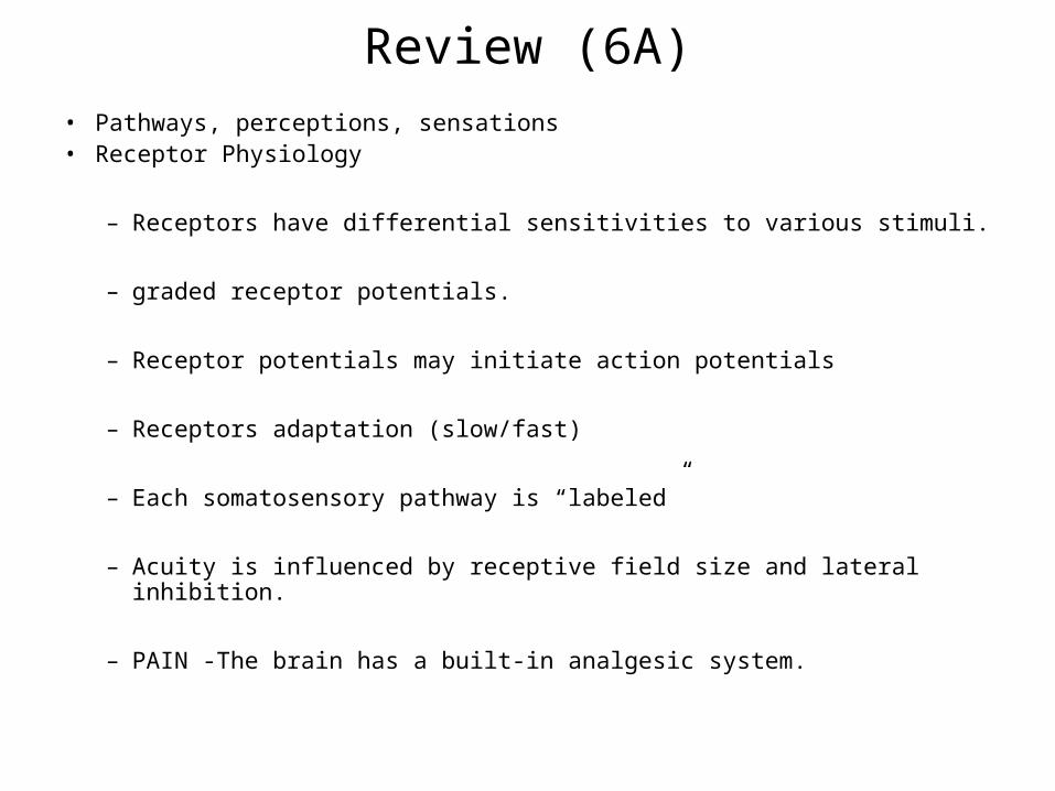
What are your special senses?
• Vision
• Hearing
• Balance and equilibrium
• Taste
• Smell
• Touch

Vision outline
• Anatomy• Muscles and light control
• Refraction and refractive structures
– Refractive problems
• Retina, photoreceptors, transduction
• Visual cortical processing
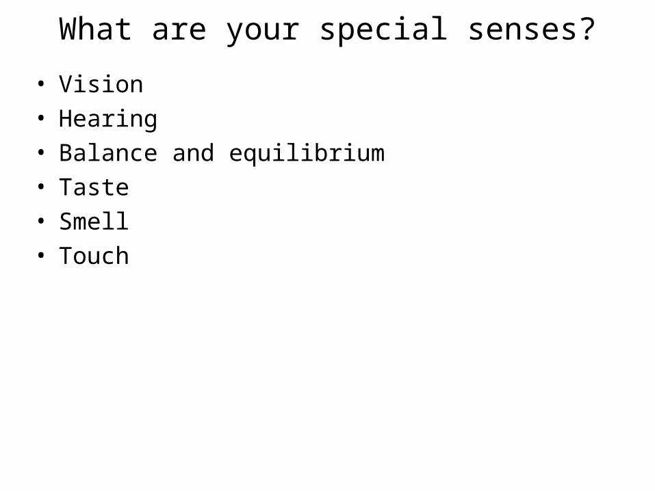
EyelidsAct like shutters to protect eye from environmental hazards
EyelashesTrap fine, airborne debris such as dust before it can fall into eye
Tears Continuously produced by lacrimal glands Lubricate, cleanse, bactericidal
EyesocketIris regulates amount of light
AnatomyEye protection
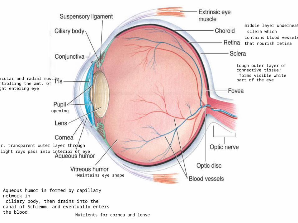
Eye
tough outer layer of connective tissue; forms visible white part of the eye
•anterior, transparent outer layer through• which light rays pass into interior of eye
middle layer underneath
sclera which
contains blood vessels
that nourish retina
Circular and radial muscle controlling the amt. of light entering eye
opening
Nutrients for cornea and lense
•Maintains eye shape
Aqueous humor is formed by capillary network in ciliary body, then drains into the canal of Schlemm, and eventually enters the blood.

Eye
• Interior consists of two fluid-filled cavities separated by the lens– Posterior cavity
• Larger cavity between lens and retina• Contains vitreous humor
– Important in maintaining the spherical shape of eyeball
– Anterior cavity• Anterior cavity between cornea and lens• Contains aqueous humor
– Carries nutrients for cornea and lens– Produced by capillary network within ciliary body
• Fovea– Pinhead-sized depression in exact center of retina– Point of most distinct vision– Has only cones
• Macula lutea– Area immediately surrounding fovea– Fairly high acuity
• Macular degeneration– Leading cause of blindness in western hemisphere

Vision outline
• Anatomy
• Light and muscle control• Refraction and refractive structures
– Refractive problems
• Retina, photoreceptors, transduction
• Visual cortical processing

Eye
• Convex structures of eye produce convergence of diverging light rays that reach eye
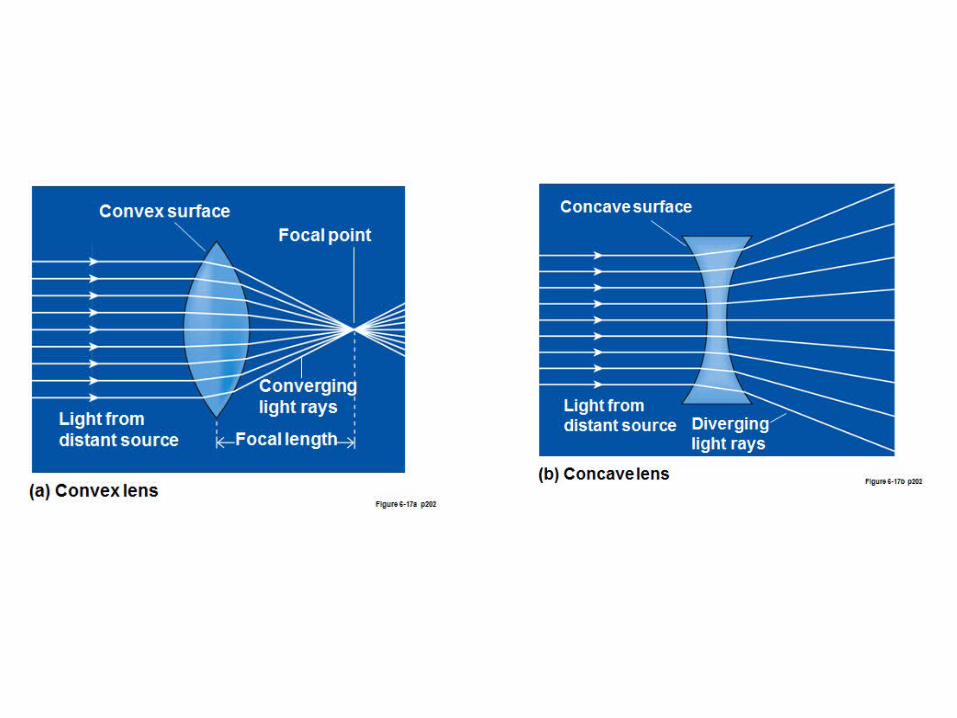

Eye
Focusing on Distant and Near Light Sources
What happens to light rays when they leave the light source?

Eye
• Two structures most important in eye’s refractive ability are– Cornea
• Contributes most extensively to eye’s total refractive ability• Refractive ability remains constant because curvature never
changes– Lens
• Refractive ability can be adjusted by changing curvature as needed for near or far vision
• Accommodation – Change in strength and shape of lens– Accomplished by action of ciliary muscle and suspensory
ligaments– Age-related reduction in accommodation ability - presbyopia

Mechanics of Accommodation
Far vision Near vision
* Light moves towards thick part of lens

Fig. 6-11, p. 193

Vision outline
• Anatomy
• Muscles and light control
• Refraction and refractive structures– Refractive problems
• Retina, photoreceptors, transduction
• Visual cortical processing

Emmetropia, Myopia, and Hyperopia
Vision outline
• Anatomy
• Muscles and light control
• Refraction and refractive structures
– Refractive problems
• Retina, photoreceptors, transduction• Visual cortical processing

Retinal Layers• Retina – receptor containing portion is actually an extension of the
CNS• Neural portion of retina consists of three layers of excitable cells
– Outermost layer containing rods and cones– Middle layer of bipolar cells– Inner layer of ganglion cells
• Axons of ganglion cells join to form optic nerve– Point on retina at which optic nerve leaves is the optic disc
» Region often called the blind spot because no image can be detected here because of lack of rods and cones

Photoreceptors• Rod and cone cells• Consist of three parts
– Outer segment• Detects light stimulus
– Inner segment• Contains metabolic
machinery of cell
– Synaptic terminal• Transmits signal generated
in photoreceptor on light stimulation to next cells in visual pathway

Photopigments
• Undergo chemical alterations when activated by light• Consists of two components
– Opsin• Protein that is integral part of disc membrane
– Retinene • Derivative of vitamin A• Light-absorbing part of photopigment
• Four different photopigments– Rod pigment
• Provide vision only in shades of gray• Rhodopsin
– Absorbs all visible wavelengths
– Cone pigments• Respond selectively to various wavelengths of light• Make color vision possible
– Red cones– Green cones– Blue cones
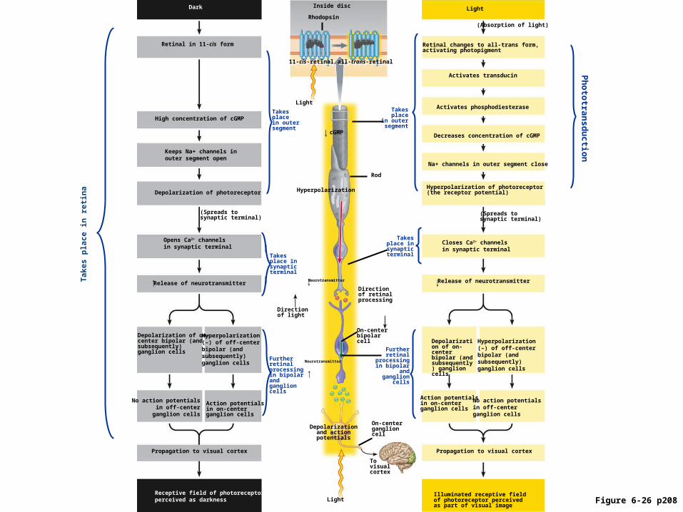
Illuminated receptive fieldof photoreceptor perceivedas part of visual image
Propagation to visual cortex
Action potentials in on-center ganglion cells
Depolarization of on-center bipolar (and subsequently) ganglion cells
Release of neurotransmitter
(Spreads tosynaptic terminal)
Hyperpolarization of photoreceptor(the receptor potential)
Ph
oto
tran
sd
uc
tion
Retinal changes to all-trans form, activating photopigment
Na+ channels in outer segment close
(Absorption of light)
Light
Takesplace
in outersegment
Takesplace in
synapticterminal
Directionof retinalprocessing
On-centerbipolarcell
Furtherretinal
processingin bipolar
andganglion
cells
On-centerganglioncell
Tovisualcortex
Depolarizationand actionpotentials
Neurotransmitter
Directionof light
Hyperpolarization
Rod
Rhodopsin
all-trans-retinalall-trans-retinal11-cis-retinal11-cis-retinal
Inside disc
Light
Hyperpolarization(–) of off-centerbipolar (andsubsequently)ganglion cells
Activates phosphodiesterase
Activates transducin
Decreases concentration of cGMP
Closes Ca2+ channelsin synaptic terminal
No action potentialsin off-centerganglion cells
Release of neurotransmitter
Opens Ca2+ channelsin synaptic terminal
(Spreads tosynaptic terminal)
Retinal in 11-cis form
High concentration of cGMP
Keeps Na+ channels inouter segment open
Depolarization of photoreceptor
Depolarization of on-center bipolar (and subsequently) ganglion cells
Hyperpolarization(–) of off-centerbipolar (andsubsequently)ganglion cells
Action potentials in on-center ganglion cells
No action potentialsin off-center
ganglion cells
Propagation to visual cortex
Receptive field of photoreceptorperceived as darkness
Dark
Light
Takesplacein outersegment
Takesplace insynapticterminal
Furtherretinalprocessingin bipolarandganglioncells
Tak
es p
lace
in
ret
ina
Neurotransmitter
cGMP
Figure 6-26 p208

Fig. 6-25, p. 202
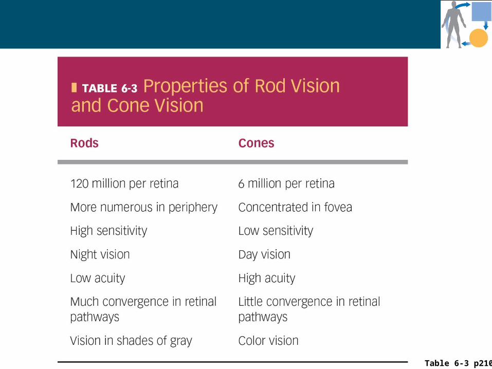
Table 6-3 p210

The sensitivity of the eyes varies through dark and light
adaptation.•Dark adaptation
•Can gradually distinguish objects as you enter a dark area. •Due to the regeneration of rod photopigments that had been broken down by previous light exposure.
•Light adaptation •Can gradually distinguish objects as you enter an area with more light. •Due to the rapid breakdown of cone photopigments.
Vision outline
• Anatomy
• Muscles and light control
• Refraction and refractive structures
– Refractive problems
• Retina, photoreceptors, transduction
• Visual fields
• Visual cortical processing

Visual Processing
• Blending color
– 3 cone types – blue, green, red
– Stimulated in a ratio to produce blends % max
• Distinguishing contours
– On center and off center ganglion cells
• Images on the retina are upside down and backwards.
• Depth perception

Hearing outline
• Anatomy– Outer, middle, inner
• Hearing
• Transmission of sound waves
• Hair cells and transduction
• Cochlea and canals/ducts
• Pitch and loudness
• Auditory cortical processing

Ear
• Consists of three parts– External ear
• Consists of pinna, external auditory meatus, and tympanum• Transmits airborne sound waves to fluid-filled inner ear• Amplifies sound energy
– Middle ear• Transmits airborne sound waves to fluid-filled inner ear• Amplifies sound energy
– Inner ear • Houses two different sensory systems
– Cochlea » Contains receptors for conversion of sound waves into
nerve impulses which makes hearing possible– Vestibular apparatus
» Necessary for sense of equilibrium

Ear

Hearing outline
• Anatomy
– Outer, middle, inner
• Hearing• Transmission of sound waves
• Pitch and loudness
• Hair cells and transduction
• Cochlea and canals/ducts
• Auditory cortical processing

Hearing • Neural perception of sound energy• Involves two aspects
– Identification of the sounds (“what”)– Localization of the sounds (“where”)
• Sound waves– Traveling vibrations of air– Consist of alternate regions of compression
and rarefaction of air molecules

Hearing
• Pitch (tone) of sound– Depends on frequency of air waves 20-20,000 cps, 1000-4000
• Intensity (loudness)– Depends on amplitude of air waves
• Timbre (quality) – Determined by overtones
Hearing outline
• Anatomy
– Outer, middle, inner
• hearing
• Transmission of sound waves• Pitch and loudness
• Hair cells and transduction
• Cochlea and canals/ducts
• auditory cortical processing

Transmission of Sound Waves
• Tympanic membrane vibrates when struck by sound waves
• Middle ear transfers vibrations through ossicles (malleus, incus, stapes) to oval window (entrance into fluid-filled cochlea)
• Waves in cochlear fluid set basilar membrane in motion
• Receptive hair cells are bent as basilar membrane is deflected up and down
• Mechanical deformation of specific hair cells is transduced into neural signals that are transmitted to auditory cortex in temporal lobe of brain for sound perception

Fig. 6-33, p. 213

Hearing outline
• Anatomy
– Outer, middle, inner
• Hearing
• Transmission of sound waves
• Pitch and loudness• Hair cells and transduction• Cochlea and canals/ducts
• Auditory cortical processing

Fig. 6-33c, p. 213
Transduction toAuditory nerve
amplification
• Inner
– Deformation and rubbing on the tectoral membrane hyper or depolarizes the cells resulting in a signal.
• Outer
– Do not signal the brain
– Fine tuning
– Accentuates movement of basilar membrane
• (lengthening and shortening)

Fig. 6-35, p. 215

Sound waves
Vibration oftympanic membrane
Vibration ofmiddle ear bones
Vibration ofoval window
Fluid movementwithin cochlea
Vibration ofbasilar membrane
Vibration ofround
window
Dissipation ofenergy (no
soundperception)
In ear
Fig. 6-36, p. 216
Bending of hairs of receptorhair cells of organ of Cortias basilar membrane move-ment displaces these hairsin relation to overlyingtectorial membrane in whichthe hairs and embedded
Graded potential changes(receptor potential) inreceptor cells
Changes in rate of actionpotentials generated inauditory nerve
Propagation of actionpotentials to auditory cortexin temporal lobe of brain forsound perception
Hearing outline
• Anatomy
– Outer, middle, inner
• hearing
• Transmission of sound waves
• Pitch and loudness• Hair cells and transduction• Cochlea and canals/ducts
• auditory cortical processing

Auditory Cortical Processing
– Primary auditory cortex is tonotopically organized
– Locations on basilar membrane map to locations in the cortex
– Pathway• Hair cells-afferent auditory nerve- synapses in
brainstem and thalamus (LGN)-higher auditory cortex

• Cortex– Higher processing
• Basal nuclei– Control of movement, inhibitory, negative
• Thalamus– Relay and processing of sensory information– Awareness, a positive screening center for information
• Hypothalamus– Hormone secretion, regulation of the internal environment
• Cerebellum– Important in balance and in planning and executing voluntary
movement
• Brain Stem– Relay station (posture and equilibrium), cranial nerves,
control centers, reticular integration, sleep control

Equilibrium outline
• Anatomy
– Semicircular canals
• otoliths
Equilibrium
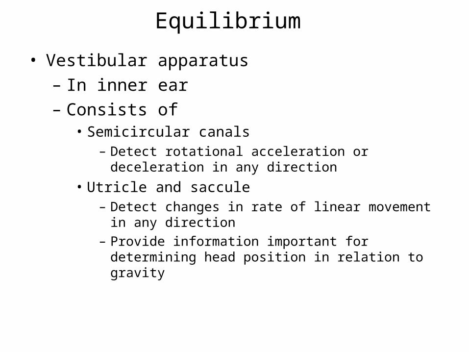
• Vestibular apparatus
– In inner ear
– Consists of• Semicircular canals
– Detect rotational acceleration or deceleration in any direction
• Utricle and saccule– Detect changes in rate of linear movement in any
direction– Provide information important for determining head
position in relation to gravity

Fig. 6-38a, p. 219
Equilibrium
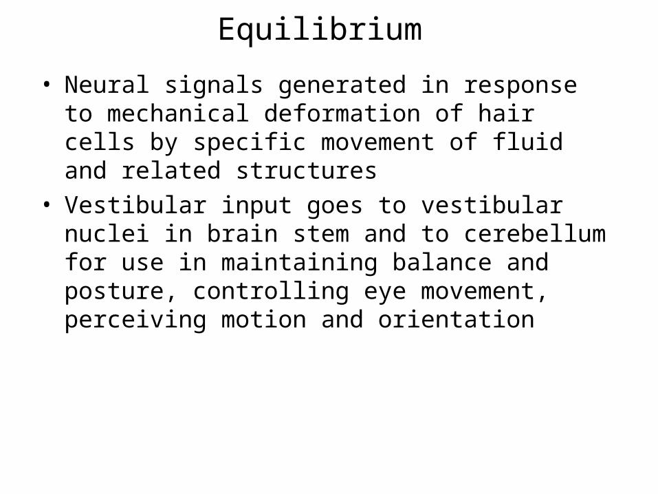
• Neural signals generated in response to mechanical deformation of hair cells by specific movement of fluid and related structures
• Vestibular input goes to vestibular nuclei in brain stem and to cerebellum for use in maintaining balance and posture, controlling eye movement, perceiving motion and orientation

• Cortex– Higher processing
• Basal nuclei– Control of movement, inhibitory, negative
• Thalamus– Relay and processing of sensory information– Awareness, a positive screening center for information
• Hypothalamus– Hormone secretion, regulation of the internal environment
• Cerebellum– Important in balance and in planning and executing voluntary
movement
• Brain Stem– Relay station (posture and equilibrium), cranial nerves,
control centers, reticular integration, sleep control

Equilibrium Semicircular canals
Detect rotational acceleration or deceleration in any direction
Utricle and sacculeDetect changes in rate of linear movement in any directionProvide information important for determining head position in relation to gravity
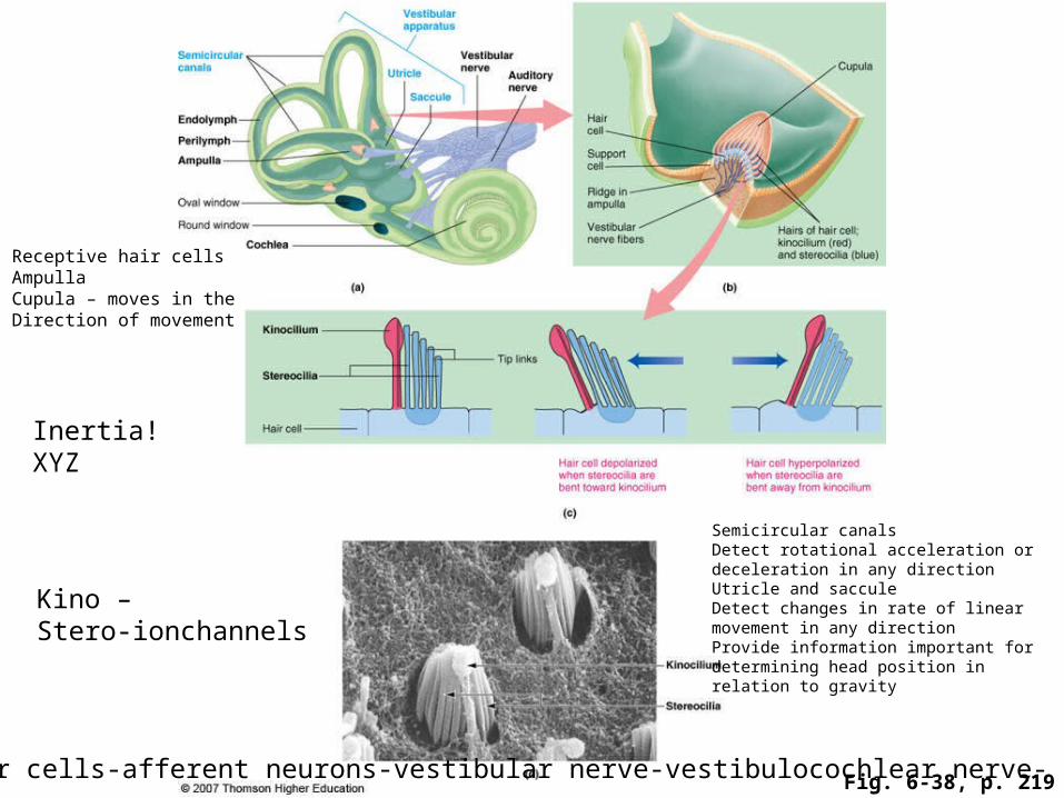
Fig. 6-38, p. 219
Receptive hair cellsAmpullaCupula – moves in the Direction of movement
Inertia!XYZ
Kino –Stero-ionchannels
Hair cells-afferent neurons-vestibular nerve-vestibulocochlear nerve-
Semicircular canalsDetect rotational acceleration or deceleration in any directionUtricle and sacculeDetect changes in rate of linear movement in any directionProvide information important for determining head position in relation to gravity

Fig. 6-38b, p. 219
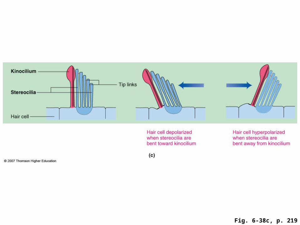
Fig. 6-38c, p. 219

Otoliths
Semicircular canalsDetect rotational acceleration or deceleration in any directionUtricle and sacculeDetect changes in rate of linear movement in any directionProvide information important for determining head position in relation to gravity
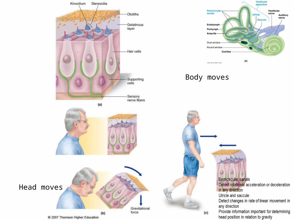
Head moves
Body moves

Chemical Senses
Taste and smell
• Receptors are chemoreceptors
• In association with food intake, influence flow of digestive juices and affect appetite
• Stimulation of receptors induces pleasurable or objectionable sensations and signals presence of something to seek or to avoid
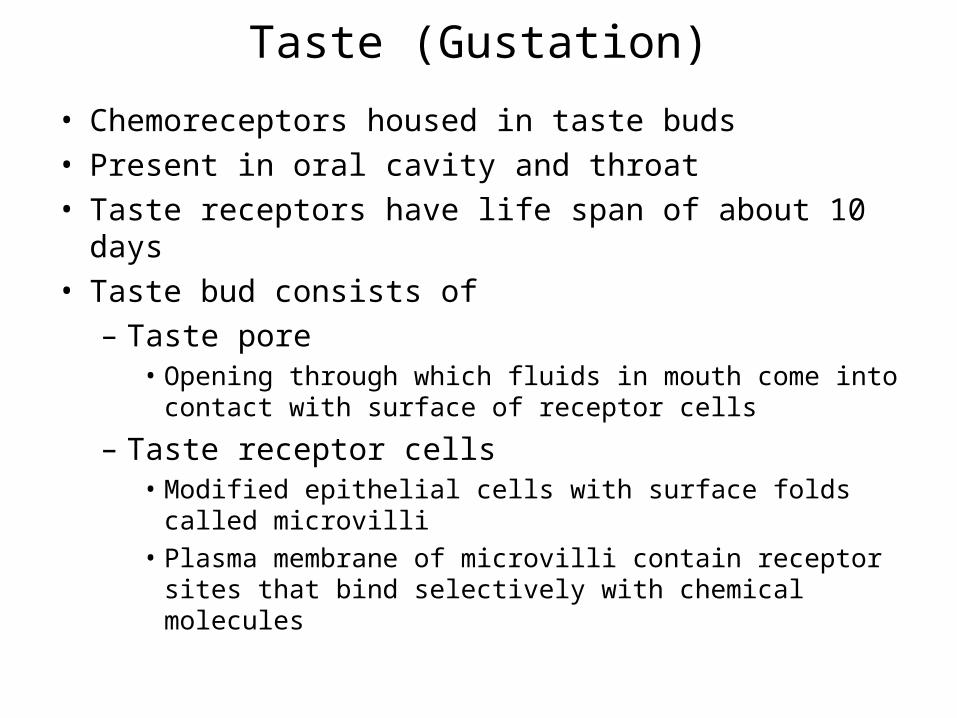
Taste (Gustation)
• Chemoreceptors housed in taste buds
• Present in oral cavity and throat
• Taste receptors have life span of about 10 days
• Taste bud consists of
– Taste pore• Opening through which fluids in mouth come into
contact with surface of receptor cells
– Taste receptor cells• Modified epithelial cells with surface folds called
microvilli
• Plasma membrane of microvilli contain receptor sites that bind selectively with chemical molecules

Location and Structure of Taste Buds

Taste
• Tastant (taste-provoking chemical)
• Binding of tastant with receptor cell alters cell’s ionic channels to produce depolarizing receptor potential
• Receptor potential initiates action potentials within terminal endings of afferent nerve fibers with which receptor cell synapses
• Terminal afferent endings of several cranial nerves synapse with taste buds in various regions of mouth
• Signals conveyed via synaptic stops in brain stem and thalamus to cortical gustatory area
Receptor cell-afferent nerve-cranial nerves- brain stem- thalamus- cortical gustatory area

Taste • Five primary tastes
– Salty• Stimulated by chemical salts, especially NaCl
– Direct entry of sodium ions thru sodium channels
– Sour• Caused by acids which contain a free hydrogen ion, H+
– hydrogen ions block potassium channels (depolarization)
– Sweet• Evoked by configuration of glucose
– G protein - cAMP pathway blockage of potassium channels (depolarization)
– Bitter• Brought about by more chemically diverse group of tastants• Examples – alkaloids, toxic plant derivatives, poisonous substances
– G protein linked
– Umani • Meaty or savory taste
– G protein linked

Taste Perception
• Influenced by information derived from other receptors, especially odor
• Temperature and texture of food influence taste
• Psychological experiences associated with past experiences with food influence taste
• How cortex accomplishes perceptual processing of taste sensation is currently unknown

Smell (Olfaction)
• Olfactory receptors in nose are specialized endings of renewable afferent neurons
• Olfactory mucosa
– 3cm2 of mucosa in ceiling of nasal cavity
– Contains three cell types• Olfactory receptor cell
– Afferent neuron whose receptor portion is in olfactory mucosa in nose and afferent axon traverses into brain
– Axons of olfactory receptor cells collectively form olfactory nerve
• Supporting cells– Secrete mucus
• Basal cells– Precursors of new olfactory receptor cells (replaced about
every two months)

Smell (Olfaction)
• Odorants
– Molecules that can be smelled
• To be smelled, substance must be
– Sufficiently volatile that some of its molecules can enter nose in inspired air
– Sufficiently water soluble that it can dissolve in mucus coating the olfactory mucosa
Smell (Olfaction)
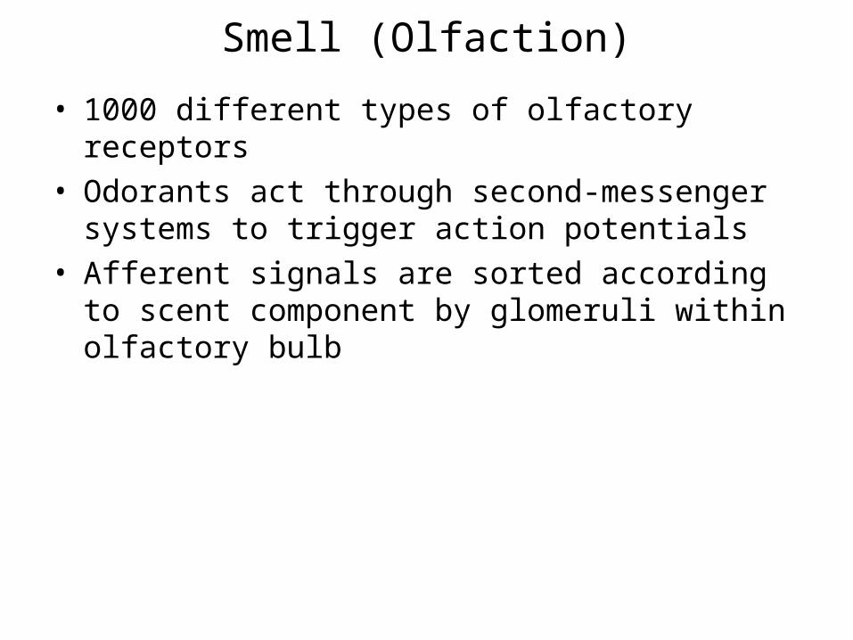
• 1000 different types of olfactory receptors
• Odorants act through second-messenger systems to trigger action potentials
• Afferent signals are sorted according to scent component by glomeruli within olfactory bulb

Fig. 6-43, p. 225

Olfactory receptor cells
• Enlarged knob bearing several cilia
• Have olfactory receptors
• Odorants
– Must be volatile
– Water soluble

Processing of Scents in Olfactory Bulb
Olfactory processing
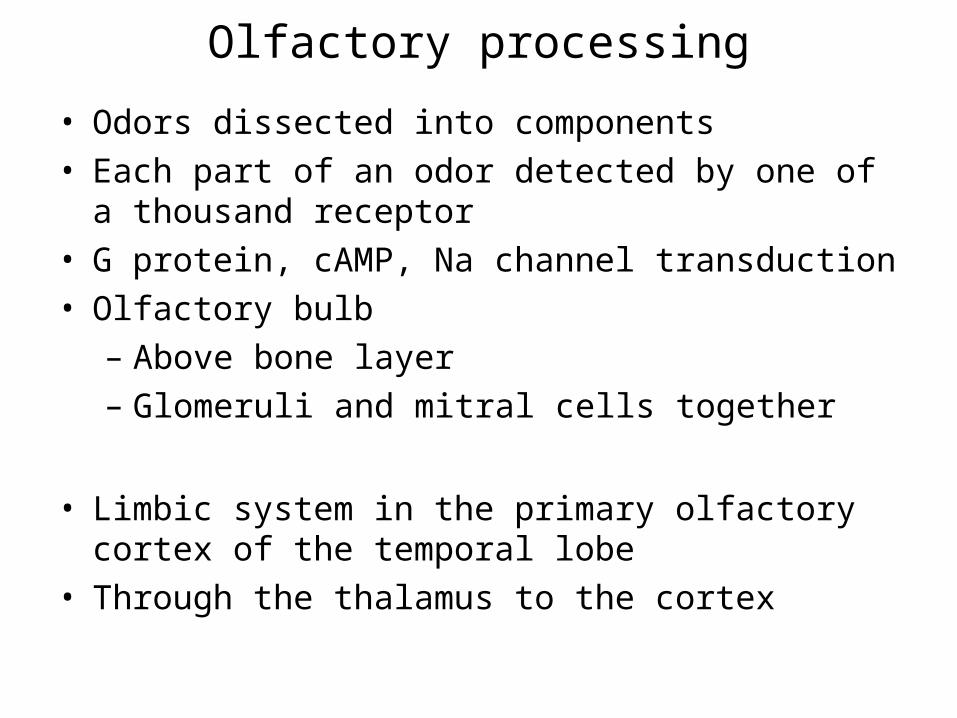
• Odors dissected into components
• Each part of an odor detected by one of a thousand receptor
• G protein, cAMP, Na channel transduction
• Olfactory bulb
– Above bone layer
– Glomeruli and mitral cells together
• Limbic system in the primary olfactory cortex of the temporal lobe
• Through the thalamus to the cortex

Processing
• Each odorant molecule activates multiple receptors and glomeruli
• Odor discrimination based on “patterns” of glomerular excitation

Vomeronasal Organ (VNO)
• Common in mammals but until recently was thought to nonexistent in humans
• Located about half an inch inside human nose next to vomer bone
• Detects pheromones
– Nonvolatile chemical signals passed subconsciously from one individual to another
• Role in human behavior has not been validated
Related Documents