CHAPTER FOUR Microbial Distribution in Soils: Physics and Scaling I. M. Young,* J. W. Crawford, † N. Nunan, ‡ W. Otten, § and A. Spiers § Contents 1. Soil as a Habitat 82 2. What Characteristics of Structure Matter and Why? 84 2.1. Moisture characteristic 85 2.2. 3D stucture–water interactions 85 2.3. Water-film thickness 88 2.4. Surface area 89 3. Spatial and Temporal Distribution of Microbes 90 3.1. Fungi in soil 95 3.2. Visualisation and quantification of fungal hyphae in soil 95 3.3. Relevance of spatial temporal dynamics to ecosystem function 97 4. Habitat–Biofilm Interactions 99 4.1. Bacterial movement 100 4.2. The biofilm environment 101 4.3. Celluose as a legacy in soil 104 4.4. Surfactants as a legacy in soil 105 5. Habitat–Microbe Interactions 105 5.1. Regulatory feedbacks in soil–microbe interactions 106 5.2. The soil–microbe complex as a complex adaptive system 108 5.3. Functional consequences 111 6. Future 112 References 113 Advances in Agronomy, Volume 100 # 2008 Elsevier Inc. ISSN 0065-2113, DOI: 10.1016/S0065-2113(08)00604-4 All rights reserved. * School of Environmental and Rural Sciences, University of New England, Armidale NSW 2351, Australia { Faculty of Agriculture, Food and Natural Resources, University of Sydney, NSW 2006, Australia { CNRS, Laboratoire BioEMCo,UMR7618, Ba ˆtiment EGER, Aile B, Campus AgroParisTech, F-78850 THIVERVAL-GRIGNON, France } SIMBIOS Centre, University of Abertay Dundee, DD1 1HG, Scotland 81

Welcome message from author
This document is posted to help you gain knowledge. Please leave a comment to let me know what you think about it! Share it to your friends and learn new things together.
Transcript

C H A P T E R F O U R
A
IS
*{
{
}
dvance
SN 0
SchoFaculCNRTHIVSIMB
Microbial Distribution in Soils:
Physics and Scaling
I. M. Young,* J. W. Crawford,† N. Nunan,‡ W. Otten,§ and
A. Spiers§
Contents
1. S
s in
065
ol oty oS, LERIOS
oil as a Habitat
Agronomy, Volume 100 # 2008
-2113, DOI: 10.1016/S0065-2113(08)00604-4 All rig
f Environmental and Rural Sciences, University of New England, Armidale NSW 23f Agriculture, Food and Natural Resources, University of Sydney, NSW 2006, Austraboratoire BioEMCo,UMR7618, Batiment EGER, Aile B, Campus AgroParisTech,VAL-GRIGNON, FranceCentre, University of Abertay Dundee, DD1 1HG, Scotland
Else
hts
51,aliaF-
82
2. W
hat Characteristics of Structure Matter and Why? 842
.1. M oisture characteristic 852
.2. 3 D stucture–water interactions 852
.3. W ater-film thickness 882
.4. S urface area 893. S
patial and Temporal Distribution of Microbes 903
.1. F ungi in soil 953
.2. V isualisation and quantification of fungal hyphae in soil 953
.3. R elevance of spatial temporal dynamics to ecosystem function 974. H
abitat–Biofilm Interactions 994
.1. B acterial movement 1004
.2. T he biofilm environment 1014
.3. C elluose as a legacy in soil 1044
.4. S urfactants as a legacy in soil 1055. H
abitat–Microbe Interactions 1055
.1. R egulatory feedbacks in soil–microbe interactions 1065
.2. T he soil–microbe complex as a complex adaptive system 1085
.3. F unctional consequences 1116. F
uture 112Refe
rences 113vier Inc.
reserved.
Australia
78850
81

82 I. M. Young et al.
Abstract
In a handful of fertile soil there are billions of microorganisms and yet, even
with a conservative estimate, the surface area covered by these organisms is
considerably less than 1%. What does this tell us about the function of the
physical structure in which soil organisms reside and function, collecting, and
separating micropopulations from each other and from resources? It would
seem that most of the soil is akin to desert regions with little life been
supported on its terrains, yet with vast communities of individuals, from an
amazing array of species, supported in small-scale habitats, connected or
disconnected by saturated or unsaturated pore space over relatively short
time-scales. The biodiversity of these communities remains impressive yet
overall functionally illusive, bar some considerations of inbuilt redundancy.
What is far more impressive is the range of habitats on offer to populations
with short-term evolutionary time frames. The availability of spatially and
temporally diverse habitats probably gives rise to the biodiversity that we see
in soil.
It is not too far fetched to state that the majority of habitats on Earth (and
indeed extraterrestrial) are revealed in that handful of soil. The key question is
what is the functional consequence of such habitat heterogeneity? To answer
this it is clear that we need to bring together a new discipline that combines the
biology and physics of the soil ecosystem. This biophysical approach, com-
bined, where required, with important mineral-microbe knowledge is needed to
help us understand the mechanisms by which soils remain productive, and to
identify the tipping-points at which there may be no return to sustainability.
This review aims to highlight the importance of addressing the soil ecosys-
tem as a dynamic heterogeneous system focusing on microbiota–habitat
interactions.
1. Soil as a Habitat
The defining features of materials that provide good habitats are con-stant: adequate supplies of food, shelter, and water; refuge from predators;and access to mates. Habitats in above ground ecosystems do not approachthe complexity of soil habitats. The spatial-temporal variation of the physi-cal, chemical, and biological structure of soil habitats is awesome even overrelatively small spatial and temporal resolutions. However, the complexity ofsoil ecosystems is also matched by the difficulty in adequately quantifying thefunctional traits of the habitats linked through to biotic activity.
Soils are opaque, fragile constructs that offer limited opportunity fornondestructive analysis. Indeed, it has only been within the last 20 yearsthat a usable molecular tool box has become available to partially quantify thesoil microbial populations, albeit destructively. Similarly, until recently,quantification of soil structure has been limited to relatively simple

Microbial Distribution in Soils: Physics and Scaling 83
qualitative assessments through measuring arbitrarily units of 2D Ped struc-tures, or infield assessments categorizing soil structure into quasigeometricunits (e.g., subangular blocky). Whilst these approaches have served tohighlight the difficulty of assessing structures, and in significant ways thespatial heterogeneity of these structures, they have offered little advance inour understanding of how the inherent heterogeneity of the structure of soilscan be linked to their functions. Standard stability tests of soil structure arerelatively useless in assessing the soil’s real stabilities as they are akin to askingquestions about the operational efficiency of a skyscraper from the debris ofthat building sieved through a 2-mm mesh. Similar issues can be raised overthe use of pore size distribution datasets as they fail to quantify pore con-nectivities, never mind the real pore geometries that control permeability.A detailed review of methodologies and modeling approaches that are usedto quantify soil structure is provided by Young et al. (1989).
Uniquely, soils are characterized by significant spatio-temporal hetero-geneities that few, if any, other porous media exhibit. The classic work ofFitzPatrick (1993) and Foster and Rovira (1976) using high quality thinsections illustrate the impressive complexity of the biophysical frameworkand mineralogy of many soils and environments. Adderley et al. (2002)present a good summary of quantification techniques used for thin sections.Figure 1 shows the typical spatial heterogeneity of soil architecture andmineralogy in soils. Across macro- and microscales, the variation andpatchiness of pore structures is demonstrated. The mineralogical diversityemphasizes the potential chemical variation, leading to, in functional terms,
Pores
Figure 1 Thin section showing highly heterogeneous pore space and mineralogy.Image width is 4 mm. Image provided by Dr. Sacha Mooney, Nottingham University.

84 I. M. Young et al.
diverse responses to a wide range of processes: sorption, flow, microbialadhesion, and plant development (Lehmann et al. 2008).
There is a growing awareness that not only are such heterogeneitiescrucial to understand in terms of accurately predicting a wide range of soilprocesses, but also, information at the small scale (<mm), where micro-organisms reside and interact with their environment, is a key area toinvestigate. At this scale, microbes are active in relatively thin films of soluteswithin a biofilm, adhering to pore surfaces.
2. What Characteristics of Structure
Matter and Why?
Soil is a porous medium with a hierarchy of pore dimensions and alarge internal surface composed of a wide variety of materials (organic andinorganic). The volume, distribution, and movement of solutes depends onthe characteristics of the solutes active surfaces and how solutes interact withthe soil surfaces. In short, surface tension and contact angles of the solute andthe capillary forces and hydrophilic nature of the soil. The moisture charac-teristic (MC) of soil arises from an integration of all these properties toprovide a unique fingerprint of how water interact with a specific soil.
Water-filled pore space and water-filmthickness decrease
CO2 moves about 1cmin 1 day when soil issaturated
CO2 moves about 1 cmin 1 hour when hydraulicconnectivity is low
Matric potential
Wat
er c
onte
nt
Air entry: drainage begins andmatrix becomes unsaturated
Large poresdrain
Point of inflection:smaller poresdrain-air connectivityincreases and hydraulicconnectivity decreases
Figure 2 Schematic of moisture characteristic of soil: moisture-matric potential rela-tion is represented by the black curve.

Microbial Distribution in Soils: Physics and Scaling 85
2.1. Moisture characteristic
The MC is the function that describes the relationship between matricpotential and water content (Brady, 1974), and is a direct consequence ofthe physical geometry of the habitat (Crawford et al., 1995) and the physico-chemistry of the composition of the habitat and the draining solute (Readet al., 2003). The MC schematic in Fig. 2 shows a succession of hydric statestypical of many soils. Soil is perhaps unique as a porous material that has asufficient hierarchy of pore sizes that permit solutes to be held over a widerange of matric potentials, thus allowing saturated and desaturated volumesof pores to coexist in close proximity. Additionally, the complex construc-tion of soils with mineral and organic components provides hydrophobicand hydrophilic materials that have properties that are polar opposites of eachother in relation to their interactions with water (Feeney et al., 2006b).In terms of habitats for microorganisms these characteristics are crucial.Many organisms (e.g., bacteria, protozoa, bacterial-feeding nematodes,etc.) are essentially aquatic, living in thin water-films adhered on poresurfaces, controlled by matric potential. Along with others, such as fungi,they rely on the happy coincidence of availablewater and connected air-filledpore spaces that permit their development and connectivity to the wider soilecosystem. Thus, the ability of soil to allow water to penetrate into it, andhold water, is the key characteristic of all soil ecosystems and highlights theMC as probably the most important relationship in Ecological Studies.
The hierarchical habitat of soil permits a gradual transition from a,saturated, aquatic system towards a fully, unsaturated, aerated systemwhere, if water does exist, it is tightly adsorbed onto mineral surfaces.This transition has been highlighted by Ghilarov (1959), Vannier (1987),and Coleman et al. (2004) who point to soil systems as ideal transitionalmediums for the evolution of many groups of terrestrial invertebrates.In essence, soil is both an aquatic and a terrestrial ecosystem.
It is our contention that much research into soil microbiology fails toaccount for the MC and where water is mentioned it is shown as a simpleabsolute moisture value or concepts such as Field Capacity or WaterHolding Capacity. Where structure is mentioned typically a surrogatemeasure of pore size is used, which is wholly inappropriate as it is thepore shape that is the key characteristic (Or et al., 2007).
2.2. 3D stucture–water interactions
Recent work by Carminati et al. (2008) reveals the intimate associationsbetween pore geometry and water. Using synchrotron X-ray tomographyCarminati et al. (2008) explored the soil–water interfaces between isolated,yet heterogeneous, aggregates within a desorbing and adsorbing soil, con-trolled by matric tensions. The monochromatic, relatively low energy,

86 I. M. Young et al.
X-rays used permitted good segmentation of water, solid, and pore spacefrom the 3D volumes (Fig. 3).
At themicron scale Fig. 3A relates to thewet end of theMCwhere air hasentered and larger pores have started to drain. The water menisci are clearlyevident, hydraulically connecting each aggregate. At this point some largepores which drain through smaller pores will remain saturated until thematric head equivalent to the pore radii of the smaller pores is reached.As the matric head increases (Fig. 3A–D) water empties out of all but thesmallest pores and the hydraulic contact between aggregates significantlydecreases. Figure 3D highlights the smaller pore retaining water and thehydraulic bridge between aggregates. In reality the volume of water withinand between aggregatesmay be higher as theX-ray tomography system had alower resolution of 5.92 mm. Any features below this spatial scale would notbe identified. This impressive work, for the first time, allows us to observeand quantify the water–soil interface near the appropriate scale of bacterialpopulations, and across scale relevant to the root–soil interface in 3D. Thistechnology and application potentially represents an unprecedented stepforward in our understanding of the soil–plant–microbial interactions.
In a further study Culligan et al. (2004), using similar technology,examine the importance of hysteresis (i.e., whether soil is wetted or driedto a specific matric potential) in the volume and distribution of water in soilin 3D. They demonstrate the importance of understanding the mechanismscontrolling the distribution of water in soil. The mechanisms of hysteresis in
[h = -0.025 m]
A
B
C
D
[h = -0.15 m]
[h = -0.43 m]
[h = -1.08 m]
Figure 3 2D tomographic segmented slices over time (A–D), showing desorbingwater within and between two heterogeneous aggregates (voxel resolution 5.92 mm).Black, grey, and white relate to solid, water, and pore, respectively. Numbers inbrackets represent matric head—the more negative the drier the soil. Arrows highlightisolated water pockets within aggregates. Circle highlights hydraulic contact betweenaggregates. Adapted from Carminati et al. (2008).

Microbial Distribution in Soils: Physics and Scaling 87
soil are given in Marshall and Holmes (1992). Figure 4 shows the impact ofhysteresis on the air–water interface and highlights the impact on potentialdiffusive and hydraulic pathways. Here we can see clearly that on thewetting curve there is a potential increase in diffusive pathways for gasand fungal hyphae spread. It is clear that, for the same structure, at thesame matric potential, the pathways are radically different, leading to poten-tially significantly different microbial functionality. In 3D such pathwayswould increase. Whitmore and Heinen (1999) provide one of the fewattempts to account for the impact of hysteresis in microbial activity,providing estimates of its impact on mineralization.
Another interesting feature of Culligens’ work (Culligan et al., 2004) isthat, in the saturated state, significant pore volumes remain air-filled, irre-spective of the initial wetting state (Fig. 4A and D). This may highlightspecific pore volumes that remain untouched by water until a physical
A B
CD
Figure 4 2D tomographic slice of saturated soil (A) initially dried to a specific matricpotential (B), then wetted to that same potential (C) and finally to saturation (D). Thebright circle highlights the main change in the distribution of water, and the impact ofsuch change is highlighted by increase in proposed diffusive pathways (dotted yellowlines). Adapted from Culligan et al. (2004).

88 I. M. Young et al.
perturbation exposes the surfaces. The lack of water ingress into thesevolumes may be due to hydrophobic substances coating the pore surfaces,acting as a barrier to water influx: perhaps old, lignified organic matterremnants acting as protected organic matter.
2.3. Water-film thickness
As seen from Fig. 2 the MC defines the hydraulic and gaseous connectivityof soil ecosystems. A key process is the alteration of water-film thicknessesheld by capillary forces at the pore surfaces. It is notoriously difficult to getgood experimental data on the thickness of water films in soil. In anexcellent review, Or et al. (2007) examine this, and related topics, in relationto bacterial habitats and activity.
Tokunaga et al. (2003) measure water-film thickness on gravel surfacesat different matric potentials. What is clear is that even under relativelywet conditions (high matric potential) thickness values are in the order of2–10 mm, which is bridging the size of single bacterium and small bacterialcolonies. Figure 5 summarizes results from Tokunaga et al. (2003) and drawsa link the work of Wallace (1958), who studied the influence of water-film
0 100
Typ
ical
wat
er fi
lm th
ickn
ess
(mic
rom
eter
s)
Distance travelled in 1 hr (mm)
12
10
8
6
4
2
00 1 2 3 4
Matric potential (-kPa)
5 6 7
20 (mm)Transverse section
of nematode
Water film
Figure 5 Typical water-film thickness versus matric potential (adapted fromTokunaga et al., 2003), and associated relation with potential nematode movement(adapted from Wallace, 1958).

Microbial Distribution in Soils: Physics and Scaling 89
thickness on nematode movement. Wallace (1958) in a seminal piece ofwork showed that for a nematode with a cross-sectional area of 20 mm, theoptimal water-film thickness for movement was approximately 4 mm. Thispermitted the nematodes to remain motile, maximized forward movementand minimized sideward movement. This work has been extended byAnderson et al. (1997) and Young et al. (2002) connecting the hydraulicand gaseous connectivity through to the chemotaxis processes ongoing insoil that permit microorganisms to search and locate food sources. The‘‘resource seeking’’ strategies of protozoa, fungi, nematodes, and bacteriaand other microbes have clearly evolved to cope with the heterogeneousmaze imposed by the soil microarchitecture.
In real heterogeneous soils the correlations found by Tokunaga et al.(2003) in absolute terms, will be rare. Due to variabilities in pore connect-edness, sizes, and roughness, at any given matric potential, we would expectnot only lager water-films, but also the presence of relatively large, yetisolated pockets of water, at low potentials. This is evident in the work ofCulligen et al. (2004).
2.4. Surface area
How connected a habitat is plays an important role in most soil microbiallycentered processes. The accessibility of surface area to microbes has obviousconsequences to a host of issues: pesticide degradation; remediation ofpollutants; carbon degradation and sequestration. The question is whatproportion of the surface area is covered by microorganisms? Taking 1 cm3
of soil the total surface area, at a conservative estimate, will be roughly 20m2.Assuming that we have 10,000 protozoa, 107 bacteria and 5 km of fungi, thetotal surface area that is covered is in the order of 10�6%. This figure of coursedependent on clay type. However, no matter what reasonable combinationof inputs may be, it is highly unlikely that, for many soil ecosystems, morethan 1% of the total surface area is available. Given the fact that most themicrobial inputs rely on the presence of aquatic environments in soils and thatmuch of the surface area is physically inaccessible, the percentage of surfacearea covered by active microbes is significantly less.
Developing on the theme of surface area and microbial activity, a recentpaper by Tarlera et al. (2008) attempts to quantify the link between bacterialdiversity and surface area. They have shown that, in certain coarse texturedimmature soils, low surface areas have a direct impact on communitycomposition. As soils age (5000–77,000 years) the percentage of surfacearea that could be occupied by prokaryotic cells dropped from 100% to 1%.No such correlation was found for fine textured soils. Whilst these resultsawait confirmation across a wider range of soil ecosystems, and a betterunderstanding of what available surface area is needs to be sought, they dooffer new insight into microhabitats and microbial diversity.
90 I. M. Young et al.
The spatial location of carbon substrate allied to the location of waterand microbes is also a key determinate of the amount and type of microbialactivity. Often organic matter provides a rich and large surface area onwhich many microbes reside. Additionally, models assume that there is aphysically protected portion of organic matter isolated from microbial andchemical degradation (e.g., Balesdenta et al., 2000). Often the concept ofsoil macroaggregates (>200 mm) and microaggregates (50–200 mm) isinvoked in an attempt to understand the importance of physical protection.These relatively arbitrary upper and lower limits ignore the importance ofunderstanding the 3D nature of soil and the fact that, typically, it does notexist as isolated aggregates (or indeed pores) but as a highly complexinterconnected system.
3. Spatial and Temporal Distribution
of Microbes
The importance of spatial variability in soil microbial ecology has longbeen recognized and the spatial distribution of microbes andmicrobial activityhas been described at scales ranging from individual bacteria in communities tothe landscape scale (Franklin and Mills, 2003; Franklin et al., 2002; Jones andGriffiths, 1964; Morris, 1999; Morris and Boerner, 1999; Nunan et al., 2001,2002; Parkin and Shelton, 1992; Robertson et al., 1997; Vieuble-Gonod et al.,2006). However, much of the work has been descriptive in nature and spatialstructure has not been fully exploited for understanding soil function nor fullyrecognized as a fundamental property of soils.
The spatial structure of soil microbial populations is the result of variousenvironmental controls operating at different scales and of intrinsic popula-tion processes such as dispersal, reproduction, mortality, or competition,that occur primarily at microbial scales (Ettema and Wardle, 2002). Becausemicrobial communities respond simultaneously to a range of variables thatdisplay different spatial patterns, the spatial patterns of microbial commu-nities are likely to be highly complex. This has been borne out by empiricalevidence. Oline and Grant (2001) have shown that the spatial pattern of soilmicrobial biomass was more complex than the spatial patterns of other soilproperties, as measured by the fractal dimension. It has been established thatsubsets of microbial communities show different spatial patterns, suggestingthat they respond differently to structuring agents or that they respond todifferent structuring agents (Ritz et al., 2004; Saetre and Baath, 2000). Thiswas achieved using principal components analysis to reduce the multivariatecommunity data (PLFA or CLPP) to a smaller set of derived variables, eachof which can be related to different patterns within the community structure(Legendre and Legendre, 1998). The derived variables (principal

Microbial Distribution in Soils: Physics and Scaling 91
components) can be considered to represent specific subsets of the microbialcommunities. A geostatistical analysis of the variables showed that subsets ofthe communities and their capacity to use different substrates were spatiallystructured over a wide range of patch sizes (Ritz et al., 2004; Saetre andBaath, 2000). Microbial communities, or subsets thereof, also display nestedscales of spatial structure, indicating that community development canrespond to several drivers at once (Franklin and Mills, 2003; Nunan et al.,2002). This results then, in an overall picture that is highly complex anddifficult to unravel.
Spatial structure can be used to help identify the external or intrinsicdrivers of population development and activity (Ettema and Wardle, 2002).However, in order to understand how external and intrinsic drivers shapemicrobial population activity, structure, and distribution it is important toaccount for the scales at which the drivers operate. Many studies haveexamined the role of environmental variables that display large or mediumscale gradients such as disturbance history (Robertson et al., 1993), moisturecontent (Morris and Boerner, 1999), organic matter content (Parkin, 1987;Vieuble-Gonod et al., 2003), distribution of plants (Klironomos et al., 1999;Saetre and Baath, 2000) or preferential flow paths (Bundt et al., 2001; Gastonand Locke, 2002) on microbial populations and activity at landscape, field,plot, or core scales. Whilst there is no doubt that all these factors influencemicrobial population development and activity, they are not related toaspects of microbial populations, such as the high levels of diversity sustainedat small scales and the noncompetitive diversity patterns found in soilmicrobial communities (Treves et al., 2003; Zhou et al., 2002), that underliefundamental properties of soil (the robustness and resilience of soil biologicalfunction for example).
Here clearly, microscale (<1 mm) processes are at work. Diffusionconstraints at the microbial cell or microhabitat scale is believed to be aprimary mechanism by which microbial diversity is sustained at small scalesin soil (Long and Or 2005; Dechesne et al., 2008a). Competition amongcells is limited by the rate of diffusion of substrate to individual cells.In unsaturated soils water usually forms thin films at the pore surface thatcan be poorly connected, limiting solute diffusion rates. When the rate ofdiffusion is lower than the consumption rate of all the competing cellsthen the more competitive cells (those that consume substrate faster) nolonger have any competitive advantage, allowing less competitive cells tosurvive (Dechesne et al., 2008a). This is akin to less competitive cells beingspatially isolated and removed from a competitive environment (Treveset al., 2003). Furthermore, when one considers that the distance to which acell can be expected to affect solute concentrations by just 5% is as little as10 mm (Franklin and Mills, 2007), it is easy to understand the coexistence ofsuch a huge diversity of differentially competitive microbial types at smallscales.

92 I. M. Young et al.
The range and variety of habitats available to microbial communities arealso likely to contribute to the maintenance of high microbial diversity asdifferent microbial types are variably adapted to the different microenvir-onments (aerobic to anaerobic, substrate rich to substrate poor. . .) that canbe encountered in soil. The physical structure of microbial habitats variesenormously in soil (Nunan et al., 2006). Soil organic matter is highlyheterogeneous at the microscale, as is the distribution of minerals(Lehmann et al., 2008). These contribute to the huge diversity of micro-environments in soil by affecting O2 and substrate availability and byproviding different levels of protection from predators or stress. Their spatialcomplexity suggests that the microenvironments can change dramaticallyover very small distances. This was demonstrated by Sexstone et al. (1985)who showed that single aggregates can have very steep oxygen gradientsbetween the surface and the centre. Aggregate surfaces tend to be harsherenvironments than the micropore environment within aggregates as theyare more prone to external stresses, such as wetting and drying cycles orexposure to pollutants. It is not surprising therefore, that aggregate surfacemicrobial communities differ from those existing in inner aggregate envir-onments (Mummey and Stahl, 2004; Ranjard et al., 2000b) and are moreresistant to chemical stress where prior exposure has occurred (Almas et al.,2005; Ranjard et al., 2000a).
Soil microbial activity is made up of the activity of individual microbes.The stimuli and constraints that are exerted on individual cells and interac-tions among cells mean that community activity is not the mere sum of theactivities of individual cells; the relationship between individual and com-munity activities is unlikely to be linear. Diffusion processes determine ratesof access to substrate (and therefore growth potential) and the distances towhich individual cells interact. Processes occurring at scales close to that ofthe individual cell can therefore have a significant impact on communityactivity and the processes are regulated by the spatial distribution ofmicrobes and the physical and chemical properties of the habitat.
Microbes release extracellular enzymes to oxidize soil organic matter forobtaining carbon and satisfying their nutritional needs. In soil, extracellularenzyme producers compete with microbes that do not, but that consumethe products, called ‘‘cheaters’’ (Allison, 2005). The energy cost of produc-ing the enzymes would normally put enzyme producers at a competitivedisadvantage. Low enzyme diffusion, on the other hand, favors enzymeproducers as the products of enzyme activity appear in close proximity tothe producer. The two can coexist in spatially structured patterns at inter-mediate enzyme production costs and diffusion rates (Allison, 2005).However, it is increasingly recognized that bacteria do not exist solely asindividual cells but are capable of cell to cell communication; certainbacterial types having the capacity to monitor cell density before expressinga phenotype (Whitehead et al., 2001). This mechanism of communication,

Microbial Distribution in Soils: Physics and Scaling 93
called quorum sensing, is believed to facilitate bacterial adaptation to altera-tions in the environment. It is possible that quorum sensing is used bybacteria in nonisogenic environments to trigger the production of extracel-lular enzymes only when their population density is such that they willprofit from the enzymes activity. Quorum sensing communication takes theform of chemical signals which are excreted from cells and elicit physiolog-ical changes within the population once a threshold value of the signalmolecule is reached. As there is a relationship between the concentration ofthe signal molecule and the cell density, the physiological response is elicitedat a certain cell density, the quorum. Although the bacterial populationdensities that one might expect to find in soil microhabitats are lower thanthe cell densities at which quorum sensing is initiated in the laboratory, ithas been found that the quorum size in environments where diffusion ratesare low can be far smaller as the diffusional losses of the signal are restricted,resulting in higher concentrations (Dulla and Lindow, 2008). The quorumsize under these conditions can be as low as 30–50 cells, well within thenumber of neighbors that bacterial cells have in soil (Nunan et al., 2003).
It has been reported that nitrification becomes pH-limited in limetreated samples at a higher pH than control samples (Strong et al., 1997).The acid produced during nitrification accumulated in the nitrifying bac-teria’s microenvironment because proton diffusion away from microsites ofactivity was slow. The bulk pH measurements did not reflect the pHexperience of the nitrifiers. The difference between bulk pH and micrositepH was greater in the treated soils due to the liming.
Microscale patchiness is a widespread feature of bulk soil, rhizosphereand rhizoplane bacterial populations, both of specific populations such asammonium oxidizers or 2,4-D degraders and of the total bacterial popula-tion (Dandurand et al., 1997; Dechesne et al., 2003; Eickhorst andTippkotter, 2008; Fisk et al., 1999; Grundmann and Debouzie, 2000;Jordan and Maier, 1999; Nunan et al., 2002, 2003; Pallud et al., 2004;Semenov et al., 1999; Nunan et al., 2007 ). The nonrandomness indicatesthat some regions are more favorable for microbial development, the porearchitecture being one of the most important determinants of where theyare located (Eickhorst and Tippkotter, 2008; Nunan et al., 2003). Thepatches appear to have internal density gradients centered around specificloci (Nunan et al., 2003). This may be due to specific sources of organicmatter as well as an integration of many or all the interactions describedabove (Gaillard et al., 1999; Semenov et al., 1999). The activity of individualcells per se is not generally of interest to ecologists as it so small it is close tobeing insignificant. Their activities as communities however, are the basicbuilding blocks of many landscape scale processes including organic matterdecomposition, nutrient cycling and greenhouse gas production. At whatscale do microbial communities exist? In the case of bacteria, can thesepatches be considered as unit communities, as coherent functional units?
94 I. M. Young et al.
To what extent do the patches interact with neighboring patches and atwhat scale, if at all, can soil be considered as a juxtaposition of suchfunctional units?
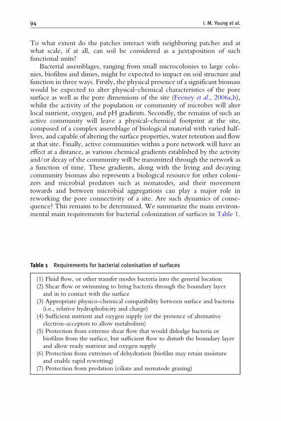
Bacterial assemblages, ranging from small microcolonies to large colo-nies, biofilms and slimes, might be expected to impact on soil structure andfunction in three ways. Firstly, the physical presence of a significant biomasswould be expected to alter physical–chemical characteristics of the poresurface as well as the pore dimensions of the site (Feeney et al., 2006a,b),whilst the activity of the population or community of microbes will alterlocal nutrient, oxygen, and pH gradients. Secondly, the remains of such anactive community will leave a physical–chemical footprint at the site,composed of a complex assemblage of biological material with varied half-lives, and capable of altering the surface properties, water retention and flowat that site. Finally, active communities within a pore network will have aneffect at a distance, as various chemical gradients established by the activityand/or decay of the community will be transmitted through the network asa function of time. These gradients, along with the living and decayingcommunity biomass also represents a biological resource for other coloni-zers and microbial predators such as nematodes, and their movementtowards and between microbial aggregations can play a major role inreworking the pore connectivity of a site. Are such dynamics of conse-quence? This remains to be determined. We summarize the main environ-mental main requirements for bacterial colonization of surfaces in Table 1.
Table 1 Requirements for bacterial colonisation of surfaces
(1) Fluid flow, or other transfer modes bacteria into the general location
(2) Shear flow or swimming to bring bacteria through the boundary layer
and in to contact with the surface
(3) Appropriate physico-chemical compatibility between surface and bacteria
(i.e., relative hydrophobicity and charge)
(4) Sufficient nutrient and oxygen supply (or the presence of alternative
electron-acceptors to allow metabolism)
(5) Protection from extreme shear flow that would dislodge bacteria or
biofilms from the surface, but sufficient flow to disturb the boundary layer
and allow ready nutrient and oxygen supply
(6) Protection from extremes of dehydration (biofilm may retain moisture
and enable rapid rewetting)
(7) Protection from predation (ciliate and nematode grazing)
Microbial Distribution in Soils: Physics and Scaling 95
3.1. Fungi in soil
Despite the widely recognized importance of the soil fungal community inregulating the structure and functioning of plant and soil ecosystems, there isstill little understanding about the origin and consequences of fungal com-munities in soil and how these communities interact with soil structure.Diversity of fungi is high with the number of species estimated around1.5 million. Yet despite their diversity, fungi share certain common featuresin that they are heterotrophs, requiring external C sources and typicallydisplay filamentous growth. The latter gives them distinct ecological advan-tages in heterogeneous soil environments and ensures they occupy a nichedifferent from those occupied by bacteria in soil. The apical growth providesthem with opportunities to locate new carbon sources which can betranslocated through themycelial network to other part of the fungal colony.The larger size of fungi (typical dimensions of 3–10 mm) relative to bacteria(0.5–1 mm)means that fungi are rarely found in micropores (Killham, 1994).The location of bacteria and fungi in the pore network is key to their survivaland activity, with the larger size for fungi making them typically morevulnerable for predation and dry–wet cycle (Denef et al., 2001). Hattori(1988) showed that 80–90% of fungi may be restricted to larger pores. This isconsistent with later work by Harris et al. (2003) and Otten et al. (1999)showing preferential fungal exploration through larger and air-filled pores.Ritz and Young (2004) review interactions between soil structure and fungi.
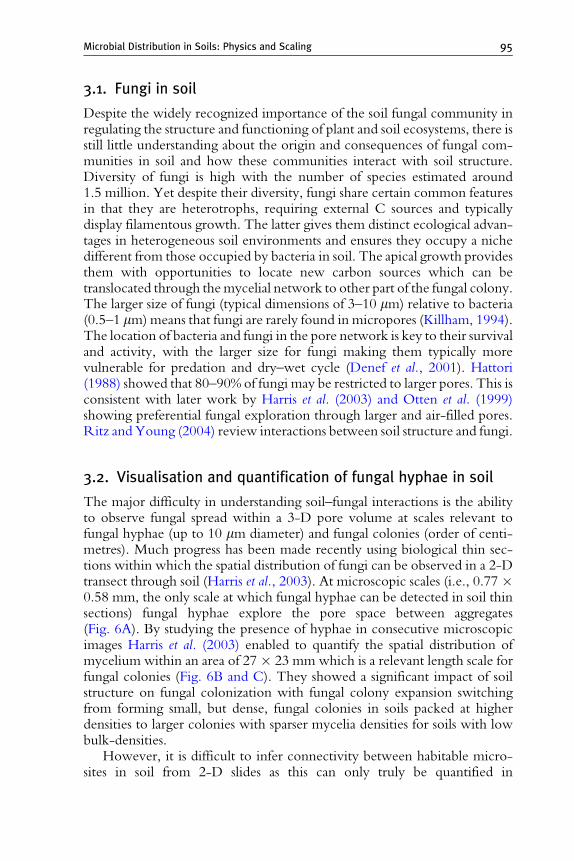
3.2. Visualisation and quantification of fungal hyphae in soil
The major difficulty in understanding soil–fungal interactions is the abilityto observe fungal spread within a 3-D pore volume at scales relevant tofungal hyphae (up to 10 mm diameter) and fungal colonies (order of centi-metres). Much progress has been made recently using biological thin sec-tions within which the spatial distribution of fungi can be observed in a 2-Dtransect through soil (Harris et al., 2003). At microscopic scales (i.e., 0.77 �0.58 mm, the only scale at which fungal hyphae can be detected in soil thinsections) fungal hyphae explore the pore space between aggregates(Fig. 6A). By studying the presence of hyphae in consecutive microscopicimages Harris et al. (2003) enabled to quantify the spatial distribution ofmycelium within an area of 27� 23 mm which is a relevant length scale forfungal colonies (Fig. 6B and C). They showed a significant impact of soilstructure on fungal colonization with fungal colony expansion switchingfrom forming small, but dense, fungal colonies in soils packed at higherdensities to larger colonies with sparser mycelia densities for soils with lowbulk-densities.
However, it is difficult to infer connectivity between habitable micro-sites in soil from 2-D slides as this can only truly be quantified in

A B C
Figure 6 Soil–fungal interactions visualized in thin soil sections at microscopic scales(A: 0.44 mm2), and at larger scales (6.2 cm2, approximately confirm size of a fungalcolony). In soil packed at a bulk-density of 1.2 mg m�3 (B), and 1.6 mg m�3 (C). At thelarger scale, presence of fungal hyphae is indicated by a dot. Details of Fig B and C canbe found in Harris et al. 2003.
Figure 7 (A) Visualization of the largest connected pore volume in a sandy loam soilpacked at a bulk-density of 1.2 mg m�3 visualized using X-ray microtomography. Porevolume exclusion occurs as pores become isolated from the largest connected cluster ascan be seen from a horizontal transect through this pore volume (B). Legend b: black:solid; grey: connected largest pore (as shown in a); white: isolated pores.
96 I. M. Young et al.
3 dimensions. This is apparent from the 2-D sections which clearly revealthat not all larger available pore voids in the loosely packed soil had becomecolonized (Fig. 6B). In addition, 2 void spaces that were colonized in soilwere separated in 2-D by a denser soil volume that was free from fungalcolonization. By scanning the resin impregnated blocks of the same soilsamples we visualized the 3-D pore volume through which fungal spreadhad occurred (Fig. 7). At the lower bulk-density of 1.2, the majority of thepore volume was part of 1 single pore (Fig. 7B). The remainder of the porevolume may be connected but only by pore necks smaller than 70 mmwhich resembled the resolution of this scan. This connectivity is importantfor fungal colonization of soil, as once within a pore volume only the pore
Microbial Distribution in Soils: Physics and Scaling 97
space connected to this volume can become colonized. It is also the volumewithin which fungal–fungal interactions are likely to occur.
Additional image analysis allows us to examine the fraction of the pore-volume visible in a 2-D plane through soil that is connected to each other in3-D. Such an analysis provides a novel look at ‘‘accessible pore space’’ insoil. It is clear that this is no longer just a matter of minimum pore diameteras has long been thought, but in addition is determined by the 3-Dconnectivity of the pore volume. Larger pores that are in close proximityof each other are not necessarily connected and hence inaccessible to aninvading fungal colony. Vice versa, larger pores at larger distance from eachother may be connected in 3-D.
3.3. Relevance of spatial temporal dynamics toecosystem function
The spatial and temporal distribution of fungi in soil has great impact onmany important ecosystem services. Below we only consider two, namelythe role in C dynamics and the role in epidemics.
There is overwhelming evidence that organic matter in smaller pores ismore protected from degradation than that in larger pores. The research inthis area has been predominantly focused on bacteria with only littlereference to fungi. Fungi and bacteria however differentially influence theformation and stabilization of different organic matter components in agri-cultural soils partly because of their different interactions with soil physicalproperties (Six et al., 2006). The interactions between organisms and theirsubstrate depends on soil structure and on the ability of the organisms toexplore the pore volume. Despite early recognition (Adu and Oades, 1978)that restrictions lead to organic matter being more protected in smaller poresthan in larger pores, the knowledge on spatial organization of fungi in soiland their role in decomposition of C is limited. Strong et al. (2004)investigated the spatial location of carbon decomposition in the soil poresystem, following earlier work by Hassink et al., (1993). Following additionof wheat husks to soil they correlated specific pore classes (derived fromwater retention curves) with fungal biomass and residue derived C-content.Their work identified over a range of soil types a considerable activity offungi (determined by ergosterol concentration) in the pore class 15–60 mmcoinciding with a higher decomposition of added organic matter.
However, our knowledge on degradation and incorporation of C into soiland the role microorganisms play in this has largely evolved from experimentsinvolving disruption of soil to obtain aggregates. Inevitably, such results maybe affected by our ability to produce aggregates from soil artificially; hence thetrue spatial distribution of C within soil is largely obscured.
The challenge we face now is to develop novel techniques such as SEM-EDX and X-ray microtomography to visualize and quantify the spatial

98 I. M. Young et al.
distribution of C in the microstructure of soil. Combined with soil thinsectioning they may offer great opportunities to quantify changes in soilmicrostructure and the spatial distribution of C during degradation of organicmatter by bacteria and fungi. Further development of such techniques willgreatly advance understanding of C in soil and will, for the first time, link thedynamics of soil structure with organic matter decomposition in situ. Figure 8highlights the potential of micro-CT in observing and quantifying theorganic matter in soil. In this case the added organic matter has an attenuationcoefficient significantly different from surrounding soil mass and pores andthus is easily isolated and segmented by image processing tools.
A second example where the microscopic heterogeneity of soil, and theway fungi explore this is of great importance, is in the development ofepidemics caused by soil borne plant pathogens. The role of soil physicalconditions in the movement and dispersal of fungal pathogens influencesepidemics in many ways and at many scales. For example, the denser soilunder wetter conditions will result in intensely colonized soil with small butdense fungal colonies. Such colonization could lead to an enhanced contactbetween pathogen and plant, giving rise to more primary infections if inocu-lum densities in soils are high. Loosely packed soil with lower water contenthas a higher percentage of air-filled pores leading to rapid colony expansion.Such conditions may result in rapidly expanding disease patches in the field.The ability of fungi to preferentially spread along cracks and surfaces in soilsalso has epidemiological consequences. Emerging seeds typically result incracking patterns, providing highways for fungal spread guiding the pathogentowards the plant. Below ground old remaining root channels are the path-ways preferentially followed by pathogens as well as roots, removing much of
A B
Figure 8 (A) Thresholded X-ray micro-CT image of soil and with (B) organic matterhighlighted in blue. Note the heterogeneous spread of organic matter in soil matrix.(Scanned by Iain Young for Valerie Pots, INRA). Voxel resolution 30 mm.
Microbial Distribution in Soils: Physics and Scaling 99
the randomness of the interactions by enhancing the likelihood of contactbetween the two. As such the preferential spread along these pathways in soilseems to offer pathogenic fungi an ecological advantage.
The ultimate challenge is to understand how the soil physical conditionsshape fungal community dynamics in soil. Whereas theories have emergedand contributed considerable to our understanding of plant ecology, inde-terminate organisms and in particular fungi have to date received compara-tively little attention. One way to progress is to develop a modelingapproach capable of linking genotype and environmental conditions tocommunity dynamics. Recently, Falconer et al. (2005, 2007, 2008) pro-posed such a model which is based on physiology of organisms, yet parsi-monious and capable of analyzing fungal dynamics in a 3-D heterogeneousstructure. The novelty of the modeling approach includes a mechanism forbiomass recycling and production of extracellular enzymes and volatilesthrough which interactions between species are regulated. Preliminaryanalysis with this model revealed significant differential behavior for fungalinteractions between 2-species on 2-D surfaces and those in a 3-Dstructured environment. Specifically, this showed the existence of a narrowrange of soil physical conditions that allow for coexistence of 2 fungalspecies, separated by the physical structure. Further development and testingof such an approach will provide a much needed understanding of howmicroscopic heterogeneities in soil physical conditions affect fungaldiversity.
4. Habitat–Biofilm Interactions
Bacteria colonize surfaces and volumes in a variety of environments,including the soil and rhizosphere, as single cells and produce a range ofassemblages ranging from microcolonies to larger colonies, biofilms, flocs,and slimes. The physicochemical characteristics of these, as well as thenumber of bacteria they contain, whether they are multispecies commu-nities or not, their metabolic status and ecological success, all impact on soilstructure and processes.
Investigations of the significance and environmental impact of bacterialassemblages in soil has largely focused on examining community structureand function, rather than the distribution and physicochemical character-istics of such communities in soil cores or simplified model systems. How-ever, parallel understanding of medical biofilms and waste-water treatmentaggregates provide some clues as to how similar soil assemblages maydevelop over time in response to the physicochemical properties of poreand particulate surfaces, nutrient, O2 and pH gradients, water availabilityand flow etc., and the processes which govern the development and success

100 I. M. Young et al.
of microbial assemblages in soil are also likely to be significant to the self-organization of soil structure.
Modern biofilm research can be traced back to the influential review byCosterton et al. (1995) (for more modern biofilm reviews, see Battin et al.,2007; Danhorn and Fuqua, 2007; Hall-Stoodley et al., 2004; Parsek andFuqua, 2003; Ramey et al., 2004; Webb et al., 2003; etc.). However,biofilm research – the examination of layers of microbes associated with asolid surface – can be traced back to the 1930s when lake and marinebacterial attachment and growth on solid supports was first noted and the‘‘surface effect’’ proposed (see Henrici, 1933; ZoBell, 1943; ZoBell andAnderson, 1936; and refs therein). This effect described the ability of solidsurfaces to absorb minute but demonstrable quantities of organic matter andthe retardation of diffusion of exocellular enzymes and hydrolysates awayfrom surface-localized bacterial cells (Van Loosdrecht et al., 1990; ZoBell,1943).
The same surface effect will apply in water-filled soil pores, wherelimited nutrients originating from plant exudates and decaying organicmatter provide the basis for bacterial growth. Periodic inundation of drypores by water will mobilize further nutrients, and diffusion and water flowwill carry these back to microbial assemblages located in distant saturatedpores. Mass transfer and diffusion of O2 and nutrients such as amino acidsand simple sugars may be altered in very small pores or thin liquid filmswhere liquid volumes, cross-sectional areas and viscosity, surface roughnessand physicochemistry, and surface-tension effects may impact on fluidmovement and diffusivity (a critical dimension might be when the porediameter/film depth is comparable to the range of intermolecular forcesoperating within the fluid). Water flow itself is unlikely to be high enoughto disrupt bacterial assemblages, as flow velocities of�2 cm s�1 are routinelyused in experimental capillary flow cells; however, at slightly higher flows/shear stress, biofilms are distorted and microcolonies can roll downstreamwith velocities of up to 55 mm h�1 (Rupp et al., 2005).
4.1. Bacterial movement
Bacterial movement vertically and horizontally through soils via waternetworks has been well studied; the transport of bacteria is dependent onthe physicochemical properties of the cell surface and soil particles, andbacteria are able to survive the high pressures of deep soil water, eventuallyresurfacing to colonize surface layers again (bacterial movement is not limitedto self-propulsion through water-saturated pores or via water flow itself, asearthworms and nematodes are known to have a big impact on bacterialdistribution in soils, and fungal hyphae provide networks through larger air-filled pores along which bacteria can migrate). Bacteria have a range ofdifferent motility mechanisms, and the most common, flagella-propelled

Microbial Distribution in Soils: Physics and Scaling 101
swimming, allows bacterial cells to overcome Brownianmotion and to followchemical gradients by chemotaxis at up to 1 mm s�1 (Armitage, 1999;Mitchell and Kogure, 2006). Bacteria such as Escherichia coli can easily move5–10 cm across the surface of soft-agar plates following a nutrient gradient,with individual cells probably moving across smaller distances with pausesduring which they are attached to a solid surface. E. coli and other peritri-chously (multi) flagellated bacteria swim and tumble by rotating the individ-ual left-handed spiral flagella filament counter-clockwise or clockwise,respectively. The hydrodynamic consequence of this can be surprising: cellsapproaching a surface are trapped in the boundary layer and swim in clock-wise circles (Lauga et al., 2006), cells in shear flow near a surface align andswimming upstream along left sidewalls or crevices (Hill et al., 2007), andarrays of funnel-shaped pores can concentrate swimming cells and separatethem from nonswimming cells (Galajda et al., 2007). Populations of swim-ming bacteria also generate bioconvective currents in which aerotaxis (fol-lowingO2 gradients) towards the surface generates self-sustaining vortices andplumes of bacteria (Pedley and Kessler, 1992).
In the absence of chemical cues in the form of nutrient and O2 gradients,and bacterial preferences represented by chemotaxis, attachment anddetachment, the movement of bacteria through the water-filled networkof a porous medium results in a random distribution: the movement ofnonmotile cells is consistent with Brownian diffusion of a similarly-sizednonmotile colloid, whilst the movement of motile cells is described by acombination of the motility of the cells in an aqueous medium plus proper-ties of the porous medium (Sherwood et al., 2003).
4.2. The biofilm environment
The distribution of bacterial assemblages, from localized regions of dispersedattached bacteria, to microcolonies and biofilms, motionless flocs andclogged slimes, as well as migrating populations of bacteria in soils canhardly be expected to be random. Variation in physical features such asthe physicochemical properties of colloid surfaces, water availability and gastransfer, nutrient and O2 gradients, interact to produce a highly heteroge-neous environment which bacteria will sense and respond to by settlementor migration (demonstrating choice and differences in life-style/ecologicalstrategies). The widespread ability to form biofilms amongst both theArchaea and Bacterial lineages suggests it is both an ancient and importantcomponent of the prokaryotic life cycle, representing a protected mode ofgrowth that enhances survival and ecological success (Hall-Stoodley et al.,2004). Recently, biofilms have been presented in terms of ‘‘microbiallandscapes’’ in which an understanding of landscape ecology provides atheoretical framework to view biofilm development and its interactionwith the local environment (Battin et al., 2007).
102 I. M. Young et al.
By acknowledging the ability of bacteria to respond to differences in theirenvironment, the distribution of bacteria within a porous medium is nolonger a simple matter of Brownian diffusion, swimming speeds and pore-size constraints. By modeling microbial distribution in porous media in whichwater flow through biofilms is allowed, the distribution of microorganismsbecomes patchy and shows a self-organized periodic pattern where somepores are filled and others are completely empty (Thullner and Baveye, 2008),whilst microbial biomass can lead to the clogging of pores which results in theredistribution of flow around such areas (Kildsgaard and Engesgaard, 2001).Such self-organization should not be viewed as an abstract result of modeling,as self-organization, complex patterning and stratification is evident withinbacterial colonies and biofilms (Ben-Jacob and Levine, 2006; Shapiro, 1998).It is not therefore unlikely that bacterial assemblages in soil pore networks arealso highly self-organized and responsive to physicochemical changes in theirimmediate environment.
In vitro studies of biofilm development, largely focused on Pseudomonasaeruginosa, suggest that bacterial assemblages may progress through the samestages of initial attachment and colonization, growth, maturation, anddispersion. Not all bacteria impacting onto a surface are irreversiblyattached, and the retraction of type IV pili by P. aeruginosa is required tostably attach cells to a surface (Toby et al., 2005). Once attached,P. aeruginosa cells have been seen to move across the surface by twitching(involving type IV pili) to form small clusters of bacteria, and cells appear toclimb one another in an effort to move away from the surface itself (this maybe an attempt to get beyond the stagnant boundary layer and be exposed tofresh nutrients and O2) (O’Toole and Kolter, 1998). The biofilm thendevelops from the cluster with growth away from the surface with factorssuch as shear flow, nutrient, and O2 gradients impacting on the size andmorphology of the mature biofilm. A network or matrix of interconnectedextracellular polymers, most often polysaccharides, provides the frameworkof the biofilm through which bacteria are distributed (Branda et al., 2005;Hall-Stoodley et al., 2004).
Bacteria are known to produce a wide range of exopolysaccharides inbiofilms, flocs, and slimes (Sutherland, 2001a,b); P. aeruginosa biofilms uti-lizes the glucose-rich Pel andmannose and galactose-rich Psl polysaccharidesand might include some alginate (Ryder et al., 2007). Related pseudomo-nads, including the soil bacterium P. putida KT2440, and soil and plant-associated P. fluorescens SBW25, are capable of expressing both alginate andcellulose (Spiers et al., 2002; Ude et al., 2006). A recent survey of soil, plant-pathogenic, and plant-associated Pseudomonas spp. isolates found that theability to produce a biofilm at the air–liquid interface was very commonwithmany of the stronger biofilms containing cellulose (Ude et al., 2006).
The physico-chemical properties of the extracellular polysaccharides(EPS) help determine the structural characteristics of the biofilm, and in

Microbial Distribution in Soils: Physics and Scaling 103
the P. fluorescens SBW25 Wrinkly Spreader (WS) strain, partial acetylationof cellulose results in a very rigid biofilm, whereas an acetylation-deficientWS mutant produces a very weak biofilm (Spiers et al., 2003). Rheologicalexamination of a variety of biofilms have shown that they are visco-elasticstructures in which the matrix components form a temporary network ofinterconnected fibers (Hall-Stoodely et al., 2004). Above the yield point ofapplied stress, the gel structure fails and the system behaves like a viscousfluid. Beyond the matrix proper, cell-surface components such as LPS,fimbrae, pili, and flagella, as well as DNA, cell debris and whole cellsthemselves contribute to the overall physical structure of the biofilm.
Quite recently biofilm research has started to use ‘‘unsaturated’’ biofilms(colonies grown on agar plates under controlled humidity conditions) toallow physical morphology and surface properties to be examined by atomicforce microscopy. The drying of P. putida colonies grown at 98–99% relativehumidity down to 75.5% humidity had little impact on morphology, rough-ness and adhesion, compared to the effects of similar drying of liquid-grownbiofilms (Auerbach et al., 2000), suggesting that biofilms could exist inregions of the soil pore network subject to variation in water levels.In response to water-limiting conditions, pseudomonads such as P. putidamay produce alginate to modify biofilm properties and facilitate the mainte-nance of a hydrated microenvironment necessary for survival (Chang andHalverson, 2003; Chang et al., 2007). Water-film thickness also has asignificant effect on the lateral expansion of P. putida colonies originatingfrom surface-attached single cells, with a matric potential of �0.5 kPaenabling faster spreading than �1.2 kPa (Dechesne et al., 2008b).
Bacteria are distributed throughout the biofilm matrix and are oftenobserved to move freely through the structure. However, considerablevariation in metabolic activity is found, indicating that the biofilm is avery heterogeneous environment with outer-surface bacteria being moreactive than those at the base on the biofilm. Such stratification can be seen inO2 consumption profiles and antibiotic sensitivity assays (Werner et al.,2004). Inducible green fluorescent protein (GFP)-gene expression tagginghas also shown that growth of P. aeruginosa in microcapillary saturatedbiofilms occurs within 25–30 mm of the surface of microcolonies, and isparticularly evident in the vicinity of interstices (Werner et al., 2004). Theobserved stratification in biofilms indicates that mass transfer of nutrientsand O2 limits microbial activity and the stereotypical biofilm structure withlarge channels running between microbial towers is an architectural struc-tural response to these limiting factors. Sections of biofilm may extendbeyond the stagnant boundary layer into regions of turbulent flow andenjoy better access to nutrients and O2. In a comparison of laminar flowand turbulent flow-grown biofilms, the latter were found to be more activemetabolically, have higher cell densities and overall mass than the former(Pereira et al., 2002; Simoes et al., 2007). In multicommunity biofilms,
104 I. M. Young et al.
bacteria with different life-styles would be expected to localise to, andcolonise those regions best suited for their needs. Notwithstanding thestratification inherent in biofilms, biofilm bacteria and free swimmingindividuals (‘‘planktonic’’ cells) have significantly different metabolism andgene-expression profiles, indicating considerable differences between thebiofilm and planktonic life-styles.
The ecological role of single and multispecies biofilms is defined by amixture of abiotic and biotic factors, including resistance to physical distur-bance, dehydration, predation and competition, and the ability to efficientlyprocess complex carbon sources which is not possible with a small numberof bacteria. Biofilm communities under succession, and even biofilmsestablished by one species undergo phenotypic diversification, generatingmutants with altered behavioral and physicochemical properties. Outer-surface located bacteria can detach individually from the biofilm, whilstshear flow and enzymatic degradation of the matrix can result in thedetachment of large sections of the biofilm. Individual bacteria and biofilmsections can then move through the water system to colonize new regions.
In vitro biofilms are probably quite large structures compared to thoseexpected in soils, simply because optimal environments are used to promotegrowth and to produce observable structures. In nutrient or O2-limitedenvironments, or those subjected to strong shear flow, smaller, more resil-ient biofilms may be produced. In a comparison of laminar flow andturbulent flow-grown biofilms, the latter were found to be more activemetabolically, have higher cell densities and overall mass than the former(Simoes et al., 2007). The expression of even small amounts of EPS issufficient to entrap small soil colloidal particles with several bacterial cells,whilst the involvement of EPS in bacterial attachment, formation, andstability of biogranules used in wastewater treatment (Liu et al., 2004;Tsuneda et al., 2003; Wang et al., 2005, 2006) provides an illustration ofhow such seed aggregates could develop into sizable aggregates in soil.
4.3. Celluose as a legacy in soil
The ability to produce cellulose-matrix based biofilms appears to be wide-spread amongst the Enterobacteriaceae and Pseudomonas, where natural Sal-monella isolates and in vitro-adapted Pseudomonas spp. produce thick‘‘floating’’ biofilms at the air–liquid (A-L) interface and rough rdar/WS-like colonies (Romling, 2005; Spiers et al., 2002; Ude et al., 2006). InEscherichia and Salmonella, the expression of cellulose in vitro protects cellsagainst desiccation (Gualdi et al., 2008; White et al., 2006). In Agrobacterium,cellulose is used to attach individual bacteria cells to plant cell as part of theinfection process as well as in biofilm formation (Mattysse et al., 1981, 2005),and in Rhizobium, it is used as part of the initial attachment in the formationof the rhizobia–legume symbiosis (Laus et al., 2005; Rodriguez-Navarro

Microbial Distribution in Soils: Physics and Scaling 105
et al., 2007). Competitive fitness assays between P. fluorescens SBW25 and amutant deficient for cellulose synthesis has shown that cellulose expression isimportant for the colonization of sugar beet leaves and roots, but not for thecolonization of bulk soil (Gal et al., 2003). This finding is surprising, giventhe number of soil and plant-associated pseudomonads now known to beable to express cellulose (Ude et al., 2006).
In addition, cellulose additions to soil have been shown to alter bothmicro and macrohabitat space (Preston et al., 2001). The precise impact onhabitat space (as measured by geometrical properties of cracks) depended onthe recalcitrance of the cellulose and the incubation period prior to anydrying of the soil. Overall the increase in general carbon production (cellu-lose and biomass) was shown to increase both crack heterogeneity andconnectivity. Thus, microorganisms are both potential degraders and pro-ducers of soil binding agents, modulating the environment they resided in.
4.4. Surfactants as a legacy in soil
Bacteria produce a wide range of multifunctional surface-active agents orbiosurfactants, including rhamnolipids, cyclic lipopeptides and long-chainedN-acylhomoserine lactones (AHLs) (Daniels et al., 2004, 2006; De Brujinet al., 2007; Nitschke et al., 2005; Van Hamme et al., 2006) (fungi alsoproduce a range of biosurfactants, e.g., yeast sophorolipids (Van Bogaertet al., 2007)). P. fluorescens SBW25 expresses the cyclic lipopeptide viscosin(De Brujin et al., 2007), and an over-night culture of SBW25 producessufficient viscosin to alter the surface tension of the growth media from48.3 to 28.8 mN m�1 (the surface tension of pure water is 72.6 mN m�1),and a survey of some 30 pseudomonads found isolates capable of decreasingsurface tension to 25.6 mN m�1 (A. Spiers and A. Koza, unpublished data).Although produced for a variety of reasons, including bacterial cell surfacemodification, biofilm dispersal, swarming motility and as a fungal antibiotic(Ron and Rosenberg, 2001), biosurfactants will have a powerful role in themodification of surface tension and wetting properties of pore water andsurfaces some distance away from production. Thus, potentially they willplay a considerable role in the resultant moisture characteristic.
5. Habitat–Microbe Interactions
The distribution of resources superimposed on the structural com-plexity of soil at all scales has consequences for the distribution and activityof soil microbes. It is the interaction between structural complexity and thedistribution of potential microbial activity that gives rise to an unprece-dented diversity of soil habitats. The connectedness of these habitats is
106 I. M. Young et al.
strongly affected by the amount of water in the system through its impact ondiffusion and convection rates of resources and microbes (Or, 2007).Changes in soil moisture content therefore has a strong effect on the degreeof spatial mixing in soil, and as has been discussed above, is known to be amajor factor in sustaining high levels of functional biodiversity.
The picture we have presented so far is a rather static one. It is one wherea given structure impacts on the amount and distribution of resourcessubject to the moisture content and supports a spatial ecology of microbiallife. The essence of soil, however, is its dynamic state. Indeed, a crucialquestion that continues to challenge soil science is whether soil can con-tinue to provide the required agricultural yields under current managementregimes indefinitely (Baveye, 2006). Given that it is the soil microbialcommunity that performs the essential nutrient recycling functions andthe pore-scale structure of soil that mediates the physical processes thatsupport agriculture, the only way we can answer this question is to under-stand what it is that drives change in the soil–microbe system at the porescale. There is a considerable literature on the biogeochemical cycle thataims to understand the dynamic processes underlying the natural fertility ofsoil. Conceptual models (Standing et al., 2007) portray it as an endogenousprocess driven by the interaction between different functional groups ofmicrobes in soil. However it is a dynamic process that is played out in a soilfabric that is implicitly assumed to be static. Aside from the biogeochemicalcycle, it is tacitly assumed that the factors that drive change in the soil–microbe system are largely exogenous. That is to say, that soil changesprimarily as a consequence of a range of factors that are external to thesystem and that include tillage (Strudley et al., 2008), compaction (Hamzaand Anderson, 2005), root growth (Watteau et al., 2006), and bioturbationby soil fauna (Meysman et al., 2006). To a large and important extent, weassume that the state of soil is a consequence of what is done to it, ratherthan being an intrinsic property of the system itself.
This focus on exogenous processes has led to less attention being givento the role of internal feedbacks in the dynamics. Any dynamical system isstabilized against exogenous forces by the counteraction between endoge-nous regulatory processes. It is the nonlinearities in these regulatory feed-backs give rise to the nature and stability of the dynamical state of soil.
5.1. Regulatory feedbacks in soil–microbe interactions
There is a growing body of evidence to suggest that the structure of soil atscales relevant to key physical and biological processes changes as a result ofendogenous factors (Young and Crawford, 2004). De Gryze et al. (2006)measured the changes that occurred in soil physical structure following theaddition of wheat residue using X-ray CT with a resolution of 27 mm. Theyfound that amendments gave rise to significant increases in porosity and that

Microbial Distribution in Soils: Physics and Scaling 107
this increase was predominantly in pores smaller than 67 mm. Feeney et al.(2006a,b) used X-ray CT imaging at the higher resolution of 4.4 mm tocompare soil in the vicinity of plant roots to that in bulk unplanted soil.They showed even larger changes in porosity resulted with structure closeto the root having a porosity more than twice that of the bulk soil.
It has been known for some time that the structure of soil at the porescale is not random. The aggregated nature of soil particles is understood tobe the origin of the broad range in pore sizes in soil that are required toensure the coexistence of air and water over a wide range of negativepressure. Attempts have been made to quantify the degree of aggregationusing fractal geometry, and a rapid burst in activity in the 1990s attempted todevelop fractal-based models to link structure to function (Anderson et al.,1998).Measurement of the fractal dimension alone is insufficient evidence ofthe existence of fractal structure, not least of all due to the difficulties ofmaking objective measures of image data (Ogawa et al., 1998). Thereforefractal models for nonrandom structure must be supported by evidence forthe possible origin of fractal behavior, that is, dynamical models for structuralgenesis. Tisdall andOades (1982) proposed a conceptual model for the originof pore-scale structure that implicated an interplay between bonding forcesof chemical and biological origin. This conceptual model was operationa-lised into a simple 2D dynamical model for structure genesis by Crawfordet al. (1997). They found that fractal-like scaling resulted from shorter-rangechemical bonding and longer-range binding by biological agents, but thatthe fractal dimension was insensitive to the relative contribution from thebiological component. However, there was evidence from this model thatthe length scale over which fractal-like aggregation occurs, the correlationlength, would increase as the relative contribution from microbial activityincreased. Evidence that this is indeed the case can be found in both DeGryze et al. (2006) and Feeney et al. (2006a,b) where the correlation lengthincreased as the amount of microbial activity also increased.
All of these factors suggest that the pore scale structure of soil is changedas a result of microbial activity. The magnitude of the observed changes issuch that it will have a profound impact on the microhabitat of soilmicrobes. Thus there is clear evidence of a feedback between habitat as itaffects microbial activity, and microbial activity as it affects habitat.
The impact of microbes on soil structure will affect transport processes insoil including convection and diffusion processes at scales that influence themicrohabitat of soil microbes. Using a simulation model, Rappoldt andCrawford (1999) demonstrated that the pore-scale structure of soil gives riseto complex gradients in oxygen concentration with regions of anoxic soilwithin mm of regions of fully oxygenated soil. Also, these gradients arestrongly affected by the amount of water in soil. Since pore scale structurealso affects the amount of water in soil through its impact on the waterrelease characteristic, we can therefore anticipate that changes in the pore

108 I. M. Young et al.
scale structure of soil will have a significant impact on the distribution ofoxygen. The distribution and diversity of these microenvironments willhave a profound impact on the amount and nature of microbial activity.
In addition to this direct effect of microbial activity on soil water,microbes indirectly affect the amount and distribution of water in soilthrough the release of by-products of activity, such as EPS into soil(Or et al., 2007). There is mounting evidence that EPS in soil can increasethe water-holding capacity (Chenu, 1993; Robertson and Firestone, 1992;Rockhold et al., 2002) This is due both to the direct effects of the high waterholding capacity of EPS, as well as to its impact on soil structure.EPS production is known to increase with the onset of drying conditions(Alvarez et al., 2004). On the other hand, EPS has also been shown todecrease the rate of infiltration of water into soil (Hallet and Young, 1999).Feeney et al. (2006a,b) demonstrated that this increase in repellency was inproportion to the amount of active fungal biomass as measured by ergosterolconcentration in the soil. The properties of EPS change depending on itshydration state, with hydrated EPS being hydrophilic and dehydrated EPSbeing hydrophobic (Or et al. 2007). Thus hydrated EPS in soil can increasethe soils ability to hold water, whilst EPS that becomes dehydrated near thesurface can actually reduce the rate of water entering into, and leaving,the soil surface (Chenu, 1993). Indeed, McHale et al. (2007) have shownthat, upon drying, a mixed hydrophobic–hydrophilic aggregate can self-organize. As a result of competition between hydrophobic and hydrophilic-coated grains for surface area on a drying water film, the grains becomesorted into a hydrophilic core surrounded by a hydrophobic shell. If EPSproduction is indeed stimulated as a response to drying conditions, then itsdual role will act to hold more water in the soil, while reducing evaporativelosses from the surface and reducing the rate of infiltration of new water intothat system, so stabilizing the soil structure against slaking.
The feedbacks between soil microbes and their habitat in soil arecomplex, however there are some conclusions we can draw. Increases inmicrobial activity lead to local changes in the structure. These changes act toincrease the local order of structure by increasing the scale over which thestructure of soil is correlated, and by increasing the local porosity. Drying ofthe microenvironment stimulates further EPS production and this drawsmore water into the local habitat. Further drying leads to EPS becominghydrophobic, reducing further water loss while slowing the rate of infiltra-tion and stabilizing the structure against slaking.
5.2. The soil–microbe complex as a complex adaptive system
There is a long history to the study of aggregation phenomena in physics,particularly with reference to the formation of particle colloids (Meakin,1999). It is known that the growing structures change the surrounding

Microbial Distribution in Soils: Physics and Scaling 109
microenvironment in a way that affects the local growth rates, and that thisleads to the development of complex correlated growth patterns withfractal-like scaling (Mandelbrot and Evertsz, 1990). Analogous growthpatterns are observed in certain bacterial cultures on 2D growth mediaand are thought to arise from similar self-organizing principles (Benjacobet al., 1994). However, aside from the study of Crawford et al. (1997), todate there has been no theoretical study of the interaction between physicaland biological processes in the growth of aggregated structures containingboth microbes and physical particles. Furthermore, Crawford et al. (1997)did not take account of the impact of the growing structures on the physicalproperties outlined above.
Given what is known about the feedbacks outlined above, it is possibleto speculate on their consequences for the dynamical properties of structureformation in soil and its consequences for function (Young and Crawford,2004). For simplicity, it is assumed that the distribution of organic resourceamongst soil grains is homogeneous and so microbial activity is limited onlyby oxygen supply. This assumption will be relaxed later. In an initiallydisorganized structure of sufficiently high porosity (strictly above the per-colation threshold (Stanley et al., 1999)), there will be regions of highoxygen concentration and other regions of anoxic soil. The relatively higherproduction of EPS and growth of hyphae associate with higher oxygenconcentration will act to preferentially consolidate the structures that sup-port the oxygen-rich environments. In anoxic regions, microbial activitywill be lower along with associated EPS production rates. The soil particlesin these regions will be less strongly bound. Under these conditions, regionsfavorable to microbial activity will be preferentially stabilized in comparisonwith those less favorable microenvironments. Perturbation, such as occursduring wetting and drying, will more likely result in the reorganization ofthe less favorable, and hence less stable regions of the structure.
Under such conditions the structure is likely to increase in porosity andto become more aerobic until the growth process is arrested. One of themost important factors that will limit the growth towards more openstructure is likely to be the drying out of the structure. As noted above,EPS production increases as soil dries and in its hydrated state increases thelocal water-holding capacity. Therefore, as soil dries out, increases in EPSproduction will stabilize the structure against further reorganization, andwill tend to draw more water into the structure. This, in turn, will tend toreduce aeration in response to further increases in porosity. If soil continuesto dry out, the EPS becomes dehydrated and changes from being hydro-philic to hydrophobic. This will prevent evaporative loss of water throughthin films and help preserve soil moisture in the more favorable environ-ments. Furthermore, should water reenter the structure, hydrophobic EPSwill slow the ingression and prevent slaking of the organized structure. Therelative binding strength of hydrated vs. dehydrated EPS is not known,

110 I. M. Young et al.
however, over time the binding effects of EPS in less favorable regions islikely to decline (through e.g., degradation) and the accompanying loss ofconsolidation of the microenvironment will maintain the dynamical state ofstructural organization.
The structural complexity of soil is generally considered to be the mainfactor underlying its uniquely high diversity (Horner-Devine et al., 2004).The arguments presented above suggest that, at a more fundamental level, itis the interaction between microbes and habitat leading to the dynamicalorganization of soil structure that is the origin of microbial diversity in soil.The ecological and evolutionary consequences of this picture of the dyna-mical state of soil is still a matter of speculation; however, it does suggest thatviewing soil as a complex adaptive system (CAS) may be a useful conceptualmodel for the evolutionary ecology of soil (Crawford et al., 2005). Levin(1998) suggests a minimal set of conditions that must be met for a system tobe regarded as a CAS. Firstly, the system should comprise a large number ofdiverse components. Secondly, these components should interact. Andfinally, there should be some process that results in the preferential amplifi-cation of a subset of these components as a consequence of their character-istics. Darwinian selection is a good example of a CAS, but there are manyother examples in diverse fields including those outside the life sciences suchas in economics and sociology (Chave and Levin, 2003). In soil, themicrobial community clearly meets the conditions of a CAS. Howeverthe particle–microbe associations that form as a result of the strong interac-tion between the physical structure and microbial activity also meet theconditions. Indeed, as it is these associations that may drive the diversity anddynamics of the system, it may be that the fundamental components of soilare neither biological nor physical, but a composite of the two.
Regarding soil as a CAS is fundamental to our understanding of soil andits sustainable management. Most importantly, the structure and physicalproperties of soil at scales impacting on plants and agriculture arises as adynamical consequence of the interactions between the physical, chemical,and biological components of soil. Soil is essentially an integrative dynami-cal system and the complexities arising from the interactions must beembraced in a systems science for soil if progress in management is to bemade. Soil as a CAS is, within limits, a self-engineering material meaningthat it reorganizes in response to external perturbation and amendments.If the concept is relevant to soil, it will underlie the capacity for soil tocontinue to function under stresses imposed by increasing demands ofagriculture and climate change – two of the most significant challengesfacing us in the future. As a CAS, soil can in principle, be engineered withpurpose towards realizing some favorable outcome by affecting its dynami-cal state. As with all self-organizing materials, the system evolves towards a
Microbial Distribution in Soils: Physics and Scaling 111
state that minimizes some potential function. It is a major challenge of soilscience to define that potential function for soil, and to determine thecontrolling parameters to realize that state. CAS can also display criticalbehavior such that the dynamics of the system changes discontinuously withcontinuous changes in the parameters. In certain cases, this can result in lossof the capacity for self-organization itself (Mandelbrot and Evertsz, 1990).In soil, this would be characterized by a loss of internal organizationaccompanied by loss in resilience and reduction in efficiency of microbiallymediated processes. Historical collapses of soil productivity such as occurredin the Dust Bowl and which have curtailed human exploitation of soilanywhere on Earth beyond 30 generations (Dale and Carter, 1955) could beconsequences of this behavior. Clearly, an understanding of this behavior interms of soil as a CAS would be significant. Finally, although the complex-ities of describing soil in terms of interactions at the microscopic level seemoverwhelming, in self-organizing systems, including CAS, the emergentbehavior is insensitive to the details of the individual interactions (Albertand Barabasi, 2002). Indeed, the effects of individual events are averaged outby the high number and frequency of interactions that are an essential factorin the emergence of higher level organization.
5.3. Functional consequences
Viewing soil as a CAS means we need to characterize its dynamical state inany reference to the management and sustainability of soil function (Craw-ford et al., 2005). At a fundamental level, soil is a structurally dynamic (andhighly complex) bioreactor operating subject to constantly changing envi-ronmental conditions. In this context, we have to consider three vitalaspects of the dynamics: the stability of the dynamical state, the efficiency ofthe supported biochemical processes, and the flux of energy through thesystem (Liu et al., 1997). In soil, and indeed any biochemical system that isfar from (linear) equilibrium, these concepts are not well defined in ther-modynamic theory (Lapidus et al., 2008). This reflects on earlier commentsabout the elusive potential function that is minimized when the systemreaches (dynamical) equilibrium. At the basis of the stability of the dynami-cal state is the interaction between physical and microbial processes. Theefficiency of the supported processes emerge as a consequence of thedynamical state. The arguments presented above suggest that the systemwill adapt towards a state where microbial activity is maximised for a givenexternal context. The more open structure will also increase flow rates ofdiffusive and convective flows. The consequences of this behavior for thesystem as a whole is not known, and is a priority for future research.

112 I. M. Young et al.
6. Future
The complexity of soil seems overwhelming. The closer we look, thegreater is the biological diversity and physical and chemical heterogeneity.And yet despite this microscopic ‘‘chaos’’, soil is stable in its macroscopicproperties across a remarkably wide range of operating conditions. Indeed itcould be argued that an understanding of the stability, as well as the originand scale of the natural fertility that underpins sustainable global foodproduction, is the future of soil science. Towards this there would seemto be no bound to the amount of data that could be collected to characterizesoil, the resident microbial community and the associated chemistry.
However, recent results on the consequences of interactions betweenthe different soil components, and in particular between structure andmicrobial activity, suggests that the introduction of additional complexityof this nature into our description of soil might be an essential step towardsreducing the demand on data. Studies of complex interaction networks arebeginning to reveal underlying universal properties (Albert and Barabassi,2002) that suggest there may indeed be a logical conclusion to the detailrequired to be incorporated. For certain patterns of networks, the macro-scopic behavior of the system can be relatively insensitive to the deep detailsof the interactions (Boccaletti et al., 2006). In other words as we develop amore comprehensive description of soil in terms of the interactions betweenits components, there will be a limit to the amount of detail we require. Soilscience should focus more on interactions and less on the individual com-ponents in isolation.
Also, there is a need to redouble our efforts to characterize soil as adynamical system. Some good work has been done in this area, but we needto understand more about how the system changes as a result of endogenousforces, and how the external environmental context affects this. Bettermicrocosms will be required to study microbiology in situ, along with keyphysical processes such as solute distribution and flow.
Soil science has long been the secondary science to disciplines such asplant science and earth science. The main issue underwriting such secondclass status is the overwhelming complexity of soil ecosystems that havedriven much research into narrow discipline, or applied avenues; simply putwe just did not have the tools to monitor, quantify and understand howsuch complexity can be utilized to help us predict important functions thatsustain life on Earth. What is required now is a new understanding of howsoils, naturally heterogeneous in nature, have a myriad of processes andmechanisms, which allow a multitude of important processes that are crucialfor the survival of the Human race.

Microbial Distribution in Soils: Physics and Scaling 113
REFERENCES
Adderley, W. P., Simpson, I. A., and Davidson, D. A. (2002). Colour description andquantification in mosaic images of soil thin sections. Geoderma 108, 181–195.
Adu, J. K., and Oades, J. M. (1978). Utilization of organic materials in soil aggregates bybacteria and fungi. Soil Biol. Biochem. 10, 117–122.
Albert, R., and Barabassi, A. L. (2002). Statistical mechanics of complex networks.Rev. Mod.Phys. 74, 47–97.
Allison, S. D. (2005). Cheaters, diffusion and nutrients constrain decomposition by microbialenzymes in spatially structured environments. Ecol. Lett. 8, 626–635.
Almas, A. R., Mulder, J., and Bakken, L. R. (2005). Trace metal exposure of soil bacteriadepends on their position in the soil matrix. Environ. Sci. Tech. 39, 5927–5932.
Alvarez, H. M., Silva, R. A., Cesari, A. C., Zamit, A. L., Peressutti, S. R., Reichelt, R.,Keller, U., Malkus, U., Rasch, C., Maskow, T., Mayer, F., and Steinbuchel, A. (2004).Physiological and morphological response of the soil bacterium Rhodococcus opacus strainPD630 to water stress. FEMS Microbiol. Ecol. 50, 75–86.
Anderson, A. R. A., Young, I. M., Sleeman, B. D., Griffiths, B. S., and Robertson, W. M.(1997). Nematode movement along a chemical gradient in a structurally heterogeneousenvironment. 1. Experiment. Fund. Appl. Nematol. 20, 157–163.
Anderson, A. N., McBratney, A. B., and Crawford, J. W. (1998). Application of fractals tosoils studies. Adv. Agron. 63, 1–76.
Armitage, J. P. (1999). Bacterial tactic responses. Adv. Microb. Physiol. 41, 229–289.Auerbach, I. D., Sorensen, C., Hansma, H. G., and Holden, P. A. (2000). Physical
morphology and surface properties of unsaturated Pseudomonas putida biofilms. J. Bacteriol.182, 3809–3815.
Balesdenta, J., Chenu, C., and Balabane, M. (2000). Relationship of soil organic matterdynamics to physical protection and tillage. Soil Till. Res. 53, 215–230.
Battin, T., Sloan, W. T., Kjelleberg, S., Daims, H., Head, I. M., Curtis, T. P., and Eberl, L.(2007). Microbial landscapes: New paths to biofilm research. Nat. Rev. Microbiol. 5,76–81.
Baveye, P. B. (2006). A future for soil science. J. Water Soil Conserv. 61, 148A–151A.Ben-Jacob, E., and Levine, H. (2006). Self-engineering capabilities of bacteria. J. R. Soc.
Interface 3, 197–214.Ben-Jacob, E., Schochet, O., Tenenbaum, A., Cohen, I., Czirok, A., and Vicsek, T. (1994).
Generic modelling of growth patterns in bacterial colonies. Nature 368, 46–49.Brady, N. C. (1974). ‘‘The Nature and Properties of Soils.’’ MacMillan Publishing, New
York.Branda, S. S., Vik, A., Friedman, L., and Kolter, R. (2005). Biofilms: The matrix revisited.
TRENDS Microbiol. 13, 20–26.Bundt, M., Widmer, F., Pesaro, M., Zeyer, J., and Blaser, P. (2001). Preferential flow paths:
Biological ‘‘hot spots’’ in soils. Soil Biol. Biochem. 33, 729–738.Carminati, A., Kaestner, A., Lehmann, P., and Fluhler, H. (2008). Unsaturated water flow
across soil aggregate contacts. Adv. Water Resour. 31, 1221–1232.Chang, W. S., and Halverson, L. J. (2003). Reduced water availability influences the
dynamics, development, and ultrastructural properties of Pseudomonas putida biofilms.J. Bacteriol. 185, 6199–6204.
Chang, W. S., van de Mortel, M., Nielsen, L., Nino de Guzman, G., Li, X., andHalverson, L. J. (2007). Alginate production by Pseudomonas putida creates a hydratedmicroenvironment and contributes to biofilm architecture and stress tolerance underwater-limiting conditions. J. Bacteriol. 189, 8290–8299.

114 I. M. Young et al.
Chave, J., and Levin, S. (2003). Scale and scaling in ecological and economic systems.Environ. Resour. Econ. 26, 527–557.
Chenu, C. (1993). Clay- or sand-polysaccharide associations as models for the interfacebetween micro-organisms and soil: water-related properties and microstructure.Geoderma 56, 143–156.
Coleman, D. C., Crossley, D. A., and Hendrix, P. F. (2004). ‘‘Fundamentals of SoilEcology.’’ Academic Press, New York.
Costerton, W., Lewandowski, Z., Caldwell, D. E., Korber, D. R., and Lappin-Scott, H. M.(1995). Microbial biofilms. Annu. Rev. Microbiol. 49, 711–745.
Crawford, J. W., Verrall, S., and Young, I. M. (1997). The origin and loss of fractal scaling insoil aggregates. Eur. J. Soil Sci. 48, 643–650.
Crawford, J. W., Matsui, N., and Young, I. M. (1995). The relation between the moisture-release curve and the structure of soil. Euro. J. Soil Sci. 46, 369–375.
Culligan, K. A., Wildenschild, D., Christensen, B. S. B., Gray, W. G., Rivers, M. L., andTompson, F. B. (2004). Interfacial area measurements for unsaturated flow through aporous medium. Water Resour. Res. 40, W12413. doi:10.1029/2004WR003278.
Dale, T., and Carter, V. G. (1955). ‘‘Topsoil and Civilization.’’ University of OklahomaPress, Oklahoma.
Dandurand, L. M., Schotzko, D. J., and Knudsen, G. R. (1997). Spatial patterns ofrhizoplane populations of Pseudomonas fluorescens. Appl. Environ. Microbiol. 63,3211–3217.
Danhorn, T., and Fuqua, C. (2007). Biofilm formation by plant-associate bacteria. Annu.Rev. Microbiol. 61, 401–422.
Daniels, R., Vanderleyden, J., and Michiels, J. (2004). Quorum sensing and swarmingmigration in bacteria. FEMS Microbiol. Rev. 28, 261–289.
Daniels, R., Reynaert, S., Hoekstra, H., Verreth, C., Janssens, J., Braeken, K., Fauvart, M.,Beullen, S. S., Heusdens, C., Lambrichts, I., De Vos, D. E., Vanderleyden, J., et al.(2006). Quorum signal molecules as biosurfactants affecting swarming in Rhizobium etli.PNAS 103, 14965–14970.
DeBrujin, I., deKock,M. J., Yang,M., deWaard, P., vanBeek, T.A., andRaaijmakers, J.M.(2007). Genome-based discovery, structure prediction and functional analysis of cycliclipopeptide antibiotics in Pseudomonas species.Mol. Microbiol. 63, 417–428.
Dechesne, A., Pallud, C., Debouzie, D., Flandrois, J. P., Vogel, T. M., Gaudet, J. P., andGrundmann, G. L. (2003). A novel method for characterizing the microscale 3D spatialdistribution of bacteria in soil. Soil Biol. Biochem. 35, 1537–1546.
Dechesne, A., Or, D., and Smets, B. F. (2008a). Limited diffusive fluxes of substrate facilitatecoexistence of two competing bacterial strains. FEMS Microbiol. Ecol. 64, 1–8.
Dechesne, A., Or, D., Gulez, G., and Smets, B. F. (2008b). The porous surface model:A novel experimental system for online quantitative observation of microbial processunder unsaturated conditions. Appl. Environ. Microbiol. 74, 5195–5200.
De Gryze, S., Jassonge, L., Six, J., Bossuyt, H., Weveres, M., and Merckx, R. (2006). Porestructure changes during decomposition of fresh residue: X-ray tomography analyses.Geoderma 134, 82–96.
Denef, K., Six, J., Bossuyt, H., Frey, S. D., Elliot, E. T., Merckx, R., and Paustian, K.(2001). Influence of dry-wet cycles on aggregate, particulate organic matter, and micro-bial community dynamics. Soil Biol. Biochem. 33, 1599–1611.
Dulla, G., and Lindow, S. E. (2008). Quorum size of Pseudomonas syringae is small anddictated by water availability on the leaf surface. PNAS 105, 3082–3087.
Eickhorst, T., and Tippkotter, R. (2008). Detection of microorganisms in undisturbed soilby combining fluorescence in situ hybridization (FISH) and micropedological methods.Soil Biol. Biochem. 40, 1284–1293.

Microbial Distribution in Soils: Physics and Scaling 115
Ettema, C. H., and Wardle, D. A. (2002). Spatial soil ecology. Trends Ecol. Evol. 17,177–183.
Falconer, R. E., Bown, J. L., White, N. A., and Crawford, J. W. (2005). Biomass recyclingand the origin of phenotype in fungal mycelia. Proc. R. Soc. 272, 1727–1734.
Falconer, R. E., Bown, J. L., White, N. A., and Crawford, J. W. (2007). Biomass recycling:A key to efficient foraging. Oikos 116, 1558–1568.
Falconer, R. E., Bown, J. L., White, N. A., and Crawford, J. W. (2008). Modellinginteractions in fungi. R. Soc. Interface 5, 603–615.
Feeney, D. S., Crawford, J. W., Daniell, T., Hallett, P. D., Nunan, N., Ritz, K., Rivers, M.,and Young, I. M. (2006a). Three-dimensional microorganization of the soil-root-microbe system. Microb. Ecol. 52, 151–158.
Feeney, D. S., Hallett, P. D., Rodger, S., Bengough, A. G., White, N. A., and Young, I. M.(2006b). Impact of fungal and bacterial biocides on microbial induced water repellency inarable soil. Geoderma 135, 72–80.
Fisk, A. C., Murphy, S. L., and Tate, R. L. (1999). Microscopic observations of bacterialsorption in soil cores. Biol. Fert. Soils 28, 111–116.
FitzPatrick, E. A. (1993). ‘‘Soil Microscopy and Micromorphology.’’ Wiley, Chichester.Foster, R. C., and Rovira, A. D. (1976). Ultrastraucture of wheat rhizosphere. New Phytol.
76, 343–352.Franklin, R. B., and Mills, A. L. (2003). Multi-scale variation in spatial heterogeneity for
microbial community structure in an eastern Virginia agricultural field. FEMS Microbiol.Ecol. 44, 335–346.
Franklin, R. B., and Mills, A. L. (2007). Introduction. In: The spatial distribution ofmicrobes in the environment (Eds. R. B. Franklin and A. L. Mills), pp: 1–30, Springer,Dordrecht, The Netherlands.
Franklin, R. B., Blum, L. K., McComb, A. C., and Mills, A. L. (2002). A geostatisticalanalysis of small-scale spatial variability in bacterial abundance and community structurein salt marsh creek bank sediments. FEMS Microbiol. Ecol. 42, 71–80.
Gaillard, V., Chenu, C., Recous, S., andRichard, G. (1999). Carbon, nitrogen andmicrobialgradients induced by plant residues decomposing in soil. Eur. J. Soil Sci. 50, 567–578.
Gal, M., Preston, G. M., Massey, R. C., Spiers, A. J., and Rainey, P. B. (2003). Genesencoding a cellulosic polymer contribute toward the ecological success of Pseudomonasfluorescens SBW25 on plant surfaces. Mol. Ecol. 12, 3109–3121.
Galajda, P., Keymer, J., Chaikin, P., and Austin, R. (2007). A wall of funnels concentratesswimming bacteria. J. Bacteriol. 189, 8704–8707.
Gaston, L. A., and Locke, M. A. (2002). Differences in microbial biomass, organic carbon,and dye sorption between flow and no-flow regions of unsaturated soil. J. Environ. Qual.31, 1406–1408.
Ghilarov, M. S. (1959). Adaptations of insects to soil dwelling. In Proceedings of the XVthInternational Congress of Zoology (16–23 July 1958), London: pp. 534–557.
Gonod, L. V., Jones, D. L., and Chenu, C. (2006). Sorption regulates the fate of the aminoacids lysine and leucine in soil aggregates. Eur. J. Soil Sci. 57, 320–329.
Grundmann, G. L., and Debouzie, D. (2000). Geostatistical analysis of the distribution ofNHþ
4 and NO�2 - oxidizing bacteria and serotypes at the millimeter scale along a soil
transect. FEMS Microbiol. Ecol. 34, 57–62.Gualdi, L., Tagliabue, L., Bertagnoli, S., Ierano, T., De Castro, C., and Landini, P. (2008).
Cellulose modulates biofilm formation by counteracting curli-mediate colonization ofsolid surfaces in Escherichia coli. Microbiology 154, 2107–2024.
Hall-Stoodley, L., Costerton, J. W., and Stoodley, P. (2004). Bacterial biofilms: From thenatural environment to infectious diseases. Nat. Rev. 2, 95–108.
Hamza, M. A., and Anderson,W. K. (2005). Soil compaction in cropping systems—a reviewof the nature, causes and possible solutions. Soil Till. Res. 82, 121–145.

116 I. M. Young et al.
Harris, K., Young, I. M., Gilligan, C. A., Otten, W., and Ritz, K. (2003). Effect of bulkdensity on the spatial organisation of the fungus Rhizoctonia solani in soil. FEMS Microbiol.Ecol. 44, 45–56.
Hassink, J., Bouwman, L. A., Zwart, K. B., and Brussard, L. (1993). Relationships betweenhabitable pore space, soil biota and mineralization rates in grassland soil. Soil Biol. Biochem.25, 47–55.
Hattori, K. (1988). Soil aggregates as microhabitats of microorganisms. Rep. Inst. Agric. Res.37, 23–36.
Henrici, A. T. (1933). Studies of freshwater bacteria. I. A direct microscopic technique.J. Bacteriol. 25, 277–286.
Hill, J., Kalkanci, O., McMurry, J. L., and Koser, H. (2007). Hydrodynamic surfaceinteractions enable Escherichia coli to seek efficient routes to swim upstream. Phys. Rev.Lett. 98(068), 101.
Horner-Devine, M. C., Carney, K. M., and Bohannan, B. J. M. (2004). An ecologicalperspective on bacterial biodiversity. Proc. R. Soc. Lond. B 271, 113–122.
Jones, D., and Griffiths, E. (1964). The use of thin sections for the study of soilmicro-organisms. Plant Soil 20, 232–240.
Jordan, F. L., and Maier, R. M. (1999). Development of an agar lift-DNA/DNA hybridiza-tion technique for use in visualization of the spatial distribution of Eubacteria on soilsurfaces. J. Microbiol. Methods 38, 107–117.
Kildsgaard, J., and Engesgaard, P. (2001). Numerical analysis of biological clogging in two-dimensional sand box experiments. J. Contam. Hydrol. 50, 261–285.
Killham, K. (1994). ‘‘Soil Ecology’’ Cambridge University Press, Cambridge.Klironomos, J. N., Rillig, M. C., and Allen, M. F. (1999). Designing belowground field
experiments with the help of semi- variance and power analyses. Appl. Soil Ecol. 12,227–238.
Lapidus, S., Han, B., and Wang, J. (2008). Intrinsic noise, dissipation cost and robustness ofcellular networks: the underlying energy landscape of MAPK signal transduction. PNAS105, 6039–6044.
Lauga, E., DiLuzio, W. R., Whitesides, G. M., and Stone, H. A. (2006). Swimming incircles: Motion of bacteria near solid boundaries. Biophys. J. 90, 400–412.
Laus, M. C., Van Brussel, A. A., and Kijne, J. W. (2005). Role of cellulose fibrils andexopolysaccharides of Rhizobium leguminosarum in attachment to and infection of Viciasativa root hairs. Mol. Plant Microbe In. 18, 533–538.
Legendre, Pierre & Louis Legendre. (1998). Numerical ecology. 2nd English edition.Elsevier Science BV, Amsterdam.
Lehmann, J., Solomon, D., Kinyangi, J., Dathe, L.,Wirick, S., and Jacobsen, C. (2008). Spatialcomplexity of soil organic matter forms at nanometer scales. Nat. Geosci. 1, 238–242.
Levin, S. A. (1998). Ecosystems and the biosphere as complex adaptive systems. Ecosystems 1,431–436.
Liu, J., Crawford, J. W., Viola, R., and Goodman, B. (1997). Prospects for advancing theunderstanding of complex biochemical systems. Plant Mol. Biol. 33, 573–581.
Liu, Y. Q., Liu, Y., and Tay, J. H. (2004). The effects of extracellular polymeric substanceson the formation and stability of biogranuales. Appl. Microbiol. Biotechnol. 65, 143–148.
Long, T., and Or, D. (2005). Aquatic habitats and diffusion constraints affecting microbialcoexistence in unsaturated porous media. Water Resour. Res. 41, W08408, doi:10.1029/2004WR003796.
Mandelbrot, B. B., and Evertsz, C. J. G. (1990). The potential distribution around growingfractal clusters. Nature 348, 143–145.
Marshall, T. J., and Holmes, J. W. (1992). ‘‘Soil Physics.’’ Cambridge University Press,Cambridge.

Microbial Distribution in Soils: Physics and Scaling 117
Mattysse, A. G., Holmes, K. V., and Gurlitz, R. H. (1981). Elaboraton of cellulose fibrils byAgrobacterium tumefaciens during attachment to carrot cells. J. Bacteriol. 145, 583–595.
Mattysse, A. G., Marry, M., Krall, L., Kaye, M., Ramey, B. E., Fuqua, C., and White, A. R.(2005). The effect of cellulose overproduction on binding and biofilm formation on rootsby Agrobacterium tumefaciens. Mol. Plant Microbe In. 18, 1002–1010.
McHale, G., Shirtcliffe, N. J., Newton, M. I., Pyatt, F. B., and Doerr, S. H. (2007). Self-organization of hydrophobic soil and granular surfaces. Appl. Phys. Lett. 90, Art. No.054110.
Meakin, P. (1999). A historical introduction to computer models for fractal aggregates. J. Sol-Gel Sci. Tech. 15, 97–117.
Meysman, F. J. R., Middelburg, J. J., and Heip, C. H. R. (2006). Bioturbation: A fresh lookat Darwins last idea. Trends Ecol. Evol. 21, 688–695.
Mitchell, J. G., and Kogure, K. (2006). Bacterial motility: Links to the environment and adriving force for microbial physics. FEMS Microbiol. Ecol. 55, 3–16.
Monierm, J. M., and Lindowm, S. E. (2003). Differential survival of solitary and aggregatedbacterial cells promotes aggregate formation on leaf surfaces. PNAS 100, 15977–15982.
Morris, S. J. (1999). Spatial distribution of fungal and bacterial biomass in southern Ohiohardwood forest soils: fine scale variability and microscale patterns. Soil Biol. Biochem. 31,1375–1386.
Morris, S. J., and Boerner, R. E. J. (1999). Spatial distribution of fungal and bacterial biomassin southern Ohio hardwood forest soils: Scale dependency and landscape patterns. SoilBiol. Biochem. 31, 887–902.
Mummey, D. L., and Stahl, P. D. (2004). Analysis of soil whole- and inner-microaggregatebacterial communities. Microb. Ecol. 48, 41–50.
Nitschke, M., Costa, S. G., and Contiero, J. (2005). Rhamnolipid surfactants: An update onthe general aspects of these remarkable biomolecules. Biotechnol. Prog. 21, 1593–1600.
Nunan, N., Ritz, K., Crabb, D., Harris, K., Wu, K. J., Crawford, J. W., and Young, I. M.(2001). Quantification of the in situ distribution of soil bacteria by large-scale imaging ofthin sections of undisturbed soil. FEMS Microbiol. Ecol. 37, 67–77.
Nunan, N., Wu, K., Young, I. M., Crawford, J. W., and Ritz, K. (2002). In situ spatialpatterns of soil bacterial populations, mapped at multiple scales, in an arable soil. Microb.Ecol. 44, 296–305.
Nunan, N., Wu, K. J., Young, I. M., Crawford, J. W., and Ritz, K. (2003). Spatialdistribution of bacterial communities and their relationships with the micro-architectureof soil. FEMS Microbiol. Ecol. 44, 203–215.
Nunan, N., Young, I. M., Crawford, J. W., and Ritz, K. (2007). Bacterial interactions at themicroscale – linking habitat to function in soil. In: The spatial distribution of microbes inthe environment (Eds. R. B. Franklin and A. L. Mills), pp: 61–85, Springer, Dordrecht,The Netherlands.
Nunan,N.,Ritz,K.,Rivers,M., Feeney,D. S., andYoung, I.M. (2006). Investigatingmicrobialmicro-habitat structure using X-ray computed tomography.Geoderma 133, 398–407.
Ogawa, S., Baveye, P., Boast, C. W., Parlange, J. Y., and Steenhuis, T. (1998). Surfacefractal characteristics of preferential flow patterns in field soils: Evaluation and effect ofimage processing. Geoderma 88, 109–136.
Oline, D. K., and Grant, M. C. (2001). Scaling patterns of biomass and soil properties:An empirical analysis. Landscape Ecol. 17, 13–26.
Or, D., Smets, B. F., Wraith, J. M., Dechesne, A., and Friedman, S. P. (2007). Physicalconstraints affecting bacterial and activity in unsaturated porous media—a review. Adv.Water Resour. 30, 1505–1527.
O’Toole, G. A., and Kolter, R. (1998). Flagella and twitching motility are necessary forPseudomonas aeruginosa biofilm development. Mol. Microbiol. 30, 295–304.

118 I. M. Young et al.
Otten, W., Gilligan, C. A., Watts, C., Dexter, A. R., and Hall, D. (1999). Continuity of air-filled pores and invasion thresholds for a soil-borne fungal pathogen, Rhizoctonia solani.Soil Biol. Biochem. 31, 1803–1810.
Pallud, C., Dechesne, A., Gaudet, J. P., Debouzie, D., and Grundmann, G. L. (2004).Modification of spatial distribution of 2,4-dichloro- phenoxyacetic acid degrader micro-habitats during growth in soil columns. Appl. Environ. Microbiol. 70, 2709–2716.
Parkin, T. B. (1987). Soil microsites as a source of denitrification variability. Soil Sci. Soc. Am.J. 51, 1194–1199.
Parkin, T. B., and Shelton, D. R. (1992). Spatial and temporal variability of carbofurandegradation in soil. J. Environ. Qual. 21, 672–678.
Parsek, M. R., and Fuqua, C. (2003). Biofilms: Emerging themes and challenges in studies ofsurface-associated microbial life. J. Bacteriol. 186, 4427–4440.
Pedley, T. J., and Kessler, J. O. (1992). Hydrodynamic phenomena in suspensions ofswimming micro-organisms. Annu. Rev. Fluid Mech. 24, 313–358.
Pereiara,M.O.,Kuehn,M.,Wuertz, S.,Neu,T., andMelo, L. F. (2002). Effect of flow regiemeon the architecture of a Pseudomonas fluorescens biofilm. Biotechnol. Bioeng. 78, 164–171.
Preston, S., Writh, S., Ritz, K., Griffiths, B. S., and Young, I. M. (2001). The role played bymicroorganisms in the biogeneiss of soil cracks: Importance of substrate quantity andquality. Soil Biol. Biochem. 33, 1851–1858.
Ramey, B. E., Koutsoudis, M., von Bodman, S. B., and Fuqua, C. (2004). Biofilm formationin plant–microbe associations. Curr. Opin. Microbiol. 7, 602–609.
Ranjard, L., Nazaret, S., Gourbiere, F., Thioulouse, J., Linet, P., and Richaume, A. (2000a).A soil microscale study to reveal the heterogeneity of Hg(II) impact on indigenousbacteria by quantification of adapted phenotypes and analysis of community DNAfingerprints. FEMS Microbiol. Ecol. 31, 107–115.
Ranjard, L., Poly, F., Combrisson, J., Richaume, A., Gourbiere, F., Thioulouse, J., andNazaret, S. (2000b). Heterogeneous cell density and genetic structure of bacterial poolsassociated with various soil microenvironments as determined by enumeration and DNAfingerprinting approach (RISA). Microb. Ecol. 39, 263–272.
Read, D. B., Bengough, A. G., Gregory, P. J., Crawford, J. W., Robinson, D.,Scrimgeour, C. M., Young, I. M., Zhang, K., and Zhang, Z. (2003). Plant roots releasephospholipid surfactants that modify the physical and chemical properties of soil. NewPhytol. 157, 315–326.
Ritz, K., and Young, I. M. (2004). Intercations between soil structure and fungi. Mycologist18, 52–59.
Ritz, K., McNicol, W., Nunan, N., Grayston, S., Millard, P., Atkinson, D., Gollotte, A.,Habeshaw, D., Boag, B., Clegg, C. D., Griffiths, B. S., Wheatley, R. E., et al. (2004).Spatial structure in soil chemical and microbiological properties in an upland grassland.FEMS Microbiol. Ecol. 49, 191–205.
Robertson, E. B., Chenu, C., and Firestone, M. K. (1993). Microstructural changes inbacterial exopolysaccharides during dessication. Soil Biol. Biochem. 25, 1299–1301.
Robertson, G. P., Klingensmith, K., Klug, M., Paul, E., Crum, J., and Ellis, B. (1997). Soilresources, microbial activity, and primary production across an agricultural ecosystem.Ecol. Appl. 7, 158–170.
Rockhold, M. L., Yarwood, R. R., Niemet, M. R., Bottomley, P. J., and Selker, J. S.(2002). Considerations for modelling bacterial-induced changes in hydraulic propertiesof variably saturated porous media. Adv. Water Resour. 25, 477–495.
Rodriguez-Navarro, D. N., Dardanelli, M. S., and Ruız-Saınz, J. E. (2007). Attachment ofbacteria to the roots of higher plants. FEMS Microbiol. Lett. 272, 127–136.
Romling, U. (2005). Characterization of the rdar morphotype, a multicellular behaviour inEnterobacteriaceae. Cell. Mol. Life Sci. 62, 1234–1246.

Microbial Distribution in Soils: Physics and Scaling 119
Ron, E. Z., and Rosenberg, E. (2001). Natural roles of biosurfactants. Environ. Microbiol.3, 229–236.
Rupp, C. J., Fux, C. A., and Stoodley, P. (2005). Viscoelasticity of Staphylococcus aureusbiofilms in response to fluid shear allows resistance to detachment and facilitates rollingmotion. Appl. Environ. Microbiol. 71, 2175–2178.
Ryder, C., Byrd, M., and Wozniak, D. J. (2007). Role of polysaccharides in Pseudomonasaeruginosa biofilm development. Curr. Opin. Microbiol. 10, 644–648.
Saetre, P., and Baath, E. (2000). Spatial variation and patterns of soil microbial communitystructure in a mixed spruce-birch stand. Soil Biol. Biochem. 32, 909–917.
Semenov, A. M., van Bruggen, A. H. C., and Zelenev, V. V. (1999). Moving waves ofbacterial populations and total organic carbon along roots of wheat. Microb. Ecol. 37,116–128.
Sexstone, A., Revsbech, N., Parkin, T., and Tiedje, J. (1985). Direct measurement of oxygenprofiles and denitrification rates in soil aggregates. Soil Sci. Soc. Am. J. 49, 645–651.
Shapiro, J. A. (1998). Thinking about bacterial populations as multicellular organisms. Annu.Rev. Microbiol. 52, 81–104.
Sherwood, J. L., Sung, J. C., Ford, R. M., Fernandez, E. J., Maneval, J. E., and Smith, J. A.(2003). Analysis of bacterial random motility in porous medium using magnetic reso-nance imaging and immunomagnetic labelling. Environ. Sci. Technol. 15, 781–785.
Sierra, J., and Renault, P. (1996). Respiratory activity and oxygen distribution in naturalaggregates in relation to anaerobiosis. Soil Sci. Soc. Am. J. 60, 1428–1438.
Sierra, J., Renault, P., and Valles, V. (1995). Anaerobiosis in saturated soil aggregates:Modelling and experiment. Eur. J. Soil Sci. 46, 519–531.
Simoes, M., Pereira, M. O., and Vieira, M. J. (2007). The role of hydrodynamic stress on thephenotypic characteristics of single and binary biofilms of Pseudomonas fluorescens. WaterSci. Technol. 55, 437–445.
Six, J., Frey, S. D., Thiet, R. K., and Batten, K. M. (2006). Bacterial and fungal contribu-tions to carbon sequestration in agroecosystems. Soil Sci. Soc. Am. J. 70, 555–569.
Spiers, A. J., Kahn, S. G., Travisano, M., Bohannon, J., and Rainey, P. B. (2002).Phenotypic evolution in Pseudomonas fluorescens. 1. Determinants of wrinkly spreaderfitness and the cause of an evolutionary transition. Genetics 161, 33–46.
Spiers, A. J., Bohannon, J., Gehrig, S., and Rainey, P. B. (2003). Biofilm formation at theair–liquid interface by the Pseudomonas fluorescens SBW25 wrinkly spreader requires anacetylated form of cellulose. Mol. Microbiol. 50, 15–27.
Standing, D., Baggs, E. M., Wattenbach, M., Smith, P., and Killham, K. (2007).Meeting the challenge of scaling up processes in the plant-soil-microbe system. Biol.Fert. Soils 44, 245–257.
Stanley, H. E., Andradem, J. S., Havlinm, S., Maksem, H. A., and Sukim, B. (1999).Percolation phenomena: a broad= brush introduction with some recent applications toporous media, liquid water and city growth. Phys. A 266, 5–16.
Strong, D. T., Sale, P. W. G., and Helyar, K. R. (1997). Initial soil ph affects the ph at whichnitrification ceases due to self-induced acidification of microbial microsites. Aus. J. SoilRes. 35, 565–570.
Strong, D. T., DeWever, H., Merckx, R., and Recous, S. (2004). Spatial location of carbondecomposition in the soil pore system. Eur. J. Soil Sci. 55, 739–750.
Strudley, M. W., Green, T. R., and Ascough, J. C. (2008). Tillage effects on soil hydraulicproperties in space and time: State of the science. Soil Till. Res. 99, 4–48.
Sutherland, I. W. (2001a). Biofilm exopolysaccharides: A strong and sticky framework.Microbiology 147, 3–9.
Sutherland, I. W. (2001b). Exoploysaccharides in biofilms, flocs and related structures.WaterSci. Technol. 43, 77–86.

120 I. M. Young et al.
Tarlera, S., Jangid, K., Ivester, A. H., Whitman, W. B., and Williams, M. A. (2008).Microbial community succession and bacterial diversity in soils during 77000 years ofecosystem development. FEMS Microbiol. Ecol. 64, 129–140.
Thieme, J., Schneider, G., and Knochel, C. (2003). X-ray tomography of a microhabitat ofbacteria and other soil colloids with sub-100 nm resolution. Micron 34, 339–344.
Thullner, M., and Baveye, P. (2008). Computational pore network modelling of theinfluence of biofilm permeability on bioclogging in porous media. Biotechnol. Bioeng.99, 1337–1351.
Tisdall, J. M., and Oades, J. (1982). Organic matter and water stable aggregates in soils. J. SoilSci. 23, 821.
Toby, A., Jenkins, A., Buckling, A., McGhee, M., and ffrench-Constant, R. H. (2005).Surface plasmon resonance shows that type IV pili are important in surface attachment byPseudomonas aeruginosa. J. R. Soc. Interface 2, 255–259.
Tokunaga, T. K., Olson, K. R., and Wan, J. (2003). Moisture characteristics of HanfordGravels: Bulk, grain-surface and intragranular components. Vadose Zone J. 2, 322–329.
Treves, D. S., Xia, B., Zhou, J., and Tiedje, J. M. (2003). A two-species test of thehypothesis that spatial isolation influences microbial diversity in soil. Microb. Ecol. 45,20–28.
Tsuneda, S., Aikawa, H., Hayashi, H., Yuasa, A., and Hirata, A. (2003). Extracellularpolymeric substances responsible for bacterial adhesion onto solid surface. FEMS Micro-biol. Lett. 27, 287–292.
Ude, S., Arnold, D. L., Moon, C. D., Timms-Wilson, T., and Spiers, A. J. (2006). Biofilmformation and cellulose expression among diverse environmental Pseudomonas isolates.Environ. Microbiol. 8, 1997–2011.
Van Bogaert, I. N. A., Saerens, K., De Muynck, C., Develter, D., Soetaert, W., andVandamme, E. J. (2007). Microbial production and application of sophorolipids. Appl.Microbiol. Biotechnol. 76, 23–34.
Van Hamme, J. D., Singh, A., and Ward, O. P. (2006). Physiological aspects. Part 1 in aseries of papers devoted to surfactants in microbiology and biotechnology. Biotechnol.Adv. 24, 604–620.
Van Loosdrecht, M. C. M., Lyklema, J., Norde, W., and Zehnder, J. B. (1990). Influence ofinterfaces on microbial activity. Microbiol. Rev. 54, 75–87.
Vannier, G. (1987). The porosphere as an ecological medium emphasized in ProfessorGhilarov’s work on soil animal adaptations. Biol. Fert. Soils 3, 39–44.
Vieuble-Gonod, L., Chenu, C., and Soulas, G. (2003). Spatial variability of 2,4-dichlor-ophenoxyacetic acid (2,4-D) mineralisation potential at a millimetre scale in soil. SoilBiol. Biochem. 35, 373–382.
Vieuble-Gonod, L., Chadoeuf, J., and Chenu, C. (2006). Spatial distribution of microbial2,4-dichlorophenoxy acetic acid mineralization from field to microhabitat scales. Soil Sci.Soc. Am. J. 70, 64–71.
Wallace, H. R. (1958). Observations on the emergence from cysts and the orientation oflarvae of three species of the genus Heterodera in the presence of host plant roots.Nematologica 3, 236–243.
Wang, Z. W., Liu, Y., and Tay, J. H. (2005). Distribution of EPS and cell surface hydro-phobicity in aerobic granules. Appl. Microbiol. Biotechnol. 69, 469–473.
Wang, Z., Liu, L., Yao, J., and Cai, W. (2006). Effects of extracellular polymeric substanceson aerobic granulaton in sequencing batch reactors. Chemosphere 63, 1728–1735.
Watteau, F., Villemin, G., Burtin, G., and Jocteur-Monrozier, L. (2006). Root impacton the stability and types of micro-aggregates in silty soil under maize. Eur. J. Soil Sci.57, 247–257.
Webb, J. S., Givskov, M., and Kjelleberg, S. (2003). Bacterial biofilms: Prokaryotic adven-tures in multicellularity. Curr. Opin. Microbiol. 6, 578–585.

Microbial Distribution in Soils: Physics and Scaling 121
Werner, E., Roe, F., Bugnicourt, A., Franklin, M. J., Heydorn, A., Molin, S., Pitts, B., andStewart, P. S. (2004). Stratified growth in Pseudomonas aeruginosa biofilms. Appl. Environ.Microbiol. 70, 6188–6196.
White, A. P., Gibson, D. L., Kim, W., Kay, W. W., and Surette, M. G. (2006). Thinaggregative fimbriae and cellulose enhance long-term survival and persistence of Salmo-nella. J. Bacteriol. 188, 3219–3227.
Whitehead, N. A., Barnard, A. M. L., Slater, H., Simpson, N. J. L., and Salmond, G. P. C.(2001). Quorum-sensing in gram-negative bacteria. FEMS Microbiol. Rev. 25, 365–404.
Whitmore, A. P., and Heinen, M. (1999). The effect of hysteresis on microbial activity incomputer simulation models. Soil Sci. Soc. Am. J. 63, 1101–1105.
Young, I. M., and Crawford, J. W. (2001). Protozoan life in a fractal world. Protist 152,123–126.
Young, I. M., and Crawford, J. W. (2004). Science.Young, I. M., Crawford, J. W., and Rappoldt, C. (1998). New methods and models for
characterising structural heterogeneity. Soil and Tillage Research, 61, 33–45.Young, I. M., Crawford, J. W., and Rappoldt, C. (2001). New methods and models for
characterising structural heterogeneity of soil. Soil Till. Res. 61, 33–45.Young, I. M., Griffiths, B. S., Robertson, W. M., and McNico, l. J. (2002). Nematode
(Caenorhabditis elegans) movement in sand as affected by particle size, moisture and thepresence of bacteria (Escherichia coli). Eur. J. Soil Sci. 49, 237–241.
Zhou, J. Z., Xia, B. C., Treves, D. S., Wu, L. Y., Marsh, T. L., O’Neill, R. V.,Palumbo, A. V., and Tiedje, J. M. (2002). Spatial and resource factors influencing highmicrobial diversity in soil. Appl. Environ. Microbiol. 68, 326–334.
ZoBell, C. E. (1943). The effect of solid surfaces upon bacterial activity. J. Bacteriol. 46,39–56.
ZoBell, C. E., and Anderson, D. Q. (1936). Observations on the multiplication of bacteria indifferent volumes of stored seas water and the influence of oxygen tension and solidsurfaces. Biol. Bull. 71, 324–342.
Related Documents