Chapter 30 Chapter 30 The Basal Ganglia The Basal Ganglia Copyright © 2014 Elsevier Inc. All rights reserved.

Chapter 30 The Basal Ganglia Copyright © 2014 Elsevier Inc. All rights reserved.
Dec 16, 2015
Welcome message from author
This document is posted to help you gain knowledge. Please leave a comment to let me know what you think about it! Share it to your friends and learn new things together.
Transcript

Chapter 30Chapter 30
The Basal GangliaThe Basal Ganglia
Copyright © 2014 Elsevier Inc. All rights reserved.
Copyright © 2014 Elsevier Inc. All rights reserved.
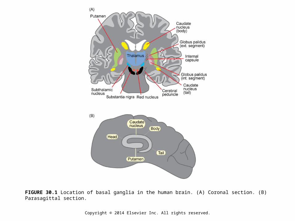
FIGURE 30.1 Location of basal ganglia in the human brain. (A) Coronal section. (B) Parasagittal section.
Copyright © 2014 Elsevier Inc. All rights reserved.
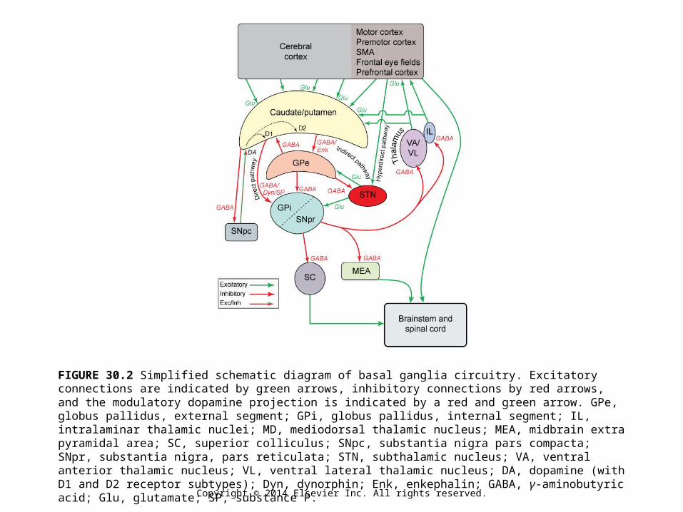
FIGURE 30.2 Simplified schematic diagram of basal ganglia circuitry. Excitatory connections are indicated by green arrows, inhibitory connections by red arrows, and the modulatory dopamine projection is indicated by a red and green arrow. GPe, globus pallidus, external segment; GPi, globus pallidus, internal segment; IL, intralaminar thalamic nuclei; MD, mediodorsal thalamic nucleus; MEA, midbrain extra pyramidal area; SC, superior colliculus; SNpc, substantia nigra pars compacta; SNpr, substantia nigra, pars reticulata; STN, subthalamic nucleus; VA, ventral anterior thalamic nucleus; VL, ventral lateral thalamic nucleus; DA, dopamine (with D1 and D2 receptor subtypes); Dyn, dynorphin; Enk, enkephalin; GABA, γ-aminobutyric acid; Glu, glutamate; SP, substance P.

Copyright © 2014 Elsevier Inc. All rights reserved.
FIGURE 30.3 Two representations of a striatal medium spiny neuron that has been filled with HRP. (A) The soma and dendritic tree with numerous dendritic spines. The thin process is the axon, which has been drawn without its collaterals. (B) The same neuron drawn to show the axonal collaterals, which branch extensively within the same field as the dendritic tree. From Wilson and Groves (1980).
Copyright © 2014 Elsevier Inc. All rights reserved.
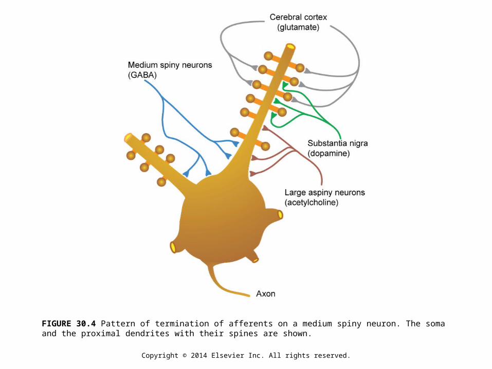
FIGURE 30.4 Pattern of termination of afferents on a medium spiny neuron. The soma and the proximal dendrites with their spines are shown.

Copyright © 2014 Elsevier Inc. All rights reserved.
FIGURE 30.5 Hypothetical parallel segregated circuits connecting the basal ganglia, thalamus, and cerebral cortex. The five circuits are named according to the primary cortical target of the output from the basal ganglia: motor, oculomotor, dorsolateral prefrontal, lateral orbitofrontal, and anterior cingulate. ACA, anterior cingulate area; APA, arcuate premotor area; CAUD, caudate; b, body; h, head; DLC, dorsolateral prefrontal cortex; EC, entorhinal cortex; FEF, frontal eye fields; GPi, internal segment of globus pallidus; HC, hippocampal cortex; ITG, inferior temporal gyrus; LOF, lateral orbitofrontal cortex; MC, motor cortex; MDpl, medialis dorsalis pars paralarnellaris; MDme, medialis dorsalis pars magnocellularis; MDpc, medialis dorsalis pars parvocellularis; PPC, posterior parietal cortex; PUT, putamen; SC, somatosensory cortex; SMA, supplementary motor area; SNr, substantia nigra pars reticulate; STG, superior temporal gyrus; VAmc, ventralis anterior pars magnocellularis; Vapc, ventralis anterior pars parvocellularis; VLm, ventralis lateralis parsmedialis; VLo, ventralis lateralis pars oralis; VP, ventral pallidum; VS, ventral striatum, cl, caudolateral; cdm, caudal dorsomedial; d1, dorsolateral; 1, lateral; 1dm, lateral dorsomedial; m, medial; mdm, medial dorsomedial; pm, posteromedial; rd, rostrodorsal; rl, rostrolateral; rm, rostromedial; vm, ventromedial; vl, ventrolateral.
Copyright © 2014 Elsevier Inc. All rights reserved.
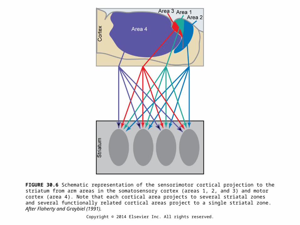
FIGURE 30.6 Schematic representation of the sensorimotor cortical projection to the striatum from arm areas in the somatosensory cortex (areas 1, 2, and 3) and motor cortex (area 4). Note that each cortical area projects to several striatal zones and several functionally related cortical areas project to a single striatal zone. After Flaherty and Graybiel (1991).

Copyright © 2014 Elsevier Inc. All rights reserved.
FIGURE 30.7 The two chemically different populations of striatal medium spiny neurons are intermixed. One population (blue) projects to the globus pallidus external segment (GPe) and contains GABA and enkephalin. The other population (red) projects to the globus pallidus internal segment (GPi) and substantia nigra pars reticulata (SNpr) and contains GABA, dynorphin, and substance P (red).
Copyright © 2014 Elsevier Inc. All rights reserved.
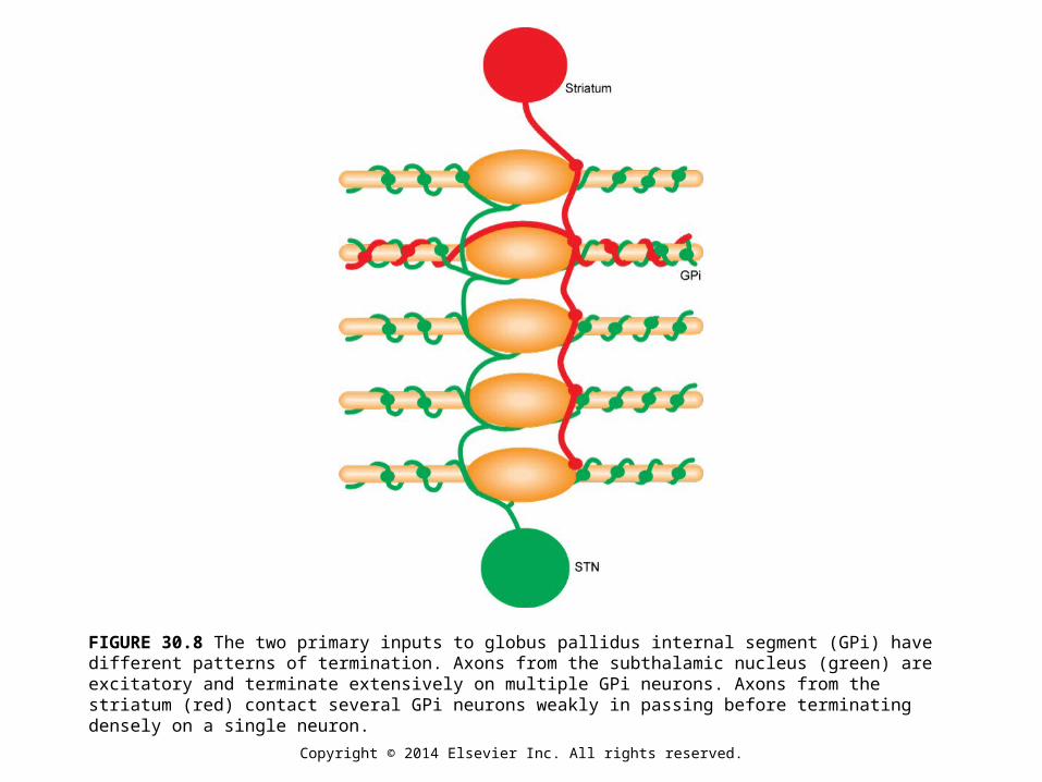
FIGURE 30.8 The two primary inputs to globus pallidus internal segment (GPi) have different patterns of termination. Axons from the subthalamic nucleus (green) are excitatory and terminate extensively on multiple GPi neurons. Axons from the striatum (red) contact several GPi neurons weakly in passing before terminating densely on a single neuron.

Copyright © 2014 Elsevier Inc. All rights reserved.
FIGURE 30.9 Representative neural discharge patterns from several basal ganglia nuclei. In the raster displays, each dot indicates the occurrence of an action potential. Each horizontal raster line represents a 1-s period of discharge. Several such periods are arranged vertically for each nucleus.

Copyright © 2014 Elsevier Inc. All rights reserved.
FIGURE 30.10 Schematic representation of the timing of neuronal activity in three nuclei of the basal ganglia in relation to limb movement and the muscle activity used to make the movement.
Copyright © 2014 Elsevier Inc. All rights reserved.
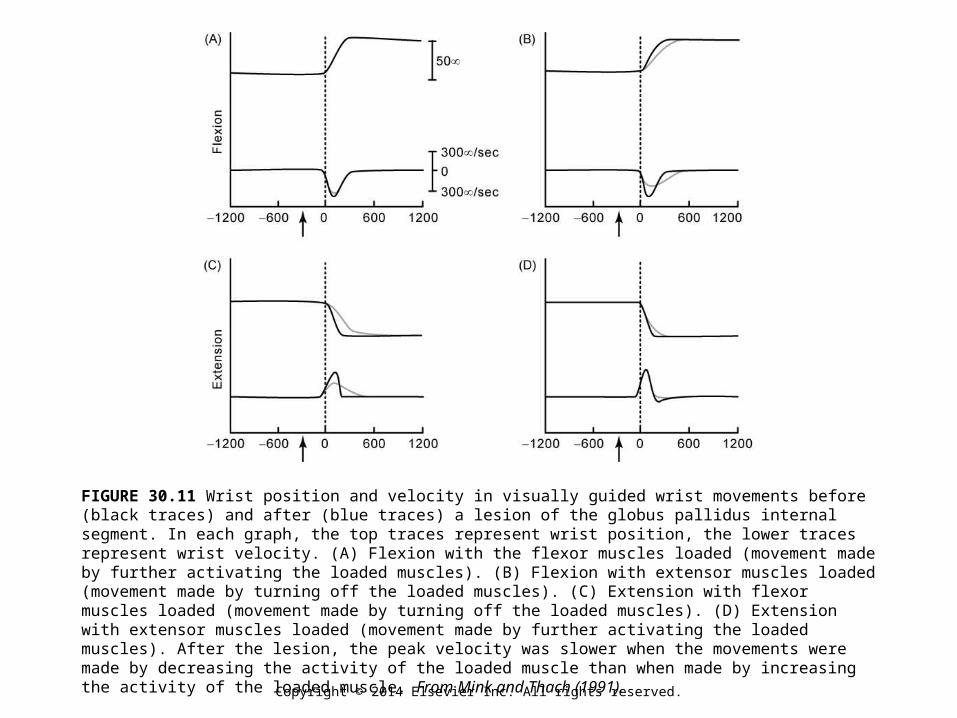
FIGURE 30.11 Wrist position and velocity in visually guided wrist movements before (black traces) and after (blue traces) a lesion of the globus pallidus internal segment. In each graph, the top traces represent wrist position, the lower traces represent wrist velocity. (A) Flexion with the flexor muscles loaded (movement made by further activating the loaded muscles). (B) Flexion with extensor muscles loaded (movement made by turning off the loaded muscles). (C) Extension with flexor muscles loaded (movement made by turning off the loaded muscles). (D) Extension with extensor muscles loaded (movement made by further activating the loaded muscles). After the lesion, the peak velocity was slower when the movements were made by decreasing the activity of the loaded muscle than when made by increasing the activity of the loaded muscle. From Mink and Thach (1991).
Copyright © 2014 Elsevier Inc. All rights reserved.
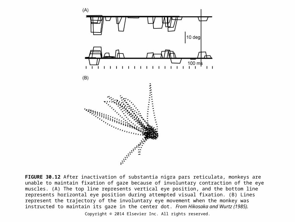
FIGURE 30.12 After inactivation of substantia nigra pars reticulata, monkeys are unable to maintain fixation of gaze because of involuntary contraction of the eye muscles. (A) The top line represents vertical eye position, and the bottom line represents horizontal eye position during attempted visual fixation. (B) Lines represent the trajectory of the involuntary eye movement when the monkey was instructed to maintain its gaze in the center dot. From Hikosaka and Wurtz (1985).
Copyright © 2014 Elsevier Inc. All rights reserved.
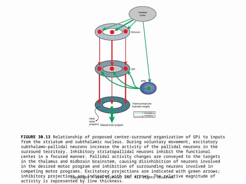
FIGURE 30.13 Relationship of proposed center-surround organization of GPi to inputs from the striatum and subthalamic nucleus. During voluntary movement, excitatory subthalamo-pallidal neurons increase the activity of the pallidal neurons in the surround territory. Inhibitory striatopallidal neurons inhibit the functional center in a focused manner. Pallidal activity changes are conveyed to the targets in the thalamus and midbrain brainstem, causing disinhibition of neurons involved in the desired motor program and inhibition of surrounding neurons involved in competing motor programs. Excitatory projections are indicated with green arrows; inhibitory projections are indicated with red arrows. The relative magnitude of activity is represented by line thickness.
Related Documents