CHAPTER 3 Preliminary Design of an Enterosoluble Multiparticulate System Incorporating Isoniazid 3.1. Introduction In this chapter, three feasible methods for the microencapsulation of INH were investigated, viz. air suspension, solvent evaporation and a novel salting-out approach for the formulation of enterogranules, microenterospheres and enterospheres respectively, as coined in this study. The methacrylic acid copolymers, methacrylic acid ethyl acrylate (MAEA) and methacrylic acid methyl methacrylate (MAMM), were instituted throughout the ensuing investigations. It is worth noting, henceforth, that the same polymer applied in a number of different ways yields exceptionally different release profiles. The rational identification of a candidate system for further optimisation was the ultimate goal of the three ensuing experimental investigations. Coherent comparison of the dissolution data of the resultant enterogranule, microenterosphere, and enterosphere formulations thus needs to be undertaken. This may be facilitated through pairwise comparison using a model-independent dissolution approach in order to determine if any one of the preliminary formulations could be considered for optimisation. The anatomical and physiological considerations with reference to enteric-release systems were delineated in the previous chapter. The following must be considered in developing the preliminary formulations: (1) reproduciblity of the production process, (2) particle size, and (3) entrapment and release of INH from the enterosoluble forms in acidic and phosphate-buffered dissolution media.

Welcome message from author
This document is posted to help you gain knowledge. Please leave a comment to let me know what you think about it! Share it to your friends and learn new things together.
Transcript
CCHHAAPPTTEERR 33
PPrreelliimmiinnaarryy DDeessiiggnn ooff aann EEnntteerroossoolluubbllee MMuullttiippaarrttiiccuullaattee SSyysstteemm IInnccoorrppoorraattiinngg IIssoonniiaazziidd
3.1. Introduction
In this chapter, three feasible methods for the microencapsulation of INH were investigated, viz.
air suspension, solvent evaporation and a novel salting-out approach for the formulation of
enterogranules, microenterospheres and enterospheres respectively, as coined in this study. The
methacrylic acid copolymers, methacrylic acid ethyl acrylate (MAEA) and methacrylic acid
methyl methacrylate (MAMM), were instituted throughout the ensuing investigations. It is worth
noting, henceforth, that the same polymer applied in a number of different ways yields
exceptionally different release profiles.
The rational identification of a candidate system for further optimisation was the ultimate goal of
the three ensuing experimental investigations. Coherent comparison of the dissolution data of the
resultant enterogranule, microenterosphere, and enterosphere formulations thus needs to be
undertaken. This may be facilitated through pairwise comparison using a model-independent
dissolution approach in order to determine if any one of the preliminary formulations could be
considered for optimisation.
The anatomical and physiological considerations with reference to enteric-release systems were
delineated in the previous chapter. The following must be considered in developing the
preliminary formulations: (1) reproduciblity of the production process, (2) particle size, and (3)
entrapment and release of INH from the enterosoluble forms in acidic and phosphate-buffered
dissolution media.

3.2. Development of a Methodology for the Fabrication of Enterogranules by the Air
Suspension Method
3.2.1. Materials and Methods
3.2.1.1. Materials
The poly (methacrylic acid co-methyl methacrylate) copolymers A and B with varying monomer
ratios (Eudragit®
L 100 and Eudragit®S 100) were received as a gift from Röhm GmbH,
Darmstadt, Germany. INH (isonicotinic acid hydrazide, 99% TLC) and triethyl citrate 99% were
purchased from Aldrich® (Sigma-Aldrich Inc., St. Louis, USA). Ethanol 90%
v/v and ammonia
solution (NH4OH, Mw=35.05g/mol) 25%v/v were obtained from Rochelle Chemicals
(Johannesburg, South Africa) and were of analytical grade. Sodium hydroxide (NaOH,
Mw=40.00g/mol) was obtained from Saarchem (Wadeville, Gauteng, South Africa). Hydrochloric
acid (HCl, 32%w/v) was purchased from Rochelle Chemicals (Johannesburg, South Africa).
Potassium dihydrogen phosphate (KH2PO4, Mw=136.09g/mol) was obtained from Riedel-de
Haen (Sigma-Aldrich Laborchemikalein GmbH, Germany). Double-deionised water (Milli-Q
System, Millipore, Bedford MA, USA) was used in the preparation of all dissolution media and
throughout the assay procedure.
3.2.1.2 Equipment
Standard laboratory-scale top-spray coating equipment having the configuration represented in
Figure 3.1 was employed for the encapsulation process: a fluidised bed coater (Aeromatic AG,
GmbH, Germany), vacuum pump (Vacutec, Johannesburg, South Africa), and a peristaltic pump
(Zero-max, USA).

Figure 3.1: Experimental set-up for the air-suspension coating process (descriptively
illustrated by Lehmann, 2001)
3.2.1.3 Identification of Processing Conditions for Successful Operation
The conditions, as elaborated henceforth, affecting the fluidised bed coating process (Table 3.1)
were set during preliminary trials by identification of settings that resulted in minimal
agglomeration and even enteric-coat application. The latter was determined following
microscopic analysis of the coated granules employing a stereomicroscope (Olympus SZX7
stereomicroscope, Olympus, Japan) connected to a digital camera (CC 12, Olympus, Japan) and
image analysis system (AnalySIS® Soft Imaging System, GmbH, Germany).
Table 3.1: Identified processing conditions
Processing Condition Setting
Product charge
Air flow rate/ fluidising air velocity
Fluid application rate
Drying temperature
Drying time
Atomising air pressure (pressure control)
15g of pre-sieved matrix granules
5m/s
5-7(x10) U/min = 5-7g/min
40oC
90min
2 bar
1 =inlet air
2 =inlet air filter
3 =air heater
4 =perforated base plate
5 =product container
6 =compressed air
7 =pneumatic spray nozzle
8 =expansion chamber
9 =exhaust air filter
10=exhaust air fan
11=peristatic pump
12=reservoir with propeller
stirrer.
2
35
4
9
8
7
6
12
10
11

3.2.1.3.1. Control of Airflow
Fluid bed coaters exert more mechanical stress on the cores as compared to coating pans. The
proper airflow rate is important for the successful operation of the process. Although increasing
the airflow rate or the temperature while maintaining the fluid application rate constant can
enhance the drying kinetics, attrition of particles must be prevented. The airflow rate was thus
adjusted to ensure adequate suspension of the granules with minimal granule agglomeration and
attrition.
3.2.1.3.2. Fluid Application Rate or Spray Rate
The fluid application rate was adjusted for minimisation of intergranular (cohesive) and granule-
fluidised bed wall (adhesive) sticking. Selecting a lower spray rate reduced any tendency to
sticking or agglomeration observed during the spray process. The application rate was thus
adjusted to the air handling capacity, such that the granules were always moist but never wet. In
the case of excessive wetness at a low spray rate, the spraying process was interrupted for 5
minutes for intermittent drying.
3.2.1.3.3. Drying Time and Temperature
These were selected in accordance with the minimum film-forming temperatures (MFT) of the
enteric-coating aqueous dispersions, having MFT of <25oC.
3.2.1.4 Formulation of Enterogranules
For the attainment of complete enterosoluble characteristics, granules incorporating INH were
prepared employing MAMM as the matrix in which the drug was embedded through institution
as a polymeric binder during the wet granulation process, and as the enteric-coating material. In
the GI tract, the coated granules were expected to behave as diffusion cells and release a constant
drug quantity per unit of time.
40%w/v solutions in 90%
v/v ethanol of the MAMM copolymer, Eudragit L
® 100, were prepared
by dispersing the polymer in the organic solvent with moderate agitation (700rpm) with a two-
blade propeller stirrer (Heidolph®, Labotec, Gauteng, South Africa) for 1 minute. Allowing the
covered solution to stand in a darkened room for 30 minutes ensured complete dissolution of the
copolymer. 10.0g INH was triturated with 75mL of the MAMM-ethanol solutions (copolymer
content of 30g) in a pestle and mortar. Formation of a coherent mass was facilitated by allowing a
degree of volatilisation of the alcohol from the moist granulate by drying under reduced pressure
at ambient conditions for 15 minutes (gravimetric weight loss: ≈5%w/w); the mass was then
passed through the 1.25cm-aperture sieve of a laboratory granulator (Erweka AR400, GmbH,
Germany). The granules were cooled on trays overnight at ambient conditions (21°C), then
screened through stainless steel sieves of a test sieve shaker (Octagon 200, Endecotts Ltd.,
London, England) and the fraction >1.00cm was used as such, or subjected to microencapsulation
by air suspension coating.
3.2.1.5 Enteric-Film Coating of Enterogranules
Granules were coated with either Eudragit L®100 (E L 100) and/ or Eudragit S
®100 (E S 100)
aqueous dispersions, prepared in accordance with Lehmann (2001) as per the formulations listed
in Table 3.2. The dispersions were prepared one day in advance. The enteric-polymeric powder
was dispersed at a steady rate in the stated amount of double-deionised water under moderate
agitation with a two-blade propeller stirrer, ensuring that the powder was rapidly wetted without
lump formation. The dispersion was stirred for 5 minutes followed by the dropwise addition of
the ammonia solution to the periphery of the vortex. After adding the ammonia solution, stirring
was continued for another 60 minutes, following the same procedure for triethyl citrate addition.

Triethyl citrate was included as a plasticiser to aid polymeric coalescence and film formation.
The dispersion was passed through a 0.25mm laboratory test sieve (Endecotts Ltd., London,
England).
Table 3.2: Formulae for Eudragit S®
100 and Eudragit L®
100 dispersions
Eudragit S®
100 dispersion Eudragit S 100
1M Ammonia solution (1.7%)
Triethyl citrate
Water
Content in dry polymer substance
Degree of neutralisation
Quantity employed for coating 15g granules
1.000g
0.500g
0.508g
5.532g
13.3%
15 mole-%
169.17g
Eudragit L®
100 dispersion Eudragit L 100
1M Ammonia solution (1.7%)
Triethyl citrate
Water
Content in dry polymer substance
Degree of neutralisation
Quantity employed for coating 15g granules
1.000g
0.500g
0.339g
5.011g
14.6%
6 mole-%
154.11g
The parameters were set for fluidising the active granules in accordance with the processing
conditions delineated in Table 3.1. Four preliminary formulations were investigated as set forth in
Table 3.3. Three batches of each formulation were prepared. Each time, 15g of accurately
weighed matrix granules were loaded into the fluidised bed. The granules were sprayed with the
quantities of dispersion as listed in Table 3.2 to achieve a theoretical coat: core ratio of 1.5:1.
When both E S 100 and E L 100 were employed, 84.59g E S 100 and 77.06g E L 100 were
blended with mild agitation (300 rpm) for 5 minutes before spray application. The duration of the
entire spraying and drying process was 90 minutes. The coated granules were evenly spread on
trays and cooled overnight under ambient conditions (21oC).

Table 3.3: Preliminary enterogranule formulationsa
Formulation E S 100 (g) E L 100 (g)
Uncoated 0 0
E L 100-coated 0 154.11g
E L 100: E S 100-coated 84.59g 77.06g
E S 100-coated 169.17g 0 aAll formulations were prepared in triplicate, n=3
3.2.1.6. Particle Size Analysis
The Feret’s diameters (df) of the coated and uncoated enterogranules were investigated by
microscopic image analysis using a stereomicroscope (Olympus SZX7, Olympus, Japan)
connected to a digital camera (CC 12, Olympus, Japan) and image analysis system (AnalySIS®
Soft Imaging System, GmbH, Germany). Feret’s diameter was determined from the mean
distance between two parallel tangents to the projected particle perimeter (Figure 3.2). The mean
diameter of each formulation was determined by measurement of the diameter of 50 randomly
selected enterogranules (n=50). Results were expressed as the mean ± standard deviation (S.D.)
of 50 measurements of the Feret’s diameter (df) for each formulation.
Figure 3.2: Feret’s diameters (df): (a) Assessment of df of an irregular particle and (b) particle
orientation for determination of shortest and longest Feret’s diameters (df) in ovoid
particles when determination of an aspect ratio is necessitated
df df (b) (a)
df
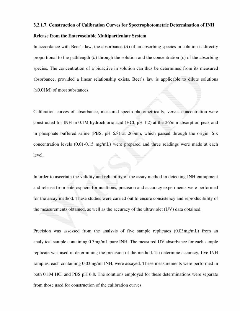
3.2.1.7. Construction of Calibration Curves for Spectrophotometric Determination of INH
Release from the Enterosoluble Multiparticulate System
In accordance with Beer’s law, the absorbance (A) of an absorbing species in solution is directly
proportional to the pathlength (b) through the solution and the concentration (c) of the absorbing
species. The concentration of a bioactive in solution can thus be determined from its measured
absorbance, provided a linear relationship exists. Beer’s law is applicable to dilute solutions
(≤0.01M) of most substances.
Calibration curves of absorbance, measured spectrophotometrically, versus concentration were
constructed for INH in 0.1M hydrochloric acid (HCl, pH 1.2) at the 265nm absorption peak and
in phosphate buffered saline (PBS, pH 6.8) at 263nm, which passed through the origin. Six
concentration levels (0.01-0.15 mg/mL) were prepared and three readings were made at each
level.
In order to ascertain the validity and reliability of the assay method in detecting INH entrapment
and release from enterosphere formualtions, precision and accuracy experiments were performed
for the assay method. These studies were carried out to ensure consistency and reproducibility of
the measurements obtained, as well as the accuracy of the ultraviolet (UV) data obtained.
Precision was assessed from the analysis of five sample replicates (0.03mg/mL) from an
analytical sample containing 0.3mg/mL pure INH. The measured UV absorbance for each sample
replicate was used in determining the precision of the method. To determine accuracy, five INH
samples, each containing 0.03mg/ml INH, were assayed. These measurements were performed in
both 0.1M HCl and PBS pH 6.8. The solutions employed for these determinations were separate
from those used for construction of the calibration curves.

Note that it is established at the outset that the methacrylic acid copolymer solution and/or latex
and all other excipients (i.e. plasticisers, electrolytes, etc.) employed in the respective
encapsulation processes did not interfere with drug analysis at the reported wavelength.
3.2.1.8. Drug Content of Enterogranules
Drug content was determined spectrophotometrically at 263nm by placing 100mg of INH-loaded
enterogranules in a 200mL conical flask containing 100mL of 0.2M PBS, pH 7.0. The
enterogranules were magnetically stirred for 5 hours after which time all the granular
formulations were microscopically observed (Olympus SZX7, Japan) to have undergone
complete erosion. This was to ensure absolute liberation and subsequent dissolution of the water-
soluble INH from the enterosoluble matrix. The resultant solutions were filtered through a
0.45µm membrane filter (Millipore®, Billerica, MD, USA). The filtrates were then made up to
200mL volumes with the PBS. Aliquots of the filtrates were subjected in triplicate to UV
spectroscopy (diode array UV spectrophotometer, Specord 40, Analytik Jena AG, Jena,
Germany) at 263nm for analysis (WinASPECT® Spectroanalytical Software, Analytik Jena AG,
Jena) following comparison with the standard calibration curves generated for INH in PBS
media.
3.2.1.9. In Vitro Release Studies on Enterogranules
Characterisation of INH release from the enterogranules was assessed using a method adapted
from the USP 24 general drug release standard for delayed release (enteric-coated) articles in
acidic and phosphate-buffered media (USP 24, 2000). Enterogranules equivalent to 10mg INH
were placed in 50mL sealed vials. For determination of the amount of INH released under acidic
conditions, 20mL of 0.1M HCl was added to the vials which were then subjected to agitation at
50rpm for 2 hours in a shaker water-bath (Labex, Stuart SBS40®, Gauteng, South Africa)

maintained at 37±0.5°C. For determination of INH release in basic media, the acid was drained
from the vials whilst retaining the enterogranules and replaced with 20mL of PBS (pH 7.0).
Agitation was continued for a further 6 hours. Balancing withdrawal of 1mL aliquots was
performed at the appropriate time intervals and samples were then analysed by UV at 263nm
following dilution for determination of the fractional INH release.
3.2.2. Results and Discussion
The regression coefficients (R2=0.9995 and 0.9997, respectively) for the calibration curves
(Figures 3.3 and 3.4) constructed for INH demonstrated linearity in acidic media (pH 1.2) and
phosphate buffered media (pH 6.8) achieved over the concentration range (0.01-0.15 mg/mL).
Figure 3.3: INH calibration curve at 265nm in 0.1M HCl (pH 1.2,) (S.D. within ±0.052 in all
cases)
Concentration (mg/mL)
0.00 0.02 0.04 0.06 0.08 0.10 0.12
Ab
sorb
an
ce
0
1
2
3
4
R = 0.9995
y = 36.015
2
y = 36.015x
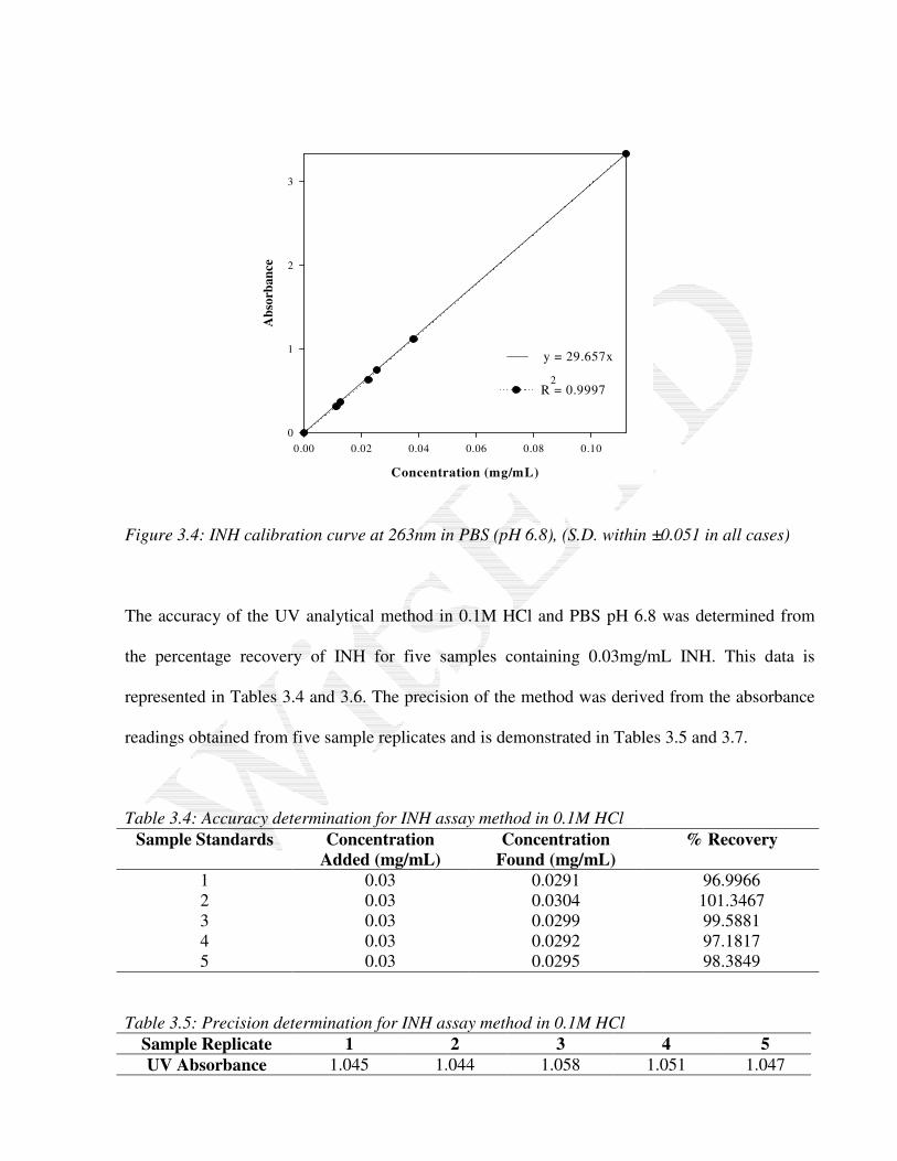
Figure 3.4: INH calibration curve at 263nm in PBS (pH 6.8), (S.D. within ±0.051 in all cases)
The accuracy of the UV analytical method in 0.1M HCl and PBS pH 6.8 was determined from
the percentage recovery of INH for five samples containing 0.03mg/mL INH. This data is
represented in Tables 3.4 and 3.6. The precision of the method was derived from the absorbance
readings obtained from five sample replicates and is demonstrated in Tables 3.5 and 3.7.
Table 3.4: Accuracy determination for INH assay method in 0.1M HCl
Sample Standards Concentration
Added (mg/mL)
Concentration
Found (mg/mL)
% Recovery
1 0.03 0.0291 96.9966
2 0.03 0.0304 101.3467
3 0.03 0.0299 99.5881
4 0.03 0.0292 97.1817
5 0.03 0.0295 98.3849
Table 3.5: Precision determination for INH assay method in 0.1M HCl
Sample Replicate 1 2 3 4 5
UV Absorbance 1.045 1.044 1.058 1.051 1.047
Concentration (mg/mL)
0.00 0.02 0.04 0.06 0.08 0.10
Ab
sorb
an
ce
0
1
2
3
R = 0.9997
y = 29.657x
2

A mean of 0.0296mg/mL and a standard deviation of 5.43x10⁻⁴
were obtained for the accuracy
determination in 0.1M HCl. The coefficient of variation, a measure of the relative variability for
this data, was 1.834%. A mean of 1.049 and a standard deviation of 5.701x10⁻³
were obtained for
the precision determination in 0.1M HCl. The coefficient of variation of this data was 0.543%.
Table 3.6: Accuracy determination for INH assay method in 0.2M PBS pH 6.8
Sample Standards Concentration
Added (mg/mL)
Concentration
Found (mg/mL)
% Recovery
1 0.03 0.03048 101.6061
2 0.03 0.02991 99.6954
3 0.03 0.02954 98.4591
4 0.03 0.02849 94.9748
5 0.03 0.02856 95.1996
Table 3.7: Precision determination for INH assay method in 0.2M PBS pH 6.8
Sample Replicate 1 2 3 4 5
UV Absorbance 0.887 0.870 0.876 0.872 0.881
A mean of 0.0294mg/mL and a standard deviation of 8.63x10⁻⁴
were obtained for the accuracy
determination assay in PBS. The coefficient of variation for this data was 2.935%. A mean of
0.877 with a standard deviation of 6.782x10⁻³
was obtained for the precision determination assay
in PBS. The coefficient of variation of this data was 0.787%.
This indicates that INH recovery using this method of detection for determination of INH release
in both 0.1M HCl and 0.2M PBS pH 6.8 is satisfactorily consistent and precise.
The morphology, mean diameter and INH release profiles in acidic media for the preliminary
enterogranules are represented in Figures 3.5, 3.6 and 3.7.

Figure 3.5: Stereomicrographs (darkfield, scale bar=1cm) of enterogranules: (a) uncoated at
40X (b) E L 100-coated at 25X and (c) E S 100-coated at 40X magnification
Figure 3.6: Feret’s diameter (mean±S.D., n=50) of the preliminary enterogranule formulations
Figure 3.7: Drug release profiles of the enterogranules in acidic media (0.1M HCl, pH 1.2),
(S.D. within ±0.039 in all cases)
a b c
X Data
Uncoated
E L 100-c
oated
E L 100: E
S 100-c
oated
E S 100-c
oated
Mea
n d
f (m
m)
0.0
0.2
0.4
0.6
0.8
1.0
1.2
1.4
1.6
1.8
2.0
2.2
Time (minutes)
0 30 60 90 120
Fra
ctio
nal
INH
Rel
ease
0.0
0.2
0.4
0.6
0.8
1.0
Uncoated
E L 100 - coated
E L 100: E S 100 - coated
E S 100 - coated
INH release from the irregular enterogranules was uncompromisingly rapid with almost the entire
entrapped amount of drug leaching out of the granule after one hour in acidic media. The
following reasons were identified for the lack of enteric control of the multiparticulate system.
• The matrix granules were insufficiently compact with resultant diffusion of INH through
the porous structure
• There was rapid diffusion of the water-soluble INH through the enteric-film coating
• The possibility of INH diffusion into the enteric coat during the coating process could not
be ruled out even under strictly controlled coating conditions.
The shortcomings associated with enteric-coated formulations made of aqueous disperse systems
or solutions is the lack of resistance against gastric fluid and the reportedly more rapid diffusion
of water-soluble drug through films prepared from aqueous solutions than through organic-
solvent-based films (Guo et al., 2002).
Bianchini et al. (1991) demonstrated the poor performance of enteric-coated dosage forms
containing a water-soluble substance; these did not pass the USP 24 test unless insulation of the
cores was undertaken by subcoating barriers or by employing twice the amount of coating. The
lack of sufficiently effective gastroresistance has been ascribed to dissolution of a small amount
of drug from the core tablet to the aqueous film during the coating process. The higher release
rates from coated pellets have thus been attributed primarily to drug diffusion into the film layer
during the coating process. If the active ingredients are freely water-soluble, as is the case with
INH, they may dissolve in the spray mist during the coating process, resulting in active
ingredients being incorporated in the film. The presence of a drug or an excipient in a film
coating is not desired as it substantially alters the mechanical adhesion and permeation
characteristics of the applied coating (Guo, 1993; Guo et al., 2002).
It is contemplated that fabrication of an optimal enteric-polymeric matrix system for
incorporation of the water-soluble drug in a single processing step, where sufficient coalescence
of the polymer is promoted, would achieve improved gastroresistance of the multiparticulate
system.
3.3. Development of a Methodology for the Fabrication of Microenterospheres by the Polar
Organic-in-Oil Emulsification Solvent Evaporation Method
3.3.1. Materials and Methods
3.3.1.1. Materials
The poly (methacrylic acid co-methyl methacrylate) copolymers with varying monomer ratios (E
L 100 and E S 100) were received as a gift from Röhm GmbH, Darmstadt, Germany. INH
(isonicotinic acid hydrazide, 99% TLC) was purchased from Aldrich® (Sigma-Aldrich Inc., St.
Louis, USA). Organic solvents (acetone, ethanol) were all of analytical grade and were purchased
from Rochelle Chemicals (Johannesburg, South Africa).
3.3.1.2 Formulation of Microenterospheres
A modified solvent evaporation emulsification method, employing an organic solution of the
MAMM copolymers, proved feasible for the formulation of uniform, spherical multiparticulates
with a narrow size distribution, capable of entrapping the water-soluble INH.
The MAMM copolymers (2.0g) were dissolved in 15.0mL acetone with 0.3mL water, or 15.0mL
ethanol, or their 1:1 combination, whilst stirring with a magnetic stirrer for 30 minutes. INH
(1.0g) was then dissolved in the MAMM copolymer solution by stirring for an additional 15
minutes. This solution was then emulsified in 40mL of a 1%w/v Span 80
®-liquid paraffin solution
at 800rpm for 3 hours at room temperature with a Heidolph®
two-blade propeller stirrer (Labotec,
Gauteng, South Africa). Once the microenterospheres were formed, they were filtered on a
Buchner funnel, washed twice with 100mL normal hexane to remove the residual oil phase, and
subsequently dried under ambient laboratory conditions (21oC) overnight.
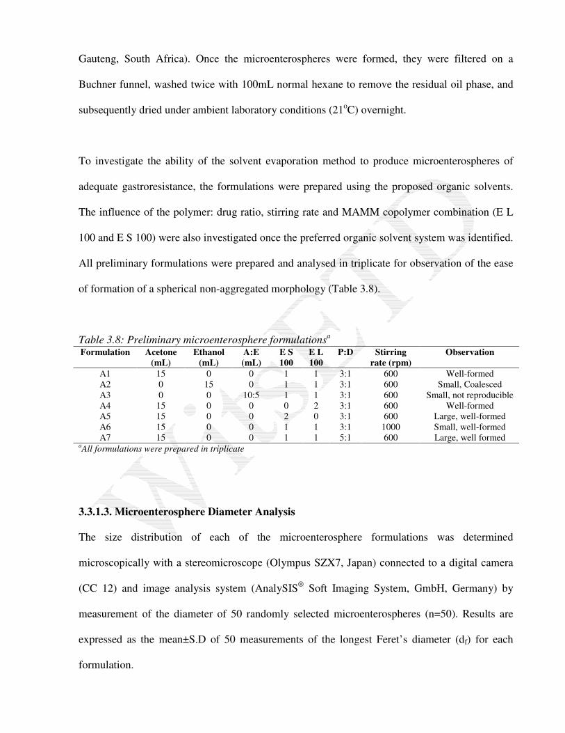
To investigate the ability of the solvent evaporation method to produce microenterospheres of
adequate gastroresistance, the formulations were prepared using the proposed organic solvents.
The influence of the polymer: drug ratio, stirring rate and MAMM copolymer combination (E L
100 and E S 100) were also investigated once the preferred organic solvent system was identified.
All preliminary formulations were prepared and analysed in triplicate for observation of the ease
of formation of a spherical non-aggregated morphology (Table 3.8).
Table 3.8: Preliminary microenterosphere formulationsa
Formulation Acetone
(mL)
Ethanol
(mL)
A:E
(mL)
E S
100
E L
100
P:D Stirring
rate (rpm)
Observation
A1
A2
A3
A4
A5
A6
A7
15
0
0
15
15
15
15
0
15
0
0
0
0
0
0
0
10:5
0
0
0
0
1
1
1
0
2
1
1
1
1
1
2
0
1
1
3:1
3:1
3:1
3:1
3:1
3:1
5:1
600
600
600
600
600
1000
600
Well-formed
Small, Coalesced
Small, not reproducible
Well-formed
Large, well-formed
Small, well-formed
Large, well formed aAll formulations were prepared in triplicate
3.3.1.3. Microenterosphere Diameter Analysis
The size distribution of each of the microenterosphere formulations was determined
microscopically with a stereomicroscope (Olympus SZX7, Japan) connected to a digital camera
(CC 12) and image analysis system (AnalySIS®
Soft Imaging System, GmbH, Germany) by
measurement of the diameter of 50 randomly selected microenterospheres (n=50). Results are
expressed as the mean±S.D of 50 measurements of the longest Feret’s diameter (df) for each
formulation.

3.3.1.4. Encapsulation Efficiency of Microenterospheres
Drug content was determined spectrophotometrically at 263nm by placing 100mg of INH-loaded
microenterospheres in a 200mL conical flask containing 100mL of 0.2M PBS, pH 7.0. The
microenterospheres were magnetically stirred for 10 hours. This period was sufficient to promote
polymeric swelling and dissolution, and the resultant complete erosion of the microenterosphere,
as observed microscopically (Olympus SZX7, Japan), for absolute liberation of the entrapped
water-soluble INH. The resultant solutions were filtered through a 0.45µm membrane filter
(Millipore®, Billerica, MD, USA). The filtrates were then made up to 200mL volumes with PBS.
Aliquots of the filtrates were subjected in triplicate to UV spectroscopy (diode array UV
spectrophotometer, Specord 40, Analytik Jena AG, Jena) at 263nm for analysis (WinASPECT®
Spectroanalytical Software, Analytik Jena AG, Jena) following comparison with the standard
calibration curves generated for INH in PBS. The amount of drug entrapped in the
microenterospheres in each formulation was compared with the amount of drug, which was
intended to be loaded in order to obtain the drug encapsulation efficiency (DEE):
100dose) loading initial (actual resenterosphe into loaded drug ofquantity lTheoretica
resenterosphein present drug ofquantity Actual(%) ×=DEE
[Equation 3.1]
3.3.1.5. In Vitro Release Studies on Microenterospheres
Characterisation of INH release from the microenterospheres was assessed using a method
adapted from the USP 24 general drug release standard for delayed release (enteric-coated)
articles in 0.1M HCl and PBS (USP 24, 2000). Microenterospheres equivalent to 10mg INH were
placed in 50mL sealed vials. For determination of the amount of INH released under acidic
conditions, 20mL of 0.1M HCl was added to the vials which were then subjected to agitation at
50rpm for 2 hours in a shaker water-bath (Labex, Stuart SBS40®, Gauteng, South Africa)
maintained at 37±0.5°C. For determination of INH release in basic media, the acid was drained
from the vials whilst carefully retaining the microenterospheres and replaced with 20mL of a
PBS (pH 7.0). Agitation was continued for a further 6 hours. Balancing withdrawal of 1mL
aliquots was performed at the appropriate time intervals and samples were then analysed by UV
spectroscopy at 263nm and 265nm following dilution for determination of the fractional INH
release.
3.3.2. Results and Discussion
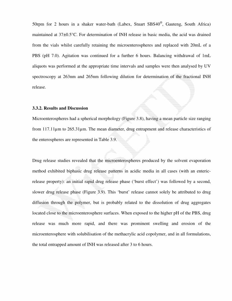
Microenterospheres had a spherical morphology (Figure 3.8), having a mean particle size ranging
from 117.11µm to 265.31µm. The mean diameter, drug entrapment and release characteristics of
the enterospheres are represented in Table 3.9.
Drug release studies revealed that the microenterospheres produced by the solvent evaporation
method exhibited biphasic drug release patterns in acidic media in all cases (with an enteric-
release property): an initial rapid drug release phase (‘burst effect’) was followed by a second,
slower drug release phase (Figure 3.9). This ‘burst’ release cannot solely be attributed to drug
diffusion through the polymer, but is probably related to the dissolution of drug aggregates
located close to the microenterosphere surfaces. When exposed to the higher pH of the PBS, drug
release was much more rapid, and there was prominent swelling and erosion of the
microenterosphere with solubilisation of the methacrylic acid copolymer, and in all formulations,
the total entrapped amount of INH was released after 3 to 6 hours.
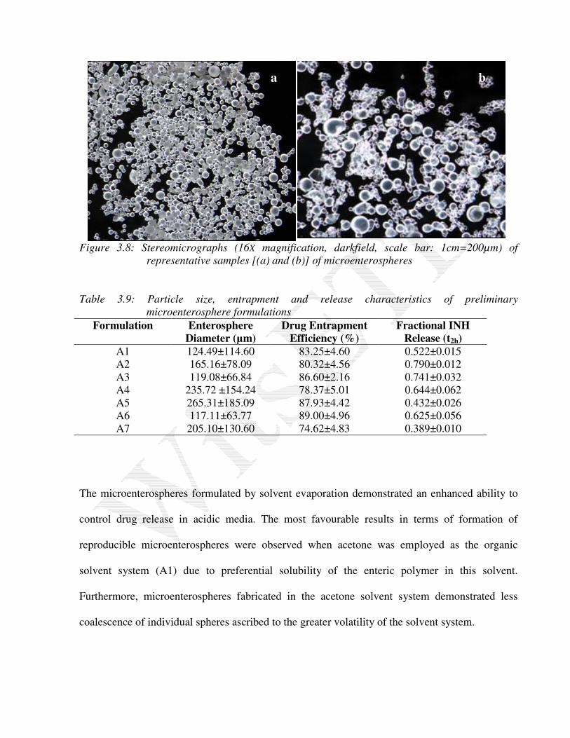
Figure 3.8: Stereomicrographs (16X magnification, darkfield, scale bar: 1cm=200µm) of
representative samples [(a) and (b)] of microenterospheres
Table 3.9: Particle size, entrapment and release characteristics of preliminary
microenterosphere formulations
Formulation Enterosphere
Diameter (µm)
Drug Entrapment
Efficiency (%)
Fractional INH
Release (t2h)
A1 124.49±114.60 83.25±4.60 0.522±0.015
A2 165.16±78.09 80.32±4.56 0.790±0.012
A3 119.08±66.84 86.60±2.16 0.741±0.032
A4 235.72 ±154.24 78.37±5.01 0.644±0.062
A5 265.31±185.09 87.93±4.42 0.432±0.026
A6 117.11±63.77 89.00±4.96 0.625±0.056
A7 205.10±130.60 74.62±4.83 0.389±0.010
The microenterospheres formulated by solvent evaporation demonstrated an enhanced ability to
control drug release in acidic media. The most favourable results in terms of formation of
reproducible microenterospheres were observed when acetone was employed as the organic
solvent system (A1) due to preferential solubility of the enteric polymer in this solvent.
Furthermore, microenterospheres fabricated in the acetone solvent system demonstrated less
coalescence of individual spheres ascribed to the greater volatility of the solvent system.
a b

Figure 3.9: Release profiles for preliminary microenterosphere formulations in acidic media
(0.1M HCl, pH 1.2), (S.D. within ±0.063 in all cases)
An increase in the polymer: drug ratio in A7 generally saw an increase in the amount of INH
entrapped within the microenterospheres. An increase in the amount of polymer incorporated into
the microenterospheres resulted in a decrease in the rate of INH release. The consequential
increase in diffusion pathways lead to slower drug diffusion rates, in accordance with Fick’s law
of diffusion. This is also ascribed to the fact that the release of INH from microenterospheres
increased with increasing proportion of the drug. This was due to an increased amount of drug
being close to the microenterosphere surface and the likelihood of a portion of the drug being
uncoated increased with higher drug loading.
Drug release from microenterospheres formulated according to the described solvent evaporation
method has shown dependency on particle size and has been demonstrated by various authors
(Beck et al., 1979; Barkai et al., 1990; de Brito Amorim and Ferreira, 2001; Perumal, 2001). This
Time (minutes)
0 30 60 90 120
Fra
ctio
na
l IN
H R
elea
se
0.0
0.2
0.4
0.6
0.8
1.0
A1
A2
A3
A4
A5
A6
A7
was the case here, as the more rapid stirring rates employed for A6 generally formed smaller
microenterospheres, which slowed the release of INH to a lesser extent, in accordance with Fick's
law of diffusion. A slower stirring rate caused a resultant increase in the wall thickness, which
increased drug diffusion pathways thereby protracting the diffusion process.
A5 incorporating only E S 100 was better able to prevent the release of INH under acidic
conditions than E L 100 (A4). This is a consequence of the lower aqueous solubility of E S 100
due to the lower ratio of carboxyl to ester groups (approximately 1:1 in E L 100 and 1:2 in E S
100). However, its use in combination with E L 100 is preferred because use of E S 100 in
isolation results in dissolution of enteric film coatings only commencing above pH 7.0 and thus
usually occurs in vivo in the lower sections of the intestines. However, since a pH of 7.0 is
frequently only just reached and not noticeably exceeded, excretion of active ingredients with the
faeces should be avoided by mixing E S 100 with E L 100.
3.4. Modifications to Overcome Burst Release from Microenterospheres
Although the microenterospheres demonstrated the ability to control drug release to a certain
extent, the initial burst release of INH was undesirably high owing to initial dissolution of drug
aggregates located close to the microenterosphere surface and then drug diffusion through the
enteric copolymer matrix. For this system, the need for double entrapment of the water-soluble
INH within a reservoir or multireservoir enterosoluble system (Figure 3.10) would be warranted.
Phase separation methods proved to be successful at depositing a polymeric coating upon the
core material.
The process involved formation of three immiscible chemical phases: a chemical liquid
manufacturing vehicle phase, a core material phase, and a coating material phase. It was therefore

essential that a polymeric phase be selected in which the core microenterosphere could not be
solubilised. Utilising one of the methods of phase-separation coacervation, the coating material
phase is induced to coalesce. In this case, a change in temperature and the addition of a soluble
inorganic salt was instituted. Deposition of the liquid polymer coating around the core material
occurs if the polymer is adsorbed at the interface formed between the core material and the liquid
vehicle phase; this is a prerequisite for effective coating. In the first case, the microenterospheres
were coated with an additional enteric coating affected by addition of the core phase and the
aqueous polymer phase to an electrolyte solution. In the second instance, ethylcellulose, a water-
insoluble polymer, was applied to the core microenterosphere following a thermal change
(Bakan, 1986).
Figure 3.10: Schematic of reservoir and multireservoir enterosphere representing double
entrapment of INH
3.4.1. Materials and Methods
3.4.1.1. Materials
The as-received methacrylic acid copolymer type C was a gift from Röhm GmbH, Darmstadt,
Germany and contains 0.7%w/w sodium lauryl sulphate and 2.3%
w/w Polysorbate 80 based on
solid substance, added to function as emulsifiers. Sodium hydroxide (NaOH, Mw=40.00g/mol)
Enteric-polymer
matrix
Enteric-polymer
film
Drug-loaded
Microenterosphere
was purchased from Saarchem (Wadeville, Gauteng, South Africa). Ethylcellulose (Ethocel®
STD 100) was purchased from Protea Industrial Chemicals (Pty) Ltd. (Wadeville, Gauteng).
Cyclohexane and sodium chloride (NaCl, Mw=58.45g/mol) were purchased from Rochelle
Chemicals (Johannesburg, Gauteng).
3.4.1.2. Double Entrapment in Methacrylic Acid Ethyl Acrylate Copolymer
The preferred (most gastroresistant) microenterosphere formulation (A7) was prepared as
described by the solvent evaporation method. An amount of microenterospheres theoretically
equivalent to 100mg INH was dispersed in 1.67mL of a 6-mole-percentage neutralised (achieved
by addition of 1M NaOH) 30%w/w MAEA copolymer aqueous dispersion equivalent to 500mg
dry MAEA. The resultant dispersion was extruded dropwise at a rate of 2.0mL/min, using a flat-
tip needle (Terumo®, GmbH, Germany) of 0.80-mm internal diameter, into 100mL of a
magnetically agitated concentrated electrolyte solution (25%w/v NaCl) and cured for 5 minutes in
the electrolyte solution for incorporation of the microenterospheres in the salted-out enterosoluble
coating for the fabrication of multireservoir enterospheres (R1). Multireservoir enterospheres
were collected following decantation of the aqueous electrolyte phase and rinsed twice with
100mL double-deionised water.
3.4.1.3. Double Entrapment in Ethylcellulose
Microenterospheres were further encapsulated in ethylcellulose employing a coacervation phase
separation by thermal change technique. The etherified cellulosic, containing a relatively high
ethoxyl content (high degree of substitution) is insoluble in cyclohexane at room temperature but
is soluble at elevated temperatures. The ethylcellulose grade selected is commonly employed for
microencapsulation purposes. The ethylcellulose coating was given on the microenterospheres by
employing cyclohexane as a solvent for ethylcellulose, in which neither MAMM or INH are
soluble, and changing the temperature from 80oC with continuous stirring at 1000rpm. 500mg of
ethylcellulose was dissolved in 5mL cyclohexane at 80oC (boiling point) with magnetic stirring
for 30 minutes. The preferred (most gastroresistant) microenterosphere formulation (A7) was
prepared as described by the solvent evaporation method. An amount of microenterospheres
theoretically equivalent to 100mg INH was dispersed in the ethylcellulose solution at 80oC. The
temperature of the solution was slowly lowered to 20oC at a rate of 1
oC/minute. Cooling to room
temperature accomplished gelation and solidification of the coating. This resulted in
microenterospheres being incorporated within the ethylcellulose matrix for the fabrication of
reservoir enterospheres (R2).
3.4.1.4. In Vitro Drug Release Studies on Reservoir Systems
R1 and R2 were prepared in triplicate for in vitro drug release testing. Drug release studies were
conducted on the resrevoir/ multireservoir enterospheres as described previously for the
microenterospheres by placing an amount of enterospheres equivalent to 10mg INH in a 50mL
vessel containing 20mL of the respective release media.
3.4.2. Results and Discussion
R1 enterospheres had a mean diameter of 2535µm and R2 enterospheres had a mean diameter of
740µm (Figure 3.11). Encapsulation by coacervation-deposition of an ethylcellulose coating on
the microenterospheres (R2) decreased the gastroresistance of the multiparticulate system with
70.4% of the entrapped INH being released at t2h. A possible explanation for this may be the
disruption of the patency of the enteric barrier created by the MAMM polymers during the
thermal deposition of the ethylcellulose due to the adsorption phenomenon. Encapsulation of
enterospheres in the salted-out MAEA coating enhanced the gastroresistance of the
multiparticulate system, with less than 3.7% of the INH being released at t2h (Figure 3.12).

Industrially, however, the process of double encapsulation for enterosphere fabrication may not
be entirely feasible as time constraints and a greater number of processing steps would limit its
application.
Figure 3.11: Stereomicrographs (darkfield, 16X magnification, scale bar: 1cm=200µm) of
representative reservoir enterospheres: (a) R1 and (b) R2
Figure 3.12: Drug release profiles of reservoir systems in acidic media (0.1M HCl, pH 1.2),
(S.D. within ±0.042 in all cases)
a b
Time (minutes)
0 30 60 90 120
Fra
ctio
na
l IN
H R
elea
se
0.0
0.2
0.4
0.6
0.8
1.0
R1
R2
Micro-
enterosphere
Microenterosphere

3.5. Development of a Methodology for the Fabrication of Enterospheres by a Novel
Salting-Out and Ionotropic Cross-Linking of MAEA
The principles inherent in colloid science and salting-out techniques formed the basis of this
approach, which employed the more environmentally benign acrylic latex rather than an organic
solution of the enteric polymer. As demonstrated previously, aqueous dispersion-coated dosage
forms of water-soluble drugs have inadequate gastroresistance. It is contemplated that fabrication
of a salted-out and cross-linked enteric-polymeric matrix system incorporating a water-soluble
drug would achieve improved gastroresistance of the methacrylic acid copolymer coating latex.
Simple coacervation and complex coacervation were defined in Chapter 2 and result in deposition
of coacervate walls from aqueous solutions of polymer by separation of a colloid-enriched phase
from a dilute colloid solution (Donbrow, 1991).
This approach exploited the ‘salting-out’ phenomenon to induce phase-separation/ coacervation,
followed by ionotropic cross-linking of the MAEA enteric copolymer. The salted-out and cross-
linked enterosphere matrices were formed by inducing separation of the anionic polyelectrolyte
as a polymer-rich enteric film (‘salting-out’) and ionotropically cross-linking the internal
enterosphere matrix following extrusion and curing of an aqueous dispersion of the polymer into
a concentrated electrolyte solution. The formation and properties of the copolymeric
enterospheres cross-linked via polyvalent ions depends on the concentrations and distribution of
the ions incorporated within the MAEA enterospheres, which is in turn also affected by the
duration of exposure of the copolymer to the salting-out solution. The copolymeric chains are
cross-linked via the cations by the formation of complexes liganded with more than one MAEA
group creating intramolecular and/ or intermolecular cross-links (Allain and Salome, 1990). The
described method precluded the use of organic solvents for dissolution of MAEA as a partially

neutralised aqueous dispersion was utilised, was highly reproducible, and the particles were
formulated in a single processing step.
The MAEA is a synthetic copolymer demonstrating excellent biocompatibility, and is suitable for
ionotropic cross-linking in this manner to form interconnected matrices. As an anionic
polyelectrolyte, it possesses charged carboxylic acidic side groups, and the water-solubilised
polymer may be cross-linked by reaction with a solution of cations. The preferred cations for
cross-linking polymers with acidic side groups are divalent and trivalent ions, divalent cations
being preferred due to lower toxicity; the higher the concentration of cation or the higher the
valency, the greater the degree of polymer cross-linking. This phenomenon is described by the
Schulze-Hardy rule, which governs the ability of an electrolyte to reduce the value of the zeta-
potential of the colloidal polymer.
Although methacrylic acid copolymers are practically insoluble in water, they are soluble in
solutions of 1M NaOH upon neutralisation of carboxyl groups, giving clear to slightly opalescent
solutions. Partial neutralisation of the aqueous polymeric dispersion of the MAEA copolymer
resulted in the formation of a latex in which the polymer particles typically had a submicron
particle-size distribution and behaved in the same manner as colloidal particles. The dispersed
phase in the latex was thus composed of spherical polymer particles with an average diameter of
200-300nm. The dispersion medium was water containing various water-soluble compounds. The
dispersions of MAEA have been demonstrated to be stabilised by a combination of electrostatic
and steric mechanisms termed as electrosteric stabilisation. The electrosteric stabilisation is
considered to arise in part from dissolved polymer chains with charged carboxylic groups
extending out into the continuous phase. The partial neutralisation and solubilisation of the
carboxyl-containing MAEA copolymer facilitated both the rapid destabilisation of MAEA in the

presence of electrolytes inducing coalescence of the colloidal particles and film formation
(Nyamweya, 2001) as well as promoting interpenentration and a degree of polymeric cross-
linking on protracted exposure to a solution of cations due to the presence of dissolved copolymer
chains in the dispersion. As described, the efficiency of coalescence due to colloidal
destabilisation is sensitive to the valency of the counterion; the concentration of counterions
required for coagulation decreases drastically with increasing valency (Lieberman et al., 1988).
3.5.1. Materials and Methods
3.5.1.1. Materials
The as-received methacrylic acid copolymer type C (E L 100-55) was a gift from Röhm GmbH,
Darmstadt, Germany and contains 0.7%w/w sodium lauryl sulphate and 2.3%
w/w Polysorbate 80
based on solid substance, added to function as emulsifiers. INH (isonicotinic acid hydrazide, 99%
TLC) and triethyl citrate (99% purity) were purchased from Aldrich® (Sigma-Aldrich Inc., St.
Louis, USA). Electrolytes were all of analytical grade and were purchased from Rochelle
Chemicals (Johannesburg, South Africa) and Saarchem (Wadeville, Gauteng, South Africa).
3.5.1.2. Formulation of Enterospheres
The novel salting-out and cross-linking method was employed for the formulation of
enterospheres, instituting a partially neutralised aqueous dispersion (latex) of MAEA copolymer
with a monomer molar ratio of 1:1. Among the anionic enteric polymers, MAEA is the only
copolymer commercially available as an aqueous dispersion (Eudragit L 30 D-55, USP/NF
methacrylic acid copolymer Type C) and as a powder for redispersion (Eudragit L 100-55,
USP/NF methacrylic acid copolymer Type C) thus facilitating industrial application of this
approach.

The formula for preparation of one batch (50mL) of an INH-loaded 6-mole-% neutralised
aqueous dispersion (latex) comprised 30g double-deionised water, 15g E L 100-55, 5.0g 1.0M
NaOH, 5.0g triethyl citrate, and 3.0g INH.
The latex was freshly prepared each time from the powder for redispersion by slow addition of
1.0M NaOH to the latex particle agglomerates in water and dispersing in accordance with
Lehmann (2001) in order to achieve neutralisation of approximately 6-mole-% of the carboxyl
groups contained in the polymer for partial solubilisation of MAEA. This was undertaken with
the aid of moderate agitation (700rpm) of the dispersion with a Heidolph® propeller stirrer
(Labotec, Gauteng, South Africa) for a period spanning 30 minutes. Triethyl citrate (10%w/w) was
included as a plasticiser. A latex-like dispersion had formed if virtually no particles were visible
in a milky liquid without any sediment formation. The pH of the dispersion thus obtained was
between 5.0 and 5.2.
Dispersion of the water-soluble INH in the aqueous dispersion was achieved under agitation at
500rpm for 15 minutes with a Heidolph® propeller stirrer to obtain a MAEA:INH ratio of 5:1
(Table 3.10). The dispersion was vortexed (Vortex Genie-2, Scientific Industries Inc., USA)
before further processing to allow for homogenisation and the dissipation of any foaming induced
during redispersal. 10mL of the dispersion was then extruded dropwise at a rate of 2.0mL/min,
using a flat-tip needle (Terumo®, GmbH, Germany) of 0.80-mm internal diameter, into 100mL of
a gently agitated 25%w/v electrolyte solution, which induced various degrees of salting-out with
the formation of spheres (Figure 3.13).
The formed enterospheres were cured in the electrolyte solution in a dark cupboard for an
additional 30 minutes to induce a degree of cross-linking of the internal matrix. The

enterospheres were then washed twice with double-deionised water (100mL) to remove any
unincorporated electrolyte and dried overnight under ambient conditions (21oC).
Figure 3.13: Schematic of salting-out process for enterosphere fabrication
It is proposed that salting-out and cross-linking of the MAEA copolymer in the presence of an
appropriately selected electrolyte would form a dense salted-out enteric film and cross-linked
interconnected enterosphere matrix that would optimally slow drug release in acidic media.
Various electrolyte solutions were initially instituted for polymer separation and matrix hardening
for identification of the single ideal salting-out and cross-linking agent from drug-release data.
Only pharmaceutically acceptable water-soluble electrolytes were considered (Table 3.10). To
evaluate the ability of anions to induce salting-out of the copolymer, corresponding sodium, zinc
and magnesium chloride and sulphate electrolyte solutions were employed in the salting-out
reaction. In order to elucidate the ability of monovalent, divalent and trivalent cations to
Syringe pump
Drug-loaded latex in
10cm3 syringe
Needle (0.8mm-bore)
Nascent enterosphere
Salted-out enterosphere
Salting-out and cross-
linking electrolyte
solution

participate in ionic cross-linking, sodium, potassium, calcium, zinc, magnesium and aluminium
cations (Na+, K
+, Ca
2+, Zn
2+, Mg
2+, Al
3+) were compared. All preliminary formulations were
prepared and analysed in triplicate for observation of the ease of formation of a spherical non-
aggregated morphology (Table 3.11).
Table 3.10: Solubility and key hydrational properties of electrolytes tested (USP 24, 2000; BP
1998; Chaplin, 2006) Electrolyte Solubility in Cold
Water (g/cm3)
Ionic Volume
Cation – Atomic
Volume
Cation ∆∆∆∆Hho
(kJ/mol)
Ionic Volume
Anion – Atomic
Volume
Anion ∆∆∆∆Hho,
(kJ/mol)
NaCl
KCl
CaCl2
ZnCl2
MgCl
AlCl3
Na2SO4
ZnSO4
MgSO4
1 in 2.8
1 in 2.8 to 1 in 3.0
1 in 0.7
1 in 0.5
1 in 0.9
1 in 2.8
1 in 2.5
1 in 0.6
1 in 0.8 to 1 in 1.5
-6.7
+3.5
-28.9
-46.0
-32.2
-58.7
-6.7
-46.0
-32.2
-406
-320
-1579
-2047
-1926
-4680
-406
-2047
-1926
+23.3
+23.3
+23.3
+23.3
+23.3
+23.3
+25
+25
+25
-76
-76
-76
-76
-76
-76
-215
-215
-215 (∆Hh
o: Absolute Enthalpy of Hydration)
Table 3.11: Preliminary enterosphere formulations Form NaCl KCl CaCl2 ZnCl2 MgCl AlCl3 Na2SO4 ZnSO4 MgSO4 Observation
B1
B2
B3
B4
B5
B6
B7
B8
B9
25
0
0
0
0
0
0
0
0
0
25
0
0
0
0
0
0
0
0
0
25
0
0
0
0
0
0
0
0
0
25
0
0
0
0
0
0
0
0
0
25
0
0
0
0
0
0
0
0
0
25
0
0
0
0
0
0
0
0
0
25
0
0
0
0
0
0
0
0
0
25
0
0
0
0
0
0
0
0
0
25
Well formed
Well formed
Well formed
Poorly formed
Well formed
V. well formed
Poorly formed
V. well formed
Well formed
3.5.1.3. Enterosphere Diameter Analysis
The size distribution of each of the enterosphere formulations was determined microscopically
with a stereomicroscope (Olympus SZX7, Japan) connected to a digital camera (CC-12,
Olympus, Japan) and image analysis system (AnalySIS® Soft Imaging System, GmbH, Germany)
by measurement of the diameter of 10 randomly selected enterospheres (n=10). Results are

expressed as the mean±S.D. of 10 measurements of the longest Feret’s diameter (df) for each
formulation.
3.5.1.4. Encapsulation Efficiency of Enterospheres
Drug content was determined spectrophotometrically at 263nm by placing 100mg of INH-loaded
enterospheres in a 200mL conical flask containing 100mL of 0.2M PBS, pH 6.8. The
enterospheres were magnetically stirred for 5 hours to promote and ensure erosion and
disentanglement of the cross-linked structure to afford liberation and subsequent dissolution of
INH. These solutions were filtered through a 0.45µm membrane filter (Millipore®, Billerica, MD,
USA). The filtrates were then made up to 200mL volumes with the PBS pH 6.8. Aliquots of these
solutions were subjected in triplicate to UV spectroscopy (diode array UV spectrophotometer,
Specord 40, Analytik Jena AG, Jena) at 263nm for analysis (WinASPECT® Spectroanalytical
Software, Analytik Jena AG, Jena) following comparison with the standard calibration curves
generated for INH in PBS media. The amount of drug entrapped in the enterospheres in each
formulation was compared with the amount of drug, which was intended to be loaded in order to
get the encapsulation efficiency as previously described in Equation 3.1.
3.5.1.5 In Vitro Release Studies on Enterospheres
Characterisation of INH release from the enterospheres was assessed using a method adapted
from the USP 24 general drug release standard for delayed release (enteric-coated) articles in
acidic and phosphate-buffered media (USP 24, 2000). Enterospheres equivalent to 10mg INH
were placed in 50mL sealed vials. For determination of the amount of INH released under acidic
conditions, 20mL of 0.1M HCl was added to the vials which were then subjected to agitation at
50rpm for 2 hours in a shaker water-bath (Labex SB040, Gauteng, South Africa) maintained at
37±0.5°C. For determination of INH release in basic media, the acid was drained from the vials

whilst retaining the enterospheres and replaced with 20mL of PBS (pH 6.8). Agitation was
continued for a further 6 hours. Balancing withdrawal of 1mL aliquots was performed at the
appropriate time intervals and samples were then analysed by UV at 263nm following
appropriate dilution for determination of the fractional INH release.
3.5.2. Results and Discussion
The resultant matrices had a spherical morphology (Figure 3.14) and narrow size distribution
(2067.5–2500.0µm) and were formulated without the use of organic solvents, harsh ingredients or
time-consuming procedures. The mean diameter, drug entrapment and drug release from the
preliminary enterospheres is represented in Table 3.12.
Figure 3.14: Stereomicrographs (darkfield, 16X magnification, scale bar=1cm) of representative
enterospheres: salted-out employing (a) KCl (b) CaCl2 (c) AlCl3 and (d) MgSO4
a b
c d

Table 3.12: Particle size, entrapment and release characteristics of preliminary enterosphere
formulations
Formulation Enterosphere
Diameter (µm)
Drug Entrapment
Efficiency (%)
Fractional INH
Release (t2h)
B1 2500.00±240.42 48.56±0.53 0.381±0.019
B2 2207.50 ±141.75 48.69±7.74 0.474±0.003
B3 2515.00 ±162.17 36.33±0.76 0.756±0.065
B4 2137.50 ±241.02 18.96±1.95 0.589±0.041
B5 2352.50±230.13 45.15±3.93 0.472±0.042
B6 2067.50±149.97 26.40±0.72 0.962±0.076
B7 2220.00±266.46 70.66±0.90 0.405±0.011
B8 2170.00±199.50 69.80±3.02 0.109±0.007
B9 2272.50±145.23 53.91±0.54 0.400±0.040
In evaluating the enterospheres formulated via the salting-out approach, consideration must be
given to the salting-out capabilities of the electrolytes investigated. The common mechanism
underlying the systematic effects of the component ions remains obscure, but thermodynamic
arguments require that the local concentration of a salting-out electrolyte be depleted in volume
elements close to the polymeric macromolecule. Possibly, the depletion effect may arise from an
incompatibility between different local hydrogen-bonding structures present in the water adjacent
to the macromolecule and in the water of the ionic hydration shells (Al-Sagheer and Hey, 2004).
The high concentrations (>1M) of neutral salts employed in this investigation result in a decrease
in the solubilty of the copolymer, due to the competition between the copolymer and the ions for
the water molecules, resulting in ‘salting-out’ of MAEA. Polymer-polymer interactions are
favoured over polymer-solvent interactions at the high electrolyte concentrations due to lack of
water molecules.
The ability of electrolytes to influence the conformation and stability of the copolymer depends
on the concentration and ionic strength of the electrolyte. In addition, the salting-out (stabilising)
action of different electrolytes increases with their hydration energy and steric hindrance.
Originally described in connection with the effect of salts on protein solubility, the Hofmeister

series ranks ions in order of their effectiveness in promoting phase separation. According to the
Hofmeister series, ions may be ordered as follows: SO42-≈HPO4
2−<F
-<CH3COO
-<Cl
-<Br
-<NO3
-
<I-<ClO4
-<SCN
-;NH4
+<K
+<Na
+<L
+<I
+<Mg
+<Ca
2+, etc. Ions to the left (SO4
2-<F
-<CH3COO
-,
NH4+, K
+) promote salting-out, aggregation and stabilisation of the native conformation. These
ions that reduce solubility are referred to as ‘structure-makers’ or ‘cosmotropes’. Ions to the right
(I-, ClO4
-, SCN
-) promote unfolding, dissociation and salting-in (Damodaran, 1996). These ions
that increase solubility are referred to as ‘structure-breakers’ or ‘chaotropes’. Chaotropes are
weakly hydrated and exhibit a smaller change in viscosity with concentration, having more
negative hydration coefficients than cosmotropes that are strongly hydrated and have positive
hydration coefficients (Chaplin, 2006).
Two general mechanisms may be proposed to account for exclusion of electrolytes from exposed
surfaces. ‘Crowding’ is based on a steric exclusion, dependent only on solute size and shape, but
not chemical nature. The spatial distribution of solute or salt concentration from the copolymer
surface is determined by the nature of the forces underlying exclusion. Alternatively, the
interaction of the enteric copolymer macromolecule and solute with water could be more
favourable than direct solute-surface interactions also resulting in a ‘preferential hydration’. In
this case, the chemical nature of the solute is additionally important. The large literature on
exclusion of small solutes (Mw< ~500g/mol) from proteins and nucleic acids indicates that
exclusion does not only depend not only on the size but also the chemical nature of the solute
(Chik et al., 2005).
With reference to Table 3.11, these effects may further be elaborated in terms of the ionic
volumes of the respective anions and cations. Small ions are strongly hydrated, with small or
negative entropies of hydration, creating local order and higher local density. Large singly

charged ions (e.g. Cl-) are able to sit comfortably within dodecahedral water clathrate shells and
produce the lowest apparent density for the water-based solution. Less large ions (e.g. K+) cause
the partial collapse of such clathrate structures through puckering. The puckered collapse of the
water clathrate structures surrounding the smallest ions (e.g. Na+), is tightly formed as these ions
hold strongly to the first shell of their hydrating water molecules; hence there is less localised
water molecule mobility and strong hydration (Dougherty, 2001). Generally, the water
surrounding anions tends to retain favourable water-water hydrogen bonding whereas that
surrounding small cations does not (Chaplin, 2006). Ultimately, the ionic volume of the
oppositely charged electrolyte ions determines the electrolytes’ solubilities.
The SO42-
anion was most effective at salting-out the enteric copolymer due to its greater
propensity to induce a salting-out action, being appropriately positioned in the Hofmeister series
as a structure-maker. For the sulphate salts, drug entrapment was satisfactory, ranging from 53.91
to 70.66% for INH, and fractional drug release at t2h was comparatively low ranging from 0.109
to 0.405. Chloride salts were less effective in promoting salting out of the methacrylic acid
copolymer than the corresponding sulphate salts. Drug entrapment ranged from 18.96 to 48.69%
and fractional drug release ranged from 0.381 to 0.756 for the corresponding salts. Na as the
chloride and sulphate-salt demonstrated similar capabilities. Zn as the chloride salt was less
effective in promoting the formation of enterospheres that effectively entrapped and controlled
the release of INH in acidic media. A similar trend was observed for Mg as the chloride salt
(Figure 3.15).
The contribution of the anion towards salting-out is greater than that of the cation of a particular
electrolyte. The effectiveness of anions in salting-out macromolecules is thus generally highest
for small, multivalent ions such as hydrogen phosphate (HPO4-) and sulphate (SO4
2-) (Hatti-Kaul,
2000). When Napper (1983) investigated the salting-out of polyvinyl acetate dispersions
stabilised by polyoxyethylene, it was reported that the order of effectiveness for anions followed
the Hofmeister series, but this was not the case for cations.
The salting-out effects of the cations employed in this investigation cannot be interpreted solely
in terms of the competition for water between the polymers and the electrolyte because highly
hydrated ions are not necessarily the best flocculants (Schick, 1987). Cations at the high order of
the series (e.g. Ca2+
) have been purported to weaken intramolecular hydrophobic interaction and
enhance the unfolding tendency of the polymer, however, certain cations may also promote
varying degrees of ionic cross-linking due to the propensity of the anionic carboxylic acid groups
of MAEA to undergo cross-linking when exposed to a suitable solution of cations. The
copolymeric chains may cross-linked via the cations by the formation of complexes liganded with
more than one polymer group creating intramolecular and/ or intermolecular cross-links. The
methacrylic acid copolymer chains act as polydentate ligands in the complexation of di- and
trivalent cations (Allain and Salome, 1990). Mg2+
and Zn2+
cations have been demonstrated to
complex with available oxygens in the polymer (Hey et al., 2005). This accounts for the observed
increase in gastroresistance of enterospheres formulated employing Mg2+
and Zn2+
cations. The
cross-linking effect of these cations was only notably promoted, however, when coupled with the
structure-making SO42-
anion. According to Hey et al. (2005) monovalent ions such as Na+ and
K+ employed here are non-complexing and induce salting-out in accordance with the standard
Hofmeister series for cations. Thus neutral salts of monovalent cations would induce a salting-out
effect additive of the capabilities of the cation and anion. Although well-formed, the trivalent
Al3+
did not prove to be effective in forming an enterosphere matrix, which favourably entrapped
and/or controlled the release of INH possibly due to unfavourable steric interactions, which
resulted in poor alignment or orientation of the copolymer chains.

An overall qualitative explanation of phase separation of the aqueous copolymeric system in the
presence of an electrolyte system relates the observed behaviour to the degree to which
substitution of water-cation hydration associations occur by MAEA-carboxylic acid oxygen-
cation interactions. Electrolytes with small multivalent anions of high-charge density are
constrained from such interactions with the copolymer chain, leading to the presence of salt-
depleted zones and consequent phase separation.
A key empirical feature both of Hofmeister effects and of neutral electrolyte exclusion from
surfaces that distinguishes these interactions from direct binding, is the approximate linear
dependence of free energy perturbations on electrolyte concentration. This linearity implies that
preferential hydration coefficients are independent of electrolyte concentration (Chik et al.,
2005).
Figure 3.15: Drug release profiles of preliminary enterosphere formulations in acidic media
(0.1M HCl, pH 1.2), (S.D. within ±0.076 in all cases)
Time (minutes)
0 30 60 90 120
Fra
ctio
nal
INH
Rel
ease
0.0
0.2
0.4
0.6
0.8
1.0
B1B2B3B4B5B6B7B8B9
ZnSO4 had the most notable effect on INH release due to the ability of the SO42-
anion to induce
the most favourable salting-out of a patent/unmitigated enteric polymeric film. Zn2+
in the
salting-out solution and cross-linking solution demonstrated superior performance in relation to
other cations for promoting gel shrinkage and the formation of intra- and intermolecular ionic
cross-links within and between the polymer chains, producing a dense, interconnected enteric
film and matrix in which drug entrapment was more likely and which retained its integrity in
acidic dissolution media, slowing the release of INH through the reduced interstices of the
enterosphere (Sriamornsak, 1999).
3.6. Treatment of Dissolution Data for Selection of a Candidate Enterosoluble Formulation
Dissolution data of the enterogranules, microenterospheres, enterospheres and a reference
multireservoir enterosphere system, R1, were subjected to pairwise comparison using a model-
independent dissolution approach in order to determine if any one of the preliminary formulations
could be considered for optimisation, or if further modifications to a fabrication method were
necessary. The resultant values generated by the model-independent approach do not depend on
the selection of a specific parameter for fitting the data, but are dependent on the sampling times.
3.6.1. Methodology
R1 was employed as the reference formulation for pairwise comparison with the dissolution data
of the enterosoluble formulations at time points 1 hour (t1h) and 2 hours (t2h).
In the pairwise approach, determination of a difference factor and a similarity factor (outlined in
the SUPAC and IVIVC guidelines) using the mean percentage released values was performed
using the following equations. The similarity factor (f2) is a logarithmic reciprocal square root
transformation of the sum of squared error and is a measurement of the similarity in the percent
dissolution between the two curves:
×−+= ∑=
− 100])(1
1[log501
5.02
2
n
i
ttt TRwn
f [Equation 3.2]
n is the number of pull points, wt is the optional weight factor, Rt is the reference assay at time
point t and Tt is the test assay at time point t. An f2 value between 50 and 100 suggests that the
dissolution profiles are similar. An f2value of 100 suggests that the test and reference values are
identical and as the value becomes smaller, the dissimilarity between release-profiles increases.
Moore and Flanner (1996) have also described a difference factor (f1) as follows:
100
1
1
1 ×
−
=
∑
∑
=
=
n
t
t
n
t
tt
R
TR
f [Equation 3.3]
f1 describes the relative error between the two curves, n is the number of time points, Rt is the
dissolution value of the reference (pre-change) batch at time t, and Tt is the dissolution value of
the test (post-change) batch at time t. The percent error is zero when the test and reference
profiles are identical and increases proportionally with the dissimilarity between the two profiles.
Generally, dissolution curves are considered equivalent when difference values are less than 15
and similarity values are greater than 50 (CDER, Center for Drug Evaluation and Research,
1997).
3.6.2. Results and Discussion
The similarity and difference factors (f2 and f1) for the preliminary formulations appear in Table
3.13. The reference system, R1, instituting double encapsulation of the INH in a multireservoir
enterosphere, enhanced the gastroresistance of the multiparticulate system, with less than 3.7% of
the INH being released at t2h (Figure 3.16). Industrially, however, the process for their fabrication
may not be entirely feasible as time constraints and a greater number of processing steps would
limit its application. Model-independent approach was thus instituted for identification of a
candidate enterosphere formulation with a similar favourable release profile for further
optimisation.
Enterosphere formulation B8, prepared by the salting-out approach and employing ZnSO4 as the
salting-out and cross-linking electrolyte, had an overall f2 value >50 suggestive of a similar
dissolution profile in acidic media for this formulation and the reference (Figure 3.16) and f1
values at each time point close to zero suggestive of only a small relative error between the
dissolution behaviour after 1 and 2 hours in acidic media of B8 and the reference. B8 proved to
be the best formulation for controlling drug release in acidic media and was selected as the
candidate formulation for further investigation and optimisation.

Table 3.13: Similarity and difference factors of the preliminary enterosoluble formulations
t1h t2h Formulation
f2 f1 f2 F1
R1 100.00 0.00 100.00 0.00
Uncoated 9.22 88.32 8.93 25.03
E L 100-coated 7.77 94.38 9.49 24.39
E L 100: E S 100-coated 10.98 81.43 9.42 24.47
E S 100-coated 10.00 85.16 8.79 25.19
A1 26.64 39.57 18.39 12.93
A2 14.40 69.57 13.68 20.11
A3 18.72 56.99 15.14 18.80
A4 22.74 47.35 23.27 16.19
A5 33.04 29.46 27.71 10.54
A6 25.00 42.68 19.06 15.70
A7 40.73 20.65 30.19 9.40
B1 36.41 25.21 30.72 9.17
B2 33.84 28.39 25.53 11.65
B3 18.48 57.64 14.69 19.20
B4 18.28 58.17 20.44 14.73
B5 40.03 21.33 25.59 11.62
B6 9.42 87.49 9.24 24.68
B7 35.83 25.90 29.27 9.81
B8 66.67 6.12 64.37 1.91
B9 36.33 25.31 29.51 9.69
Figure 3.16: Composite drug release profiles for A7, B8 and R1 representative of the degree of
similarity between R1 and the candidate formulation, B8
Time (minutes)
0 30 60 90 120 150 180 210 240 270 300
Fra
ctio
na
l IN
H R
elea
se
0.0
0.1
0.2
0.3
0.4
0.5
0.6
0.7
0.8
0.9
1.0
A7
B8
R1
pH 1.2 pH 7.0
3.7. Concluding Remarks
Three microencapsulation methods for the formulation of enterosoluble multiparticulate
formulations incorporating the water-soluble anti-TB drug, INH, were investigated for
identification of a candidate formulation for further development and optimisation. The
enterosoluble formulations entrapped and controlled the release of INH in acidic media to
varying degrees. The enterogranules provided unsatisfactory bioactive release control in acidic
media.
The initial burst release of INH was fairly high for microenterospheres formulated by the solvent
evaporation method owing to initial dissolution of drug aggregates located close to the
microparticle surface and then drug diffusion through the enteric matrix. For this system, the
need for double entrapment of the water-soluble INH within a reservoir/multireservoir system
would be warranted. Double encapsulation within a MAEA matrix employing a salting-out
approach has demonstrated the ability to improve the gastroresistance of the system and
succeeded in the achievement of a drug release profile in acidic media within the USP 24
specifications for drug release from enteric-coated dosage forms (<5% drug release after 1 hour
and <10% drug release after 2 hours in acidic media) (USP 24, 2000). The large number of
processing steps implicated in this method may escalate the cost of manufacturing the system, but
its favourable release profile validated its implementation as a model system for fitting of drug
release data.
The enterospheres formulated by the salting-out and cross-linking approach demonstrated
varying degrees of gastroresistance, which showed dependence on the salting-out and cross-
linking electrolyte employed. Use of the appropriate electrolyte succeeded in the fabrication of an
enteric-release system of adequate gastroresistance (10.9% INH release at t2h). Institution of a
model-independent approach aided in identifying a candidate formulation for optimisation.
The advantages of the selected candidate device over traditional MAMM- and MAEA-coated
dosage forms can be anticipated, such as (i) the replacement of the coating process by a simpler
less time consuming treatment without solvent vapours, (ii) cross-linking cations impart the
enterosphere with a network structure and physical integrity, which is of utmost significance in
drug delivery and dosage form design and (iii) there is a more gradual delivery of the dose to the
designed site which, for many drugs and therapies, is more suitable than the dose dumping,
typical of the traditional dosage forms. On the other hand, potential disadvantages of the present
enterosphere matrices should be recognised, such as (i) the necessity of a drug load not exceeding
the percolation threshold, and (ii) an incomplete inhibition of release in the protected GI zones
(i.e. the stomach), due to some drug diffusion in the hydrated matrix (Carelli et al. 2000),
however, this can be minimised by careful optimisation of the enterosphere to ensure adequate
polymeric coalescence and cross-linking.
Furthermore, scale-up can be performed by the appropriate equipment currently available (i.e.
extrusion apparatus) or by employing a spray-drying approach with various atomisation, drying
and separation techniques.
Related Documents