57 Chapter 3 FLUIDS AND ELECTROLYTES Claire Brett, MD Managing perioperative fluids and electrolytes requires an in-depth understanding of a wide range of topics, including renal, hepatic, cardiorespiratory, endocrine, and central nervous system physiology. Electrolyte derangements both evolve from and lead to multi-organ dysfunction. For example, electrolyte and acid base abnormalities accompany renal, neurologic, hematologic, and/or hepatic disorders and often accompany myocardial or pulmonary failure. On the other hand, fluid and electrolyte imbalance often leads to electrophysiological abnormalities that affect multiple organ systems simultaneously. Age-related differences in function (e.g., cardio-respiratory, renal, hepatic) and pharmacologic responses, coupled with congenital anomalies and genetic abnormalities add to the complexity of fluid and electrolyte management in infants and children. In this chapter, the newborn is discussed separately because managing perioperative fluids and electrolytes in this age group requires understanding of the unique aspects of distribution of total body water as well as specific renal, hepatic, cardiorespiratory, and central nervous system physiology. In all age groups, meticulously assessing and stabilizing the pre-operative status of the patient and promptly responding to intra-operative events sets the stage for a smooth transition to stable post-operative fluid and electrolyte balance. General Considerations Intravenous fluids replace water and electrolyte losses when oral intake is inadequate or impossible. Administration of intravenous fluid has at least four components: 1) replacing deficits secondary to NPO status or excessive losses (i.e., treating dehydration/hypovolemia); 2) calculating basal requirements, also called “maintenance” water and electrolytes (i.e., primarily from insensible and urinary losses and normal metabolism); 3) estimating ongoing fluid losses (i.e., diarrhea/vomiting, bleeding, intra-operative events); and 4) identifying additional derangements [e.g., hypo- or hyperglycemia, acid-base abnormalities, pathophysiologic states such as birth asphyxia, excessive secretion of arginine vasopressin (AVP)]. Although this framework for analyzing fluid delivery applies to all ages from newborn to the elderly, the unique developmental physiology of the newborn and young infant alters aspects of each component. For example, estimating “maintenance” fluid depends on gestational and postnatal age, and normal requirements for glucose change with postnatal age. Finally, responses to a similar insult may vary depending on developmental status.

Welcome message from author
This document is posted to help you gain knowledge. Please leave a comment to let me know what you think about it! Share it to your friends and learn new things together.
Transcript

57
Chapter 3
FLUIDS AND ELECTROLYTES
Claire Brett, MD
Managing perioperative fluids and electrolytes requires an in-depth understanding of a wide range of topics, including renal, hepatic, cardiorespiratory, endocrine, and central nervous system physiology. Electrolyte derangements both evolve from and lead to multi-organ dysfunction. For example, electrolyte and acid base abnormalities accompany renal, neurologic, hematologic, and/or hepatic disorders and often accompany myocardial or pulmonary failure. On the other hand, fluid and electrolyte imbalance often leads to electrophysiological abnormalities that affect multiple organ systems simultaneously. Age-related differences in function (e.g., cardio-respiratory, renal, hepatic) and pharmacologic responses, coupled with congenital anomalies and genetic abnormalities add to the complexity of fluid and electrolyte management in infants and children.
In this chapter, the newborn is discussed separately because managing perioperative fluids and electrolytes in this age group requires understanding of the unique aspects of distribution of total body water as well as specific renal, hepatic, cardiorespiratory, and central nervous system physiology. In all age groups, meticulously assessing and stabilizing the pre-operative status of the patient and promptly responding to intra-operative events sets the stage for a smooth transition to stable post-operative fluid and electrolyte balance.
General Considerations
Intravenous fluids replace water and electrolyte losses when oral intake is inadequate or impossible. Administration of intravenous fluid has at least four components: 1) replacing deficits secondary to NPO status or excessive losses (i.e., treating dehydration/hypovolemia); 2) calculating basal requirements, also called “maintenance” water and electrolytes (i.e., primarily from insensible and urinary losses and normal metabolism); 3) estimating ongoing fluid losses (i.e., diarrhea/vomiting, bleeding, intra-operative events); and 4) identifying additional derangements [e.g., hypo- or hyperglycemia, acid-base abnormalities, pathophysiologic states such as birth asphyxia, excessive secretion of arginine vasopressin (AVP)]. Although this framework for analyzing fluid delivery applies to all ages from newborn to the elderly, the unique developmental physiology of the newborn and young infant alters aspects of each component. For example, estimating “maintenance” fluid depends on gestational and postnatal age, and normal requirements for glucose change with postnatal age. Finally, responses to a similar insult may vary depending on developmental status.
Chapter 3: FLUIDS AND ELECTROLYTES
58
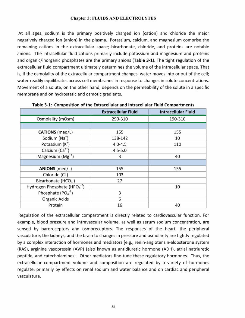
At all ages, sodium is the primary positively charged ion (cation) and chloride the major negatively charged ion (anion) in the plasma. Potassium, calcium, and magnesium comprise the remaining cations in the extracellular space; bicarbonate, chloride, and proteins are notable anions. The intracellular fluid cations primarily include potassium and magnesium and proteins and organic/inorganic phosphates are the primary anions (Table 3-1). The tight regulation of the extracellular fluid compartment ultimately determines the volume of the intracellular space. That is, if the osmolality of the extracellular compartment changes, water moves into or out of the cell; water readily equilibrates across cell membranes in response to changes in solute concentrations. Movement of a solute, on the other hand, depends on the permeability of the solute in a specific membrane and on hydrostatic and osmotic gradients.
Table 3-1: Composition of the Extracellular and Intracellular Fluid Compartments Extracellular Fluid Intracellular Fluid
Osmolality (mOsm) 290-310 190-310
CATIONS (meq/L) 155 155 Sodium (Na+) 138-142 10
Potassium (K+) 4.0-4.5 110 Calcium (Ca++) 4.5-5.0
Magnesium (Mg++) 3 40
ANIONS (meq/L) 155 155 Chloride (Cl-) 103
Bicarbonate (HCO3-) 27
Hydrogen Phosphate (HPO4-2) 10
Phosphate (PO4-2) 3
Organic Acids 6 Protein 16 40
Regulation of the extracellular compartment is directly related to cardiovascular function. For example, blood pressure and intravascular volume, as well as serum sodium concentration, are sensed by baroreceptors and osmoreceptors. The responses of the heart, the peripheral vasculature, the kidneys, and the brain to changes in pressure and osmolarity are tightly regulated by a complex interaction of hormones and mediators [e.g., renin-angiotensin-aldosterone system (RAS), arginine vasopressin (AVP) (also known as antidiuretic hormone (ADH), atrial natriuretic peptide, and catecholamines]. Other mediators fine-tune these regulatory hormones. Thus, the extracellular compartment volume and composition are regulated by a variety of hormones regulate, primarily by effects on renal sodium and water balance and on cardiac and peripheral vasculature.

Anesthesia Care of Pediatric Patients (George A. Gregory & Dean B. Andropoulos)
59
The Newborn: Developmental Aspects of Fluids and Electrolytes
Developmental Changes in Fluid Compartments and Distribution of Water
Water accounts for 78 ± 5% of a newborn’s body weight, decreasing to 60% (mostly from the extracellular fraction) by six months of age, and to 57% later in childhood. Total body water is composed of intracellular (ICW) and extracellular (ECW) fluid compartments (Figure 3-1). In the fetus the extracellular compartment predominates, contributing approximately 62% of body weight during the first trimester of pregnancy but only about 43% at term. At the same time, ICW increases from 25%-to-32% of body weight.1,2 The change in total body water distribution, as a function of weight, continues postnatally so that by three months of life, the intracellular compartment is larger than the extracellular compartment. The adult ratio of ECW to ICW is achieved by about one year of age. ECW is divided into plasma and interstitial compartments. The plasma compartment remains constant throughout life, comprising approximately 5% of body weight. Although two additional body compartments [slowly exchangeable areas (e.g., bone and cartilage) and transcellular] are actually extracellular, neither contributes to clinically important sites for water exchange under normal circumstances.
With their larger surface area, premature infants have a higher percentage of their body weight as total body water and a larger ECW compartment than infants born at term. Blood volume gradually decreases (as a function of weight) with growth and development.3,4 That of a premature infant is approximately 100ml/kg, that of a full term infant approximately 90ml/kg, that of the two year old approximately 80ml/kg, and that of the older child and adolescent approximately 75-80ml/kg.

Chapter 3: FLUIDS AND ELECTROLYTES
60
Figure 3-1: Body Compartments: Fetus to Adult
Total body water, extracellular water, and intracellular water as a percent of body weight. From: Friis-Hansen B. (1961) Body water compartments in children: changes during growth and related changes in body composition. Pediatrics 28:169-181.
Insensible Water loss.
Insensible water loss occurs primarily by evaporation from the skin (approximately 70%) and from the respiratory tract (approximately 30%). The total volume and source of this loss varies with ambient temperature and relative humidity5 and is directly proportional to the amount of exposed surface area as well as metabolic rate. Because the ratio of skin surface area to body weight increases as gestational age decreases, premature infants lose the most water transcutaneously. Other factors, including increased permeability of and blood flow to the skin, as well as lower amount of subcutaneous fat, increase transcutaneous water loss.6 The higher minute ventilation of the newborn magnifies the insensible water loss from the lungs. Tachypnea caused by acidosis or cardiorespiratory disease further exaggerates transpulmonary water loss.

Anesthesia Care of Pediatric Patients (George A. Gregory & Dean B. Andropoulos)
61
Neutral Thermal Environment
In a neutral thermal environment (skin temperature 36-36.5˚C in an environment that has a temperature between 32˚-to-34˚C),7 newborns expend the least amount of energy to maintain a normal body temperature. Minimizing basal expenditure of energy improves growth.8
Surgery in the intensive care nursery or the operating room often necessitates moving the patient from a protected and/or humidified environment to an open bed. Overhead warming and enclosed humidifying devices can seldom be used during surgery (they limit access to the patient) and during transport of infants to an operating room (inoperable without electrical power). To minimize heat and fluid loss during transport and surgery, newborns should be covered (other than the surgical site) with plastic shields, polyethylene blankets, or semi-occlusive skin barriers (including Seran Wrap™), and a hat. In addition, portable warmers that rest under the infant will help maintain the child’s temperature during transport to and from the operating area. A variety of electrical warming devices are utilized intraoperatively on operating room beds.
Developmental Physiology
Various aspects of age-related characteristics of renal, cardiac, hepatic, hematologic, and neurologic function directly impact fluid and electrolyte balance in the newborn. Each of these factors will be discussed briefly.
Renal Physiology: Glomerular Filtration Rate (GFR), Tubular Function
Maintenance of normal extracellular fluid volume, electrolyte concentrations, and water balance are interrelated and undergo significant postnatal changes that are highly dependent on renal function. With nephrogenesis completed by 34-36 weeks gestation (60% occurring during the last trimester of pregnancy), at birth the number of nephrons approximates that of the adult.9 Thereafter the size of both the glomeruli and tubules increase, but the number of nephrons does not increase more. The renal vasculature matures in parallel with the nephrons. In general, fetal and neonatal renal function is characterized by low renal blood flow (high vascular resistance), low GFR, low excretion of solids, and limited ability to concentrate the urine.
Glomerular filtration is present as early as the tenth week of gestation, increases steadily throughout fetal and postnatal life (10-13ml/min/1.73 m2 at 25-28 weeks gestation and 20-25ml/min/1.73 m2 after 34 weeks gestation,10 reaching adult levels between 1-2 years of life (approximately 125ml/min/1.73 m2, although values vary between men and women). Urine production also increases steadily throughout gestation (2-5ml/hr. at 20 weeks gestation, 10-12ml/hr. at 35 weeks gestation, and 35-50 ml/hr. at 40 weeks gestation).11
At the same time that GFR and renal blood flow are increasing dramatically during fetal and postnatal life, the renal tubules are simultaneously maturing. The plasma ultrafiltrate formed as a
Chapter 3: FLUIDS AND ELECTROLYTES
62
result of glomerular filtration is presented to the proximal tubules, which play a major role in regulating the serum concentrations of a wide variety of molecules (e.g., sodium, chloride, potassium, phosphate, acid, glucose, amino acids) and excreting water. Neonates (especially premature infants) neither excrete nor reabsorb sodium as well as more mature infants.
Tubular immaturity accounts for other acid base and metabolic features of the newborn. For example, immaturity of the renal tubules impairs re-absorption of glucose; glucosuria in preterm infants may occur at serum glucose concentrations as low as 100mg/dl. Similarly, bicarbonate loss caused by incomplete tubular reabsorption leads to decreased “normal” serum bicarbonate concentrations in both premature and term newborns (12-16mEq/l, <26-28 weeks gestation; 18-20mEq/l, 30-35 week gestation; 20-22mEq/, term infant; 25-28mEq/l, adult).12 As usual, mild hyperkalemia co-exists with metabolic acidosis (i.e., intracellular potassium moves extracellularly in exchange for protons moving intracellularly in response to acidosis). Neonatal renal function changes with both gestational and postnatal age. Due to rapid maturation after birth, the three-week old, ex-27 week gestation infant usually demonstrates more mature renal function than a one-day old term infant.
Cardiovascular Physiology: Cardiac Output, Heart Rate, Flux of Calcium
At birth, elimination of the placental circulation and initiation of breathing decreases pulmonary vascular resistance and increases pulmonary blood flow. At the same time, systemic vascular resistance increases. Left atrial pressure rises, functionally closing the foramen ovale. These physiologic events plus the closure of the ductus arteriosus and ductus venosus are the most critical aspects of the cardiovascular transition from intra- to extrauterine life. Cardiac output per kilogram body weight is higher in the newborn than at any other age. At the same time, volume loading of the immature ventricle increases cardiac output, but to a far lesser degree than occurs at older ages.13 Finally, since the resting heart rate of the newborn is high, increasing the heart rate above normal has less effect on cardiac output. Decreasing the heart rate drastically reduces cardiac output.
Differences in myocardial ultrastructure (e.g., receptors, channels, transporters, pumps, contractile proteins) and the immaturity of various intracellular structures (e.g., myofibrils, sarcoplasmic reticulum, microtubules) influence clinical management of fluids and electrolytes, especially in the often-unstable perioperative period. For example, the volume of sarcoplasmic reticulum, as well as its ability to pump calcium, increases in utero and postnatally. In addition, the various sub-types of sarcoplasmic reticulum are less differentiated functionally in the immature heart.14 These differences, compared to the adult, are probably the reason immature hearts are more sensitive to calcium channel antagonists,15 and why maximal contractility is more dependent on extracellular calcium.16
Anesthesia Care of Pediatric Patients (George A. Gregory & Dean B. Andropoulos)
63
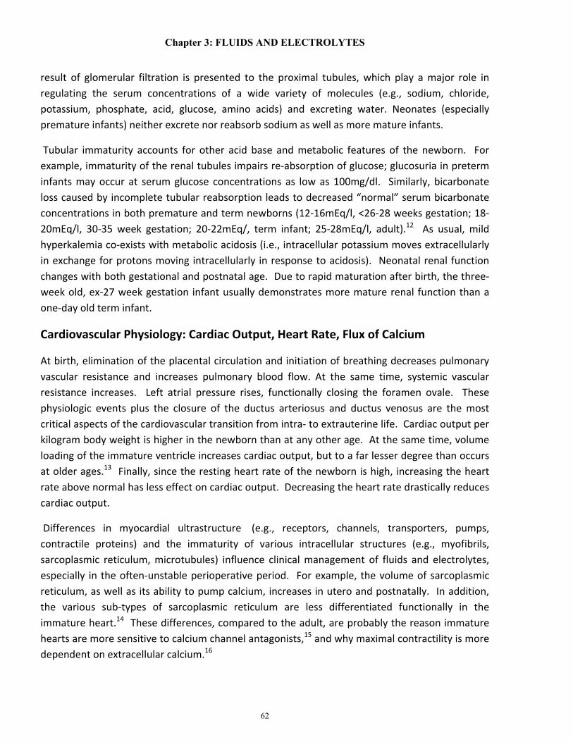
The relationship between blood pressure, cardiac output (CO), and systemic vascular resistance (SVR) remains the same throughout life: BP = CO X SVR. That is, pressure and blood flow are not equal but are related to each other by resistance. Thus, flow to an organ may increase, decrease, or remain constant over a wide range of blood pressure depending on changes in vascular resistance. In fact, defining hypotension in the newborn is not simple.17,18 Although the normal ranges for blood pressure correlate with gestational age,19,20 the definition of hypotension remains elusive (Figure 3-2). Nonetheless, the variability in “normal” for blood pressure and heart rate among infants of the same gestational and/or postnatal age creates a dilemma in the setting of clinical care, especially in the tumultuous perioperative period.
Figure 3-2: Predicted Lower Limit of Mean Blood Pressure in Newborns (initial 72 hours of life).
Engle WD. (2008) Definition of normal blood pressure range: The elusive target. In: Kleinman CS & Seri I (eds.) Hemodynamics and Cardiology. Copyright, Saunders Elsevier Publishing. Philadelphia, p 48. Data from: Nuntnarumit P, Yang W, Gada-Ellzey HS. Blood pressure measurements in the newborn. Clin Perinatol 1999; 26:981-996. With permission.
Thus, until bedside monitoring allows easy monitoring of cardiac output and/or direct measurement of blood flow to specific organs, acid base status and intravascular volume must serve as indirect guides for appropriate fluid administration. That is, rational intravascular fluid therapy requires integrating readily available cardiovascular parameters (e.g., blood pressure, heart rate) with renal, hepatic, respiratory, and central nervous system function. For example, electrolyte concentrations, pH, pCO2, and urine output must be interpreted in the context of an infant’s overall clinical condition (e.g., sepsis, bleeding, neurologic status, urine output) as well specific trends in arterial blood pressure, heart rate, and peripheral perfusion over 1-6 hours or longer. Changes over time provide more and better information than an isolated measurement.
Chapter 3: FLUIDS AND ELECTROLYTES
64
Central Nervous System Physiology: Autoregulation of Cerebral Blood Flow
Although cerebrovascular autoregulation is present in preterm and term neonates, the range of arterial blood pressures over which cerebral blood flow is regulated is narrower for preterm infants and seems to be easily disrupted.21,22 Of importance, autoregulation seems to be disturbed or disrupted by hypoxia, acidosis, seizures, and by the low diastolic blood pressures of patients with a patent ductus arteriosus.23,24 Rapid increases in arterial blood pressure (e.g., from overly-aggressive administration of fluid boluses) can abruptly increase flow to the fragile vessels of the immature brain, while hypotension (e.g., inadequate intravascular volume) and low cerebral perfusion pressures may cause cerebral ischemia. More than at any other developmental stage, the intravascular volume status of the neonate, especially the premature infant, can have rapid and significant effects on the brain.
Hepatic Physiology: Glucose and Coagulation
To discuss hepatic function from the perspective of neonatal fluids and electrolytes, two topics are most relevant: glucose homeostasis and coagulation (See Chapter 4).
Glucose Homeostasis
The transplacental supply of glucose to the fetus abruptly stops at birth, requiring neonates to convert the glycogen stored in their livers to glucose by glycolysis or to produce glucose by gluconeogenesis. Compared to the adult, the liver of term infants is larger and has a greater store of glycogen25 than that of adults. Thus, after the first day of life, most term infants are able to maintain a normal serum glucose concentration during a 10-12 hour fast. Since glycogen storage and the capacity for its degradation mostly occur during the last trimester of pregnancy, infants born in the second or early third trimester of pregnancy often develop hypoglycemia if they do not receive oral or parenteral glucose (i.e., fasting), especially in the first 24-48 hours of life.
Fetal glucose concentrations remain constant (approximately 50-55mg/dl) during the third trimester of pregnancy if maternal carbohydrate metabolism is normal. In experimental animals, glucose utilization at mid-gestation is about 9.5mg/kg/min and decreases to approximately 5mg/kg/min at term.26 These data provide estimates of normal serum glucose requirements for premature infants of similar gestational ages. A surge in the concentration of catecholamines, glucagon, and cortisol (e.g., during birth or stress) increases glycogenolysis and gluconeogenesis, often resulting in hyperglycemia. In high-risk newborns (e.g., large or small for gestational age infants, premature infants, asphyxiated infants), glucose concentrations must be monitored frequently and infusion rates for glucose adjusted to maintain normoglycemia (approximately 60-90mg/dl).
Anesthesia Care of Pediatric Patients (George A. Gregory & Dean B. Andropoulos)
65
Initially, glucose should be infused at rates of 5-6mg/kg/min in NPO newborns (e.g., 10% dextrose, 4ml/kg/hr. = 6.6mg/kg/min of glucose) and the infusion rates adjusted to maintain normal serum glucose concentrations of 60-90mg/dl. For infants who are stable on a well-established regimen, the infusion rate of glucose in the operating room should mimic that used in the intensive care nursery and should only be altered if hypo- or hypergylcemia develop. In many cases, especially in small infants, it may be difficult to obtain a blood sample without an arterial or central venous catheter in place, but currently available devices for measuring glucose only require a drop of blood, which can be obtained from a finger, toe, or ear lobe.
Hypoglycemia can have devastating effects on the central nervous system, primarily mediated by its effects on cerebral blood flow. MRIs of the brains of newborns with documented hypoglycemia demonstrate white matter abnormalities, and clinical evaluation at 18 months reveals persistent central nervous system impairment.27 Older pediatric patients who also have abnormal glucose metabolism also have increased morbidity and mortality.28
Coagulation
Coagulation factors do not efficiently cross the placenta. (See Chapter 4) Although these factors are produced by the fetus, plasma concentrations and the laboratory tests used to monitor function [prothrombin time (PT); activated partial thromboplastin time (APTT)] differ markedly in both normal term and preterm infants compared to adults (Table 3-2). While the concentrations of vitamin K dependent factors (II, VII, IX, X), factors XI and XII, prekallikrein, kininogen, and thrombi are approximately 50% less than those of adults, the levels of fibrinogen and factors V and VIII are similar to those found in adults.29,30 Coagulation disturbances associated with liver dysfunction are linked to decreased synthesis of clotting and fibrinolytic factors and with abnormal platelet function. The PT correlates with availability of factor VII. APTT primarily reflects the amount of thrombin generated.

Chapter 3: FLUIDS AND ELECTROLYTES
66
Table 3-2: Normal Values of Coagulation Function in Newborns Compared with Adults.
Prothrombin Time (sec)
Partial Thromboplastin Time
(sec)
Thrombin Time (sec)
Adults 13.0 (11.2-14.5) 44.0 (36.8-50.0) 10 (9.0-11.1)
Term Infants 13.6 (12.8-14.4) 65.2 (50.0-84.0) 12.5 (10.0-15.0)
ELBW Infants 15.4 14.6-16.9) 108 (80.0-168) 14.9 (11.0-17.8)
Barnard DR, Simmons MA, Hathaway WE. Coagulation studies in extremely premature infants Pediat Res 1979; 13:1330-1335.
In spite of the differences in liver function between newborns and adults, clinically significant bleeding is uncommon in normal neonates whose have adequate vitamin K levels. On the other hand, sepsis and/or asphyxia-induced disseminated intravascular coagulation (DIC) increase the risk for bleeding. Both asphyxia and sepsis deplete coagulation factors (e.g., fibrinogen, factors V and VIII) and platelets and produce fibrin degradation products. To compensate for abnormal levels of coagulation factors, especially if accompanied by cardiovascular instability and ongoing blood loss, administration of specific blood components not only improves clotting function but can also replete intravascular volume.
Hematologic Physiology
In otherwise normal appropriate for gestational age (AGA) fetuses, hemoglobin concentrations vary with gestational age (14gm/dl at 25 weeks gestation, 16gm/dl at 30 weeks, 17gm/dl at 35 weeks, 18gm/dl at term).31 The normal postnatal decrease in hemoglobin is more rapid in premature infants.32
Indications for transfusion of red blood cells or other blood components pre-, intra-, or post-operatively must be determined in the context of the patient’s cardiorespiratory status, ongoing blood and fluid losses, and response to delivery of crystalloid and colloid.
Neonatal Perioperative Fluids: General Concepts
For the first 2-3 hours of life, the neonate’s electrolyte concentrations reflect those of the mother and of perinatal events (e.g., asphyxia, placental or umbilical cord hemorrhage). Afterwards, the electrolyte concentrations reflect a balance among normal metabolism, cardiovascular, renal and hepatic function, and ongoing metabolic derangements (e.g., sepsis, congenital metabolic diseases, complex cyanotic congenital heart disease, etc.). Until adequate tissue perfusion is

Anesthesia Care of Pediatric Patients (George A. Gregory & Dean B. Andropoulos)
67
established and washout of accumulated acid and other metabolic byproducts of anaerobic metabolism from tissues have occurred, metabolic acidosis may persist in depressed newborns, especially those who were severely asphyxiated and required resuscitation.
Sodium is seldom added to intravenous fluids of term and late preterm infants until day two of extra-uterine life. Then 2-6meq/kg/day of sodium is added. In the ELBW infant, sodium-containing fluid is often required as early as 12-24 hours of life to maintain adequate intravascular volume, especially with excessive transcutaneous fluid losses (See section, Insensible Water Loss). After the first few days of life, adequate sodium intake is essential for infants of all gestational ages to maintain normal growth and appropriate weight gain. Poor skeletal and tissue growth, as well as adverse neurodevelopment, are associated with chronic sodium deficiency.33
Ensuring normal fluid and electrolyte balance in the newborn in the presence of the high metabolic rate is associated with growth, marked insensible water loss, and limited ability to salvage and excrete water and solutes requires accurate monitoring of the newborn’s fluid loss (i.e., urine, gastrointestinal, cerebrospinal fluid, blood sampling) and electrolyte concentrations to guide the quantity and composition of replacement fluid. Based on the complex interactions of illness and immaturity, the anesthetist must consider the following when determining appropriate fluid and electrolyte management in the newborn:
1. The normal postnatal diuresis contracts the extracellular space in all newborns but does so to a greater degree in preterm infants. During the first few days after birth, negative fluid and electrolyte balance occurs normally in healthy term infants, but prematurity and/or hemodynamic instability may demand aggressive intravascular fluid administration in these patients.
2. Transepidermal fluid loss is inversely related to gestational age and can be as much as 60-100ml/kg/day in ELBW infants. During the first few postnatal days, naked preterm infants lose 15 times more water by evaporation than naked term infants.34
3. Providing a warm, humidified environment and inspired gases and/or the use of plastic shields reduce transepidermal fluid loss, especially in the ELBW infant. Shielding devices are difficult or impossible to use during surgery but should be used during transport of the patient to and from the operating room and in the operating room before and after surgery.
4. The hypothesis that over-hydration or hyper- or hyponatremia increase the incidence of patent ductus arteriosus, necrotizing enterocolitis, and chronic lung disease should be considered, but the data documenting a strong correlation are inconsistent.
5. An intravenous infusion of calcium is usually required in preterm, asphyxiated, or LGA or SGA infants until they establish adequate enteral nutrition. Ideally, calcium is infused into

Chapter 3: FLUIDS AND ELECTROLYTES
68
a central venous catheter, but most often only peripheral intravenous access is justified in an uncomplicated SGA or LGA newborn. The site of peripheral infusion of calcium must be meticulously monitored, since subcutaneous infiltration of calcium can cause extensive tissue necrosis. As stated above, calcium should be administered through a central line if possible.
Preoperative Evaluation of the Neonate
Understanding how fluids and electrolytes have been managed before surgery will help avoid dangerous and unnecessary alterations in therapy (e.g., rate of glucose delivery). Of course, intra-operative events, such as general anesthesia, blood loss, increased insensible losses (e.g., open abdomen with exposed intestine), often require changes in both the volume and type of fluid delivered. Developing an effective relationship with the intensive care nursery staff facilitates intraoperative consultation with the primary medical team.
The following summarizes the most significant aspects of pre-operative evaluation of the newborn:
1. Review of labor and delivery: a. Documentation of appropriate fetal growth (AGA, LGA, or SGA)
2. Review of systems:
a. Trend in weight (daily or more frequent) b. Trend in intravenous and oral intake and urine output/urine specific gravity c. Trend in other output (gastrointestinal, cerebrospinal fluid, etc.) d. Composition of intravenous fluid (glucose, sodium, calcium) correlated with
corresponding trends in plasma concentrations of these electrolytes e. Hemodynamic instability: trends in heart rate, arterial blood pressure, peripheral
perfusion; presence and effect of patent ductus arteriosus or other cardiovascular dysfunction (e.g., tricuspid regurgitation after asphyxia)
f. Current and recent trend in hemoglobin concentration correlated with hemodynamic function; determine if the patient “requires” a minimum level of hemoglobin (e.g., increased heart rate or acidosis if the hematocrit is <40%)
g. Central nervous system insult (presence of intraventricular hemorrhage) h. Current coagulation status, history of bleeding, and trends in requirements for blood
components; availability of blood products 3. Adequacy of intravenous access and monitors (functioning of arterial, venous, or central
venous lines)
Anesthesia Care of Pediatric Patients (George A. Gregory & Dean B. Andropoulos)
69
4. Estimate “allowable blood loss” as part of the pre-operative evaluation. For example, blood volume in a 3-day old, 1 kg infant is 100 ml/kg, or 100ml. A loss of 20 ml of blood is 20% of the blood volume. Anticipating the need for transfusion depends on current hemoglobin concentration and history of tolerance for decreasing blood volume and/or a decrease in hemoglobin.
Intra-Operative Fluid Management
During transport from the intensive care nursery to the operating room, intravenous infusions, cardiorespiratory support, and body temperature must be maintained, and hemodynamic status and oxygenation monitored. At a minimum, heart rate and oxygen saturation should be continuously monitored, and more invasive hemodynamic monitoring (e.g., arterial blood pressure) may be required, depending on the clinical status of the patient. The infant’s head and body should be covered with semi-occlusive material and/or blankets/hat. Portable warming devices (e.g., a heating pad set on low) are often placed under the infant. Intravenous infusions for convenient and effective use in the operating room should be set up before the patient leaves the intensive care nursery. For example, the extensions on the intravenous and intra-arterial tubing should be long enough to allow easy access to the lines when surgical drapes are in place.
Initially, glucose is infused in the operating room at the same rate that provided normoglycemia in the intensive care nursery. Often, simply continuing the infusion of “maintenance” fluid or total parenteral nutrition is appropriate. In surgical procedures lasting longer than one hour, monitoring glucose concentrations should be considered to ensure normoglycemia. In most cases, additional non-glucose-containing fluid must be delivered to compensate for intra-operative events and blood and fluid losses. To avoid hyperglycemia and its side effects (e.g., osmotic diuresis, central nervous system injury), glucose-containing solutions should never be used to replace intraoperative intravascular losses (i.e., increased insensible losses during abdominal surgery, blood loss, etc). In some cases (e.g., newborns, patients totally dependent on intravenous alimentation, some diabetics) glucose is delivered intraoperatively as part of maintenance fluid. All other losses should be replaced with non-glucose containing fluids.
As always, during induction of anesthesia, anesthetic agents should be titrated to maintain an appropriate hemodynamic status. Often, dramatic hemodynamic responses occur with the administration of anesthetics (e.g., hypotension, tachycardia) and require prompt rapid delivery of a bolus of a normal saline or plasmalyte (5-10ml/kg or more).
As surgery proceeds, blood loss and coagulation abnormalities may occur and require the administration of various components of blood to maintain intravascular volume and hemodynamic stability and to restore normal levels of hemoglobin and/or coagulation factors. Monitoring blood loss requires meticulous observation of the surgical field, since loss of small volumes of blood is difficult to determine and is easily hidden in drapes and body cavities. Since

Chapter 3: FLUIDS AND ELECTROLYTES
70
the blood volume of a 1kg infant is only 100ml, a 10-20ml blood loss can induce significant hemodynamic instability. Theoretically, urine output should be monitored, but in the smallest of infants, it may be difficult or impossible to measure small volumes of urine accurately. For example, after placing a tube into the bladder and attaching it to a drainage system, at least 5ml or more of urine must be produced before the anesthetist can detect and measure any urine in the drainage device. In addition, the small urinary catheter and drainage system are frequently soft and easily compressed, kinked, or occluded during positioning of the patient.
Inspired gases should be warmed and humidified to near body temperature whenever possible to reduce insensible fluid losses and help maintain body temperature. Overhead, servo-controlled warmers should be used during pre-operative preparation (e.g., inserting or adjusting intravenous lines and ventilation devices, preparation of the surgical site). The surgical bed should include a warming device (varies among institutions), and the room temperature should be adjusted to minimize development of hypothermia. Once the child is draped for surgery, the room temperature can usually be decreased, depending on the response of the infant’s body temperature to the surgery and to decreases in the environmental temperature. Intravenous fluids and blood products should be warmed to 37°C when possible, especially if large volumes of either are required. However, some blood warming devices can overheat blood and cause hemolysis.
The following summarizes the most significant aspects of intraoperative fluid management in the newborn (See case study, 3-3):
1. Ensure that the intravascular volume is adequate before induction of anesthesia.
2. Estimate the need for blood products in the operating room before starting anesthesia/surgery. (See Chapter 4) This process should begin in the intensive care nursery to ensure availability of specific blood products when they are needed.
3. In spite of normovolemia, the newborn may respond to judicious doses of either inhaled or intravenous anesthetic agents with dramatic changes in heart rate, arterial blood pressure, and/or peripheral perfusion. To maintain hemodynamic stability, fluid and/or colloid boluses or infusions may be required.
4. The infusion of glucose should be continued at the same rate that was associated with normoglycemia in the intensive care nursery. This can be considered the “maintenance” component of fluid administration. The infusion of this solution should be adjusted, based on intraoperative measurements of glucose.
5. In most cases, delivering crystalloid compensates adequately for insensible losses, but the required rate of delivery depends on the type of surgery. For example, during a laparotomy to treat necrotizing enterocolitis, 10-50ml/kg/hr. (or more) of non-glucose
Anesthesia Care of Pediatric Patients (George A. Gregory & Dean B. Andropoulos)
71
containing, isotonic crystalloid (normal saline or plamalyte) may be required to compensate for ongoing insensible losses from exposed bowel and peritoneum. During less invasive procedures (e.g., inguinal hernia repair), less crystalloid is required (0-5ml/kg/hr.), since ongoing losses are minimal to none. At some point after delivering large volumes of crystalloid (30-40ml/kg/hr.) for several hours (sometimes longer, sometimes shorter, depending on cardiovascular and clotting status), colloid may be a more appropriate intravenous therapy (See #s 6, 7 below).
6. Critically-ill newborns, especially premature newborns, are often more hemodynamically stable (e.g., less apnea and bradycardia) when their hematocrit is >40%. In those cases, frequent packed red blood cell transfusions (and/or erythropoietin) are required in the intensive care nursery. In contrast, after days to weeks in the intensive care nursery a stable infant who undergoes elective surgery (e.g., inguinal hernia repair) often tolerates lower concentrations of hemoglobin. For example, an ex-26 week gestation infant undergoing an inguinal hernia repair at 10 weeks of age (36 weeks gestation) may have no cardiorespiratory problems (i.e., no apnea/bradycardia, tachycardia, or hypotension) despite a hematocrit of 30%. In contrast, perfusion and metabolic status of a 5-day old, 26-week gestation infant with a patent ductus arteriosus and necrotizing enterocolitis, who is receiving mechanical ventilatory support, usually improves when the hematocrit is >40%. The hematocrit associated with a stable preoperative cardiorespiratory status provides the initial guideline for the appropriate perioperative level.
7. Many newborns, especially critically ill newborns, respond more promptly to the administration of colloid than to crystalloid after losing between 10-20% of their blood volume. Colloid may include packed red blood cells, fresh frozen plasma, or fibrinogen, depending on the coagulation status and the patient’s hemoglobin concentration.
8. Monitoring of urine output, arterial blood pressure and/or central venous pressure helps guide intravenous fluid therapy.
9. Heat loss and insensible fluid loss should be minimized during transport of the infant to and from the operating room.

Chapter 3: FLUIDS AND ELECTROLYTES
72
Table 3-3. Intraoperative Fluids: Newborn
12 h old, 3.2 kg, 39-week gestation infant, noted to have increasing abdominal distension. Abdominal imaging consistent with bowel obstruction, but free air also present. Nasogastric tube in place, with bilious drainage. She is breathing spontaneously (50 breaths per minute). BP= 58/34, Heart rate=160 bpm, Urine output has decreased from 2ml/kg/h to 0.5ml/kg/h over prior 6h. Two peripheral IV’s are in place. D10 in NS infusing, 15 ml/h
• Arterial blood gas: pH=7.30, pO2=62, pCO2=47 (blow-by supplemental oxygen). • Electrolytes: Na+, 135; K+, 4.6; Cl-, 106; HCO3
-=16; Ca++, 1.0; Glucose, 75 mg/dl • Renal Function: BUN 32mg/dl/creatinine 0.7mg/dl • Hemoglobin, 15g; Platelets, 125,000; PT, 17 seconds; PTT 48 seconds; Fibrinogen
120mg/dl • Before transport to the operating room to treat bowel obstruction/perforation, 3
additional boluses of 10ml/kg administered: BP 68/40, HR 150 • Before transport, packed RBC’s available.
Deficit Maintenance Ongoing Losses • 0 ml
• Allowable blood loss: about 30-40ml
• 15ml/hr D10 infusion
• Blood products available in operating room: RBC’s, fresh frozen plasma available
• Infuse crystalloid, 10-50ml/kg/hr or more; boluses to maintain hemodynamic instability
• RBC’s for blood loss (1ml for each 2-3ml of blood loss). Fresh frozen plasma infusion/boluses in response to bleeding/laboratory values
• Monitor Hg, PT/PTT, platelets

Anesthesia Care of Pediatric Patients (George A. Gregory & Dean B. Andropoulos)
73
The same patient, now 6 m old, presents for elective take down of ileostomy. She tolerates a combination of breast milk and formula and now weighs 6.1 kg. Normal physical examination, with ileostomy in place.
Hemoglobin, 10g/dl; glucose, 90mg/dl; Normal electrolytes. Parents report that she has been NPO 8 hours.
Deficit Maintenance Ongoing Losses 4 X 6.1 kg X 8 h =
195 ml • 4 X 6.1 kg =
25ml/h, isotonic fluid during surgery
• Open abdomen, but without acute injury (e.g., no ischemia)
• Consider 20-30ml/kg of isotonic fluid to infuse over initial 2 hours
• Adjust rate in response to hemodynamic status
• Continue isotonic fluids in PACU and on ward; measure Na+ after 24h
• Transfusion unnecessary intra- or postoperatively unless unexpected blood loss/anemia
The Older Infant and Child
Unlike the newborn, older infants and children often present for elective surgery with a clearly defined medical and/or surgical history. That is, congenital anomalies and metabolic/genetic abnormalities have been identified and a specific, stable treatment plan has been established. Outside of trauma, accidents, or an acute/new illness (e.g., malignancy, gastrointestinal events, systemic or localized infection, etc.), emergent/urgent surgery is usually required to treat complications of a recognized anomaly or because diagnostic studies are needed to evaluate an underlying illness.
As in all age groups, pre-operative evaluation of the fluid and electrolyte status of older infants/children requires assessing intravascular volume by integrating findings from the physical examination, review of cardiorespiratory/renal/hepatic/central nervous system status, and relevant laboratory studies. And, similar to the newborn infant, when devising a rational plan for intra-operative fluid management of older children, the clinician must consider maintenance fluid requirements, replacement for ongoing losses, and the need to correct specific abnormalities (e.g., glucose, calcium, coagulation). Fine-tuning fluid and electrolyte abnormalities must be balanced against the urgency of the surgery. In some cases, monitoring laboratory values during

Chapter 3: FLUIDS AND ELECTROLYTES
74
surgery demands ensuring easy access to central venous, arterial, or a large peripheral intravenous catheter during surgery.
Maintenance Fluid: Classic Concepts
Maintenance fluids should contain appropriate amounts of water and electrolytes to replace urine, gastrointestinal, sweat, breathing, and skin evaporation losses. Oral, intravenous, and other routes (e.g., intraosseous) can be used to administer maintenance fluid.
Figure 3-3: Comparison of Energy Expenditure in Basal and Ideal State
Based on Holiday and Segar’s data, energy expended (calories) is the approximately the same as the requirement for fluid (milliliters). Although caloric expenditure varies (basal rate vs. normal activity) three linear weight categories are easily identified (0-10 kg, 10-20 kg, >20 kg). Energy expended by hospitalized patients was estimated to be approximately between that for basal and normal activity. From the curve for the hospitalized patient, a 10kg infant expends 1,000 calories (requires 1,000ml/day, 100ml/kg) and a 20kg child, 1,500 calories (requires 1,500 ml/day, 100ml/kg for the first 10kg + 50 ml/kg for each kilogram between 10 and 20kg. if the child weighs >20kg, he/she requires 1500ml + 1ml/kg he/she is above 20kg). From: Holliday MA, Segar WE. The maintenance need for water in parenteral fluid therapy. Pediatrics. 1957; 19:823-832

Anesthesia Care of Pediatric Patients (George A. Gregory & Dean B. Andropoulos)
75
Any discussion of “maintenance” fluids and electrolytes inevitably notices Holliday and Segar’s classic paper, “The maintenance need for water in parenteral fluid therapy”.35 These authors simplified the calculation of fluid requirements by correlating insensible loses with the metabolic rate of healthy children at rest and during activity (Figure 3-3). They noted that the requirements for water paralleled those for energy and that the metabolic rate correlated with weight: ≤10kg, 100kcal/kg; 10-20kg, 1000+5kcal/kg; >20 kg, 1500+2kcal/kg. For a 10kg child, they calculated a net loss of approximately 34ml/kg/day of insensible loss (i.e., evaporative loss of water from the skin and respiratory tract) and approximately 66ml/kg/day of urine, for a total of 100ml/100kcal/day (approximately 4cc/kg/hour) of maintenance fluid for the <10kg patient. Based on curves generated from plots of weight versus estimated calories expended/day, the authors estimated that required maintenance fluid is 100ml/kg/day (approximately 4ml/kg/hour) up to 10kg, an additional 50ml/kg/day (approximately 2ml/kg/hour) for each kilogram between 10-20 kilograms, and 20ml/kg/day (approximately 1ml/kg/hour) for each kilogram beyond 20 kilograms. This is the basis for the “4-2-1” formula widely used to calculate rates for hourly maintenance intravenous fluid (Table 3-4).
Table 3-4. Calculating Maintenance Fluid: The “4-2-1 Rule”.
Weight (kg) Hourly Rate Daily Volume
<10 4ml/kg 100ml/kg
10-20 40ml + 2ml/kg (for each kg between 10-20)
1000ml + 50ml/kg (for each kg between 10-20)
>20 60 ml + 1 ml/kg (for each kg >20) 1500ml + 20ml/kg (for each kg > 20)
To characterize the composition of maintenance intravenous fluid, Holliday and Segar assumed that electrolytes in intravenous fluid should mimic those in breast milk. They estimated the amount of sodium and potassium in 100mls of human milk and extrapolated those data to determine the concentration of electrolytes needed in maintenance fluid (i.e., approximately 3-5meq/kg/day for sodium and approximately 2-3meq/kg/day for potassium). This led to the practice of adding 0.2-0.45% saline to 5% dextrose in water for routine intravenous fluid. Holliday and Segar’s estimated intravenous fluids for patients was based on data derived from normal children.

Chapter 3: FLUIDS AND ELECTROLYTES
76
However, over the last two decades, severe neurologic injury and death have been reported in both pediatric and adult patients who develop hyponatremia while receiving hypotonic intravenous solution, suggesting that Holliday and Segar’s data are not easily extrapolated to the acutely ill infant/child,36 especially after surgery.37 Moritz’s recent review emphasizes the enormous impact of hyponatremia (serum sodium <135mEq/l) since he found that approximately 25% of hospitalized children were hyponatremic.38 Severe adverse outcomes (death or neurologic injury) have been reported in 30% of severely hyponatremic children.39
Thus, although Holliday and Segar’s principles for calculating routine intravenous fluid have been incorporated into clinical care for over 60 years, the high incidence of hyponatremia occurring with administering intravenous hypotonic fluids has prompted many experts to strongly recommend 0.9% normal saline for the vast majority of hospitalized children, especially peri-operatively. In addition, some authors have suggested that in hospitalized patients, predicting fluid excretion/reabsorption) are not prompt or are incomplete.
Arginine vasopressin (AVP or ADH) modulates normal osmolality by increasing free water reabsorption by inducing water channel [vasopressin-sensitive water channel (aquaporin 2)] insertion into the distal tubule and collecting ducts.33,42 In most cases, hyponatremia is linked to an excess of water rather than sodium imbalance. That is, hyponatremia evolves with or without a deficit of sodium, and with either hypovolemia or euvolemia, but in the presence of AVP.
Hypovolemia (i.e., inadequate intravascular volume) appropriately elicits a stimulus to decrease free water excretion (i.e., release of AVP is normal). Regardless of the etiology (e.g., salt wasting states, sepsis, blood/fluid loss), hypovolemia initially is usually treated with boluses of an isotonic crystalloid (e.g., 0.9% normal saline), and, in some cases, followed by colloid or blood products. Treatment is guided by various parameters, such as the hemodynamic response, acid-base status, and urine output. In most cases, after the initial repletion of the intravascular volume (with normal underlying renal, hepatic, and cardiorespiratory function), isotonic fluid should be continued at maintenance rates during the transition to oral hydration/feeds. At that point, if intravenous supplement is required, intravenous hypotonic fluid might be considered.
In the absence of renal pathology, congestive heart failure, or hepatic disease (cirrhosis), there is euvolemic non-osmotic, un-physiologic (excessive) release of requirements (i.e., fluid losses) ased on energy expenditure overestimates the volume needed for maintenance fluid40 (See section, Intravenous Fluids and Hyponatremia: Current Controversy).
In all cases, intravenous fluid should be considered an “invasive therapy” or a “medication” rather than an exact replacement for breast milk, formula, or other
oral fluids. Patients who require intravenous fluid often have a significant illness that is associated with hormonal imbalance or physiologic derangements (e.g., renal or hepatic disease,

Anesthesia Care of Pediatric Patients (George A. Gregory & Dean B. Andropoulos)
77
chronic lung disease) that complicate maintaining adequate intravascular volume and electrolyte balance. Holliday reminds us, “when fluid therapy extends beyond the first day, monitoring is needed to adapt orders to special cases, avoiding the consequences of ‘one plan fits all”.41
Physiology of Hyponatremia: Clinically Relevant Concepts
More than any other molecule, sodium establishes osmolality (normal, 280-290 mOsm/l) in the extracellular space (Plasma Osmolality = 2 x [Na+] + [glucose]/18 + [BUN]/2.8), and consequently contributes significantly to maintaining extracellular fluid volume, blood volume, and perfusion pressure. Sodium cannot freely move across cell membranes. On the other hand, water does freely pass through cell membranes in response to osmolar gradients. Arginine vasopressin (AVP) is secreted by the pituitary gland in response to low circulating blood volume and results in the retention of free water by increasing water reabsorption in the collecting ducts of the kidney nephron.
The release of AVP is often a response to central nervous system abnormalities (brain tumors, meningitis, trauma), pulmonary disease (asthma, pneumonia), various non-CNS malignancies (lung, leukemia, lymphoma, thymoma), or other non-specific pathology (e.g., nausea, stress due to pain, post-operative state). In these cases, the etiology of hyponatremia is attributed to abnormal release of and response to AVP, despite the absence of both osmotic and baroreceptor mediated stimuli.26 Consequently, the high incidence of hyponatremia is not surprising in perioperative patients who commonly encounters disorders associated with both physiologic and non-physiologic excessive levels of AVP.
SUMMARY: Perioperative Intravenous Fluids: Current Controversy
Reports of hyponatremia-induced morbidity and mortality (e.g., death, permanent brain injury) have elicited heated debate about whether hypotonic or isotonic crystalloid should be the routine maintenance fluid, especially in the perioperative period (SeeGeneral Considerations). Thus, isotonic solutions do not change cell volume, but intravenous hypotonic solutions may increase (water moves intracellularly) and hypertonic solutions decrease (water moves extracellularly) cell volume. Infants and children who require surgery have an especially high risk for hyponatremia from a combination of events:
1. Co-existing significant medical illnesses that predispose to AVP levels (e.g., leukemia/chemotherapy, congenital heart disease/diuretic therapy, chronic lung disease/ex-premature infant).
2. Non-osmotic stimuli for AVP secretion are commonly encountered perioperatively (e.g., pain, nausea and vomiting, narcotic administration, inhaled anesthetic agents, positive pressure ventilation), and increase the risk for hyponatremia.
Chapter 3: FLUIDS AND ELECTROLYTES
78
3. When the concentration of AVP is high, hypotonic solutions greatly predispose to hyponatremia.42,43
4. Specific surgical patients are at high risk for hyponatremia (those with central nervous system injury44 or scoliosis repair).45,46
Administering hypotonic intravenous fluid in the presence of AVP has been considered to be the primary etiology of postoperative hyponatremia. Burrows45 noted that serum sodium concentrations decreased postoperatively in pediatric patients who had received either hypotonic or isotonic fluids, but more so when hypotonic solutions were administered. Similarly, Eulmesekian47 reported hyponatremia (Na<135mmol/L) in 21% (by 12 hours) and 31% (by 24 hours) of patients admitted to a PICU after orthopedic surgery.
Several prospective randomized studies,37,48-52 as well as smaller observational reports53 and recent reviews,38,54 document that administering isotonic intravenous fluid minimizes perioperative hyponatremia but does not induce hypernatremia. The rate of fluid delivery is apparently less important than the concentration of sodium delivered.49,50 Large or even normal volumes of hypotonic intravenous fluid are an obvious source of excess water.
Minimizing the risk for hyponatremia requires meticulous attention throughout the perioperative period (See case study, 3- 5):
1. Preoperative: • Ensure adequate intravascular volume • Measure serum sodium if intravenous therapy is required for >12-24 hours, especially
during intravenous therapy with hypotonic intravenous fluid • Maintain a low threshold for symptoms of hyponatremia, especially with high-risk
physiology/disease states.
2. Intraoperative
• Since NPO guidelines now allow clear liquids orally up to two hours before the induction of anesthesia, children are ideally fasted for no more than 3-4 hours.55 Thus, pre-operative dehydration is more likely linked to an underlying disease state. If the pre-operative fast was appropriate and without evidence for intravascular deficit or hemodynamic response to induction of general anesthesia, intravenous fluid administration beyond those required for maintenance rates may not be necessary during non-invasive surgery or may be deliberately avoided (e.g., stable patient undergoing neurosurgery). The clinician must assess the physical status of the patient and the nature of the surgery rather than simply delivering intravenous fluid to compensate for the hours of NPO.

Anesthesia Care of Pediatric Patients (George A. Gregory & Dean B. Andropoulos)
79
• On the other hand, families may not follow NPO instructions and/or the operating room schedule may be delayed. If the fasting period has been prolonged or there is a high-risk disease state, the anesthetist should consider giving a bolus of 20-40ml/kg of isotonic fluid over the initial 1-2 hours of surgery and then adjust the rate of administration based on maintenance plus ongoing fluid losses. This may inhibit excessive AVP release and avoid hypovolemia. After initial vigorous fluid delivery, “maintenance fluids” might be decreased to half the classic rate recommended by Holiday and Segar (“2-1-0.5” ml/kg/h).56
3. Postoperative Isotonic solutions are appropriate for postoperative maintenance fluids and for replace ongoing losses. Frequent monitoring of the patient’s clinical status and serum electrolytes should guide both the rate and electrolyte composition of the intravenous fluid.
As Bailey notes, “no single IV fluid can be used safely in all situations”.54 Excessive delivery of any solution to patients with cardiovascular or renal failure could be disastrous. Finally, hypotonic intravenous solutions may be required when attempting to compensate for excessive free water loss (e.g. diabetes insipidus).
Summary: Perioperative Fluids and Electrolytes
During pre-, intra-, and post-operative periods, the clinician must differentiate between delivery of intravenous fluid to treat hypovolemia (e.g., to compensate for NPO status) and delivery of “maintenance” fluids. Boluses of isotonic fluid are appropriate to correct hypovolemia (i.e., hemodynamic instability, poor perfusion). In the perioperative period, one indication for hypotonic fluids is excessive loss of free water, which must be diagnosed and monitored by simultaneously measuring serum and urine electrolyte concentrations. At all times, meticulous monitoring of the patient’s clinical status and serum electrolytes is essential to avoid perpetuating or inducing electrolyte abnormalities.
Intra-Operative Glucose Delivery in the Older Infant/Child (See Case Study, Table 3-5)
In the era of prolonged preoperative fasting, hypoglycemia was common.57,58 Now, with more liberal NPO regimens, the incidence of hypoglycemia ranges between 0-2.5% and occurs almost exclusively in patients who have had an inappropriately long fast (8-19 hours).59 Nonetheless, in spite of its rare occurrence, when hypo- and hyperglycemia do occur, the consequences can be catastrophic. For example, administering glucose-containing fluid at high rates during surgery may cause hyperglycemia, glucosuria, and an osmotic diuresis. Of greater significance, hyperglycemia has been associated with increased mortality and infection in both adult and pediatric patients in intensive care units.60,61 In the absence of an underlying metabolic requirement (e.g., diabetes, methylmalonic acidemia) intraoperative fluids do not routinely include glucose.
Chapter 3: FLUIDS AND ELECTROLYTES
80
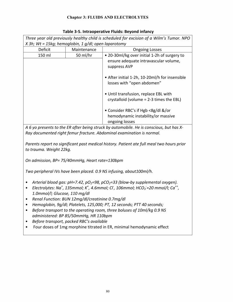
Table 3-5. Intraoperative Fluids: Beyond infancy Three year old previously healthy child is scheduled for excision of a Wilm’s Tumor. NPO X 3h; Wt = 15kg; hemoglobin, 1 g/dl; open laparotomy
Deficit Maintenance Ongoing Losses 150 ml 50 ml/hr • 20-30ml/kg over initial 1-2h of surgery to
ensure adequate intravascular volume, suppress AVP
• After initial 1-2h, 10-20ml/h for insensible losses with “open abdomen”
• Until transfusion, replace EBL with crystalloid (volume = 2-3 times the EBL)
• Consider RBC’s if Hgb <8g/dl &/or hemodynamic instability/or massive ongoing losses
A 6 yo presents to the ER after being struck by automobile. He is conscious, but has X-Ray documented right femur fracture. Abdominal examination is normal.
Parents report no significant past medical history. Patient ate full meal two hours prior to trauma. Weight 22kg.
On admission, BP= 75/40mmHg, Heart rate=130bpm
Two peripheral IVs have been placed. 0.9 NS infusing, about100ml/h.
• Arterial blood gas: pH=7.42, pO2=98, pCO2=33 (blow-by supplemental oxygen). • Electrolytes: Na+, 135mmol; K+, 4.6mmol; Cl-, 106mmol; HCO3
-=20 mmol/l; Ca++, 1.0mmol/l; Glucose, 110 mg/dl
• Renal Function: BUN 12mg/dl/creatinine 0.7mg/dl • Hemoglobin, 9g/dl; Platelets, 125,000; PT, 12 seconds; PTT 40 seconds; • Before transport to the operating room, three boluses of 10ml/kg 0.9 NS
administered: BP 85/50mmHg, HR 110bpm • Before transport, packed RBC’s available • Four doses of 1mg morphine titrated in ER, minimal hemodynamic effect

Anesthesia Care of Pediatric Patients (George A. Gregory & Dean B. Andropoulos)
81
Table 3-5. (Continued)
Deficit Maintenance Ongoing Losses • 10ml/kg (200ml,
0.9 NS) infused rapidly prior to rapid sequence induction
• 60ml/h, isotonic fluid (no glucose)
• BP decreased from 90/50-to-65/40, heart rate 110bpm, after bolus of 20mg propofol, 25mg rocuronium
• 20ml/kg of isotonic fluid to infused rapidly, BP increased to 80/50, HR stable at 100-110bpm
• Sevoflurane/fentanyl titrated while delivering additional 0.9 NS; no glucose
• Decrease isotonic fluid delivery as hemodynamic status improves
• Measure hemoglobin and ongoing blood loss to assess need for PRBC
• Continue isotonic fluids in PACU and on ward; measure Na+ after about 24h, if NPO
• transfusion unnecessary intra- or postoperatively if blood loss controlled and hemodynamic status stable

Chapter 3: FLUIDS AND ELECTROLYTES
82
Refernces
1. Friis-Hansen B. Changes in body water compartments during growth. Acta Paediatr 46 suppl 1957; 110, 1-68.
2. Friis-Hansen B. Body water compartments in children: changes during growth and related changes in body composition. Pediatrics 1961; 28:169-81.
3. Linderkamp O, Versmold HR, Riegel KP. Estimation and prediction of blood volume in infants and children. Eur J Pediatr 1977; 125:227-34.
4. Casady G: Plasma volume studies in low birth weight infants. Pediatrics; 1966; 38:1020-17.
5. Sulyok E, Jequier E, Prod’hom LS. Respiratory contribution to the thermal balance of the newborn infant under various ambient conditions. Pediatrics 1973; 51:641-50.
6. Evans NJ, Rutter N. Development of the epidermis in the newborn. Biol Neonate 1986; 49:74-80.
7. Adamson SK, Towell ME. Thermal homeostasis in the fetus and newborn. Anesthesiology 1965; 26:531-48.
8. Glass L, Silverman WA, Sinclair JC. Effect of the thermal environment on cold resistance and growth of small infants after the first week of life. Pediatrics 1968; 41:1033-46.
9. Hinchliffe, S. A., Sargent, P. H., Howard, C. V., Chan, Y. F. & van Velzen, D. Human intrauterine renal growth expressed in absolute number of glomeruli assessed by the dissector method and Cavalieri principle. Lab Invest 1991; 64: 777–84.
10. Svenningsen NW, Aronson AS. Postnatal development of renal concentration capacity as estimated by DDAVP-test in normal and asphyxiated neonates. Biol Neonate 1974; 25:230-41.
11. Schwartz GJ, Haycock JB, Edelmann CM Jr, et al. Late metabolic acidosis: a reassessment of the definition. J Pediatr 1979; 95:102-7.
12. Klopfenstein HS, Rudolph AM. Postnatal changes in the circulation and responses to volume loading in sheep. Circ Res 1978; 42:839-45.
13. Nakanishi T, Seguchi M, Takao A. Development of the myocardial contractile system. Experientia 1988; 44:936-44.
14. Boucek RJ Jr, Shelton M, Artman M, et al. Comparative effects of verapamil, nifedipine, and diltiazem on contractile function in the isolated immature and adult rabbit heart. Pediatr Res 1984; 18:948-52.

Anesthesia Care of Pediatric Patients (George A. Gregory & Dean B. Andropoulos)
83
15. Jarmakani JM, Nakanishi T, George BL, et al. Effect of extracellular calcium on myocardial mechanical function in the neonatal rabbit. Dev Pharmacol Ther 1982; 5:1-13.
16. Short BL, Van Meurs K, Evans JR, et al. Summary proceedings from the cardiology group on cardiovascular instability in preterm infants. Pediatrics 2006; 117:S34-S39.
17. Evans N, Osborn D, Kluckow M. Preterm circulatory support is more complex than just blood pressure. Pediatrics 2005; 115:1114-5.
18. Hegyi T, Anwar M, Carbone MT, et al. Blood pressure ranges in premature infants: II. The first week of life. Pediatrics 1996; 97:336-42.
19. Hegyi T, Carbone MT, Anwar M, et al. Blood pressure ranges in premature infants. I. The first hours of life. J Pediatr 1994; 124:627-33.
20. Tweed WA, Cote J, Pash M, et al. Arterial oxygenation determines autoregulation of cerebral blood flow in the fetal lamb. Pediatr Res 1983; 17:246-9.
21. Greisen G. To autoregulate or not to autoregulate--that is no longer the question. Semin Pediatr Neurol 2009; 16:207-15.
22. Lou HC, Lassen NA, Friis-Hansen B. Impaired autoregulation of cerebral blood flow in the distressed newborn infant. J Pediatr 1979; 94:118-21.
23. Lou HC. Autoregulation of cerebral blood flow and brain lesions in newborn infants. Lancet 1998; 352:1406.
24. Kalhan S, Parimi P. Gluconeogenesis in the fetus and neonate. Semin Perinatol 2000; 24:94-106.
25. Molina RD, Meschia G, Battaglia FC, et al. Gestational maturation of placental glucose transfer capacity in sheep. Am J Physiol 1991; 261:R697-704.
26. Burns CM, Rutherford MA, Boardman JP, et al. Pattern of cerebral injury and neurodevelopmental outcomes after symptomatic neonatal hypoglycemia. Pediatrics 2008; 122:65-74.
27. Windergerst KA, Buckingham B, Gandrud L, et al. Association of hypoglycemia, hyperglycemia, and glucose variability with morbidity and death in the pediatric intensive care unit. Pediatrics 2006; 118:173-9.
28. Andrew M, Paes B, Milner R, et al. Development of the human coagulation system in the full-term infant. Blood 1987; 70:165-72.
Chapter 3: FLUIDS AND ELECTROLYTES
84
29. Andrew M, Paes B, Milner R, et al. Development of the human coagulation system in the healthy premature infant. Blood 1988; 72:1651-7.
30. Jopling J, Henry E, Wiedmeier SE, et al. Reference ranges for hematocrit and blood hemoglobin concentration during the neonatal period: data from a multihospital health care system. Pediatrics 2009; 123:e333-7.
31. Obladen M, Diepold K, Maier RF. Venous and arterial hematologic profiles of very low birth weight infants. European Multicenter rhEPO Study Group. Pediatrics 2000; 106:707-11.
32. Modi N. Hyponatraemia in the newborn. Arch Dis Child Fetal Neonatal Ed 1998; 78:F81-F84.
33. Hammarlund K, Sedin G, Strömberg B. Transepidermal water loss in newborn infants. VIII. Relation to gestational age and post-natal age in appropriate and small for gestational age infants. Acta Paediatr Scand 1983; 172:721-8.
34. Holliday MA, Segar WE. The maintenance need for water in parenteral fluid therapy. Pediatrics 1957; 19:823-32.
35. Moritz ML, Ayus JC. Intravenous fluid management for the acutely ill child. Curr Opin Pediatr 2011; 23:186-93.
36. Choong K, Arora S, Cheng J, et al. Hypotonic versus isotonic maintenance fluids after surgery for children: A randomized controlled trial. Pediatrics 2011; 128:857-866.
37. Moritz ML, Ayus JC. Prevention of hospital-acquired hyponatremia: do we have the answers? Pediatrics 2011; 128:980-3.
38. Halberthal M, Halperin ML, Bohn D. Acute hyponatremia in children admitted to hospital: retrospective analysis of factors contributing to its development and resolution. BMJ 2001; 322:780-2.
39. Shaifee MAS, Bohn D, Horn E, et al. How to select optimal maintenance intravenous fluid therapy. Q J Med 2003; 96:601-610.
40. Holliday MA, Freidman AL, Segar, et al. Hospital-Induced hyponatremia (reply). J Pediatr 2005; 147:274-5.
41. Moritz ML, Ayus JC. Water water everywhere: Standardizing postoperative fluid therapy with 0.9% normal saline. Anesth Anal 2010; 110:293-5.

Anesthesia Care of Pediatric Patients (George A. Gregory & Dean B. Andropoulos)
85
42. Bohn D. The problem of acute hyponatremia in hospitalized children: the solution is in the solution. Pediatr Crit Care Med 2008; 9:658-9.
43. Moritz ML, Ayus JC. Hospital-acquired hyponatremia—why are hyponatremic parenteral fluids still being used? Nat Clin Pract Nephrol 2007; 3:374-82.
44. Burrows FA, Shutack JG, Crone RK. Inappropriate secretion of antidiuretic hormone in a post surgical pediatric population. Crit Care Med 1983; 11:527-31.
45. Lieh-Lai MW, Stanitski DF, Sarnaik AP, et al. Syndrome of inappropriate antidiuretic hormone secretion in children following spinal fusion. Crit Care Med 1999; 27:622-7.
46. Eulmesekian PG, Pérez A, Minces PG, Bohn D. Hospital-acquired hyponatremia in postoperative pediatric patients: prospective observational study. Pediatr Crit Care Med 2010; 11:479-83.
47. Montanana PA, Modesto I Alapont V, et al. The use of isotonic fluid as maintenance therapy prevents iatrogenic hyponatremia in pediatrics: a randomized, controlled open study. Pediatr Crit Care Med 2008; 9:589-597.
48. Yung M, Keeley S. Randomized controlled trial of intravenous maintenance fluids. J Paediatr Child Health 2009; 45:9-14.
49. Neville KA, Sandeman DJ, Rubinstein A, et al. Prevention of hyponatremia during maintenance intravenous fluid administration: a prospective randomized study of fluid type versus fluid rate. J Pediatr 2010; 156:313-319.e1-2.
50. Kannan L, Lodha R, Vivekanandhan S, Bagga A, et al. Intravenous fluid regimen and hyponatraemia among children: a randomized controlled trial. Pediatr Nephrol 2010; 25:2303-9.
51. Neville KA, Sandeman DJ, Rubinstein A, Henry GM, et al. Prevention of hyponatremia during maintenance intravenous fluid administration: a prospective randomized study of fluid type versus fluid rate. J Pediatr 2010; 156:313-9.
52. Hoorne EJ, Geary D, Robb M, et al. Acute Hyponatremia Related to Intravenous Fluid Administration in Hospitalized Children: An Observational Study. Pediatrics 2004; 113:1279-84.
53. Bailey AG, McNaull PP, Jooste E, et al. Perioperative crystalloid and colloid fluid management in children: where are we and how did we get here? Anesth Anal 2010; 110:375-90.

Chapter 3: FLUIDS AND ELECTROLYTES
86
54. ASA Task Force on preoperative fasting. Practice guidelines for preoperative fasting and the use of pharmacologic agents to reduce the risk of pulmonary aspiration: application to healthy patients undergoing elective procedures. Anesthesiology 1999; 90:896-905.
55. Gueli SL1, Lerman J. Controversies in pediatric anesthesia: sevoflurane and fluid management. Curr Opin Anaesthesiol 2013; 3:310-7.
56. Kelnar CJ. Hypoglycaemia in children undergoing adenotonsillectomy. Br Med J 1976; 1:751-2.
57. Thomas DKM. Hypoglycemia in children before operation: its incidence and prevention. Br J Anaesth 1974; 46:66-68.
58. Leelanukrom R, Cunliffe M. Intraoperative fluid and glucose management in children. Paediar Anaesth 2000; 10:353-359.
59. Fahy BG, Sheehy AM, Douglas B, et al. Glucose control in the intensive care unit. Crit Care Med 2009; 37:1769-76.
60. Srinivasan V, Spinella PC, Drott HR, et al. (2004) Association of timing, duration, and intensity of hyperglycemia with intensive care unit mortality in critically ill children. Pediatr Crit Care Med 5:329-36.
Related Documents