Chapter 3 CELLS

Chapter 3 CELLS. How we study cells Light MicroscopesLight Microscopes Electron MicroscopesElectron Microscopes.
Dec 27, 2015
Welcome message from author
This document is posted to help you gain knowledge. Please leave a comment to let me know what you think about it! Share it to your friends and learn new things together.
Transcript
Chapter 3
CELLS
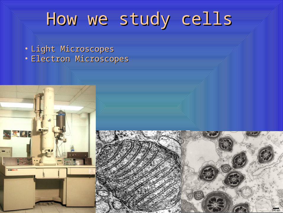
How we study cellsHow we study cells
• Light MicroscopesLight Microscopes• Electron MicroscopesElectron Microscopes
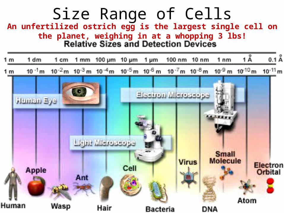
Size Range of CellsAn unfertilized ostrich egg is the largest single cell on the planet,
weighing in at a whopping 3 lbs!
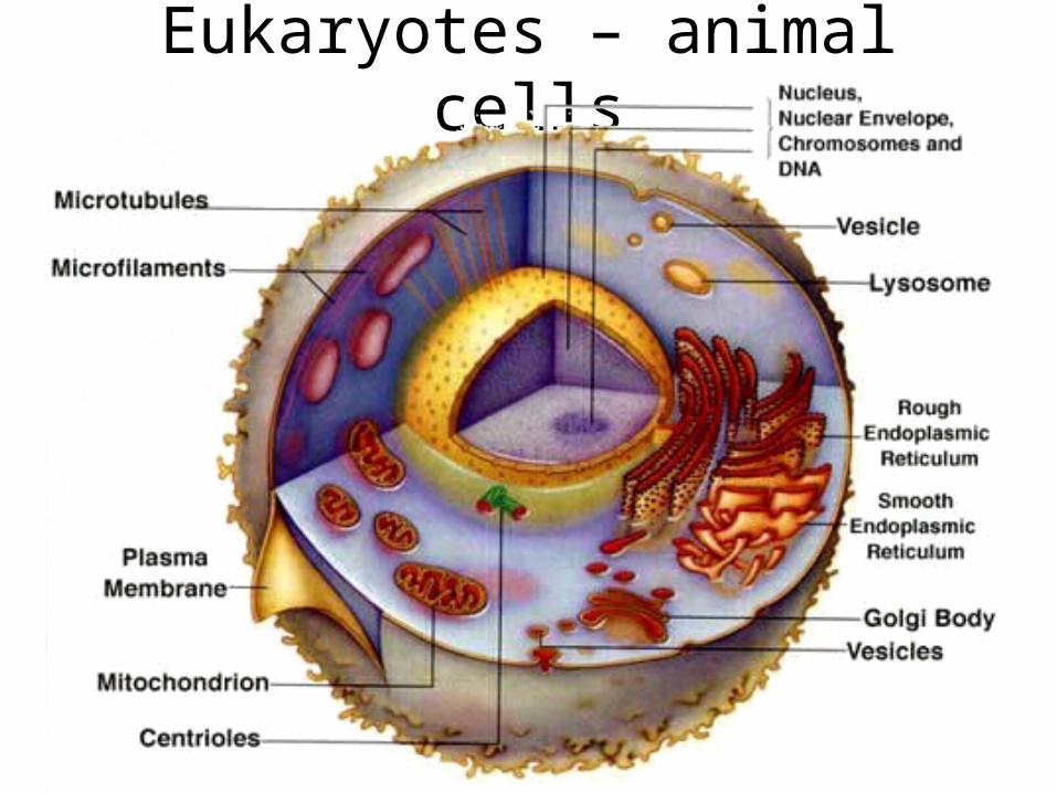
Eukaryotes – animal cells
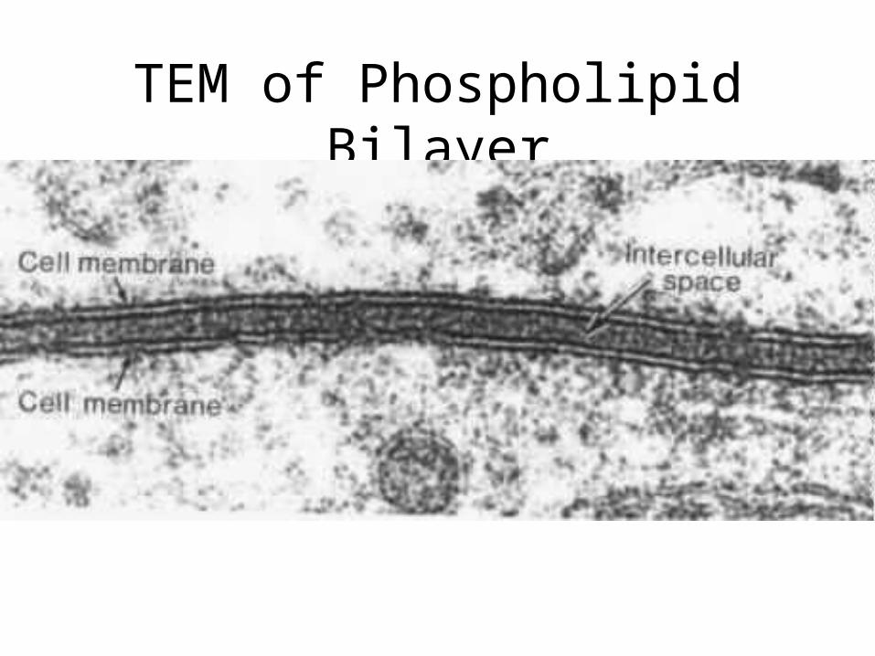
TEM of Phospholipid Bilayer
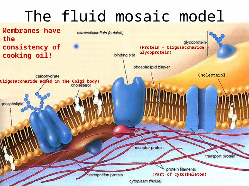
The fluid mosaic model
(Part of cytoskeleton)
(Oligosaccharide added in the Golgi body)
(Protein + Oligosaccharide = Glycoprotein)
Cholesterol
Membranes have the consistency of cooking oil!
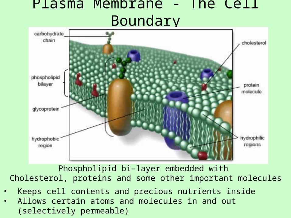
Plasma Membrane - The Cell Boundary
Phospholipid bi-layer embedded with Cholesterol, proteins and some other important molecules
• Keeps cell contents and precious nutrients inside• Allows certain atoms and molecules in and out (selectively permeable)
Cytosol• Semi-fluid medium
• Contains all organelles
• Contains water, nutrients and building blocks (carbohydrates, lipids, amino acids, nucleotides, ATP, enzymes, ions such as Ca++, Na+, Cl-, H+, OH-, K+ and many others)
Cytoplasm: The cytosol-containing area outside the nucleusNucleoplasm: The cytosol-containing area inside the nucleus
Extracellular fluid (Interstitial fluid): The watery liquid that surrounds cells
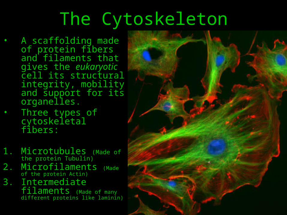
The Cytoskeleton• A scaffolding made of
protein fibers and filaments that gives the eukaryotic cell its structural integrity, mobility and support for its organelles.
• Three types of cytoskeletal fibers:
1. Microtubules (Made of the protein Tubulin)
2. Microfilaments (Made of the protein Actin)
3. Intermediate filaments (Made of many different proteins like laminin)
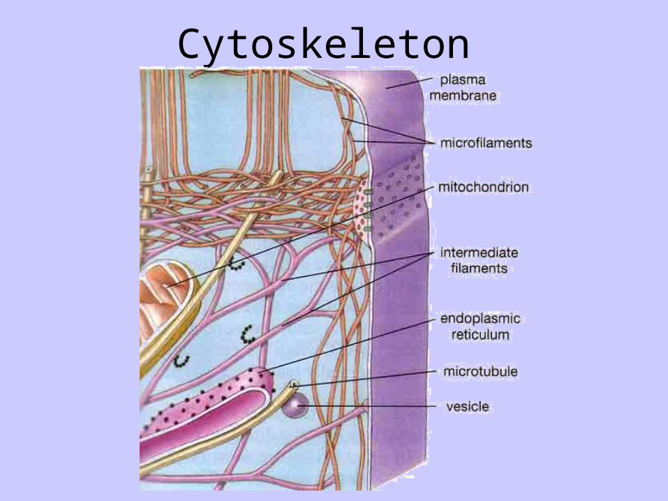
Cytoskeleton
Types of Membrane Proteins
• Receptor Proteins – Receive and transmit signals
• Integral Proteins – Form pores, channels, and carriers in cell membranes
• Cellular Adhesion Molecules (CAMS) – Enable cells to stick together
• Cell Surface Proteins – Establish “SELF”
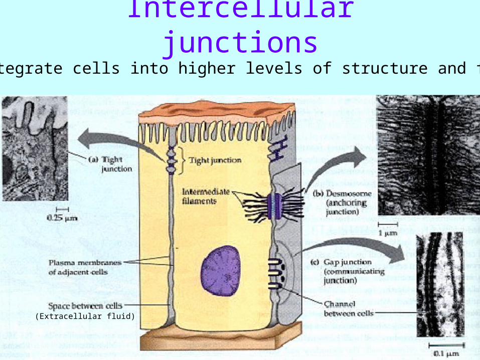
Intercellular junctionsHelp integrate cells into higher levels of structure and function
(Extracellular fluid)
Intercellular Junctions, cont’d.
• Tight Junctions – Extremely tight, belt-like areas of fusion– Cells of the digestive tract have this junction– Cells of capillaries of the brain have this –
preventing passage of many chemicals – “Blood-Brain Barrier”
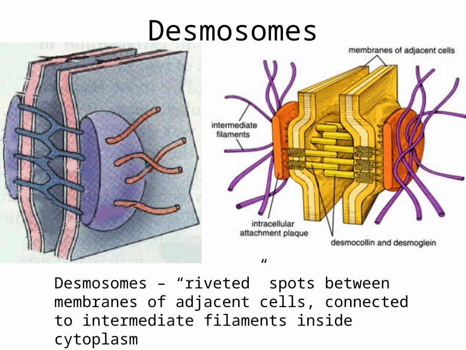
• Desmosomes – “riveted” spots between membranes of adjacent cells, connected to intermediate filaments inside cytoplasm
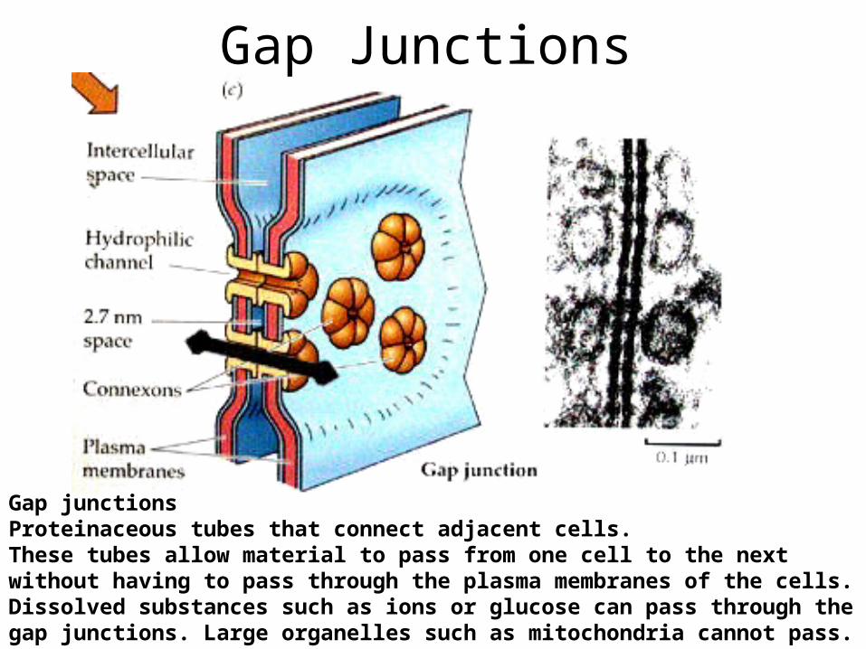
• Gap Junctions – tubular channels found between membranes of adjacent cells which allow the passage of nutrients and other small molecules between cells
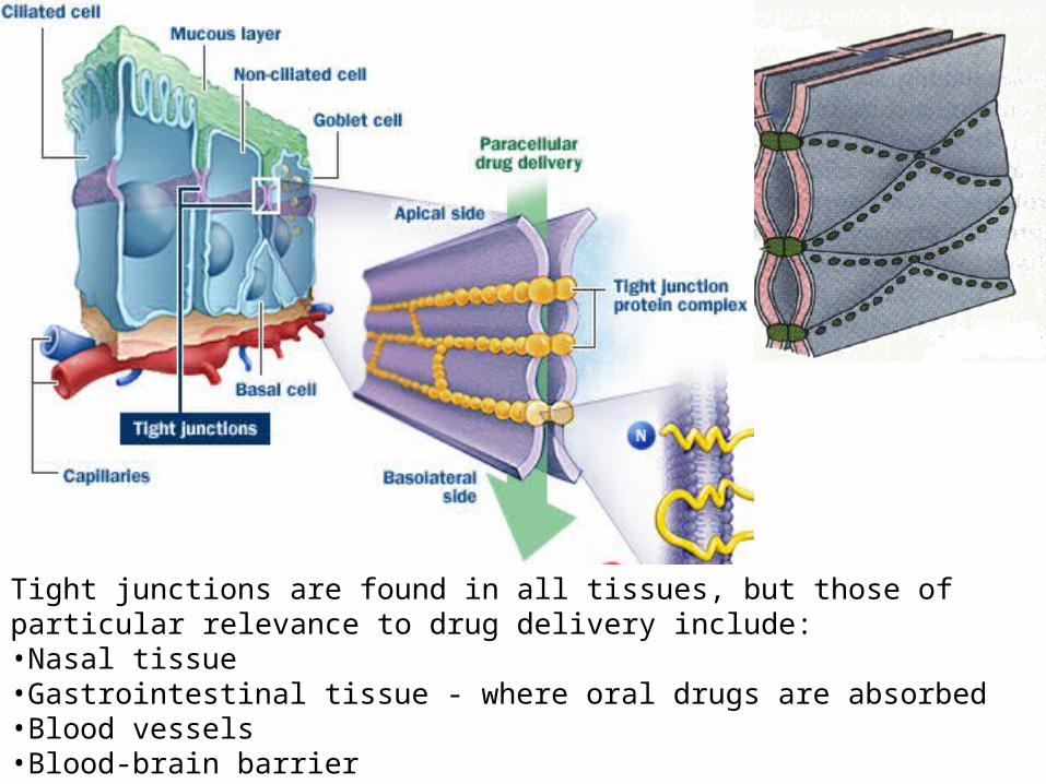
Tight junctions are found in all tissues, but those of particular relevance to drug delivery include:•Nasal tissue •Gastrointestinal tissue - where oral drugs are absorbed •Blood vessels •Blood-brain barrier
Desmosomes
Desmosomes – “riveted” spots between membranes of adjacent cells, connected to intermediate filaments inside cytoplasm
Gap Junctions
Gap junctionsProteinaceous tubes that connect adjacent cells. These tubes allow material to pass from one cell to the next without having to pass through the plasma membranes of the cells. Dissolved substances such as ions or glucose can pass through the gap junctions. Large organelles such as mitochondria cannot pass.
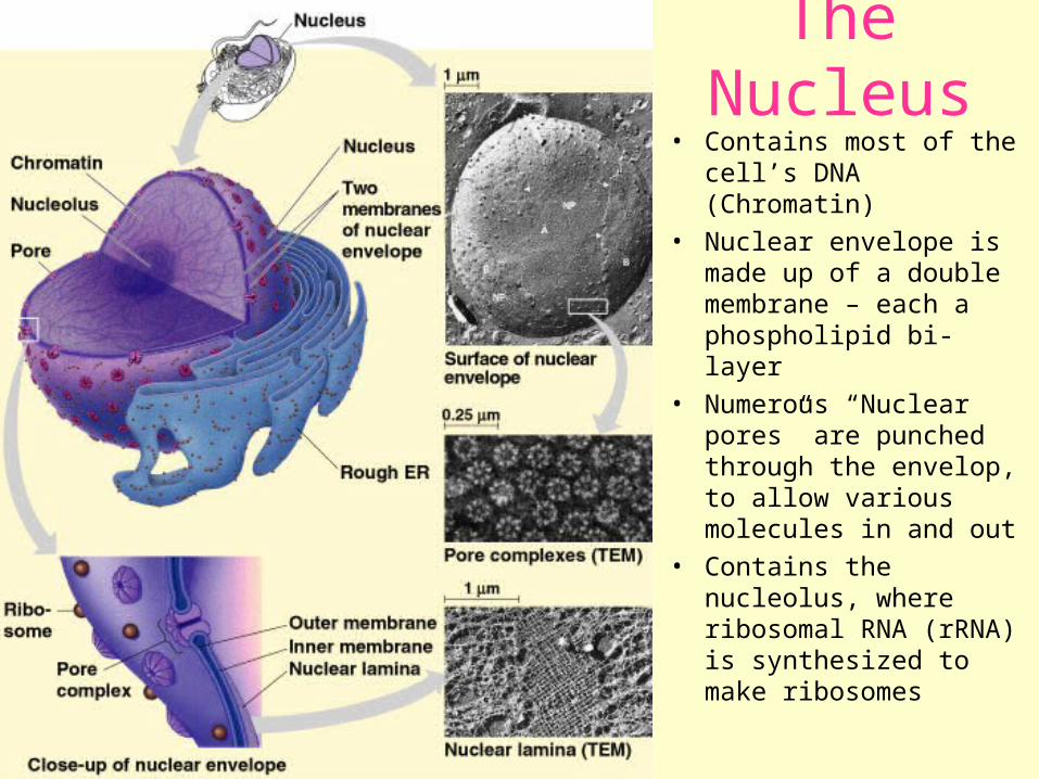
• Contains most of the cell’s DNA (Chromatin)
• Nuclear envelope is made up of a double membrane – each a phospholipid bi-layer
• Numerous “Nuclear pores” are punched through the envelop, to allow various molecules in and out
• Contains the nucleolus, where ribosomal RNA (rRNA) is synthesized to make ribosomes
The Nucleus
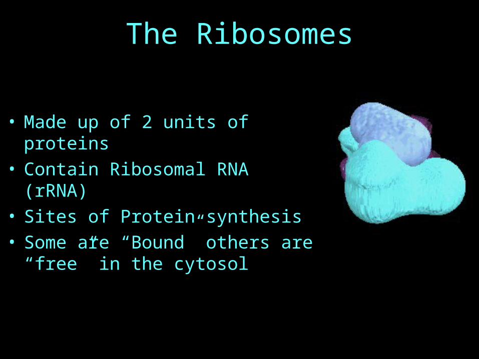
The Ribosomes
• Made up of 2 units of proteins• Contain Ribosomal RNA (rRNA)• Sites of Protein synthesis• Some are “Bound” others are “free”
in the cytosol
The Endomembrane System
• A network of sac-like membranes that are spread through-out the cell cytoplasm
• Consists of:
1. The Nuclear envelop
2. The Rough Endoplasmic Reticulum
3. The Smooth Endoplasmic Reticulum
4. The Golgi Apparatus or Body
5. Lysosomes, vacuoles and other Transport Vesicles
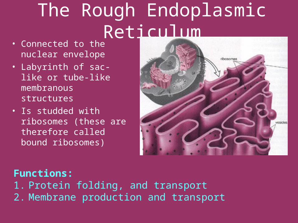
The Rough Endoplasmic Reticulum• Connected to the
nuclear envelope• Labyrinth of sac-like or
tube-like membranous structures
• Is studded with ribosomes (these are therefore called bound ribosomes)
Functions: 1. Protein folding, and transport2. Membrane production and transport
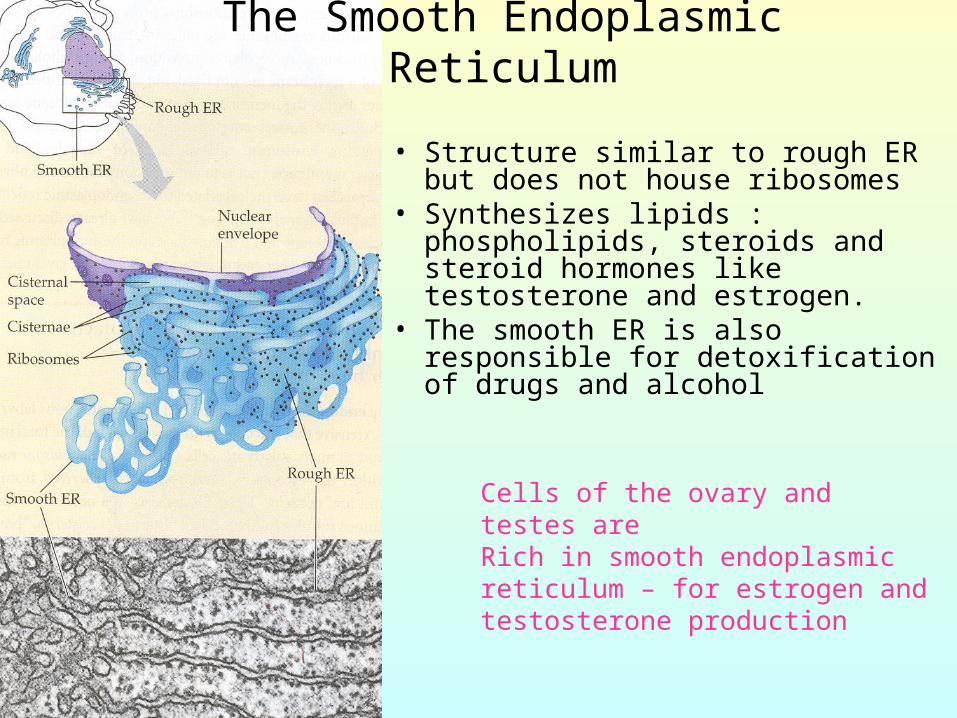
• Structure similar to rough ER but does not house ribosomes
• Synthesizes lipids : phospholipids, steroids and steroid hormones like testosterone and estrogen.
• The smooth ER is also responsible for detoxification of drugs and alcohol
The Smooth Endoplasmic Reticulum
Cells of the ovary and testes areRich in smooth endoplasmic reticulum – for estrogen and testosterone production
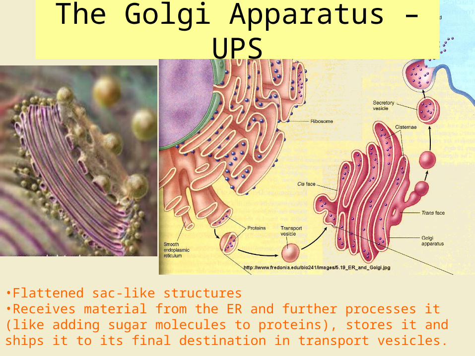
•Flattened sac-like structures•Receives material from the ER and further processes it (like adding sugar molecules to proteins), stores it and ships it to its final destination in transport vesicles.
The Golgi Apparatus – UPS
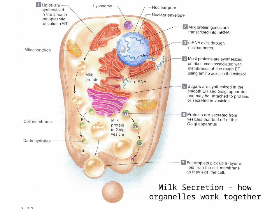
Milk Secretion – how organelles work
together
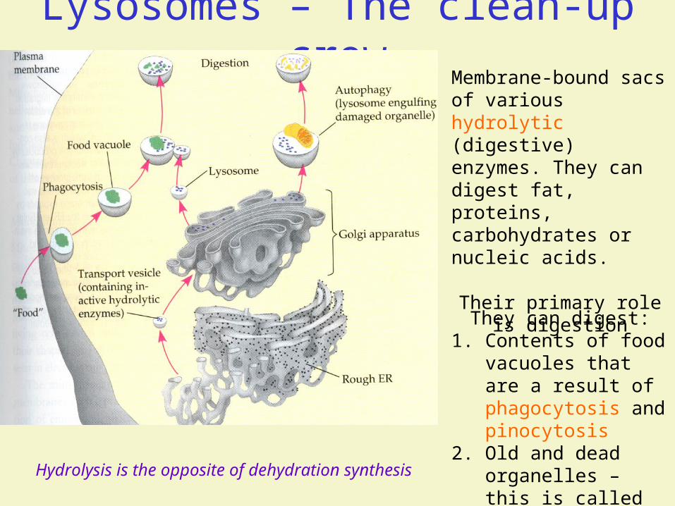
Lysosomes – The clean-up crewMembrane-bound sacs of various hydrolytic (digestive) enzymes. They can digest fat, proteins, carbohydrates or nucleic acids.
Their primary role is digestion
They can digest:1. Contents of food
vacuoles that are a result of phagocytosis and pinocytosis
2. Old and dead organelles – this is called autophagyHydrolysis is the opposite of dehydration synthesis
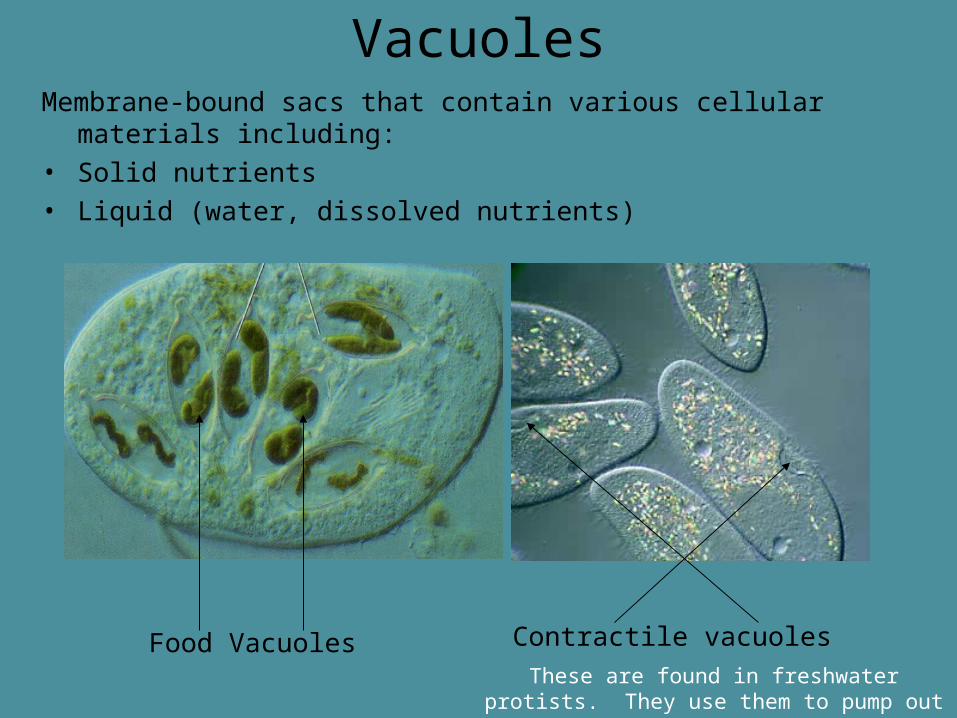
VacuolesMembrane-bound sacs that contain various cellular materials
including:• Solid nutrients• Liquid (water, dissolved nutrients)
Contractile vacuolesFood VacuolesThese are found in freshwater protists. They use
them to pump out excess water
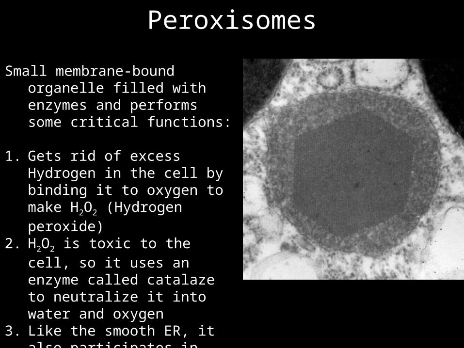
Peroxisomes
Small membrane-bound organelle filled with enzymes and performs some critical functions:
1. Gets rid of excess Hydrogen in the cell by binding it to oxygen to make H2O2 (Hydrogen peroxide)
2. H2O2 is toxic to the cell, so it uses an enzyme called catalaze to neutralize it into water and oxygen
3. Like the smooth ER, it also participates in detoxifying alcohol, drugs and other toxins
4. It breaks down long fatty acids5. Makes bile acids in liver cells
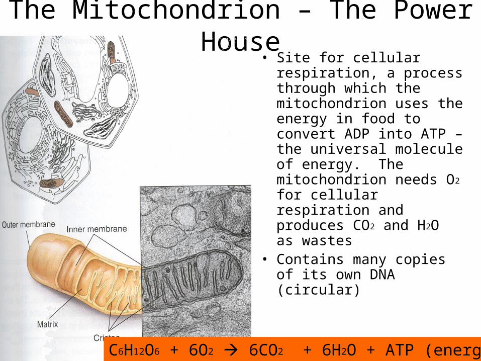
The Mitochondrion – The Power House• Site for cellular
respiration, a process through which the mitochondrion uses the energy in food to convert ADP into ATP – the universal molecule of energy. The mitochondrion needs O2 for cellular respiration and produces CO2 and H2O as wastes
• Contains many copies of its own DNA (circular)
C6H12O6 + 6O2 6CO2 + 6H2O + ATP (energy)
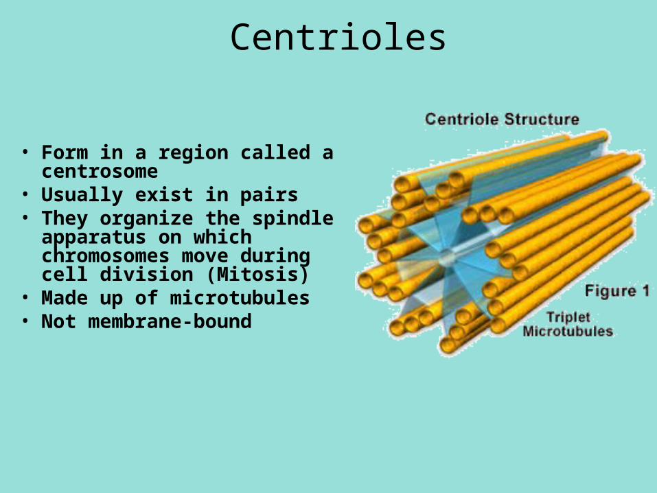
Centrioles
• Form in a region called a centrosome
• Usually exist in pairs• They organize the spindle
apparatus on which chromosomes move during cell division (Mitosis)
• Made up of microtubules• Not membrane-bound
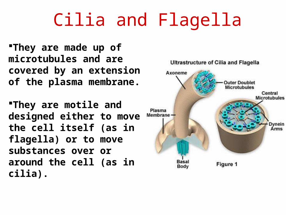
Cilia and FlagellaThey are made up of microtubules and are covered by an extension of the plasma membrane.
They are motile and designed either to move the cell itself (as in flagella) or to move substances over or around the cell (as in cilia).
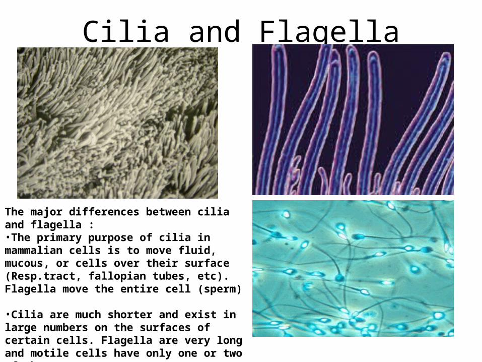
Cilia and Flagella
The major differences between cilia and flagella : •The primary purpose of cilia in mammalian cells is to move fluid, mucous, or cells over their surface (Resp.tract, fallopian tubes, etc). Flagella move the entire cell (sperm)
•Cilia are much shorter and exist in large numbers on the surfaces of certain cells. Flagella are very long and motile cells have only one or two of them.
Movement Into and Out of the Cell
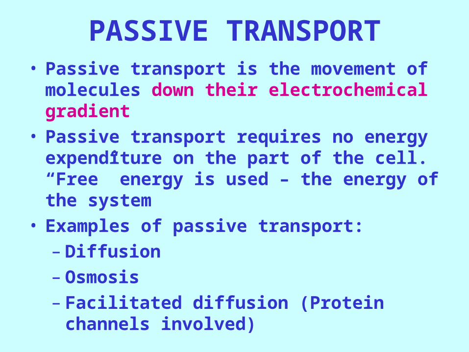
PASSIVE TRANSPORT• Passive transport is the movement of molecules
down their electrochemical gradient• Passive transport requires no energy expenditure
on the part of the cell. “Free” energy is used – the energy of the system
• Examples of passive transport:– Diffusion– Osmosis– Facilitated diffusion (Protein channels involved)
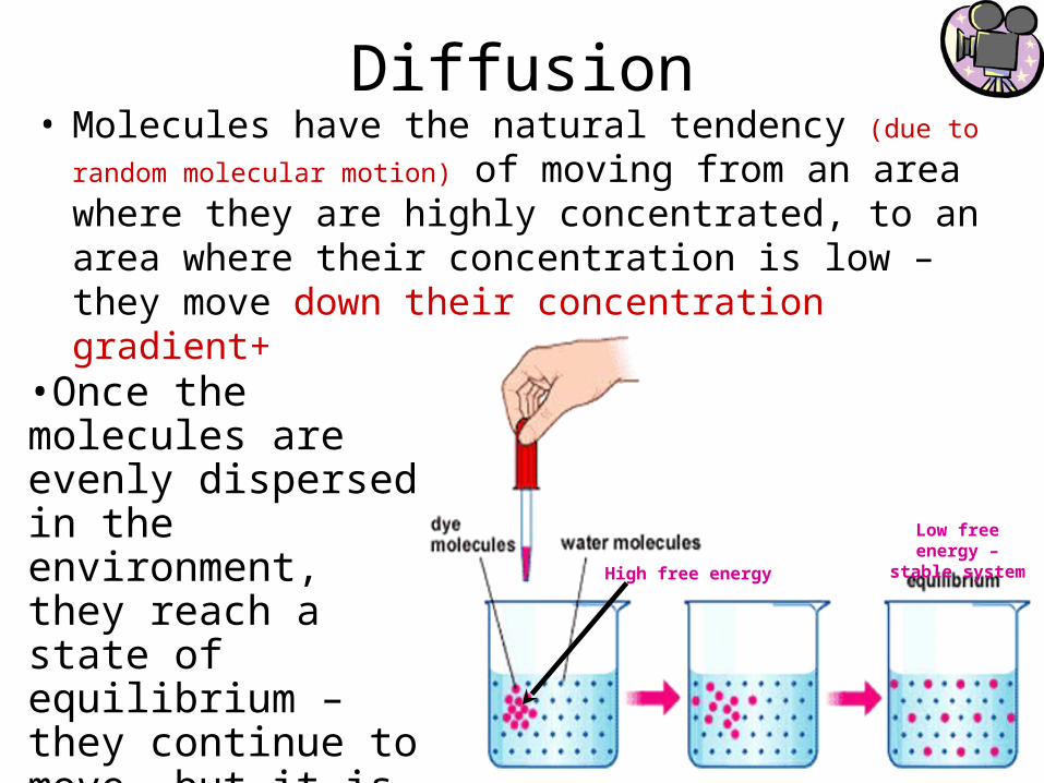
Diffusion• Molecules have the natural tendency (due to random molecular
motion) of moving from an area where they are highly concentrated, to an area where their concentration is low – they move down their concentration gradient+
•Once the molecules are evenly dispersed in the environment, they reach a state of equilibrium – they continue to move, but it is equal in every direction – so no net change
High free energy
Low free energy – stable system
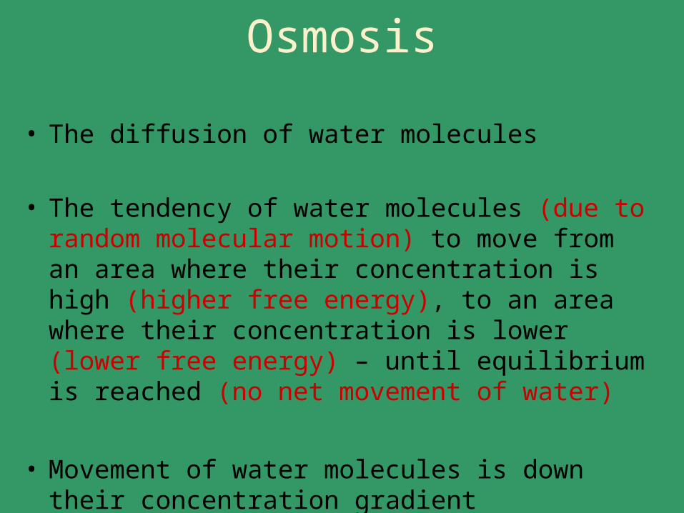
Osmosis
• The diffusion of water molecules
• The tendency of water molecules (due to random molecular motion) to move from an area where their concentration is high (higher free energy), to an area where their concentration is lower (lower free energy) – until equilibrium is reached (no net movement of water)
• Movement of water molecules is down their concentration gradient
(of water molecules)
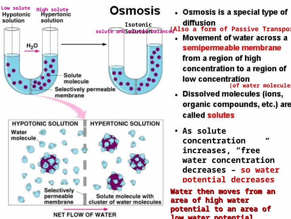
As solute concentration increases, “free” water concentration decreases – so water potential decreases
Water then moves from an area Water then moves from an area of high water potential to an of high water potential to an area of low water potentialarea of low water potential
Low solute High solute
Isotonic Solutionsolute and solvent balanced (Also a form of Passive Transport)
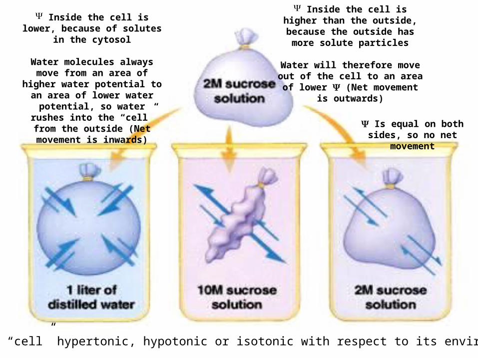
Inside the cell is lower, because of solutes in the
cytosol
Water molecules always move from an area of higher water potential to an area of lower
water potential, so water rushes into the “cell” from the outside
(Net movement is inwards)
Is the “cell” hypertonic, hypotonic or isotonic with respect to its environment?
Inside the cell is higher than the outside, because the outside
has more solute particles
Water will therefore move out of the cell to an area of lower (Net
movement is outwards)
Is equal on both sides, so no net movement
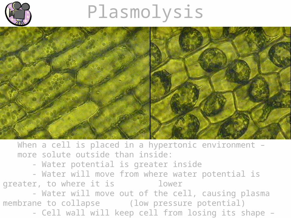
Plasmolysis
When a cell is placed in a hypertonic environment – more solute outside than inside:
- Water potential is greater inside- Water will move from where water potential is greater, to where it is lower- Water will move out of the cell, causing plasma membrane to collapse (low pressure potential)- Cell wall will keep cell from losing its shape – animal cell loses shape
Facilitated Diffusion
• Ions and small polar (Hydrophilic) molecules use facilitated diffusion
• Membrane channel proteins are used
• Requires no cellular energy (ATP, GTP, etc.)
• Diffusion is down concentration gradient
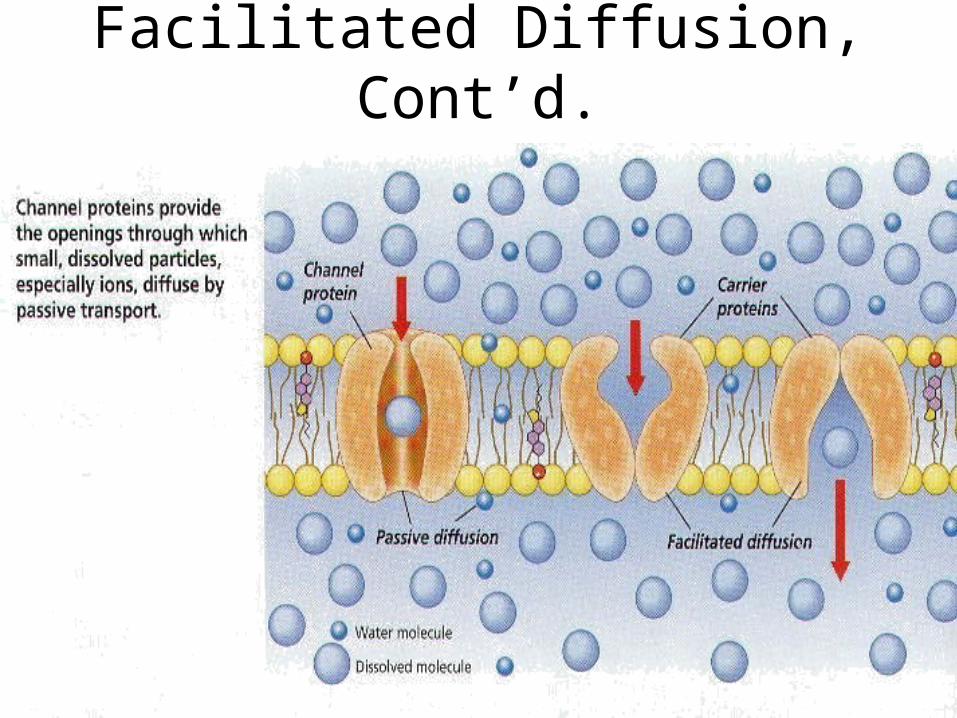
Facilitated Diffusion, Cont’d.
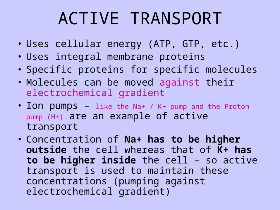
ACTIVE TRANSPORT• Uses cellular energy (ATP, GTP, etc.)• Uses integral membrane proteins• Specific proteins for specific molecules• Molecules can be moved against their
electrochemical gradient• Ion pumps – like the Na+ / K+ pump and the Proton pump (H+) are
an example of active transport • Concentration of Na+ has to be higher outside the
cell whereas that of K+ has to be higher inside the cell – so active transport is used to maintain these concentrations (pumping against electrochemical gradient)
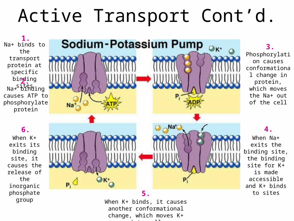
Active Transport Cont’d.Na+ binds to the transport protein
at specific binding sites
Na+ binding causes ATP to phosphorylate
protein
Phosphorylation causes
conformational change in protein, which moves the
Na+ out of the cell
When Na+ exits the binding site, the binding site for K+ is made accessible and
K+ binds to sites
When K+ binds, it causes another conformational change, which
moves K+ into cell
When K+ exits its binding site, it
causes the release of the
inorganic phosphate group
1.
2.
3.
4.
5.
6.
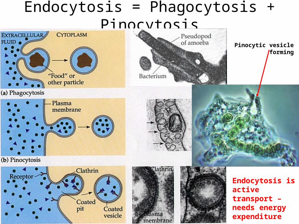
Endocytosis = Phagocytosis + Pinocytosis
Endocytosis is active transport – needs energy expenditure
Pinocytic vesicle forming
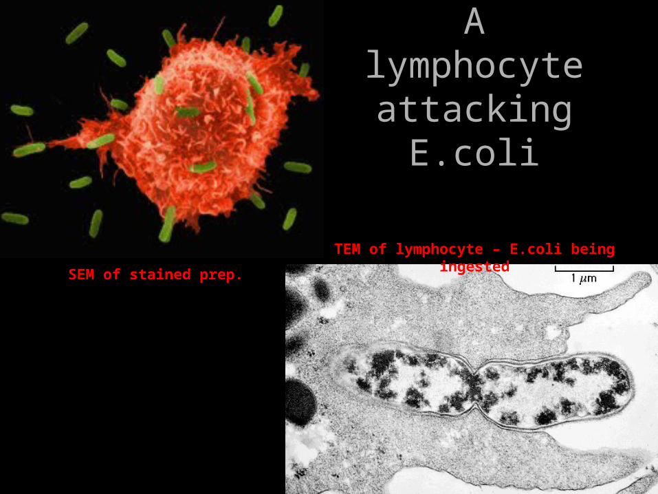
A lymphocyte attacking E.coli
SEM of stained prep.
TEM of lymphocyte – E.coli being ingested
All somatic cells reproduce mitoticallyAll somatic cells reproduce mitotically
• Somatic cells are Somatic cells are allall the cells of the body, the cells of the body, exceptexcept the gametes (egg and sperm) the gametes (egg and sperm)
• Skin cells, liver cells, cells that line the G.I. Skin cells, liver cells, cells that line the G.I. tract, etc. are constantly dividing, to replace tract, etc. are constantly dividing, to replace dead cellsdead cells
• Other cells such as neurons, adipose cells, Other cells such as neurons, adipose cells, muscle cells, etc. never or rarely dividemuscle cells, etc. never or rarely divide
The Cell CycleThe Cell Cycle• A A typicaltypical human cell undergoes a division about every 24 human cell undergoes a division about every 24
hours (there are many exceptions!)hours (there are many exceptions!)
• The cell cycle is basically an alternation of 2 major phases – The cell cycle is basically an alternation of 2 major phases – Mitosis and InterphaseMitosis and Interphase
• Interphase is the phase in which the cell spends 23 of the 24 Interphase is the phase in which the cell spends 23 of the 24 hours – the cell grows, carries out its “housekeeping duties” hours – the cell grows, carries out its “housekeeping duties” and its specialized activitiesand its specialized activities
• Mitosis takes about 1 hourMitosis takes about 1 hour
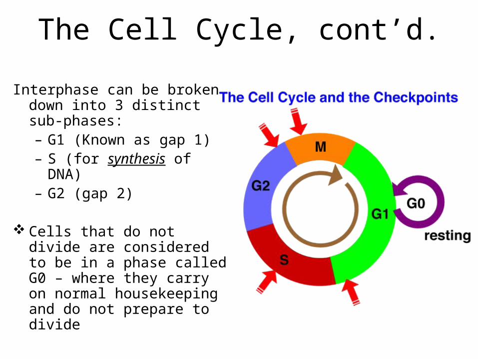
The Cell Cycle, cont’d.
Interphase can be broken down into 3 distinct sub-phases:– G1 (Known as gap 1)– S (for synthesis of DNA)– G2 (gap 2)
Cells that do not divide are considered to be in a phase called G0 – where they carry on normal housekeeping and do not prepare to divide
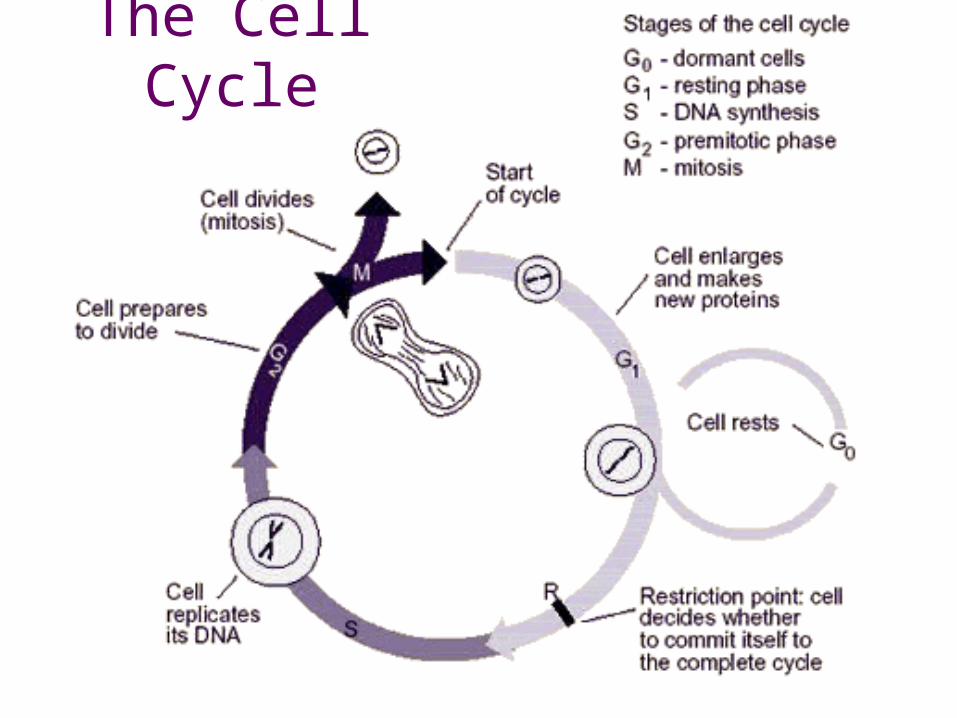
The Cell Cycle
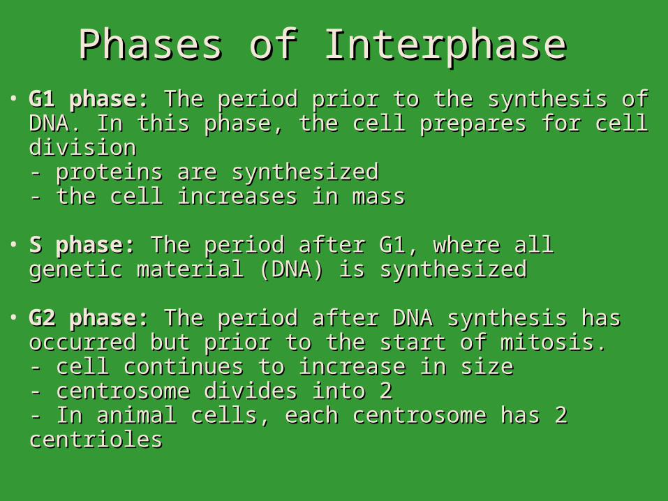
Phases of Interphase Phases of Interphase • G1 phase:G1 phase: The period prior to the synthesis of DNA. In this The period prior to the synthesis of DNA. In this
phase, the cell prepares for cell divisionphase, the cell prepares for cell division- proteins are synthesized- proteins are synthesized- the cell increases in mass - the cell increases in mass
• S phase: S phase: The period after G1, where all genetic material The period after G1, where all genetic material (DNA) is synthesized(DNA) is synthesized
• G2 phase:G2 phase: The period after DNA synthesis has occurred but The period after DNA synthesis has occurred but prior to the start of mitosis. prior to the start of mitosis. - cell continues to increase in size- cell continues to increase in size- centrosome divides into 2- centrosome divides into 2- In animal cells, each centrosome has 2 centrioles- In animal cells, each centrosome has 2 centrioles
Phases of Mitosis
• Prophase
• Prometaphase
• Metaphase
• Anaphase
• Telophase (followed immediately by cytokinesis)
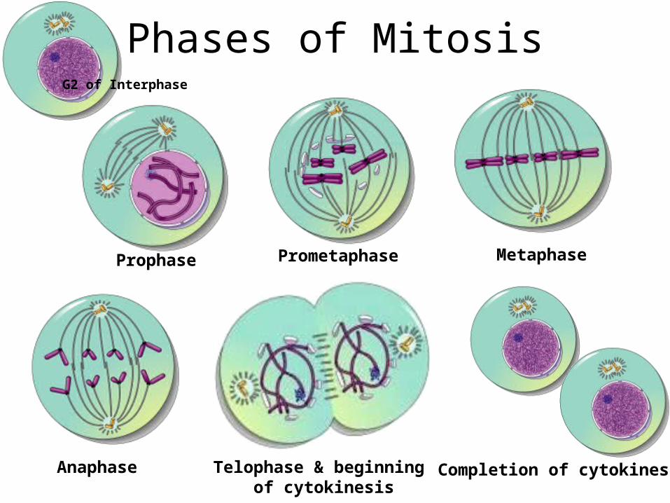
Phases of Mitosis
Prophase Prometaphase
Anaphase
Metaphase
Telophase & beginning of cytokinesis
Completion of cytokinesis
G2 of Interphase
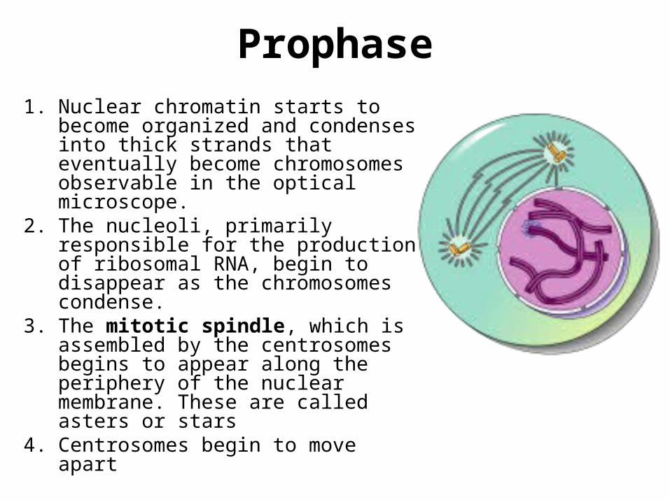
Prophase
1. Nuclear chromatin starts to become organized and condenses into thick strands that eventually become chromosomes observable in the optical microscope.
2. The nucleoli, primarily responsible for the production of ribosomal RNA, begin to disappear as the chromosomes condense.
3. The mitotic spindle, which is assembled by the centrosomes begins to appear along the periphery of the nuclear membrane. These are called asters or stars
4. Centrosomes begin to move apart
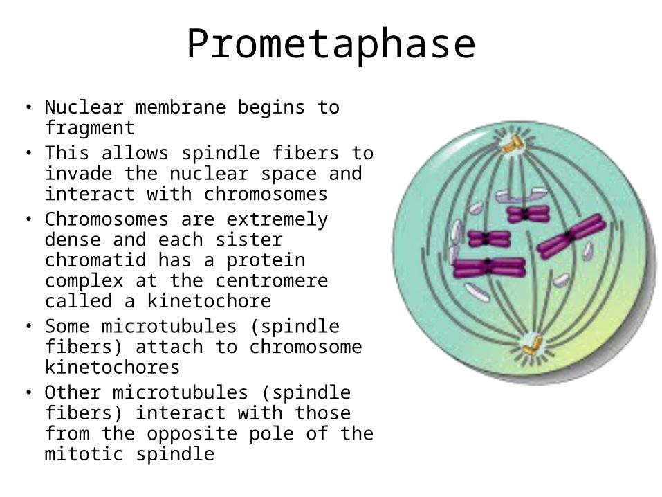
Prometaphase
• Nuclear membrane begins to fragment• This allows spindle fibers to invade
the nuclear space and interact with chromosomes
• Chromosomes are extremely dense and each sister chromatid has a protein complex at the centromere called a kinetochore
• Some microtubules (spindle fibers) attach to chromosome kinetochores
• Other microtubules (spindle fibers) interact with those from the opposite pole of the mitotic spindle
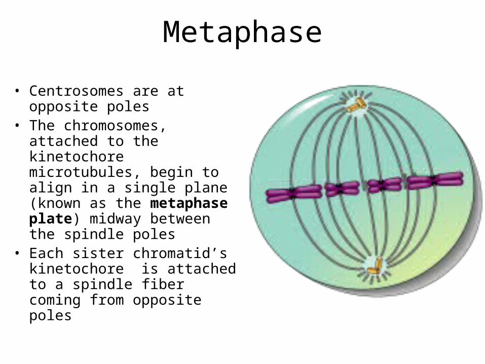
Metaphase
• Centrosomes are at opposite poles
• The chromosomes, attached to the kinetochore microtubules, begin to align in a single plane (known as the metaphase plate) midway between the spindle poles
• Each sister chromatid’s kinetochore is attached to a spindle fiber coming from opposite poles
Anaphase
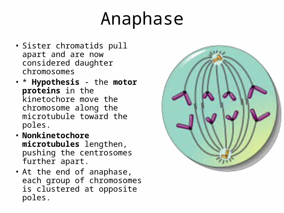
• Sister chromatids pull apart and are now considered daughter chromosomes
• * Hypothesis - the motor proteins in the kinetochore move the chromosome along the microtubule toward the poles.
• Nonkinetochore microtubules lengthen, pushing the centrosomes further apart.
• At the end of anaphase, each group of chromosomes is clustered at opposite poles.
Telophase
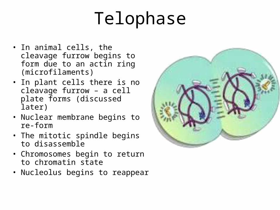
• In animal cells, the cleavage furrow begins to form due to an actin ring (microfilaments)
• In plant cells there is no cleavage furrow – a cell plate forms (discussed later)
• Nuclear membrane begins to re-form
• The mitotic spindle begins to disassemble
• Chromosomes begin to return to chromatin state
• Nucleolus begins to reappear
Interphase
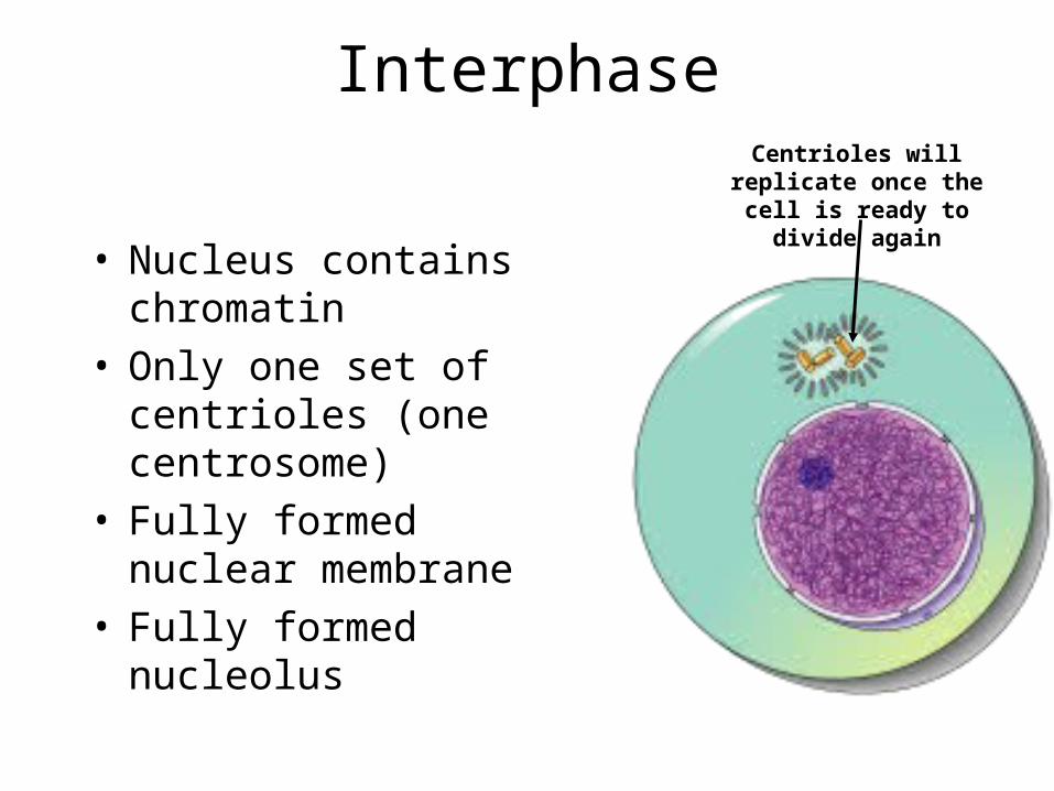
• Nucleus contains chromatin• Only one set of centrioles
(one centrosome)• Fully formed nuclear
membrane• Fully formed nucleolus
Centrioles will replicate once the cell is ready to
divide again
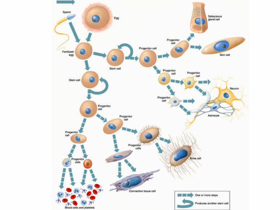
Stem & Progenitor Cells
• Stem cells are totipotent
• Stem cells can divide and give rise to more stem cells or one daughter stem cell and a progenitor cell (partially specialized)
• A Progenitor cell is pluripotent a.k.a. committed. It can only become a set number of cells (restricted to a number of cell types)

THE END
Related Documents