Chapter 28 Chapter 28 Lung Cancer Lung Cancer

Chapter 28 Lung Cancer. Mosby items and derived items © 2009 by Mosby, Inc., an affiliate of Elsevier Inc. 2 Objectives Describe the epidemiology of.
Dec 27, 2015
Welcome message from author
This document is posted to help you gain knowledge. Please leave a comment to let me know what you think about it! Share it to your friends and learn new things together.
Transcript
Chapter 28Chapter 28
Lung CancerLung Cancer
Mosby items and derived items © 2009 by Mosby, Inc., an affiliate of Elsevier Inc. 2
ObjectivesObjectives Describe the epidemiology of lung cancer in the Describe the epidemiology of lung cancer in the
United States, particularly current trends.United States, particularly current trends.
Describe risk factors for lung cancer. Describe risk factors for lung cancer.
Describe the classification of lung cancer types and Describe the classification of lung cancer types and the cellular features of the four common types of lung the cellular features of the four common types of lung cancer. cancer.
Describe current understanding of the Describe current understanding of the pathophysiology of lung cancer.pathophysiology of lung cancer.
Mosby items and derived items © 2009 by Mosby, Inc., an affiliate of Elsevier Inc. 3
Objectives (cont.)Objectives (cont.)
Describe the clinical features of the common Describe the clinical features of the common types of lung cancer.types of lung cancer.
Describe the diagnostic approach to lung Describe the diagnostic approach to lung cancer.cancer.
Describe the staging system for lung cancer.Describe the staging system for lung cancer.
Mosby items and derived items © 2009 by Mosby, Inc., an affiliate of Elsevier Inc. 4
Objectives (cont.)Objectives (cont.)
Describe the treatment and outcomes for the Describe the treatment and outcomes for the common types of lung cancer by stage. common types of lung cancer by stage.
Describe the role of the respiratory therapist Describe the role of the respiratory therapist in managing patients with lung cancer.in managing patients with lung cancer.
Mosby items and derived items © 2009 by Mosby, Inc., an affiliate of Elsevier Inc. 5
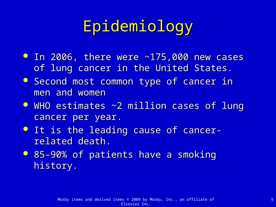
EpidemiologyEpidemiology
In 2006, there were ~175,000 new cases of lung In 2006, there were ~175,000 new cases of lung cancer in the United States.cancer in the United States.
Second most common type of cancer in men and Second most common type of cancer in men and womenwomen
WHO estimates ~2 million cases of lung cancer per WHO estimates ~2 million cases of lung cancer per year.year.
It is the leading cause of cancer-related death.It is the leading cause of cancer-related death. 85–90% of patients have a smoking history.85–90% of patients have a smoking history.
Mosby items and derived items © 2009 by Mosby, Inc., an affiliate of Elsevier Inc. 6
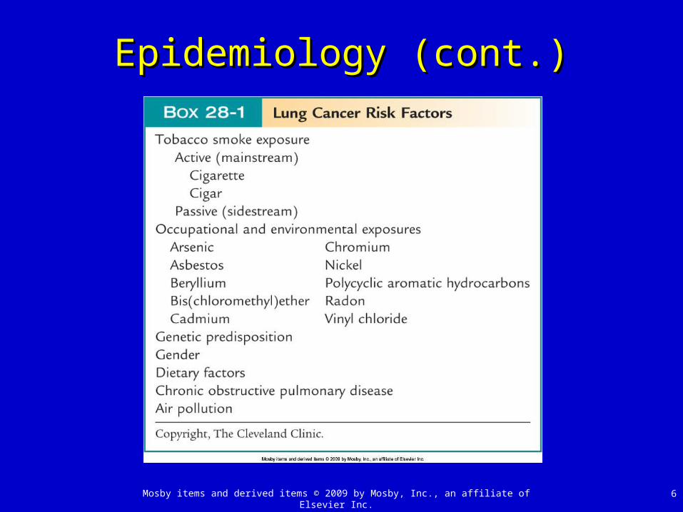
Epidemiology (cont.)Epidemiology (cont.)

Mosby items and derived items © 2009 by Mosby, Inc., an affiliate of Elsevier Inc. 7
Lung Cancer ClassificationLung Cancer Classification
Classified as small cell or non–small cell carcinomaClassified as small cell or non–small cell carcinoma
Non–small cell lung carcinoma (NSCLC) consists ofNon–small cell lung carcinoma (NSCLC) consists of Adenocarcinoma: most common type, ~40% of all lung Adenocarcinoma: most common type, ~40% of all lung
cancers in United Statescancers in United States Squamous cell carcinoma: 2Squamous cell carcinoma: 2ndnd most common type most common type Large cell carcinoma: rarest form of lung cancerLarge cell carcinoma: rarest form of lung cancer
Small cell lung carcinoma (SCLC): ~20% of U.S. Small cell lung carcinoma (SCLC): ~20% of U.S. cases cases
Mosby items and derived items © 2009 by Mosby, Inc., an affiliate of Elsevier Inc. 8
PathophysiologyPathophysiology
Poorly understoodPoorly understood
Genetic material in lung cells damaged secondary to Genetic material in lung cells damaged secondary to exposure to carcinogens, i.e., those in tobacco exposure to carcinogens, i.e., those in tobacco smokesmoke
There may be a genetic predisposition.There may be a genetic predisposition.
The more genetic activation of the following pathways The more genetic activation of the following pathways occurs; more likely, lung cancer’s growth isoccurs; more likely, lung cancer’s growth is Stimulation of cell growth, differentiation, apoptosis, Stimulation of cell growth, differentiation, apoptosis,
angiogenesis, tumor progression, immune regulationangiogenesis, tumor progression, immune regulation
Mosby items and derived items © 2009 by Mosby, Inc., an affiliate of Elsevier Inc. 9
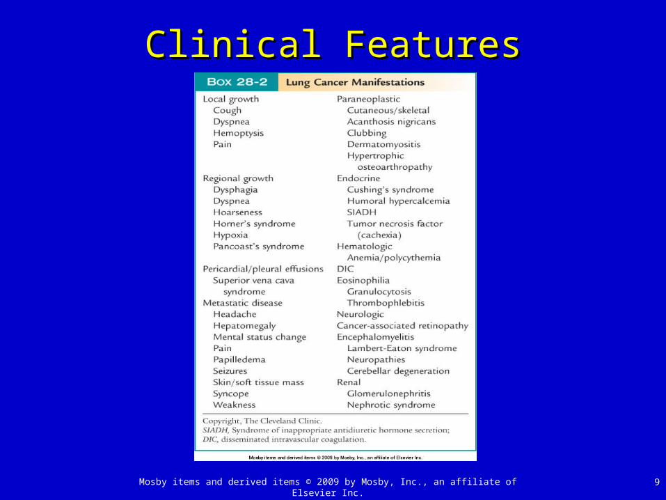
Clinical FeaturesClinical Features

Mosby items and derived items © 2009 by Mosby, Inc., an affiliate of Elsevier Inc. 10
DiagnosisDiagnosis
~85% of patients will be symptomatic (see Box 28-2).~85% of patients will be symptomatic (see Box 28-2).
Remainder detected by radiographic evaluationRemainder detected by radiographic evaluation Chest radiograph and CT scan initial evaluationChest radiograph and CT scan initial evaluation Will show nodules (<3 cm) and masses (>3 cm)Will show nodules (<3 cm) and masses (>3 cm) Other findings: enlarged lymph nodes, effusionsOther findings: enlarged lymph nodes, effusions
If radiograph, symptoms, history are very suggestive of If radiograph, symptoms, history are very suggestive of malignancy may move straight to surgerymalignancy may move straight to surgery
If unsure if malignant or benign, further testing If unsure if malignant or benign, further testing indicatedindicated
Mosby items and derived items © 2009 by Mosby, Inc., an affiliate of Elsevier Inc. 11
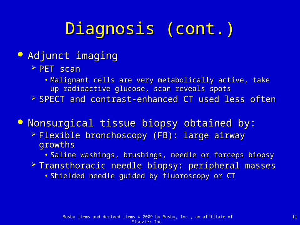
Diagnosis (cont.)Diagnosis (cont.)
Adjunct imagingAdjunct imaging PET scanPET scan
• Malignant cells are very metabolically active, take up Malignant cells are very metabolically active, take up radioactive glucose, scan reveals spotsradioactive glucose, scan reveals spots
SPECT and contrast-enhanced CT used less oftenSPECT and contrast-enhanced CT used less often
Nonsurgical tissue biopsy obtained by:Nonsurgical tissue biopsy obtained by: Flexible bronchoscopy (FB): large airway growthsFlexible bronchoscopy (FB): large airway growths
• Saline washings, brushings, needle or forceps biopsySaline washings, brushings, needle or forceps biopsy Transthoracic needle biopsy: peripheral massesTransthoracic needle biopsy: peripheral masses
• Shielded needle guided by fluoroscopy or CT Shielded needle guided by fluoroscopy or CT
Mosby items and derived items © 2009 by Mosby, Inc., an affiliate of Elsevier Inc. 12
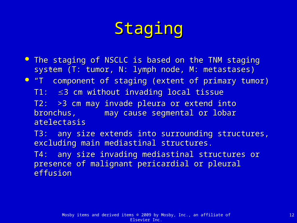
StagingStaging
The staging of NSCLC is based on the TNM staging The staging of NSCLC is based on the TNM staging system (T: tumor, N: lymph node, M: metastases)system (T: tumor, N: lymph node, M: metastases)
““T” component of staging (extent of primary tumor)T” component of staging (extent of primary tumor)
T1:T1: 3 cm without invading local tissue3 cm without invading local tissue
T2:T2: >3 cm may invade pleura or extend into >3 cm may invade pleura or extend into bronchus, bronchus, may cause segmental or lobar atelectasis may cause segmental or lobar atelectasis
T3:T3: any size extends into surrounding structures, any size extends into surrounding structures, excluding main mediastinal structures. excluding main mediastinal structures.
T4:T4: any size invading mediastinal structures or any size invading mediastinal structures or presence of malignant pericardial or pleural effusionpresence of malignant pericardial or pleural effusion
Mosby items and derived items © 2009 by Mosby, Inc., an affiliate of Elsevier Inc. 13
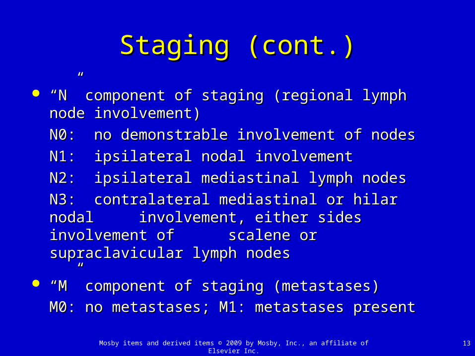
Staging (cont.)Staging (cont.)
““N” component of staging (regional lymph node N” component of staging (regional lymph node involvement)involvement)
N0:N0: no demonstrable involvement of nodesno demonstrable involvement of nodes
N1:N1: ipsilateral nodal involvementipsilateral nodal involvement
N2:N2: ipsilateral mediastinal lymph nodesipsilateral mediastinal lymph nodes
N3:N3: contralateral mediastinal or hilar nodal contralateral mediastinal or hilar nodal involvement, either sides involvement of involvement, either sides involvement of scalene scalene or supraclavicular lymph nodesor supraclavicular lymph nodes
““M” component of staging (metastases)M” component of staging (metastases)
M0: no metastases; M1: metastases presentM0: no metastases; M1: metastases present
Mosby items and derived items © 2009 by Mosby, Inc., an affiliate of Elsevier Inc. 14
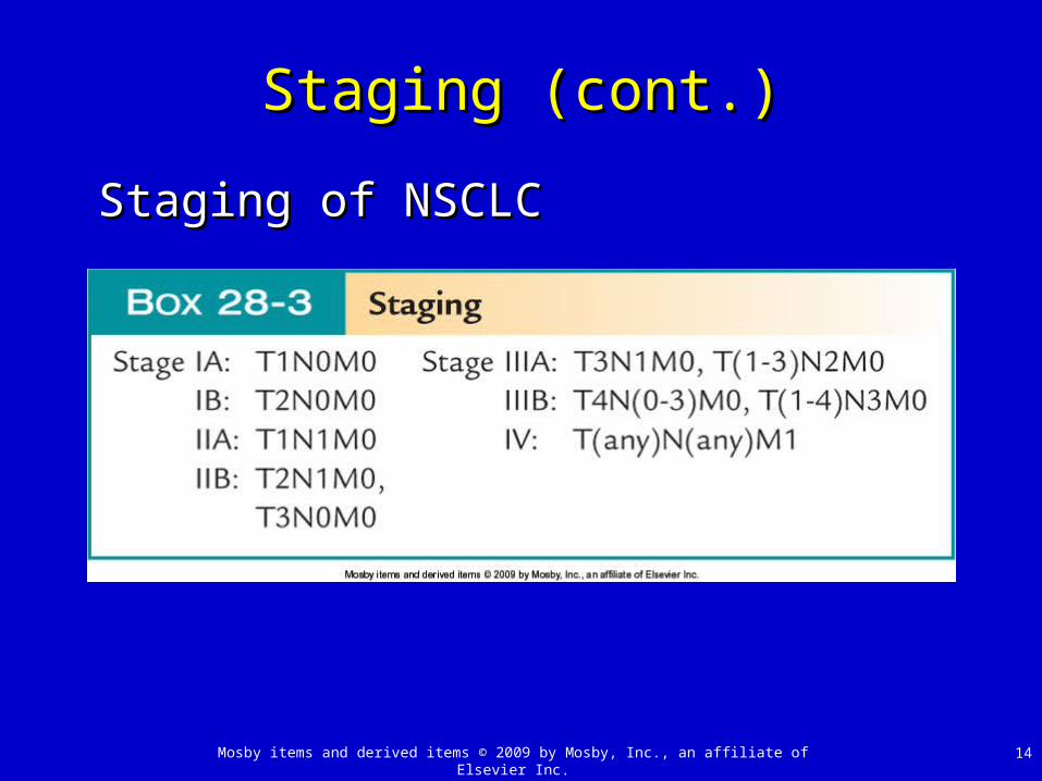
Staging (cont.)Staging (cont.)
Staging of NSCLCStaging of NSCLC
Mosby items and derived items © 2009 by Mosby, Inc., an affiliate of Elsevier Inc. 15
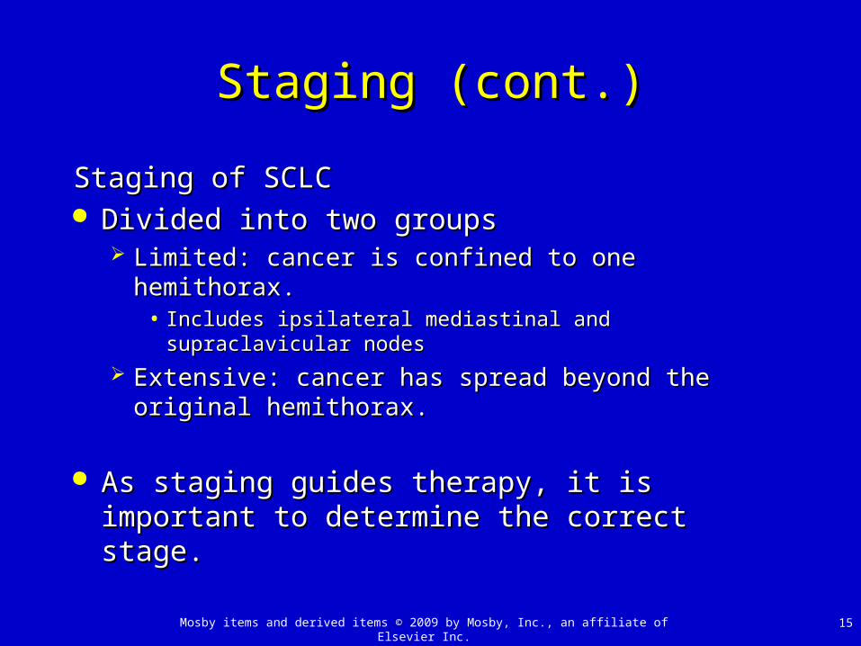
Staging (cont.)Staging (cont.)
Staging of SCLCStaging of SCLC Divided into two groupsDivided into two groups
Limited: cancer is confined to one hemithorax. Limited: cancer is confined to one hemithorax. • Includes ipsilateral mediastinal and supraclavicular Includes ipsilateral mediastinal and supraclavicular
nodesnodes
Extensive: cancer has spread beyond the original Extensive: cancer has spread beyond the original hemithorax.hemithorax.
As staging guides therapy, it is important to As staging guides therapy, it is important to determine the correct stage.determine the correct stage.

Mosby items and derived items © 2009 by Mosby, Inc., an affiliate of Elsevier Inc. 16
Staging (cont.)Staging (cont.)
Determination of staging for all lung cancers:Determination of staging for all lung cancers: CT of chest and upper abdomen is ordered for all.CT of chest and upper abdomen is ordered for all. MRI only superior to CT scan for a Pancoast tumorMRI only superior to CT scan for a Pancoast tumor FDG-PET best to determine staging of mediastinal nodesFDG-PET best to determine staging of mediastinal nodes FB with transbronchial needle aspiration help for mediastinal FB with transbronchial needle aspiration help for mediastinal
stagingstaging Gold standard remains surgical resection and mediastinal Gold standard remains surgical resection and mediastinal
dissection.dissection. Patient performance status is important in determining Patient performance status is important in determining
prognosis and ability to tolerate surgery.prognosis and ability to tolerate surgery.

Mosby items and derived items © 2009 by Mosby, Inc., an affiliate of Elsevier Inc. 17
Screening for Lung CancerScreening for Lung Cancer
Due to the high proportion of patients who present Due to the high proportion of patients who present with advanced lung cancer and its associated with advanced lung cancer and its associated mortality, screening is very attractive.mortality, screening is very attractive.
TechniquesTechniques Chest radiograph and/or sputum exam Chest radiograph and/or sputum exam
• Studies did not support beneficial outcome.Studies did not support beneficial outcome.
Low-dose CT imaging Low-dose CT imaging • No proof it is of any benefitNo proof it is of any benefit
• May be useful in high-risk individualsMay be useful in high-risk individuals

Mosby items and derived items © 2009 by Mosby, Inc., an affiliate of Elsevier Inc. 18
Treatment and OutcomesTreatment and Outcomes
Mosby items and derived items © 2009 by Mosby, Inc., an affiliate of Elsevier Inc. 19
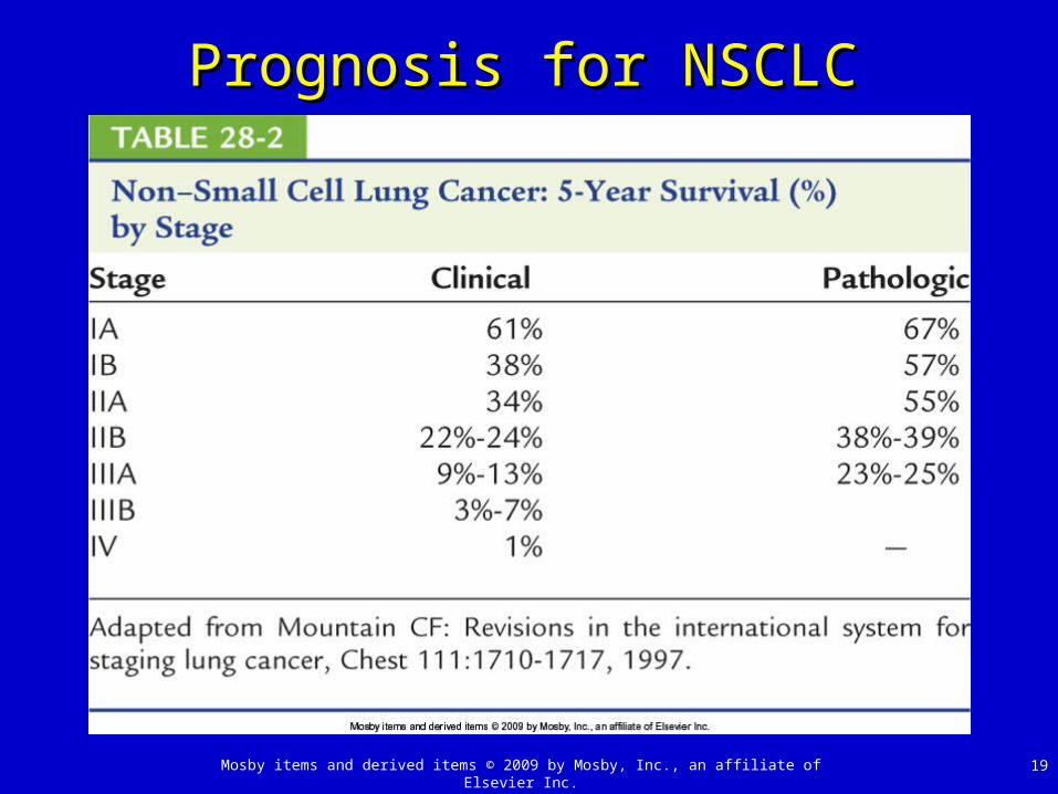
Prognosis for NSCLCPrognosis for NSCLC
Related Documents