© 2012 Pearson Education, Inc. Lecture by Edward J. Zalisko PowerPoint Lectures for Campbell Biology: Concepts & Connections, Seventh Edition Reece, Taylor, Simon, and Dickey Chapter 28 Chapter 28 Nervous Systems Introduction Spinal cord injuries disrupt communication between – the central nervous system (brain and spinal cord) and – the rest of the body. © 2012 Pearson Education, Inc. Over 250,000 Americans are living with spinal cord injuries. Spinal cord injuries – happen more often to men, – happen mostly to people in their teens and 20s, – are caused by vehicle accidents, gunshots, and falls, and – are usually permanent because the spinal cord cannot be repaired. Introduction © 2012 Pearson Education, Inc. Figure 28.0_1 Chapter 28: Big Ideas Nervous System Structure and Function The Human Brain An Overview of Animal Nervous Systems Nerve Signals and Their Transmission Figure 28.0_2 NERVOUS SYSTEM STRUCTURE AND FUNCTION © 2012 Pearson Education, Inc.

Welcome message from author
This document is posted to help you gain knowledge. Please leave a comment to let me know what you think about it! Share it to your friends and learn new things together.
Transcript

© 2012 Pearson Education, Inc. Lecture by Edward J. Zalisko
PowerPoint Lectures for
Campbell Biology: Concepts & Connections, Seventh Edition
Reece, Taylor, Simon, and Dickey
Chapter 28Chapter 28 Nervous SystemsIntroduction
Spinal cord injuries disrupt communication between
– the central nervous system (brain and spinal cord) and
– the rest of the body.
© 2012 Pearson Education, Inc.
Over 250,000 Americans are living with spinal cordinjuries.
Spinal cord injuries
– happen more often to men,
– happen mostly to people in their teens and 20s,
– are caused by vehicle accidents, gunshots, and falls, and
– are usually permanent because the spinal cord cannot berepaired.
Introduction
© 2012 Pearson Education, Inc.
Figure 28.0_1
Chapter 28: Big Ideas
Nervous SystemStructure and Function
The Human BrainAn Overview of AnimalNervous Systems
Nerve Signals andTheir Transmission
Figure 28.0_2
NERVOUS SYSTEMSTRUCTURE
AND FUNCTION
© 2012 Pearson Education, Inc.

28.1 Nervous systems receive sensory input,interpret it, and send out appropriatecommands
The nervous system
– obtains sensory information, sensory input,
– processes sensory information, integration, and
– sends commands to effector cells (muscles) that carry outappropriate responses, motor output.
© 2012 Pearson Education, Inc.
Figure 28.1A
Sensory receptor
Effector cells
Sensory input
Motor output
Integration
Brain and spinal cord
Central nervoussystem (CNS)
Peripheral nervoussystem (PNS)
The central nervous system (CNS) consists of the
– brain and
– spinal cord (vertebrates).
The peripheral nervous system (PNS)
– is located outside the CNS and
– consists of
– nerves (bundles of neurons wrapped in connective tissue) and
– ganglia (clusters of neuron cell bodies).
28.1 Nervous systems receive sensory input,interpret it, and send out appropriatecommands
© 2012 Pearson Education, Inc.
Sensory neurons
– convey signals from sensory receptors
– to the CNS.
Interneurons
– are located entirely in the CNS,
– integrate information, and
– send it to motor neurons.
Motor neurons convey signals to effector cells.
28.1 Nervous systems receive sensory input,interpret it, and send out appropriatecommands
© 2012 Pearson Education, Inc.
Figure 28.1B
Sensoryreceptor
21
3
4
Sensoryneuron
Brain
Spinalcord
Interneuron
CNSPNS
NerveFlexormuscles
Quadricepsmuscles
Ganglion
Motorneuron
28.2 Neurons are the functional units of nervoussystems
Neurons are
– cells specialized for carrying signals and
– the functional units of the nervous system.
A neuron consists of
– a cell body and
– two types of extensions (fibers) that conduct signals,
– dendrites and
– axons.
© 2012 Pearson Education, Inc.

Myelin sheaths
– enclose axons,
– form a cellular insulation, and
– speed up signal transmission.
28.2 Neurons are the functional units of nervoussystems
© 2012 Pearson Education, Inc.
Figure 28.2
Signal direction
Nucleus
Myelinsheath
Schwanncell
Dendrites Cellbody Axon
Nodes ofRanvier
Signalpathway
Node of Ranvier
Synapticterminals
NucleusSchwann
cell
Cell body
Layers ofmyelin
NERVE SIGNALSAND THEIR TRANSMISSION
© 2012 Pearson Education, Inc.
28.3 Nerve function depends on charge differencesacross neuron membranes
At rest, a neuron’s plasma membrane has potentialenergy—the membrane potential, in which
– just inside the cell is slightly negative and
– just outside the cell is slightly positive.
The resting potential is the voltage across theplasma membrane of a resting neuron.
© 2012 Pearson Education, Inc.
The resting potential exists because of differences inion concentration of the fluids inside and outside theneuron.
– Inside the neuron
– K+ is high and
– Na+ is low.
– Outside the neuron
– K+ is low and
– Na+ is high.
28.3 Nerve function depends on charge differencesacross neuron membranes
© 2012 Pearson Education, Inc.
Animation: Resting Potential
Figure 28.3
Neuron Axon
Plasmamembrane
Plasmamembrane
Na
channel
Outside of neuron
K channel
Inside of neuron
Na
Na
Na
Na
Na
KK
K
K
KK
K
K
K
K
K
Na Na
Na
Na
Na
Na Na
Na
Na
Na Na
NaNa
NaK
K
KKK
Na-K
pumpATP
K

28.4 A nerve signal begins as a change in themembrane potential
A stimulus is any factor that causes a nerve signalto be generated. A stimulus
– alters the permeability of a portion of the membrane,
– allows ions to pass through, and
– changes the membrane’s voltage.
© 2012 Pearson Education, Inc.
A nerve signal, called an action potential, is
– a change in the membrane voltage,
– from the resting potential,
– to a maximum level, and
– back to the resting potential.
28.4 A nerve signal begins as a change in themembrane potential
© 2012 Pearson Education, Inc.
Animation: Action Potential
Figure 28.4
Na Na
K
Additional Na channelsopen, K channels areclosed; interior of cellbecomes more positive.
Na Na
2
K
A stimulus opens some Na
channels; if threshold is reached,an action potential is triggered.
Na Na
K
1 Resting state: Voltage-gated Na
and K channels are closed;resting potential is maintained byungated channels (not shown).
Sodiumchannel
Potassiumchannel Outside
of neuron
Plasma membrane
Inside of neuron
Actionpotential
Threshold
Resting potential
Time (msec)
Mem
bra
ne
po
ten
tial
(mV
)
50
0
50
100
2
3
1
34
51
Na Na
K
4 Na channels closeand inactivate; K
channels open, andK rushes out;interior of cell is morenegative than outside.
Na Na
K
The K channelsclose relativelyslowly, causing abrief undershoot.
5
1 Return to restingstate.
28.5 The action potential propagates itself alongthe axon
Action potentials are
– self-propagated in a one-way chain reaction along aneuron and
– all-or-none events.
© 2012 Pearson Education, Inc.
Figure 28.5
Axon
Plasmamembrane
Axonsegment
ActionpotentialNa
Na
Na
Action potential
Na
Na
Action potential
K
K
K
K
Na
1
2
3
The frequency of action potentials (but not theirstrength) changes with the strength of the stimulus.
28.5 The action potential propagates itself alongthe axon
© 2012 Pearson Education, Inc.

28.6 Neurons communicate at synapses
Synapses are junctions where signals aretransmitted between
– two neurons or
– between neurons and effector cells.
© 2012 Pearson Education, Inc.
Electrical signals pass between cells at electricalsynapses.
At chemical synapses
– the ending (presynaptic) cell secretes a chemical signal, aneurotransmitter,
– the neurotransmitter crosses the synaptic cleft, and
– the neurotransmitter binds to a specific receptor on thesurface of the receiving (postsynaptic) cell.
28.6 Neurons communicate at synapses
© 2012 Pearson Education, Inc.
Animation: Synapse
Figure 28.6
Axon ofsendingcell
Synapticterminalof sendingcell
Dendriteof receivingcell
Sending cell
Synapticvesicles
Synapticterminal
Synapticcleft
Vesicle fuseswith plasmamembrane
Actionpotentialarrives
Neurotransmitteris released intosynaptic cleft
Neurotransmitterbinds to receptor
Neurotransmittermolecules
Neurotransmitter brokendown and released
Ion channel closes
Ions
Receptor
Receivingcell
Neurotransmitter
Ion channels
Ion channel opens5 6
4
32
1
28.7 Chemical synapses enable complexinformation to be processed
Some neurotransmitters
– excite a receiving cell, and
– others inhibit a receiving cell’s activity by decreasing itsability to develop action potentials.
© 2012 Pearson Education, Inc.
A receiving neuron’s membrane may receive signals
– that are both excitatory and inhibitory and
– from many different sending neurons.
The summation of excitation and inhibitiondetermines if a neuron will transmit a nerve signal.
28.7 Chemical synapses enable complexinformation to be processed
© 2012 Pearson Education, Inc.
Figure 28.7
Dendrites
Myelinsheath
Axon
Synaptic terminals
Inhibitory Excitatory
Receivingcell body
Synapticterminals

28.8 A variety of small molecules function asneurotransmitters
Many small, nitrogen-containing molecules areneurotransmitters.
– Acetylcholine is a neurotransmitter
– in the brain and
– at synapses between motor neurons and muscle cells.
– Biogenic amines
– are important neurotransmitters in the CNS and
– include serotonin and dopamine, which affect sleep, mood, andattention.
© 2012 Pearson Education, Inc.
– Many neuropeptides
– consist of relatively short chains of amino acids important in theCNS and
– include endorphins, decreasing our perception of pain.
– Nitric oxide
– is a dissolved gas and
– triggers erections during sexual arousal in men.
28.8 A variety of small molecules function asneurotransmitters
© 2012 Pearson Education, Inc.
28.9 CONNECTION: Many drugs act at chemicalsynapses
Many psychoactive drugs
– act at synapses and
– affect neurotransmitter action.
Caffeine counters the effect of inhibitoryneurotransmitters.
Nicotine acts as a stimulant by binding toacetylcholine receptors.
Alcohol is a depressant.
© 2012 Pearson Education, Inc.
Figure 28.9
AN OVERVIEWOF ANIMAL
NERVOUS SYSTEMS
© 2012 Pearson Education, Inc.
28.10 EVOLUTION CONNECTION: Theevolution of animal nervous systems reflectschanges in body symmetry
Radially symmetrical animals have a nervous systemarranged in a weblike system of neurons called anerve net.
© 2012 Pearson Education, Inc.

Figure 28.10A
Nervenet
Neuron
Hydra (cnidarian)
Most bilaterally symmetrical animals evolved
– cephalization, the concentration of the nervous systemat the head end, and
– centralization, the presence of a central nervous systemdistinct from a peripheral nervous system.
28.10 EVOLUTION CONNECTION: Theevolution of animal nervous systems reflectschanges in body symmetry
© 2012 Pearson Education, Inc.
Figure 28.10B
Eyespot
Brain
Nervecord
Transversenerve
Flatworm (planarian)
Figure 28.10C
Brain
Ventralnervecord
Segmentalganglion
Leech (annelid)
Figure 28.10D
Brain
Ventralnervecord
Ganglia
Insect (arthropod)
Figure 28.10E
Brain
Giantaxon
Squid (mollusc)

In the vertebrates, the central nervous system (CNS)
– consists of the brain and spinal cord and
– includes spaces filled with cerebrospinal fluid
– forming ventricles of the brain,
– forming the central canal of the spinal cord, and
– surrounding the brain.
The vertebrate peripheral nervous system (PNS)consists of
– cranial nerves,
– spinal nerves, and
– ganglia.
28.11 Vertebrate nervous systems are highlycentralized
© 2012 Pearson Education, Inc.
Figure 28.11A
Centralnervoussystem(CNS)
Peripheralnervoussystem(PNS)
Spinalcord
Cranialnerves
GangliaoutsideCNS
Spinalnerves
Brain
Figure 28.11B
Brain Cerebrospinal fluid
Meninges
Gray matter
Whitematter
Dorsal rootganglion(part of PNS)
Spinal nerve(part of PNS)Central canal
Spinal cord(cross section)
Ventricles
Central canalof spinal cord
Spinal cord
28.12 The peripheral nervous system ofvertebrates is a functional hierarchy
The PNS can be divided into two functionalcomponents:
1. the motor system, mostly voluntary, and
2. the autonomic nervous system, mostly involuntary.
© 2012 Pearson Education, Inc.
The motor nervous system
– carries signals to and from skeletal muscles and
– mainly responds to external stimuli.
The autonomic nervous system
– regulates the internal environment and
– controls smooth and cardiac muscle and organs andglands of the digestive, cardiovascular, excretory, andendocrine systems.
28.12 The peripheral nervous system ofvertebrates is a functional hierarchy
© 2012 Pearson Education, Inc.
Figure 28.12A
Peripheral nervous system(to and from the central
nervous system)
Motor system(voluntary and
involuntary; to and fromskeletal muscles)
Autonomic nervous system(involuntary; smooth and
cardiac muscles, various glands)
Parasympatheticdivision
(“Rest and digest”)
Sympatheticdivision
(“Flight and fight”)
Enteric division(muscles and glands
of the digestive system)
28.12 The peripheral nervous system ofvertebrates is a functional hierarchy
The autonomic nervous system is composed ofthree divisions.
1. The parasympathetic division primes the body foractivities that gain and conserve energy for the body.
2. The sympathetic division prepares the body for intense,energy-consuming activities.
3. The enteric division consists of networks of neurons inthe digestive tract, pancreas, and gallbladder that controlsecretion and peristalsis.
© 2012 Pearson Education, Inc.
Figure 28.12B
Brain
Parasympathetic division
Eye
Constricts pupil
Lung
Constrictsbronchi
Stimulatessalivasecretion
Stimulatesstomach,pancreas,and intestines
Salivaryglands
Sympathetic division
Dilates pupil
Inhibitssalivasecretion
Relaxesbronchi
AcceleratesheartHeart
Liver
Stomach
Adrenalgland
Stimulatesepinephrineand norepi-nephrine release
Pancreas
Intestines
Bladder
Stimulatesglucose release
Inhibitsstomach,pancreas,and intestines
Inhibitsurination
Slowsheart
Stimulatesurination
Spinalcord
Genitalia
Promoteserection ofgenitalia
Promotes ejacu-lation and vaginalcontractions
Figure 28.12B_1
Parasympathetic division
Eye
Constricts pupil
LungConstrictsbronchi
Stimulatessalivasecretion
Salivaryglands
Dilates pupil
Inhibitssalivasecretion
Relaxesbronchi
AcceleratesheartHeart
Slowsheart
Sympathetic division
Figure 28.12B_2
Parasympathetic division Sympathetic division
Stimulatesstomach,pancreas,and intestines
Liver
Stomach
Adrenalgland
Stimulatesepinephrineand norepi-nephrine release
Pancreas
Intestines
Bladder
Stimulatesglucose release
Inhibitsstomach,pancreas,and intestines
Inhibitsurination
Stimulatesurination
Genitalia
Promoteserection ofgenitalia
Promotes ejacu-lation and vaginalcontractions
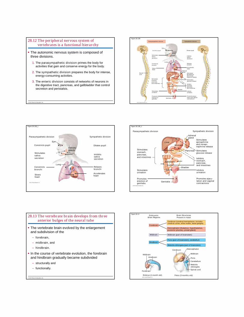
28.13 The vertebrate brain develops from threeanterior bulges of the neural tube
The vertebrate brain evolved by the enlargementand subdivision of the
– forebrain,
– midbrain, and
– hindbrain.
In the course of vertebrate evolution, the forebrainand hindbrain gradually became subdivided
– structurally and
– functionally.
© 2012 Pearson Education, Inc.
Figure 28.13Embryonic
Brain RegionsBrain StructuresPresent in Adult
Forebrain
Midbrain
Hindbrain
Hindbrain
Midbrain
Forebrain
Embryo (1 month old) Fetus (3 months old)
Cerebrum (cerebral hemispheres; includescerebral cortex, white matter, basal ganglia)
Diencephalon (thalamus, hypothalamus,posterior pituitary, pineal gland)
Pons (part of brainstem), cerebellum
Medulla oblongata (part of brainstem)
CerebrumDiencephalon
Midbrain
Pons
Cerebellum
Medullaoblongata
Spinal cord
Midbrain (part of brainstem)

In birds and mammals the cerebrum
– is much larger and
– correlates with their sophisticated behavior.
28.13 The vertebrate brain develops from threeanterior bulges of the neural tube
© 2012 Pearson Education, Inc.
THE HUMAN BRAIN
© 2012 Pearson Education, Inc.
28.14 The structure of a living supercomputer:The human brain
The human brain is
– more powerful than the most sophisticated computer and
– composed of three main parts:
1. forebrain,
2. midbrain, and
3. hindbrain.
© 2012 Pearson Education, Inc.
Figure 28.14A
Cerebral cortex(outer regionof cerebrum)
Cerebrum
Thalamus
Hypothalamus
Pituitary gland
Midbrain
Forebrain
Hindbrain
Pons
Medullaoblongata
Cerebellum
Spinalcord
The midbrain, subdivisions of the hindbrain, thethalamus, and the hypothalamus
– conduct information to and from higher brain centers,
– regulate homeostatic functions,
– keep track of body position, and
– sort sensory information.
28.14 The structure of a living supercomputer:The human brain
© 2012 Pearson Education, Inc.
Figure 28.14B
Left cerebralhemisphere
Right cerebralhemisphere
Thalamus
Basalnuclei
Medullaoblongata
Corpuscallosum
Cerebrum
Cerebellum

Table 28.14
The cerebrum is
– part of the forebrain and
– the largest and most complex part of the brain.
– Most of the cerebrum’s integrative power resides in thecerebral cortex of the two cerebral hemispheres.
28.14 The structure of a living supercomputer:The human brain
© 2012 Pearson Education, Inc.
28.15 The cerebral cortex is a mosaic ofspecialized, interactive regions
The cerebral cortex
– is less than 5 mm thick and
– accounts for 80% of the total human brain mass.
Specialized integrative regions of the cerebral cortexinclude
– the somatosensory cortex and
– centers for vision, hearing, taste, and smell.
© 2012 Pearson Education, Inc.
The motor cortex directs responses.
Association areas
– make up most of the cerebrum and
– are concerned with higher mental activities such asreasoning and language.
In a phenomenon known as lateralization, right andleft cerebral hemispheres tend to specialize indifferent mental tasks.
28.15 The cerebral cortex is a mosaic ofspecialized, interactive regions
© 2012 Pearson Education, Inc.
Figure 28.15
Frontal lobe Parietal lobe
Occipital lobeTemporal lobe
Frontalassociationarea
Speech
Smell
Speech
Moto
rco
rtex
Hearing
Reading
Vision
Visualassociationarea
Somatosensoryassociationarea
Auditoryassociationarea
So
mat
ose
nso
ryco
rtex
28.16 CONNECTION: Injuries and brainoperations provide insight into brainfunction
Brain injuries and surgeries reveal brain functions.
– After a 13-pound steel rod pierced his skull, PhineasGage appeared to have an intact intellect but hisassociates noted negative changes to his personality.
– Stimulation of the cerebral cortex during surgeries causedpatients to recall sensations and memories.
– Cutting the corpus callosum revealed information aboutbrain lateralization.
© 2012 Pearson Education, Inc.

Figure 28.16A Figure 28.16B
28.17 CONNECTION: fMRI scans provide insightinto brain structure and function
Functional magnetic resonance imaging (fMRI) is
– a scanning and imaging technology used to study brainfunctions,
– used on conscious patients,
– monitors changes in blood oxygen usage in the brain, and
– correlates to regions of intense brain function.
© 2012 Pearson Education, Inc.
Figure 28.17
28.18 Several parts of the brain regulate sleep andarousal
Sleep and arousal involve activity by the
– hypothalamus,
– medulla oblongata,
– pons, and
– neurons of the reticular formation.
© 2012 Pearson Education, Inc.
Sleep
– is essential for survival,
– is an active state, and
– may be involved in consolidating learning and memory.
28.18 Several parts of the brain regulate sleep andarousal
© 2012 Pearson Education, Inc.

28.19 The limbic system is involved in emotions,memory, and learning
The limbic system is
– a functional group of integrating centers in the
– cerebral cortex,
– thalamus,
– hypothalamus, and
– involved in
– emotions, such as nurturing infants and bonding emotionally toother people,
– memory, and
– learning.
© 2012 Pearson Education, Inc.
Figure 28.19
Cerebrum
HippocampusAmygdalaOlfactorybulb
Thalamus
Hypothalamus
Prefrontalcortex
Smell
28.20 CONNECTION: Changes in brainphysiology can produce neurologicaldisorders
Many neurological disorders can be linked tochanges in brain physiology, including
– schizophrenia,
– major depression,
– Alzheimer’s disease, and
– Parkinson’s disease.
© 2012 Pearson Education, Inc.
28.20 CONNECTION: Changes in brainphysiology can produce neurologicaldisorders
Schizophrenia is
– a severe mental disturbance and
– characterized by psychotic episodes in which patientslose the ability to distinguish reality.
© 2012 Pearson Education, Inc.
28.20 CONNECTION: Changes in brainphysiology can produce neurologicaldisorders
Depression
– Two broad forms of depressive illness have beenidentified:
1. major depression and
2. bipolar disorder, manic-depressive disorder.
– Treatments may include selective serotonin reuptakeinhibitors (SSRIs), which increase the amount of timeserotonin is available to stimulate certain neurons in thebrain.
© 2012 Pearson Education, Inc.
Figure 28.20A

Figure 28.20B
140
120
100
80
60
40
095 96
Year
Pre
scri
pti
on
s(m
illio
ns)
20
97 98 99 00 01 02 03 04 05 06 07 08
Alzheimer’s disease is
– characterized by confusion, memory loss, and personalitychanges and
– difficult to diagnose.
28.20 CONNECTION: Changes in brainphysiology can produce neurologicaldisorders
© 2012 Pearson Education, Inc.
Parkinson’s disease is
– a motor disorder and
– characterized by
– difficulty in initiating movements,
– slowness of movement, and
– rigidity.
28.20 CONNECTION: Changes in brainphysiology can produce neurologicaldisorders
© 2012 Pearson Education, Inc.
Figure 28.20C
1. Describe the structural and functional subdivisionsof the nervous system.
2. Describe the three parts of a reflex, distinguishingthe three types of neurons that may be involved inthe reaction.
3. Describe the structures and functions of neuronsand myelin sheaths.
4. Define a resting potential and explain how it iscreated.
You should now be able to
© 2012 Pearson Education, Inc.
5. Explain how an action potential is produced andthe resting membrane potential restored.
6. Explain how an action potential propagates itselfalong a neuron.
7. Compare the structures, functions, and locations ofelectrical and chemical synapses.
8. Compare excitatory and inhibitoryneurotransmitters.
9. Describe the types and functions ofneurotransmitters known in humans.
You should now be able to
© 2012 Pearson Education, Inc.

10. Explain how drugs can alter chemical synapses.
11. Describe the diversity of animal nervous systemsand provide examples.
12. Describe the general structure of the brain, spinalcord, and associated nerves of vertebrates.
13. Compare the functions of the motor nervoussystem and autonomic nervous system.
You should now be able to
© 2012 Pearson Education, Inc.
14. Compare the structures, functions, andinterrelationships of the parasympathetic,sympathetic, and enteric divisions of theperipheral nervous system.
15. Explain how the vertebrate brain develops from anembryonic tube.
16. Describe the main parts and functions of thehuman brain.
17. Explain how injuries, illness, and surgery provideinsight into the functions of the brain.
You should now be able to
© 2012 Pearson Education, Inc.
18. Explain how fMRI scans help us understand brainfunctions.
19. Explain how the brain regulates sleep andarousal.
20. Describe the structure and functions of the limbicsystem.
21. Describe the causes, symptoms, and treatmentsof schizophrenia, depression, Alzheimer’sdisease, and Parkinson’s disease.
You should now be able to
© 2012 Pearson Education, Inc.
Related Documents