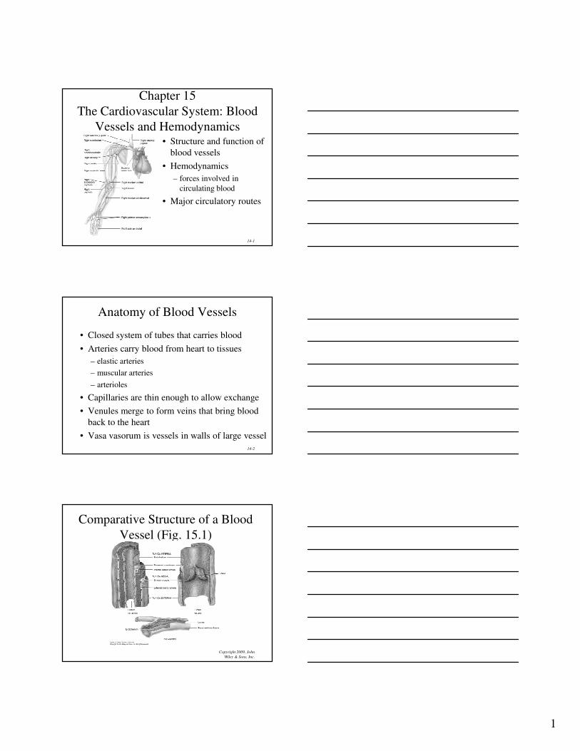
1 14-1 Chapter 15 The Cardiovascular System: Blood Vessels and Hemodynamics • Structure and function of blood vessels • Hemodynamics – forces involved in circulating blood • Major circulatory routes 14-2 Anatomy of Blood Vessels • Closed system of tubes that carries blood • Arteries carry blood from heart to tissues – elastic arteries – muscular arteries – arterioles • Capillaries are thin enough to allow exchange • Venules merge to form veins that bring blood back to the heart • Vasa vasorum is vessels in walls of large vessel Copyright 2009, John Wiley & Sons, Inc. Comparative Structure of a Blood Vessel (Fig. 15.1)

Welcome message from author
This document is posted to help you gain knowledge. Please leave a comment to let me know what you think about it! Share it to your friends and learn new things together.
Transcript
1
14-1
Chapter 15
The Cardiovascular System: Blood
Vessels and Hemodynamics• Structure and function of
blood vessels
• Hemodynamics
– forces involved in
circulating blood
• Major circulatory routes
14-2
Anatomy of Blood Vessels
• Closed system of tubes that carries blood
• Arteries carry blood from heart to tissues
– elastic arteries
– muscular arteries
– arterioles
• Capillaries are thin enough to allow exchange
• Venules merge to form veins that bring blood
back to the heart
• Vasa vasorum is vessels in walls of large vessel
Copyright 2009, John
Wiley & Sons, Inc.
Comparative Structure of a Blood
Vessel (Fig. 15.1)
2
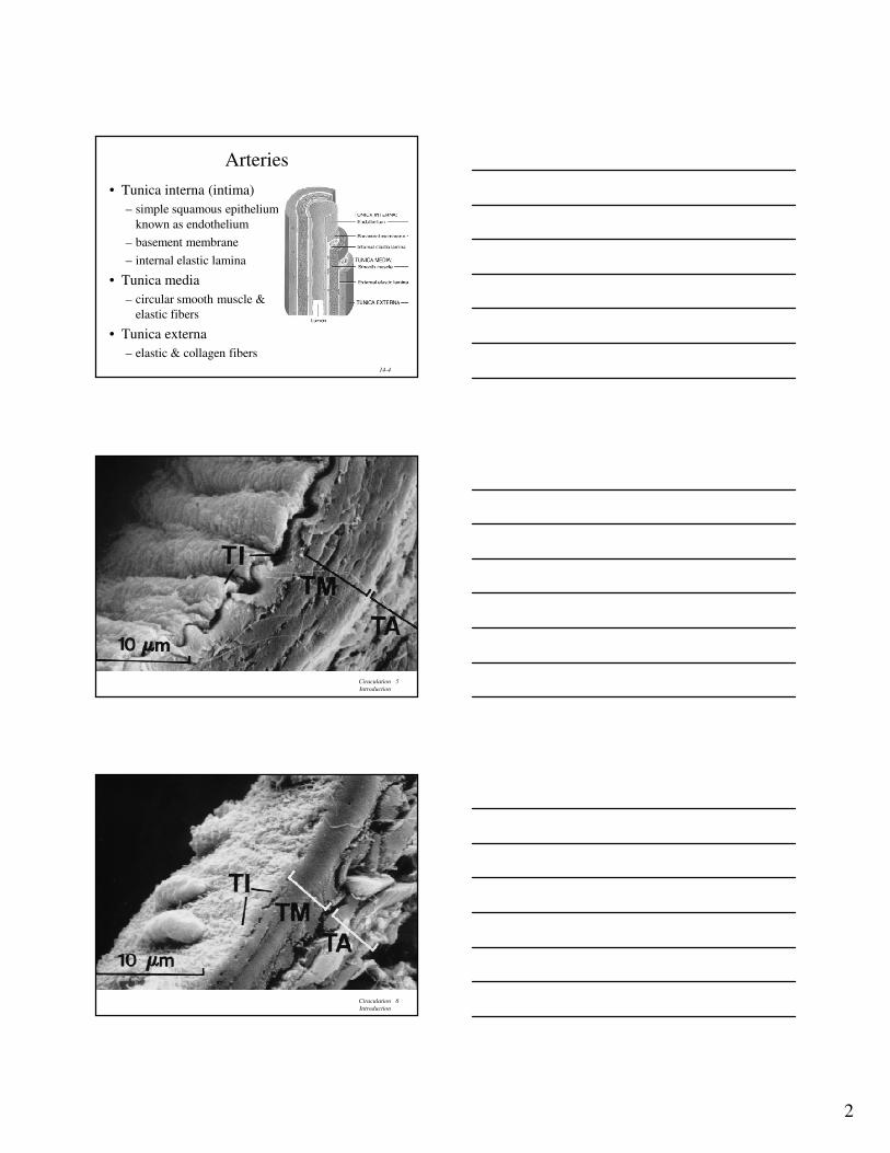
14-4
Arteries
• Tunica interna (intima)
– simple squamous epithelium
known as endothelium
– basement membrane
– internal elastic lamina
• Tunica media
– circular smooth muscle &
elastic fibers
• Tunica externa
– elastic & collagen fibers
Ciraculation
Introduction
5
Ciraculation
Introduction
6

3
Ciraculation
Introduction
7
Ciraculation
Introduction
8
14-9
Elastic Arteries• Largest-diameter arteries have lot of elastic fibers
in tunica media
• Help propel blood onward despite ventricular
relaxation (stretch and recoil -- pressure reservoir)

4
14-10
Muscular Arteries
• Medium-sized arteries with more muscle
than elastic fibers in tunica media
• Capable of greater vasoconstriction and
vasodilation to adjust rate of flow
– walls are relatively thick
– called distributing arteries because they direct
blood flow
14-11
Arterioles• Small arteries delivering blood
to capillaries
– tunica media containing few
layers of muscle
• Metarterioles form branches
into capillary bed
– to bypass capillary bed,
precapillary sphincters close &
blood flows out of bed in
thoroughfare channel
– vasomotion is intermittent
contraction & relaxation of
sphincters that allow filling of
capillary bed 5-10 times/minute
14-12
Capillaries form Microcirculation• Microscopic vessels that connect arterioles to venules
• Found near every cell in the body but more extensive in
highly active tissue (muscles, liver, kidneys & brain)
– entire capillary bed fills with blood when tissue is active
– lacking in epithelia, cornea and lens of eye & cartilage
• Function is exchange of nutrients & wastes between
blood and tissue fluid
• Structure is single layer of simple squamous epithelium
and its basement membrane
5
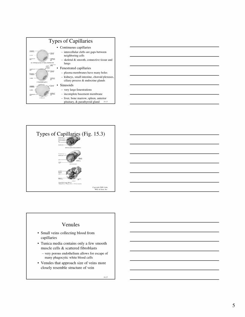
14-13
Types of Capillaries• Continuous capillaries
– intercellular clefts are gaps between
neighboring cells
– skeletal & smooth, connective tissue and
lungs
• Fenestrated capillaries
– plasma membranes have many holes
– kidneys, small intestine, choroid plexuses,
ciliary process & endocrine glands
• Sinusoids
– very large fenestrations
– incomplete basement membrane
– liver, bone marrow, spleen, anterior
pituitary, & parathyroid gland
Copyright 2009, John
Wiley & Sons, Inc.
Types of Capillaries (Fig. 15.3)
14-15
Venules
• Small veins collecting blood from
capillaries
• Tunica media contains only a few smooth
muscle cells & scattered fibroblasts
– very porous endothelium allows for escape of
many phagocytic white blood cells
• Venules that approach size of veins more
closely resemble structure of vein
6
14-16
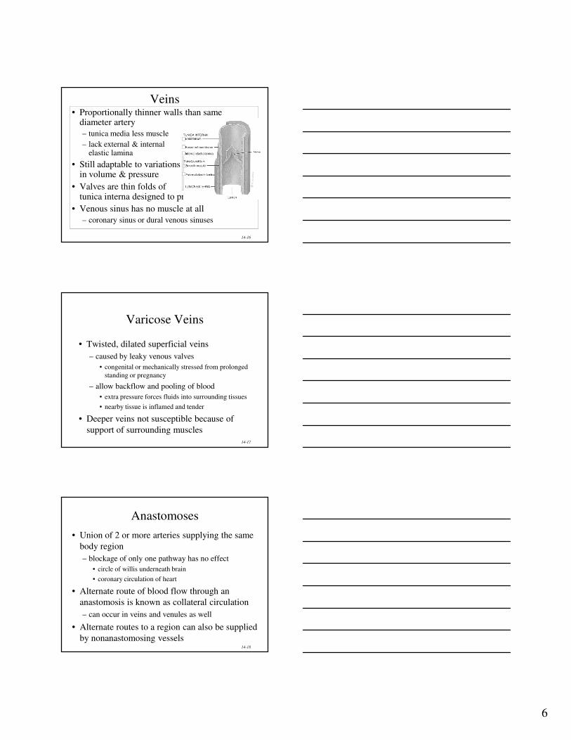
Veins• Proportionally thinner walls than same
diameter artery
– tunica media less muscle
– lack external & internalelastic lamina
• Still adaptable to variationsin volume & pressure
• Valves are thin folds of tunica interna designed to prevent backflow
• Venous sinus has no muscle at all
– coronary sinus or dural venous sinuses
14-17
Varicose Veins
• Twisted, dilated superficial veins
– caused by leaky venous valves
• congenital or mechanically stressed from prolonged
standing or pregnancy
– allow backflow and pooling of blood
• extra pressure forces fluids into surrounding tissues
• nearby tissue is inflamed and tender
• Deeper veins not susceptible because of
support of surrounding muscles
14-18
Anastomoses
• Union of 2 or more arteries supplying the same
body region
– blockage of only one pathway has no effect
• circle of willis underneath brain
• coronary circulation of heart
• Alternate route of blood flow through an
anastomosis is known as collateral circulation
– can occur in veins and venules as well
• Alternate routes to a region can also be supplied
by nonanastomosing vessels
7
14-19
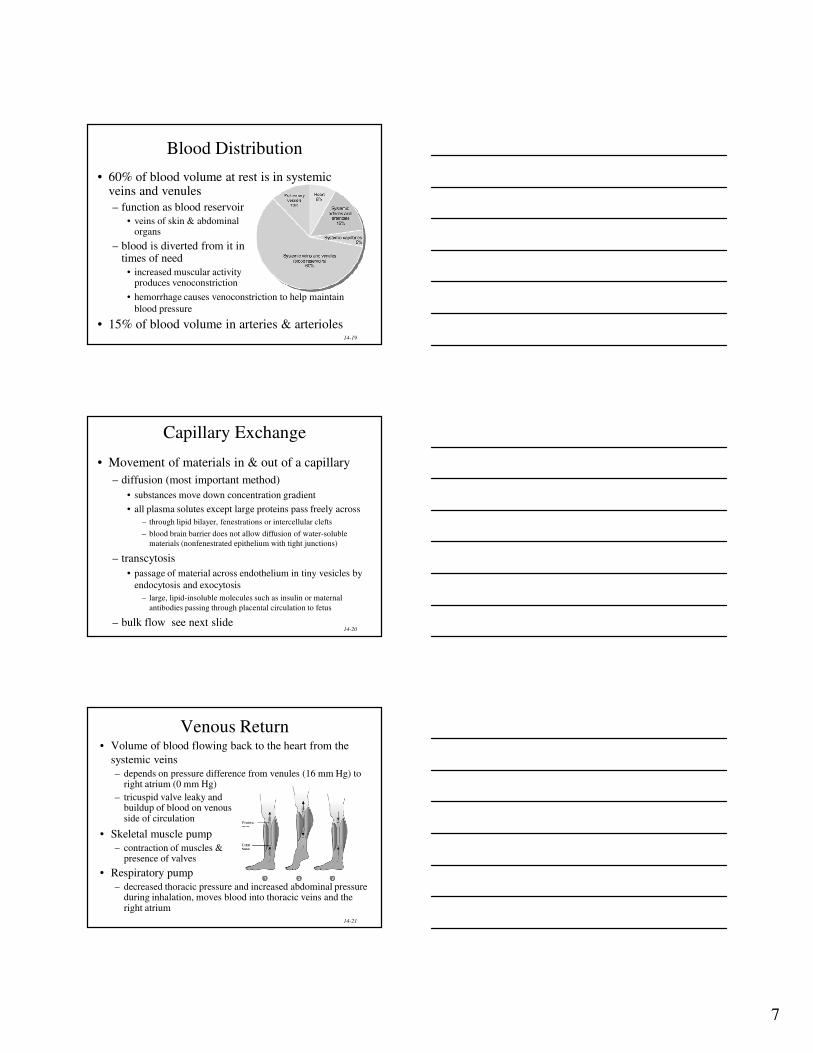
Blood Distribution
• 60% of blood volume at rest is in systemic veins and venules
– function as blood reservoir
• veins of skin & abdominalorgans
– blood is diverted from it intimes of need
• increased muscular activityproduces venoconstriction
• hemorrhage causes venoconstriction to help maintain
blood pressure
• 15% of blood volume in arteries & arterioles
14-20
Capillary Exchange
• Movement of materials in & out of a capillary
– diffusion (most important method)
• substances move down concentration gradient
• all plasma solutes except large proteins pass freely across
– through lipid bilayer, fenestrations or intercellular clefts
– blood brain barrier does not allow diffusion of water-soluble
materials (nonfenestrated epithelium with tight junctions)
– transcytosis
• passage of material across endothelium in tiny vesicles by
endocytosis and exocytosis
– large, lipid-insoluble molecules such as insulin or maternal
antibodies passing through placental circulation to fetus
– bulk flow see next slide
14-21
Venous Return• Volume of blood flowing back to the heart from the
systemic veins
– depends on pressure difference from venules (16 mm Hg) to right atrium (0 mm Hg)
– tricuspid valve leaky andbuildup of blood on venousside of circulation
• Skeletal muscle pump
– contraction of muscles & presence of valves
• Respiratory pump
– decreased thoracic pressure and increased abdominal pressure during inhalation, moves blood into thoracic veins and the right atrium

8
14-22
Syncope
• Fainting or a sudden, temporary loss of
consciousness not due to trauma
– due to cerebral ischemia or lack of blood flow to the
brain
• Causes
– vasodepressor syncope = sudden emotional stress
– situational syncope = pressure stress of coughing, defecation, or urination
– drug-induced syncope = antihypertensives, diuretics, vasodilators and tranquilizers
– orthostatic hypotension = decrease in BP upon standing
14-23
Pulse Points
Copyright 2009, John
Wiley & Sons, Inc.
Circulatory Routes
• The circulatory routes for blood flow are parallel.
• Each organ receives its own supply of freshly oxygenated blood.
The two basic routes for blood flow:
– The systemic circulation includes all the arteries and arterioles that
carry oxygenated blood from the left ventricle to systemic capillaries.
– The pulmonary circulation carries deoxygenated blood from the right
ventricle to the air sacs within the lungs and returns oxygenated blood
from the air sacs to the left atrium.
9
14-25
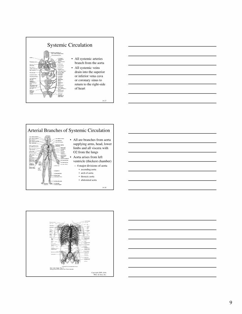
Systemic Circulation
• All systemic arteries
branch from the aorta
• All systemic veins
drain into the superior
or inferior vena cava
or coronary sinus to
return to the right-side
of heart
14-26
Arterial Branches of Systemic Circulation
• All are branches from aorta
supplying arms, head, lower
limbs and all viscera with
O2 from the lungs
• Aorta arises from left
ventricle (thickest chamber)
– 4 major divisions of aorta
• ascending aorta
• arch of aorta
• thoracic aorta
• abdominal aorta
Copyright 2009, John
Wiley & Sons, Inc.

10
14-28
Aorta and Its Superior Branches
• Aorta is largest artery of the body
– ascending aorta
• 2 coronary arteries supply myocardium
– arch of aorta -- branches to the arms & head• brachiocephalic trunk branches into right common carotid and right
subclavian
• left subclavian & left carotid arise independently
– thoracic aorta supplies branches to pericardium, esophagus, bronchi, diaphragm, intercostal & chest muscles, mammary gland, skin, vertebrae and spinal cord
Copyright 2009, John
Wiley & Sons, Inc.
Arch of Aorta and its Branches
Copyright 2009, John
Wiley & Sons, Inc.
Arch of Aorta and its Branches
11
14-31
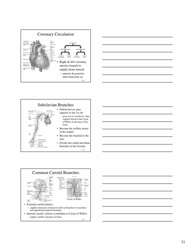
Coronary Circulation
• Right & left coronary
arteries branch to
supply heart muscle
– anterior & posterior
interventricular aa.
14-32
Subclavian Branches • Subclavian aa. pass
superior to the 1st rib
– gives rise to vertebral a. that
supplies blood to the Circle
of Willis on the base of the
brain
• Become the axillary artery
in the armpit
• Become the brachial in the
arm
• Divide into radial and ulnar
branches in the forearm
14-33
Common Carotid Branches
• External carotid arteries
– supplies structures external to skull as branches of maxillary and superficial temporal branches
• Internal carotid arteries (contribute to Circle of Willis)
– supply eyeballs and parts of brain
Circle of Willis

12
14-34
Abdominal Aorta and Its Branches
• Supplies abdominal & pelvic viscera & lower extremities
– celiac aa. supplies liver, stomach, spleen & pancreas
– superior & inferior mesenteric aa. supply intestines
– renal aa supply kidneys
– gonadal aa. supply ovaries
and testes
• Splits into common iliac
aa at 4th lumbar vertebrae
– external iliac aa supply
lower extremity
– internal iliac aa supply
pelvic viscera
Copyright 2009, John
Wiley & Sons, Inc.
Abdominal Aorta
• The abdominal aorta ends by
dividing into the right and left
common iliac arteries.
• These, divide into the internal
and external iliac arteries.
• The external iliacs become the
femoral arteries in the thighs,
the popliteal arteries posterior
to the knee, and the anterior
and posterior tibial arteries in
the legs.
14-36
Visceral Branches off Abdominal Aorta
• Celiac artery is first branch inferior to diaphragm
– left gastric artery, splenic artery, common hepatic artery
• Superior mesenteric artery lies in mesentery
– pancreaticoduodenal, jejunal, ileocolic, ascending & middle colic aa.
• Inferior mesenteric artery
– descending colon, sigmoid colon & rectal aa
13
14-37
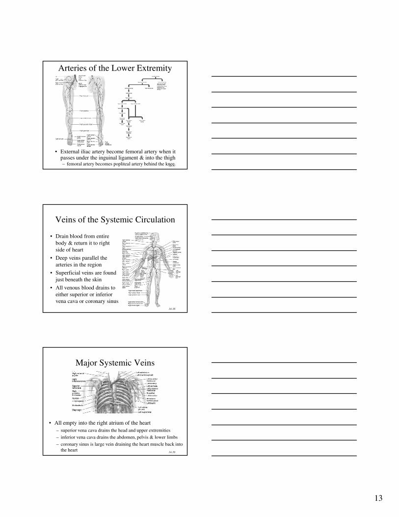
Arteries of the Lower Extremity
• External iliac artery become femoral artery when it passes under the inguinal ligament & into the thigh– femoral artery becomes popliteal artery behind the knee
14-38
Veins of the Systemic Circulation
• Drain blood from entire
body & return it to right
side of heart
• Deep veins parallel the
arteries in the region
• Superficial veins are found
just beneath the skin
• All venous blood drains to
either superior or inferior
vena cava or coronary sinus
14-39
Major Systemic Veins
• All empty into the right atrium of the heart
– superior vena cava drains the head and upper extremities
– inferior vena cava drains the abdomen, pelvis & lower limbs
– coronary sinus is large vein draining the heart muscle back into
the heart

14
14-40
Veins of the Head and Neck
• External and
Internal jugular
veins drain the
head and neck
into the superior
vena cava
• Dural venous
sinuses empty
into internal
jugular vein
Copyright 2009, John
Wiley & Sons, Inc.
Superficial and Deep Venous Return
from Upper Body
• Superficial veins are located
just deep to the skin and are
often visible.
• Deep veins are located deep in
the body. They usually
accompany arteries and have
the same names as the
corresponding arteries.
• Both superficial and deep veins
have valves, but valves are
more numerous in the deep
veins.
Copyright 2009, John
Wiley & Sons, Inc.
Veins of Right Upper Limb
15
Copyright 2009, John
Wiley & Sons, Inc.
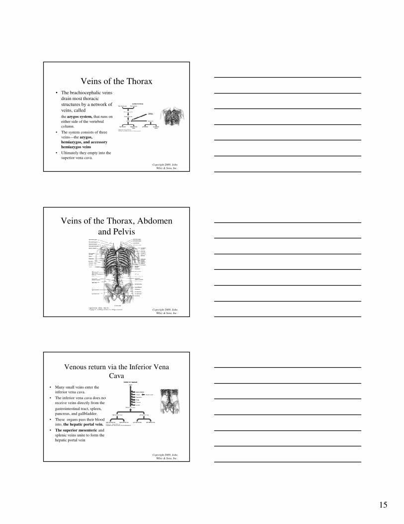
Veins of the Thorax
• The brachiocephalic veins
drain most thoracic
structures by a network of
veins, called
the azygos system, that runs on
either side of the vertebral
column.
• The system consists of three
veins—the azygos,
hemiazygos, and accessory
hemiazygos veins
• Ultimately they empty into the
superior vena cava.
Copyright 2009, John
Wiley & Sons, Inc.
Veins of the Thorax, Abdomen
and Pelvis
Copyright 2009, John
Wiley & Sons, Inc.
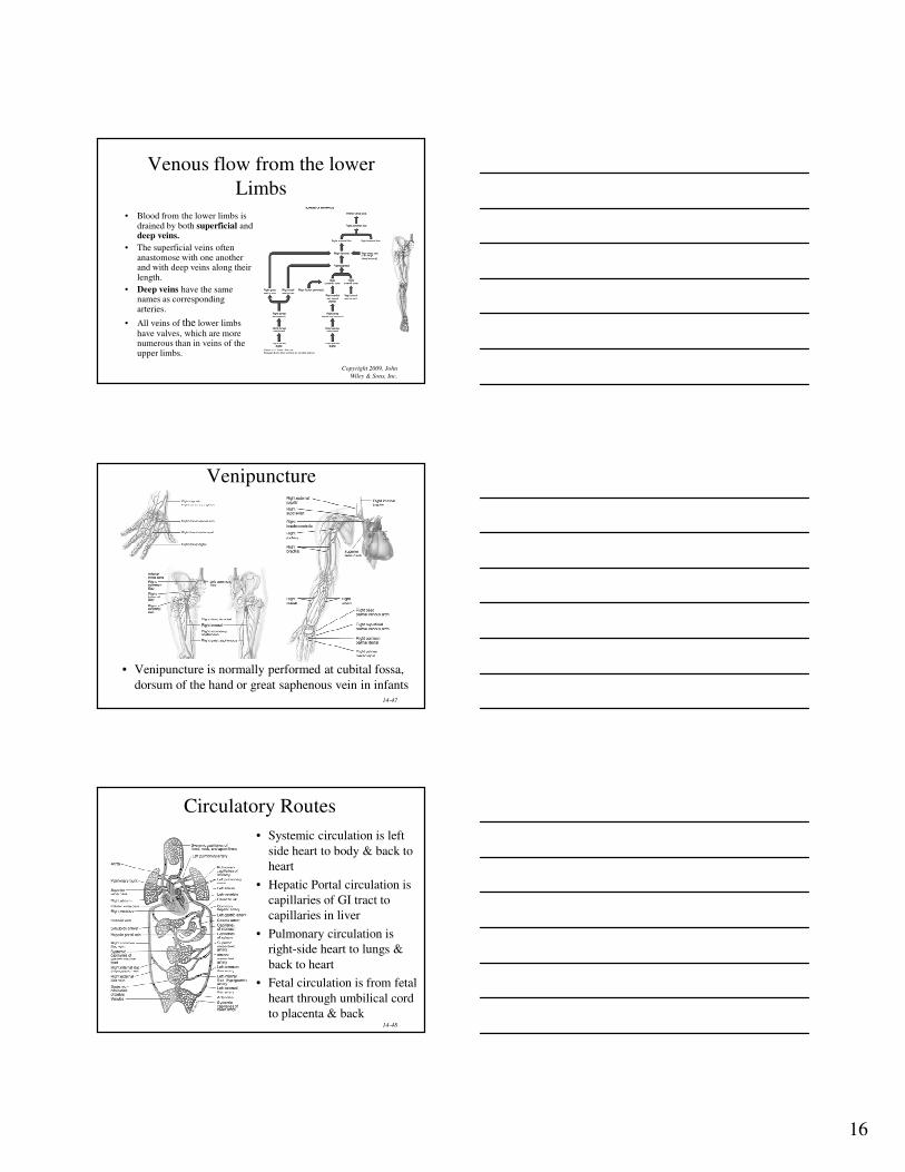
Venous return via the Inferior Vena
Cava
• Many small veins enter the
inferior vena cava.
• The inferior vena cava does not
receive veins directly from the
gastrointestinal tract, spleen,
pancreas, and gallbladder.
• These organs pass their blood
into, the hepatic portal vein.
• The superior mesenteric and
splenic veins unite to form the
hepatic portal vein
16
Copyright 2009, John
Wiley & Sons, Inc.
Venous flow from the lower
Limbs
• Blood from the lower limbs is drained by both superficial anddeep veins.
• The superficial veins often anastomose with one another and with deep veins along their length.
• Deep veins have the same names as corresponding arteries.
• All veins of the lower limbs
have valves, which are more numerous than in veins of the upper limbs.
14-47
Venipuncture
• Venipuncture is normally performed at cubital fossa,
dorsum of the hand or great saphenous vein in infants
14-48
Circulatory Routes
• Systemic circulation is left
side heart to body & back to
heart
• Hepatic Portal circulation is
capillaries of GI tract to
capillaries in liver
• Pulmonary circulation is
right-side heart to lungs &
back to heart
• Fetal circulation is from fetal
heart through umbilical cord
to placenta & back
17
14-49
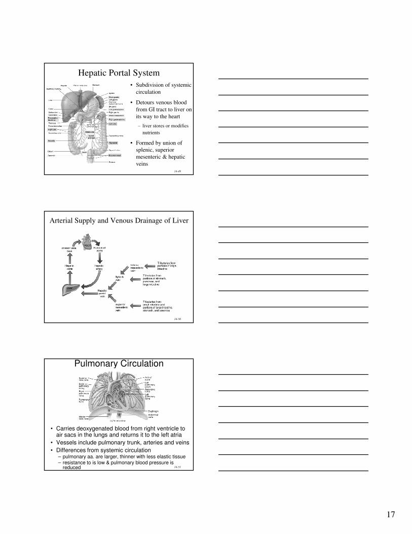
Hepatic Portal System
• Subdivision of systemic
circulation
• Detours venous blood
from GI tract to liver on
its way to the heart
– liver stores or modifies
nutrients
• Formed by union of
splenic, superior
mesenteric & hepatic
veins
14-50
Arterial Supply and Venous Drainage of Liver
14-51
Pulmonary Circulation
• Carries deoxygenated blood from right ventricle to air sacs in the lungs and returns it to the left atria
• Vessels include pulmonary trunk, arteries and veins
• Differences from systemic circulation– pulmonary aa. are larger, thinner with less elastic tissue– resistance to is low & pulmonary blood pressure is
reduced
18
14-52
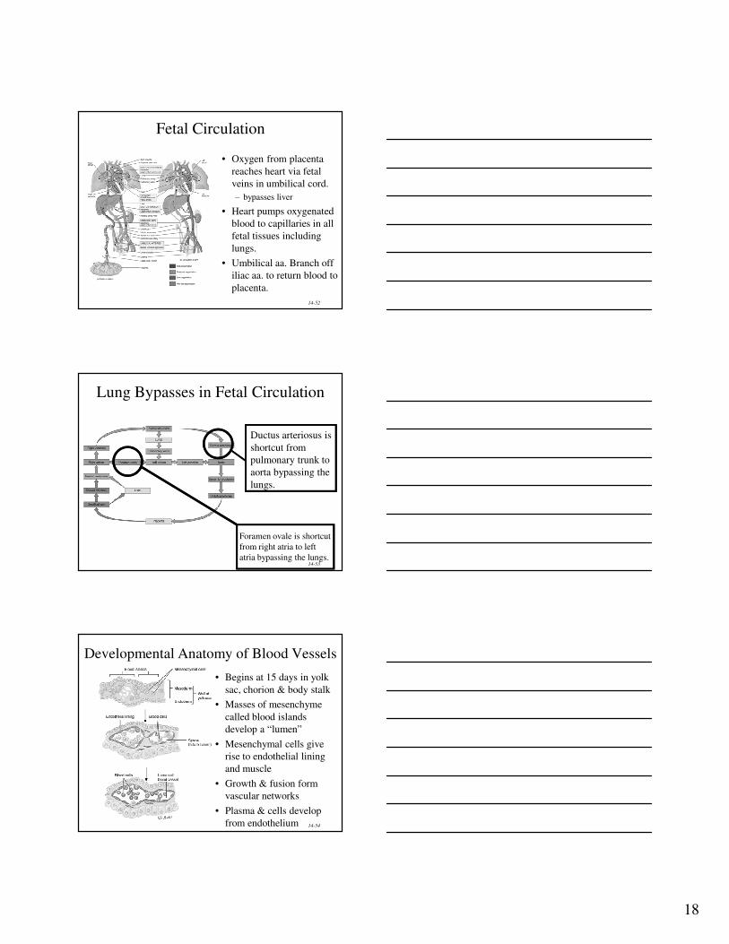
Fetal Circulation
• Oxygen from placenta
reaches heart via fetal
veins in umbilical cord.
– bypasses liver
• Heart pumps oxygenated
blood to capillaries in all
fetal tissues including
lungs.
• Umbilical aa. Branch off
iliac aa. to return blood to
placenta.
14-53
Ductus arteriosus is
shortcut from
pulmonary trunk to
aorta bypassing the
lungs.
Lung Bypasses in Fetal Circulation
Foramen ovale is shortcut
from right atria to left
atria bypassing the lungs.
14-54
Developmental Anatomy of Blood Vessels
• Begins at 15 days in yolk
sac, chorion & body stalk
• Masses of mesenchyme
called blood islands
develop a “lumen”
• Mesenchymal cells give
rise to endothelial lining
and muscle
• Growth & fusion form
vascular networks
• Plasma & cells develop
from endothelium
19
14-55
Aging and the Cardiovascular System
• General changes associated with aging
– decreased compliance of aorta
– reduction in cardiac muscle fiber size
– reduced cardiac output & maximum heart rate
– increase in systolic pressure
• Total cholesterol & LDL increases, HDL
decreases
• Congestive heart failure, coronary artery disease
and atherosclerosis more likely
Related Documents