Chapter 10 Biochemistry of the Genome Figure 10.1 Siblings within a family share some genes with each other and with each parent. Identical twins, however, are genetically identical. Bacteria like Escherichia coli may acquire genes encoding virulence factors, converting them into pathogenic strains, like this uropathogenic E. coli. (credit left: modification of work by Pellegrini C, Fargnoli MC, Suppa M, Peris K; credit right: modification of work by American Society for Microbiology) Chapter Outline 10.1 Using Microbiology to Discover the Secrets of Life 10.2 Structure and Function of DNA 10.3 Structure and Function of RNA 10.4 Structure and Function of Cellular Genomes Introduction Children inherit some characteristics from each parent. Siblings typically look similar to each other, but not exactly the same—except in the case of identical twins. How can we explain these phenomena? The answers lie in heredity (the transmission of traits from one generation to the next) and genetics (the science of heredity). Because humans reproduce sexually, 50% of a child’s genes come from the mother’s egg cell and the remaining 50% from the father’s sperm cell. Sperm and egg are formed through the process of meiosis, where DNA recombination occurs. Thus, there is no predictable pattern as to which 50% comes from which parent. Thus, siblings have only some genes, and their associated characteristics, in common. Identical twins are the exception, because they are genetically identical. Genetic differences among related microbes also dictate many observed biochemical and virulence differences. For example, some strains of the bacterium Escherichia coli are harmless members of the normal microbiota in the human gastrointestinal tract. Other strains of the same species have genes that give them the ability to cause disease. In bacteria, such genes are not inherited via sexual reproduction, as in humans. Often, they are transferred via plasmids, small circular pieces of double-stranded DNA that can be exchanged between prokaryotes. Chapter 10 | Biochemistry of the Genome 405

Welcome message from author
This document is posted to help you gain knowledge. Please leave a comment to let me know what you think about it! Share it to your friends and learn new things together.
Transcript

Chapter 10
Biochemistry of the Genome
Figure 10.1 Siblings within a family share some genes with each other and with each parent. Identical twins,however, are genetically identical. Bacteria like Escherichia coli may acquire genes encoding virulence factors,converting them into pathogenic strains, like this uropathogenic E. coli. (credit left: modification of work by PellegriniC, Fargnoli MC, Suppa M, Peris K; credit right: modification of work by American Society for Microbiology)
Chapter Outline
10.1 Using Microbiology to Discover the Secrets of Life
10.2 Structure and Function of DNA
10.3 Structure and Function of RNA
10.4 Structure and Function of Cellular Genomes
IntroductionChildren inherit some characteristics from each parent. Siblings typically look similar to each other, but not exactlythe same—except in the case of identical twins. How can we explain these phenomena? The answers lie in heredity(the transmission of traits from one generation to the next) and genetics (the science of heredity). Because humansreproduce sexually, 50% of a child’s genes come from the mother’s egg cell and the remaining 50% from the father’ssperm cell. Sperm and egg are formed through the process of meiosis, where DNA recombination occurs. Thus, thereis no predictable pattern as to which 50% comes from which parent. Thus, siblings have only some genes, and theirassociated characteristics, in common. Identical twins are the exception, because they are genetically identical.
Genetic differences among related microbes also dictate many observed biochemical and virulence differences. Forexample, some strains of the bacterium Escherichia coli are harmless members of the normal microbiota in the humangastrointestinal tract. Other strains of the same species have genes that give them the ability to cause disease. Inbacteria, such genes are not inherited via sexual reproduction, as in humans. Often, they are transferred via plasmids,small circular pieces of double-stranded DNA that can be exchanged between prokaryotes.
Chapter 10 | Biochemistry of the Genome 405
10.1 Using Microbiology to Discover the Secrets of Life
Learning Objectives• Describe the discovery of nucleic acid and nucleotides
• Explain the historical experiments that led to the characterization of DNA
• Describe how microbiology and microorganisms have been used to discover the biochemistry of genes
• Explain how scientists established the link between DNA and heredity
Through the early 20th century, DNA was not yet recognized as the genetic material responsible for heredity, thepassage of traits from one generation to the next. In fact, much of the research was dismissed until the mid-20thcentury. The scientific community believed, incorrectly, that the process of inheritance involved a blending of parentaltraits that produced an intermediate physical appearance in offspring; this hypothetical process appeared to be correctbecause of what we know now as continuous variation, which results from the action of many genes to determine aparticular characteristic, like human height. Offspring appear to be a “blend” of their parents’ traits when we look atcharacteristics that exhibit continuous variation. The blending theory of inheritance asserted that the original parentaltraits were lost or absorbed by the blending in the offspring, but we now know that this is not the case.
Two separate lines of research, begun in the mid to late 1800s, ultimately led to the discovery and characterization ofDNA and the foundations of genetics, the science of heredity. These lines of research began to converge in the 1920s,and research using microbial systems ultimately resulted in significant contributions to elucidating the molecular basisof genetics.
Discovery and Characterization of DNA
Modern understanding of DNA has evolved from the discovery of nucleic acid to the development of the double-helix model. In the 1860s, Friedrich Miescher (1844–1895), a physician by profession, was the first person to isolatephosphorus-rich chemicals from leukocytes (white blood cells) from the pus on used bandages from a local surgicalclinic. He named these chemicals (which would eventually be known as RNA and DNA) “nuclein” because they wereisolated from the nuclei of the cells. His student Richard Altmann (1852–1900) subsequently termed it “nucleic acid”20 years later when he discovered the acidic nature of nuclein. In the last two decades of the 19th century, Germanbiochemist Albrecht Kossel (1853–1927) isolated and characterized the five different nucleotide bases composingnucleic acid. These are adenine, guanine, cytosine, thymine (in DNA), and uracil (in RNA). Kossell received theNobel Prize in Physiology or Medicine in 1910 for his work on nucleic acids and for his considerable work onproteins, including the discovery of histidine.
Part 1
Alex is a 22-year-old college student who vacationed in Puerta Vallarta, Mexico, for spring break. Unfortunately,two days after flying home to Ohio, he began to experience abdominal cramping and extensive waterydiarrhea. Because of his discomfort, he sought medical attention at a large Cincinnati hospital nearby.
• What types of infections or other conditions may be responsible?
Jump to the next Clinical Focus box.
Clinical Focus
406 Chapter 10 | Biochemistry of the Genome
This OpenStax book is available for free at http://cnx.org/content/col12087/1.5

Foundations of Genetics
Despite the discovery of DNA in the late 1800s, scientists did not make the association with heredity for many moredecades. To make this connection, scientists, including a number of microbiologists, performed many experiments onplants, animals, and bacteria.
Mendel’s Pea Plants
While Miescher was isolating and discovering DNA in the 1860s, Austrian monk and botanist Johann Gregor Mendel(1822–1884) was experimenting with garden peas, demonstrating and documenting basic patterns of inheritance, nowknown as Mendel’s laws.
In 1856, Mendel began his decade-long research into inheritance patterns. He used the diploid garden pea, Pisumsativum, as his primary model system because it naturally self-fertilizes and is highly inbred, producing “true-breeding” pea plant lines—plants that always produce offspring that look like the parent. By experimenting withtrue-breeding pea plants, Mendel avoided the appearance of unexpected traits in offspring that might occur if heused plants that were not true-breeding. Mendel performed hybridizations, which involve mating two true-breedingindividuals (P generation) that have different traits, and examined the characteristics of their offspring (first filialgeneration, F1) as well as the offspring of self-fertilization of the F1 generation (second filial generation, F2) (Figure10.2).
Figure 10.2 In one of his experiments on inheritance patterns, Mendel crossed plants that were true- breeding forviolet flower color with plants true-breeding for white flower color (the P generation). The resulting hybrids in the F1generation all had violet flowers. In the F2 generation, approximately three-quarters of the plants had violet flowers,and one-quarter had white flowers.
In 1865, Mendel presented the results of his experiments with nearly 30,000 pea plants to the local natural historysociety. He demonstrated that traits are transmitted faithfully from parents to offspring independently of other traits.In 1866, he published his work, “Experiments in Plant Hybridization,”[1] in the Proceedings of the Natural HistorySociety of Brunn. Mendel’s work went virtually unnoticed by the scientific community, which believed, incorrectly,in the theory of blending of traits in continuous variation.
He was not recognized for his extraordinary scientific contributions during his lifetime. In fact, it was not until 1900that his work was rediscovered, reproduced, and revitalized by scientists on the brink of discovering the chromosomalbasis of heredity.
1. J.G. Mendel. “Versuche uber Pflanzenhybriden.” Verhandlungen des naturforschenden Vereines in Brunn, Bd. Abhandlungen 4
(1865):3–7. (For English translation, see http://www.mendelweb.org/Mendel.plain.html)
Chapter 10 | Biochemistry of the Genome 407
The Chromosomal Theory of Inheritance
Mendel carried out his experiments long before chromosomes were visualized under a microscope. However, withthe improvement of microscopic techniques during the late 1800s, cell biologists could stain and visualize subcellularstructures with dyes and observe their actions during meiosis. They were able to observe chromosomes replicating,condensing from an amorphous nuclear mass into distinct X-shaped bodies and migrating to separate cellular poles.The speculation that chromosomes might be the key to understanding heredity led several scientists to examineMendel’s publications and re-evaluate his model in terms of the behavior of chromosomes during mitosis and meiosis.
In 1902, Theodor Boveri (1862–1915) observed that in sea urchins, nuclear components (chromosomes) determinedproper embryonic development. That same year, Walter Sutton (1877–1916) observed the separation of chromosomesinto daughter cells during meiosis. Together, these observations led to the development of the Chromosomal Theoryof Inheritance, which identified chromosomes as the genetic material responsible for Mendelian inheritance.
Despite compelling correlations between the behavior of chromosomes during meiosis and Mendel’s observations, theChromosomal Theory of Inheritance was proposed long before there was any direct evidence that traits were carriedon chromosomes. Thomas Hunt Morgan (1866–1945) and his colleagues spent several years carrying out crosses withthe fruit fly, Drosophila melanogaster. They performed meticulous microscopic observations of fly chromosomes andcorrelated these observations with resulting fly characteristics. Their work provided the first experimental evidence tosupport the Chromosomal Theory of Inheritance in the early 1900s. In 1915, Morgan and his “Fly Room” colleaguespublished The Mechanism of Mendelian Heredity, which identified chromosomes as the cellular structures responsiblefor heredity. For his many significant contributions to genetics, Morgan received the Nobel Prize in Physiology orMedicine in 1933.
In the late 1920s, Barbara McClintock (1902–1992) developed chromosomal staining techniques to visualize anddifferentiate between the different chromosomes of maize (corn). In the 1940s and 1950s, she identified a breakageevent on chromosome 9, which she named the dissociation locus (Ds). Ds could change position within thechromosome. She also identified an activator locus (Ac). Ds chromosome breakage could be activated by an Acelement (transposase enzyme). At first, McClintock’s finding of these jumping genes, which we now call transposons,was not accepted by the scientific community. It wasn’t until the 1960s and later that transposons were discoveredin bacteriophages, bacteria, and Drosophila. Today, we know that transposons are mobile segments of DNA that canmove within the genome of an organism. They can regulate gene expression, protein expression, and virulence (abilityto cause disease).
Microbes and Viruses in Genetic Research
Microbiologists have also played a crucial part in our understanding of genetics. Experimental organisms such asMendel’s garden peas, Morgan’s fruit flies, and McClintock’s corn had already been used successfully to pave theway for an understanding of genetics. However, microbes and viruses were (and still are) excellent model systemsfor the study of genetics because, unlike peas, fruit flies, and corn, they are propagated more easily in the laboratory,growing to high population densities in a small amount of space and in a short time. In addition, because of theirstructural simplicity, microbes and viruses are more readily manipulated genetically.
Fortunately, despite significant differences in size, structure, reproduction strategies, and other biologicalcharacteristics, there is biochemical unity among all organisms; they have in common the same underlying moleculesresponsible for heredity and the use of genetic material to give cells their varying characteristics. In the words ofFrench scientist Jacques Monod, “What is true for E. coli is also true for the elephant,” meaning that the biochemistryof life has been maintained throughout evolution and is shared in all forms of life, from simple unicellular organismsto large, complex organisms. This biochemical continuity makes microbes excellent models to use for genetic studies.
In a clever set of experiments in the 1930s and 1940s, German scientist Joachim Hämmerling (1901–1980), usingthe single-celled alga Acetabularia as a microbial model, established that the genetic information in a eukaryotic cellis housed within the nucleus. Acetabularia spp. are unusually large algal cells that grow asymmetrically, forming a“foot” containing the nucleus, which is used for substrate attachment; a stalk; and an umbrella-like cap—structuresthat can all be easily seen with the naked eye. In an early set of experiments, Hämmerling removed either the cap or
408 Chapter 10 | Biochemistry of the Genome
This OpenStax book is available for free at http://cnx.org/content/col12087/1.5
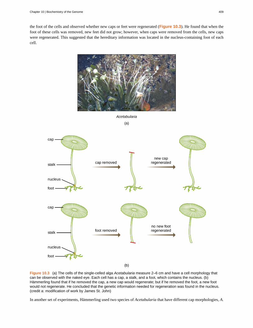
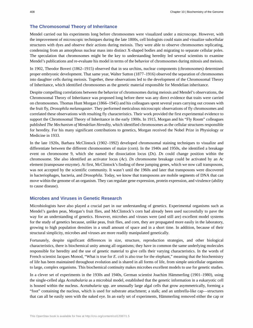
the foot of the cells and observed whether new caps or feet were regenerated (Figure 10.3). He found that when thefoot of these cells was removed, new feet did not grow; however, when caps were removed from the cells, new capswere regenerated. This suggested that the hereditary information was located in the nucleus-containing foot of eachcell.
Figure 10.3 (a) The cells of the single-celled alga Acetabularia measure 2–6 cm and have a cell morphology thatcan be observed with the naked eye. Each cell has a cap, a stalk, and a foot, which contains the nucleus. (b)Hämmerling found that if he removed the cap, a new cap would regenerate; but if he removed the foot, a new footwould not regenerate. He concluded that the genetic information needed for regeneration was found in the nucleus.(credit a: modification of work by James St. John)
In another set of experiments, Hämmerling used two species of Acetabularia that have different cap morphologies, A.
Chapter 10 | Biochemistry of the Genome 409
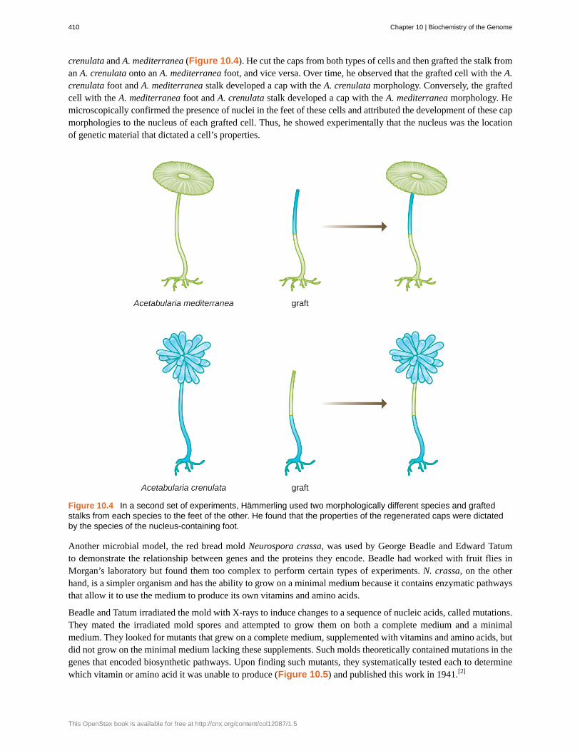
crenulata and A. mediterranea (Figure 10.4). He cut the caps from both types of cells and then grafted the stalk froman A. crenulata onto an A. mediterranea foot, and vice versa. Over time, he observed that the grafted cell with the A.crenulata foot and A. mediterranea stalk developed a cap with the A. crenulata morphology. Conversely, the graftedcell with the A. mediterranea foot and A. crenulata stalk developed a cap with the A. mediterranea morphology. Hemicroscopically confirmed the presence of nuclei in the feet of these cells and attributed the development of these capmorphologies to the nucleus of each grafted cell. Thus, he showed experimentally that the nucleus was the locationof genetic material that dictated a cell’s properties.
Figure 10.4 In a second set of experiments, Hämmerling used two morphologically different species and graftedstalks from each species to the feet of the other. He found that the properties of the regenerated caps were dictatedby the species of the nucleus-containing foot.
Another microbial model, the red bread mold Neurospora crassa, was used by George Beadle and Edward Tatumto demonstrate the relationship between genes and the proteins they encode. Beadle had worked with fruit flies inMorgan’s laboratory but found them too complex to perform certain types of experiments. N. crassa, on the otherhand, is a simpler organism and has the ability to grow on a minimal medium because it contains enzymatic pathwaysthat allow it to use the medium to produce its own vitamins and amino acids.
Beadle and Tatum irradiated the mold with X-rays to induce changes to a sequence of nucleic acids, called mutations.They mated the irradiated mold spores and attempted to grow them on both a complete medium and a minimalmedium. They looked for mutants that grew on a complete medium, supplemented with vitamins and amino acids, butdid not grow on the minimal medium lacking these supplements. Such molds theoretically contained mutations in thegenes that encoded biosynthetic pathways. Upon finding such mutants, they systematically tested each to determinewhich vitamin or amino acid it was unable to produce (Figure 10.5) and published this work in 1941.[2]
410 Chapter 10 | Biochemistry of the Genome
This OpenStax book is available for free at http://cnx.org/content/col12087/1.5

Figure 10.5 Beadle and Tatum’s experiment involved the mating of irradiated and nonirradiated mold spores. Thesespores were grown on both complete medium and a minimal medium to determine which amino acid or vitamin themutant was unable to produce on its own.
Subsequent work by Beadle, Tatum, and colleagues showed that they could isolate different classes of mutants thatrequired a particular supplement, like the amino acid arginine (Figure 10.6). With some knowledge of the argininebiosynthesis pathway, they identified three classes of arginine mutants by supplementing the minimal medium withintermediates (citrulline or ornithine) in the pathway. The three mutants differed in their abilities to grow in each of themedia, which led the group of scientists to propose, in 1945, that each type of mutant had a defect in a different genein the arginine biosynthesis pathway. This led to the so-called one gene–one enzyme hypothesis, which suggested thateach gene encodes one enzyme.
Subsequent knowledge about the processes of transcription and translation led scientists to revise this to the “onegene–one polypeptide” hypothesis. Although there are some genes that do not encode polypeptides (but rather encodefor transfer RNAs [tRNAs] or ribosomal RNAs [rRNAs], which we will discuss later), the one gene–one enzymehypothesis is true in many cases, especially in microbes. Beadle and Tatum’s discovery of the link between genes andcorresponding characteristics earned them the 1958 Nobel Prize in Physiology and Medicine and has since becomethe basis for modern molecular genetics.
2. G.W. Beadle, E.L. Tatum. “Genetic Control of Biochemical Reactions in Neurospora.” Proceedings of the National Academy of Sciences
27 no. 11 (1941):499–506.
Chapter 10 | Biochemistry of the Genome 411
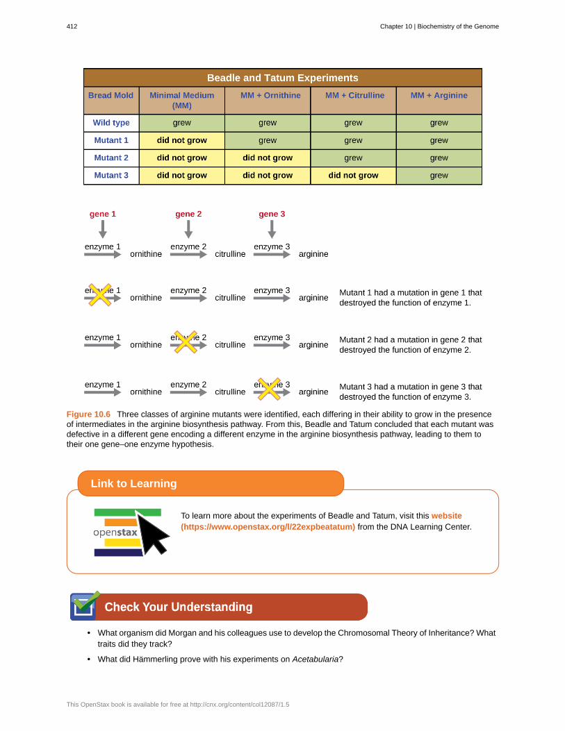
Figure 10.6 Three classes of arginine mutants were identified, each differing in their ability to grow in the presenceof intermediates in the arginine biosynthesis pathway. From this, Beadle and Tatum concluded that each mutant wasdefective in a different gene encoding a different enzyme in the arginine biosynthesis pathway, leading to them totheir one gene–one enzyme hypothesis.
To learn more about the experiments of Beadle and Tatum, visit this website(https://www.openstax.org/l/22expbeatatum) from the DNA Learning Center.
• What organism did Morgan and his colleagues use to develop the Chromosomal Theory of Inheritance? Whattraits did they track?
• What did Hämmerling prove with his experiments on Acetabularia?
Link to Learning
412 Chapter 10 | Biochemistry of the Genome
This OpenStax book is available for free at http://cnx.org/content/col12087/1.5

DNA as the Molecule Responsible for Heredity
By the beginning of the 20th century, a great deal of work had already been done on characterizing DNA andestablishing the foundations of genetics, including attributing heredity to chromosomes found within the nucleus.Despite all of this research, it was not until well into the 20th century that these lines of research converged andscientists began to consider that DNA could be the genetic material that offspring inherited from their parents. DNA,containing only four different nucleotides, was thought to be structurally too simple to encode such complex geneticinformation. Instead, protein was thought to have the complexity required to serve as cellular genetic informationbecause it is composed of 20 different amino acids that could be combined in a huge variety of combinations.Microbiologists played a pivotal role in the research that determined that DNA is the molecule responsible forheredity.
Griffith’s Transformation Experiments
British bacteriologist Frederick Griffith (1879–1941) was perhaps the first person to show that hereditary informationcould be transferred from one cell to another “horizontally” (between members of the same generation), rather than“vertically” (from parent to offspring). In 1928, he reported the first demonstration of bacterial transformation, aprocess in which external DNA is taken up by a cell, thereby changing its characteristics.[3] He was working withtwo strains of Streptococcus pneumoniae, a bacterium that causes pneumonia: a rough (R) strain and a smooth (S)strain. The R strain is nonpathogenic and lacks a capsule on its outer surface; as a result, colonies from the R strainappear rough when grown on plates. The S strain is pathogenic and has a capsule outside its cell wall, allowing itto escape phagocytosis by the host immune system. The capsules cause colonies from the S strain to appear smoothwhen grown on plates.
In a series of experiments, Griffith analyzed the effects of live R, live S, and heat-killed S strains of S. pneumoniae onlive mice (Figure 10.7). When mice were injected with the live S strain, the mice died. When he injected the micewith the live R strain or the heat-killed S strain, the mice survived. But when he injected the mice with a mixture oflive R strain and heat-killed S strain, the mice died. Upon isolating the live bacteria from the dead mouse, he onlyrecovered the S strain of bacteria. When he then injected this isolated S strain into fresh mice, the mice died. Griffithconcluded that something had passed from the heat-killed S strain into the live R strain and “transformed” it intothe pathogenic S strain; he called this the “transforming principle.” These experiments are now famously known asGriffith’s transformation experiments.
3. F. Griffith. “The Significance of Pneumococcal Types.” Journal of Hygiene 27 no. 2 (1928):8–159.
Chapter 10 | Biochemistry of the Genome 413
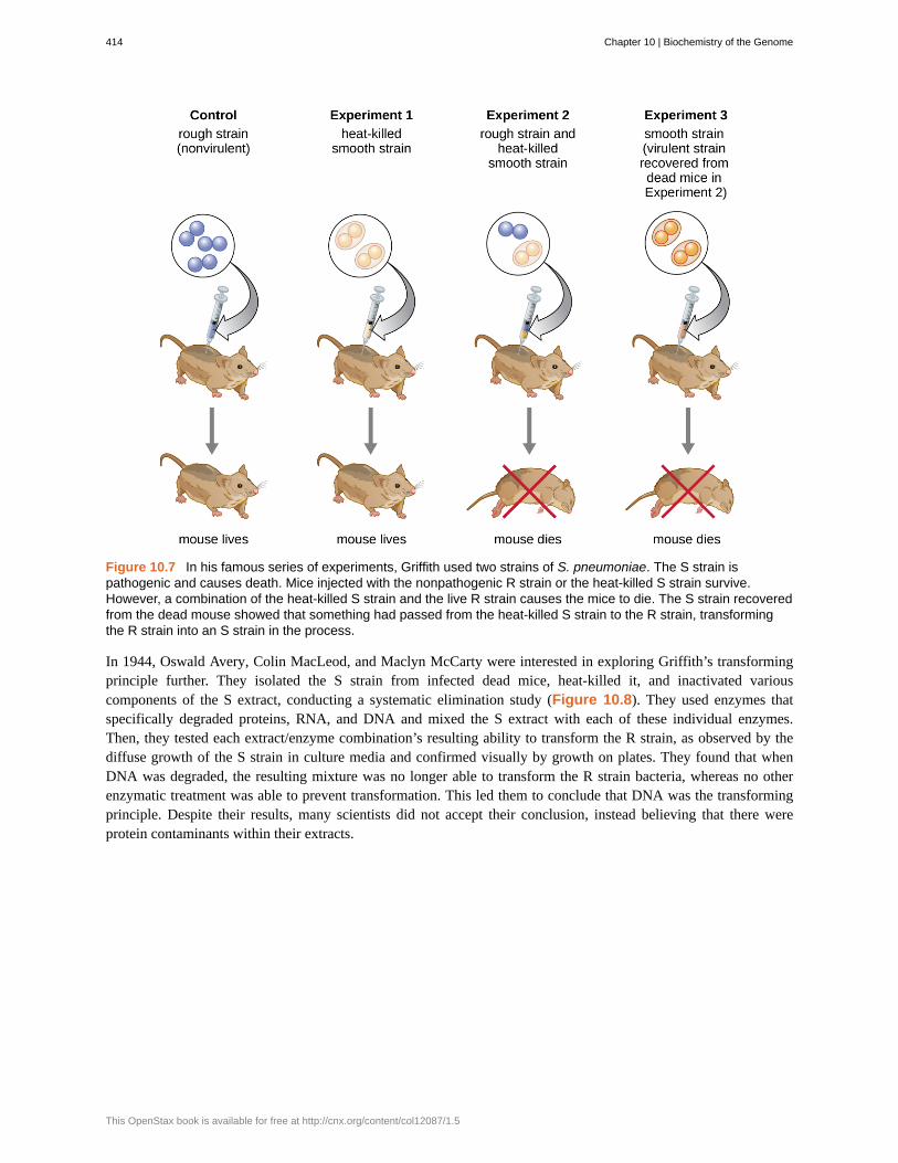
Figure 10.7 In his famous series of experiments, Griffith used two strains of S. pneumoniae. The S strain ispathogenic and causes death. Mice injected with the nonpathogenic R strain or the heat-killed S strain survive.However, a combination of the heat-killed S strain and the live R strain causes the mice to die. The S strain recoveredfrom the dead mouse showed that something had passed from the heat-killed S strain to the R strain, transformingthe R strain into an S strain in the process.
In 1944, Oswald Avery, Colin MacLeod, and Maclyn McCarty were interested in exploring Griffith’s transformingprinciple further. They isolated the S strain from infected dead mice, heat-killed it, and inactivated variouscomponents of the S extract, conducting a systematic elimination study (Figure 10.8). They used enzymes thatspecifically degraded proteins, RNA, and DNA and mixed the S extract with each of these individual enzymes.Then, they tested each extract/enzyme combination’s resulting ability to transform the R strain, as observed by thediffuse growth of the S strain in culture media and confirmed visually by growth on plates. They found that whenDNA was degraded, the resulting mixture was no longer able to transform the R strain bacteria, whereas no otherenzymatic treatment was able to prevent transformation. This led them to conclude that DNA was the transformingprinciple. Despite their results, many scientists did not accept their conclusion, instead believing that there wereprotein contaminants within their extracts.
414 Chapter 10 | Biochemistry of the Genome
This OpenStax book is available for free at http://cnx.org/content/col12087/1.5

Figure 10.8 Oswald Avery, Colin MacLeod, and Maclyn McCarty followed up on Griffith’s experiment andexperimentally determined that the transforming principle was DNA.
Chapter 10 | Biochemistry of the Genome 415

• How did Avery, MacLeod, and McCarty’s experiments show that DNA was the transforming principle firstdescribed by Griffith?
Hershey and Chase’s Proof of DNA as Genetic Material
Alfred Hershey and Martha Chase performed their own experiments in 1952 and were able to provide confirmatoryevidence that DNA, not protein, was the genetic material (Figure 10.9).[4] Hershey and Chase were studying abacteriophage, a virus that infects bacteria. Viruses typically have a simple structure: a protein coat, called thecapsid, and a nucleic acid core that contains the genetic material, either DNA or RNA (see Viruses). The particularbacteriophage they were studying was the T2 bacteriophage, which infects E. coli cells. As we now know today, T2attaches to the surface of the bacterial cell and then it injects its nucleic acids inside the cell. The phage DNA makesmultiple copies of itself using the host machinery, and eventually the host cell bursts, releasing a large number ofbacteriophages.
Hershey and Chase labeled the protein coat in one batch of phage using radioactive sulfur, 35S, because sulfur is foundin the amino acids methionine and cysteine but not in nucleic acids. They labeled the DNA in another batch usingradioactive phosphorus, 32P, because phosphorus is found in DNA and RNA but not typically in protein.
Each batch of phage was allowed to infect the cells separately. After infection, Hershey and Chase put each phagebacterial suspension in a blender, which detached the phage coats from the host cell, and spun down the resultingsuspension in a centrifuge. The heavier bacterial cells settled down and formed a pellet, whereas the lighter phageparticles stayed in the supernatant. In the tube with the protein labeled, the radioactivity remained only in thesupernatant. In the tube with the DNA labeled, the radioactivity was detected only in the bacterial cells. Hershey andChase concluded that it was the phage DNA that was injected into the cell that carried the information to producemore phage particles, thus proving that DNA, not proteins, was the source of the genetic material. As a result of theirwork, the scientific community more broadly accepted DNA as the molecule responsible for heredity.
4. A.D. Hershey, M. Chase. “Independent Functions of Viral Protein and Nucleic Acid in Growth of Bacteriophage.” Journal of General
Physiology 36 no. 1 (1952):39–56.
416 Chapter 10 | Biochemistry of the Genome
This OpenStax book is available for free at http://cnx.org/content/col12087/1.5
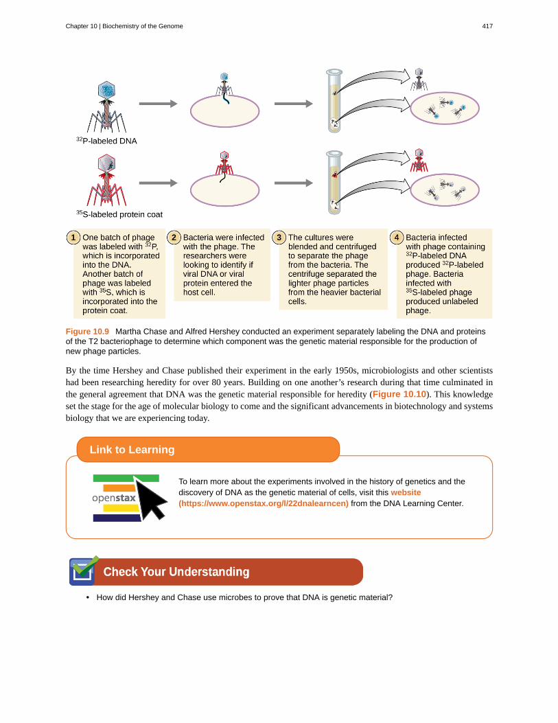
Figure 10.9 Martha Chase and Alfred Hershey conducted an experiment separately labeling the DNA and proteinsof the T2 bacteriophage to determine which component was the genetic material responsible for the production ofnew phage particles.
By the time Hershey and Chase published their experiment in the early 1950s, microbiologists and other scientistshad been researching heredity for over 80 years. Building on one another’s research during that time culminated inthe general agreement that DNA was the genetic material responsible for heredity (Figure 10.10). This knowledgeset the stage for the age of molecular biology to come and the significant advancements in biotechnology and systemsbiology that we are experiencing today.
To learn more about the experiments involved in the history of genetics and thediscovery of DNA as the genetic material of cells, visit this website(https://www.openstax.org/l/22dnalearncen) from the DNA Learning Center.
• How did Hershey and Chase use microbes to prove that DNA is genetic material?
Link to Learning
Chapter 10 | Biochemistry of the Genome 417
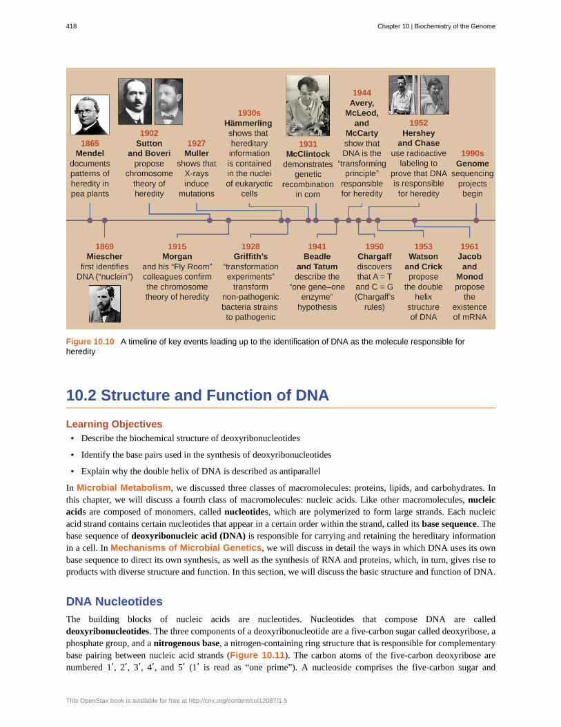
Figure 10.10 A timeline of key events leading up to the identification of DNA as the molecule responsible forheredity
10.2 Structure and Function of DNA
Learning Objectives• Describe the biochemical structure of deoxyribonucleotides
• Identify the base pairs used in the synthesis of deoxyribonucleotides
• Explain why the double helix of DNA is described as antiparallel
In Microbial Metabolism, we discussed three classes of macromolecules: proteins, lipids, and carbohydrates. Inthis chapter, we will discuss a fourth class of macromolecules: nucleic acids. Like other macromolecules, nucleicacids are composed of monomers, called nucleotides, which are polymerized to form large strands. Each nucleicacid strand contains certain nucleotides that appear in a certain order within the strand, called its base sequence. Thebase sequence of deoxyribonucleic acid (DNA) is responsible for carrying and retaining the hereditary informationin a cell. In Mechanisms of Microbial Genetics, we will discuss in detail the ways in which DNA uses its ownbase sequence to direct its own synthesis, as well as the synthesis of RNA and proteins, which, in turn, gives rise toproducts with diverse structure and function. In this section, we will discuss the basic structure and function of DNA.
DNA Nucleotides
The building blocks of nucleic acids are nucleotides. Nucleotides that compose DNA are calleddeoxyribonucleotides. The three components of a deoxyribonucleotide are a five-carbon sugar called deoxyribose, aphosphate group, and a nitrogenous base, a nitrogen-containing ring structure that is responsible for complementarybase pairing between nucleic acid strands (Figure 10.11). The carbon atoms of the five-carbon deoxyribose arenumbered 1ʹ, 2ʹ, 3ʹ, 4ʹ, and 5ʹ (1ʹ is read as “one prime”). A nucleoside comprises the five-carbon sugar and
418 Chapter 10 | Biochemistry of the Genome
This OpenStax book is available for free at http://cnx.org/content/col12087/1.5
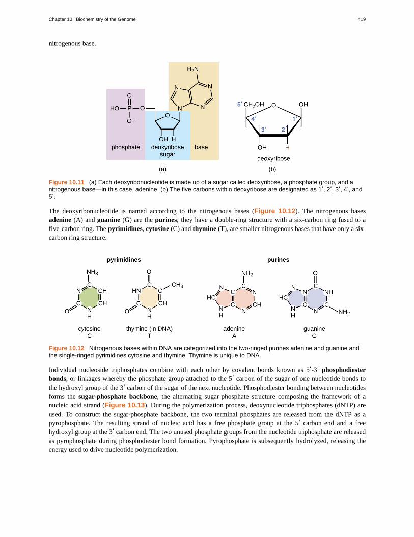
nitrogenous base.
Figure 10.11 (a) Each deoxyribonucleotide is made up of a sugar called deoxyribose, a phosphate group, and anitrogenous base—in this case, adenine. (b) The five carbons within deoxyribose are designated as 1ʹ, 2ʹ, 3ʹ, 4ʹ, and5ʹ.
The deoxyribonucleotide is named according to the nitrogenous bases (Figure 10.12). The nitrogenous basesadenine (A) and guanine (G) are the purines; they have a double-ring structure with a six-carbon ring fused to afive-carbon ring. The pyrimidines, cytosine (C) and thymine (T), are smaller nitrogenous bases that have only a six-carbon ring structure.
Figure 10.12 Nitrogenous bases within DNA are categorized into the two-ringed purines adenine and guanine andthe single-ringed pyrimidines cytosine and thymine. Thymine is unique to DNA.
Individual nucleoside triphosphates combine with each other by covalent bonds known as 5ʹ-3ʹ phosphodiesterbonds, or linkages whereby the phosphate group attached to the 5ʹ carbon of the sugar of one nucleotide bonds tothe hydroxyl group of the 3ʹ carbon of the sugar of the next nucleotide. Phosphodiester bonding between nucleotidesforms the sugar-phosphate backbone, the alternating sugar-phosphate structure composing the framework of anucleic acid strand (Figure 10.13). During the polymerization process, deoxynucleotide triphosphates (dNTP) areused. To construct the sugar-phosphate backbone, the two terminal phosphates are released from the dNTP as apyrophosphate. The resulting strand of nucleic acid has a free phosphate group at the 5ʹ carbon end and a freehydroxyl group at the 3ʹ carbon end. The two unused phosphate groups from the nucleotide triphosphate are releasedas pyrophosphate during phosphodiester bond formation. Pyrophosphate is subsequently hydrolyzed, releasing theenergy used to drive nucleotide polymerization.
Chapter 10 | Biochemistry of the Genome 419

Figure 10.13 Phosphodiester bonds form between the phosphate group attached to the 5ʹ carbon of one nucleotideand the hydroxyl group of the 3ʹ carbon in the next nucleotide, bringing about polymerization of nucleotides in tonucleic acid strands. Note the 5ʹ and 3ʹ ends of this nucleic acid strand.
• What is meant by the 5ʹ and 3ʹ ends of a nucleic acid strand?
Discovering the Double Helix
By the early 1950s, considerable evidence had accumulated indicating that DNA was the genetic material of cells,and now the race was on to discover its three-dimensional structure. Around this time, Austrian biochemist ErwinChargaff[5] (1905–2002) examined the content of DNA in different species and discovered that adenine, thymine,guanine, and cytosine were not found in equal quantities, and that it varied from species to species, but not betweenindividuals of the same species. He found that the amount of adenine was very close to equaling the amount ofthymine, and the amount of cytosine was very close to equaling the amount of guanine, or A = T and G = C. Theserelationships are also known as Chargaff’s rules.
Other scientists were also actively exploring this field during the mid-20th century. In 1952, American scientist LinusPauling (1901–1994) was the world’s leading structural chemist and odds-on favorite to solve the structure of DNA.Pauling had earlier discovered the structure of protein α helices, using X-ray diffraction, and, based upon X-raydiffraction images of DNA made in his laboratory, he proposed a triple-stranded model of DNA.[6] At the same time,
5. N. Kresge et al. “Chargaff's Rules: The Work of Erwin Chargaff.” Journal of Biological Chemistry 280 (2005):e21.
420 Chapter 10 | Biochemistry of the Genome
This OpenStax book is available for free at http://cnx.org/content/col12087/1.5
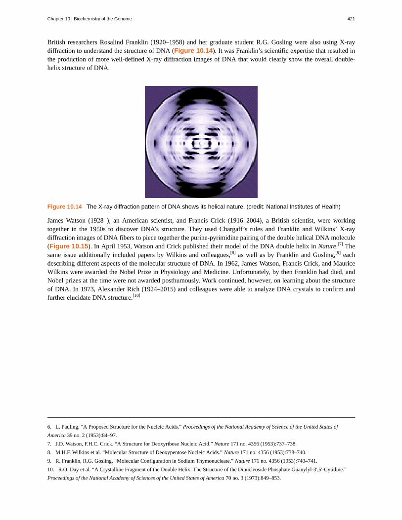
British researchers Rosalind Franklin (1920–1958) and her graduate student R.G. Gosling were also using X-raydiffraction to understand the structure of DNA (Figure 10.14). It was Franklin’s scientific expertise that resulted inthe production of more well-defined X-ray diffraction images of DNA that would clearly show the overall double-helix structure of DNA.
Figure 10.14 The X-ray diffraction pattern of DNA shows its helical nature. (credit: National Institutes of Health)
James Watson (1928–), an American scientist, and Francis Crick (1916–2004), a British scientist, were workingtogether in the 1950s to discover DNA’s structure. They used Chargaff’s rules and Franklin and Wilkins’ X-raydiffraction images of DNA fibers to piece together the purine-pyrimidine pairing of the double helical DNA molecule(Figure 10.15). In April 1953, Watson and Crick published their model of the DNA double helix in Nature.[7] Thesame issue additionally included papers by Wilkins and colleagues,[8] as well as by Franklin and Gosling,[9] eachdescribing different aspects of the molecular structure of DNA. In 1962, James Watson, Francis Crick, and MauriceWilkins were awarded the Nobel Prize in Physiology and Medicine. Unfortunately, by then Franklin had died, andNobel prizes at the time were not awarded posthumously. Work continued, however, on learning about the structureof DNA. In 1973, Alexander Rich (1924–2015) and colleagues were able to analyze DNA crystals to confirm andfurther elucidate DNA structure.[10]
6. L. Pauling, “A Proposed Structure for the Nucleic Acids.” Proceedings of the National Academy of Science of the United States of
America 39 no. 2 (1953):84–97.
7. J.D. Watson, F.H.C. Crick. “A Structure for Deoxyribose Nucleic Acid.” Nature 171 no. 4356 (1953):737–738.
8. M.H.F. Wilkins et al. “Molecular Structure of Deoxypentose Nucleic Acids.” Nature 171 no. 4356 (1953):738–740.
9. R. Franklin, R.G. Gosling. “Molecular Configuration in Sodium Thymonucleate.” Nature 171 no. 4356 (1953):740–741.
10. R.O. Day et al. “A Crystalline Fragment of the Double Helix: The Structure of the Dinucleoside Phosphate Guanylyl-3',5'-Cytidine.”
Proceedings of the National Academy of Sciences of the United States of America 70 no. 3 (1973):849–853.
Chapter 10 | Biochemistry of the Genome 421

Figure 10.15 In 1953, James Watson and Francis Crick built this model of the structure of DNA, shown here ondisplay at the Science Museum in London.
• Which scientists are given most of the credit for describing the molecular structure of DNA?
DNA Structure
Watson and Crick proposed that DNA is made up of two strands that are twisted around each other to form a right-handed helix. The two DNA strands are antiparallel, such that the 3ʹ end of one strand faces the 5ʹ end of theother (Figure 10.16). The 3ʹ end of each strand has a free hydroxyl group, while the 5ʹ end of each strand has afree phosphate group. The sugar and phosphate of the polymerized nucleotides form the backbone of the structure,whereas the nitrogenous bases are stacked inside. These nitrogenous bases on the interior of the molecule interactwith each other, base pairing.
Analysis of the diffraction patterns of DNA has determined that there are approximately 10 bases per turn in DNA.The asymmetrical spacing of the sugar-phosphate backbones generates major grooves (where the backbone is farapart) and minor grooves (where the backbone is close together) (Figure 10.16). These grooves are locations whereproteins can bind to DNA. The binding of these proteins can alter the structure of DNA, regulate replication, orregulate transcription of DNA into RNA.
422 Chapter 10 | Biochemistry of the Genome
This OpenStax book is available for free at http://cnx.org/content/col12087/1.5

Figure 10.16 Watson and Crick proposed the double helix model for DNA. (a) The sugar-phosphate backbones areon the outside of the double helix and purines and pyrimidines form the “rungs” of the DNA helix ladder. (b) The twoDNA strands are antiparallel to each other. (c) The direction of each strand is identified by numbering the carbons (1through 5) in each sugar molecule. The 5ʹ end is the one where carbon #5 is not bound to another nucleotide; the 3ʹend is the one where carbon #3 is not bound to another nucleotide.
Base pairing takes place between a purine and pyrimidine. In DNA, adenine (A) and thymine (T) are complementarybase pairs, and cytosine (C) and guanine (G) are also complementary base pairs, explaining Chargaff’s rules (Figure10.17). The base pairs are stabilized by hydrogen bonds; adenine and thymine form two hydrogen bonds betweenthem, whereas cytosine and guanine form three hydrogen bonds between them.
Figure 10.17 Hydrogen bonds form between complementary nitrogenous bases on the interior of DNA.
In the laboratory, exposing the two DNA strands of the double helix to high temperatures or to certain chemicalscan break the hydrogen bonds between complementary bases, thus separating the strands into two separate singlestrands of DNA (single-stranded DNA [ssDNA]). This process is called DNA denaturation and is analogous toprotein denaturation, as described in Proteins. The ssDNA strands can also be put back together as double-strandedDNA (dsDNA), through reannealing or renaturing by cooling or removing the chemical denaturants, allowing these
Chapter 10 | Biochemistry of the Genome 423
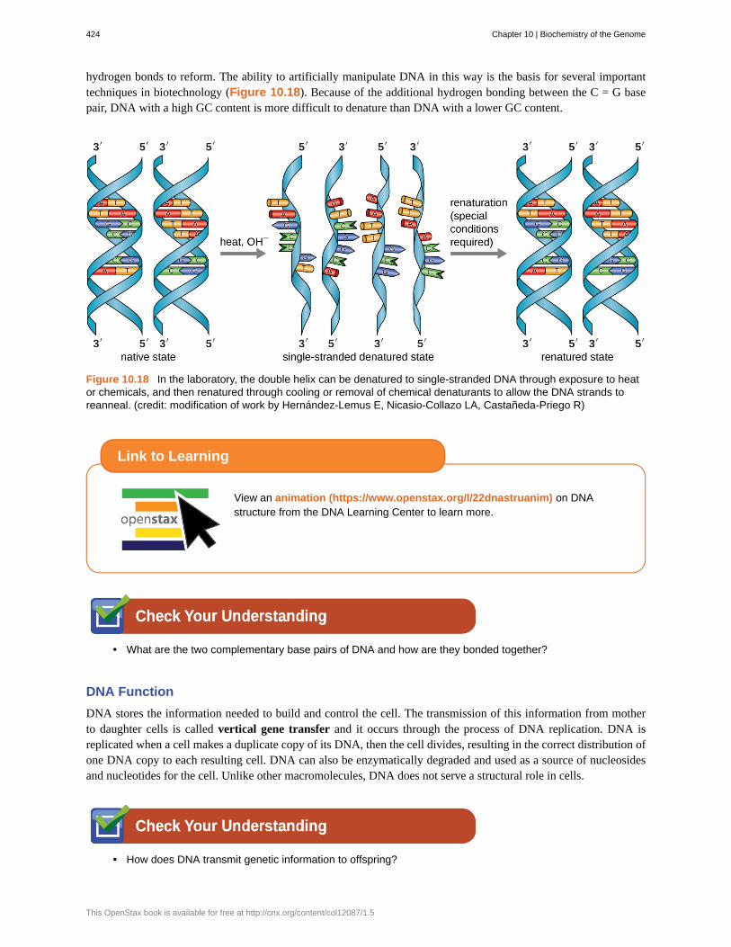
hydrogen bonds to reform. The ability to artificially manipulate DNA in this way is the basis for several importanttechniques in biotechnology (Figure 10.18). Because of the additional hydrogen bonding between the C = G basepair, DNA with a high GC content is more difficult to denature than DNA with a lower GC content.
Figure 10.18 In the laboratory, the double helix can be denatured to single-stranded DNA through exposure to heator chemicals, and then renatured through cooling or removal of chemical denaturants to allow the DNA strands toreanneal. (credit: modification of work by Hernández-Lemus E, Nicasio-Collazo LA, Castañeda-Priego R)
View an animation (https://www.openstax.org/l/22dnastruanim) on DNAstructure from the DNA Learning Center to learn more.
• What are the two complementary base pairs of DNA and how are they bonded together?
DNA Function
DNA stores the information needed to build and control the cell. The transmission of this information from motherto daughter cells is called vertical gene transfer and it occurs through the process of DNA replication. DNA isreplicated when a cell makes a duplicate copy of its DNA, then the cell divides, resulting in the correct distribution ofone DNA copy to each resulting cell. DNA can also be enzymatically degraded and used as a source of nucleosidesand nucleotides for the cell. Unlike other macromolecules, DNA does not serve a structural role in cells.
• How does DNA transmit genetic information to offspring?
Link to Learning
424 Chapter 10 | Biochemistry of the Genome
This OpenStax book is available for free at http://cnx.org/content/col12087/1.5
Paving the Way for Women in Science and Health Professions
Historically, women have been underrepresented in the sciences and in medicine, and often their pioneeringcontributions have gone relatively unnoticed. For example, although Rosalind Franklin performed the X-raydiffraction studies demonstrating the double helical structure of DNA, it is Watson and Crick who becamefamous for this discovery, building on her data. There still remains great controversy over whether theiracquisition of her data was appropriate and whether personality conflicts and gender bias contributed to thedelayed recognition of her significant contributions. Similarly, Barbara McClintock did pioneering work in maize(corn) genetics from the 1930s through 1950s, discovering transposons (jumping genes), but she was notrecognized until much later, receiving a Nobel Prize in Physiology or Medicine in 1983 (Figure 10.19).
Today, women still remain underrepresented in many fields of science and medicine. While more than halfof the undergraduate degrees in science are awarded to women, only 46% of doctoral degrees in scienceare awarded to women. In academia, the number of women at each level of career advancement continuesto decrease, with women holding less than one-third of the positions of Ph.D.-level scientists in tenure-trackpositions, and less than one-quarter of the full professorships at 4-year colleges and universities.[11] Even inthe health professions, like nearly all other fields, women are often underrepresented in many medical careersand earn significantly less than their male counterparts, as shown in a 2013 study published by the Journal ofthe American Medical Association.[12]
Why do such disparities continue to exist and how do we break these cycles? The situation is complex andlikely results from the combination of various factors, including how society conditions the behaviors of girlsfrom a young age and supports their interests, both professionally and personally. Some have suggested thatwomen do not belong in the laboratory, including Nobel Prize winner Tim Hunt, whose 2015 public commentssuggesting that women are too emotional for science[13] were met with widespread condemnation.
Perhaps girls should be supported more from a young age in the areas of science and math (Figure 10.19).Science, technology, engineering, and mathematics (STEM) programs sponsored by the American Associationof University Women (AAUW)[14] and National Aeronautics and Space Administration (NASA)[15] are excellentexamples of programs that offer such support. Contributions by women in science should be made knownmore widely to the public, and marketing targeted to young girls should include more images of historicallyand professionally successful female scientists and medical professionals, encouraging all bright young minds,including girls and women, to pursue careers in science and medicine.
Eye on Ethics
11. N.H. Wolfinger “For Female Scientists, There's No Good Time to Have Children.” The Atlantic July 29, 2013.
http://www.theatlantic.com/sexes/archive/2013/07/for-female-scientists-theres-no-good-time-to-have-children/278165/.
12. S.A. Seabury et al. “Trends in the Earnings of Male and Female Health Care Professionals in the United States, 1987 to 2010.” Journal
of the American Medical Association Internal Medicine 173 no. 18 (2013):1748–1750.
13. E. Chung. “Tim Hunt, Sexism and Science: The Real 'Trouble With Girls' in Labs.” CBC News Technology and Science, June 12, 2015.
http://www.cbc.ca/news/technology/tim-hunt-sexism-and-science-the-real-trouble-with-girls-in-labs-1.3110133. Accessed 8/4/2016.
14. American Association of University Women. “Building a STEM Pipeline for Girls and Women.” http://www.aauw.org/what-we-do/
stem-education/. Accessed June 10, 2016.
15. National Aeronautics and Space Administration. “Outreach Programs: Women and Girls Initiative.” http://women.nasa.gov/outreach-
programs/. Accessed June 10, 2016.
Chapter 10 | Biochemistry of the Genome 425

Figure 10.19 (a) Barbara McClintock’s work on maize genetics in the 1930s through 1950s resulted in thediscovery of transposons, but its significance was not recognized at the time. (b) Efforts to appropriatelymentor and to provide continued societal support for women in science and medicine may someday helpalleviate some of the issues preventing gender equality at all levels in science and medicine. (credit a:modification of work by Smithsonian Institution; credit b: modification of work by Haynie SL, Hinkle AS, JonesNL, Martin CA, Olsiewski PJ, Roberts MF)
Part 2
Based upon his symptoms, Alex’s physician suspects that he is suffering from a foodborne illness that heacquired during his travels. Possibilities include bacterial infection (e.g., enterotoxigenic E. coli, Vibrio cholerae,Campylobacter jejuni, Salmonella), viral infection (rotavirus or norovirus), or protozoan infection (Giardialamblia, Cryptosporidium parvum, or Entamoeba histolytica).
His physician orders a stool sample to identify possible causative agents (e.g., bacteria, cysts) and to look forthe presence of blood because certain types of infectious agents (like C. jejuni, Salmonella, and E. histolytica)are associated with the production of bloody stools.
Alex’s stool sample showed neither blood nor cysts. Following analysis of his stool sample and based upon hisrecent travel history, the hospital physician suspected that Alex was suffering from traveler’s diarrhea causedby enterotoxigenic E. coli (ETEC), the causative agent of most traveler’s diarrhea. To verify the diagnosis andrule out other possibilities, Alex’s physician ordered a diagnostic lab test of his stool sample to look for DNAsequences encoding specific virulence factors of ETEC. The physician instructed Alex to drink lots of fluids toreplace what he was losing and discharged him from the hospital.
ETEC produces several plasmid-encoded virulence factors that make it pathogenic compared with typical E.coli. These include the secreted toxins heat-labile enterotoxin (LT) and heat-stabile enterotoxin (ST), as wellas colonization factor (CF). Both LT and ST cause the excretion of chloride ions from intestinal cells to theintestinal lumen, causing a consequent loss of water from intestinal cells, resulting in diarrhea. CF encodes abacterial protein that aids in allowing the bacterium to adhere to the lining of the small intestine.
• Why did Alex’s physician use genetic analysis instead of either isolation of bacteria from the stoolsample or direct Gram stain of the stool sample alone?
Clinical Focus
426 Chapter 10 | Biochemistry of the Genome
This OpenStax book is available for free at http://cnx.org/content/col12087/1.5

Jump to the next Clinical Focus box. Go back to the previous Clinical Focus box.
10.3 Structure and Function of RNA
Learning Objectives• Describe the biochemical structure of ribonucleotides
• Describe the similarities and differences between RNA and DNA
• Describe the functions of the three main types of RNA used in protein synthesis
• Explain how RNA can serve as hereditary information
Structurally speaking, ribonucleic acid (RNA), is quite similar to DNA. However, whereas DNA molecules aretypically long and double stranded, RNA molecules are much shorter and are typically single stranded. RNAmolecules perform a variety of roles in the cell but are mainly involved in the process of protein synthesis (translation)and its regulation.
RNA Structure
RNA is typically single stranded and is made of ribonucleotides that are linked by phosphodiester bonds. Aribonucleotide in the RNA chain contains ribose (the pentose sugar), one of the four nitrogenous bases (A, U, G, andC), and a phosphate group. The subtle structural difference between the sugars gives DNA added stability, makingDNA more suitable for storage of genetic information, whereas the relative instability of RNA makes it more suitablefor its more short-term functions. The RNA-specific pyrimidine uracil forms a complementary base pair with adenineand is used instead of the thymine used in DNA. Even though RNA is single stranded, most types of RNA moleculesshow extensive intramolecular base pairing between complementary sequences within the RNA strand, creating apredictable three-dimensional structure essential for their function (Figure 10.20 and Figure 10.21).
Figure 10.20 (a) Ribonucleotides contain the pentose sugar ribose instead of the deoxyribose found indeoxyribonucleotides. (b) RNA contains the pyrimidine uracil in place of thymine found in DNA.
Chapter 10 | Biochemistry of the Genome 427
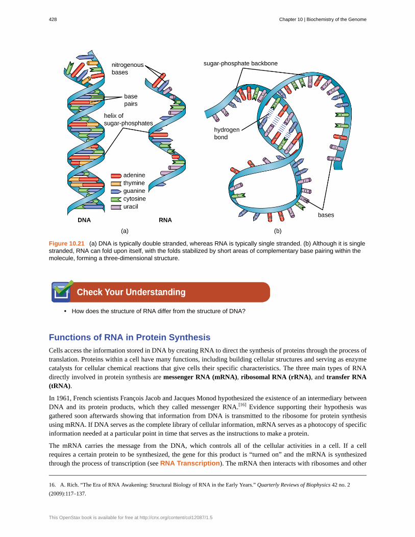
Figure 10.21 (a) DNA is typically double stranded, whereas RNA is typically single stranded. (b) Although it is singlestranded, RNA can fold upon itself, with the folds stabilized by short areas of complementary base pairing within themolecule, forming a three-dimensional structure.
• How does the structure of RNA differ from the structure of DNA?
Functions of RNA in Protein Synthesis
Cells access the information stored in DNA by creating RNA to direct the synthesis of proteins through the process oftranslation. Proteins within a cell have many functions, including building cellular structures and serving as enzymecatalysts for cellular chemical reactions that give cells their specific characteristics. The three main types of RNAdirectly involved in protein synthesis are messenger RNA (mRNA), ribosomal RNA (rRNA), and transfer RNA(tRNA).
In 1961, French scientists François Jacob and Jacques Monod hypothesized the existence of an intermediary betweenDNA and its protein products, which they called messenger RNA.[16] Evidence supporting their hypothesis wasgathered soon afterwards showing that information from DNA is transmitted to the ribosome for protein synthesisusing mRNA. If DNA serves as the complete library of cellular information, mRNA serves as a photocopy of specificinformation needed at a particular point in time that serves as the instructions to make a protein.
The mRNA carries the message from the DNA, which controls all of the cellular activities in a cell. If a cellrequires a certain protein to be synthesized, the gene for this product is “turned on” and the mRNA is synthesizedthrough the process of transcription (see RNA Transcription). The mRNA then interacts with ribosomes and other
16. A. Rich. “The Era of RNA Awakening: Structural Biology of RNA in the Early Years.” Quarterly Reviews of Biophysics 42 no. 2
(2009):117–137.
428 Chapter 10 | Biochemistry of the Genome
This OpenStax book is available for free at http://cnx.org/content/col12087/1.5
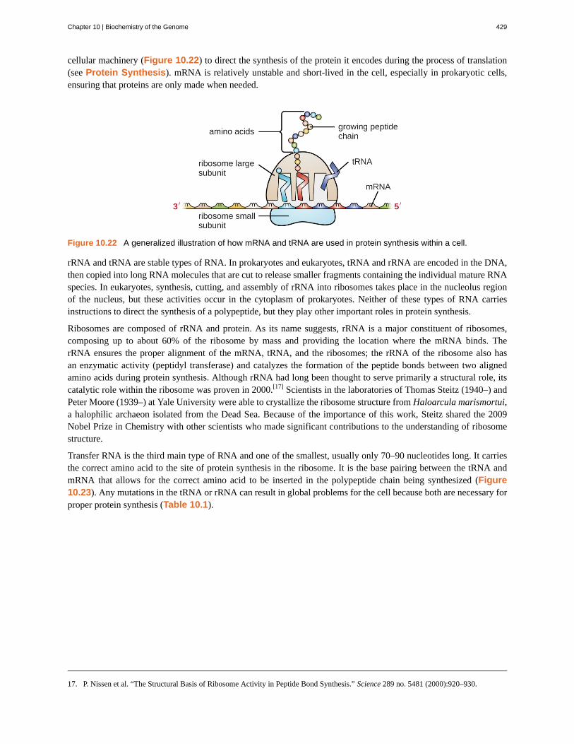
cellular machinery (Figure 10.22) to direct the synthesis of the protein it encodes during the process of translation(see Protein Synthesis). mRNA is relatively unstable and short-lived in the cell, especially in prokaryotic cells,ensuring that proteins are only made when needed.
Figure 10.22 A generalized illustration of how mRNA and tRNA are used in protein synthesis within a cell.
rRNA and tRNA are stable types of RNA. In prokaryotes and eukaryotes, tRNA and rRNA are encoded in the DNA,then copied into long RNA molecules that are cut to release smaller fragments containing the individual mature RNAspecies. In eukaryotes, synthesis, cutting, and assembly of rRNA into ribosomes takes place in the nucleolus regionof the nucleus, but these activities occur in the cytoplasm of prokaryotes. Neither of these types of RNA carriesinstructions to direct the synthesis of a polypeptide, but they play other important roles in protein synthesis.
Ribosomes are composed of rRNA and protein. As its name suggests, rRNA is a major constituent of ribosomes,composing up to about 60% of the ribosome by mass and providing the location where the mRNA binds. TherRNA ensures the proper alignment of the mRNA, tRNA, and the ribosomes; the rRNA of the ribosome also hasan enzymatic activity (peptidyl transferase) and catalyzes the formation of the peptide bonds between two alignedamino acids during protein synthesis. Although rRNA had long been thought to serve primarily a structural role, itscatalytic role within the ribosome was proven in 2000.[17] Scientists in the laboratories of Thomas Steitz (1940–) andPeter Moore (1939–) at Yale University were able to crystallize the ribosome structure from Haloarcula marismortui,a halophilic archaeon isolated from the Dead Sea. Because of the importance of this work, Steitz shared the 2009Nobel Prize in Chemistry with other scientists who made significant contributions to the understanding of ribosomestructure.
Transfer RNA is the third main type of RNA and one of the smallest, usually only 70–90 nucleotides long. It carriesthe correct amino acid to the site of protein synthesis in the ribosome. It is the base pairing between the tRNA andmRNA that allows for the correct amino acid to be inserted in the polypeptide chain being synthesized (Figure10.23). Any mutations in the tRNA or rRNA can result in global problems for the cell because both are necessary forproper protein synthesis (Table 10.1).
17. P. Nissen et al. “The Structural Basis of Ribosome Activity in Peptide Bond Synthesis.” Science 289 no. 5481 (2000):920–930.
Chapter 10 | Biochemistry of the Genome 429

Figure 10.23 A tRNA molecule is a single-stranded molecule that exhibits significant intracellular base pairing,giving it its characteristic three-dimensional shape.
Structure and Function of RNA
mRNA rRNA tRNA
Structure Short, unstable, single-stranded RNAcorresponding to a geneencoded within DNA
Longer, stable RNA moleculescomposing 60% of ribosome’smass
Short (70-90 nucleotides), stableRNA with extensiveintramolecular base pairing;contains an amino acid bindingsite and an mRNA binding site
Function Serves as intermediarybetween DNA andprotein; used byribosome to directsynthesis of protein itencodes
Ensures the proper alignment ofmRNA, tRNA, and ribosomeduring protein synthesis;catalyzes peptide bond formationbetween amino acids
Carries the correct amino acid tothe site of protein synthesis in theribosome
Table 10.1
• What are the functions of the three major types of RNA molecules involved in protein synthesis?
430 Chapter 10 | Biochemistry of the Genome
This OpenStax book is available for free at http://cnx.org/content/col12087/1.5
RNA as Hereditary Information
Although RNA does not serve as the hereditary information in most cells, RNA does hold this function for manyviruses that do not contain DNA. Thus, RNA clearly does have the additional capacity to serve as genetic information.Although RNA is typically single stranded within cells, there is significant diversity in viruses. Rhinoviruses, whichcause the common cold; influenza viruses; and the Ebola virus are single-stranded RNA viruses. Rotaviruses, whichcause severe gastroenteritis in children and other immunocompromised individuals, are examples of double-strandedRNA viruses. Because double-stranded RNA is uncommon in eukaryotic cells, its presence serves as an indicator ofviral infection. The implications for a virus having an RNA genome instead of a DNA genome are discussed in moredetail in Viruses.
10.4 Structure and Function of Cellular Genomes
Learning Objectives• Define gene and genotype and differentiate genotype from phenotype
• Describe chromosome structure and packaging
• Compare prokaryotic and eukaryotic chromosomes
• Explain why extrachromosomal DNA is important in a cell
Thus far, we have discussed the structure and function of individual pieces of DNA and RNA. In this section, we willdiscuss how all of an organism’s genetic material—collectively referred to as its genome—is organized inside of thecell. Since an organism’s genetics to a large extent dictate its characteristics, it should not be surprising that organismsdiffer in the arrangement of their DNA and RNA.
Genotype versus Phenotype
All cellular activities are encoded within a cell’s DNA. The sequence of bases within a DNA molecule representsthe genetic information of the cell. Segments of DNA molecules are called genes, and individual genes contain theinstructional code necessary for synthesizing various proteins, enzymes, or stable RNA molecules.
The full collection of genes that a cell contains within its genome is called its genotype. However, a cell does notexpress all of its genes simultaneously. Instead, it turns on (expresses) or turns off certain genes when necessary.The set of genes being expressed at any given point in time determines the cell’s activities and its observablecharacteristics, referred to as its phenotype. Genes that are always expressed are known as constitutive genes; someconstitutive genes are known as housekeeping genes because they are necessary for the basic functions of the cell.
While the genotype of a cell remains constant, the phenotype may change in response to environmental signals (e.g.,changes in temperature or nutrient availability) that affect which nonconstitutive genes are expressed. For example,the oral bacterium Streptococcus mutans produces a sticky slime layer that allows it to adhere to teeth, forming dentalplaque; however, the genes that control the production of the slime layer are only expressed in the presence of sucrose(table sugar). Thus, while the genotype of S. mutans is constant, its phenotype changes depending on the presence andabsence of sugar in its environment. Temperature can also regulate gene expression. For example, the gram-negativebacterium Serratia marcescens, a pathogen frequently associated with hospital-acquired infections, produces a redpigment at 28 °C but not at 37 °C, the normal internal temperature of the human body (Figure 10.24).
Chapter 10 | Biochemistry of the Genome 431
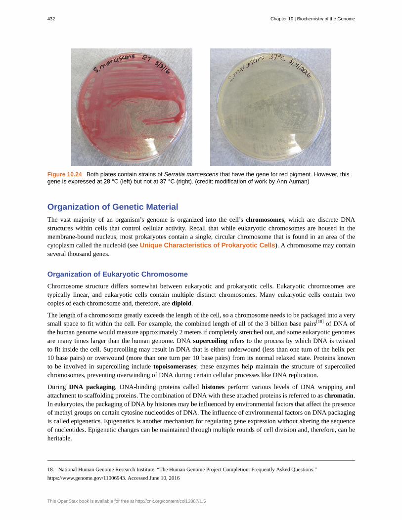
Figure 10.24 Both plates contain strains of Serratia marcescens that have the gene for red pigment. However, thisgene is expressed at 28 °C (left) but not at 37 °C (right). (credit: modification of work by Ann Auman)
Organization of Genetic Material
The vast majority of an organism’s genome is organized into the cell’s chromosomes, which are discrete DNAstructures within cells that control cellular activity. Recall that while eukaryotic chromosomes are housed in themembrane-bound nucleus, most prokaryotes contain a single, circular chromosome that is found in an area of thecytoplasm called the nucleoid (see Unique Characteristics of Prokaryotic Cells). A chromosome may containseveral thousand genes.
Organization of Eukaryotic Chromosome
Chromosome structure differs somewhat between eukaryotic and prokaryotic cells. Eukaryotic chromosomes aretypically linear, and eukaryotic cells contain multiple distinct chromosomes. Many eukaryotic cells contain twocopies of each chromosome and, therefore, are diploid.
The length of a chromosome greatly exceeds the length of the cell, so a chromosome needs to be packaged into a verysmall space to fit within the cell. For example, the combined length of all of the 3 billion base pairs[18] of DNA ofthe human genome would measure approximately 2 meters if completely stretched out, and some eukaryotic genomesare many times larger than the human genome. DNA supercoiling refers to the process by which DNA is twistedto fit inside the cell. Supercoiling may result in DNA that is either underwound (less than one turn of the helix per10 base pairs) or overwound (more than one turn per 10 base pairs) from its normal relaxed state. Proteins knownto be involved in supercoiling include topoisomerases; these enzymes help maintain the structure of supercoiledchromosomes, preventing overwinding of DNA during certain cellular processes like DNA replication.
During DNA packaging, DNA-binding proteins called histones perform various levels of DNA wrapping andattachment to scaffolding proteins. The combination of DNA with these attached proteins is referred to as chromatin.In eukaryotes, the packaging of DNA by histones may be influenced by environmental factors that affect the presenceof methyl groups on certain cytosine nucleotides of DNA. The influence of environmental factors on DNA packagingis called epigenetics. Epigenetics is another mechanism for regulating gene expression without altering the sequenceof nucleotides. Epigenetic changes can be maintained through multiple rounds of cell division and, therefore, can beheritable.
18. National Human Genome Research Institute. “The Human Genome Project Completion: Frequently Asked Questions.”
https://www.genome.gov/11006943. Accessed June 10, 2016
432 Chapter 10 | Biochemistry of the Genome
This OpenStax book is available for free at http://cnx.org/content/col12087/1.5

View this animation (https://www.openstax.org/l/22dnapackanim) from theDNA Learning Center to learn more about on DNA packaging in eukaryotes.
Organization of Prokaryotic Chromosomes
Chromosomes in bacteria and archaea are usually circular, and a prokaryotic cell typically contains only a singlechromosome within the nucleoid. Because the chromosome contains only one copy of each gene, prokaryotes arehaploid. As in eukaryotic cells, DNA supercoiling is necessary for the genome to fit within the prokaryotic cell. TheDNA in the bacterial chromosome is arranged in several supercoiled domains. As with eukaryotes, topoisomerasesare involved in supercoiling DNA. DNA gyrase is a type of topoisomerase, found in bacteria and some archaea, thathelps prevent the overwinding of DNA. (Some antibiotics kill bacteria by targeting DNA gyrase.) In addition, histone-like proteins bind DNA and aid in DNA packaging. Other proteins bind to the origin of replication, the location inthe chromosome where DNA replication initiates. Because different regions of DNA are packaged differently, someregions of chromosomal DNA are more accessible to enzymes and thus may be used more readily as templates forgene expression. Interestingly, several bacteria, including Helicobacter pylori and Shigella flexneri, have been shownto induce epigenetic changes in their hosts upon infection, leading to chromatin remodeling that may cause long-termeffects on host immunity.[19]
• What is the difference between a cell’s genotype and its phenotype?
• How does DNA fit inside cells?
Noncoding DNA
In addition to genes, a genome also contains many regions of noncoding DNA that do not encode proteins or stableRNA products. Noncoding DNA is commonly found in areas prior to the start of coding sequences of genes as wellas in intergenic regions (i.e., DNA sequences located between genes) (Figure 10.25).
Link to Learning
19. H. Bierne et al. “Epigenetics and Bacterial Infections.” Cold Spring Harbor Perspectives in Medicine 2 no. 12 (2012):a010272.
Chapter 10 | Biochemistry of the Genome 433

Figure 10.25 Chromosomes typically have a significant amount of noncoding DNA, often found in intergenicregions.
Prokaryotes appear to use their genomes very efficiently, with only an average of 12% of the genome being takenup by noncoding sequences. In contrast, noncoding DNA can represent about 98% of the genome in eukaryotes, asseen in humans, but the percentage of noncoding DNA varies between species.[20] These noncoding DNA regionswere once referred to as “junk DNA”; however, this terminology is no longer widely accepted because scientists havesince found roles for some of these regions, many of which contribute to the regulation of transcription or translationthrough the production of small noncoding RNA molecules, DNA packaging, and chromosomal stability. Althoughscientists may not fully understand the roles of all noncoding regions of DNA, it is generally believed that they dohave purposes within the cell.
• What is the role of noncoding DNA?
Extrachromosomal DNA
Although most DNA is contained within a cell’s chromosomes, many cells have additional molecules of DNA outsidethe chromosomes, called extrachromosomal DNA, that are also part of its genome. The genomes of eukaryoticcells would also include the chromosomes from any organelles such as mitochondria and/or chloroplasts that thesecells maintain (Figure 10.26). The maintenance of circular chromosomes in these organelles is a vestige of theirprokaryotic origins and supports the endosymbiotic theory (see Foundations of Modern Cell Theory). Insome cases, genomes of certain DNA viruses can also be maintained independently in host cells during latentviral infection. In these cases, these viruses are another form of extrachromosomal DNA. For example, the humanpapillomavirus (HPV) may be maintained in infected cells in this way.
20. R.J. Taft et al. “The Relationship between Non-Protein-Coding DNA and Eukaryotic Complexity.” Bioessays 29 no. 3 (2007):288–299.
434 Chapter 10 | Biochemistry of the Genome
This OpenStax book is available for free at http://cnx.org/content/col12087/1.5
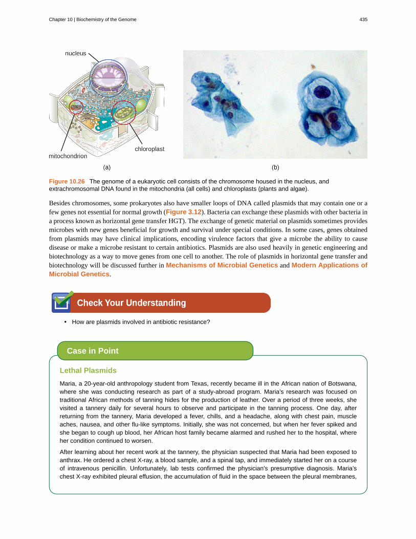
Figure 10.26 The genome of a eukaryotic cell consists of the chromosome housed in the nucleus, andextrachromosomal DNA found in the mitochondria (all cells) and chloroplasts (plants and algae).
Besides chromosomes, some prokaryotes also have smaller loops of DNA called plasmids that may contain one or afew genes not essential for normal growth (Figure 3.12). Bacteria can exchange these plasmids with other bacteria ina process known as horizontal gene transfer HGT). The exchange of genetic material on plasmids sometimes providesmicrobes with new genes beneficial for growth and survival under special conditions. In some cases, genes obtainedfrom plasmids may have clinical implications, encoding virulence factors that give a microbe the ability to causedisease or make a microbe resistant to certain antibiotics. Plasmids are also used heavily in genetic engineering andbiotechnology as a way to move genes from one cell to another. The role of plasmids in horizontal gene transfer andbiotechnology will be discussed further in Mechanisms of Microbial Genetics and Modern Applications ofMicrobial Genetics.
• How are plasmids involved in antibiotic resistance?
Lethal Plasmids
Maria, a 20-year-old anthropology student from Texas, recently became ill in the African nation of Botswana,where she was conducting research as part of a study-abroad program. Maria’s research was focused ontraditional African methods of tanning hides for the production of leather. Over a period of three weeks, shevisited a tannery daily for several hours to observe and participate in the tanning process. One day, afterreturning from the tannery, Maria developed a fever, chills, and a headache, along with chest pain, muscleaches, nausea, and other flu-like symptoms. Initially, she was not concerned, but when her fever spiked andshe began to cough up blood, her African host family became alarmed and rushed her to the hospital, whereher condition continued to worsen.
After learning about her recent work at the tannery, the physician suspected that Maria had been exposed toanthrax. He ordered a chest X-ray, a blood sample, and a spinal tap, and immediately started her on a courseof intravenous penicillin. Unfortunately, lab tests confirmed the physician’s presumptive diagnosis. Maria’schest X-ray exhibited pleural effusion, the accumulation of fluid in the space between the pleural membranes,
Case in Point
Chapter 10 | Biochemistry of the Genome 435
and a Gram stain of her blood revealed the presence of gram-positive, rod-shaped bacteria in short chains,consistent with Bacillus anthracis. Blood and bacteria were also shown to be present in her cerebrospinalfluid, indicating that the infection had progressed to meningitis. Despite supportive treatment and aggressiveantibiotic therapy, Maria slipped into an unresponsive state and died three days later.
Anthrax is a disease caused by the introduction of endospores from the gram-positive bacterium B. anthracisinto the body. Once infected, patients typically develop meningitis, often with fatal results. In Maria’s case, sheinhaled the endospores while handling the hides of animals that had been infected.
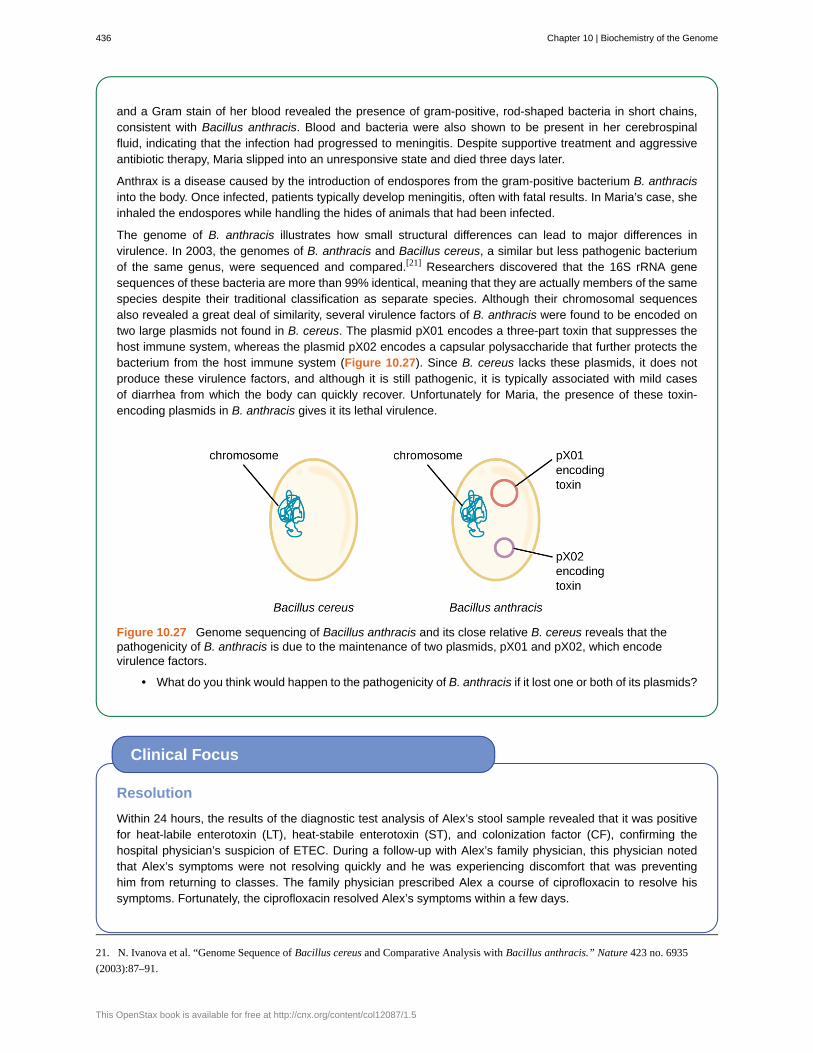
The genome of B. anthracis illustrates how small structural differences can lead to major differences invirulence. In 2003, the genomes of B. anthracis and Bacillus cereus, a similar but less pathogenic bacteriumof the same genus, were sequenced and compared.[21] Researchers discovered that the 16S rRNA genesequences of these bacteria are more than 99% identical, meaning that they are actually members of the samespecies despite their traditional classification as separate species. Although their chromosomal sequencesalso revealed a great deal of similarity, several virulence factors of B. anthracis were found to be encoded ontwo large plasmids not found in B. cereus. The plasmid pX01 encodes a three-part toxin that suppresses thehost immune system, whereas the plasmid pX02 encodes a capsular polysaccharide that further protects thebacterium from the host immune system (Figure 10.27). Since B. cereus lacks these plasmids, it does notproduce these virulence factors, and although it is still pathogenic, it is typically associated with mild casesof diarrhea from which the body can quickly recover. Unfortunately for Maria, the presence of these toxin-encoding plasmids in B. anthracis gives it its lethal virulence.
Figure 10.27 Genome sequencing of Bacillus anthracis and its close relative B. cereus reveals that thepathogenicity of B. anthracis is due to the maintenance of two plasmids, pX01 and pX02, which encodevirulence factors.
• What do you think would happen to the pathogenicity of B. anthracis if it lost one or both of its plasmids?
Resolution
Within 24 hours, the results of the diagnostic test analysis of Alex’s stool sample revealed that it was positivefor heat-labile enterotoxin (LT), heat-stabile enterotoxin (ST), and colonization factor (CF), confirming thehospital physician’s suspicion of ETEC. During a follow-up with Alex’s family physician, this physician notedthat Alex’s symptoms were not resolving quickly and he was experiencing discomfort that was preventinghim from returning to classes. The family physician prescribed Alex a course of ciprofloxacin to resolve hissymptoms. Fortunately, the ciprofloxacin resolved Alex’s symptoms within a few days.
Clinical Focus
21. N. Ivanova et al. “Genome Sequence of Bacillus cereus and Comparative Analysis with Bacillus anthracis.” Nature 423 no. 6935
(2003):87–91.
436 Chapter 10 | Biochemistry of the Genome
This OpenStax book is available for free at http://cnx.org/content/col12087/1.5

Alex likely got his infection from ingesting contaminated food or water. Emerging industrialized countries likeMexico are still developing sanitation practices that prevent the contamination of water with fecal material.Travelers in such countries should avoid the ingestion of undercooked foods, especially meats, seafood,vegetables, and unpasteurized dairy products. They should also avoid use of water that has not been treated;this includes drinking water, ice cubes, and even water used for brushing teeth. Using bottled water for thesepurposes is a good alternative. Good hygiene (handwashing) can also aid the prevention of an ETEC infection.Alex had not been careful about his food or water consumption, which led to his illness.
Alex’s symptoms were very similar to those of cholera, caused by the gram-negative bacterium Vibrio cholerae,which also produces a toxin similar to ST and LT. At some point in the evolutionary history of ETEC, anonpathogenic strain of E. coli similar to those typically found in the gut may have acquired the genes encodingthe ST and LT toxins from V. cholerae. The fact that the genes encoding those toxins are encoded onextrachromosomal plasmids in ETEC supports the idea that these genes were acquired by E. coli and are likelymaintained in bacterial populations through horizontal gene transfer.
Go back to the previous Clinical Focus box.
Viral Genomes
Viral genomes exhibit significant diversity in structure. Some viruses have genomes that consist of DNA as theirgenetic material. This DNA may be single stranded, as exemplified by human parvoviruses, or double stranded, asseen in the herpesviruses and poxviruses. Additionally, although all cellular life uses DNA as its genetic material,some viral genomes are made of either single-stranded or double-stranded RNA molecules, as we have discussed.Viral genomes are typically smaller than most bacterial genomes, encoding only a few genes, because they rely ontheir hosts to carry out many of the functions required for their replication. The diversity of viral genome structuresand their implications for viral replication life cycles are discussed in more detail in The Viral Life Cycle.
• Why do viral genomes vary widely among viruses?
Genome Size Matters
There is great variation in size of genomes among different organisms. Most eukaryotes maintain multiplechromosomes; humans, for example have 23 pairs, giving them 46 chromosomes. Despite being largeat 3 billion base pairs, the human genome is far from the largest genome. Plants often maintain verylarge genomes, up to 150 billion base pairs, and commonly are polyploid, having multiple copies of eachchromosome.
The size of bacterial genomes also varies considerably, although they tend to be smaller than eukaryoticgenomes (Figure 10.28). Some bacterial genomes may be as small as only 112,000 base pairs. Often, thesize of a bacterium’s genome directly relates to how much the bacterium depends on its host for survival.When a bacterium relies on the host cell to carry out certain functions, it loses the genes encoding the abilitiesto carry out those functions itself. These types of bacterial endosymbionts are reminiscent of the prokaryoticorigins of mitochondria and chloroplasts.
From a clinical perspective, obligate intracellular pathogens also tend to have small genomes (some around
Micro Connections
Chapter 10 | Biochemistry of the Genome 437
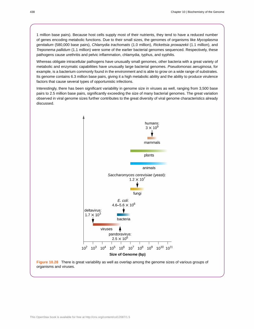
1 million base pairs). Because host cells supply most of their nutrients, they tend to have a reduced numberof genes encoding metabolic functions. Due to their small sizes, the genomes of organisms like Mycoplasmagenitalium (580,000 base pairs), Chlamydia trachomatis (1.0 million), Rickettsia prowazekii (1.1 million), andTreponema pallidum (1.1 million) were some of the earlier bacterial genomes sequenced. Respectively, thesepathogens cause urethritis and pelvic inflammation, chlamydia, typhus, and syphilis.
Whereas obligate intracellular pathogens have unusually small genomes, other bacteria with a great variety ofmetabolic and enzymatic capabilities have unusually large bacterial genomes. Pseudomonas aeruginosa, forexample, is a bacterium commonly found in the environment and is able to grow on a wide range of substrates.Its genome contains 6.3 million base pairs, giving it a high metabolic ability and the ability to produce virulencefactors that cause several types of opportunistic infections.
Interestingly, there has been significant variability in genome size in viruses as well, ranging from 3,500 basepairs to 2.5 million base pairs, significantly exceeding the size of many bacterial genomes. The great variationobserved in viral genome sizes further contributes to the great diversity of viral genome characteristics alreadydiscussed.
Figure 10.28 There is great variability as well as overlap among the genome sizes of various groups oforganisms and viruses.
438 Chapter 10 | Biochemistry of the Genome
This OpenStax book is available for free at http://cnx.org/content/col12087/1.5

Visit the genome database (https://www.openstax.org/l/22NCBIgendata) ofthe National Center for Biotechnology Information (NCBI) to see the genomesthat have been sequenced and their sizes.
Summary
10.1 Using Microbiology to Discover the Secrets of Life• DNA was discovered and characterized long before its role in heredity was understood. Microbiologists
played significant roles in demonstrating that DNA is the hereditary information found within cells.
• In the 1850s and 1860s, Gregor Mendel experimented with true-breeding garden peas to demonstrate theheritability of specific observable traits.
• In 1869, Friedrich Miescher isolated and purified a compound rich in phosphorus from the nuclei of whiteblood cells; he named the compound nuclein. Miescher’s student Richard Altmann discovered its acidicnature, renaming it nucleic acid. Albrecht Kossell characterized the nucleotide bases found within nucleicacids.
• Although Walter Sutton and Theodor Boveri proposed the Chromosomal Theory of Inheritance in 1902, itwas not scientifically demonstrated until the 1915 publication of the work of Thomas Hunt Morgan and hiscolleagues.
• Using Acetabularia, a large algal cell, as his model system, Joachim Hämmerling demonstrated in the 1930sand 1940s that the nucleus was the location of hereditary information in these cells.
• In the 1940s, George Beadle and Edward Tatum used the mold Neurospora crassa to show that each protein’sproduction was under the control of a single gene, demonstrating the “one gene–one enzyme” hypothesis.
• In 1928, Frederick Griffith showed that dead encapsulated bacteria could pass genetic information to livenonencapsulated bacteria and transform them into harmful strains. In 1944, Oswald Avery, Colin McLeod,and Maclyn McCarty identified the compound as DNA.
• The nature of DNA as the molecule that stores genetic information was unequivocally demonstrated in theexperiment of Alfred Hershey and Martha Chase published in 1952. Labeled DNA from bacterial virusesentered and infected bacterial cells, giving rise to more viral particles. The labeled protein coats did notparticipate in the transmission of genetic information.
10.2 Structure and Function of DNA• Nucleic acids are composed of nucleotides, each of which contains a pentose sugar, a phosphate group, and
a nitrogenous base. Deoxyribonucleotides within DNA contain deoxyribose as the pentose sugar.
• DNA contains the pyrimidines cytosine and thymine, and the purines adenine and guanine.
• Nucleotides are linked together by phosphodiester bonds between the 5ʹ phosphate group of one nucleotideand the 3ʹ hydroxyl group of another. A nucleic acid strand has a free phosphate group at the 5ʹ end and afree hydroxyl group at the 3ʹ end.
• Chargaff discovered that the amount of adenine is approximately equal to the amount of thymine in DNA,and that the amount of the guanine is approximately equal to cytosine. These relationships were laterdetermined to be due to complementary base pairing.
• Watson and Crick, building on the work of Chargaff, Franklin and Gosling, and Wilkins, proposed the doublehelix model and base pairing for DNA structure.
• DNA is composed of two complementary strands oriented antiparallel to each other with the phosphodiesterbackbones on the exterior of the molecule. The nitrogenous bases of each strand face each other and
Link to Learning
Chapter 10 | Biochemistry of the Genome 439
complementary bases hydrogen bond to each other, stabilizing the double helix.
• Heat or chemicals can break the hydrogen bonds between complementary bases, denaturing DNA. Cooling orremoving chemicals can lead to renaturation or reannealing of DNA by allowing hydrogen bonds to reformbetween complementary bases.
• DNA stores the instructions needed to build and control the cell. This information is transmitted from parentto offspring through vertical gene transfer.
10.3 Structure and Function of RNA• Ribonucleic acid (RNA) is typically single stranded and contains ribose as its pentose sugar and the
pyrimidine uracil instead of thymine. An RNA strand can undergo significant intramolecular base pairing totake on a three-dimensional structure.
• There are three main types of RNA, all involved in protein synthesis.
• Messenger RNA (mRNA) serves as the intermediary between DNA and the synthesis of protein productsduring translation.
• Ribosomal RNA (rRNA) is a type of stable RNA that is a major constituent of ribosomes. It ensures theproper alignment of the mRNA and the ribosomes during protein synthesis and catalyzes the formation of thepeptide bonds between two aligned amino acids during protein synthesis.
• Transfer RNA (tRNA) is a small type of stable RNA that carries an amino acid to the corresponding site ofprotein synthesis in the ribosome. It is the base pairing between the tRNA and mRNA that allows for thecorrect amino acid to be inserted in the polypeptide chain being synthesized.
• Although RNA is not used for long-term genetic information in cells, many viruses do use RNA as theirgenetic material.
10.4 Structure and Function of Cellular Genomes• The entire genetic content of a cell is its genome.
• Genes code for proteins, or stable RNA molecules, each of which carries out a specific function in the cell.
• Although the genotype that a cell possesses remains constant, expression of genes is dependent onenvironmental conditions.
• A phenotype is the observable characteristics of a cell (or organism) at a given point in time and results fromthe complement of genes currently being used.
• The majority of genetic material is organized into chromosomes that contain the DNA that controls cellularactivities.
• Prokaryotes are typically haploid, usually having a single circular chromosome found in the nucleoid.Eukaryotes are diploid; DNA is organized into multiple linear chromosomes found in the nucleus.
• Supercoiling and DNA packaging using DNA binding proteins allows lengthy molecules to fit inside a cell.Eukaryotes and archaea use histone proteins, and bacteria use different proteins with similar function.
• Prokaryotic and eukaryotic genomes both contain noncoding DNA, the function of which is not wellunderstood. Some noncoding DNA appears to participate in the formation of small noncoding RNA moleculesthat influence gene expression; some appears to play a role in maintaining chromosomal structure and in DNApackaging.
• Extrachromosomal DNA in eukaryotes includes the chromosomes found within organelles of prokaryoticorigin (mitochondria and chloroplasts) that evolved by endosymbiosis. Some viruses may also maintainthemselves extrachromosomally.
• Extrachromosomal DNA in prokaryotes is commonly maintained as plasmids that encode a few nonessentialgenes that may be helpful under specific conditions. Plasmids can be spread through a bacterial communityby horizontal gene transfer.
• Viral genomes show extensive variation and may be composed of either RNA or DNA, and may be eitherdouble or single stranded.
440 Chapter 10 | Biochemistry of the Genome
This OpenStax book is available for free at http://cnx.org/content/col12087/1.5
Multiple Choice
1. Frederick Griffith infected mice with a combinationof dead R and live S bacterial strains. What was theoutcome, and why did it occur?
a. The mice will live. Transformation was notrequired.
b. The mice will die. Transformation of geneticmaterial from R to S was required.
c. The mice will live. Transformation of geneticmaterial from S to R was required.
d. The mice will die. Transformation was notrequired.
2. Why was the alga Acetabularia a good modelorganism for Joachim Hämmerling to use to identify thelocation of genetic material?
a. It lacks a nuclear membrane.b. It self-fertilizes.c. It is a large, asymmetrical, single cell easy to see
with the naked eye.d. It makes a protein capsid.
3. Which of the following best describes the resultsfrom Hershey and Chase’s experiment using bacterialviruses with 35S-labeled proteins or 32P-labeled DNAthat are consistent with protein being the moleculeresponsible for hereditary?
a. After infection with the 35S-labeled viruses andcentrifugation, only the pellet would beradioactive.
b. After infection with the 35S-labeled viruses andcentrifugation, both the pellet and thesupernatant would be radioactive.
c. After infection with the 32P-labeled viruses andcentrifugation, only the pellet would beradioactive.
d. After infection with the 32P-labeled viruses andcentrifugation, both the pellet and thesupernatant would be radioactive.
4. Which method did Morgan and colleagues use toshow that hereditary information was carried onchromosomes?
a. statistical predictions of the outcomes of crossesusing true-breeding parents
b. correlations between microscopic observationsof chromosomal movement and thecharacteristics of offspring
c. transformation of nonpathogenic bacteria topathogenic bacteria
d. mutations resulting in distinct defects inmetabolic enzymatic pathways
5. According to Beadle and Tatum’s “one gene–oneenzyme” hypothesis, which of the following enzymeswill eliminate the transformation of hereditary materialfrom pathogenic bacteria to nonpathogenic bacteria?
a. carbohydrate-degrading enzymesb. proteinasesc. ribonucleasesd. deoxyribonucleases
6. Which of the following is not found within DNA?a. thymineb. phosphodiester bondsc. complementary base pairingd. amino acids
7. If 30% of the bases within a DNA molecule areadenine, what is the percentage of thymine?
a. 20%b. 25%c. 30%d. 35%
8. Which of the following statements about basepairing in DNA is incorrect?
a. Purines always base pairs with pyrimidines.b. Adenine binds to guanine.c. Base pairs are stabilized by hydrogen bonds.d. Base pairing occurs at the interior of the double
helix.
9. If a DNA strand contains the sequence 5ʹ-ATTCCGGATCGA-3ʹ, which of the following is thesequence of the complementary strand of DNA?
a. 5ʹ-TAAGGCCTAGCT-3ʹb. 5ʹ-ATTCCGGATCGA-3ʹc. 3ʹ-TAACCGGTACGT-5ʹd. 5ʹ-TCGATCCGGAAT-3ʹ
Review Questions
Chapter 10 | Biochemistry of the Genome 441
10. During denaturation of DNA, which of thefollowing happens?
a. Hydrogen bonds between complementary basesbreak.
b. Phosphodiester bonds break within the sugar-phosphate backbone.
c. Hydrogen bonds within the sugar-phosphatebackbone break.
d. Phosphodiester bonds between complementarybases break.
11. Which of the following types of RNA codes for aprotein?
a. dsRNAb. mRNAc. rRNAd. tRNA
12. A nucleic acid is purified from a mixture. Themolecules are relatively small, contain uracil, and mostare covalently bound to an amino acid. Which of thefollowing was purified?
a. DNAb. mRNAc. rRNAd. tRNA
13. Which of the following types of RNA is known forits catalytic abilities?
a. dsRNAb. mRNAc. rRNAd. tRNA
14. Ribosomes are composed of rRNA and what othercomponent?
a. proteinb. carbohydratesc. DNAd. mRNA
15. Which of the following may use RNA as itsgenome?
a. a bacteriumb. an archaeonc. a virusd. a eukaryote
16. Which of the following correctly describes thestructure of the typical eukaryotic genome?
a. diploidb. linearc. singulard. double stranded
17. Which of the following is typically found as part ofthe prokaryotic genome?
a. chloroplast DNAb. linear chromosomesc. plasmidsd. mitochondrial DNA
18. Serratia marcescens cells produce a red pigment atroom temperature. The red color of the colonies is anexample of which of the following?
a. genotypeb. phenotypec. change in DNA base compositiond. adaptation to the environment
19. Which of the following genes would not likely beencoded on a plasmid?
a. genes encoding toxins that damage host tissueb. genes encoding antibacterial resistancec. gene encoding enzymes for glycolysisd. genes encoding enzymes for the degradation of
an unusual substrate
20. Histones are DNA binding proteins that areimportant for DNA packaging in which of thefollowing?
a. double-stranded and single-stranded DNAviruses
b. archaea and bacteriac. bacteria and eukaryotesd. eukaryotes and archaea
True/False
21. The work of Rosalind Franklin and R.G. Gosling was important in demonstrating the helical nature of DNA.
442 Chapter 10 | Biochemistry of the Genome
This OpenStax book is available for free at http://cnx.org/content/col12087/1.5
22. The A-T base pair has more hydrogen bonding than the C-G base pair.
23. Ribosomes are composed mostly of RNA.
24. Double-stranded RNA is commonly found inside cells.
25. Within an organism, phenotypes may change while genotypes remain constant.
26. Noncoding DNA has no biological purpose.
Matching
27. Match the correct molecule with its description:
___tRNA
___rRNA
___mRNA
A. is a major component of ribosome
B. is a copy of the information in a gene
C. carries an amino acid to the ribosome
Fill in the Blank
28. The element ____________ is unique to nucleic acids compared with other macromolecules.
29. In the late 1800s and early 1900s, the macromolecule thought to be responsible for heredity was______________.
30. The end of a nucleic acid strand with a free phosphate group is called the ________.
31. Plasmids are typically transferred among members of a bacterial community by ________ gene transfer.
Short Answer
32. Why do bacteria and viruses make good model systems for various genetic studies?
33. Why was nucleic acid disregarded for so long as the molecule responsible for the transmission of hereditaryinformation?
34. Bacteriophages inject their genetic material into host cells, whereas animal viruses enter host cells completely.Why was it important to use a bacteriophage in the Hershey–Chase experiment rather than an animal virus?
35. What is the role of phosphodiester bonds within the sugar-phosphate backbone of DNA?
36. What is meant by the term “antiparallel?”
37. Why is DNA with a high GC content more difficult to denature than that with a low GC content?
38. What are the differences between DNA nucleotides and RNA nucleotides?
39. How is the information stored within the base sequence of DNA used to determine a cell’s properties?
40. How do complementary base pairs contribute to intramolecular base pairing within an RNA molecule?
41. If an antisense RNA has the sequence 5ʹAUUCGAAUGC3ʹ, what is the sequence of the mRNA to which it willbind? Be sure to label the 5ʹ and 3ʹ ends of the molecule you draw.
42. Why does double-stranded RNA (dsRNA) stimulate RNA interference?
43. What are some differences in chromosomal structures between prokaryotes and eukaryotes?
44. How do prokaryotes and eukaryotes manage to fit their lengthy DNA inside of cells? Why is this necessary?
45. What are some functions of noncoding DNA?
46. In the chromatin of eukaryotic cells, which regions of the chromosome would you expect to be more compact:the regions that contain genes being actively copied into RNA or those that contain inactive genes?
Chapter 10 | Biochemistry of the Genome 443
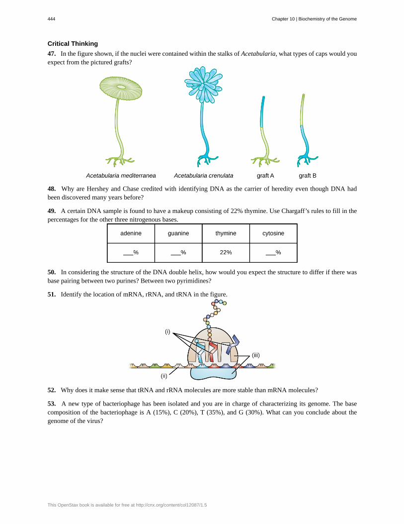
Critical Thinking
47. In the figure shown, if the nuclei were contained within the stalks of Acetabularia, what types of caps would youexpect from the pictured grafts?
48. Why are Hershey and Chase credited with identifying DNA as the carrier of heredity even though DNA hadbeen discovered many years before?
49. A certain DNA sample is found to have a makeup consisting of 22% thymine. Use Chargaff’s rules to fill in thepercentages for the other three nitrogenous bases.
50. In considering the structure of the DNA double helix, how would you expect the structure to differ if there wasbase pairing between two purines? Between two pyrimidines?
51. Identify the location of mRNA, rRNA, and tRNA in the figure.
52. Why does it make sense that tRNA and rRNA molecules are more stable than mRNA molecules?
53. A new type of bacteriophage has been isolated and you are in charge of characterizing its genome. The basecomposition of the bacteriophage is A (15%), C (20%), T (35%), and G (30%). What can you conclude about thegenome of the virus?
444 Chapter 10 | Biochemistry of the Genome
This OpenStax book is available for free at http://cnx.org/content/col12087/1.5
Related Documents