1 CHAPTER 1: INTRODUCTION 1.1 History of mandibular fractures and treatment There are two books that describe, in detail, historical aspects of maxillofacial and oral surgery 1, 2 . The facts in this section are based on these writings. The earliest record, describing mandibular fractures, dates back to 1650 BC. This was known as the ‘Edwin Smith Papyrus’, which was translated in 1930 by Breasted. The first description of the treatment of jaw fractures however, has to be credited to the ‘Father of Medicine’… Hippocrates. He made use of bandages and single jaw fixation, to manually reduce fractures of the jaws. Celsus (30 BC – 50 AD), a Roman physician, was one of the earliest to recognize the importance of establishing the occlusion in the treatment of fractures. His principle of fracture immobilization was the forerunner of intermaxillary fixation (IMF), still in use today. The Eleventh Century Italian influence on the treatment of mandibular fractures continued, primarily due to the work of clinicians in Salerno. In 1275 Guglielmo Salicetti wrote his book Cyrurgia, in which he recommended the wiring together of teeth adjacent to the fracture (tension band principle) followed by the wiring together of both jaws (IMF). This technique was crude by our methods, but remarkably insightful for the Thirteenth Century. By the late 1700s and early 19 th century, the development and use of extraoral splint techniques were in favour. Thomas Gunning, an Irish-American dentist was credited with the first intraoral splint; and ironically made one for himself after sustaining a mandibular fracture in a riding accident, in 1862. The concept of closed reduction of fractures persisted until the early 1900s. Buck, Kinlock, and Gilmer were probably the earliest to attempt open reductions;

Welcome message from author
This document is posted to help you gain knowledge. Please leave a comment to let me know what you think about it! Share it to your friends and learn new things together.
Transcript

1
CHAPTER 1: INTRODUCTION
1.1 History of mandibular fractures and treatment
There are two books that describe, in detail, historical aspects of maxillofacial and oral
surgery 1, 2. The facts in this section are based on these writings. The earliest record,
describing mandibular fractures, dates back to 1650 BC. This was known as the ‘Edwin Smith
Papyrus’, which was translated in 1930 by Breasted. The first description of the treatment of
jaw fractures however, has to be credited to the ‘Father of Medicine’… Hippocrates. He made
use of bandages and single jaw fixation, to manually reduce fractures of the jaws.
Celsus (30 BC – 50 AD), a Roman physician, was one of the earliest to recognize the
importance of establishing the occlusion in the treatment of fractures. His principle of fracture
immobilization was the forerunner of intermaxillary fixation (IMF), still in use today. The
Eleventh Century Italian influence on the treatment of mandibular fractures continued,
primarily due to the work of clinicians in Salerno. In 1275 Guglielmo Salicetti wrote his book
Cyrurgia, in which he recommended the wiring together of teeth adjacent to the fracture
(tension band principle) followed by the wiring together of both jaws (IMF). This technique
was crude by our methods, but remarkably insightful for the Thirteenth Century.
By the late 1700s and early 19th century, the development and use of extraoral splint
techniques were in favour. Thomas Gunning, an Irish-American dentist was credited with the
first intraoral splint; and ironically made one for himself after sustaining a mandibular fracture
in a riding accident, in 1862. The concept of closed reduction of fractures persisted until the
early 1900s. Buck, Kinlock, and Gilmer were probably the earliest to attempt open reductions;

2
whilst Schede is credited with the first use in 1888, of a true bone plate made of steel, and
secured with four screws. In the 1960s, Luhr developed a Vitallium mandibular compression
plate as a result of his work on rigid fixation. The 1970s saw Spiessl bringing the
modifications of the orthopaedic principles to the discipline of maxillofacial trauma, under the
auspices of Arbeitsgemeinschaft fur Ostesynthesefragen/Association for the Study of Internal
Fixation (AO/ASIF). Champy also introduced his principles of rigid fixation, along lines of
ideal osteosynthesis, using malleable non-compression plates; in the mid ‘70s. These
principles and treatment modalities remain the benchmark, thirty years on.
1.2 Classification of fractures
Classification of disease or injuries forms the cornerstone of understanding and
communicating, among all health personnel. Not surprisingly, there exists a vast selection of
classification schemes for bony injuries; and the mandibular fracture is no exception. It is
unfortunate that no singular universal classification exists, but some are more pragmatic than
others; and hence, are more widely used. Perhaps the most important aspect of the mandibular
fracture classification schemes available is that they suggest which modalities of treatment are
the most appropriate. The following four systems are perhaps the most widely used in
maxillofacial and oral surgery.

3
1.2.1 Generic classification of bone fractures 1
Simple or Closed: A single fracture line through the bone; that does not communicate with
the external environment or oral cavity.
Compound or Open: The fracture line does communicate with the external environment,
and/or the oral cavity.
Greenstick: The fracture involves only one cortical plate, whilst the opposing cortical
plate is bent.
Comminuted: There exists several fracture lines producing multiple fragments of bone.
Complicated: The fracture produces significant injury to adjacent soft tissue or structures.
Pathologic: The fracture line passes through an area previously weakened by some
disease process.
Dislocation Fracture: A fracture of a bone near an articulation, with resultant
disarticulation.
Direct (coup): A fracture occurring at the point of impact.
Indirect (contre coup): A fracture occurring at a point distant to the impact point.
Impacted: One fractured segment is driven into another.
Incomplete: The fracture line does not traverse the entire bone.
Multiple: Two or more lines of fracture, independent from each other, but occurring in the
same bone.
Unstable: The segments of the fracture have a tendency to displace away from each other
after reduction.

4
1.2.2 Classification of fractures by anatomic site (Fig. 1.1)
Symphysis / Parasymphysis: The Symphyseal fracture is a linear fracture in the midline of
the mandible. The Parasymphysis is the anterior part of the mandible, bounded posteriorly
by a line distal to the canines.
Alveolar Process: That part of the bone encasing the teeth.
Body: This extends from the parasmphyseal line to the angle of the mandible (anterior
border of masseter muscle).
Angle: A triangular region extending from anterior border of masseter muscle to a line
drawn from the 3rd molar, to the posterosuperior attachment of masseter muscle.
Ramus: Area superior to angle but inferior to the angle formed by 2 lines, originating at
the sigmoid notch.
Coronoid: Area superior, to the anterior line from sigmoid notch to anterior border of
mandible.
Condyle: Region superior, to the posterior line from sigmoid notch to posterior border of
mandible.
Fig. 1.1 Classification of fracture by anatomic site 2

5
1.2.3 Classification of fractures by reducibility (Figs. 1.2; 1.3)
Horizontally Favourable: The actions of masseter and temporalis muscles tend to reduce
the fracture segments.
Horizontally Unfavourable: The actions of masseter and temporalis muscle tend to
displace the fracture segments.
Vertically Favourable: The actions of the pterygoids tend to reduce the fracture segments.
Vertically Unfavourable: The actions of the pterygoids tend to displace the fracture
segments.
Fig.1.2. Horizontally favourable fracture (left) and horizontally unfavourable fracture (right).2
Fig. 1.3 Vertically favourable fracture (left) and vertically unfavourable
fracture (right).2

6
1.2.4 Classification based on dentate/edentate segments (Fig. 1.4)
Class I: Teeth are present on both sides of the fracture line.
Class II: Teeth are present on one side of fracture line only.
Class III: Both sides of the fracture are edentate.
Fig.1.4 Classification of fractures by dentate/edentate segments 3

7
1.3 Treatment of fractures
The treatment of mandibular fractures is varied, and depends primarily on several
characteristics of the fracture. The anatomic location, degree of displacement, favourability,
continuity or defects, the number of fractures, and age of the patient; have all to be considered
prior to treatment planning. The actual principles of treatment however, are universal and
remain unchanged for all fractures involving the facial skeleton. These principles are
reduction, alignment, fixation and immobilization. The reduction and alignment of
mandibular fractures hinges on the establishing of a pre-injury occlusion in the dentate
patient. In edentulous states, this is more subjective and depends largely on a visual
anatomical reduction.
Treatments may vary from conservative management, involving several weeks of liquid or
semi-solid food; to closed reduction, involving intermaxillary fixation (IMF); and finally, to
open reduction with or without internal fixation. Modalities of treatment, of historic interest
include intraoral or extraoral splints, and external pin fixation. Adjunctive treatments such as
autogenous bone grafting with the aid of ‘reconstruction plates’, has also an important role in
the rehabilitation of the severely fractured mandible. The use of compression plates, mini
bone plates, intraosseous wires, reconstruction plates, and lag screws, are common in
techniques of internal fixation. Whilst, Erich arch bars, Ivy loops and Cap stem screws,
Gunning splints and Barton’s bandage; have all been used to secure IMF, with varying
degrees of immobilization. The above treatment options may be applied singularly, or in
combination with each other.

8
1.4 Review of relevant literature
The Division of Maxillofacial and Oral Surgery of the University of the Witwatersrand
provide services to the Chris Hani Baragwanath, Helen Joseph and Johannesburg Hospitals.
These hospitals, in turn, accept referrals and patient transfers from virtually the entire Gauteng
Province south of the Jukskei River as well as neighbouring provinces without maxillofacial
and oral surgery services. The Division renders a full spectrum of maxillofacial services, but
trauma cases comprise the bulk of the total case load. The treatment of mandibular fractures,
in turn, constitutes the majority of trauma cases. Because of this it becomes crucial that the
epidemiology of mandibular fractures be well recorded, understood and frequently updated.
This unfortunately has not been the case since the most recent data on mandibular fractures
dates back to studies done by Rosenberg & Smith in 1976 4 and to a lesser degree Beaumont
et al. 1985 5.
Beaumont et al.5 did a retrospective analysis of 389 Johannesburg patients with facial
fractures and found that 81 % of all patients were male and that 74% were black. Among
female patients, coloured and Indian ethnic groups made up the largest percentage (19%). Of
the facial fractures, 75% were mandibular fractures caused in descending order by assault,
motor vehicle accidents and sport injuries. Rosenberg & Smith 4 undertook a study on
fractured mandibles and reported that the male to female patient ratio was 8.5:1. They also
studied the relationship between site of impact and the resulting type of fracture and degree of
displacement produced. Their results indicate that site, force and direction of impact were
more important than the action of muscle pull on fractured segments; when trying to predict
favourability of fractures.

9
In 1985 Ellis and co-workers 6 reported their findings of a retrospective 10 year audit of 2137
Scottish patients with traumatic injuries; their findings had striking similarities to previous
South African studies. They found that fractured mandibles comprised 45.4% of all
maxillofacial injuries and 76% of these were in male patients. Injuries peaked in the second
and third decades for males and in the third to fourth for females. On average they saw 200
fractured mandibles per annum, with a peak during the summer month of July. Interpersonal
violence was the cause of more than half of the fractures seen, and 75% of assaults occurred
at drinking establishments. Ellis et al. 6 also noted a mean of 1.6 fractures per mandible, with
the majority occurring in the body region, followed by condylar and angle fractures. Perhaps
the most interesting finding is that roughly a third of fractures required no active treatment
save for observation, whilst the other two- thirds were treated either with closed reductions or
open, with internal fixation.
Passeri 7 highlighted the importance of understanding mandibular fracture patterns in order to
plan correctly for treatment. He reported a mean time of 3 days between injury and
presentation, 0.8 days between presentation and treatment and an average of 1.5 days post-
surgical hospitalization. Therefore patients were seen, treated and discharged after a mean of
5.3 days at a Texas hospital. In 1999, Oji 8 conducted a ten year retrospective analysis of
mandibular fractures, in Nigeria. His sample size comprised 900 patients, and 83 % were due
to road traffic accidents; whilst only 8.4 % were as a result of interpersonal violence. Sport
and occupational injuries only accounted for 4.3 % of the total. Most fractures occurred in the
21-30 years age group, and 75 % were male.

10
Bochlogyros 9 examined a series of 1,512 mandibular fractures in West Germany, and he too
reported a 3:1 ratio of male to female patients. Remarkably only 6.8 % of these fractures were
attributed to interpersonal altercations. Again, the peak age for fractures occurred in the 20-29
years category. Haug 10 and his colleagues from Cleveland, Ohio; reported that mandibular
outranked zygomatic and maxillary fractures (6:2:1). The anatomic order of frequency of
mandibular fractures was the body (29.5%), angle (27.3%), condyle (21.1%), symphysis
(19.5%), ramus (2.4%) and coronoid (0.2%) 10.
Snijman11 and Duvenhage 12 conducted studies in the Pretoria district of Gauteng, South
Africa. They reported similar statistics to those of the Johannesburg studies, but these results
too are well over twenty years old.
There has been a perceived increase in crime, violence and violent crimes throughout South
Africa over the last decade. Johannesburg is the financial hub of the country and is the most
densely populated city; so clearly there is a need for a study on facial trauma in our
department, hospital, and city, to determine current maxillofacial trauma rates. With this in
mind, specific aims and objectives were conceived, to study fractures of the mandible in the
Johannesburg region.
Aims and objectives
• To determine the prevalence of mandibular fractures in the Johannesburg region.
• To record the association between mandibular fractures, and the nature and
mechanism of the causative injury.

11
• To provide data that may be used to predict treatment requirements, thereby allowing
for an improved matching of financial, and professional resources to patients treatment
needs.
CHAPTER 2: MATERIALS AND METHODS
2.1 Ethics approval
This study involved the clinical evaluation and treatment of patients, hence an approval from
the Committee for Research on Human Subjects (Medical) of the University of the
Witwatersrand was sought; and received - protocol no. M040324 (Appendix C). Patients, who
fulfilled the inclusion criteria for the study, were given a written and verbal explanation of the
study. A signed consent was then obtained from each individual participating in the study.
(Appendix A)
2.2 The clinical study
The study was undertaken in the Division of Oral and Maxillofacial Surgery, Department of
Surgery, University of the Witwatersrand; at the Johannesburg Hospital, Johannesburg, South
Africa.
This was a prospective study of a sample of 133 adult patients that presented to the out-patient
clinic of the maxillofacial and oral surgery division. This sample represented approximately
70% of the total number of patients with mandibular fractures seen in the division over the
period of data collection. Individuals 16 years (age when physical maturity is largely
complete) and older, both male and female, with fractured mandibles were included in the

12
study. The data was collected and recorded by a single clinician (to ensure data quality), over
a six month period (March to August 2004).
All patients received a detailed clinical examination that included a history taking, physical
examination, and a viewing of radiographs. The radiographs included Orthopantomographs
(OPG) (Fig. 2.1), Posterior-Anterior views of the Mandible (PA mandible) (Fig. 2.2), and
Reverse Towne’s view (Fig. 2.3). Reverse Towne’s views were used only in those patients
where a suspected condyle fracture was not discernable on the OPG or PA views. All relevant
findings were then recorded on the patient information data sheet. (Appendix B)
Figure 2.1 Orthopantomograph (OPG) is used for an overall assessment of the
mandible, and for fractures that are horizontally
favourable/unfavourable.

13
Figure 2.2 Postero-Anterior mandible (PA) is used for assessing fractures
that are vertically favourable/unfavourable.
Figure 2.3 Reverse Towne’s view is used to detect fractures as well as
displacement of the mandibular condylar processes.

14
The histories recorded included details about the injury, and also a comprehensive medical
and surgical history. The clinical examination started with a general evaluation, and
proceeded to a specific orofacial assessment. This orofacial evaluation looked at the soft and
hard tissues as well as a neurological profile of cranial nerves I – VII. Intra-orally, any
occlusal steps, mobility of individual teeth and segments, mucosae and tongue; were all
assessed and documented. The radiographs were scrutinized for fracture lines, and
favourability of fractures was noted in the horizontal and vertical planes. Teeth occurring in
fracture lines were so noted. Fractures were classified as per simple/closed, open/compound,
coup/contrecoup; and horizontally/vertically favourable or unfavourable. Patients were then
admitted to our hospital ward, and the appropriate treatment rendered.
2.3 Data analysis
Data was analysed with SAS for Windows version 9.02 (SAS Institute Inc, Cary NC, USA)
and Instat version 3 (Graphpad Software Inc, San Diego, CA, USA). Descriptive statistics are
presented.

15
CHAPTER 3: RESULTS
3.1 Demography of sample
Of the 133 patients with fractured mandibles examined 75% were Black. Male patients
comprised the bulk of the patient sample with a male to female ratio of 6.4:1. Most of the
patients were in their 3rd or 4th decades (Table 3.1). The age of patients ranged from a
minimum of 16 to a maximum of 58 years. Coloured patients on average were younger;
whilst the Indian sample was the oldest (Table 3.2). The bulk of our patient pool, was either
unemployed, worked as manual labour, or was employed in an informal private sector (Table
3.3). From the patient histories, most of the injuries occurred after dark, and at drinking
establishments; especially on weekends.
Table 3.1 Frequency distribution by age in decades; gender and race group (N=133)
N % Decade 10-19 6 4.5 (years) 20-29 51 38.4
30-39 52 39.1 40-49 21 15.8 50-59 3 2.3 Race group black 100 75.2 other 33 24.8 Gender male 115 86.5 female 18 13.5

16
Table 3.2 Details of age in years by race group (N=133)
Black Coloured Indian White Frequency 100 10 2 21 Mean 31.5 27.5 38.5 32.3 Std Dev 8.5 7.8 4.9 10.0 Minimum 18 16 35 20 Maximum 53 41 42 58
3.2 Nature and mechanism of injury
A total of 115 patients (86.5%) sustained their injuries as a result of inter-personal altercation,
whilst 18 (13.5%) were accidental injuries (rta-16; sport-2) (table 3.3). Only 6/115 patients
had sustained their injuries as a result of penetrating trauma (5- high-velocity gun shots; 1-
low-velocity knife wound). A total of 127/133 patients were injured as a consequence of blunt
trauma (16- high-velocity rta; 111- blunt objects or body parts). Single and multiple (2 or
more) fractures had similar prevalences (Table 3.4).
Table 3.3 Frequency distribution by occupation and nature of injury (N=133)
N % Occupation labourer 34 25.6 state 4 3.0 private 38 28.6 professional 1 0.8 unemployed 46 34.6 student 10 7.5 Nature of Injury accidental 18 13.5 inter-personal 115 86.5
17
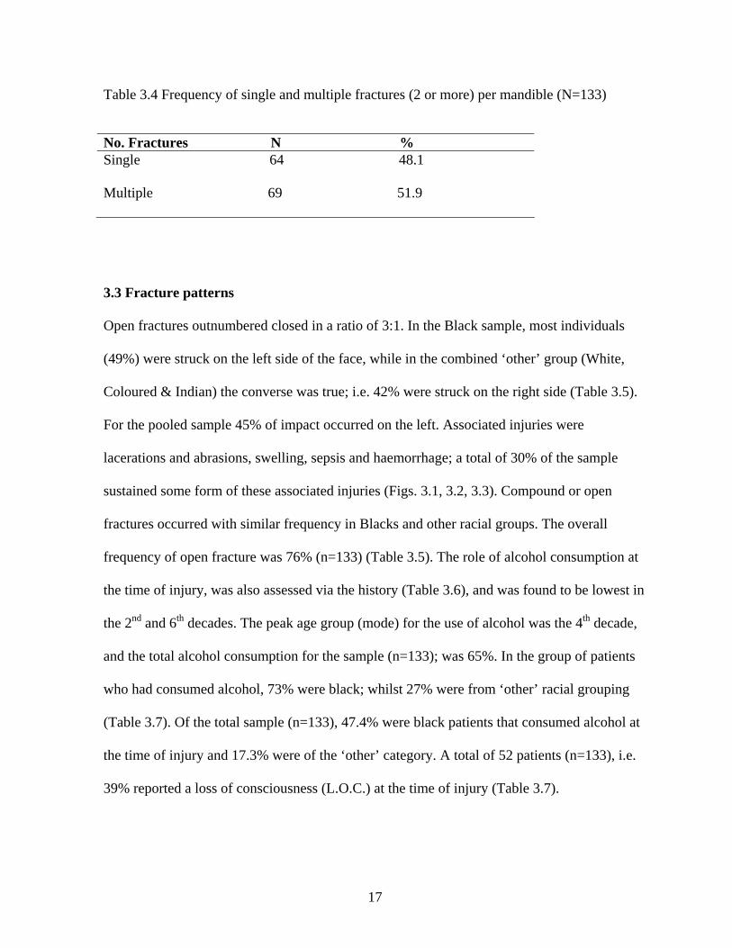
Table 3.4 Frequency of single and multiple fractures (2 or more) per mandible (N=133)
No. Fractures N % Single 64 48.1 Multiple 69 51.9
3.3 Fracture patterns
Open fractures outnumbered closed in a ratio of 3:1. In the Black sample, most individuals
(49%) were struck on the left side of the face, while in the combined ‘other’ group (White,
Coloured & Indian) the converse was true; i.e. 42% were struck on the right side (Table 3.5).
For the pooled sample 45% of impact occurred on the left. Associated injuries were
lacerations and abrasions, swelling, sepsis and haemorrhage; a total of 30% of the sample
sustained some form of these associated injuries (Figs. 3.1, 3.2, 3.3). Compound or open
fractures occurred with similar frequency in Blacks and other racial groups. The overall
frequency of open fracture was 76% (n=133) (Table 3.5). The role of alcohol consumption at
the time of injury, was also assessed via the history (Table 3.6), and was found to be lowest in
the 2nd and 6th decades. The peak age group (mode) for the use of alcohol was the 4th decade,
and the total alcohol consumption for the sample (n=133); was 65%. In the group of patients
who had consumed alcohol, 73% were black; whilst 27% were from ‘other’ racial grouping
(Table 3.7). Of the total sample (n=133), 47.4% were black patients that consumed alcohol at
the time of injury and 17.3% were of the ‘other’ category. A total of 52 patients (n=133), i.e.
39% reported a loss of consciousness (L.O.C.) at the time of injury (Table 3.7).

18
Fig.3.1 Swelling of the face. Fig.3.2 Abrasions of the torso.
Fig.3.3 Laceration of the face associated with fractured mandible.

19
Fig. 3.4 Mandibular fracture sites and frequencies in 133 patients.
Key: Total number of fractures= 203 (100%)
Blue= condyle 33 (16.8%), Orange= coronoid 0 (0%)
Green= ramus 0 (0%), Red= angle 79 (38.9%)
Pink= body 36 (17.7%), Brown= dentoalveolus 5 (2.5%)
Yellow= Parasymphysis 39 (19.2%), Black= symphysis 11 (5.4%)
A total of 203 fractures were recorded in the sample, a mean of 1.5 fractures per
mandible. Angle fractures were most common, followed by parasymphyseal; body; condyle;
symphysis and dentoalveolar, respectively (Fig. 3.4). Ramus and coronoid fractures were not
seen in this sample group.

20
Table 3.5 Fracture characteristics by race group (N=133)
Black n=100 N %
Other n=33 N %
Total n=133 N %
Impact Site left midline right
49 49 19 19 32 32
10 30 9 27 14 42
59 45 28 21 46 34
Fracture open Type closed
75 75 25 25
26 79 7 21
101 76 32 24
Associated injuries 31 31 9 27 40 30
Table 3.6 Frequency distribution of fracture type, associated injury and alcohol
consumption by decade. (N=133)
Decade (years)
Fracture Type open closed N % N %
Associated Injury N %
Alcohol Consumed N %
2(10-19) 3(20-29) 4(30-39) 5(40-49) 6(50-59)
6 5.9 0 0 38 37.6 13 37.5 39 38.6 13 37.5 16 15.8 5 15.6 2 2.0 1 3.1
1 2.5 15 37.5 15 37.5 8 20.0 1 2.5
3 3.5 30 34.9 35 40.7 16 18.6 2 2.3
Total 101 32 40 86

21
Table 3.7 Frequency distribution of alcohol consumption and loss of consciousness (LOC) by race group (N=133)
Race group Alcohol
N % LOC N %
Black Other
63 47.4
23 17.3
35 26 17 13
Total
86 64.7
52 39
Table 3.8 Frequency distribution of tooth in fracture line; nerve damage; displaced
fracture; fracture type, and treatment rendered (N=133)
N % Tooth in fracture line 102 76.7 Nerve damage 77 57.9 Displaced fracture 98 73.7 Fracture type open 101 75.9 closed 32 24.1 Treatment none 7 5.3 closed reduction 27 20.3 open reduction 99 74.4

22
3.4 Treatments and cost of hardware
Fractures of the mandible were displaced in 73.7% of cases; with associated nerve
paraesthesia in 57.9% of cases, and 76.7% of fractures contained a tooth within the line of
fracture (Table 3.8). Furthermore, 5.3% of patients required no form of surgery, whilst 20.3%
were treated by closed reduction and fixation of mandible (CRFM) and 74.4% were managed
via open reduction with internal fixation (ORIF).
An audit of departmental records for the statistics on fractured mandibles for the years 2002-
2004, confirmed that a total of 198 (2002); 133 (2003); and 217 (2004) ORIF’s were done at
the Johannesburg hospital. This equates to a total of 548 ORIF’s for the last three years, a
mean of 182.6 ORIF’s per annum.
The rigid fixation systems of four companies were used in these open reductions (ORIF’s).
All comprised of 4-hole extended mini-plates and 2.0mm diameter screws (Fig 3.5). Due to a
verbal undertaking not to divulge the details of the cost of the products of individual
suppliers, an average costing across all four is presented: screws @ R97.79 per unit, 4-hole
extended plate @ R313.56 per unit.
From the current study (Table 3.4), 48.1% of ORIF’s were single and 51.9% were multiple
i.e. a minimum of two fracture sites per mandible. Thus, of the 182.6 ORIF’s done per annum
(average no. ORIF’s per annum from department statistics 2002-4) 87.8 (182.6 x 48.1%) will
require a single mini-plate and 4 screws; whilst 94.7 (182.6 x 51.9%) will require a minimum

23
of 2 mini-plates and 8 screws. The trends of fracture number have been applied to department
stats. 2002-4. Therefore direct cost of ‘hardware’ is as follows:
87.8 x 1 mini-plate @ R313.56 each = R27 530.57
87.8 x 4 screws @ R97.79 each = R34 343.85
94.7 x 2 mini-plates @ R313.56 each = R59 388.26
94.7 x 4 screws @ R97.79 each = R37 042.85
Therefore mean cost of ‘hardware’ for ORIF’s per annum = R158 305.53 or US$ 25 048 (rate
US$ 1 = R 6.32). It should be emphasized that the aforementioned costing does not include
surgical, anaesthetic, hospitalization, and consumables other than plates and screws.
Fig. 3.5 ‘ORIF’ shows two 4-hole extended mini-plates and their 2.0mm screws.

24
CHAPTER 4: DISCUSSION
4.1 General
The Johannesburg Hospital provides care for patients without private health insurance, and
thus mainly caters for the health needs of the poorer members of our communities. At least
three quarters of the patients treated in this study were Black. Historically, the Black
population has been underprivileged and although there appears to be an emerging Black
middle-class since democratic reformation in South Africa; the bulk of the Black population
still appears to be the neediest in our country.
Furthermore, young healthy males comprise the bulk of the current study sample; a finding
that has not changed since earlier studies 4,5,11,12,13,16. The obvious explanation for this would
be that these males are of working age and are interacting amongst each other, with
altercations being common. The reality however, is that a staggering amount of mandibular
fractures are as a result of criminally motivated trauma. The level of inter-personal violence is
an unacceptable 86.5%; with most of these being robberies. From my interaction with the
patients, and from the social history taking; it is almost uniformly the same modus operandi
by the criminal element. Patients are most frequently attacked on Friday and Saturday
evenings (when most labourers receive their weekly wage) after a spell of alcohol
consumption at informal drinking establishments (known as Shebeens). The victims are
ambushed and struck in the face with blunt objects (usually rocks, sticks or the butt of a
handgun). This has the effect of dazing or rendering the victim unconscious, so that the
robbing of money and possessions proceeds unhindered. Often, these attacks are so brutal that
they result in significant co-injury, such as head injury; extensive soft tissue laceration and

25
limb fracture (Figures 3.1-3.3). This is of importance, as some of these patients will be kept at
peripheral hospitals and only referred once the other injuries have been stabilized (up to 2
weeks at times). Abiose 14 from Nigeria, reports that in the western states of his country,
4.81% of maxillofacial trauma was due to violent crime, whilst 80.77% was attributed to road
traffic accidents. In our sample the converse was true, with only 13.5% being accidental.
Returning to the current South African sample; the potential role that alcohol plays in the
contribution to the trauma rate, can not be emphasized enough. At least 65% of all patients
who sustained fractured mandibles did so whilst intoxicated. Almost the entire spectrum of
the sample admitted to the frequent consumption of alcohol, and whether those that denied
intoxication at time of injury, were being sincere or not we will never know. A further
dilemma regarding the role of alcohol becomes apparent, when the head injury statistic is
closely scrutinized. The transient loss of consciousness (LOC) implies at the very least, a mild
head injury 17. Approximately 39% of patients reported a LOC. with many unable to specify
the duration of LOC. As many of these patients were also intoxicated at the time, the patient’s
interpretation of what constituted a LOC; and a mere ‘passing out’, may have been confused;
thereby making the head injury statistic an unreliable one.
Another interesting statistic is the low prevalence of sporting injury in the sample 2/133
(1.5%). One may speculate that sporting injury may be higher among the private medical
facilities; or that fewer youth in the poorer communities are participating in contact sport, or
that soccer (the preferred sport of the Black population) has a much lower prevalence of facial
trauma than say rugby or cricket (traditionally favoured by ‘other’ population groups). In

26
Europe, the prevalence of sport related fractures seems to be much higher. For example, in
Greece soccer accounts for up to 64% of sport related facial fractures in which the zygoma
was more commonly fractured than the mandible 15.
In 1978, Reitzik and co-workers 18 performed experiments on the force required to fracture
monkey mandibles. The findings of their study suggest that mandibles with unerrupted third
molars, require 40% less force to cause a fracture in the angle region. In fact, the more severe
the impaction of the third molar, the greater the chance of fracture as a consequence of blunt
trauma 19. Since the bulk of our patients were in their 3rd and 4th decades, and mostly did not
have lower 3rd molars; and since angle fractures were the most common type, it may be
deduced that the blows received must have been of considerable magnitude. The blows
received were forceful enough, as 76% of all fractures in the series were open (compound);
and these open fractures out-numbered closed (simple) fractures by a ratio of 3:1. Open
fractures need not be frankly compound into the oral cavity, or onto the skin; but even
extension of the fracture line into the periodontium of a tooth qualifies as an open fracture. In
this study, at least 77% of teeth were in the line of fracture; and this invariably incorporated
lower anterior teeth in symphyseal or parasymphyseal fractures. Unfortunately, these teeth
were all removed as part of treatment protocol in the prevention of post surgical sepsis.
Edward Ellis III 20 and Alpert 21 both report that there is a greater chance of sepsis if a tooth is
retained, but this risk is not significant. Admittedly, our patients do not display the finest oral
hygiene, but perhaps aesthetically important anterior teeth should be retained in open fractures
that are rigidly fixed; and the post-operative vitality of these teeth should be monitored.

27
Another statistic that appears to correlate closely is the displacement of fracture segments and
the presence of neurological fall out; although this is not statistically significant (table 3.4.1).
The larger the displacement of proximal and distal fracture segments, the more likely one is to
find nerve damage, as tugging or even severing of the inferior alveolar nerve will produce
paraesthesia or anaesthesia respectively.
The treatment of fractures in this series ranged from conservative to CRFM, to ORIF.
Conservative management, in essence, is a soft diet with no surgical intervention; and is the
treatment of choice for fractures that are undisplaced, primarily asymptomatic and with no
neurological compromise. Closed reductions (CRFM) were used in class I fractures that were
only mildly displaced, but had to be no older than 72 hrs; or for grossly comminuted fractures.
The indications for open reductions (ORIF) in this series were: nerve paraesthesia; moderate
to severely displaced fracture segments; older fractures (>1 week post-injury); class III
fractures; bilateral fractures, especially one with a high condylar fracture requiring early
mobilization; and where CRFM was contraindicated e.g. epileptics. The bulk of our patients
presented late; then, were kept waiting for surgery due to overcrowded operating lists, and
hence were mandatory open reductions. The Champy miniplate placed along the line of ideal
osteosynthesis 22, using monocortical screws; was the preferred method of open rigid fixation.
Alternative techniques include the AO/ASIF system of compression or reconstruction plates
with bicortical screws; the bicortical Luhr system, using vitallium plates 23; and lag-screw
osteosynthesis 24.

28
The role of human immunodeficiency virus (HIV) on the postoperative healing of treated
fractures was not studied in this research but should be evaluated in future. In South Africa, as
in most of Sub-Saharan Africa, HIV has become a major factor in the pathogenesis of disease
and trauma. In our institution, anecdotal evidence is suggestive of increased postoperative
sepsis in open reductions of mandibular fractures. Credibility to the aforementioned statement
is offered by a study done on HIV patients in San Francisco 25. In this study, an overall sepsis
rate of 30% for HIV positive patients was recorded, opposed to 9.5% in the HIV negative
control. Sepsis when it did occur was statistically significant for bone rather than soft tissue.
The rate of sepsis in both sample and control were higher for ORIF (16%) than for CRFM
(3.1%). Further, the overall sepsis rate for ORIF in HIV positive patients; was 45% as
compared to 13.9% in the control group. These statistics also mitigate in favour of early
intervention, by way of CRFM, in our patients.
4.2 Matching resources to treatment needs
From this study, indications are that there is an upward trend in the number of mandibular
fractures treated at the Johannesburg Hospital. The concomitant rise in other trauma
especially the life and limb threatening variety; means that fractured mandibles have to
compete for operating theatre time, and are thus treated ‘cold’. There can be no justification in
general, to give fractured mandibles priority on after-hours emergency theatre lists.
Furthermore, we at the Johannesburg hospital are short on emergency theatre staff, including
anaesthetists. Thus, the elective repair of mandibular fractures means that a large proportion
of these cases require ORIF. Were these cases treated within 72 hrs of injury, then the bulk of
them would be done as CRFM; a considerably cheaper treatment modality. The closed

29
reductions (CRFM) require little of the costly hardware (see section 3.4) that ORIF’s do, and
the overall hospitalization is about half as long. Further, the CRFM may be done in half the
time that it takes to complete an ORIF, and may be done by one operator; whereas the open
reduction requires two surgeons. Because of the number of patients awaiting surgery, often a
fractured mandible can only be treated 4-6 weeks post-injury. In the 70’s and 80’s,
maxillofacial surgeons were stationed at fully functional units located at peripheral hospitals.
This meant that facial trauma was being attended to sooner, with presumably larger savings
on direct treatment costs. These posts have in more recent years been discontinued. Is there
perhaps an argument for the recreation of posts, and equipping of facilities at peripheral
hospitals? Perhaps state patients could be treated in the private sector at a negotiated tariff, to
alleviate the burden placed on state hospitals?
4.3 Socio-economic upliftment strategies
A total of 18/133 patients, or 13.5% in this series were female. This is an increase in the
number of females treated, when compared to previous Johannesburg studies 5, 13. A male to
female ratio of 6.4:1 was recorded in this study, and this is a considerable increase in the
number of female patients, on the previous ratio of 8.5:1 documented by Rosenberg and
Smith in 1975 4. A staggering 88% of women (n=16) were victims of domestic abuse; being
assaulted by their partners. In a society where the abuse of women and children has been
attracting an increase in attention; this statistic does not flatter! Even more discouraging, was
the unwillingness of these patients to report the abuse to the South African Police Services
(SAPS) or to be referred to social services at the hospital. Whether it is the ignorance of the
victims or intimidation by the abusers, that protects these perpetrators; something has to be

30
done. The women of our country have to be educated regarding their rights to safety, and the
law enforcement agencies need to guarantee protection to women, that report their abusers to
the authorities.
Other members of our society that are particularly vulnerable to criminal abuse are the
informal labour of our country. These individuals are often illiterate, impoverished and are
paid by their employers on a weekly basis-usually with banknotes as tender. The lack of
formal payment into banking accounts, make these individuals a predictably soft target for
muggers. Finally, the informal townships that lay scattered around our province are usually
the setting for criminally motivated trauma. These settlements are to be targeted by local
government, to improve amenities such as street lighting etc. thereby creating a safer
environment for those that dwell in them.
4.4 Conclusion
The findings of this report suggest that the proportions of causes of mandibular fractures are
largely unchanged from earlier audits 5, 11, 13. There is however an increase in the total number
of fractures to the mandible, with particular increase in the female population. From the data
collected, fractures were being treated after varying delays, which ultimately resulted in more
complicated and costly treatment modalities.
The unacceptably high incidence of crime and alcohol related trauma is no doubt placing
enormous strain on the financial and personnel resources, of both provincial and state health
departments. Local government are mandated to provide safety and security for its

31
constituents, and endeavours in this regard may alleviate an already overburdened public
health sector. Clinicians working in these public health institutions, have also their part to
play in providing adequate services to trauma patients. It is upon the judgement of the treating
clinician to decide on the most appropriate methods to be used in the management of
maxillofacial fractures. This decision, has to consider many factors such as the class of
fracture, the state of the bone and dentition, the co-morbidities of the patient and also the
‘hardware’ at the disposal of the surgeon. The soaring costs of materials in use today, mandate
surgeons to be circumspect and to engineer appropriate yet cost-efficient treatment protocols
for our patients.
4.5 Further research requirements
Further research is required, to identify feasible methods of decreasing the number of
mandatory ORIF’s done in our units. Furthermore, research into alternative techniques of
internal fixation of jaw fractures, is conceivable. And finally, medium to longer-term
prospective studies on the post-ORIF sepsis rates, when teeth within the line of fracture are
retained; would be an invaluable study; as would formal post-operative sepsis rates on HIV
positive patients, undergoing mandibular repair.
Related Documents


