Changes in the hypothalamo-pituitary-adrenocortical and hypothalamo-pituitary-thyroid axes in diabetic rats acclimated to moderate hyperthermic environment Suzana Dinevska-Kjovkarovska a, , Teuta Guladin b , Biljana Miova a , Slavco Mitev a , Katerina Gerazova a a Department of Physiology and Biochemistry, Faculty of Natural Sciences and Mathematics, Gazi Baba bb, 1000 Skopje, Republic of Macedonia b State University of Tetovo, Republic of Macedonia article info Article history: Received 23 September 2008 Accepted 4 February 2009 Keywords: Experimental diabetes High environmental temperature Insulin ACTH Corticosterone TSH T 3 T 4 Endocrine glands Rats abstract We examined the effect of acclimation to moderate hyperthermic environment on the ACTH, TSH, T 3 ,T 4 and corticosterone level, as well as the relative weight of hypophysis, thyroid and adrenal glands in streptozotocin-diabetic rats. Increased activity of the hypothalamo-pituitary-adrenocortical (HPA) axis has been demonstrated in diabetic animals, whereas insulin treatment restores the changes. Heat acclimation reduces the level of ACTH and corticosterone in control animals and moderates the hormonal disturbances caused by diabetes. Simultaneously, our study revealed impairment in the activity of the hypothalamo-pituitary-thyroid (HPT) axis. Acclimation to 3571 1C resulted in significantly lower T 3 and T 4 levels in control, diabetic and insulin-treated animals. Relative weight of the hypophysis, thyroid and adrenal glands is decreased in heat-acclimated rats. Our assumption is that there might be a cross tolerance between diabetes and heat acclimation on a hormonal level. & 2009 Elsevier Ltd. All rights reserved. 1. Introduction and literature As a complex and heterogenic metabolic syndrome, diabetes mellitus is characterized by extensive alterations in different metabolic pathways constituting the catabolic as well as the anabolic processes of the energy-providing substances (Delaunay et al., 1997), which are under the neuroendocrine regulation, also disarranged once diabetes occurs. Regarding this, in their previous studies, Chan et al. (2005) demonstrated that basal HPA function is up-regulated in streptozotocin (STZ)-diabetic rats and that this is associated with increases in hypothalamic corticotrophin-releasing hormone mRNA, plasma ACTH and corticosterone levels (Chan et al., 2001). Based on the study of Gonzalez et al. (1980), STZ-diabetes causes impairment of the activity of the hypothalamic-pituitary-thyroid (HPT) axis, resulting primarily in reduction of the hypothalamic thyrotropin- releasing hormone level, which subsequently leads to decreased plasma levels of thyroid hormones in diabetic rats. We wanted to examine not only the hormonal distur- bances caused by experimental diabetes, but also the cross tolerance—the phenomenon where adaptation to one environ- mental stressor sometimes provides protection against an additional type of stress (Horowitz, 2003). According to Collins and Weiner (1968), exposure of a homeothermic organism to high environmental temperature, as a form of stress, induces, primarily, its neural reflex responses, whereas the prolonged exposure results in activating secondary adaptive mechanisms including modified endocrine activity. Regarding the above-mentioned findings, we investigated the effect of prolonged exposure to moderate high environmental temperature (3571 1C, 28 days) in STZ-diabetic and insulin- treated rats over several hormonal parameters (ACTH, TSH, T 3 ,T 4 and corticosterone) and the relative mass weight of the some endocrine glands, hoping that the results would give a better understanding on the effect of the altered concentration of these hormones in animals with diabetes mellitus both in euthermic and hyperthermic environment. 2. Materials and methods 2.1. Experimental animals and treatment This experimental study was performed on adult (3–4 months old), male Wistar rats (n ¼ 60, 10 animals in each group), with an ARTICLE IN PRESS Contents lists available at ScienceDirect journal homepage: www.elsevier.com/locate/jtherbio Journal of Thermal Biology 0306-4565/$ - see front matter & 2009 Elsevier Ltd. All rights reserved. doi:10.1016/j.jtherbio.2009.02.001 Corresponding author. Tel.: +3892 3249602; fax: +389 23228141. E-mail addresses: [email protected] (S. Dinevska-Kjovkarovska), [email protected] (T. Guladin), [email protected] (B. Miova), [email protected] (S. Mitev), [email protected] (K. Gerazova). Journal of Thermal Biology 34 (2009) 200–205

Welcome message from author
This document is posted to help you gain knowledge. Please leave a comment to let me know what you think about it! Share it to your friends and learn new things together.
Transcript

ARTICLE IN PRESS
Journal of Thermal Biology 34 (2009) 200–205
Contents lists available at ScienceDirect
Journal of Thermal Biology
0306-45
doi:10.1
� Corr
E-m
(S. Dine
bmiova
(S. Mite
journal homepage: www.elsevier.com/locate/jtherbio
Changes in the hypothalamo-pituitary-adrenocorticaland hypothalamo-pituitary-thyroid axes in diabeticrats acclimated to moderate hyperthermic environment
Suzana Dinevska-Kjovkarovska a,�, Teuta Guladin b, Biljana Miova a, Slavco Mitev a, Katerina Gerazova a
a Department of Physiology and Biochemistry, Faculty of Natural Sciences and Mathematics, Gazi Baba bb, 1000 Skopje, Republic of Macedoniab State University of Tetovo, Republic of Macedonia
a r t i c l e i n f o
Article history:
Received 23 September 2008
Accepted 4 February 2009
Keywords:
Experimental diabetes
High environmental temperature
Insulin
ACTH
Corticosterone
TSH
T3
T4
Endocrine glands
Rats
65/$ - see front matter & 2009 Elsevier Ltd. A
016/j.jtherbio.2009.02.001
esponding author. Tel.: +389 2 3249602; fax:
ail addresses: [email protected]
vska-Kjovkarovska), [email protected]
@yahoo.com (B. Miova), [email protected]
v), [email protected] (K. Gerazova).
a b s t r a c t
We examined the effect of acclimation to moderate hyperthermic environment on the ACTH, TSH, T3, T4
and corticosterone level, as well as the relative weight of hypophysis, thyroid and adrenal glands in
streptozotocin-diabetic rats. Increased activity of the hypothalamo-pituitary-adrenocortical (HPA) axis
has been demonstrated in diabetic animals, whereas insulin treatment restores the changes. Heat
acclimation reduces the level of ACTH and corticosterone in control animals and moderates the
hormonal disturbances caused by diabetes. Simultaneously, our study revealed impairment in the
activity of the hypothalamo-pituitary-thyroid (HPT) axis. Acclimation to 3571 1C resulted in
significantly lower T3 and T4 levels in control, diabetic and insulin-treated animals. Relative weight of
the hypophysis, thyroid and adrenal glands is decreased in heat-acclimated rats. Our assumption is that
there might be a cross tolerance between diabetes and heat acclimation on a hormonal level.
& 2009 Elsevier Ltd. All rights reserved.
1. Introduction and literature
As a complex and heterogenic metabolic syndrome, diabetesmellitus is characterized by extensive alterations in differentmetabolic pathways constituting the catabolic as well as the anabolicprocesses of the energy-providing substances (Delaunay et al., 1997),which are under the neuroendocrine regulation, also disarrangedonce diabetes occurs. Regarding this, in their previous studies, Chanet al. (2005) demonstrated that basal HPA function is up-regulated instreptozotocin (STZ)-diabetic rats and that this is associated withincreases in hypothalamic corticotrophin-releasing hormone mRNA,plasma ACTH and corticosterone levels (Chan et al., 2001). Based onthe study of Gonzalez et al. (1980), STZ-diabetes causes impairmentof the activity of the hypothalamic-pituitary-thyroid (HPT) axis,resulting primarily in reduction of the hypothalamic thyrotropin-releasing hormone level, which subsequently leads to decreasedplasma levels of thyroid hormones in diabetic rats.
We wanted to examine not only the hormonal distur-bances caused by experimental diabetes, but also the cross
ll rights reserved.
+389 2 3228141.
.mk
k (T. Guladin),
kim.edu.mk
tolerance—the phenomenon where adaptation to one environ-mental stressor sometimes provides protection against anadditional type of stress (Horowitz, 2003). According to Collinsand Weiner (1968), exposure of a homeothermic organism tohigh environmental temperature, as a form of stress, induces,primarily, its neural reflex responses, whereas the prolongedexposure results in activating secondary adaptive mechanismsincluding modified endocrine activity.
Regarding the above-mentioned findings, we investigated theeffect of prolonged exposure to moderate high environmentaltemperature (3571 1C, 28 days) in STZ-diabetic and insulin-treated rats over several hormonal parameters (ACTH, TSH, T3, T4
and corticosterone) and the relative mass weight of the someendocrine glands, hoping that the results would give a betterunderstanding on the effect of the altered concentration of thesehormones in animals with diabetes mellitus both in euthermicand hyperthermic environment.
2. Materials and methods
2.1. Experimental animals and treatment
This experimental study was performed on adult (3–4 monthsold), male Wistar rats (n ¼ 60, 10 animals in each group), with an
ARTICLE IN PRESS
S. Dinevska-Kjovkarovska et al. / Journal of Thermal Biology 34 (2009) 200–205 201
estimated weight of 200–250 g. The animals were fed ad libitumwith free access to food and water. They were kept at 12 h lightcycle (lights on between 06 and 18 h).
The animals were divided into two major groups:
1.
TabPro
(20
I gr
II g
III g
TabPro
I gr
II g
III g
Animals kept at room temperature of 2072 1C.
2. Animals exposed to moderate hyperthermic environment(3571 1C) for a period of 28 days. The acclimation to atemperature of 3571 1C was carried out using a special heatchamber with regulated air temperature and air humidity(30–40%).
The animals of each thermal group were divided into threesubgroups:
1.
control animals (C); 2. diabetic animals (D); 3. insulin-treated diabetic animals (DI).The induction of the experimental diabetes mellitus wasperformed by a single intraperitoneal injection of streptozotocin(STZ, 55 mg/kg body weight) dissolved in 0.1 M citrate buffer,pH ¼ 4.5. All animals with clear diabetic symptoms (fastingglycemia levels higher than 15 mmol/L) 48 h after the inductionof the experimental diabetes, were used for the purpose of thisexperiment. In order to treat the diabetic symptoms, the thirdsubgroup of animals (DI) of each thermal group, was given NovoNordisk insulin solution (2 IU/100 g body weight) for a period of14 days, 7 days after the induction of the STZ-diabetes. Insulin wasgiven subcutaneously, twice a day, for a period of 12 h, after themeasurement of the blood glucose levels (Abbott, Medisense).
2.2. Experimental procedure
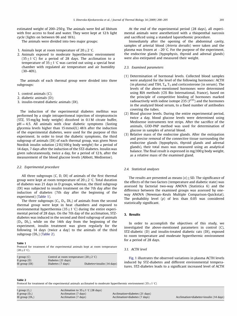
All three subgroups (C, D, DI) of animals of the first thermalgroup were kept at room temperature of 2072 1C. Total durationof diabetes was 21 days in D groups, whereas, the third subgroup(DI) was subjected to insulin treatment on the 7th day after theinduction of diabetes (7th day after the beginning of theexperiment) (Table 1).
The three subgroups (C1, D1, DI1) of animals from the secondthermal group were kept in heat chambers and exposed toenvironmental hyperthermia (3571 1C) during the entire experi-mental period of 28 days. On the 7th day of the acclimation, STZ-diabetes was induced in the second and third subgroup of animals(D1, DI1), while on the 14th day from the beginning of theexperiment, insulin treatment was given regularly for thefollowing 14 days (twice a day) to the animals of the thirdsubgroup (DI1) (Table 2).
le 1tocol for treatment of the experimental animals kept at room temperature
72 1C).
oup (C) Control at room temperature (2072 1C)
roup (D) Diabetes (21 days)
roup (DI) Diabetes (7 days) Diabetes+insulin (14 days)
le 2tocol for treatment of the experimental animals acclimated to moderate hypertherm
oup (C1) Acclimation to 3571 1C (28 days)
roup (D1) Acclimation (7 days) Acclimat
roup (DI1) Acclimation (7 days) Acclimat
At the end of the experimental period (28 days), all experi-mental animals were anesthetized with a thiopenthal narcosisand sacrificed using a standard laparothomic procedure.
Immediately after the opening of the abdominal cavity,samples of arterial blood (Arteria dorsalis) were taken and theplasma was frozen at �20 1C. For the purpose of the experiment,the endocrine glands (hypophysis, thyroid and adrenal glands)were also extirpated and measured their weight.
2.3. Examined parameters
(1)
ic e
ion+d
ion+d
Determination of hormonal levels. Collected blood sampleswere analyzed for the level of the following hormones: ACTH(in plasma) and TSH, T4, T3 and corticosterone (in serum). Thelevels of the above-mentioned hormones were determinedusing RIA methods (CIS Bio International, France), based onthe principle of competitive binding of hormones markedradioactively with iodine isotope 235 (I235) and the hormonesin the analyzed blood serum, to a fixed number of antibodiescovering the tubes.
(2)
Blood glucose levels. During the entire experimental period,twice a day, blood glucose levels were determined usingMedisense instruments test strips. After the sacrifice of theanimals, GOD-PAP method was used for determination ofglucose in samples of arterial blood.(3)
Relative mass of the endocrine glands. After the extirpationand precise removal of the connective tissue surrounding theendocrine glands (hypophysis, thyroid glands and adrenalglands), their total mass was measured using an analyticalbalance. The final result is expressed in mg/100 g body weight,as a relative mass of the examined gland.2.4. Statistical analyses
The results are presented as means (x)7SD. The significance ofthe effects of the two factors (temperature and diabetic state) wasassessed by factorial two-way ANOVA (Statistica 6) and thedifference between the examined groups was assessed by one-way ANOVA (Newman–Keuls Multiple Comparison-Quickstat).The probability level (p) of less than 0.05 was consideredstatistically significant.
3. Results
In order to accomplish the objectives of this study, weinvestigated the above-mentioned parameters in control (C),STZ-diabetic (D) and insulin-treated diabetic rats (DI), exposedto room temperature and moderate hyperthermic environmentfor a period of 28 days.
3.1. ACTH level
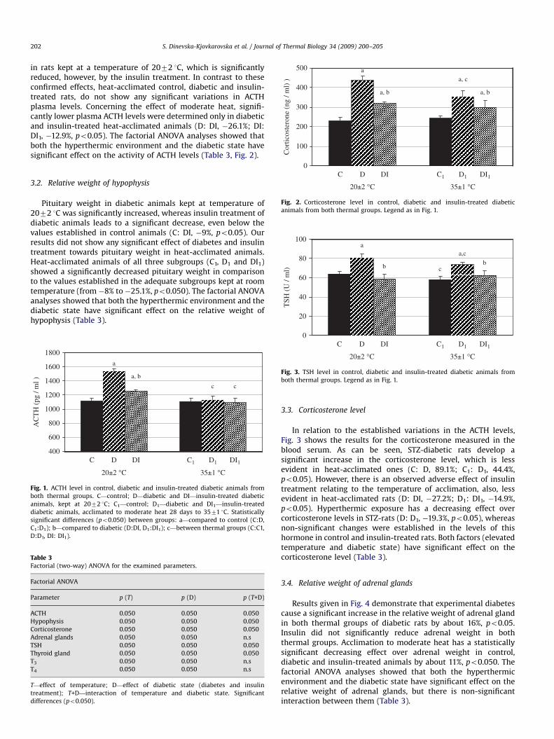
Fig. 1 illustrates the observed variations in plasma ACTH levelsinduced by STZ-diabetes and different environmental tempera-tures. STZ-diabetes leads to a significant increased level of ACTH
nvironment (3571 1C)
iabetes (21 days)
iabetes (7 days) Acclimation+diabetes+insulin (14 days)
ARTICLE IN PRESS
100
200
300
400
500
Cor
ticos
tero
ne (
ng /
ml)
)
a
a, b
a, c
a, b
S. Dinevska-Kjovkarovska et al. / Journal of Thermal Biology 34 (2009) 200–205202
in rats kept at a temperature of 2072 1C, which is significantlyreduced, however, by the insulin treatment. In contrast to theseconfirmed effects, heat-acclimated control, diabetic and insulin-treated rats, do not show any significant variations in ACTHplasma levels. Concerning the effect of moderate heat, signifi-cantly lower plasma ACTH levels were determined only in diabeticand insulin-treated heat-acclimated animals (D: DI, �26.1%; DI:DI1, �12.9%, po0.05). The factorial ANOVA analyses showed thatboth the hyperthermic environment and the diabetic state havesignificant effect on the activity of ACTH levels (Table 3, Fig. 2).
0
20±2 °C
C DI1D1C1DID
35±1 °C
Fig. 2. Corticosterone level in control, diabetic and insulin-treated diabetic
animals from both thermal groups. Legend as in Fig. 1.
a,ca
bb
20
40
60
80
100
TSH
(U
/ m
l) c
3.2. Relative weight of hypophysis
Pituitary weight in diabetic animals kept at temperature of2072 1C was significantly increased, whereas insulin treatment ofdiabetic animals leads to a significant decrease, even below thevalues established in control animals (C: DI, �9%, po0.05). Ourresults did not show any significant effect of diabetes and insulintreatment towards pituitary weight in heat-acclimated animals.Heat-acclimated animals of all three subgroups (C1, D1 and DI1)showed a significantly decreased pituitary weight in comparisonto the values established in the adequate subgroups kept at roomtemperature (from �8% to�25.1%, po0.050). The factorial ANOVAanalyses showed that both the hyperthermic environment and thediabetic state have significant effect on the relative weight ofhypophysis (Table 3).
400
600
800
1000
1200
1400
1600
1800
AC
TH
(pg
/ m
l )
20±2 °C
D1 DI1
a
a, b
cc
C1DIDC
35±1 °C
Fig. 1. ACTH level in control, diabetic and insulin-treated diabetic animals from
both thermal groups. C—control; D—diabetic and DI—insulin-treated diabetic
animals, kept at 2072 1C; C1—control; D1—diabetic and DI1—insulin-treated
diabetic animals, acclimated to moderate heat 28 days to 3571 1C. Statistically
significant differences (po0.050) between groups: a—compared to control (C:D,
C1:D1); b—compared to diabetic (D:DI, D1:DI1); c—between thermal groups (C:C1,
D:D1, DI: DI1).
Table 3Factorial (two-way) ANOVA for the examined parameters.
Factorial ANOVA
Parameter p (T) p (D) p (T+D)
ACTH 0.050 0.050 0.050
Hypophysis 0.050 0.050 0.050
Corticosterone 0.050 0.050 0.050
Adrenal glands 0.050 0.050 n.s
TSH 0.050 0.050 0.050
Thyroid gland 0.050 0.050 0.050
T3 0.050 0.050 n.s
T4 0.050 0.050 n.s
T—effect of temperature; D—effect of diabetic state (diabetes and insulin
treatment); T+D—interaction of temperature and diabetic state. Significant
differences (po0.050).
0
20±2 °C
C D1 DI1C1
35±1 °C
DID
Fig. 3. TSH level in control, diabetic and insulin-treated diabetic animals from
both thermal groups. Legend as in Fig. 1.
3.3. Corticosterone level
In relation to the established variations in the ACTH levels,Fig. 3 shows the results for the corticosterone measured in theblood serum. As can be seen, STZ-diabetic rats develop asignificant increase in the corticosterone level, which is lessevident in heat-acclimated ones (C: D, 89.1%; C1: D1, 44.4%,po0.05). However, there is an observed adverse effect of insulintreatment relating to the temperature of acclimation, also, lessevident in heat-acclimated rats (D: DI, �27.2%; D1: DI1, �14.9%,po0.05). Hyperthermic exposure has a decreasing effect overcorticosterone levels in STZ-rats (D: D1, �19.3%, po0.05), whereasnon-significant changes were established in the levels of thishormone in control and insulin-treated rats. Both factors (elevatedtemperature and diabetic state) have significant effect on thecorticosterone level (Table 3).
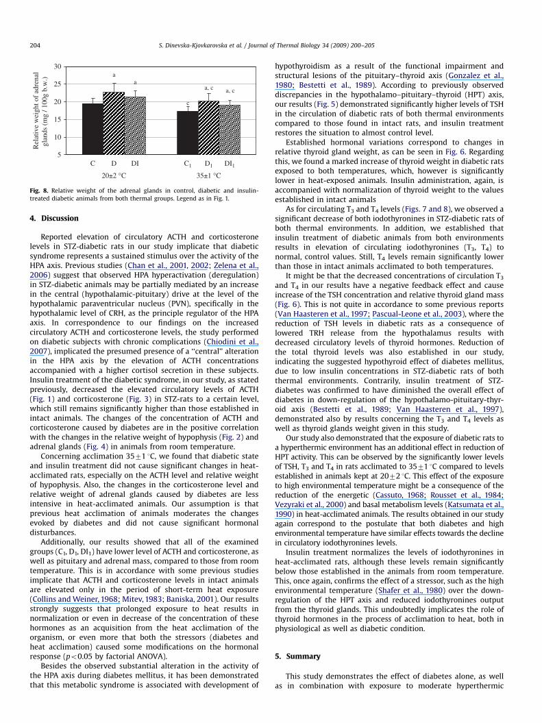
3.4. Relative weight of adrenal glands
Results given in Fig. 4 demonstrate that experimental diabetescause a significant increase in the relative weight of adrenal glandin both thermal groups of diabetic rats by about 16%, po0.05.Insulin did not significantly reduce adrenal weight in boththermal groups. Acclimation to moderate heat has a statisticallysignificant decreasing effect over adrenal weight in control,diabetic and insulin-treated animals by about 11%, po0.050. Thefactorial ANOVA analyses showed that both the hyperthermicenvironment and the diabetic state have significant effect on therelative weight of adrenal glands, but there is non-significantinteraction between them (Table 3).
ARTICLE IN PRESS
a,c
a
b
a, b, c
0.0
0.1
0.2
0.3
0.4
0.5
0.6
0.7
T3
(nm
ol /
L)
35±1 °C
c
DI1D1C1DIDC
20±2 °C
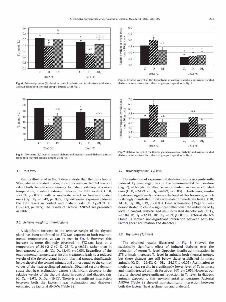
Fig. 4. Triiodothyronine (T3) level in control diabetic and insulin-treated diabetic
animals from both thermal groups. Legend as in Fig. 1.
c
aa
a, b, c
a, b
0
10
20
30
40
50
60
70
T4
(nm
ol /
L)
35±1 °C
C DI1D1C1DID
20±2 °C
Fig. 5. Thyroxine (T4) level in control diabetic and insulin-treated diabetic animals
from both thermal groups. Legend as in Fig. 1.
cc
a
c
a, b
3.0
3.5
4.0
4.5
5.0
5.5
6.0
Rel
ativ
e w
eigh
t of
hypo
phys
is
(mg
/ 100
g b.
w.)
20±2 °C
C D1 DI1C1DID
35±1 °C
Fig. 6. Relative weight of the hypophysis in control, diabetic and insulin-treated
diabetic animals from both thermal groups. Legend as in Fig. 1.
c
a, c
a
ba, b
3.0
3.5
4.0
4.5
5.0
5.5
6.0
6.5
7.0
Rel
ativ
e w
eigh
t of
thyr
oid
glan
d (m
g / 1
00g
b.w
.)
20±2 °C
C DI1D1C1DID
35±1 °C
Fig. 7. Relative weight of the thyroid glands in control, diabetic and insulin-treated
diabetic animals from both thermal groups. Legend as in Fig. 1.
S. Dinevska-Kjovkarovska et al. / Journal of Thermal Biology 34 (2009) 200–205 203
3.5. TSH level
Results illustrated in Fig. 5 demonstrate that the induction ofSTZ-diabetes is related to a significant increase in the TSH levels inrats of both thermal environments. In diabetic rats kept at a roomtemperature, insulin treatment reduces the TSH levels (D: DI,�27.5%, po0.05), with a moderate effect in heat-acclimatedones (D1: DI1, �15.4%, po0.05). Hyperthermic exposure reducesthe TSH levels in control and diabetic rats (C: C1,�9.5%, D:D1,�8.6%, po0.05). The results of factorial ANOVA are presentedin Table 3.
3.6. Relative weight of thyroid gland
A significant increase in the relative weight of the thyroidgland has been confirmed in STZ-rats exposed to both environ-mental temperatures, as can be seen in Fig. 6. However, thisincrease is more distinctly observed in STZ-rats kept at atemperature of 2072 1C (C: D, 28.1%, po0.05), rather than inheat-exposed animals (C1: D1, 13.4%, po0.05). Regardless of theenvironmental temperature, insulin treatment leads to a reducedweight of the thyroid gland in both thermal groups, significantlybelow those of the control animals and almost equal to the controlvalues of the heat-acclimated animals. Obtained results demon-strate that heat acclimation causes a significant decrease in therelative weight of the thyroid gland in control and diabetic rats(C: C1, �8.6%; D: D1, �19.1%). There is a significant interactionbetween both the factors (heat acclimation and diabetes)estimated by factorial ANOVA (Table 3).
3.7. Triiodothyronine (T3) level
The induction of experimental diabetes results in significantlyreduced T3 level regardless of the environmental temperature(Fig. 7), although the effect is more evident in heat-acclimatedones (C: D, �24.2%; C1: D1, �49.8%, po0.05). In both cases, insulintreatment significantly increases the level of this hormone, whichis strongly manifested in rats acclimated to moderate heat (D: DI,34.3%; D1: DI1, 63%, po0.05). Heat acclimation (3571 1C) wasdemonstrated to cause a significant effect over the reduction of T3
level in control, diabetic and insulin-treated diabetic rats (C: C1,�13.8%, D: D1, �32.4%; DI: DI1, �18%, po0.05). Factorial ANOVA(Table 3) showed non-significant interaction between both thefactors (heat acclimation and diabetes).
3.8. Thyroxine (T4) level
The obtained results illustrated in Fig. 8, showed thestatistically significant effect of induced diabetes over thereduction of serum T4 level. Opposite, insulin administration inSTZ-animals increases T4 level in animals both thermal groups,but these changes are still below those established in intactanimals (C: DI, �26.4%; C1: DI1, �24.3%, po0.05). Acclimation tomoderate heat results in significantly lower level of T4 in intactand insulin-treated animals for about 14% (po0.05). However, ourresults showed non-significant reduction in T4 level in diabeticanimals exposed to this environmental temperature. FactorialANOVA (Table 3) showed non-significant interaction betweenboth the factors (heat acclimation and diabetes).
ARTICLE IN PRESS
a, c
a
a, ca
5
10
15
20
25
30
Rel
ativ
e w
eigh
t of
adre
nal
glan
ds (
mg
/ 100
g b.
w.)
20±2 °C
C D1 DI1
c
C1DID
35±1 °C
Fig. 8. Relative weight of the adrenal glands in control, diabetic and insulin-
treated diabetic animals from both thermal groups. Legend as in Fig. 1.
S. Dinevska-Kjovkarovska et al. / Journal of Thermal Biology 34 (2009) 200–205204
4. Discussion
Reported elevation of circulatory ACTH and corticosteronelevels in STZ-diabetic rats in our study implicate that diabeticsyndrome represents a sustained stimulus over the activity of theHPA axis. Previous studies (Chan et al., 2001, 2002; Zelena et al.,2006) suggest that observed HPA hyperactivation (deregulation)in STZ-diabetic animals may be partially mediated by an increasein the central (hypothalamic-pituitary) drive at the level of thehypothalamic paraventricular nucleus (PVN), specifically in thehypothalamic level of CRH, as the principle regulator of the HPAaxis. In correspondence to our findings on the increasedcirculatory ACTH and corticosterone levels, the study performedon diabetic subjects with chronic complications (Chiodini et al.,2007), implicated the presumed presence of a ‘‘central’’ alterationin the HPA axis by the elevation of ACTH concentrationsaccompanied with a higher cortisol secretion in these subjects.Insulin treatment of the diabetic syndrome, in our study, as statedpreviously, decreased the elevated circulatory levels of ACTH(Fig. 1) and corticosterone (Fig. 3) in STZ-rats to a certain level,which still remains significantly higher than those established inintact animals. The changes of the concentration of ACTH andcorticosterone caused by diabetes are in the positive correlationwith the changes in the relative weight of hypophysis (Fig. 2) andadrenal glands (Fig. 4) in animals from room temperature.
Concerning acclimation 3571 1C, we found that diabetic stateand insulin treatment did not cause significant changes in heat-acclimated rats, especially on the ACTH level and relative weightof hypophysis. Also, the changes in the corticosterone level andrelative weight of adrenal glands caused by diabetes are lessintensive in heat-acclimated animals. Our assumption is thatprevious heat acclimation of animals moderates the changesevoked by diabetes and did not cause significant hormonaldisturbances.
Additionally, our results showed that all of the examinedgroups (C1, D1, DI1) have lower level of ACTH and corticosterone, aswell as pituitary and adrenal mass, compared to those from roomtemperature. This is in accordance with some previous studiesimplicate that ACTH and corticosterone levels in intact animalsare elevated only in the period of short-term heat exposure(Collins and Weiner, 1968; Mitev, 1983; Baniska, 2001). Our resultsstrongly suggests that prolonged exposure to heat results innormalization or even in decrease of the concentration of thesehormones as an acquisition from the heat acclimation of theorganism, or even more that both the stressors (diabetes andheat acclimation) caused some modifications on the hormonalresponse (po0.05 by factorial ANOVA).
Besides the observed substantial alteration in the activity ofthe HPA axis during diabetes mellitus, it has been demonstratedthat this metabolic syndrome is associated with development of
hypothyroidism as a result of the functional impairment andstructural lesions of the pituitary–thyroid axis (Gonzalez et al.,1980; Bestetti et al., 1989). According to previously observeddiscrepancies in the hypothalamo–pituitary–thyroid (HPT) axis,our results (Fig. 5) demonstrated significantly higher levels of TSHin the circulation of diabetic rats of both thermal environmentscompared to those found in intact rats, and insulin treatmentrestores the situation to almost control level.
Established hormonal variations correspond to changes inrelative thyroid gland weight, as can be seen in Fig. 6. Regardingthis, we found a marked increase of thyroid weight in diabetic ratsexposed to both temperatures, which, however is significantlylower in heat-exposed animals. Insulin administration, again, isaccompanied with normalization of thyroid weight to the valuesestablished in intact animals
As for circulating T3 and T4 levels (Figs. 7 and 8), we observed asignificant decrease of both iodothyronines in STZ-diabetic rats ofboth thermal environments. In addition, we established thatinsulin treatment of diabetic animals from both environmentsresults in elevation of circulating iodothyronines (T3, T4) tonormal, control values. Still, T4 levels remain significantly lowerthan those in intact animals acclimated to both temperatures.
It might be that the decreased concentrations of circulation T3
and T4 in our results have a negative feedback effect and causeincrease of the TSH concentration and relative thyroid gland mass(Fig. 6). This is not quite in accordance to some previous reports(Van Haasteren et al., 1997; Pascual-Leone et al., 2003), where thereduction of TSH levels in diabetic rats as a consequence oflowered TRH release from the hypothalamus results withdecreased circulatory levels of thyroid hormones. Reduction ofthe total thyroid levels was also established in our study,indicating the suggested hypothyroid effect of diabetes mellitus,due to low insulin concentrations in STZ-diabetic rats of boththermal environments. Contrarily, insulin treatment of STZ-diabetes was confirmed to have diminished the overall effect ofdiabetes in down-regulation of the hypothalamo-pituitary-thyr-oid axis (Bestetti et al., 1989; Van Haasteren et al., 1997),demonstrated also by results concerning the T3 and T4 levels aswell as thyroid glands weight given in this study.
Our study also demonstrated that the exposure of diabetic rats toa hyperthermic environment has an additional effect in reduction ofHPT activity. This can be observed by the significantly lower levelsof TSH, T3 and T4 in rats acclimated to 3571 1C compared to levelsestablished in animals kept at 2072 1C. This effect of the exposureto high environmental temperature might be a consequence of thereduction of the energetic (Cassuto, 1968; Rousset et al., 1984;Vezyraki et al., 2000) and basal metabolism levels (Katsumata et al.,1990) in heat-acclimated animals. The results obtained in our studyagain correspond to the postulate that both diabetes and highenvironmental temperature have similar effects towards the declinein circulatory iodothyronines levels.
Insulin treatment normalizes the levels of iodothyronines inheat-acclimated rats, although these levels remain significantlybelow those established in the animals from room temperature.This, once again, confirms the effect of a stressor, such as the highenvironmental temperature (Shafer et al., 1980) over the down-regulation of the HPT axis and reduced iodothyronines outputfrom the thyroid glands. This undoubtedly implicates the role ofthyroid hormones in the process of acclimation to heat, both inphysiological as well as diabetic condition.
5. Summary
This study demonstrates the effect of diabetes alone, as wellas in combination with exposure to moderate hyperthermic

ARTICLE IN PRESS
S. Dinevska-Kjovkarovska et al. / Journal of Thermal Biology 34 (2009) 200–205 205
environment over the up-regulation of the hypothalamo-pituitary-adrenal axis and the down-regulation of the hypothalamo-pituitary-thyroid axis. Insulin administration however, was con-firmed to restore their hormonal activity to a normal level,regardless of previous thermal acclimation. Heat acclimation causessome modifications on both the hypothalamo-pituitary-adrenaland hypothalamo-pituitary-thyroid axis which can be generallyobserved as a down-regulation of the most of the hormonalchanges. Our assumption is that there might be a cross tolerancebetween diabetes and heat acclimation on the hormonal level.
References
Baniska, A., 2001. Effect of temperature as an external factor over hormonalmeasurements and the levels of several hormones. Master Thesis, Skopje (inMacedonian).
Bestetti, g.e., Reymond, m.j., Boujon, c.e., Lemarchand-beraud, t., Rossi, g.l., 1989.Functional and morphological aspects of impaired TRH release by mediobasalhypothalamus of STZ-induced diabetic rats. Diabetes 381, 351–1356.
Cassuto, Y., 1968. Metabolic adaptations to chronic heat exposure in the goldenhamster. Am. J. Physiol. 214, 1147–1151.
Chan, O., chan, S., Inouye, K., Vranic, M., Matthews, S.G., 2001. Molecular regulationof the hypothalamic-pituitary-adrenal axis in streptozotocin-induced diabetes:effects of insulin treatment. Endocrinology 142, 4872–4879.
Chan, O., Inouye, K., Vranic, M., Matthews, S.G., 2002. Hyperactivation of thehypothalamo-pituitary-adrenocortical axis in streptozotocin-diabetes is asso-ciated with reduced stress responsiveness and decreased pituitary and adrenalsensitivity. Endocrinology 143, 1761–1768.
Chan, O., Inouye, K., Akirav, E.M., Park, E., Riddell, M.C., Matthews, S.G., Vranic, M.,2005. Hyperglycemia does not increase basal hypothalamo-pituitary-adrenalactivity in diabetes but it does impair the HPA response to insulin-inducedhypoglycemia. Am. J. Physiol Regul. Integrative Comp. Physiol. 289, 235–246.
Chiodini, I., Adda, G., Scillitani, A., Coletti, F., Morelli, V., Lembo, S., Epaminonda, P.,Masserini, B., Beck-Peccoz, P., Orsi, E., Ambrosi, B., Arosio, M., 2007. Cortisol
secretion in patients with type 2 diabetes-relationship with chronic complica-tions. Diabetes Care 30, 83–88.
Collins, K.J., Weiner, J.S., 1968. Endocrinological aspects of exposure to highenvironmental temperatures. Physiol. Rev. 48, 785–839.
Delaunay, F., Khan, A., Cintra, A., Davani, B., Ling, Z.C., Andersson, A., Ostenson, C.G.,Gustafsson, J.A., Efendic, S., Okret, S., 1997. Pancreatic b cells are importanttargets for the diabetogenic effects of glucocorticoids. J. Clin. Invest. 100 (8),2094–2098.
Gonzalez, C., Montoya, E., Jolin, T., 1980. Effect of streptozotocin diabetes on thehypothalamic-pituitary-thyroid axis in the rat. Endocrinology 107, 2099–2103.
Horowitz, M., 2003. Matching the heart to heat-induced circulatory load: heat-acclimatory responses. News Physiol. Sci. 18, 215–221.
Van Haasteren, G.A., Sleddens-Linkels, E., Van Toor, H., Klootwijk, W., de Jong, F.H.,Visser, T.J., de Greef, W.J., 1997. Possible role of corticosterone in the down-regulation of the hypothalamo-hypophysial-thyroid axis in streptozotocin-induced diabetes mellitus in rats. J. Endocrinol. 153 (2), 259–267.
Katsumata, M., Yano, H., Ishida, N., Miyazaki, A., 1990. Influence of a high ambienttemperature and administration of clenbuterol on the body composition inrats. J. Nutr. Sci. Vitaminol. 36, 569–578.
Mitev, S., 1983. Influence of high environmental temperature and some endocrinefactors on the liver glycogen content and some other parameters in white rat.Doctoral Thesis, Skopje (in Macedonian).
Pascual-Leone, A.M., Ramos, S., Goya, L., Alvarez, C., Escriva, F., Obregon, M.J., 2003.Age-dependent adaptation of the liver thyroid status and recovery of serumlevels and hepatic insulin-like growth factor-I expression in neonatal and adultdiabetic rats. Metabolism 52 (9), 1117–1125.
Rousset, B., Jordan, D., Kervran, A., Borne, t.H., 1984. Metabolic alterations inducedby chronic heat exposure in the rat: the involvement of thyroid function.Pflugers. Arch. 401, 64–70.
Shafer, R.B., Oken, M.M., Elson, M.K., 1980. Effects of fever and hyperthermia onthyroid function. J. Nucl. Med. 21, 1158–1169.
Zelena, D., Filaretova, L., Mergl, Z., Barna, I., Toth, Z.E., Makara, G.B., 2006.Hypothalamic paraventricular nucleus, but not vasopressin, participates inchronic hyperactivity of the HPA axis in diabetic rats. Am. J. Physiol.Endocrinol. Metab. 290, E243–E250.
Vezyraki, P., Kalfakakou, V., Evangelou, A., 2000. Atrial natriuretic peptide andthyroid hormones’ relation to plasma and heart calcium and magnesiumconcentrations of Wistar rats exposed to cold and hot ambients. Biol. TraceElem. Res. 73 (2), 163–173.
Related Documents