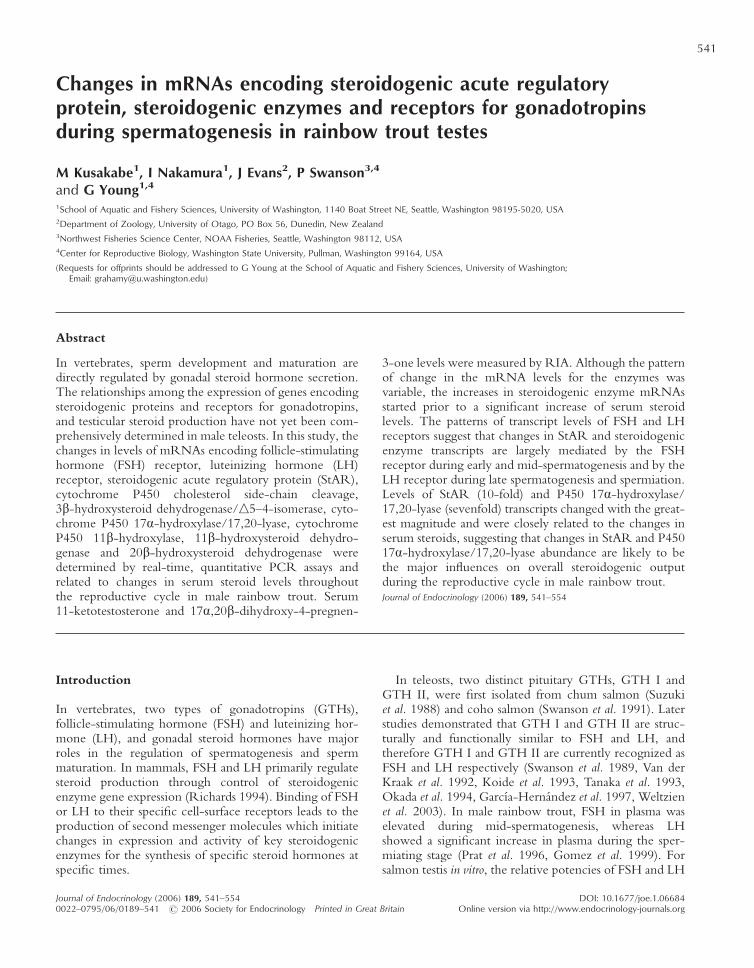
Changes in mRNAs encoding steroidogenic acute regulatory protein, steroidogenic enzymes and receptors for gonadotropins during spermatogenesis in rainbow trout testes M Kusakabe 1 , I Nakamura 1 , J Evans 2 , P Swanson 3,4 and G Young 1,4 1 School of Aquatic and Fishery Sciences, University of Washington, 1140 Boat Street NE, Seattle, Washington 98195-5020, USA 2 Department of Zoology, University of Otago, PO Box 56, Dunedin, New Zealand 3 Northwest Fisheries Science Center, NOAA Fisheries, Seattle, Washington 98112, USA 4 Center for Reproductive Biology, Washington State University, Pullman, Washington 99164, USA (Requests for offprints should be addressed to G Young at the School of Aquatic and Fishery Sciences, University of Washington; Email: grahamy@u.washington.edu) Abstract In vertebrates, sperm development and maturation are directly regulated by gonadal steroid hormone secretion. The relationships among the expression of genes encoding steroidogenic proteins and receptors for gonadotropins, and testicular steroid production have not yet been com- prehensively determined in male teleosts. In this study, the changes in levels of mRNAs encoding follicle-stimulating hormone (FSH) receptor, luteinizing hormone (LH) receptor, steroidogenic acute regulatory protein (StAR), cytochrome P450 cholesterol side-chain cleavage, 3-hydroxysteroid dehydrogenase/n5–4-isomerase, cyto- chrome P450 17-hydroxylase/17,20-lyase, cytochrome P450 11-hydroxylase, 11-hydroxysteroid dehydro- genase and 20-hydroxysteroid dehydrogenase were determined by real-time, quantitative PCR assays and related to changes in serum steroid levels throughout the reproductive cycle in male rainbow trout. Serum 11-ketotestosterone and 17,20-dihydroxy-4-pregnen- 3-one levels were measured by RIA. Although the pattern of change in the mRNA levels for the enzymes was variable, the increases in steroidogenic enzyme mRNAs started prior to a significant increase of serum steroid levels. The patterns of transcript levels of FSH and LH receptors suggest that changes in StAR and steroidogenic enzyme transcripts are largely mediated by the FSH receptor during early and mid-spermatogenesis and by the LH receptor during late spermatogenesis and spermiation. Levels of StAR (10-fold) and P450 17-hydroxylase/ 17,20-lyase (sevenfold) transcripts changed with the great- est magnitude and were closely related to the changes in serum steroids, suggesting that changes in StAR and P450 17-hydroxylase/17,20-lyase abundance are likely to be the major influences on overall steroidogenic output during the reproductive cycle in male rainbow trout. Journal of Endocrinology (2006) 189, 541–554 Introduction In vertebrates, two types of gonadotropins (GTHs), follicle-stimulating hormone (FSH) and luteinizing hor- mone (LH), and gonadal steroid hormones have major roles in the regulation of spermatogenesis and sperm maturation. In mammals, FSH and LH primarily regulate steroid production through control of steroidogenic enzyme gene expression (Richards 1994). Binding of FSH or LH to their specific cell-surface receptors leads to the production of second messenger molecules which initiate changes in expression and activity of key steroidogenic enzymes for the synthesis of specific steroid hormones at specific times. In teleosts, two distinct pituitary GTHs, GTH I and GTH II, were first isolated from chum salmon (Suzuki et al. 1988) and coho salmon (Swanson et al. 1991). Later studies demonstrated that GTH I and GTH II are struc- turally and functionally similar to FSH and LH, and therefore GTH I and GTH II are currently recognized as FSH and LH respectively (Swanson et al. 1989, Van der Kraak et al. 1992, Koide et al. 1993, Tanaka et al. 1993, Okada et al. 1994, García-Hernández et al. 1997, Weltzien et al. 2003). In male rainbow trout, FSH in plasma was elevated during mid-spermatogenesis, whereas LH showed a significant increase in plasma during the sper- miating stage (Prat et al. 1996, Gomez et al. 1999). For salmon testis in vitro, the relative potencies of FSH and LH 541 Journal of Endocrinology (2006) 189, 541–554 0022–0795/06/0189–541 2006 Society for Endocrinology Printed in Great Britain DOI: 10.1677/joe.1.06684 Online version via http://www.endocrinology-journals.org

Welcome message from author
This document is posted to help you gain knowledge. Please leave a comment to let me know what you think about it! Share it to your friends and learn new things together.
Transcript
Changes in mRNAs encoding steroidogenic acute regulatoryprotein, steroidogenic enzymes and receptors for gonadotropinsduring spermatogenesis in rainbow trout testes
M Kusakabe1, I Nakamura1, J Evans2, P Swanson3,4
and G Young1,4
1School of Aquatic and Fishery Sciences, University of Washington, 1140 Boat Street NE, Seattle, Washington 98195-5020, USA2Department of Zoology, University of Otago, PO Box 56, Dunedin, New Zealand3Northwest Fisheries Science Center, NOAA Fisheries, Seattle, Washington 98112, USA4Center for Reproductive Biology, Washington State University, Pullman, Washington 99164, USA
(Requests for offprints should be addressed to G Young at the School of Aquatic and Fishery Sciences, University of Washington;Email: [email protected])
Abstract
In vertebrates, sperm development and maturation aredirectly regulated by gonadal steroid hormone secretion.The relationships among the expression of genes encodingsteroidogenic proteins and receptors for gonadotropins,and testicular steroid production have not yet been com-prehensively determined in male teleosts. In this study, thechanges in levels of mRNAs encoding follicle-stimulatinghormone (FSH) receptor, luteinizing hormone (LH)receptor, steroidogenic acute regulatory protein (StAR),cytochrome P450 cholesterol side-chain cleavage,3�-hydroxysteroid dehydrogenase/n5–4-isomerase, cyto-chrome P450 17�-hydroxylase/17,20-lyase, cytochromeP450 11�-hydroxylase, 11�-hydroxysteroid dehydro-genase and 20�-hydroxysteroid dehydrogenase weredetermined by real-time, quantitative PCR assays andrelated to changes in serum steroid levels throughoutthe reproductive cycle in male rainbow trout. Serum11-ketotestosterone and 17�,20�-dihydroxy-4-pregnen-
3-one levels were measured by RIA. Although the patternof change in the mRNA levels for the enzymes wasvariable, the increases in steroidogenic enzyme mRNAsstarted prior to a significant increase of serum steroidlevels. The patterns of transcript levels of FSH and LHreceptors suggest that changes in StAR and steroidogenicenzyme transcripts are largely mediated by the FSHreceptor during early and mid-spermatogenesis and by theLH receptor during late spermatogenesis and spermiation.Levels of StAR (10-fold) and P450 17�-hydroxylase/17,20-lyase (sevenfold) transcripts changed with the great-est magnitude and were closely related to the changes inserum steroids, suggesting that changes in StAR and P45017�-hydroxylase/17,20-lyase abundance are likely to bethe major influences on overall steroidogenic outputduring the reproductive cycle in male rainbow trout.Journal of Endocrinology (2006) 189, 541–554
Introduction
In vertebrates, two types of gonadotropins (GTHs),follicle-stimulating hormone (FSH) and luteinizing hor-mone (LH), and gonadal steroid hormones have majorroles in the regulation of spermatogenesis and spermmaturation. In mammals, FSH and LH primarily regulatesteroid production through control of steroidogenicenzyme gene expression (Richards 1994). Binding of FSHor LH to their specific cell-surface receptors leads to theproduction of second messenger molecules which initiatechanges in expression and activity of key steroidogenicenzymes for the synthesis of specific steroid hormones atspecific times.
In teleosts, two distinct pituitary GTHs, GTH I andGTH II, were first isolated from chum salmon (Suzukiet al. 1988) and coho salmon (Swanson et al. 1991). Laterstudies demonstrated that GTH I and GTH II are struc-turally and functionally similar to FSH and LH, andtherefore GTH I and GTH II are currently recognized asFSH and LH respectively (Swanson et al. 1989, Van derKraak et al. 1992, Koide et al. 1993, Tanaka et al. 1993,Okada et al. 1994, García-Hernández et al. 1997, Weltzienet al. 2003). In male rainbow trout, FSH in plasma waselevated during mid-spermatogenesis, whereas LHshowed a significant increase in plasma during the sper-miating stage (Prat et al. 1996, Gomez et al. 1999). Forsalmon testis in vitro, the relative potencies of FSH and LH
541
Journal of Endocrinology (2006) 189, 541–5540022–0795/06/0189–541 � 2006 Society for Endocrinology Printed in Great Britain
DOI: 10.1677/joe.1.06684Online version via http://www.endocrinology-journals.org
in stimulating steroid synthesis did not change before thelate spermatogenesis stage and spermiation (Planas &Swanson 1995). However, during the later stages ofspermatogenesis and spermiation, the sensitivity of thetestis to the steroidogenic effects of LH on progestogenproduction increased, whereas the sensitivity of the testisto the effects of FSH decreased (Planas & Swanson 1995).These results imply that the differences in the steroido-genic actions of FSH and LH that are associated withparticular stages of gonadal development are probably dueto changes in expression of genes encoding their recep-tors. Although cDNAs for the FSH receptor (FSH-R) andthe LH receptor (LH-R) have been isolated from severalsalmonid FSH-R – amago salmon (Oba et al. 1999a),rainbow trout (GenBank accession no. AF439405) andAtlantic salmon (GenBank accession no. AJ567667) – andLH-R – amago salmon (Oba et al. 1999b), rainbow trout(GenBank accession no. AF439404) and Atlantic salmon(GenBank accession no. AJ579790) – the intracellularmechanisms of steroidogenesis that are regulated byFSH and LH through their receptors in teleosts are notunderstood to the same degree as in mammals.
For male salmonids, the potent androgen,11-ketotestosterone (11-KT) and the progestogen, 17�,20�dihydroxy-4-pregnen-3-one (17,20�-P), have key rolesin the regulation of spermatogenesis. A distinct shift in thesteroidogenic pathway from 11-KT to 17,20�-P synthesisoccurs in the testes of salmonid species around the onset ofspermiation (Nagahama 1994). Studies on male salmonidsindicate that spermiation is associated with a decrease inplasma 11-KT and an increase in plasma 17,20�-P (Baynes& Scott 1985, Lou et al. 1986, King & Young 2001).Using Japanese eel testis fragments in vitro, Miura et al.(1991) demonstrated that 11-KT can induce all stages ofspermatogenesis, including spermatogonial proliferation,meiotic division and spermiogenesis. These results indi-cate that 11-KT has important roles during spermato-genesis in teleost species (Schulz & Miura 2002). Althoughthe capacity of the testis to produce 17,20�-P is lowduring spermatogenesis, 17,20�-P production sharplyincreases during spermiation (Ueda et al. 1983, 1984,Sakai et al. 1989). Two successive injections of 17,20�-Pinduced precocious spermiation in nonspermiating amagosalmon and goldfish (Ueda et al. 1985). These resultssuggest that 17,20�-P plays a role in the process of finalsperm maturation and spermiation in teleosts.
Previous studies have shown that the long-termchanges in steroid secretion in mammalian ovaries areregulated by changes in steroidogenic enzyme geneexpression (Doody et al. 1990a, 1990b, Aspden et al.1998). It is likely that steroid hormone synthesis in teleostsis similarly regulated by changes in the activity of indi-vidual steroidogenic enzymes through the action of GTHsand/or endogenous steroids. Salmonids are excellent studyanimals to examine the changes in steroidogenic enzymegene expression in relation to steroid hormone synthesis,
since germ cell development progresses in a synchronousfashion and dynamic changes in steroid hormone secretionoccur during gametogenesis. In rainbow trout, cDNAs formost of the steroidogenic enzymes responsible for thesynthesis of sex steroids have already been isolated: cyto-chrome P450 cholesterol side-chain cleavage (P450scc)(Takahashi et al. 1993), cytochrome P450 17�-hydroxylase/17,20-lyase (P450C17) (Sakai et al. 1992),3�-hydroxysteroid dehydrogenase/n5–4-isomerase (3�-HSD) (Sakai et al. 1994), cytochrome P450 aromatase(P450 arom) (Tanaka et al. 1992), cytochrome P45011�-hydroxylase (P45011�) (Liu et al. 2000, Kusakabeet al. 2002a), 11�-hydroxysteroid dehydrogenase (11�-HSD) (Kusakabe et al. 2003) and 20�-hydroxysteroiddehydrogenase (20�-HSD) (Guan et al. 1999). Recentstudies reported changes in mRNA levels for key ovariansteroidogenic enzymes during a reproductive cycle inchannel catfish (Kumar et al. 2000), Arctic char (vonHofsten et al. 2002) and rainbow trout (Nakamura et al.2005), and a few recent studies in male teleosts reportedseasonal changes in transcript levels of a single steroido-genic enzyme (Liu et al. 2000, Kusakabe et al. 2002a,2003). However, comprehensive studies on the expressionof testicular steroidogenic enzyme genes during sperma-togenesis in teleosts have not been reported.
The rate-limiting step of tropic hormone-inducedsteroidogenesis in mammals is the rate of the delivery ofcholesterol to P450scc by steroidogenic acute regulatoryprotein (StAR) (Stocco 2000). Studies on mammalianStARs have mainly concentrated on the short-term(acute) changes in steroidogenesis with little informationavailable on the regulation of StAR during prolongedseasonal cycles. Recently, cDNAs encoding StAR inbrook trout and rainbow trout were isolated (Kusakabeet al. 2002b), and an increase in ovarian StAR geneexpression during the late vitellogenesis, maturation andpost-ovulatory stages in rainbow trout was demonstrated(Bobe et al. 2004, Nakamura et al. 2005). However, thechanges occurring in testicular StAR gene expression inteleosts during spermatogenesis remain to be determined.
The aim of this study was to determine the changes inexpression of the gene encoding steroidogenic enzymesand StAR in rainbow trout testes during spermatogenesisin relation to androgens and 17,20�-P production, and tochanges in mRNAs encoding FSH-R and LH-R. In thecourse of the study, we also had to address the problem ofnormalization of mRNA levels by expression of house-keeping genes. The trout testis changes 20-fold in sizeduring spermatogenesis and has proven to be problematicin this regard. Kusakabe et al. (2002a) reported theprogressive reduction of mRNA levels for the commonlyused housekeeping gene, �-actin, during mid-spermatogenesis. In this study, we conducted furtherinvestigations of the stability of two other housekeepinggenes, acidic ribosomal phosphoprotein P0 (ARP) and18S ribosomal RNA.
M KUSAKABE and others · Changes in steroidogenic protein mRNAs in trout542
www.endocrinology-journals.orgJournal of Endocrinology (2006) 189, 541–554
Materials and Methods
Study animals
Two-year-old, male rainbow trout were obtainedmonthly from Wanaka Trout Hatchery (Wanaka, NewZealand) from January to November in 2001. For eachsampling, 3–5 fish were anesthetized with 300 mg/lMS222 (3-aminobenzoic acid ethyl ester; Sigma) bufferedin sodium bicarbonate. Testes were isolated by dissection,and blood was sampled from the caudal vein with syringes.Blood was allowed to clot, and serum was obtained bycentrifugation and stored at –20 �C for steroid assay. Testisfragments were frozen with liquid nitrogen and stored at–80 �C for RNA extraction. Testis fragments were alsofixed with Bouin’s fixative to determine the developmen-tal stage. Animal care and use were approved by theUniversity of Otago Committee on Care and Use ofAnimals.
Histologic analysis of rainbow trout testes
The fixed testis fragments were dehydrated and embeddedin paraffin, sections (8 µm) were stained by hematoxylinand eosin, and the developmental stages in each samplewere determined, according to Grier (1981), as follows:
1. Early spermatogenesis – spermatogonia and sperma-tocytes are dominant.
2. Mid-spermatogenesis – spermatogonia, spermato-cytes, spermatids and sperm are present.
3. Late spermatogenesis – sperm become dominant andvery few spermatogonia and/or spermatocytes are present.
4. After spawning – the testes size declines and nodeveloping germ cells are present.
Serum sex steroids
Sex steroids were extracted from serum with ethyl ether,as previously described by Kagawa et al. (1981). Levels of11-KT and 17�,20�-P were measured by RIA, as pre-viously described (Kagawa et al. 1981, Ueda et al. 1985,Young et al. 1986).
Expression of genes encoding steroidogenic proteins andgonadotropin receptors
Previous studies have shown that the sites of expression ofsteroidogenic enzymes and StAR in rainbow trout testesare restricted to Leydig cells (Kobayashi et al. 1998,Kusakabe et al. 2002a, 2002b, 2003). Furthermore, thelocalization of Leydig cells in testis during spermatogenesischanges with the maturational stage (Kusakabe et al.2003). Therefore, in order to avoid biasing the number ofLeydig cells in each sample, testes samples for RNAextraction were collected as 300 mg cross-sections takenfrom the widest point, approximately 3 cm from the
anterior end. Testis fragments from 3–5 males werecollected for each month. Relative expression of tran-scripts was measured by real-time, quantitative RT–PCRas described below.
Total RNA was isolated from the testis fragmentswith Trizol reagent, as described by the manufacturer(Invitrogen). Concentrations of total RNA were carefullymeasured at least three times with NanoDrop ND-100(NanoDrop Technologies, Wilmington, DE, USA),and equal amounts of RNA were subjected to reversetranscription to synthesize cDNAs.
An amount of 300 ng total RNA was incubated with 0·6unit of DNase I, Amp Grade (Invitrogen) at room tempera-ture for 15 min to eliminate genomic DNA contamination.DNase I was inactivated by heat denaturation at 65 �Cfor 10 min. Subsequently, single-strand cDNAs weresynthesized from the DNase I-treated RNA with theHigh-Capacity cDNA archive kit as described by themanufacturer (Applied Biosystems, Foster City, CA, USA).
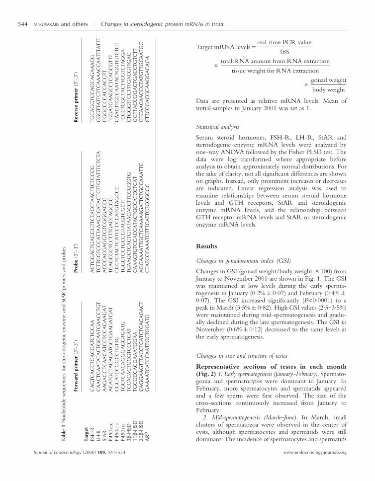
For the relative quantitation of StAR, steroidogenicenzyme and GTH receptor gene expression, real-time,quantitative RT–PCR was performed with the ABIPRISM 7300 sequence detection system (Applied Bio-systems). Primers and TaqMan probes for real-time PCRwere designed with Primer Express, Version 1·5 (AppliedBiosystems). The nucleotide sequences for the primersand probes are listed in Table 1. For normalization of data,an 18S ribosomal gene was measured with commerciallyavailable primers and probes purchased from AppliedBiosystems.
Each real-time PCR reaction contained 25 µl PCRmixture made from 12·5 µl ABI Universal PCR MasterMix, 0·9 µM forward primer, 0·9 µM reverse primer,0·2 µM fluorescent-labeled probe and 3·0 µl cDNA tem-plate. Real-time PCR cycling conditions were 50 �C for2 min and 95 �C for 10 min, followed by 45 cycles of95 �C for 15 s and 60 �C for 1 min. A standard curve wasgenerated by serial dilutions of cDNA for each PCRreaction to determine the levels of transcripts. Standardcurve dilutions were run in triplicate, and correlationcoefficients of the standard curves ranged from 0·99 to1·00. RNA samples not subject to reverse transcriptionwere used to check for genomic DNA contaminationduring RNA preparation.
In order to present changes in steroidogenic enzymeand StAR mRNA levels on the basis of absolute amountsper testes, the total testes weight in each fish and theweight of frozen tissue used for total RNA extractionwere measured. The yield of total RNA extracted fromeach testis sample was calculated by measuring the con-centration of total RNA. Therefore, it was possible toestimate the total amount of total RNA contained withinthe testes. Given the result obtained from real-time PCRin the formula, the relative amount of target mRNA peranimal normalized to relative gonad size was determinedby the following formula:
Changes in steroidogenic protein mRNAs in trout · M KUSAKABE and others 543
www.endocrinology-journals.org Journal of Endocrinology (2006) 189, 541–554
Target mRNA levels =real-time PCR value
18S
�total RNA amount from RNA extraction
tissue weight for RNA extraction
�gonad weight
body weight
Data are presented as relative mRNA levels. Mean ofinitial samples in January 2001 was set as 1.
Statistical analysis
Serum steroid hormones, FSH-R, LH-R, StAR andsteroidogenic enzyme mRNA levels were analyzed byone-way ANOVA followed by the Fisher PLSD test. Thedata were log transformed where appropriate beforeanalysis to obtain approximately normal distributions. Forthe sake of clarity, not all significant differences are shownon graphs. Instead, only prominent increases or decreasesare indicated. Linear regression analysis was used toexamine relationships between serum steroid hormonelevels and GTH receptors, StAR and steroidogenicenzyme mRNA levels, and the relationship betweenGTH receptor mRNA levels and StAR or steroidogenicenzyme mRNA levels.
Results
Changes in gonadosomatic index (GSI)
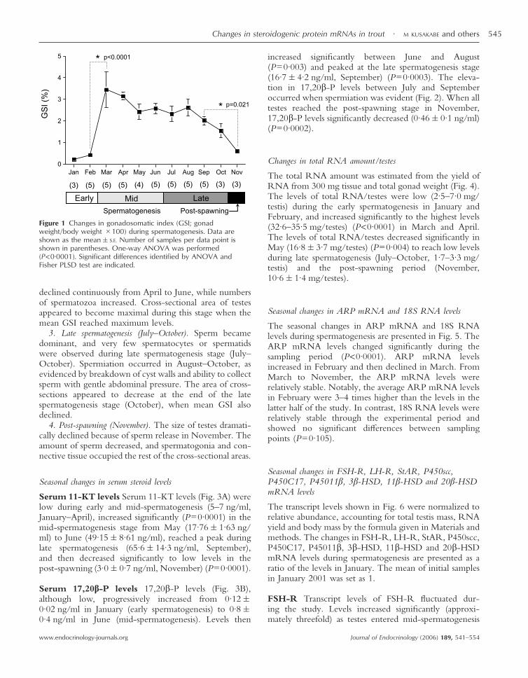
Changes in GSI (gonad weight/body weight �100) fromJanuary to November 2001 are shown in Fig. 1. The GSIwas maintained at low levels during the early sperma-togenesis in January (0·2%�0·07) and February (0·4%�0·07). The GSI increased significantly (P<0·0001) to apeak in March (3·5%�0·82). High GSI values (2·5–3·5%)were maintained during mid-spermatogenesis and gradu-ally declined during the late spermatogenesis. The GSI inNovember (0·6%�0·12) decreased to the same levels asthe early spermatogenesis.
Changes in size and structure of testes
Representative sections of testes in each month(Fig. 2) 1. Early spermatogenesis (January–February). Spermato-gonia and spermatocytes were dominant in January. InFebruary, more spermatocytes and spermatids appearedand a few sperm were first observed. The size of thecross-sections continuously increased from January toFebruary.
2. Mid-spermatogenesis (March–June). In March, smallclusters of spermatozoa were observed in the center ofcysts, although spermatocytes and spermatids were stilldominant. The incidence of spermatocytes and spermatidsTa
ble
1N
ucle
otid
ese
quen
ces
for
ster
oido
geni
cen
zym
ean
dSt
AR
prim
ers
and
prob
es
Forw
ard
prim
er(5
�–3�
)Pr
obe
(5�–
3�)
Rev
erse
prim
er(5
�–3�
)
Targ
etFS
H-R
CA
GTC
AC
CTG
AC
GA
TCTG
CA
AA
CTG
GA
CTG
AG
GG
TTC
TAC
CTA
AC
TTC
TCC
CG
TGC
AG
GTC
CA
GC
AG
AA
AC
GLH
-RC
AA
CTG
AA
TATA
CTG
CA
ATG
AA
CC
TGT
TCTT
GG
TCC
CA
TTA
AA
GG
CA
TAG
TCTT
GTA
TTTC
TCTA
CG
GTT
ATT
CTT
CA
AA
AC
CA
ATT
TATT
TSt
AR
AA
GA
GG
TCA
AG
ATC
CTC
CA
GA
AG
AT
AC
CC
AC
GA
GG
TGTC
GG
GA
CC
CC
GG
CC
CC
AC
CA
CG
TTP4
50sc
cA
CA
TGC
TAC
AG
ATG
CTG
AA
GA
TGA
TTC
AG
CG
CTC
CTT
TGA
CC
AG
CG
GTG
GA
TGA
AG
CC
TCA
GC
GTT
TP4
50C
17C
GC
ATC
CG
GC
CTG
TTG
CC
CTC
TAC
TCA
TCC
CC
CA
TGTA
GC
CC
GA
AC
TTG
CC
AA
TAC
TGG
TGTC
TGT
P450
11�
TGC
TCA
AC
AG
GG
AG
GTG
ATG
TGG
CTC
CTG
CC
GTA
CG
TCG
CTT
TCC
CTC
GC
TAC
TTC
GTC
TAG
GA
3�-H
SDTC
CA
CA
CTG
CG
TCC
CTC
AT
TGA
AG
CTC
AC
TGTA
TAA
CA
CC
TTC
CC
GG
TGC
TGG
GTT
CC
TTTG
AC
GTT
GA
C11
�-H
SDTG
CG
CC
AC
GA
AA
TGG
AA
CA
AA
GTG
TCC
AC
CA
TAC
TGC
CA
TCC
TCA
TG
GTT
AC
CG
GA
CTG
AC
CTG
TCTT
20�-
HSD
CA
GG
AA
GTT
TAC
CTC
ATC
TCA
CA
GA
CT
AG
GA
AA
ATT
GC
TCA
AA
AA
GA
TTC
TGC
CA
AA
TTC
GTC
AA
CA
AC
CC
TATG
TTG
CA
ATG
CA
RPG
AA
AA
TCA
TCC
AA
TTG
CTG
GA
TGC
TATC
CC
AA
ATG
TTTC
ATT
GTC
GG
CG
CC
TTC
CC
AC
GC
AA
GG
AC
AG
A
M KUSAKABE and others · Changes in steroidogenic protein mRNAs in trout544
www.endocrinology-journals.orgJournal of Endocrinology (2006) 189, 541–554
declined continuously from April to June, while numbersof spermatozoa increased. Cross-sectional area of testesappeared to become maximal during this stage when themean GSI reached maximum levels.
3. Late spermatogenesis (July–October). Sperm becamedominant, and very few spermatocytes or spermatidswere observed during late spermatogenesis stage (July–October). Spermiation occurred in August–October, asevidenced by breakdown of cyst walls and ability to collectsperm with gentle abdominal pressure. The area of cross-sections appeared to decrease at the end of the latespermatogenesis stage (October), when mean GSI alsodeclined.
4. Post-spawning (November). The size of testes dramati-cally declined because of sperm release in November. Theamount of sperm decreased, and spermatogonia and con-nective tissue occupied the rest of the cross-sectional areas.
Seasonal changes in serum steroid levels
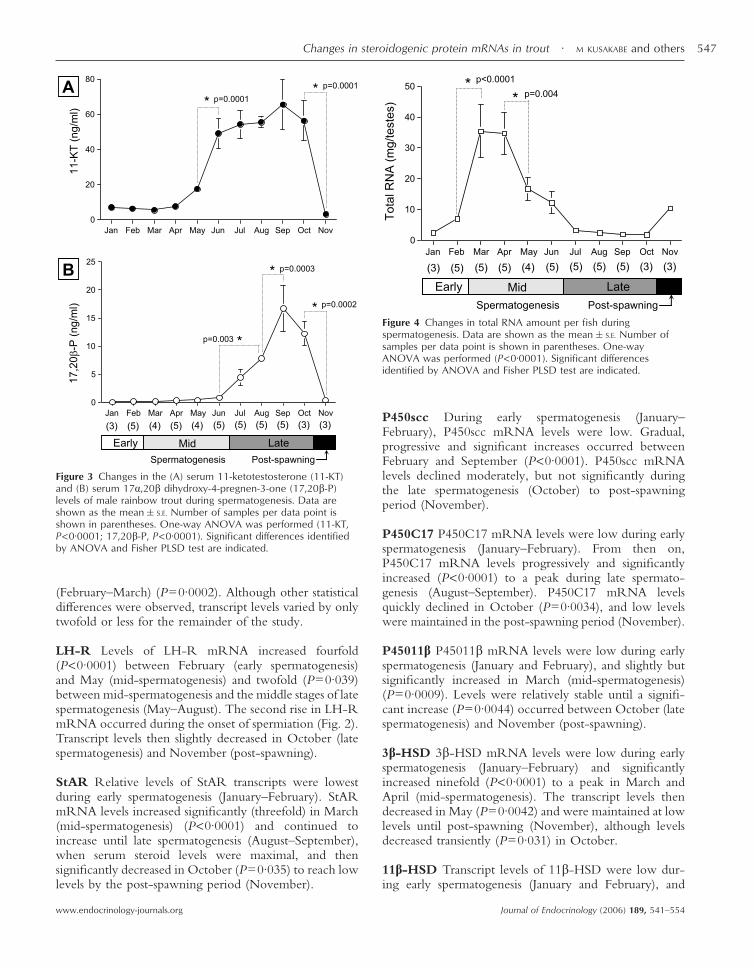
Serum 11-KT levels Serum 11-KT levels (Fig. 3A) werelow during early and mid-spermatogenesis (5–7 ng/ml,January–April), increased significantly (P=0·0001) in themid-spermatogenesis stage from May (17·76�1·63 ng/ml) to June (49·15�8·61 ng/ml), reached a peak duringlate spermatogenesis (65·6�14·3 ng/ml, September),and then decreased significantly to low levels in thepost-spawning (3·0�0·7 ng/ml, November) (P=0·0001).
Serum 17,20�-P levels 17,20�-P levels (Fig. 3B),although low, progressively increased from 0·12�0·02 ng/ml in January (early spermatogenesis) to 0·8�0·4 ng/ml in June (mid-spermatogenesis). Levels then
increased significantly between June and August(P=0·003) and peaked at the late spermatogenesis stage(16·7�4·2 ng/ml, September) (P=0·0003). The eleva-tion in 17,20�-P levels between July and Septemberoccurred when spermiation was evident (Fig. 2). When alltestes reached the post-spawning stage in November,17,20�-P levels significantly decreased (0·46�0·1 ng/ml)(P=0·0002).
Changes in total RNA amount/testes
The total RNA amount was estimated from the yield ofRNA from 300 mg tissue and total gonad weight (Fig. 4).The levels of total RNA/testes were low (2·5–7·0 mg/testis) during the early spermatogenesis in January andFebruary, and increased significantly to the highest levels(32·6–35·5 mg/testes) (P<0·0001) in March and April.The levels of total RNA/testes decreased significantly inMay (16·8�3·7 mg/testes) (P=0·004) to reach low levelsduring late spermatogenesis (July–October, 1·7–3·3 mg/testis) and the post-spawning period (November,10·6�1·4 mg/testes).
Seasonal changes in ARP mRNA and 18S RNA levels
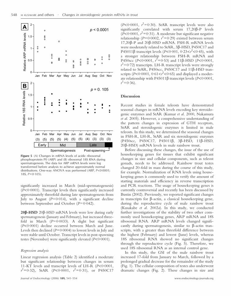
The seasonal changes in ARP mRNA and 18S RNAlevels during spermatogenesis are presented in Fig. 5. TheARP mRNA levels changed significantly during thesampling period (P<0·0001). ARP mRNA levelsincreased in February and then declined in March. FromMarch to November, the ARP mRNA levels wererelatively stable. Notably, the average ARP mRNA levelsin February were 3–4 times higher than the levels in thelatter half of the study. In contrast, 18S RNA levels wererelatively stable through the experimental period andshowed no significant differences between samplingpoints (P=0·105).
Seasonal changes in FSH-R, LH-R, StAR, P450scc,P450C17, P45011�, 3�-HSD, 11�-HSD and 20�-HSDmRNA levels
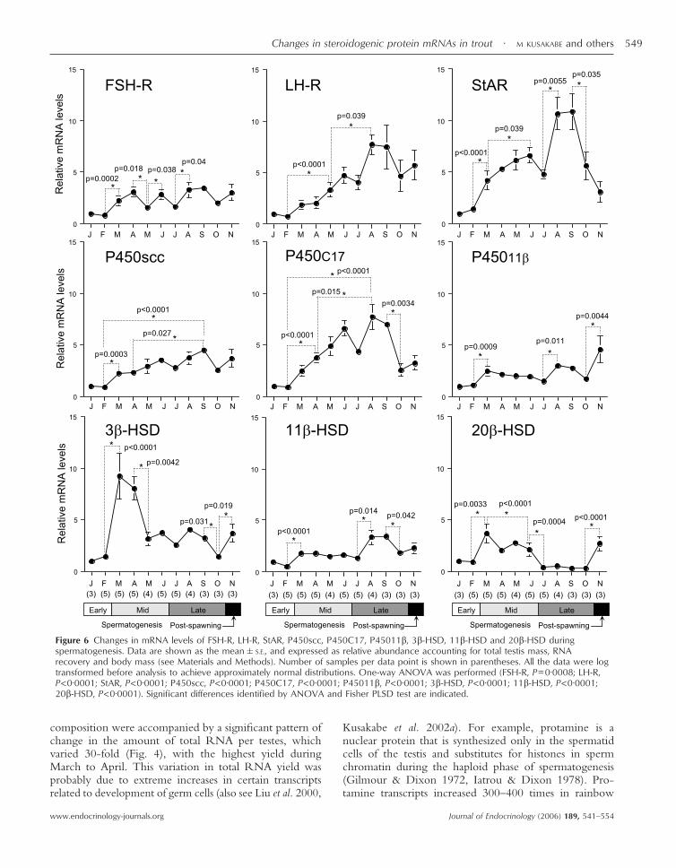
The transcript levels shown in Fig. 6 were normalized torelative abundance, accounting for total testis mass, RNAyield and body mass by the formula given in Materials andmethods. The changes in FSH-R, LH-R, StAR, P450scc,P450C17, P45011�, 3�-HSD, 11�-HSD and 20�-HSDmRNA levels during spermatogenesis are presented as aratio of the levels in January. The mean of initial samplesin January 2001 was set as 1.
FSH-R Transcript levels of FSH-R fluctuated dur-ing the study. Levels increased significantly (approxi-mately threefold) as testes entered mid-spermatogenesis
Figure 1 Changes in gonadosomatic index (GSI; gonadweight/body weight �100) during spermatogenesis. Data areshown as the mean�S.E. Number of samples per data point isshown in parentheses. One-way ANOVA was performed(P<0·0001). Significant differences identified by ANOVA andFisher PLSD test are indicated.
Changes in steroidogenic protein mRNAs in trout · M KUSAKABE and others 545
www.endocrinology-journals.org Journal of Endocrinology (2006) 189, 541–554
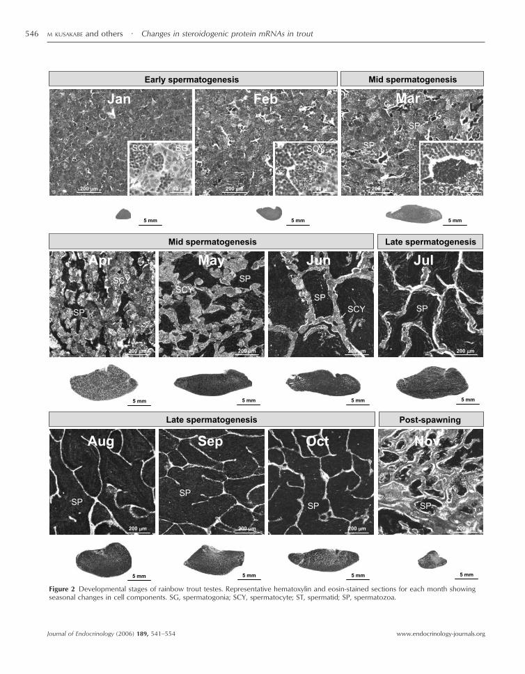
Figure 2 Developmental stages of rainbow trout testes. Representative hematoxylin and eosin-stained sections for each month showingseasonal changes in cell components. SG, spermatogonia; SCY, spermatocyte; ST, spermatid; SP, spermatozoa.
M KUSAKABE and others · Changes in steroidogenic protein mRNAs in trout546
www.endocrinology-journals.orgJournal of Endocrinology (2006) 189, 541–554
(February–March) (P=0·0002). Although other statisticaldifferences were observed, transcript levels varied by onlytwofold or less for the remainder of the study.
LH-R Levels of LH-R mRNA increased fourfold(P<0·0001) between February (early spermatogenesis)and May (mid-spermatogenesis) and twofold (P=0·039)between mid-spermatogenesis and the middle stages of latespermatogenesis (May–August). The second rise in LH-RmRNA occurred during the onset of spermiation (Fig. 2).Transcript levels then slightly decreased in October (latespermatogenesis) and November (post-spawning).
StAR Relative levels of StAR transcripts were lowestduring early spermatogenesis (January–February). StARmRNA levels increased significantly (threefold) in March(mid-spermatogenesis) (P<0·0001) and continued toincrease until late spermatogenesis (August–September),when serum steroid levels were maximal, and thensignificantly decreased in October (P=0·035) to reach lowlevels by the post-spawning period (November).
P450scc During early spermatogenesis (January–February), P450scc mRNA levels were low. Gradual,progressive and significant increases occurred betweenFebruary and September (P<0·0001). P450scc mRNAlevels declined moderately, but not significantly duringthe late spermatogenesis (October) to post-spawningperiod (November).
P450C17 P450C17 mRNA levels were low during earlyspermatogenesis (January–February). From then on,P450C17 mRNA levels progressively and significantlyincreased (P<0·0001) to a peak during late spermato-genesis (August–September). P450C17 mRNA levelsquickly declined in October (P=0·0034), and low levelswere maintained in the post-spawning period (November).
P45011� P45011� mRNA levels were low during earlyspermatogenesis (January and February), and slightly butsignificantly increased in March (mid-spermatogenesis)(P=0·0009). Levels were relatively stable until a signifi-cant increase (P=0·0044) occurred between October (latespermatogenesis) and November (post-spawning).
3�-HSD 3�-HSD mRNA levels were low during earlyspermatogenesis (January–February) and significantlyincreased ninefold (P<0·0001) to a peak in March andApril (mid-spermatogenesis). The transcript levels thendecreased in May (P=0·0042) and were maintained at lowlevels until post-spawning (November), although levelsdecreased transiently (P=0·031) in October.
11�-HSD Transcript levels of 11�-HSD were low dur-ing early spermatogenesis (January and February), and
Figure 3 Changes in the (A) serum 11-ketotestosterone (11-KT)and (B) serum 17�,20� dihydroxy-4-pregnen-3-one (17,20�-P)levels of male rainbow trout during spermatogenesis. Data areshown as the mean�S.E. Number of samples per data point isshown in parentheses. One-way ANOVA was performed (11-KT,P<0·0001; 17,20�-P, P<0·0001). Significant differences identifiedby ANOVA and Fisher PLSD test are indicated.
Figure 4 Changes in total RNA amount per fish duringspermatogenesis. Data are shown as the mean�S.E. Number ofsamples per data point is shown in parentheses. One-wayANOVA was performed (P<0·0001). Significant differencesidentified by ANOVA and Fisher PLSD test are indicated.
Changes in steroidogenic protein mRNAs in trout · M KUSAKABE and others 547
www.endocrinology-journals.org Journal of Endocrinology (2006) 189, 541–554
significantly increased in March (mid-spermatogenesis)(P<0·0001). Transcript levels then significantly increasedapproximately threefold during late spermatogenesis fromJuly to August (P=0·014), with a significant declinebetween September and October (P=0·042).
20�-HSD 20�-HSD mRNA levels were low during earlyspermatogenesis (January and February), but increased three-fold in March (P=0·0033). A slight but significant(P<0·0001) decline occurred between March and June.Levels then declined (P=0·0004) to lowest levels in July andwere stable until October. Transcript levels in post-spawningtestes (November) were significantly elevated (P<0·0001).
Regression analysis
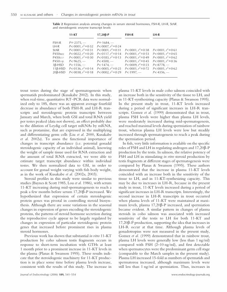
Linear regression analysis (Table 2) identified a moderatebut significant relationship between changes in serum11-KT levels and transcript levels of LH-R (P<0·0001,r2=0·32), StAR (P<0·0001, r2=0·31), or P450C17
(P<0·0001, r2=0·30). StAR transcript levels were alsosignificantly correlated with serum 17,20�-P levels(P<0·0001, r2=0·31). A moderate but significant negativerelationship (P=0·0002, r2=0·29) existed between serum17,20�-P and 20�-HSD mRNA. FSH-R mRNA levelswere moderately related to StAR, 3�-HSD, P450C17 andP45011� transcript levels (P<0·001, 0·23<r2<0·45), witha stronger relationship between FSH-R mRNA andP450scc (P<0·0001, r2=0·53) and 11�-HSD (P<0·0001,r2=0·72) transcripts. LH-R transcript levels were stronglyrelated to StAR, P450scc, P450C17 and 11�-HSD tran-scripts (P<0·0001, 0·61<r2<0·65) and displayed a moder-ate relationship with P45011� transcript levels (P<0·0001,r2=0·36).
Discussion
Recent studies in female teleosts have demonstratedseasonal changes in mRNA levels encoding key steroido-genic enzymes and StAR (Kumar et al. 2000, Nakamuraet al. 2005). However, a comprehensive understanding ofthe pattern changes in expression of GTH receptors,StAR and steroidogenic enzymes is limited in maleteleosts. In this study, we determined the seasonal changesin FSH-R, LH-R, StAR and six steroidogenic enzymes(P450scc, P450C17, P45011�, 3�-HSD, 11�-HSD,20�-HSD) mRNA levels in male rainbow trout.
Before discussing these changes, the issue of the use ofhousekeeping genes for tissues that exhibit significantchanges in size and cellular components, such as teleostgonads, needs to be addressed. Rainbow trout testeschanged 20-fold in mass during the course of this study,for example. Normalization of RNA levels using house-keeping genes is commonly used to verify the amount ofstarting materials and efficiency in reverse transcriptionand PCR reactions. The usage of housekeeping genes iscurrently controversial and recently has been discussed byBustin (2002). Previously, we reported significant changesin transcripts for �-actin, a classical housekeeping gene,during the reproductive cycle of male rainbow trout(Kusakabe et al. 2002a). In this study, we conductedfurther investigations of the stability of two other com-monly used housekeeping genes, ARP mRNA and 18Sribosomal RNA. ARP mRNA levels changed signifi-cantly during spermatogenesis, similar to �-actin tran-scripts, with a greater than threefold difference betweenthe highest (February) and lowest (June) levels, whereas18S ribosomal RNA showed no significant changesthrough the reproductive cycle (Fig. 5). Therefore, weused 18S ribosomal RNA as an internal control gene.
In this study, the GSI of the male rainbow troutincreased 17-fold from January to March, followed by aprolonged gradual decrease for the remainder of the study(Fig. 1). The cellular composition of testes also underwentdramatic changes (Fig. 2). These changes in size and
Figure 5 (A) Changes in mRNA levels of acidic ribosomalphosphoprotein P0 (ARP) and (B) ribosomal 18S RNA duringspermatogenesis. The data for ARP mRNA levels were logtransformed before analysis to achieve approximately normaldistributions. One-way ANOVA was performed (ARP, P<0·0001;18S, P=0·105).
M KUSAKABE and others · Changes in steroidogenic protein mRNAs in trout548
www.endocrinology-journals.orgJournal of Endocrinology (2006) 189, 541–554
composition were accompanied by a significant pattern ofchange in the amount of total RNA per testes, whichvaried 30-fold (Fig. 4), with the highest yield duringMarch to April. This variation in total RNA yield wasprobably due to extreme increases in certain transcriptsrelated to development of germ cells (also see Liu et al. 2000,
Kusakabe et al. 2002a). For example, protamine is anuclear protein that is synthesized only in the spermatidcells of the testis and substitutes for histones in spermchromatin during the haploid phase of spermatogenesis(Gilmour & Dixon 1972, Iatrou & Dixon 1978). Pro-tamine transcripts increased 300–400 times in rainbow
Figure 6 Changes in mRNA levels of FSH-R, LH-R, StAR, P450scc, P450C17, P45011�, 3�-HSD, 11�-HSD and 20�-HSD duringspermatogenesis. Data are shown as the mean�S.E., and expressed as relative abundance accounting for total testis mass, RNArecovery and body mass (see Materials and Methods). Number of samples per data point is shown in parentheses. All the data were logtransformed before analysis to achieve approximately normal distributions. One-way ANOVA was performed (FSH-R, P=0·0008; LH-R,P<0·0001; StAR, P<0·0001; P450scc, P<0·0001; P450C17, P<0·0001; P45011�, P<0·0001; 3�-HSD, P<0·0001; 11�-HSD, P<0·0001;20�-HSD, P<0·0001). Significant differences identified by ANOVA and Fisher PLSD test are indicated.
Changes in steroidogenic protein mRNAs in trout · M KUSAKABE and others 549
www.endocrinology-journals.org Journal of Endocrinology (2006) 189, 541–554
trout testes during the stage of spermatogenesis whenspermatids predominated (Kusakabe 2002). In this study,when real-time, quantitative RT–PCR data were normal-ized only to 18S, there was an apparent average fourfolddecrease in abundance of both FSH-R and LH-R tran-scripts and steroidogenic protein transcripts betweenJanuary and March, when both GSI and total RNA yieldper testes peaked (data not shown), an effect probably dueto the dilution of Leydig cell target mRNAs by mRNA,such as protamine, that are expressed in the multiplyingand differentiating germ cells (Liu et al. 2000, Kusakabeet al. 2002a). To assess the functional importance ofchanges in transcript abundance (i.e. potential gonadalsteroidogenic capacity of an individual animal), knowingthe weight of sample tissue used for RNA extraction andthe amount of total RNA extracted, we were able toestimate target transcript abundance within individualtestes. We then normalized data to GSI, in order toaccount for gonad weight varying with fish body weight,as in the work of Kusakabe et al. (2002a, 2003).
Steroid profiles in this study were similar to previousstudies (Baynes & Scott 1985, Lou et al. 1986), with serum11-KT increasing during mid-spermatogenesis to reach apeak a few months before serum 17,20�-P increased. Wehypothesized that expression of certain steroidogenicprotein genes was pivotal in controlling steroid biosyn-thesis. Although there are some variations in the seasonalchanges in expression of genes encoding the steroidogenicproteins, the patterns of steroid hormone secretion duringthe reproductive cycle appear to be largely regulated bychanges in expression of particular steroidogenic proteingenes that increased before prominent rises in plasmasteroid hormones.
Previous work has shown that substantial in vitro 11-KTproduction by coho salmon testis fragments occurs inresponse to short-term incubation with GTHs at least1 month prior to a prominent increase in 11-KT levels inthe plasma (Planas & Swanson 1995). These results indi-cate that the steroidogenic machinery for 11-KT produc-tion is in place some time before plasma levels increase,consistent with the results of this study. The increase in
plasma 11-KT levels in male coho salmon coincided withan increase both in the sensitivity of the tissue to LH, andin 11-KT-synthesizing capacity (Planas & Swanson 1995).In the present study in trout, 11-KT levels increasedduring a period of significant increases in LH-R tran-scripts. Gomez et al. (1999) demonstrated that in trout,plasma FSH levels were higher than plasma LH levels,were moderately increased during mid-spermatogenesis,and reached maximal levels during spermiation of rainbowtrout, whereas plasma LH levels were low but steadilyincreased through spermatogenesis to reach a peak duringthe spermiation period.
In fish, very little information is available on the specificroles of FSH and LH in regulating androgen and 17,20�-Pproduction by the testis. In salmon, the relative potency ofFSH and LH in stimulating in vitro steroid production bytestis fragments at different stages of spermatogenesis werecompared by Planas & Swanson (1995). These authorsdemonstrated that the increase in plasma 11-KT levelscoincided with an increase both in the sensitivity of thetissue to LH, and in 11-KT-synthesizing capacity. Thismay be due to increases in LH-R, because in the presentstudy in trout, 11-KT levels increased during a period ofsignificant increases in LH-R transcripts. Interestingly, thesecond increase in LH-R transcripts in trout occurredwhen plasma levels of 11-KT were maintained at maxi-mum levels, plasma 17,20�-P increased, and spermiationbecame evident. A similar pattern in changes of plasmasteroids in coho salmon was associated with increasedsensitivity of the testis to LH for both 11-KT and17,20�-P production, supporting the idea that increases inLH-R occur at that time. Although plasma levels ofgonadotropins were not measured in the present study,Gomez et al. (1999) demonstrated that in rainbow trout,plasma LH levels were generally low (less than 1 ng/ml)compared with FSH (2–10 ng/ml), and first detectablewhen spermatocytes were the predominant germ cell stage(comparable to the March samples in the present study).Plasma LH increased 15-fold as numbers of spermatids andspermatozoa increased, although maximum levels werestill less than 1 ng/ml at spermiation. Thus, increases in
Table 2 Regression analysis among changes in serum steroid hormones, FSH-R, LH-R, StARand steroidogenic enzyme transcript levels
11-KT 17,20�-P FSH-R LH-R
FSH-R P= ·2375, — P= ·1684, —LH-R P< ·0001, r2=0·32 P< ·0007, r2=0·24StAR P< ·0001, r2=0·31 P< ·0001, r2=0·31 P< ·0001, r2=0·38 P< ·0001, r2=0·61P450scc P< ·0022, r2=0·20 P< ·0117, r2=0·14 P< ·0001, r2=0·53 P< ·0001, r2=0·65P450C17 P< ·0001, r2=0·30 P< ·0183, r2=0·13 P< ·0001, r2=0·49 P< ·0001, r2=0·62P45011� P< ·9625, — P< ·4500, — P< ·0001, r2=0·45 P< ·0001, r2=0·363�-HSD P< ·1556, — P< ·1474, — P< ·0009, r2=0·23 P< ·8778, —11�-HSD P< ·0136, r2=0·14 P< ·0005, r2=0·25 P< ·0001, r2=0·72 P< ·0001, r2=0·6220�-HSD P< ·0038, r2=0·18 P< ·0002, r2=0·29 P< ·1997, — P< ·4356, —
M KUSAKABE and others · Changes in steroidogenic protein mRNAs in trout550
www.endocrinology-journals.orgJournal of Endocrinology (2006) 189, 541–554

plasma LH levels in trout during spermatogenesis appearto occur in parallel with increases in LH-R transcripts, andprobably LH-R protein. Even though plasma FSH levelsare substantially higher than LH during spermatogenesis introut (Gomez et al. 1999), a number of studies in fish haveshown that the LH receptor is specific for LH (reviewedby Bogerd et al. 2005), and autoradiography studies insalmon have localized an LH-specific receptor in Leydigcells (Miwa et al. 1991). Taken together, these data suggestthat, in salmonids, a major regulatory point for steroidproduction by the testis could be increases in LH signal-ing, induction of LH receptor in Leydig cells of the testis,and increases in plasma LH.
In contrast to the pattern of LH-R gene expression,FSH-R transcripts fluctuated during spermatogenesis andwere only 2–3-fold higher than initial levels toward theend of spermatogenesis. The initial rise in FSH-R tran-scripts (February–March) occurred prior to any significantchange in LH-R gene expression and was associated withhigh meiotic activity in the testis (increase in spermato-cytes and spermatids), but without a major change inplasma 11-KT levels. At this stage, Gomez et al. (1999)reported significant increases in plasma FSH levels introut. Few data are available on specific functions of FSHin spermatogenesis in fish; however, studies suggest thatFSH stimulates spermatogonial proliferation in trout (Loir1999) and 11-KT production in vitro (Planas & Swanson1995). Furthermore, autoradiography studies in salmonlocalized FSH-R to presumed Sertoli cells, but could notdetermine whether Leydig cells contained FSH-R (Miwaet al. 1994). More recently, Schulz et al. (2003) reportedthat FSH also stimulates proliferation of Sertoli cells inAfrican catfish. Thus, as in mammals, a major target of fishFSH appears to be Sertoli cells, and FSH plays importantroles in regulating spermatogenesis. But, because fish FSHalso stimulates androgen production, it is possible thateither FSH-R is present in Leydig cells or other FSH-induced Sertoli cell products affect steroid biosynthesis.Thus, increases in transcripts for StAR and several steroido-genic enzymes that occur early in spermatogenesis in troutmay be due to increases in FSH signaling (see discussionbelow).
Abundance of P450scc, P45011� and 11�-HSD tran-scripts changed significantly but only moderately duringthe reproductive cycle. These three transcripts increasedsignificantly in March, 3 months before serum 11-KTincreased significantly. von Hofsten et al. (2002) reporteda significant rise of transcripts for P450scc in Arctic chartestes when plasma 11-KT levels were high in vivo. Thisdifference may be due to the different ways of quantifyingdata, since the P450scc mRNA levels of Arctic char werepresented on a unit RNA base, whereas the P450sccmRNA levels in this study were normalized by total testesRNA and GSI (Fig. 6). Although the magnitude ofchanges in transcript levels of P45011� was lower (three-fold increase) than seen in the previous study, where
transcript levels were determined by Northern blotting(Kusakabe et al. 2002a), transcript levels were highestwhen serum 11-KT levels increased significantly. FSH-Rand LH-R transcript levels exhibited moderately tostrongly significant linear relationships with P450scc,P45011� and 11�-HSD transcripts, suggesting that theexpression of the P450scc, P45011� and 11�-HSD genesis at least partially regulated by FSH and/or LH. It appearsthat there is a distinct gender difference with regard to thechanges in 3�-HSD and 20�-HSD mRNA levels duringgametogenesis in rainbow trout. In male rainbow trout,testicular 3�-HSD mRNA levels showed a prominent8–9-fold increase to peak levels in the early stage ofmid-spermatogenesis (March and April), followed by arapid decline to levels 2–3-fold higher than the initialsample for the remainder of the study. In contrast, ovarian3�-HSD mRNA levels in female rainbow trout increasedduring late vitellogenesis and post-ovulation (Nakamuraet al. 2005). These observations imply that expression ofthe testicular 3�-HSD gene may be regulated differentlyfrom those for other key steroidogenic enzymes, althoughthe functional significance of the rapid increase of testicu-lar 3�-HSD transcripts during mid-spermatogenesis isstill unclear. In female rainbow trout, 20�-HSD mRNAlevels significantly increased sevenfold during the latereproductive stages (Nakamura et al. 2005). Ovarian20�-HSD transcripts in ayu (Tanaka et al. 2002) and tilapia(Senthilkumaran et al. 2002) also showed a distinctincrease during late oogenesis stages. In contrast, 20�-HSDtranscripts in rainbow trout testes appeared to have aninverse relationship with seasonal serum 17,20�-P levels,20�-HSD mRNA levels decreasing during late spermato-genesis when serum 17,20�-P levels peaked. One possibleexplanation for the weak relationship is that there may bemore than one type of 20�-HSD in some teleosts. In maleamago salmon, an in vitro study demonstrated that spermconverted 17�-hydroxyprogesterone to 17,20�-P in rain-bow trout (Ueda et al. 1984, Sakai et al. 1989), suggestingthe existence of 20�-HSD enzyme in the salmonid sperm.However, Vizziano et al. (1996) reported 20�-HSDactivity in nonflagellated germ cells obtained from earlyspermatogenic rainbow trout testes, although serum17,20�-P levels are very low at this stage. In this study,20�-HSD mRNA levels were high during the mid-spermatogenesis stages, whereas levels during latespermatogenesis were almost nondetectable. Histologicanalysis showed that mid-spermatogenic testes are occu-pied with a considerable proportion of nonflagellatedgerm cells (Fig. 2). Therefore, the 20�-HSD mRNAsdetected in the rainbow trout testes may be derived fromnonflagellated germ cells. Further investigation is requiredto clarify the regulation of 20�-HSD associated withtesticular 17,20�-P production. All in all, it appears thatthe moderate changes in P450scc, P45011�, 11�-HSD,3�-HSD and 20�-HSD transcripts may have only minoreffects on the changes in seasonal steroid production.
Changes in steroidogenic protein mRNAs in trout · M KUSAKABE and others 551
www.endocrinology-journals.org Journal of Endocrinology (2006) 189, 541–554
StAR mRNA correlated moderately with serum11-KT levels (r2=0·31, P<0·0001). Of all the transcriptsmeasured, the 10-fold increase in StAR mRNA was thegreatest change observed. During the later stages ofspermatogenesis, StAR transcripts increased significantly,corresponding to the increases of serum sex-steroid levels.These results suggest that StAR abundance at least par-tially determines the levels of steroid production duringspermatogenesis in rainbow trout. Previous mammalianstudies have demonstrated an induction of StAR proteinby LH treatment in rat Leydig cells (Luo et al. 1998).Interestingly, there was a significant linear relationshipbetween LH-R and StAR mRNA levels (r2=0·61,P<0·0001), which was higher than that of FSH-R andStAR mRNA (r2=0·38, P<0·0001). These results, alongwith data from other studies on plasma FSH and LHlevels, suggest that expression of the StAR gene maybe under the control of FSH during early stages ofspermatogenesis, and of LH during the later stages ofspermatogenesis.
Like StAR transcripts, P450C17 transcripts had amoderately linear relationship with serum 11-KT levels(r2=0·30, P=0·0001). Both StAR and P450C17 startedincreasing in March and April when spermatocyte abun-dance started to increase (Fig. 5). P450C17 is a singlemicrosomal enzyme that catalyzes two distinct steroidbiosynthesis activities, 17�-hydroxylase and 17,20-lyase.Phosphorylation of P450C17 and the presence of cyto-chrome b5 are important factors determining whether theenzyme has 17,20-lyase activity (Katagiri et al. 1995,Zhang et al. 1995). These factors partly explain thetissue-specific differences in P450C17 activities betweenadrenal gland and gonads (Miller et al. 1997). As rainbowtrout P450C17 also has two distinct steroid biosyntheticactivities (Sakai et al. 1992), the relative enzymatic activityof 17�-hydroxylase and 17,20-lyase probably regulates thebiosynthesis of androgens and progestogen. The lack of17,20�-P production during the early and mid-spermatogenesis stages, despite the presence of 20�-HSDmRNA (this study) and evidence of protein (Vizzianoet al. 1996) in the testes, may be due to high C17-C20lyase activity resulting in low availability of the immedi-ate precursor for 17,20�-P, 17�-hydroxyprogesterone.Knowledge of the regulation of the enzymatic activities ofP450C17 is important to understand the mechanisms oftesticular 17,20�-P production in teleosts.
This study provides an overall picture of changes insteroidogenic enzyme gene expression changes duringspermatogenesis and their relationships to steroid produc-tion. The patterns of steroid hormone secretion during thereproductive cycle appear to be largely regulated bychanges in expression of genes encoding StAR, P450C17,and gonadotropin receptors. Expression of genes encod-ing various steroidogenic enzymes increased beforeprominent rises in levels of plasma steroid hormones thatwere presumably initiated by FSH and/or LH signals.
Planas et al. (1997) reported that two intracellular signalingpathways, cAMP/protein kinase A and protein kinaseC/Ca2+, mediate the effects of FSH and LH on ovariansteroidogenesis in brook trout. How these signaling path-ways affect gene expression of steroidogenic enzymes andrelated factors has not yet been clarified. Experimentalapproaches will be required for further understanding ofthe regulation of steroidogenesis at the level of geneexpression, such as analyzing the effects on expression ofgenes encoding these proteins through in vitro tissueculture with GTHs.
Acknowledgements
Jon Dickey (National Marine Fisheries Service), KimGarrett (University of Otago), Ken Miller (University ofOtago) and Gerald Stokes (University of Otago) providedexcellent technical assistance. Dr Munetaka Shimizu(University of Washington) provided statistical assistance.
Funding
This work was supported by the National ResearchInitiative Competitive Grant no. 2003–35203–13602from the USDA Cooperative State Research, Education,and Extension Service (to G Y), Bonneville PowerAdministration Project no. 93–056 (to P S) and a JapanSociety for the Promotion of Science Fellowship (toM K). The authors declare that there is no conflict ofinterest that would prejudice the impartiality of thisscientific work.
References
Aspden WJ, Rodgers RJ, Stocco DM, Scott PT, Wreford NG,Trigg TE, Walsh J & D’Occhio MJ 1998 Changes in testicularsteroidogenic acute regulatory (StAR) protein, steroidogenicenzymes and testicular morphology associated with increasedtestosterone secretion in bulls receiving the luteinizing hormonereleasing hormone agonist deslorelin. Domestic Animal Endocrinology15 227–238.
Baynes SM & Scott AP 1985 Seasonal variations in parameters of miltproduction and in plasma concentration of sex steroids of malerainbow trout (Salmo gairdneri). General and ComparativeEndocrinology 57 150–160.
Bobe J, Nguyen T & Jalabert B 2004 Targeted gene expressionprofiling in the rainbow trout (Oncorhynchus mykiss) ovary duringmaturational competence acquisition and oocyte maturation. Biologyof Reproduction 71 73–82.
Bogerd J, Granneman JC, Schulz RW & Vischer HF 2005 Fish FSHreceptors bind LH: how to make the human FSH receptor to bemore fishy? General and Comparative Endocrinology 142 34–43.
Bustin SA 2002 Quantification of mRNA using real-time reversetranscription PCR (RT–PCR): trends and problems. Journal ofMolecular Endocrinology 29 23–39.
Doody KJ, Lephart ED, Stirling D, Lorence MC, Magness RR,McPhaul MJ & Simpson ER 1990a Expression of mRNA speciesencoding steroidogenic enzymes in the rat ovary. Journal ofMolecular Endocrinology 6 153–162.
M KUSAKABE and others · Changes in steroidogenic protein mRNAs in trout552
www.endocrinology-journals.orgJournal of Endocrinology (2006) 189, 541–554
Doody KJ, Lorence MC, Mason JI & Simpson ER 1990b Expressionof messenger ribonucleic acid species encoding steroidogenicenzymes in human follicles and corpora lutea throughout themenstrual cycle. Journal of Clinical Endocrinology and Metabolism70 1041–1045.
García-Hernández MP, Koide Y, Díaz MV & Kawauchi H 1997Isolation and characterization of two distinct gonadotropins fromthe pituitary gland of Mediterranean yellowtail, Seriola dumerilii(Risso, 1810). General and Comparative Endocrinology 106 389–399.
Gilmour RS & Dixon GH 1972 Biosynthesis of protamine in trouttestis. Journal of Biological Chemistry 247 4621–4627.
Gomez JM, Weil C, Ollitrault M, Le Bail PY, Breton B & Le Gac F1999 Growth hormone (GH) and gonadotropin subunit geneexpression and pituitary and plasma changes during spermatogenesisand oogenesis in rainbow trout (Oncorhynchus mykiss). General andComparative Endocrinology 113 413–428.
Grier HJ 1981 Cellular organization of the testis and spermatogenesisin fishes. American Zoologist 21 345–357.
Guan G, Tanaka M, Todo T, Young G, Yoshikuni M & Nagahama Y1999 Cloning and expression of two carbonyl reductase-like20�-hydroxysteroid dehydrogenase cDNAs in ovarian follicles ofrainbow trout (Oncorhynchus mykiss). Biochemical and BiophysicalResearch Communications 255 123–128.
Iatrou K & Dixon GH 1978 Protamine messenger RNA: its lifehistory during spermatogenesis in rainbow trout. FederationProceedings 37 2526–2533.
Kagawa H, Takano K & Nagahama Y 1981 Correlation of plasmaestradiol-17� and progesterone levels with ultrastructure andhistochemistry of ovarian follicles in the white-spotted char,Salvelinus leucomaenis. Cell and Tissue Research 218 315–329.
Katagiri M, Kagawa N & Waterman MR 1995 The role ofcytochrome b5 in the biosynthesis of androgens by humanP450c17. Archives of Biochemistry and Biophysics 317 343–347.
King HR & Young G 2001 Milt production by non-spermiating maleAtlantic salmon (Salmo salar) after injection of a commercialgonadotropin releasing hormone analog preparation,17�-hydroxyprogesterone or 17�,20�-dihydroxy-4-pregnen-3-one,alone or in combination. Aquaculture 193 179–195.
Kobayashi T, Nakamura M, Kajiura-Kobayashi H, Young G &Nagahama Y 1998 Immunolocalization of steroidogenic enzymes(P450scc, P450C17, P450 arom, and 3�-HSD) in immature andmature testes of rainbow trout (Oncorhynchus mykiss). Cell and TissueResearch 292 573–577.
Koide Y, Itoh H & Kawauchi H 1993 Isolation and characterizationof two distinct gonadotropins, GTHI and GTHII, from bonito(Katsuwonus plelamis) pituitary glands. International Journal of Peptideand Protein Research 41 52–65.
Kumar RS, Ijiri S & Trant JM 2000 Changes in the expression ofgenes encoding steroidogenic enzymes in the channel catfish(Ictalurus punctatus) ovary throughout a reproductive cycle. Biology ofReproduction 63 1676–1682.
Kusakabe M 2002 Steroidogenic acute regulatory protein (StAR) andsteroidogenic enzymes in rainbow trout (Oncorhynchus mykiss):cDNA cloning and expression during spermatogenesis. Thesis,University of Otago, Dunedin, New Zealand.
Kusakabe M, Kobayashi T, Todo T, Lokman PM, Nagahama Y &Young G 2002a Molecular cloning and expression duringspermatogenesis of a cDNA encoding testicular 11�-hydroxylase(P45011�) in rainbow trout (Oncorhynchus mykiss). MolecularReproduction and Development 62 456–469.
Kusakabe M, Todo T, McQuillan HJ, Goetz FW & Young G 2002bCharacterization and expression of steroidogenic acute regulatoryprotein and MLN64 cDNAs in trout. Endocrinology 143 2062–2070.
Kusakabe M, Nakamura I & Young G 2003 11�-Hydroxysteroiddehydrogenase complementary deoxyribonucleic acid in rainbowtrout: cloning, sites of expression, and seasonal changes in gonads.Endocrinology 144 2534–2545.
Liu S, Govoroun M, D’Cotta H, Ricordel MJ, Lareyre JJ,McMeel OM, Smith T, Nagahama Y & Guiguen Y 2000Expression of cytochrome P45011� (11�-hydroxylase) geneduring gonadal sex differentiation and spermatogenesis in rainbowtrout, Oncorhynchus mykiss. Journal of Steroid Biochemistry andMolecular Biology 75 291–298.
Loir M 1999 Spermatogonia of rainbow trout. II. In vitro study ofthe influence of pituitary hormones, growth factors, and steroids onmitotic activity. Molecular Reproduction and Development 53 434–442.
Lou SW, Aida K, Hanyu I, Sakai K, Nomura M, Tanaka M &Tazaki S 1986 Endocrine profiles in the males of a twice-annuallyspawning strain of rainbow trout, Salmo gairdneri. General andComparative Endocrinology 64 212–219.
Luo L, Chen H, Stocco DM & Zirkin BR 1998 Leydig cell proteinsynthesis and steroidogenesis in response to acute stimulation byluteinizing hormone in rats. Biology of Reproduction 59 263–270.
Miller WL, Auchus RJ & Geller DH 1997 The regulation of 17,20lyase activity. Steroids 62 133–142.
Miura T, Yamauchi K, Takahashi H & Nagahama Y 1991 Hormonalinduction in vitro of all stages of spermatogenesis in the maleJapanese eel (Anguilla japonica). PNAS 88 5774–5778.
Miwa S, Yan L & Swanson P 1994 Localization of two gonadotropinreceptors in the salmon gonad by in vitro ligand autoradiography.Biology of Reproduction 50 629–642.
Nagahama Y 1994 Endocrine regulation of gametogenesis in fish.International Journal of Developmental Biology 38 217–229.
Nakamura I, Evans CJ, Kusakabe M & Young G 2005 Changes insteroidogenic enzyme and steroidogenic acute regulatory proteinmessenger RNAs in ovarian follicles during ovarian development ofrainbow trout (Oncorhynchus mykiss). General and ComparativeEndocrinology 144 224–231.
Oba Y, Hirai T, Yoshiura Y, Yoshikuni M, Kawauchi H &Nagahama Y 1999a Cloning, functional characterization, andexpression of a gonadotropin receptor cDNA in the ovary andtestis of amago salmon (Oncorhynchus rhodurus). Biochemical andBiophysical Research Communications 263 584–590.
Oba Y, Hirai T, Yoshiura Y, Yoshikuni M, Kawauchi H &Nagahama Y 1999b The duality of fish gonadotropin receptors:cloning and functional characterization of a second gonadotropinreceptor cDNA expressed in the ovary and testis of amago salmon(Oncorhynchus rhodurus). Biochemical and Biophysical ResearchCommunications 265 366–371.
Okada T, Kawazoe I, Kimura S, Sasamoto Y, Aida K & Kawauchi H1994 Purification and characterization of gonadotropin I and IIfrom pituitary glands of tuna (Thunnus obesus). International Journal ofPeptide and Protein Research 43 69–80.
Planas J & Swanson P 1995 Maturation-associated changes in theresponse of the salmon testis to the steroidogenic actions ofgonadotropins (GTH I and GTH II) in vitro. Biology of Reproduction52 697–704.
Planas J, Goetz FW & Swanson P 1997 Stimulation of brook troutovarian steroidogenesis by gonadotropins I and II is mediated bythe cyclic adenosine 3�,5�-monophosphate/protein kinase Apathway. Biology of Reproduction 57 647–654.
Prat F, Sumpter JP & Tyler CR 1996 Validation ofradioimmunoassays for two salmon gonadotropins (GTH I andGTH II) and their plasma concentrations throughout thereproductive cycle in male and female rainbow trout (Oncorhynchusmykiss). Biology of Reproduction 54 1375–1382.
Richards JS 1994 Hormonal control of gene expression in the ovary.Endocrine Reviews 15 725–751.
Sakai N, Ueda H, Suzuki N & Nagahama Y 1989 Steroid productionby amago salmon (Oncorhynchus rhodurus) testes at differentdevelopmental stages. General and Comparative Endocrinology75 231–240.
Sakai N, Tanaka M, Adachi S, Miller WL & Nagahama Y 1992Rainbow trout cytochrome P-450c17 (17�-hydroxylase/
Changes in steroidogenic protein mRNAs in trout · M KUSAKABE and others 553
www.endocrinology-journals.org Journal of Endocrinology (2006) 189, 541–554

17,20-lyase). cDNA cloning, enzymatic properties and temporalpattern of ovarian P-450c17 mRNA expression during oogenesis.FEBS Letters 301 60–64.
Sakai N, Tanaka M, Takahashi M, Fukada S, Mason JI & Nagahama Y1994 Ovarian 3�-hydroxysteroid dehydrogenase/n5–4-isomeraseof rainbow trout: its cDNA cloning and properties of the enzymeexpressed in a mammalian cell. FEBS Letters 350 309–313.
Schulz RW & Miura T 2002 Spermatogenesis and its endocrineregulation. Fish Physiology and Biochemistry 26 43–56.
Schulz RW, van Dijk W & Bogerd J 2003 Sertoli cell proliferationand FSH signaling in African catfish, Clarias garipinus. FishPhysiology and Biochemistry 28 223–224.
Senthilkumaran B, Sudhakumari CC, Chang XT, Kobayashi T,Oba Y, Guan G, Yoshiura Y, Yoshikuni M & Nagahama Y 2002Ovarian carbonyl reductase-like 20�-hydroxysteroid dehydrogenaseshows distinct surge in messenger RNA expression during naturaland gonadotropin-induced meiotic maturation in nile tilapia.Biology of Reproduction 67 1080–1086.
Stocco DM 2000 The role of StAR protein in steroidogenesis:challenges for the future. Journal of Endocrinology 164 247–253.
Suzuki K, Kawauchi H & Nagahama Y 1988 Isolation andcharacterization of two distinct gonadotropins from chum salmonpituitary glands. General and Comparative Endocrinology 71 292–301.
Swanson P, Bernard M, Nozaki M, Suzuki K, Kawauchi H &Dickhoff WW 1989 Gonadotropins I and II in juvenile cohosalmon. Fish Physiology and Biochemistry 7 169–176.
Swanson P, Suzuki K, Kawauchi H & Dickhoff WW 1991 Isolationand characterization of two coho salmon gonadotropins, GtH I andGtH II. Biology of Reproduction 44 29–38.
Takahashi M, Tanaka M, Sakai N, Adachi S, Miller WL & Nagahama Y1993 Rainbow trout ovarian cholesterol side-chain cleavagecytochrome P450 (P450scc). cDNA cloning and mRNAexpression during oogenesis. FEBS Letters 319 45–48.
Tanaka H, Kagawa H, Okuzawa K & Hirose K 1993 Purification ofgonadotropins (Pm GTH I and II) from red seabream (Pagrus major)and development of a homologous radioimmunoassay for Pm GTHII. Fish Physiology and Biochemistry 10 409–418.
Tanaka M, Telecky TM, Fukada S, Adachi S, Chen S & Nagahama Y1992 Cloning and sequence analysis of the cDNA encoding P-450aromatase (P450 arom) from a rainbow trout (Oncorhynchus mykiss)ovary; relationship between the amount of P450 arom mRNA andthe production of oestradiol-17� in the ovary. Journal of MolecularEndocrinology 8 53–61.
Tanaka M, Nakajin S, Kobayashi D, Fukada S, Guan G, Todo T,Senthilkumaran B & Nagahama Y 2002 Teleost ovarian carbonylreductase-like 20�-hydroxysteroid dehydrogenase: potential role in
the production of maturation-inducing hormone during finaloocyte maturation. Biology of Reproduction 66 1498–1504.
Ueda H, Young G, Crim LM, Kambegawa A & Nagahama Y 198317�,20�-Dihydroxy-4-pregnen-3-one: plasma levels during sexualmaturation, and in vitro production by the testes of amago salmon(Oncorhynchus rhodurus) and rainbow trout (Salmo gairdneri). Generaland Comparative Endocrinology 51 106–112.
Ueda H, Kambegawa A & Nagahama Y 1984 In vitro11-ketotestosterone and 17�,20�-dihydroxy-4-pregnen-3-oneproduction by testicular fragments and isolated sperm ofrainbow trout, Salmo gairdneri. Journal of Experimental Zoology231 435–439.
Ueda H, Kambegawa A & Nagahama Y 1985 Involvement ofgonadotrophin and steroid hormones in spermiation in the amagosalmon, Oncorhynchus rhodurus, and goldfish, Carassius auratus.General and Comparative Endocrinology 59 24–30.
Van der Kraak G, Suzuki K, Peter RE, Itoh H & Kawauchi H 1992Properties of common carp gonadotropin I and gonadotropin II.General and Comparative Endocrinology 85 217–229.
Vizziano D, Fostier A, Le Gac F & Loir M 1996 20�-hydroxysteroiddehydrogenase activity in nonflagellated germ cells of rainbow trouttestis. Biology of Reproduction 54 1–7.
von Hofsten J, Karlsson J, Jones I & Olsson PE 2002 Expression andregulation of fushi tarazu factor-1 and steroidogenic genes duringreproduction in Arctic char (Salvelinus alpinus). Biology ofReproduction 67 1297–1304.
Weltzien FA, Norberg B & Swanson P 2003 Isolation andcharacterization of FSH and LH from pituitary glands of Atlantichalibut (Hippoglossus hippoglossus L.). General and ComparativeEndocrinology 131 97–105.
Young G, Adachi S & Nagahama Y 1986 Role of ovarian thecal andgranulosa layers in gonadotropin-induced synthesis of a salmonidmaturation-inducing substance(17�,20�-Dihydroxy-4-pregnen-3-one). Developmental Biology118 1–8.
Zhang LH, Rodriguez H, Ohno S & Miller WL 1995 Serinephosphorylation of human P450c17 increases 17,20-lyase activity:implications for adrenarche and the polycystic ovary syndrome.PNAS 92 10619–10623.
Received in final form 17 February 2006Accepted 3 March 2006Made available online as an Accepted Preprint13 March 2006
M KUSAKABE and others · Changes in steroidogenic protein mRNAs in trout554
www.endocrinology-journals.orgJournal of Endocrinology (2006) 189, 541–554
Related Documents