Changes in Culture Expanded Human Amniotic Epithelial Cells: Implications for Potential Therapeutic Applications Gita Pratama 1. , Vijesh Vaghjiani 1. , Jing Yang Tee 1 , Yu Han Liu 2 , James Chan 2 , Charmaine Tan 3 , Padma Murthi 4 , Caroline Gargett 3 , Ursula Manuelpillai 1 * 1 Centre for Reproduction & Development, Monash Institute of Medical Research, Monash University, Clayton, Victoria, Australia, 2 Centre for Inflammatory Diseases, Department of Medicine, Monash University, Clayton, Victoria, Australia, 3 Department of Obstetrics & Gynecology and The Ritchie Centre, Monash Institute of Medical Research, Monash University, Clayton, Victoria, Australia, 4 Department of Obstetrics & Gynecology, University of Melbourne and Department of Perinatal Medicine, Pregnancy Research Centre, Royal Women’s Hospital, Melbourne, Victoria, Australia Abstract Human amniotic epithelial cells (hAEC) isolated from term placenta have stem cell-like properties, differentiate into tissue specific cells and reduce lung and liver inflammation and fibrosis following transplantation into disease models established in mice. These features together with their low immunogenicity and immunosuppressive properties make hAEC an attractive source of cells for potential therapeutic applications. However, generation of large cell numbers required for therapies through serial expansion in xenobiotic-free media may be a limiting factor. We investigated if hAEC could be expanded in xenobiotic-free media and if expansion altered their differentiation capacity, immunophenotype, immunosuppressive properties and production of immunomodulatory factors. Serial expansion in xenobiotic-free media was limited with cumulative cell numbers and population doubling times significantly lower than controls maintained in fetal calf serum. The epithelial morphology of primary hAEC changed into mesenchymal-stromal like cells by passage 4–5 (P4–P5) with down regulation of epithelial markers CK7, CD49f, EpCAM and E-cadherin and elevation of mesenchymal- stromal markers CD44, CD105, CD146 and vimentin. The P5 hAEC expanded in xenobiotic-free medium differentiated into osteocyte and alveolar epithelium-like cells, but not chondrocyte, hepatocyte, a- and b-pancreatic-like cells. Expression of HLA Class IA, Class II and co-stimulatory molecules CD80, CD86 and CD40 remained unaltered. The P5 hAEC suppressed mitogen stimulated T cell proliferation, but were less suppressive compared with primary hAEC at higher splenocyte ratios. Primary and P5 hAEC did not secrete the immunosuppressive factors IL-10 and HGF, whereas TGF-b1 and HLA-G were reduced and IL-6 elevated in P5 hAEC. These findings suggest that primary and expanded hAEC may be suitable for different cellular therapeutic applications. Citation: Pratama G, Vaghjiani V, Tee JY, Liu YH, Chan J, et al. (2011) Changes in Culture Expanded Human Amniotic Epithelial Cells: Implications for Potential Therapeutic Applications. PLoS ONE 6(11): e26136. doi:10.1371/journal.pone.0026136 Editor: Che John Connon, University of Reading, United Kingdom Received July 26, 2011; Accepted September 20, 2011; Published November 2, 2011 Copyright: ß 2011 Pratama et al. This is an open-access article distributed under the terms of the Creative Commons Attribution License, which permits unrestricted use, distribution, and reproduction in any medium, provided the original author and source are credited. Funding: UM is funded by: the Australian National Health & Medical Research Council (NHMRC) project grant #606473; CG by NHMRC Career Development Award #465121 and project grant #545992; PM by NHMRC project grant #509140; JC by NHMRC program grant #436634. GP was supported by an Australian Partnership Scholarship, AusAID. Study was supported by the Victorian Government’s Operational Infrastructure Support Program. The funders had no role in study design, data collection and analysis, decision to publish, or preparation of manuscript. Competing Interests: The authors have declared that no competing interests exist. * E-mail: ursula.manuelpillai@ monash.edu . These authors contributed equally to this work. Introduction Human amniotic epithelial cells (hAEC) line the inner of two fetal derived membranes attached to the placenta. hAEC arise from pluripotent epiblast cells of the embryo and are among the first cells to differentiate in the conceptus [1]. Studies have shown that even at term pregnancy, primary hAEC isolated from amnion membranes retain some of the features of their founder cells, expressing pluripotency associated genes and differentiating into lineages derived from each of the three primary embryonic germ layers in vitro [2,3]. Primary hAEC also display similarities to multipotent mesenchymal stromal/stem cells (MSC) expressing some of the surface antigens defining MSC, and like MSC lack hematopoietic and monocytic lineage markers [4,5,6]. Importantly, primary hAEC have several features that make them most attractive for cellular therapies. Compared with adult tissue derived stem cells, hAEC are plentiful and obtained without invasive and expensive procedures from term placenta, a widely accepted non-controversial source of stem cells. Replacement of cells damaged by disease, injury and aging remains a key goal in many therapeutic applications. In this context, hAEC have been shown to differentiate into functional neurons in spinal cord injury models [7,8], insulin secreting pancreatic b-islet like-cells that normalized blood glucose in diabetic mice [9] and recently into surfactant producing alveolar epithelial cells in the lung [6]. Therapies aimed at reducing tissue inflammation and scarring to promote host tissue repair are another important potential application of stem cells. Studies in murine models of lung and liver fibrosis have shown that primary hAEC reduce inflammation and fibrosis and induce tissue remodeling and repair [6,10,11]. Further, hAEC transplantation appears to be safe and tumour or teratoma formation has not been demonstrated in spite of Oct-4, Sox-2 and Nanog expression that are linked to teratoma formation by embryonic and induced pluripotent stem cells [2,3,6,11]. PLoS ONE | www.plosone.org 1 November 2011 | Volume 6 | Issue 11 | e26136

Welcome message from author
This document is posted to help you gain knowledge. Please leave a comment to let me know what you think about it! Share it to your friends and learn new things together.
Transcript
Changes in Culture Expanded Human Amniotic EpithelialCells: Implications for Potential Therapeutic ApplicationsGita Pratama1., Vijesh Vaghjiani1., Jing Yang Tee1, Yu Han Liu2, James Chan2, Charmaine Tan3, Padma
Murthi4, Caroline Gargett3, Ursula Manuelpillai1*
1 Centre for Reproduction & Development, Monash Institute of Medical Research, Monash University, Clayton, Victoria, Australia, 2 Centre for Inflammatory Diseases,
Department of Medicine, Monash University, Clayton, Victoria, Australia, 3 Department of Obstetrics & Gynecology and The Ritchie Centre, Monash Institute of Medical
Research, Monash University, Clayton, Victoria, Australia, 4 Department of Obstetrics & Gynecology, University of Melbourne and Department of Perinatal Medicine,
Pregnancy Research Centre, Royal Women’s Hospital, Melbourne, Victoria, Australia
Abstract
Human amniotic epithelial cells (hAEC) isolated from term placenta have stem cell-like properties, differentiate into tissuespecific cells and reduce lung and liver inflammation and fibrosis following transplantation into disease models establishedin mice. These features together with their low immunogenicity and immunosuppressive properties make hAEC anattractive source of cells for potential therapeutic applications. However, generation of large cell numbers required fortherapies through serial expansion in xenobiotic-free media may be a limiting factor. We investigated if hAEC could beexpanded in xenobiotic-free media and if expansion altered their differentiation capacity, immunophenotype,immunosuppressive properties and production of immunomodulatory factors. Serial expansion in xenobiotic-free mediawas limited with cumulative cell numbers and population doubling times significantly lower than controls maintained infetal calf serum. The epithelial morphology of primary hAEC changed into mesenchymal-stromal like cells by passage 4–5(P4–P5) with down regulation of epithelial markers CK7, CD49f, EpCAM and E-cadherin and elevation of mesenchymal-stromal markers CD44, CD105, CD146 and vimentin. The P5 hAEC expanded in xenobiotic-free medium differentiated intoosteocyte and alveolar epithelium-like cells, but not chondrocyte, hepatocyte, a- and b-pancreatic-like cells. Expression ofHLA Class IA, Class II and co-stimulatory molecules CD80, CD86 and CD40 remained unaltered. The P5 hAEC suppressedmitogen stimulated T cell proliferation, but were less suppressive compared with primary hAEC at higher splenocyte ratios.Primary and P5 hAEC did not secrete the immunosuppressive factors IL-10 and HGF, whereas TGF-b1 and HLA-G werereduced and IL-6 elevated in P5 hAEC. These findings suggest that primary and expanded hAEC may be suitable for differentcellular therapeutic applications.
Citation: Pratama G, Vaghjiani V, Tee JY, Liu YH, Chan J, et al. (2011) Changes in Culture Expanded Human Amniotic Epithelial Cells: Implications for PotentialTherapeutic Applications. PLoS ONE 6(11): e26136. doi:10.1371/journal.pone.0026136
Editor: Che John Connon, University of Reading, United Kingdom
Received July 26, 2011; Accepted September 20, 2011; Published November 2, 2011
Copyright: � 2011 Pratama et al. This is an open-access article distributed under the terms of the Creative Commons Attribution License, which permitsunrestricted use, distribution, and reproduction in any medium, provided the original author and source are credited.
Funding: UM is funded by: the Australian National Health & Medical Research Council (NHMRC) project grant #606473; CG by NHMRC Career DevelopmentAward #465121 and project grant #545992; PM by NHMRC project grant #509140; JC by NHMRC program grant #436634. GP was supported by an AustralianPartnership Scholarship, AusAID. Study was supported by the Victorian Government’s Operational Infrastructure Support Program. The funders had no role instudy design, data collection and analysis, decision to publish, or preparation of manuscript.
Competing Interests: The authors have declared that no competing interests exist.
* E-mail: ursula.manuelpillai@ monash.edu
. These authors contributed equally to this work.
Introduction
Human amniotic epithelial cells (hAEC) line the inner of two
fetal derived membranes attached to the placenta. hAEC arise
from pluripotent epiblast cells of the embryo and are among the
first cells to differentiate in the conceptus [1]. Studies have shown
that even at term pregnancy, primary hAEC isolated from amnion
membranes retain some of the features of their founder cells,
expressing pluripotency associated genes and differentiating into
lineages derived from each of the three primary embryonic germ
layers in vitro [2,3]. Primary hAEC also display similarities to
multipotent mesenchymal stromal/stem cells (MSC) expressing
some of the surface antigens defining MSC, and like MSC lack
hematopoietic and monocytic lineage markers [4,5,6].
Importantly, primary hAEC have several features that make
them most attractive for cellular therapies. Compared with adult
tissue derived stem cells, hAEC are plentiful and obtained without
invasive and expensive procedures from term placenta, a widely
accepted non-controversial source of stem cells. Replacement of
cells damaged by disease, injury and aging remains a key goal in
many therapeutic applications. In this context, hAEC have been
shown to differentiate into functional neurons in spinal cord injury
models [7,8], insulin secreting pancreatic b-islet like-cells that
normalized blood glucose in diabetic mice [9] and recently into
surfactant producing alveolar epithelial cells in the lung [6].
Therapies aimed at reducing tissue inflammation and scarring to
promote host tissue repair are another important potential
application of stem cells. Studies in murine models of lung and
liver fibrosis have shown that primary hAEC reduce inflammation
and fibrosis and induce tissue remodeling and repair [6,10,11].
Further, hAEC transplantation appears to be safe and tumour or
teratoma formation has not been demonstrated in spite of Oct-4,
Sox-2 and Nanog expression that are linked to teratoma formation
by embryonic and induced pluripotent stem cells [2,3,6,11].
PLoS ONE | www.plosone.org 1 November 2011 | Volume 6 | Issue 11 | e26136
Another key feature is that primary hAEC appear to be
amenable to allogeneic transplantation and indeed have been
successfully transplanted into non-related human recipients during
trials for lysosomal storage diseases [12]. Successful transplantation
across histocompatibility barriers is probably facilitated by low
HLA Class IA antigen expression and absence of HLA Class II
antigens [2,4,13,14]. Primary hAEC have also been shown to exert
potent immunosuppressive properties inhibiting T cell prolifera-
tion [13,14,15], although the mechanisms remain unclear.
Approximately 50–100 million hAEC can be harvested from
each term amnion membrane [3,16]. However, cellular therapies
would require several billion cells from each cell line for multiple
dosing regimens and, importantly, to prevent micro-chimerism and
potential immune responses arising from cells that have been pooled
from several unrelated donors. For clinical applications, large
numbers of MSC are generated by serial expansion under
xenobiotic-free conditions to comply with good manufacturing
practices (GMP) [17]. hAEC do not appear to be amenable to
extensive expansion in animal serum supplemented media.
Expression of pluripotency genes was suppressed during expansion
in fetal calf serum (FCS) accompanied by changes in phenotype and
surface antigen expression suggestive of an epithelial to mesenchy-
mal transition [4,5]. While hAEC expanded in FCS differentiated
into osteocytes in vitro [5], whether expanded cells retain the ability
to differentiate into lineages having therapeutic potential, such as
hepatocytes and pancreatic b-islet cells, remains unknown.
Importantly, the immunogenicity, immunosuppressive and secre-
tory properties of expanded hAEC are unknown. We investigated if
hAEC could be expanded in xenobiotic-free media and compared
the differentiation, immunophenotype and immunosuppressive
properties of cells expanded in xenobiotic-free medium with FCS
supplemented medium and primary cells. The findings showed that
expansion led to significant differences in the expression of markers,
capacity to differentiate, ability to suppress T cell proliferation and
the secretion of immunosuppressive factors by hAEC.
Materials and Methods
Ethics StatementThe study was approved by Southern Health and Royal
Women’s Hospital Human Research Ethics Committees and
Institutional Review Boards of Monash University and University
of Melbourne. Informed, written consent was obtained from each
patient prior to amnion tissue collection. Amnion membranes
were retrieved from placentae delivered by healthy women with a
normal singleton pregnancy undergoing elective cesarean section
at term (37–40 weeks gestation) for breech presentation or prior
section (n = 30). Membranes were collected in DMEM/F12
medium containing 100 U/ml penicillin, 100 mg/ml streptomy-
cin, 0.25 mg/ml amphotericin B and 2 mM L-glutamine (Gibco,
Grand Island, NY). Isolation of splenocytes from C57BL/6 mice
was approved by the Animal Ethics Committee, Monash
University (approval number MMCB 2009/16).
Isolation and Characterization of hAECCells were isolated using a method described previously [2].
Briefly, tissue was digested twice in 0.25% trypsin containing
0.5 mM EDTA in Hanks Balanced Salt Solution (HBSS) for
15 min at 37uC with gentle shaking. Trypsin was inactivated with
newborn calf serum, solution filtered and centrifuged at 1756g.
The cells were washed in DMEM/F12 and contaminating
erythrocytes lysed in hypotonic solution (8% ammonium chloride,
0.84% sodium bicarbonate and 0.37% EDTA) for 10 min at
37uC. Media and reagents were purchased from Gibco. Purity of
the isolates was determined by flow analysis for the epithelial
marker cytokeratin-7 (CK7; Dako, Carpentaria, CA), as described
earlier [2]. Isolates that were .99% positive for CK7 and
exhibiting a cobblestone appearance in primary culture were used
in the experiments described below. Each of the following studies
were carried out on hAEC isolated from (n = 4–6) amnion
membranes.
hAEC ExpansionTo determine if hAEC could be expanded under xenobiotic-free
conditions, commercially available serum free media and human
serum were tested. Freshly isolated hAEC (1.56106) were plated in
25 cm2 flasks and cultured in the following: 1) Epilife medium with
xenobiotic-free S7 supplement (Cascade Biologics, Portland, OR);
2) PC-1 medium (Lonza, Walkersville, MD); 3) Stempro MSC
medium (Gibco); 4) CnT22 medium (Millipore, Billerica, MA); and
5) 2–10% heat inactivated human serum (Gibco) in DMEM/F12.
Comparisons were made against hAEC grown in DMEM/F12 with
10% FCS. Preliminary studies demonstrated better growth with
addition of recombinant human epidermal growth factor (rhEGF).
Therefore, all culture media were supplemented with 10 ng/ml
rhEGF (Invitrogen, Carlsbad, CA). Primary cultures were desig-
nated as passage 0 (P0). Media were changed thrice weekly and cells
passaged at ,80% confluence using a split ratio of 1:2. Cells were
lifted using TrypLE (Gibco) and counted (CountessTM Automated
Cell Counter, Invitrogen). Cumulative cell numbers (CCN) [4] and
cumulative population doublings (CPD) were determined. CPD was
calculated using the formula: CPD = [ln (cumulative cell number)]/
ln 2 [18]. Where possible, cells were maintained until P7.
To monitor changes in morphology with expansion, P2 hAEC
with cobblestone epithelial appearance were labeled with CFSE
(Invitrogen) following manufacturer’s instructions and then
expanded. Cultures were passaged twice after reaching ,80%
confluence and photographed under an inverted fluorescence
microscope (Olympus IX71, Melville, NY).
Flow CytometryhAEC (approximately 16105 cells) suspended in 100 ml of PBS/
2% FCS/0.01% sodium azide were incubated with directly
conjugated or unconjugated anti-human primary antibodies or
matched-isotype control IgG (Table 1) for 45 min at 4uC. After
several washes cells were incubated with phycoerythrin (PE)-
conjugated goat anti-mouse Ig F(ab9)2 fragments (10 ml/ml;
Chemicon, Melbourne, Australia), Alexa Fluor (AF) 647-conju-
gated goat anti-mouse IgG (10 mg/ml; Molecular Probes, Eugene,
OR) or AF 488-conjugated chicken anti-rat IgG (10 mg/ml) for
30 min at 4uC except for CD31, CD45 and CD90. Blocking
serum (5 ml chicken serum for CD29 and CD49f; 5 ml goat serum
for the balance) was also included during incubation with primary
and secondary antibodies. Cells were analyzed by flow cytometry
using Cyclops SUMMIT software (Version 5.0; Dako Cytomation,
Fort Collins, CO).
ImmunocytochemistryhAEC cultured in 8-well chamber slides (26104 cells/well) were
fixed in 4% paraformaldehyde for 20 min. Endogenous peroxi-
dase activity was quenched in 0.3% H2O2 (Orion Laboratories,
Balcatta, Australia) in methanol and non-specific binding blocked
in PBA solution (Shandon, Pittsburgh, PA) for 15 min. Cells were
incubated with antibodies against HLA-G (1:100; BD Biosciences,
San Jose, CA) or CK7 (1:100; Dako) in DPBS containing 0.2%
Triton X-100 for 1 h at 37uC. Negative controls were incubated
with mouse IgG1,k (BD Biosciences). Cells were washed and
incubated with anti-mouse-biotinylated secondary antibody
Properties of Expanded Amniotic Epithelial Cells
PLoS ONE | www.plosone.org 2 November 2011 | Volume 6 | Issue 11 | e26136

(1:200; Vector Laboratories, Burlingame, CA) for 30 min followed
by ABC kit reagents (Vector Laboratories). Immunostaining was
visualized using DAB chromogen (Sigma-Aldrich, St. Louis, MO).
Karyotype AnalysisChromosomal analysis using G-banding was performed by
Southern Cross Pathology, Monash Medical Centre. After 4–6 h
treatment in colchicine, cells were harvested and fixed in
methanol/acetic acid. G-Bands were visualized by 0.025% trypsin
(BD Difco, Sparks, MD) treatment for 5–20 sec, followed by
incubation in 0.04% Leishman’s stain (Sigma-Aldrich, Castle Hill,
Australia) for 5 min.
Clonal CultureP0 hAEC have been shown to form clonal colonies [2]. To
determine if P5 hAEC were clonogenic, cells were seeded at low
density (,30–50 cells/cm2 in 100 mm diameter petri dishes).
Cultures were maintained in DMEM/F12 with 10% FCS or
Epilife with 10 ng/ml rhEGF for up to 21 d with media replaced
once weekly. Clusters containing more than five cells were
considered to be colonies. Cloning efficiency was calculated using
the formula: cloning efficiency (%) = (number of colonies/number
of cells seeded)6100 [2].
Alkaline Phosphatase ActivityhAEC were seeded in 8-well chamber slides (26104/well) and
fixed in 4% paraformaldehyde for 1–2 min at room temperature
(RT). Alkaline phosphatase activity was detected using a kit
(Millipore), following manufacturer’s instruction and then counter-
stained with haematoxylin for 10–15 sec. A human embryonal
carcinoma (hEC) cell line (provided by Prof. Martin Pera, University
of Southern California, USA), served as a positive control [19].
Transmission Electron Microscopy (TEM)Cultures were fixed in 2.5% glutaraldehyde for 2 h at RT,
washed in 0.1 M cacodylate buffer and post-fixed in 1% osmium
tetroxide for 2 h. Cells were dehydrated through ethanol,
infiltrated, embedded in resin-araldite mixture and polymerized
at 60uC for 24 h. Ultrathin sections (90 nm) were stained with
uranyl acetate for 10 min and Reynolds lead for 2 min. Sections
were viewed on Hitachi H-7500 (Tokyo, Japan) transmission
electron microscope and images acquired digitally.
Differentiation and CharacterizationP0 and P5 hAEC were plated in 8-well chamber slides (26104
cells/well) or 6-well plates (2.56105 cells/well). Cells were cultured
in Small Airway Growth Medium (SAGM; Lonza, Walkersville,
MD; Table 2) to induce differentiation into alveolar epithelium-
like cells [6]. Supplements listed in Table 2 were added to basal
medium to differentiate cells into mesodermal and endodermal
derived lineages. Chondrocytic differentiation was induced by
pelleting hAEC (36105 cells) and adding supplements (Table 2).
Treated cultures and non-stimulated controls in basal medium
were maintained for up to four weeks with media changes thrice
weekly.
Secreted insulin was measured by Southern Cross Pathology,
Monash Medical Centre using the Access/DXI Ultrasensitive
Insulin assay (Beckmann-Coulter, Sydney, Australia). Immunocy-
tochemistry was carried out to identify the hormone glucagon
(GCG), produced by pancreatic a-cells. hAEC were fixed with 4%
paraformaldehyde, endogenous peroxidase activity quenched in
methanol and non-specific binding blocked in PBA. Cells were
incubated anti-GCG (1:50; R&D Systems; Minneapolis, MN) in
PBS containing 0.2% Triton-X, overnight at 4uC. Mouse IgG2a
(Dako) was applied to negative controls. Antibody binding was
detected with DAB.
Differentiation into alveolar epithelium-like cells was also tested
by immunocytochemistry for prosurfactant protein-C (proSP-C;
Millipore). hAEC were fixed in ethanol and incubated with
antibody against proSP-C (1:200) in PBS containing 0.01% Tween
20, overnight at 4uC. Rabbit IgG (Dako) was applied to negative
controls.
Table 1. Antibodies Used to Phenotype hAEC by Flow Cytometry.
Primary Antibodies/Fluorochrome Isotype Working Concentration Source of primary antibodies
E-cadherin Mouse IgG1 (supernatant) Hans-Jorg Buhring, Tbingen, Germany
CD49f Rat IgG2a 5 mg/ml BD Biosciences, San Jose, CA
EpCAM Mouse IgG1 11.8 mg/ml Dako, Glostrup, Denmark
CD44 Mouse IgG2b 1 mg/ml BD Biosciences, San Jose, CA
CD90/APC Mouse IgG1 1 mg/ml BD Biosciences, San Jose, CA
CD105 Mouse IgG1 10 mg/ml BD Biosciences, San Jose, CA
CD146 Mouse IgG2a (supernatant) Stem Cell Centre, Melbourne, Australia
Vimentin Mouse IgG1 0.28 mg/ml Invitrogen, Camarillo, CA
PDGFR-b Mouse IgG1 20 mg/ml R&D Systems, Minneapolis, MN
CD29 Rat IgG2a 1 mg/ml BD Biosciences, San Jose, CA
CD45/FITC Mouse IgG1 10 mg/ml Caltag, Burlingame, CA
CD31/PE Mouse IgG1 4 mg/ml Dako, Glostrup, Denmark
HLA-A-B-C Mouse IgG1 0.5 mg/ml BD Biosciences, San Jose, CA
HLA-DR-DP-DQ Mouse IgG2a 2 mg/ml BD Biosciences, San Jose, CA
CD40 Mouse IgG2a 0.4 mg/ml Abcam, Cambridge, UK
CD80 Mouse IgG1 4 mg/ml Abcam, Cambridge, UK
CD86 Mouse IgG1 0.4 mg/ml Abcam, Cambridge, UK
doi:10.1371/journal.pone.0026136.t001
Properties of Expanded Amniotic Epithelial Cells
PLoS ONE | www.plosone.org 3 November 2011 | Volume 6 | Issue 11 | e26136

Differentiation into hepatocyte-like cells was tested by immu-
nofluorescence for hepatocyte nuclear factor-4a (HNF-4a; Cell
Signaling Technology, Danvers, MA) and albumin (R&D
Systems). hAEC were fixed in ethanol and incubated with
antibodies against HNF-4a (1:6000) in PBS containing 0.01%
Tween 20, overnight at 4uC. Controls were incubated with
corresponding concentration of rabbit IgG. Antibody binding was
detected using AF 488-conjugated goat anti-rabbit secondary
antibody (1:1000; Molecular Probes). Albumin antibody (1:10 in
0.1% triton X-100) was applied overnight at 4uC. Mouse IgG2a
was added to controls. Binding was detected using goat anti-mouse
AF-568 (1:1000; Molecular Probes). Cells were washed and
mounted in Vectorshield containing DAPI nuclear stain (Vector
Laboratories).
Osteocytic differentiation was assessed by identifying calcium
deposition using Alizarin red staining. Cells were fixed in 10%
neutral buffered formalin for 15 min at RT, washed twice in
distilled water and incubated in 1 ml of 1% of Alizarin red
(pH 4.1). Cultures were washed with distilled water and dried.
Chondrocytic differentiation was assessed by Alcian blue staining.
Cell pellets were fixed in 10% formalin for 1 h and embedded in
paraffin. Sections were incubated in 1% Alcian blue in 0.1 M
hydrochloric acid for 30 min, dehydrated and mounted in DPX.
T cell Proliferation AssayTo compare the immunosuppressive properties of P0 and P5
hAEC, T cell proliferation assays were carried out as described
previously [20]. In brief, splenocytes from C57BL/6 mice were
seeded in 96-well plates (5.06105 cells/well) in complete RPMI
1640 medium (Gibco) supplemented with 10% FCS. P0 and P5
hAEC were irradiated (20 Gy) and added to splenocytes in
different hAEC stimulator : splenocyte responder cell ratios. Then,
10 mg/ml Concanavelin A (Con A) was added to each well. Con A
stimulated splenocytes minus hAEC served as a positive control.
After 72 h incubation at 37uC in 10% CO2, 2 mCi [3H]-thymidine
in a volume of 20 ml was added to each well and incubated for a
further 24 h. Cells were harvested using a Packard Micromate
196-cell harvester (Packard Biosciences, Meriden, CT) and
incorporated [3H]-thymidine measured using a Packard Tri-Carb
1900TR liquid scintillation analyzer. Measurements were taken
from triplicate wells from each sample tested. Data were expressed
as the percentage suppression of [3H]-thymidine uptake relative to
Con A stimulated splenocytes.
Measurement of Cytokines and Growth FactorsThe production of cytokines and growth factors associated with
immunosuppression [interleukin-(IL)-6, IL-10, transforming
growth factor (TGF)-b and hepatocyte growth factor (HGF)] by
P0 and P5 hAEC were measured using ELISAs (R&D Systems)
following manufacturer’s instructions. Samples were assayed in
duplicate. The co-efficients of variation between sample duplicates
was ,8%.
Migration AssayTo compare the migratory properties of P0 and P5 cells, hAEC
were seeded in 6-well plates (2.56105 cells/well) and maintained
in DMEM/F12+10% FCS or Epilife until confluent. Cross shaped
scratch wounds were made using plastic pasteur pipettes and
cultures washed several times to remove the dislodged cells. Cell
migration over the wound areas was observed using phase contrast
microscopy and images captured at regular intervals.
To investigate if CXCR4 played a role in migration, P0 and P5
hAEC were plated in 8-well chamber slides (2.06104 cells/well),
fixed in methanol for 5 min and incubated with Image-iTTM FX
signal enhancer (Molecular Probes) for 30 min at RT. Primary
antibody against CXCR4 (1:200; Abcam, Cambridge, UK) was
applied and left for 1 h at RT. Corresponding concentration of
goat serum was added to the negative controls. AF 488-conjugated
rabbit anti-goat secondary antibody (1:1000) was applied for 1 h at
RT. Cells were washed and mounted in Vectorshield containing
DAPI nuclear stain.
Statistical AnalysisData are shown as mean6SEM and analyzed using ANOVA
followed by Tukey’s post hoc and paired comparisons by the
Student’s t test (GraphPad Prism software, v5.02, San Diego, CA).
Significance was accorded when p,0.05.
Results
hAEC Expansion in Xenobiotic Free MediaTo investigate whether hAEC could be expanded under
xenobiotic supplement free conditions, the cells were cultured in
commercially available serum free media and human serum
supplemented with rhEGF. hAEC were readily expanded until P5
and plateaued thereafter in Epilife culture medium supplemented
with S7 additive (Fig. 1A). A similar trend was seen in control
Table 2. Differentiation media and supplements.
Lineage Differentiation medium/supplements/duration/references Characterized by
Endodermal
Alveolar Epithelium SAGM medium containing hydrocortisone, BSA-fatty acid free serum,bovine pituitary extract, rhEGF, epinephrine, transferrin, insulin, retinoicacid and tri-iodothyronine. Four weeks [6].
Prosurfactant Protein-C
Pancreatic Nicotinamide (10 mM), retinoic acid (1 mM), N2 supplement, rhEGF (10 ng/ml),exendin-4 (10 nM). Two weeks [42].
Insulin/glucagon (GCG)
Hepatic rhEGF (10 ng/ml, 5 days), then dexamethasone (0.1 mM), insulin (0.1 mM)for 3 weeks [2,3].
Hepatocyte Nuclear Factor-4a,Albumin
Mesodermal
Osteocytic 1,25-Dihydroxyvitamin D3 (0.01 mM), ascorbic acid (50 mM), b-glycerophosphate(10 mM). Four weeks [2].
Alizarin Red Stain
Chondrocytic Insulin (6.25 mg/ml), ascorbic acid-2-phosphate (50 mM), transforminggrowth factor-b1 (10 ng/ml). Four weeks [43].
Alcian Blue
doi:10.1371/journal.pone.0026136.t002
Properties of Expanded Amniotic Epithelial Cells
PLoS ONE | www.plosone.org 4 November 2011 | Volume 6 | Issue 11 | e26136
cultures expanded in DMEM/F12 with 10% FCS and rhEGF.
Cultures expanded in PC-1 media showed signs of senescence by
P4. The cells failed to grow in serum free DMEM/F12, CnT22 and
Stempro MSC media. hAEC maintained in DMEM/F12 contain-
ing 2–10% human serum could not be expanded beyond P2.
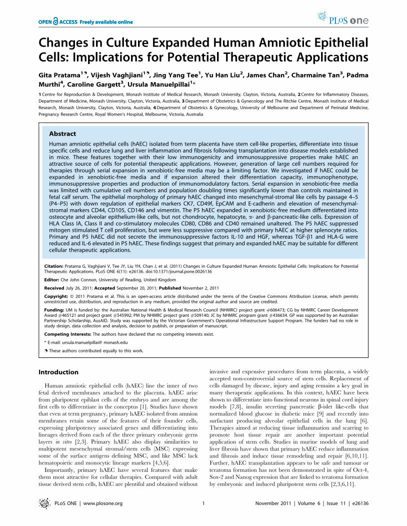
After an initial seeding density of 1.56106 cells, the CCN in
Epilife reached 37.4462.956106 cells by P5 but was significantly
lower than cultures grown in DMEM/F12 containing 10% FCS
(8064.336106 cells; p = 0.002; Fig. 1A). In PC-1 and 10% human
serum the CCN reached 10.8060.886106 and 2.0460.126106
cells by P4 and P2, respectively. The time needed for hAEC to
reach P5 in Epilife was 64.7561.6 d compared with
45.5062.02 d in DMEM/F12 containing 10% FCS (p,0.0001).
The CPD of 5.2160.12 for hAEC cultured in Epilife by P5 was
also significantly lower than in DMEM/F12 with 10% FCS
(6.3260.08; p = 0.0026; Fig. 1B).
During expansion, the cells changed their phenotype from a
typical epithelial morphology in P0–P2 to transitional epithelial-
stromal cells in P3–P4 and completely stromal-like cells by P5 in
Epilife and DMEM/F12 containing 10% FCS (Fig. 1C). Cells
Figure 1. Expansion of hAEC. The hAEC grew well in the xenobiotic-free media Epilife until passage 5 (P5) and plateaued thereafter. A similartrend was seen in control cultures grown in DMEM/F12+10% FCS but the cumulative cell number in Epilife was lower than controls (*p = 0.002; A).Cumulative population doubling of hAEC cultured in Epilife media was lower than DMEM/F12+10% FCS (*p = 0.0026; B). hAEC appeared stromal-likeat P5 in Epilife and DMEM/F12+10% FCS (C). Stromal-like cells at P4 retained the CFSE label suggesting the stromal cells arose from the labeled P2epithelial cells (D). Scale bars = 100 mm.doi:10.1371/journal.pone.0026136.g001
Properties of Expanded Amniotic Epithelial Cells
PLoS ONE | www.plosone.org 5 November 2011 | Volume 6 | Issue 11 | e26136
cultured in PC-1 were stromal like at P1 (data not shown). We
labeled P2 hAEC growing in Epilife and DMEM/F12+10% FCS
showing typical epithelial cobblestone morphology with the
intracellular dye CFSE to investigate whether the stromal-like
cells at P4 would retain the dye. At P4, the stromal-like cells were
labeled with CFSE (Fig. 1D), suggesting that these cells arose from
the epithelial cells.
We compared features of P5 cells expanded in Epilife and
DMEM/F12+10% FCS (DF) with the P0 cells. Analyses of P0 cells
cultured in Epilife and DF showed no significant differences for
any of the parameters tested; hence data for cultures grown in DF
are shown in Figs. 2–6.
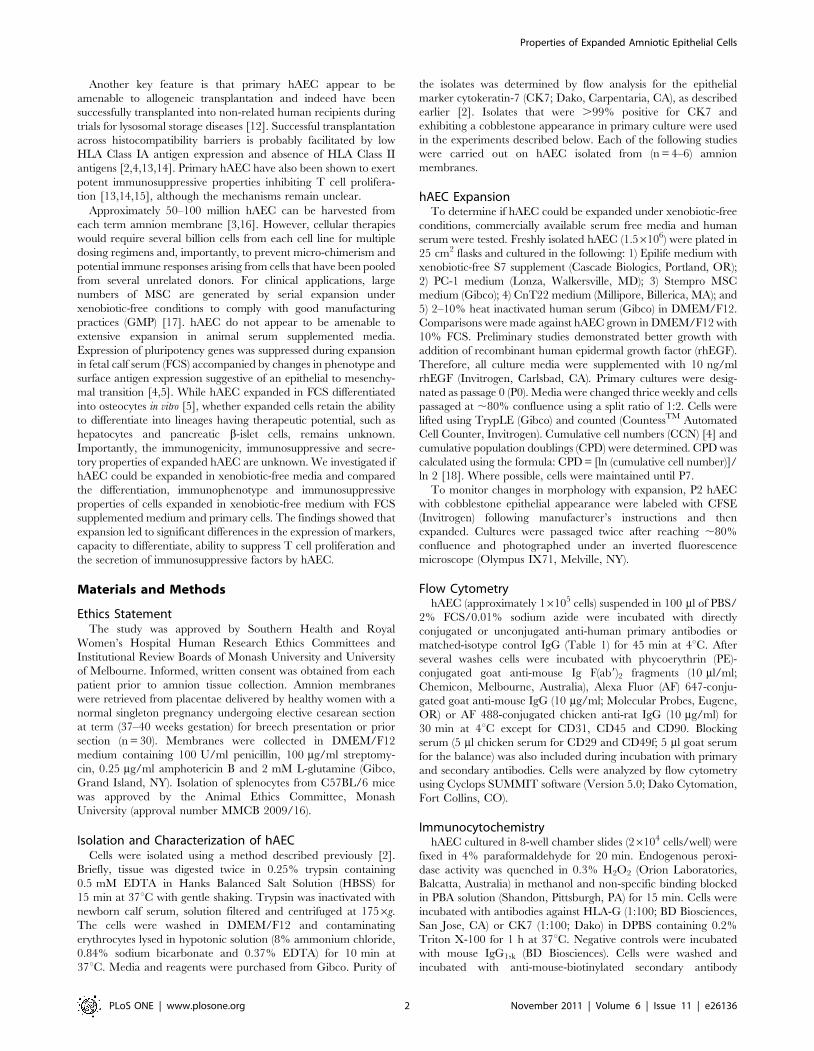
Properties of Expanded hAECKaryotype analyses were carried out to determine if chromo-
somal abnormalities arose during expansion. P5 cells expanded in
Epilife and DF were found to retain the normal autosomal and
XX or XY sex chromosome complement of the P0 cells (Fig. 2A).
We also compared the ultrastructural features of the P0 and
expanded cells by TEM. P0 and P5 hAEC had a high nuclear :
cytoplasmic ratio and prominent nucleoli (Fig. 2B). P0 hAEC were
typically roundish cells with surface microvilli and cytoplasmic
blebs. A multiloculated peripheral appearance with well developed
intercellular junctions, particularly desmosomes and small
amounts of rough endoplasmic reticulum (rER) were also seen in
the P0 cells. In contrast, P5 hAEC expanded in Epilife and DF had
extensive rER and associated Golgi complexes and very few
surface villi compared to P0 cells.
Investigating clonal colony formation, small clusters containing
.5 cells were observed in P0 cultures seeded at low density within
two weeks (Fig. 2C). Large clonal colonies were evident by three
weeks. The percentage cloning efficiency of P0 cells was
1.8960.17. Cells from the primary epithelial colonies were sub-
cloned 3–4 times but changed into stromal-like cells (data not
shown). The expanded P5 hAEC failed to form clonal colonies.
Next, we tested for alkaline phosphatase activity. The P0 and P5
hAEC lacked alkaline phosphatase activity measured using a
commercial assay, unlike the hEC cell line used as a positive
control (Fig. 2D).
Expression of Epithelial and Mesenchymal Markers withhAEC Expansion
Given the phenotypic changes observed with expansion, we
ascertained if there were changes in epithelial (E-cadherin, CD49f,
CK7, EpCAM), stromal (vimentin) and MSC associated markers
(CD44, CD90, CD105, CD146, PDGFR-b, CD29). P5 DF-hAEC
had reduced numbers of cells with epithelial markers compared to
P0 (E-cadherin, CK7 and CD49f; P,0.001; Fig. 3A), and more
cells with stromal/MSC markers (vimentin, CD90, CD146, CD44
and CD105; P,0.001). P5 Epilife cells also expressed stromal
Figure 2. Features of cultured primary (P0) and cells expanded in Epilife medium to passage 5 (P5). Normal karyotype of P0 hAEC wasretained at P5 (A). Transmission electron micrograph of P0 hAEC showed intercellular junctions (IJ), a multiloculated peripheral appearance comparedto P5 hAEC. P5 cells had extensive rough endoplasmic reticulum (rER) and fewer cell surface projections. P0 and P5 cells showed high nuclear (N) tocytoplasmic ratio (B). P0 hAEC seeded at low density formed clonal colonies unlike P5 hAEC (C). hAEC lacked alkaline phosphatase activity unlikehuman embryonal carcinoma (hEC) cell line used as a positive control (D). Scale bars = 100 mm (A–B); 5 mm and 1 mm (C).doi:10.1371/journal.pone.0026136.g002
Properties of Expanded Amniotic Epithelial Cells
PLoS ONE | www.plosone.org 6 November 2011 | Volume 6 | Issue 11 | e26136
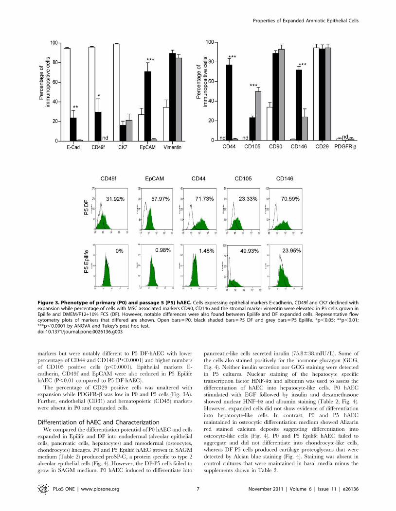
markers but were notably different to P5 DF-hAEC with lower
percentage of CD44 and CD146 (P,0.0001) and higher numbers
of CD105 positive cells (p,0.0001). Epithelial markers E-
cadherin, CD49f and EpCAM were also reduced in P5 Epilife
hAEC (P,0.01 compared to P5 DF-hAEC).
The percentage of CD29 positive cells was unaltered with
expansion while PDGFR-b was low in P0 and P5 cells (Fig. 3A).
Further, endothelial (CD31) and hematopoietic (CD45) markers
were absent in P0 and expanded cells.
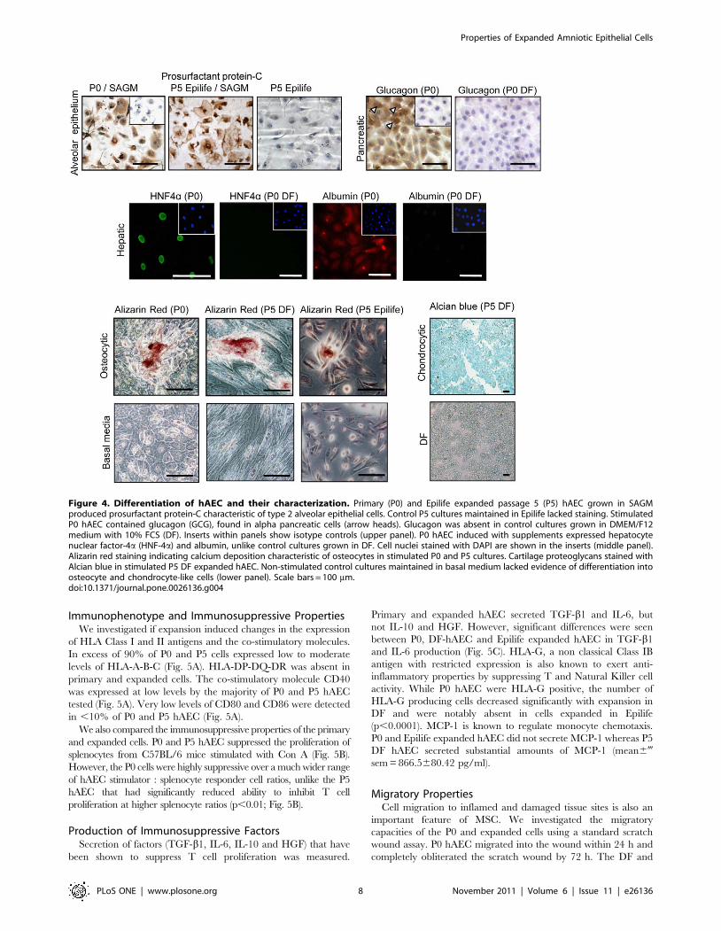
Differentiation of hAEC and CharacterizationWe compared the differentiation potential of P0 hAEC and cells
expanded in Epilife and DF into endodermal (alveolar epithelial
cells, pancreatic cells, hepatocytes) and mesodermal (osteocytes,
chondrocytes) lineages. P0 and P5 Epilife hAEC grown in SAGM
medium (Table 2) produced proSP-C, a protein specific to type 2
alveolar epithelial cells (Fig. 4). However, the DF-P5 cells failed to
grow in SAGM medium. P0 hAEC induced to differentiate into
pancreatic-like cells secreted insulin (75.8638.mIU/L). Some of
the cells also stained positively for the hormone glucagon (GCG,
Fig. 4). Neither insulin secretion nor GCG staining were detected
in P5 cultures. Nuclear staining of the hepatocyte specific
transcription factor HNF-4a and albumin was used to assess the
differentiation of hAEC into hepatocyte-like cells. P0 hAEC
stimulated with EGF followed by insulin and dexamethasone
showed nuclear HNF-4a and albumin staining (Table 2; Fig. 4).
However, expanded cells did not show evidence of differentiation
into hepatocyte-like cells. In contrast, P0 and P5 hAEC
maintained in osteocytic differentiation medium showed Alizarin
red stained calcium deposits suggesting differentiation into
osteocyte-like cells (Fig. 4). P0 and P5 Epilife hAEC failed to
aggregate and did not differentiate into chondrocyte-like cells,
whereas DF-P5 cells produced cartilage proteoglycans that were
detected by Alcian blue staining (Fig. 4). Staining was absent in
control cultures that were maintained in basal media minus the
supplements shown in Table 2.
Figure 3. Phenotype of primary (P0) and passage 5 (P5) hAEC. Cells expressing epithelial markers E-cadherin, CD49f and CK7 declined withexpansion while percentage of cells with MSC associated markers CD90, CD146 and the stromal marker vimentin were elevated in P5 cells grown inEpilife and DMEM/F12+10% FCS (DF). However, notable differences were also found between Epilife and DF expanded cells. Representative flowcytometry plots of markers that differed are shown. Open bars = P0, black shaded bars = P5 DF and grey bars = P5 Epilife. *p,0.05; **p,0.01;***p,0.0001 by ANOVA and Tukey’s post hoc test.doi:10.1371/journal.pone.0026136.g003
Properties of Expanded Amniotic Epithelial Cells
PLoS ONE | www.plosone.org 7 November 2011 | Volume 6 | Issue 11 | e26136
Immunophenotype and Immunosuppressive PropertiesWe investigated if expansion induced changes in the expression
of HLA Class I and II antigens and the co-stimulatory molecules.
In excess of 90% of P0 and P5 cells expressed low to moderate
levels of HLA-A-B-C (Fig. 5A). HLA-DP-DQ-DR was absent in
primary and expanded cells. The co-stimulatory molecule CD40
was expressed at low levels by the majority of P0 and P5 hAEC
tested (Fig. 5A). Very low levels of CD80 and CD86 were detected
in ,10% of P0 and P5 hAEC (Fig. 5A).
We also compared the immunosuppressive properties of the primary
and expanded cells. P0 and P5 hAEC suppressed the proliferation of
splenocytes from C57BL/6 mice stimulated with Con A (Fig. 5B).
However, the P0 cells were highly suppressive over a much wider range
of hAEC stimulator : splenocyte responder cell ratios, unlike the P5
hAEC that had significantly reduced ability to inhibit T cell
proliferation at higher splenocyte ratios (p,0.01; Fig. 5B).
Production of Immunosuppressive FactorsSecretion of factors (TGF-b1, IL-6, IL-10 and HGF) that have
been shown to suppress T cell proliferation was measured.
Primary and expanded hAEC secreted TGF-b1 and IL-6, but
not IL-10 and HGF. However, significant differences were seen
between P0, DF-hAEC and Epilife expanded hAEC in TGF-b1
and IL-6 production (Fig. 5C). HLA-G, a non classical Class IB
antigen with restricted expression is also known to exert anti-
inflammatory properties by suppressing T and Natural Killer cell
activity. While P0 hAEC were HLA-G positive, the number of
HLA-G producing cells decreased significantly with expansion in
DF and were notably absent in cells expanded in Epilife
(p,0.0001). MCP-1 is known to regulate monocyte chemotaxis.
P0 and Epilife expanded hAEC did not secrete MCP-1 whereas P5
DF hAEC secreted substantial amounts of MCP-1 (mean6-
sem = 866.5680.42 pg/ml).
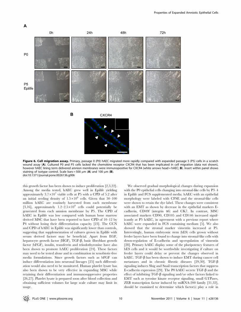
Migratory PropertiesCell migration to inflamed and damaged tissue sites is also an
important feature of MSC. We investigated the migratory
capacities of the P0 and expanded cells using a standard scratch
wound assay. P0 hAEC migrated into the wound within 24 h and
completely obliterated the scratch wound by 72 h. The DF and
Figure 4. Differentiation of hAEC and their characterization. Primary (P0) and Epilife expanded passage 5 (P5) hAEC grown in SAGMproduced prosurfactant protein-C characteristic of type 2 alveolar epithelial cells. Control P5 cultures maintained in Epilife lacked staining. StimulatedP0 hAEC contained glucagon (GCG), found in alpha pancreatic cells (arrow heads). Glucagon was absent in control cultures grown in DMEM/F12medium with 10% FCS (DF). Inserts within panels show isotype controls (upper panel). P0 hAEC induced with supplements expressed hepatocytenuclear factor-4a (HNF-4a) and albumin, unlike control cultures grown in DF. Cell nuclei stained with DAPI are shown in the inserts (middle panel).Alizarin red staining indicating calcium deposition characteristic of osteocytes in stimulated P0 and P5 cultures. Cartilage proteoglycans stained withAlcian blue in stimulated P5 DF expanded hAEC. Non-stimulated control cultures maintained in basal medium lacked evidence of differentiation intoosteocyte and chondrocyte-like cells (lower panel). Scale bars = 100 mm.doi:10.1371/journal.pone.0026136.g004
Properties of Expanded Amniotic Epithelial Cells
PLoS ONE | www.plosone.org 8 November 2011 | Volume 6 | Issue 11 | e26136

Epilife expanded hAEC showed reduced migration with the
scratch wound still visible after 72 h (Fig. 6A). The chemokine
receptor CXCR4 has been widely implicated in regulating the
migration of MSC in response to CXCL12 [21]. Immunolocal-
ization studies showed that CXCR4 was absent in the cultured P0
and expanded P5 cells. However, hAEC lining amnion membrane
stained positively for CXCR4 (Fig. 6B).
Discussion
We showed that hAEC can be expanded in xenobiotic-free
media, but that cell expansion was limited and that the hAEC
underwent phenotypic changes consistent with an epithelial-
mesenchymal transition (EMT). Further, we found notable
differences in differentiation capacity, migration, immunosuppres-
sive properties and secretion of immunomodulatory factors
between the P0 and P5 Epilife expanded hAEC. These changes
would need to be taken into account as they would have a marked
impact on the potential therapeutic applications of the expanded
hAEC.
Bilic et al. [4], reported that hAEC from term fetal membranes
showed limited expansion in FCS supplemented DMEM/F12
medium. Since stem cells need to be expanded in xenobiotic-free
media to comply with GMP for therapeutic applications [17], we
expanded hAEC in commercially available xenobiotic-free and
human serum containing media with rhEGF supplementation as
Figure 5. Immunogenicity of hAEC and effects on T cell proliferation. Primary (P0) and passage 5 (P5) hAEC expressed HLA-A-B-C but lackedHLA-DP-DQ-DR. Co-stimulatory molecules CD40, CD80 and CD86 remained unaltered at P5. Representative flow plots for HLA antigens and co-stimulatory molecules are shown (A). P0 and P5 hAEC suppressed Concanavelin A stimulated splenocytes from C57/BL6 mice, however P5 cellsshowed reduced suppression at higher splenocyte ratios (B). P0 and expanded cells produced the immunosuppressive factors TGF-b and IL-6. HLA-Gthat was abundant in P0 cells declined significantly in P5 grown in DMEM/F12+10%FCS (DF), while hAEC expanded in Epilife lacked HLA-G. Openbars = P0, black shaded bars = P5 cells expanded in DF and grey bars = P5 cells expanded in Epilife. Scale bars = 100 mm. *p,0.05; **p,0.01 and***p,0.001.doi:10.1371/journal.pone.0026136.g005
Properties of Expanded Amniotic Epithelial Cells
PLoS ONE | www.plosone.org 9 November 2011 | Volume 6 | Issue 11 | e26136
this growth factor has been shown to induce proliferation [2,3,22].
Among the media tested, hAEC grew well in Epilife yielding
approximately 3.76107 viable cells at P5 with a CPD of 5.2 after
an initial seeding density of 1.56106 cells. Given that 50–100
million hAEC are routinely harvested from each membrane
[3,16], approximately 1.2–2.56109 cells could potentially be
generated from each amnion membrane by P5. The CPD of
hAEC in Epilife was low compared with human bone marrow
derived MSC that have been reported to have CPD of 10–12 by
P4 without losing their differentiation capacity [23]. The CCN
and CPD of hAEC in Epilife was significantly lower than controls,
suggesting that supplementation of cultures grown in Epilife with
serum derived factors may be beneficial. Apart from EGF,
hepatocyte growth factor (HGF), TGF-b, basic fibroblast growth
factor (bFGF), insulin, transferrin and triiodothyronine have also
been shown to promote hAEC proliferation [24]. These factors
may need to be tested alone and in combination in xenobiotic-free
media formulations. Since growth factors such as bFGF can
induce differentiation into neuronal lineages [25] such differenti-
ation would also need to be monitored. Human platelet lysate has
also been shown to be very effective in expanding MSC while
retaining their differentiation and immunosuppressive properties
[26,27]. Platelet lysate is prepared soon after blood collection and
obtaining sufficient volumes for large scale culture may limit its
usage.
We observed gradual morphological changes during expansion
with the P0 epithelial cells changing into stromal-like cells by P3–4
in Epilife and FCS supplemented media. hAEC with an epithelial
morphology were labeled with CFSE and the stromal-like cells
were shown to retain the dye label. These changes were consistent
with an EMT as shown by decrease in the epithelial markers E-
cadherin, CD49f (integrin a6) and CK7. In contrast, MSC
associated markers CD90, CD105 and CD146 increased signif-
icantly at P5 hAEC, in agreement with a previous report where
hAEC were expanded in FCS containing medium [5]. We also
showed that the stromal marker vimentin increased at P5.
Interestingly, human embryonic stem (hES) cells grown without
feeder layers have been found to change into stromal-like cells with
down-regulation of E-cadherin and up-regulation of vimentin
[28]. Primary hAEC display some of the pluripotency features of
hES cells and it would be worthwhile investigating if culture on
feeder layers could delay or prevent the changes observed in
hAEC. TGF-b has been shown to induce EMT during cancer cell
metastases and in chronic fibrotic diseases [29,30]. TGF-bsignaling induces Slug and Snail transcription factors that suppress
E-cadherin expression [29]. The P0 hAEC secrete TGF-b and the
effect of inhibiting TGF-b signaling and/or other factors linked to
EMT such as tyrosine kinase receptor signaling, small GTPases,
ZEB transcription factor induced by miRNA-200 family [31,32],
should be examined to determine which factor(s) play a role in
Figure 6. Cell migration assay. Primary, passage 0 (P0) hAEC migrated more rapidly compared with expanded passage 5 (P5) cells in a scratchwound assay (A). Cultured P0 and P5 cells lacked the chemokine receptor CXCR4 that has been implicated in cell migration (data not shown),however hAEC lining term delivered amnion membranes were immunopositive for CXCR4 (white arrows head = hAEC; B). Insert within panel showsstaining of isotype control. Scale bars = 500 mm (A) and 100 mm (B).doi:10.1371/journal.pone.0026136.g006
Properties of Expanded Amniotic Epithelial Cells
PLoS ONE | www.plosone.org 10 November 2011 | Volume 6 | Issue 11 | e26136
changes observed during expansion of hAEC. Further, EGF also
has been reported to induce EMT in some cancer cell lines
[33,34], but enhanced expression of MSC-related antigens in
hAEC occur in cultures without addition of EGF [5]. In
preliminary experiments addition of EGF to P0–P2 cultures did
not stimulate EMT (data not shown), suggesting that EGF does not
play a role.
Mature, polarized epithelial cells that undergo EMT display
migratory properties [29,35]. Interestingly, using a scratch wound
assay we found that not only the stromal-like P5 hAEC were able
to migrate, but that the P0 hAEC had a higher migratory capacity.
hAEC lining the amnion membrane have no known migratory
properties and hence factors that could regulate the migration of
hAEC have not been investigated. Interactions between chemo-
kines and their receptors, in particular CXCR4, are believed to
play important roles in the migration of MSC [21]. We localized
CXCR4 to hAEC lining the amnion membranes but neither P0
nor P5 hAEC expressed CXCR4. Expression of CXCR4 has been
shown to decline in MSC during culture [21] and this may
account for the loss of CXCR4 in the P0 cells. Since chemotactic
and adhesion factors play important roles in regulating migration
of stem cells to target sites of tissue inflammation and damage, it
would be important to identify these factors in assessing the
therapeutic applications of hAEC of the primary and expanded
hAEC.
Changes in morphology and reduced differentiation capacity
due to senescence have been reported in porcine and human MSC
expanded in long term culture [23,36,37]. We found that unlike
the P0 cells, P5 hAEC failed to differentiate into important
endodermal lineages such hepatocytes and pancreatic cells and
would limit derivation of these lineages for potential cell
replacement therapies to primary hAEC and cells from early
passages. However, as P5 Epilife expanded cells differentiated into
osteocyte and surfactant producing alveolar epithelial-like cells, it
suggests a functional alteration rather than senescence being
responsible for changes in differentiation, and it would be
important to determine if the expanded cells can undergo tissue
specific differentiation in vivo as has been demonstrated for P0
hAEC [6,9,11]. Indeed, the TEM studies showed that the
expanded hAEC had well developed rER and Golgi complexes
consistent with maturation and a well developed secretory profile
and not senescence. Down regulation of ES markers TRA1-60
and TRA1-81 has been reported during expansion [5] and it is
possible that expression of lineage specification and differentiation
pathways also alter during hAEC expansion. Expansion may also
lead to selection of sub-populations within the primary isolates as
notable differences in both marker expression, secretory profile
and differentiation was found between FCS supplemented and
Epilife expanded hAEC.
The low immunogenicity exhibited by expanded MSC from
bone marrow and gestational tissue have enabled clinical trials
involving allogeneic transplantation. We showed that P0 hAEC
expressed low to moderate levels of HLA class IA and lack HLA
class II antigens, consistent with previous reports [4,13,14].
Expression of HLA and the co-stimulatory molecules CD80,
CD86 and CD40 is required to activate T cells and subsequent
immune rejection of the transplanted cells. We found CD40
expressed by P0 cells, while both CD80 and CD86 were negligible.
There were no significant differences in the expression of these
antigens in the P5 hAEC. These findings may explain the survival
of P0 hAEC following xeno-transplantation into immune-compe-
tent animals over prolonged periods [8,11] and also suggest that
P0 and P5 hAEC are unlikely to be rejected following xeno-
transplantation.
We also examined the immunosuppressive properties of the P0
and expanded hAEC. Consistent with previous reports, P0 hAEC
suppressed T cell proliferation [13,14,15]. The P5 hAEC also
suppressed T cell proliferation, however the P0 cells were more
effective at higher splenocyte ratios. The immunosuppressive
properties of MSC are well established and HLA-G, IL-6 and
TGF-b [38,39,40] among the factors known to play a role. Djouad
et al [38] proposed that IL-6 secreted by MSC inhibits dendritic
cell maturation and subsequently impairs T cell proliferation and
induces tolerance. TGF-b1 has also been shown to inhibit T cell
proliferation [39]. We found that IL-6 and TGF-b1 were secreted
by P0 and expanded hAEC and these factors may partly
contribute towards the suppression of T cell proliferation. On
the other hand, a high percentage of P0 cells were HLA-G positive
compared with P5 hAEC with cells cultured in Epilife lacking this
non-polymorphic Class IB antigen. HLA-G has been shown to
inhibit proliferation by binding to killer immunoglobulin-like
receptors and/or immunoglobulin-like transcript on CD4+ and
CD8+ T cells [41]. HLA-G is also known to modulate the
cytotoxic activity of Natural Killer cells. MSC secrete other anti-
inflammatory factors such as IL-10 and HGF. Interestingly,
neither the P0 nor expanded hAEC secreted IL-10 or HGF.
Recent studies show that transplantation of P0 hAEC reduces
tissue inflammation and fibrosis in murine liver and lungs [6,11],
although the mechanisms remain largely unknown. P5-DF
expanded hAEC secreted significant amounts of MCP-1 that
could induce the recruitment of monocytes and promote
fibrogenesis. In addition to the immuno-modulatory effects,
TGF-b1 and IL-6 play an important role in promoting
fibrogenesis. Therefore, the effects of expanded P5 hAEC on
tissue inflammation, monocyte chemotaxis and fibrosis would need
to be tested in animal models.
In conclusion, we have shown that expanded hAEC have
different properties to the primary cells. P0 hAEC may be useful
for generating hepatocyte and pancreatic–like cells for therapeutic
applications and expanded cells for mending bone fractures and
contributing towards the alveolar epithelial cell population
damaged in lung diseases. Further, the P0 cells may be more
useful in suppressing tissue inflammation and fibrosis and as a
treatment for autoimmune diseases and graft vs host disease where
it would be important to limit T cell activation. Characterization
of the transitional hAEC at passages 2–3 and testing expanded
hAEC in vivo models would be beneficial in assessing the suitability
of the expanded hAEC for cellular therapeutic applications.
Acknowledgments
We gratefully acknowledge assistance given by P Temple-Smith for
analyzing TEM images and Ding Oh for Alcian blue and Alizarin red
staining.
Author Contributions
Conceived and designed the experiments: GP VV CG UM. Performed the
experiments: GP VV JYT YHL. Analyzed the data: GP VV JC UM.
Contributed reagents/materials/analysis tools: JYT YHL CT PM UM.
Wrote the paper: GP VV UM.
Properties of Expanded Amniotic Epithelial Cells
PLoS ONE | www.plosone.org 11 November 2011 | Volume 6 | Issue 11 | e26136
References
1. Ilancheran S, Moodley Y, Manuelpillai U (2009) Human fetal membranes: asource of stem cells for tissue regeneration and repair? Placenta 30: 2–10.
2. Ilancheran S, Michalska A, Peh G, Wallace EM, Pera M, et al. (2007) Stem cellsderived from human fetal membranes display multilineage differentiation
potential. Biol Reprod 77: 577–588.3. Miki T, Lehmann T, Cai H, Stolz DB, Strom SC (2005) Stem cell characteristics
of amniotic epithelial cells. Stem Cells 23: 1549–1559.
4. Bilic G, Zeisberger SM, Mallik AS, Zimmermann R, Zisch AH (2008)Comparative characterization of cultured human term amnion epithelial and
mesenchymal stromal cells for application in cell therapy. Cell Transplant 17:955–968.
5. Stadler G, Hennerbichler S, Lindenmair A, Peterbauer A, Hofer K, et al. (2008)
Phenotypic shift of human amniotic epithelial cells in culture is associated withreduced osteogenic differentiation in vitro. Cytotherapy 10: 743–752.
6. Moodley Y, Ilancheran S, Samuel C, Vaghjiani V, Atienza D, et al. (2010)Human amnion epithelial cell transplantation abrogates lung fibrosis and
augments repair. Am J Respir Crit Care Med 182: 643–651.
7. Meng XT, Li C, Dong ZY, Liu JM, Li W, et al. (2008) Co-transplantation ofbFGF-expressing amniotic epithelial cells and neural stem cells promotes
functional recovery in spinal cord-injured rats. Cell Biol Int 32: 1546–1558.8. Sankar V, Muthusamy R (2003) Role of human amniotic epithelial cell
transplantation in spinal cord injury repair research. Neuroscience 118: 11–17.9. Wei JP, Zhang TS, Kawa S, Aizawa T, Ota M, et al. (2003) Human amnion-
isolated cells normalize blood glucose in streptozotocin-induced diabetic mice.
Cell Transplant 12: 545–552.10. Cargnoni A, Gibelli L, Tosini A, Signoroni PB, Nassuato C, et al. (2009)
Transplantation of allogeneic and xenogeneic placenta-derived cells reducesbleomycin-induced lung fibrosis. Cell Transplant 18: 405–422.
11. Manuelpillai U, Tchongue J, Lourensz D, Vaghjiani V, Samuel CS, et al. (2010)
Transplantation of human amnion epithelial cells reduces hepatic fibrosis inimmunocompetent CCl4 treated mice. Cell Transplant 19: 1157–1168.
12. Yeager AM, Singer HS, Buck JR, Matalon R, Brennan S, et al. (1985) Atherapeutic trial of amniotic epithelial cell implantation in patients with
lysosomal storage diseases. Am J Med Genet 22: 347–355.13. Banas RA, Trumpower C, Bentlejewski C, Marshall V, Sing G, et al. (2008)
Immunogenicity and immunomodulatory effects of amnion-derived multipotent
progenitor cells. Hum Immunol 69: 321–328.14. Wolbank S, Peterbauer A, Fahrner M, Hennerbichler S, van Griensven M, et al.
(2007) Dose-dependent immunomodulatory effect of human stem cells fromamniotic membrane: a comparison with human mesenchymal stem cells from
adipose tissue. Tissue Eng 13: 1173–1183.
15. Li H, Niederkorn JY, Neelam S, Mayhew E, Word RA, et al. (2005)Immunosuppressive factors secreted by human amniotic epithelial cells. Invest
Ophthalmol Vis Sci 46: 900–907.16. Toda A, Okabe M, Yoshida T, Nikaido T (2007) The potential of amniotic
membrane/amnion-derived cells for regeneration of various tissues. J PharmacolSci 105: 215–228.
17. Felka T, Schafer R, De Zwart P, Aicher WK (2010) Animal serum-free
expansion and differentiation of human mesenchymal stromal cells. Cytotherapy12: 143–153.
18. Gargett CE, Schwab KE, Zillwood RM, Nguyen HP, Wu D (2009) Isolation andculture of epithelial progenitors and mesenchymal stem cells from human
endometrium. Biol Reprod 80: 1136–1145.
19. Pera MF, Reubinoff B, Trounson A (2000) Human embryonic stem cells. J CellSci 113(Pt 1): 5–10.
20. Chan J, Ban EJ, Chun KH, Wang S, McQualter J, et al. (2008)Methylprednisolone induces reversible clinical and pathological remission and
loss of lymphocyte reactivity to myelin oligodendrocyte glycoprotein inexperimental autoimmune encephalomyelitis. Autoimmunity 41: 405–413.
21. Kollar K, Cook MM, Atkinson K, Brooke G (2009) Molecular mechanisms
involved in mesenchymal stem cell migration to the site of acute myocardialinfarction. Int J Cell Biol 2009: 1–8.
22. Terada S, Matsuura K, Enosawa S, Miki M, Hoshika A, et al. (2000) Inducingproliferation of human amniotic epithelial (HAE) cells for cell therapy. Cell
Transplant 9: 701–704.
23. Siddappa R, Licht R, van Blitterswijk C, de Boer J (2007) Donor variation andloss of multipotency during in vitro expansion of human mesenchymal stem cells
for bone tissue engineering. J Orthop Res 25: 1029–1041.24. Ochsenbein-Kolble N, Bilic G, Hall H, Huch R, Zimmermann R (2003)
Inducing proliferation of human amnion epithelial and mesenchymal cells forprospective engineering of membrane repair. J Perinat Med 31: 287–294.
25. Yang H, Xia Y, Lu SQ, Soong TW, Feng ZW (2008) Basic fibroblast growth
factor-induced neuronal differentiation of mouse bone marrow stromal cellsrequires FGFR-1, MAPK/ERK, and transcription factor AP-1. J Biol Chem
283: 5287–5295.26. Doucet C, Ernou I, Zhang Y, Llense JR, Begot L, et al. (2005) Platelet lysates
promote mesenchymal stem cell expansion: a safety substitute for animal serum
in cell-based therapy applications. J Cell Physiol 205: 228–236.27. Bernardo ME, Avanzini MA, Perotti C, Cometa AM, Moretta A, et al. (2007)
Optimization of in vitro expansion of human multipotent mesenchymal stromalcells for cell-therapy approaches: further insights in the search for a fetal calf
serum substitute. J Cell Physiol 211: 121–130.
28. Ullmann U, In’t Veld P, Gilles C, Sermon K, De Rycke M, et al. (2007)Epithelial-mesenchymal transition process in human embryonic stem cells
cultured in feeder-free conditions. Mol Hum Reprod 13: 21–32.29. Zavadil J, Bottinger EP (2005) TGF-beta and epithelial-to-mesenchymal
transitions. Oncogene 24: 5764–5774.30. Willis BC, Liebler JM, Luby-Phelps K, Nicholson AG, Crandall ED, et al. (2005)
Induction of epithelial-mesenchymal transition in alveolar epithelial cells by
transforming growth factor-beta1: potential role in idiopathic pulmonaryfibrosis. Am J Pathol 166: 1321–1332.
31. Korpal M, Kang Y (2008) The emerging role of miR-200 family of microRNAsin epithelial-mesenchymal transition and cancer metastasis. RNA Biol 5:
115–119.
32. Thiery JP (2003) Epithelial-mesenchymal transitions in development andpathologies. Curr Opin Cell Biol 15: 740–746.
33. Ahmed N, Maines-Bandiera S, Quinn MA, Unger WG, Dedhar S, et al. (2006)Molecular pathways regulating EGF-induced epithelio-mesenchymal transition
in human ovarian surface epithelium. Am J Physiol Cell Physiol 290:C1532–1542.
34. Ackland ML, Newgreen DF, Fridman M, Waltham MC, Arvanitis A, et al.
(2003) Epidermal growth factor-induced epithelio-mesenchymal transition inhuman breast carcinoma cells. Lab Invest 83: 435–448.
35. Thiery JP (2002) Epithelial-mesenchymal transitions in tumour progression. NatRev Cancer 2: 442–454.
36. Vacanti V, Kong E, Suzuki G, Sato K, Canty JM, et al. (2005) Phenotypic
changes of adult porcine mesenchymal stem cells induced by prolongedpassaging in culture. J Cell Physiol 205: 194–201.
37. Banfi A, Bianchi G, Notaro R, Luzzatto L, Cancedda R, et al. (2002) Replicativeaging and gene expression in long-term cultures of human bone marrow stromal
cells. Tissue Eng 8: 901–910.38. Djouad F, Charbonnier LM, Bouffi C, Louis-Plence P, Bony C, et al. (2007)
Mesenchymal stem cells inhibit the differentiation of dendritic cells through an
interleukin-6-dependent mechanism. Stem Cells 25: 2025–2032.39. Di Nicola M, Carlo-Stella C, Magni M, Milanesi M, Longoni PD, et al. (2002)
Human bone marrow stromal cells suppress T-lymphocyte proliferation inducedby cellular or nonspecific mitogenic stimuli. Blood 99: 3838–3843.
40. Nasef A, Mathieu N, Chapel A, Frick J, Francois S, et al. (2007)
Immunosuppressive effects of mesenchymal stem cells: involvement of HLA-G. Transplantation 84: 231–237.
41. Bahri R, Hirsch F, Josse A, Rouas-Freiss N, Bidere N, et al. (2006) Soluble HLA-G inhibits cell cycle progression in human alloreactive T lymphocytes. J Immunol
176: 1331–1339.42. Tamagawa T, Ishiwata I, Sato K, Nakamura Y (2009) Induced in vitro
differentiation of pancreatic-like cells from human amnion-derived fibroblast-like
cells. Hum Cell 22: 55–63.43. Peister A, Mellad JA, Larson BL, Hall BM, Gibson LF, et al. (2004) Adult stem
cells from bone marrow (MSCs) isolated from different strains of inbred micevary in surface epitopes, rates of proliferation, and differentiation potential.
Blood 103: 1662–1668.
Properties of Expanded Amniotic Epithelial Cells
PLoS ONE | www.plosone.org 12 November 2011 | Volume 6 | Issue 11 | e26136
Related Documents