Changes in Cerebral Glucose Metabolism during Early Abstinence from Chronic Methamphetamine Abuse Steven M. Berman, Bradley Voytek, Mark A. Mandelkern, B. Daniel Hassid, Andrew Isaacson, John Monterosso, Karen Miotto, Walter Ling, and Edythe D. London Departments of Psychiatry and Biobehavioral Sciences (Drs. Berman, Monterosso, Miotto, Ling and London; Mr. Voytek, Mr. Hassid and Mr. Isaacson), Molecular and Medical Pharmacology (Dr. London), and the Brain Research Institute (Drs. Berman and London), David Geffen School of Medicine, University of California Los Angeles, Los Angeles, CA; and Department of Physics, University of California Irvine, Irvine, CA (Dr. Mandelkern) Abstract Changes in brain function during the initial weeks of abstinence from chronic methamphetamine abuse may substantially affect clinical outcome, but are not well understood. We used positron emission tomography with [F-18]fluorodeoxyglucose (FDG) to quantify regional cerebral glucose metabolism, an index of brain function, during performance of a vigilance task. Ten methamphetamine-dependent subjects were tested after 5-9 days of abstinence, and after 4 additional weeks of supervised abstinence. Twelve healthy control subjects were tested at corresponding times. Global glucose metabolism increased between tests (p = .01), more in methamphetamine-dependent (10.9%, p = .02) than control subjects (1.9%, NS). Glucose metabolism did not change in subcortical regions of methamphetamine-dependent subjects, but increased in neocortex, with maximal increase (> 20%) in parietal regions. Changes in reaction time and self-reports of negative affect varied more in methamphetamine-dependent than in control subjects, and correlated both with the increase in parietal glucose metabolism, and decrease in relative activity (after scaling to the global mean) in some regions. A robust relationship between change in self-reports of depressive symptoms and relative activity in the ventral striatum may have great relevance to treatment success because of the role of this region in drug abuse-related behaviors. Shifts in cortical-subcortical metabolic balance either reflect new processes that occur during early abstinence, or the unmasking of effects of chronic methamphetamine abuse that are obscured by suppression of cortical glucose metabolism that continues for at least 5-9 days after cessation of methamphetamine self-administration. Keywords methamphetamine; drug abuse; brain metabolism; fluorodeoxyglucose; PET imaging; continuous performance test; abstinence Human subjects who previously abused methamphetamine (MA) exhibit disturbances of mood and cognition during abstinence 1-5 . These deficits appear to reflect regional cerebral dysfunction. When abstinent for one week, MA abusers studied with [F-18] fluorodeoxyglucose (FDG) and positron emission tomography (PET) have more severe self- reports of depressive symptoms than control subjects, and these self-reports covary with relative uptake of the radiotracer in anterior cingulate cortex and amygdala 1 . Cingulate and Send correspondence to: Edythe D. London, PhD, Professor of Psychiatry and Biobehavioral Sciences, Semel Institute for Neuroscience and Human Behavior, University of California Los Angeles, 760 Westwood Plaza, Box 175919, Los Angeles, California 90024-1759 (310)8250606 fax (310)8250812 ([email protected]).. NIH Public Access Author Manuscript Mol Psychiatry. Author manuscript; available in PMC 2009 December 1. Published in final edited form as: Mol Psychiatry. 2008 September ; 13(9): 897–908. doi:10.1038/sj.mp.4002107. NIH-PA Author Manuscript NIH-PA Author Manuscript NIH-PA Author Manuscript

Welcome message from author
This document is posted to help you gain knowledge. Please leave a comment to let me know what you think about it! Share it to your friends and learn new things together.
Transcript
Changes in Cerebral Glucose Metabolism during Early Abstinencefrom Chronic Methamphetamine Abuse
Steven M. Berman, Bradley Voytek, Mark A. Mandelkern, B. Daniel Hassid, AndrewIsaacson, John Monterosso, Karen Miotto, Walter Ling, and Edythe D. LondonDepartments of Psychiatry and Biobehavioral Sciences (Drs. Berman, Monterosso, Miotto, Ling andLondon; Mr. Voytek, Mr. Hassid and Mr. Isaacson), Molecular and Medical Pharmacology (Dr.London), and the Brain Research Institute (Drs. Berman and London), David Geffen School ofMedicine, University of California Los Angeles, Los Angeles, CA; and Department of Physics,University of California Irvine, Irvine, CA (Dr. Mandelkern)
AbstractChanges in brain function during the initial weeks of abstinence from chronic methamphetamineabuse may substantially affect clinical outcome, but are not well understood. We used positronemission tomography with [F-18]fluorodeoxyglucose (FDG) to quantify regional cerebral glucosemetabolism, an index of brain function, during performance of a vigilance task. Tenmethamphetamine-dependent subjects were tested after 5-9 days of abstinence, and after 4 additionalweeks of supervised abstinence. Twelve healthy control subjects were tested at corresponding times.Global glucose metabolism increased between tests (p = .01), more in methamphetamine-dependent(10.9%, p = .02) than control subjects (1.9%, NS). Glucose metabolism did not change in subcorticalregions of methamphetamine-dependent subjects, but increased in neocortex, with maximal increase(> 20%) in parietal regions. Changes in reaction time and self-reports of negative affect varied morein methamphetamine-dependent than in control subjects, and correlated both with the increase inparietal glucose metabolism, and decrease in relative activity (after scaling to the global mean) insome regions. A robust relationship between change in self-reports of depressive symptoms andrelative activity in the ventral striatum may have great relevance to treatment success because of therole of this region in drug abuse-related behaviors. Shifts in cortical-subcortical metabolic balanceeither reflect new processes that occur during early abstinence, or the unmasking of effects of chronicmethamphetamine abuse that are obscured by suppression of cortical glucose metabolism thatcontinues for at least 5-9 days after cessation of methamphetamine self-administration.
Keywordsmethamphetamine; drug abuse; brain metabolism; fluorodeoxyglucose; PET imaging; continuousperformance test; abstinence
Human subjects who previously abused methamphetamine (MA) exhibit disturbances of moodand cognition during abstinence 1-5. These deficits appear to reflect regional cerebraldysfunction. When abstinent for one week, MA abusers studied with [F-18]fluorodeoxyglucose (FDG) and positron emission tomography (PET) have more severe self-reports of depressive symptoms than control subjects, and these self-reports covary withrelative uptake of the radiotracer in anterior cingulate cortex and amygdala 1. Cingulate and
Send correspondence to: Edythe D. London, PhD, Professor of Psychiatry and Biobehavioral Sciences, Semel Institute for Neuroscienceand Human Behavior, University of California Los Angeles, 760 Westwood Plaza, Box 175919, Los Angeles, California 90024-1759(310)8250606 fax (310)8250812 ([email protected])..
NIH Public AccessAuthor ManuscriptMol Psychiatry. Author manuscript; available in PMC 2009 December 1.
Published in final edited form as:Mol Psychiatry. 2008 September ; 13(9): 897–908. doi:10.1038/sj.mp.4002107.
NIH
-PA Author Manuscript
NIH
-PA Author Manuscript
NIH
-PA Author Manuscript

insular dysfunctions may also contribute to impaired vigilance in MA-dependent subjects whorecently stopped taking the drug 6.
Anterior cingulate dysfunction has also been observed in subjects who maintained abstinencefrom MA. Deficiencies were seen in activation during performance of a decision-making taskafter one month of abstinence 2. In another study, cingulate perfusion was lower than controllevels after 3 months of abstinence, but was normal in subjects abstinent for 36 months 7.
Abnormalities in cerebral glucose metabolism during the first week of abstinence from MAdiffer from those observed in a sample abstinent between .5-35 months. Cortical glucosemetabolism was above control levels, most markedly in parietal cortex, with lower relativeradioactivity (after global scaling) in the striatum and thalamus 8. The thalamic, but not thestriatal deficits in relative activity, normalized after the first year 9.
Although the first weeks of abstinence from MA abuse are important for retention in treatment10, we know of no reports on cerebral metabolic changes during this period. Such effects mighthave important implications for therapies that involve specific brain circuits, and therefore arethe subject of the present report. We hypothesized: 1) that abnormalities during the initial weekof abstinence from chronic MA abuse (i.e., lower relative glucose metabolism in the anteriorcingulate gyrus, higher relative glucose metabolism in the striatum) 1 would shift, after anadditional month of abstinence, to the pattern previously reported after longer abstinence 8,9through increases in cortical activity, particularly in the parietal lobes, and 2) that changes inactivity would be associated with concurrent changes in cognitive performance, as measuredby a vigilance task, and in self-reports of depressive symptoms.
MethodsGeneral experimental design
Two groups of research subjects participated. MA-dependent participants resided on a researchward during the study. Cerebral glucose metabolism was assayed using FDG with PET 11
during an auditory vigilance task. Comparison subjects participated on a nonresidential basis.MA-dependent subjects were tested after 5-9 days (mean ± SD = 6.7 ± 1.6 days) and afteranother month of supervised abstinence (mean ± SD = 27.6 ± 0.96 days). The test interval wasanalogous for comparison subjects (mean ± SD = 33.1 ± 7.20 days). Participants each provideda self-rating of depressive symptoms (Beck Depression Inventory, BDI) 12 and a urine samplenegative for illegal drugs of abuse on the day of each PET scan.
Research participantsThe current sample of 10 MA-dependent and 12 comparison subjects overlapped with 17 MA-dependent and 18 comparison subjects in a prior study of mood and cerebral glucosemetabolism 1. Only subjects who completed both PET sessions were included. Abstinence wasconfirmed by urine drug screens during hospitalization. All subjects gave informed consent asapproved by the institutional review boards of UCLA and Long Beach VAMC, and werehealthy according to medical history, physical examination, and laboratory tests. Left-handedness 13, current use of psychoactive medications, and seropositive status for humanimmunodeficiency virus were exclusionary.
The groups did not differ significantly on age, gender, ethnicity, education, or mother’seducation (Table 1). During the first week of abstinence, subjects completed a self-report intakequestionnaire and drug-use survey. Current Axis I diagnoses of dependencies on substancesother than nicotine and lifetime Axis I diagnoses unrelated to drug abuse (Structured ClinicalInterview for DSM-IV; SCID-I) 14, were exclusionary, except that MA-dependence and abuse
Berman et al. Page 2
Mol Psychiatry. Author manuscript; available in PMC 2009 December 1.
NIH
-PA Author Manuscript
NIH
-PA Author Manuscript
NIH
-PA Author Manuscript
before enrollment (verified by urine screen) were required for the MA group. Alcohol use <7.5 drinks per week was permitted.
Image acquisitionT2 and T1-weighted whole-brain MRI images were acquired (3-T, General Electric),respectively, for exclusion of individuals with frank structural brain abnormalities and for co-registration of PET scans (Siemens ECAT EXACT HR+, Knoxville, TN), acquired inconjunction with an auditory vigilance task. Details on the methods of data acquisition andquantitative analysis of the PET scans have been published 1.
Subjects performed a 30-min auditory vigilance task that required a button press after hearinga tone of designated pitch within a sequence of nontarget tones (inter-stimulus interval = 2sec). FDG (< 5 mCi) was injected shortly after the vigilance task began. PET images integrateddecay-corrected activity from 50-80 min after FDG injection, used as an index of radiotraceruptake during the vigilance task. An input function, using data from arterial samples, was usedto determine cerebral metabolic rate for glucose (CMRglc, mg glucose per 100 g tissue permin) at each voxel 15. Voxels exhibiting values < 4 mg/100 g/min were excluded to minimizecontributions from cerebrospinal fluid, and remaining voxels averaged to calculate globalCMRglc. Although all subjects provided analyzable images of decay-corrected activity,CMRglc could not be calculated for one MA-dependent and five control subjects due to catheterfailure or improper instrument calibration.
Statistical analysisVigilance performance was quantified by target accuracy and reaction time (RT). Two-tailedt-tests compared the groups on vigilance and continuous demographic variables. Chi squareanalyses were used for frequency data. The statistical threshold was set at p < .05.
Group comparisons to assess regional relative radioactivity from FDG (regional radioactivitydivided by whole-brain radioactivity, used as a surrogate measure of regional – CMRglc), andabsolute regional CMRglc (from modeled images) were performed by Statistical ParametricMapping software (SPM2; Wellcome Department of Cognitive Neurology, London;www.fil.ion.ucl.ac.uk/spm). PET images were spatially normalized into a standard coordinatesystem (Montreal Neurological Institute, MNI space), and smoothed with an 8-mm (FWHM)isotropic Gaussian kernel.
Ten regions of interest (ROIs) were selected because of prior demonstrations of functionalabnormalities associated with MA abuse: parietal cortex (Brodmann areas [BAs] 1, 2, 3, 5, 7,19, 39, 40), thalamus, dorsal striatum, ventral striatum, medial orbitofrontal cortex (gyrusrectus & medial orbital gyrus; BA 11), lateral orbitofrontal cortex (lateral, posterior orbital &inferior frontal gyri; BAs 47, 11), infragenual anterior cingulate cortex (ACC) (BAs 25, 32),supragenual ACC (BAs 24, 32, 33), insula (BA 13), and amygdala. 1, 6, 8, 9
ROIs were drawn on the structural MR template provided in SPM2, using MEDx Software(Sensor Systems, Sterling, VA). Bilateral sampling of these regions provided data on activitychanges across sessions within 20 ROIs (i.e., 40 comparisons). Separate analyses wereperformed for analyses of the cerebral metabolic rate for glucose (CMRglc) and regionalrelative radioactivity. Additional analyses assessed covariation between changes in regionalbrain activity across sessions and simultaneous changes in cognitive performance (vigilancetask reaction time and depressive symptoms; BDI score).
We set an initial voxel-height threshold of p = .01 (uncorrected) for inclusion in statisticalparametric maps. However, to correct for multiple comparisons within a volume of assessedvoxels, we considered as significant only findings that also retained a p value < .05 after
Berman et al. Page 3
Mol Psychiatry. Author manuscript; available in PMC 2009 December 1.
NIH
-PA Author Manuscript
NIH
-PA Author Manuscript
NIH
-PA Author Manuscript
correction for the volume of the ROI (SPM2 familywise error correction; WellcomeDepartment of Cognitive Neurology, London; www.fil.ion.ucl.ac.uk/spm). As an additionalmeasure of confidence, effects that retained statistical significance after Bonferroni correctionfor the number of regions were also identified. Using this approach, the nominal alpha levelfor a particular region must be < 0.05 divided by the number of comparisons (i.e., .05 / 40 = .0012) in order to exhibit a true alpha level of 0.05. This evidentiary criterion is highlyconservative as it assumes that tests of the individual regions are independent of one another.This assumption is certainly untrue because of connectivity between different brain regions,especially brain regions selected for study on the basis of prior reports of a common functionalabnormality.
RESULTSSelf-report of drug use
Self report data are given in Table 1. The MA group had used the drug, on average, for > 8years; consumed about 2-4 g/week, and had used MA on most of the 30 days before enteringthe study.
Self-report of depressive symptomsControl subjects reported low ratings of depressive symptoms at the initial measurement (eightBDI scores of 0, two scores of 1, and two scores of 3). Participants in the MA group gave higherself-ratings than control subjects at 5-9 days of abstinence (mean [SD] 5.8 [6.4], (t = 2.73, p= .01). Four weeks later, one control subject reported a 1-unit increase in BDI score. No otherscores changed. In contrast, half of the MA group showed a BDI change ≥ 4 units at retest (2increases, 3 decreases). Although the MA group as a whole reported a 29.3% reduction in BDIscore after 4 weeks (from 5.8 to 4.1 [6.1]), this reduction was not significant (t = 0.93, p = .38). At retest, there was no longer a significant difference between groups (t = 1.88, p = .08).
Vigilance task performanceAccuracy on the vigilance task decreased in both groups at retest (MA initial mean [SD] 96.0%[4.4], retest 94.1% [5.6]; control 98.1% [2.3], retest 96.1% [3.3]). Although the Session-by-Group interaction was not significant (F 1, 20 = .01, p = .93), the decrease attained significancein the control group (p = .02) but not in the MA group (p = .37). Since the mean decrease wassimilar (MA 1.9%; control 2.0%), this finding appeared to be due to larger variance of thechange score in the MA group (SD = 6.0%) vs control group (2.5%); Levene’s Test for equalityof variance (F = 3.85, p = .06).
Mean RT was 4 ms shorter at retest in the control group (Mean [SD] 664 ms [206] vs. 660 ms[181]) and 43 ms longer in the MA group (714 ms [194] vs. 757 ms [243]). These effects andthe Session-by-Group interaction (F 1, 20 = 0.40, p = .53) were not significant. However, therewas significantly greater variance in the RT change score for the MA group (43 ms [243]) thanin the control group (4 ms [78]), as indicated by Levene’s Test for equality of variance (F =5.68, p = .03).
Global CMRglcThe total sample showed a significant increase in global CMRglc from the first to the secondmeasurement (F 1, 14 = 8.67, p = .01). The Session-by-Group interaction bordered onsignificance (F 1, 14 = 4.31, p = .06). Planned comparisons indicated that in the MA group, a10.9% increase in CMRglc was significant (9.84 vs. 10.91, t 8 = 2.94, p = .02), whereas in thecontrol group the 1.9% increase was not significant (10.19 vs. 10.38, t 6 = 1.71, p = .14).
Berman et al. Page 4
Mol Psychiatry. Author manuscript; available in PMC 2009 December 1.
NIH
-PA Author Manuscript
NIH
-PA Author Manuscript
NIH
-PA Author Manuscript

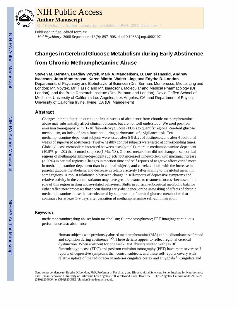
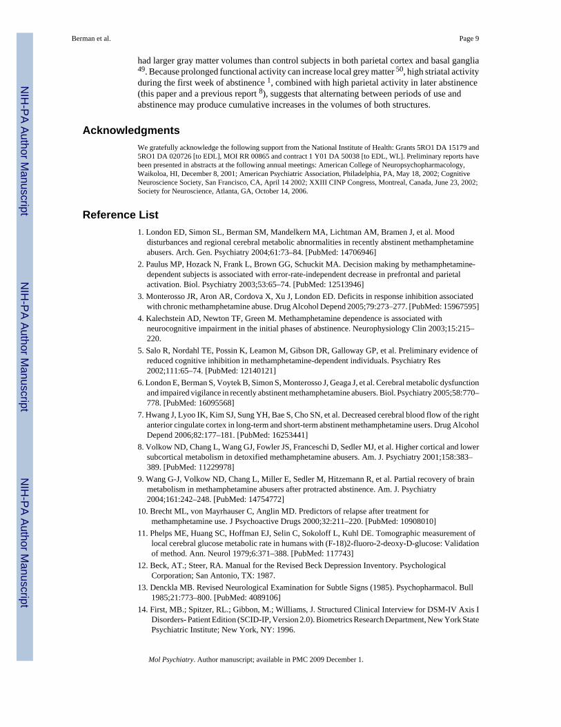
Regional CMRglcAnalyses of the interaction of group with session indicated larger increases at retest in the MAgroup or decreases in the control group based on volume-corrected spatial-extent p-values inboth right and left parietal lobe ROIs (Table 2). These effects maintained statistical significanceafter Bonferroni correction. Peak voxel effects were significant in the parietal lobes, the lateralorbitofrontal cortex, supragenual cingulate cortex, insula, and thalamus, although voxel effectsdid not maintain statistical significance after Bonferroni correction. Figure 1 depicts thisinteraction over much of the cerebral grey matter, including prominent effects in parietal cortex.
Separate analysis of the change across session within each group confirmed that the effectsdepicted in Figure 1 were due solely to widespread increases in the MA group. They attainedBonferroni-corrected spatial-extent significance in bilateral parietal lobe, lateral orbitofrontalcortex, insula and supragenual cingulate gyrus (Table 2). Peak voxel effects attainedBonferroni-corrected significance in bilateral parietal lobe, right lateral orbitofrontal cortexand right insula. Whole brain statistical analysis revealed that no voxel in the MA group showeda decrease in CMRglc of p < .01, even prior to correction for multiple comparisons.
Regional relative radioactivityThe interaction of group with session produced evidence for greater increase in the MA groupor decrease in the control group by the criterion of spatial-extent in the right parietal lobe (p= .033), and of peak height in the parietal lobes (left p = .029; right p = .023), right lateralorbitofrontal cortex (p = .017), and right insula (p = .028). A peak voxel in the left dorsalstriatum had a greater decrease in the MA group, or increase in the Control group, (p = .017).No effects survived Bonferroni correction.
However, separate analysis of the MA group confirmed an increase in the bilateral parietallobes (left and right spatial-extent p <.0005; both peak voxel z > 4.8, both p = .001), and rightlateral orbitofrontal cortex (spatial-extent p <.003; peak voxel z = 4.21, p = .003). All parietaleffects retained significance after Bonferroni correction, but the orbitofrontal cortex effectsdid not.
Relationship between changes in regional CMRglc or relative radioactivity and vigilance taskperformance
In the control group, there was no relationship between RT change and either regional CMRglcor relative radioactivity.
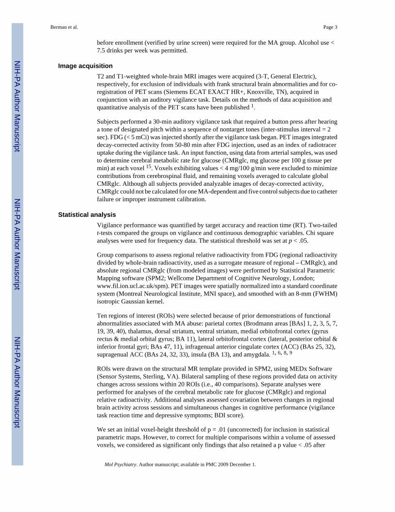
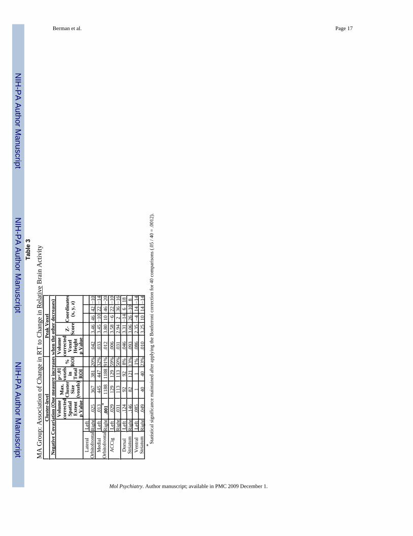
In the MA group, the amount of RT slowing was associated with increases in regional CMRglcby the criterion of spatial extent for clusters in the left (32 voxels, p = .047) and right parietallobes (166 voxels, p = .004). The peak voxel effect in the right parietal lobe (14, −62, 48) wasalso significant (Z = 4.38, p = .024). Although the amount of RT slowing was not associatedwith increased relative radioactivity in any ROI, it was associated with a decrease in 7 of the20 ROIs by the criterion of peak voxel-height (Table 3). Six of these ROIs were also significantfor spatial extent, with the largest effects in ventromedial orbitofrontal cortex (Figure 2). Thespatial-extent effect in the right medial orbitofrontal cortex retained significance afterBonferroni correction.
Relationship between changes in regional CMRglc or relative radioactivity and depressivesymptoms in MA Group
As BDI scores were negligible, with no meaningful change between assessments in the controlgroup, these analyses were only conducted for the MA group. The extent of the increase inregional CMRglc in the left parietal lobe was directly associated with BDI score by the criteriaof spatial extent (p = .045) and peak voxel height (x,y,z = −32, −40, 64; Z = 4.57, p = .005).
Berman et al. Page 5
Mol Psychiatry. Author manuscript; available in PMC 2009 December 1.
NIH
-PA Author Manuscript
NIH
-PA Author Manuscript
NIH
-PA Author Manuscript

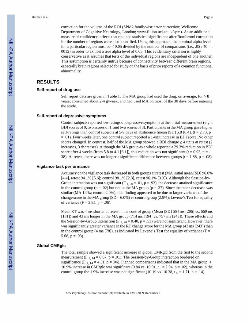
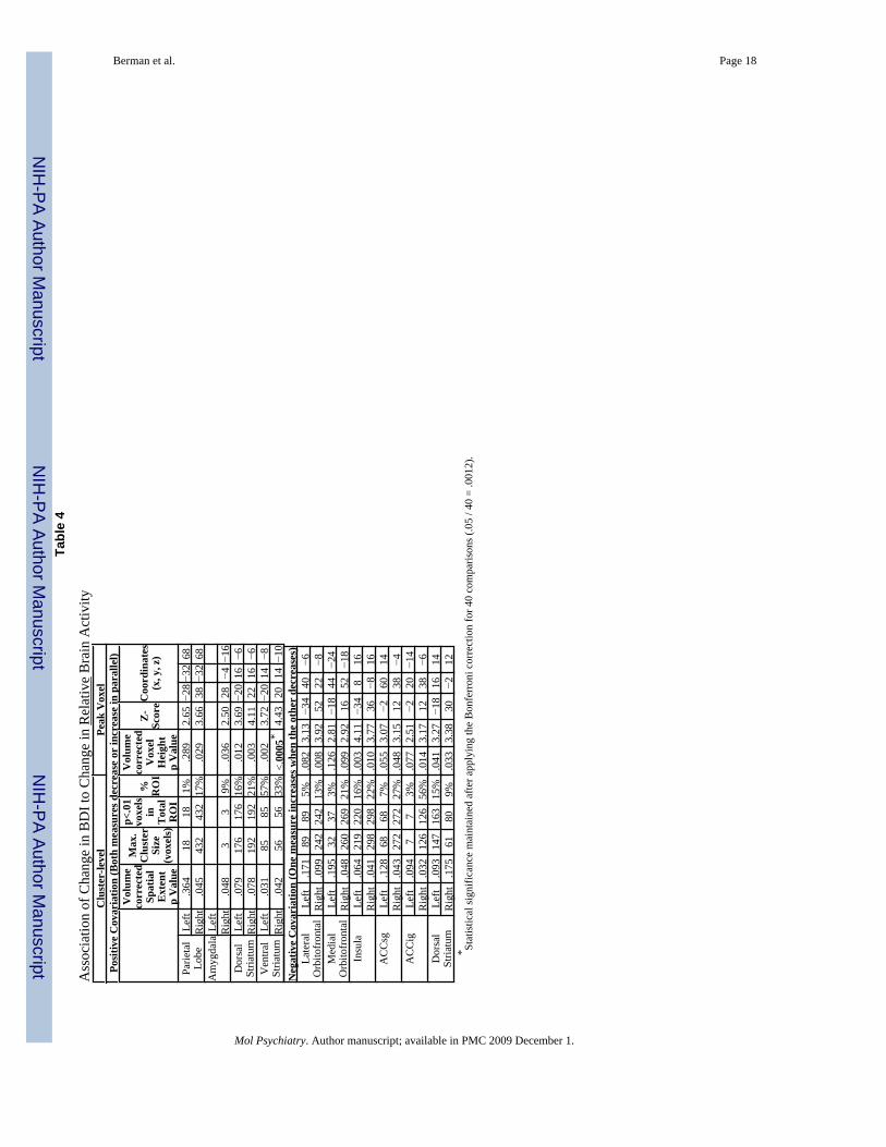
BDI score was correlated with change in relative radioactivity in at least one cerebralhemisphere for all ROIs with the exception of the thalamus (Table 4). BDI score was positivelycorrelated with the change in relative radioactivity by criteria for both spatial extent and peakvoxel effect in the right parietal lobe, right amygdala, and bilateral ventral striatum, and in thebilateral dorsal striatum by the criterion of peak voxel effect. The peak effect in the right ventralstriatum retained significance after Bonferroni correction (Figure 3).
BDI score was negatively correlated with the change in relative radioactivity (one measureincreased when the other decreased) by criteria for both spatial extent and peak voxel effect inthe right infragenual cingulate cortex, right supragenual cingulate cortex and right insula. Theindirect associations were significant by the peak voxel criterion only in the left insula, rightlateral orbitofrontal cortex, and bilateral dorsal striatum, and by the spatial extent criterion onlyin the right medial orbitofrontal cortex (Table 4).
DiscussionWidespread, large increases in cortical CMRglc (>20% in parietal lobes) develop during theinitial month of MA-abstinence. Notably, parietal glucose metabolism during the initial weekof abstinence from MA does not differ from that in healthy comparison subjects but reportedlyis higher after prolonged abstinence. This combination of findings may indicate that residualeffects of MA on CMRglc persist for at least a week after initiating abstinence, making anunderlying chronic effect; or session of chronic MA administration produces new effects thatevolve after the first week of abstinence form the drug. At retest, participants who weremaintaining abstinence from MA varied more than healthy subjects on task performance andglobal CMRglc. Such heterogeneity is consistent with considerable variation in the magnitudeor timecourse of physiological abnormalities during early MA abstinence.
Importance of absolute measuresAlthough many functional brain imaging studies have used only measures of relative activity,our results underscore the value of absolute measures of cerebral glucose metabolism. Duringthe first month without MA, CMRglc increased in most of the superior cortex. Thesewidespread increases changed the global mean, and therefore the relative measures scaled tothat mean. Despite the greater sample size for analysis of changes in regional relativeradioactivity, scaling reduced the power to detect significant differences between groups. Thepattern of higher relative radioactivity in striata of MA abusers during the first week ofabstinence 6 was reversed four weeks later. With only relative measures, this might beinterpreted as a loss of striatal function, but there was no absolute change in this region.Absolute measures suggest that relative decreases in striatal radioactivity may actually measurethe degree of global increase in CMRglc driven by widespread cortical change.
With only relative measures, one might interpret the relationship between slowing of vigilancereaction time and decreased relative ventromedial frontal radioactivity depicted in Figure 2 asa frontomedial metabolic deficit with cognitive consequences. However, comparison of Figure2 to Figure 1 shows that the ventromedial frontal region where decreased relative radioactivityis reliably related to slowed vigilance reaction time is precisely that part of cortex where theactual level of metabolic activity changed least. The largest metabolic change, the degree ofincreased parietal CMRglc, was associated with both slowed vigilance reaction time and withless reduced (or higher) self-report of depressive symptoms. Subjects with larger metabolicincreases in parietal cortex will have larger whole-brain increases. Larger whole-brainincreases generate relative activity decrease in areas where absolute activity increases less thanthe global index. Since no brain area showed a decrease in absolute CMRglc over the firstweeks of abstinence, the magnitude of CMRglc increase in the parietal lobes may constitute ameasure of the severity of brain insult, or the inability to recover normal brain function.
Berman et al. Page 6
Mol Psychiatry. Author manuscript; available in PMC 2009 December 1.
NIH
-PA Author Manuscript
NIH
-PA Author Manuscript
NIH
-PA Author Manuscript
Distractibility and anhedonia are two deficits associated with abstinence from chronic MAabuse 17. This explanation is, therefore, consistent with negative covariation of both vigilancereaction time and BDI score with relative activity in the medial and lateral orbitofrontal cortex,ventral cingulate, and dorsal striatum (Tables 3 and 4), areas where CMRglc increased lessthan in the parietal lobes.
Behavioral observations during early withdrawalMA withdrawal has an early phase with severity greatest at 24 h after final use, decreasingover the next week 16, 17 and stabilizing at 7-10 days. The initial week is characterized bydepression, craving, anxiety, increased sleeping and eating. Together with prior results 1, 6, 8,9, the current results suggest that during this phase, subcortical and limbic radiotracer uptakeare abnormally high and cortical uptake is abnormally low, whereas later abstinence isassociated with higher CMRglc in posterior, particularly parietal cortices.
Possibly reflecting reduced novelty to some degree, there was evidence for reduced vigilanceat retest, i.e. – accuracy decreased by 2% in both groups. Mean reaction time was 41 ms longerat retest with higher variance in the MA group than the control group. BDI scores also werehigher and more variable in the MA group than in the control group, with negligible BDI scoreor change across sessions in the control group, but notable change in half of the MA group. Asdiscussed above, increased parietal CMRglc was associated with both slower vigilance reactiontime and less drop in BDI score, and this may have produced the negative covariations withrelative activity in Tables 3 and 4.
In contrast, the robust positive covariation between the change in BDI score and relative activityin the ventral striatum suggests that although the MA-group had no overall change in activityof the ventral striatum at retest, subjects with lower BDI scores also had less relative activity,subjects with higher BDI scores also had more relative activity, or both. The association ofventral striatal activity with the change in BDI score during the critical early weeks ofabstinence from MA is particularly important because of the role of the nucleus accumbens indrug-mediated reinforcement 18, withdrawal 19, and relapse 20.
The dorsal striatum yielded evidence for areas with both positive and negative covariationbetween change in BDI score and relative activity. Visual inspection of the statisticalparametric maps indicated the positive covariation represented extension of effects in theventral striatum into the inferior dorsal putamen (see Figure 3), whereas the negativecovariation occurred about 2 cm superiorly in the most dorsal 10% of the dorsal striatum.
Possible interpretations of changes in cortical glucose metabolismAlthough MA abuse produces the most profound neurotoxicity in the dopamine-rich basalganglia 21-23, it also increases proliferation of cortical microglia and astrocytes 24. Increasingthe numbers of both types of glia are thought to increase CMRglc 25. Therefore, Volkow andassociates 8 suggested that higher parietal activity in MA abusers could reflect reactive gliosisafter excitotoxic damage to parietal neurons. Notably, the rat brain shows MA-associatedneurodegeneration in parietal cortices 26.
Magnetic resonance spectroscopy (MRS) investigations of MA abuse have reported basalganglia and frontal cortical metabolite abnormalities consistent with gliosis 27-30, including anabnormally high choline signal, a potential index of glial density, in the anterior cingulatecortex of MA abusers abstinent less than six months 31. However, the signal was negativelycorrelated with duration of abstinence, and had returned to control values after more than oneyear. The authors suggested the first 8 months following MA neurotoxicity are characterizedby axon sprouting and gliosis, whereas later abstinence features axon pruning and recovery.
Berman et al. Page 7
Mol Psychiatry. Author manuscript; available in PMC 2009 December 1.
NIH
-PA Author Manuscript
NIH
-PA Author Manuscript
NIH
-PA Author Manuscript

Our preliminary MRS investigations of the parietal lobes 32 did not find evidence for thisabnormally high choline signal at 5-9 days of abstinence.
In rats, three daily exposures to MA are sufficient to induce reactive gliosis lasting 14-21 days21. Our subjects used MA regularly for many years. If the elevated parietal metabolism wereport at one month of abstinence, and previously reported later in abstinence 8, derives fromparietal gliosis, either gliosis did not occur until after the initial week of abstinence, or someprocess masks the gliosis-mediated increase in parietal CMRglc during the initial week.
Cortical/subcortical homeostasisMetabolic abnormalities in the MA group at one week abstinence 1 resemble those observedin a previous human study of acute MA effects; less cortical activity and more subcorticalactivity 33. Reduction of cortical CMRglc is a common response to acute administration ofdrugs of abuse 34, as observed with opiates 35, barbiturates 36, benzodiazepines 37, cocaine38, and amphetamine 39. This commonality is consistent with a classic theory proposing thatactivity in subcortical emotion circuits alters human consciousness more when corticalinhibition is suppressed 40. In this view, metabolic effects of intoxication represent release ofsubcortical regions from cortical inhibition, resulting in heightened emotions.
If chronic MA use inhibits cortical activity, compensatory adjustment might work to up-regulate cortical activity. Although the time-course of inhibition of cortical activity after acuteMA intoxication has not been well-studied, compensatory changes after chronic abuse shouldbe unmasked after a sufficient period of abstinence. The time-course and reversibility of anysuch adjustments are also unknown.
Unmasking increased noradrenergic toneOur vigilance task required detection of rare events, termed “oddballs.” Oddball detection inall sensory modalities activates frontoparietal attention networks 41, which generate the P300event-related scalp potential. Parietal activity during oddball detection, has been linked tonoradrenergic modulation 42 and frontal activity to dopaminergic modulation 43. Therefore,another possible explanation of the changes observed during the first month of abstinence fromMA is that progressive unmasking of increased noradrenergic tone due to long-term MA abuseand accompanying dopaminergic neurotoxicity has shifted frontoparietal attention networkstoward relatively more parietal activity, and that lowered efficiency of frontal executive arousalnetworks has increased vigilance reaction time. This idea is consistent with reports that a ratanalog of P300 was reduced by 15 days of MA followed by over a week of abstinence 44, justas the human P300 is reduced during recovery from MA-abuse 45.
LimitationsThis study has a small sample size of abstaining MA users. However, most previous studiesof regional brain function during abstinence from chronic MA have not considered longitudinalchanges, or have compared subjects tested once during broad periods of early abstinence,(usually < 6 months), with subjects tested during later abstinence. 46, 7, 8, 9 As the only previousreport on repeated assessments of brain function compared 5 subjects tested at < 6 monthsabstinence to reassessment between 12 and 17 months 9, the current report represents the largestsample of subjects tested twice during abstinence and the first study to compare two narrowlyspecified periods of abstinence (5-9 days vs 32-36 days).
Although chronic MA-use has been associated with lesions in both gray 47 and white matter48, we did not consider structural influences on our functional measures. A previous study of22 MA-abusers and 21 controls from which the current subjects were drawn did not findstructural changes in the parietal lobes 47. However, a larger group of 43 former MA-abusers
Berman et al. Page 8
Mol Psychiatry. Author manuscript; available in PMC 2009 December 1.
NIH
-PA Author Manuscript
NIH
-PA Author Manuscript
NIH
-PA Author Manuscript
had larger gray matter volumes than control subjects in both parietal cortex and basal ganglia49. Because prolonged functional activity can increase local grey matter 50, high striatal activityduring the first week of abstinence 1, combined with high parietal activity in later abstinence(this paper and a previous report 8), suggests that alternating between periods of use andabstinence may produce cumulative increases in the volumes of both structures.
AcknowledgmentsWe gratefully acknowledge the following support from the National Institute of Health: Grants 5RO1 DA 15179 and5RO1 DA 020726 [to EDL], MOI RR 00865 and contract 1 Y01 DA 50038 [to EDL, WL]. Preliminary reports havebeen presented in abstracts at the following annual meetings: American College of Neuropsychopharmacology,Waikoloa, HI, December 8, 2001; American Psychiatric Association, Philadelphia, PA, May 18, 2002; CognitiveNeuroscience Society, San Francisco, CA, April 14 2002; XXIII CINP Congress, Montreal, Canada, June 23, 2002;Society for Neuroscience, Atlanta, GA, October 14, 2006.
Reference List1. London ED, Simon SL, Berman SM, Mandelkern MA, Lichtman AM, Bramen J, et al. Mood
disturbances and regional cerebral metabolic abnormalities in recently abstinent methamphetamineabusers. Arch. Gen. Psychiatry 2004;61:73–84. [PubMed: 14706946]
2. Paulus MP, Hozack N, Frank L, Brown GG, Schuckit MA. Decision making by methamphetamine-dependent subjects is associated with error-rate-independent decrease in prefrontal and parietalactivation. Biol. Psychiatry 2003;53:65–74. [PubMed: 12513946]
3. Monterosso JR, Aron AR, Cordova X, Xu J, London ED. Deficits in response inhibition associatedwith chronic methamphetamine abuse. Drug Alcohol Depend 2005;79:273–277. [PubMed: 15967595]
4. Kalechstein AD, Newton TF, Green M. Methamphetamine dependence is associated withneurocognitive impairment in the initial phases of abstinence. Neurophysiology Clin 2003;15:215–220.
5. Salo R, Nordahl TE, Possin K, Leamon M, Gibson DR, Galloway GP, et al. Preliminary evidence ofreduced cognitive inhibition in methamphetamine-dependent individuals. Psychiatry Res2002;111:65–74. [PubMed: 12140121]
6. London E, Berman S, Voytek B, Simon S, Monterosso J, Geaga J, et al. Cerebral metabolic dysfunctionand impaired vigilance in recently abstinent methamphetamine abusers. Biol. Psychiatry 2005;58:770–778. [PubMed: 16095568]
7. Hwang J, Lyoo IK, Kim SJ, Sung YH, Bae S, Cho SN, et al. Decreased cerebral blood flow of the rightanterior cingulate cortex in long-term and short-term abstinent methamphetamine users. Drug AlcoholDepend 2006;82:177–181. [PubMed: 16253441]
8. Volkow ND, Chang L, Wang GJ, Fowler JS, Franceschi D, Sedler MJ, et al. Higher cortical and lowersubcortical metabolism in detoxified methamphetamine abusers. Am. J. Psychiatry 2001;158:383–389. [PubMed: 11229978]
9. Wang G-J, Volkow ND, Chang L, Miller E, Sedler M, Hitzemann R, et al. Partial recovery of brainmetabolism in methamphetamine abusers after protracted abstinence. Am. J. Psychiatry2004;161:242–248. [PubMed: 14754772]
10. Brecht ML, von Mayrhauser C, Anglin MD. Predictors of relapse after treatment formethamphetamine use. J Psychoactive Drugs 2000;32:211–220. [PubMed: 10908010]
11. Phelps ME, Huang SC, Hoffman EJ, Selin C, Sokoloff L, Kuhl DE. Tomographic measurement oflocal cerebral glucose metabolic rate in humans with (F-18)2-fluoro-2-deoxy-D-glucose: Validationof method. Ann. Neurol 1979;6:371–388. [PubMed: 117743]
12. Beck, AT.; Steer, RA. Manual for the Revised Beck Depression Inventory. PsychologicalCorporation; San Antonio, TX: 1987.
13. Denckla MB. Revised Neurological Examination for Subtle Signs (1985). Psychopharmacol. Bull1985;21:773–800. [PubMed: 4089106]
14. First, MB.; Spitzer, RL.; Gibbon, M.; Williams, J. Structured Clinical Interview for DSM-IV Axis IDisorders- Patient Edition (SCID-IP, Version 2.0). Biometrics Research Department, New York StatePsychiatric Institute; New York, NY: 1996.
Berman et al. Page 9
Mol Psychiatry. Author manuscript; available in PMC 2009 December 1.
NIH
-PA Author Manuscript
NIH
-PA Author Manuscript
NIH
-PA Author Manuscript

15. Huang S-C, Phelps ME, Hoffman EJ, Sideris K, Selin CJ, Kuhl DE. Noninvasive determination oflocal cerebral metabolic rate of glucose in man. Am. J. Physiol 1980;238:E69–E82. [PubMed:6965568]
16. McGregor C, Srisurapanont M, Jittiwutikarn J, Laobhripatr S, Wongtan T, White JM. The nature,time course and severity of methamphetamine withdrawal. Addiction 2005;100:1320–1329.[PubMed: 16128721]
17. Newton TF, Kalechstein AD, Duran S, Vansluis N, Ling W. Methamphetamine abstinence syndrome:Preliminary findings. American Journal on Addictions 2002;13:248–255. [PubMed: 15370944]
18. Koob GF. Neural mechanisms of drug reinforcement. Ann. N. Y. Acad. Sci 1992;654:171–191.[PubMed: 1632582]
19. Hammer RP Jr. Pires WS, Markou A, Koob GF. Withdrawal following cocaine self-administrationdecreases regional cerebral metabolic rate in critical brain reward regions. Synapse 1993;14:73–80.[PubMed: 8511720]
20. Weiss F, Ciccocioppo R, Parsons LH, Katner S, Liu X, Zorrilla EP, et al. Compulsive drug-seekingbehavior and relapse. Neuroadaptation, stress, and conditioning factors. Ann. N. Y. Acad. Sci2001;937:1–26. [PubMed: 11458532]
21. Pennypacker KR, Kassed CA, Eidizadeh S, O’Callaghan JP. Brain injury: prolonged induction oftranscription factors. Acta Neurobiol. Exp. (Wars. ) 2000;60:515–530. [PubMed: 11200181]
22. McCann UD, Wong DF, Yokoi F, Villemagne V, Dannals RF, Ricaurte GA. Reduced striataldopamine transporter density in abstinent methamphetamine and methcathinone users: evidence frompositron emission tomography studies with [11C]WIN-35,428. J Neurosci 1998;18:8417–8422.[PubMed: 9763484]
23. Volkow ND, Chang L, Wang G-J, Fowler JS, Franceschi D, Sedler M, et al. Loss of dopaminetransporters in methamphetamine abusers recovers with protracted abstinence. J. Neurosci2001;21:9414–9418. [PubMed: 11717374]
24. LaVoie MJ, Card JP, Hastings TG. Microglial activation precedes dopamine terminal pathology inmethamphetamine-induced neurotoxicity. Exp. Neurol 2004;187:47–57. [PubMed: 15081587]
25. Roh JK, Nam H, Lee MC. A case of central pontine and extrapontine myelinolysis with earlyhypermetabolism on 18FDG-PET scan. J Korean Med. Sci 1998;13:99–102. [PubMed: 9539329]
26. Bowyer JF, Clausing P, Gough B, Slikker W Jr. Holson RR. Nitric oxide regulation ofmethamphetamine-induced dopamine release in caudate/putamen. Brain Res 1995;699:62–70.[PubMed: 8616614]
27. Ernst T, Chang L, Leonido-Yee M, Speck O. Evidence for long-term neurotoxicity associated withmethamphetamine abuse: A 1H MRS study. Neurology 2000;54:1344–1349. [PubMed: 10746608]
28. Nordahl TE, Salo R, Possin K, Gibson DR, Flynn N, Leamon M, et al. Low N-acetyl-aspartate andhigh choline in the anterior cingulum of recently abstinent methamphetamine-dependent subjects: apreliminary proton MRS study. Magnetic resonance spectroscopy. Psychiatry Res 2002;116:43–52.[PubMed: 12426033]
29. Sekine Y, Iyo M, Ouchi Y, Matsunaga T, Tsukada H, Okada H, et al. Methamphetamine-relatedpsychiatric symptoms and reduced brain dopamine transporters studied with PET. Am J Psychiatry2001;158:1206–1214. [PubMed: 11481152]
30. Smith LM, Chang L, Yonekura ML, Grob C, Osborn D, Ernst T. Brain proton magnetic resonancespectroscopy and imaging in children exposed to methamphetamine in utero. Neurology2001;57:255–260. [PubMed: 11468309]
31. Nordahl TE, Salo R, Natsuaki Y, Galloway GP, Waters C, Moore CD, et al. Methamphetamine usersin sustained abstinence: a proton magnetic resonance spectroscopy study. Arch Gen Psychiatry2005;62:444–452. [PubMed: 15809412]
32. O’Neill, JO.; Monterosso, J.; Walker, MK.; London, ED. 1H MRSI brain findings in chronicmethamphetamine abuse. Program 392.9, 2006 Neuroscience Meeting Planner, Atlanta, Georgia,Society for Neuroscience; 2006. online
33. Gouzoulis-Mayfrank E, Schreckenberger M, Sabri O, Arning C, Thelen B, Spitzer M, et al.Neurometabolic effects of psilocybin, 3,4-methylenedioxyethylamphetamine (MDE) and d-methamphetamine in healthy volunteers. Neuropsychopharmacology 1999;20:565–581. [PubMed:10327426]
Berman et al. Page 10
Mol Psychiatry. Author manuscript; available in PMC 2009 December 1.
NIH
-PA Author Manuscript
NIH
-PA Author Manuscript
NIH
-PA Author Manuscript

34. London, ED.; Grant, SJ.; Morgan, MJ.; Zukin, SR. Neurobiology of drug abuse. In: Fogel, BS.;Schiffer, RB.; Rao, SM., editors. Neuropsychiatry. William & Wilkins; Baltimore: 1996. p. 635-678.
35. London ED, Broussolle EP, Links JM, Wong DF, Cascella NG, Dannals RF, et al. Morphine-inducedmetabolic changes in human brain. Studies with positron emission tomography and [fluorine 18]fluorodeoxyglucose. Arch. Gen. Psychiatry 1990;47:73–81. [PubMed: 2403775]
36. Theodore WH, DiChiro G, Margolin R, Fishbein D, Porter RJ, Brooks RA. Barbiturates reduce humancerebral glucose metabolism. Neurology 1986;36:60–64. [PubMed: 3484547]
37. Foster NL, VanDerSpek AFL, Aldrich MS, Berent S, Hichwa RH, Sackellares JC, et al. The effectof diazepam sedation on cerebral glucose metabolism in Alzheimer’s disease as measured usingpositron emission tomography. J. Cereb. Blood Flow Metab 1987;7:415–420. [PubMed: 3497161]
38. London ED, Cascella NG, Wong DF, Phillips RL, Dannals RF, Links JM, et al. Cocaine-inducedreduction of glucose utilization in human brain. A study using positron emission tomography and[fluorine 18]-fluorodeoxyglucose. Arch. Gen. Psychiatry 1990;47:567–574. [PubMed: 2350209]
39. Wolkin A, Angrist B, Wolf A, Brodie J, Wolkin B, Jaeger J, et al. Effects of amphetamine on localcerebral metabolism in normal and schizophrenic subjects as determined by positron emissiontomography. Psychopharmacology (Berl) 1987;92:241–246. [PubMed: 3110848]
40. Cannon WB. The James-Lange theory of emotions: A critical examination and an alternative theory.Am. J. Psychol 1927;39:106–124.
41. Huang MX, Lee RR, Miller GA, Thoma RJ, Hanlon FM, Paulson KM, et al. A parietal-frontal networkstudied by somatosensory oddball MEG responses, and its cross-modal consistency. Neuroimage2005;28:99–114. [PubMed: 15979344]
42. Nieuwenhuis S, Aston-Jones G, Cohen JD. Decision making, the P3, and the locus coeruleus-norepinephrine system. Psychol. Bull 2005;131:510–532. [PubMed: 16060800]
43. Polich J, Criado JR. Neuropsychology and neuropharmacology of P3a and P3b. Int. J. Psychophysiol2006;60:172–185. [PubMed: 16510201]
44. Takeuchi S, Jodo E, Suzuki Y, Matsuki T, Niwa S, Kayama Y. Effects of Repeated Administrationof Methamphetamine on P3-like Potentials in Rats. Int. J. Psychophysiol 1999;32:183–192.[PubMed: 10437630]
45. Iwanami A, Suga I, Kaneko T, Sugiyama A, Nakatani Y. P300 Component of Event-related Potentialsin Methamphetamine Psychosis and Schizophrenia. Prog Neuro-Psychopharmacol & Biol Psychiat1994;18:465–475.
46. Nordahl TE, Salo R, Natsuaki Y, Galloway GP, Waters C, Moore CD, et al. Methamphetamine usersin sustained abstinence: a proton magnetic resonance spectroscopy study. Arch Gen Psychiatry2005;62:444–452. [PubMed: 15809412]
47. Thompson PM, Hayashi K, Simon SL, Geaga JA, Hong MS, Sui Y, et al. Structural abnormalities inthe brains of human subjects who use methamphetamine. J Neurosci 2004;24:6028–6036. [PubMed:15229250]
48. Oh JS, Lyoo IK, Sung YH, Hwang J, Kim J, Chung A, et al. Shape changes of the corpus callosumin abstinent methamphetamine users. Neurosci. Lett 2005;384:76–81. [PubMed: 15913890]
49. Jernigan TL, Gamst AC, Archibald SL, Fennema-Notestine C, Mindt MR, Marcotte TD, et al. Effectsof methamphetamine dependence and HIV infection on cerebral morphology. Am J Psychiatry2005;162:1461–1472. [PubMed: 16055767]
50. Draganski B, Gaser C, Busch V, Schuierer G, Bogdahn U, May A. Neuroplasticity: changes in greymatter induced by training. Nature 2004;427:311–312. [PubMed: 14737157]
Berman et al. Page 11
Mol Psychiatry. Author manuscript; available in PMC 2009 December 1.
NIH
-PA Author Manuscript
NIH
-PA Author Manuscript
NIH
-PA Author Manuscript
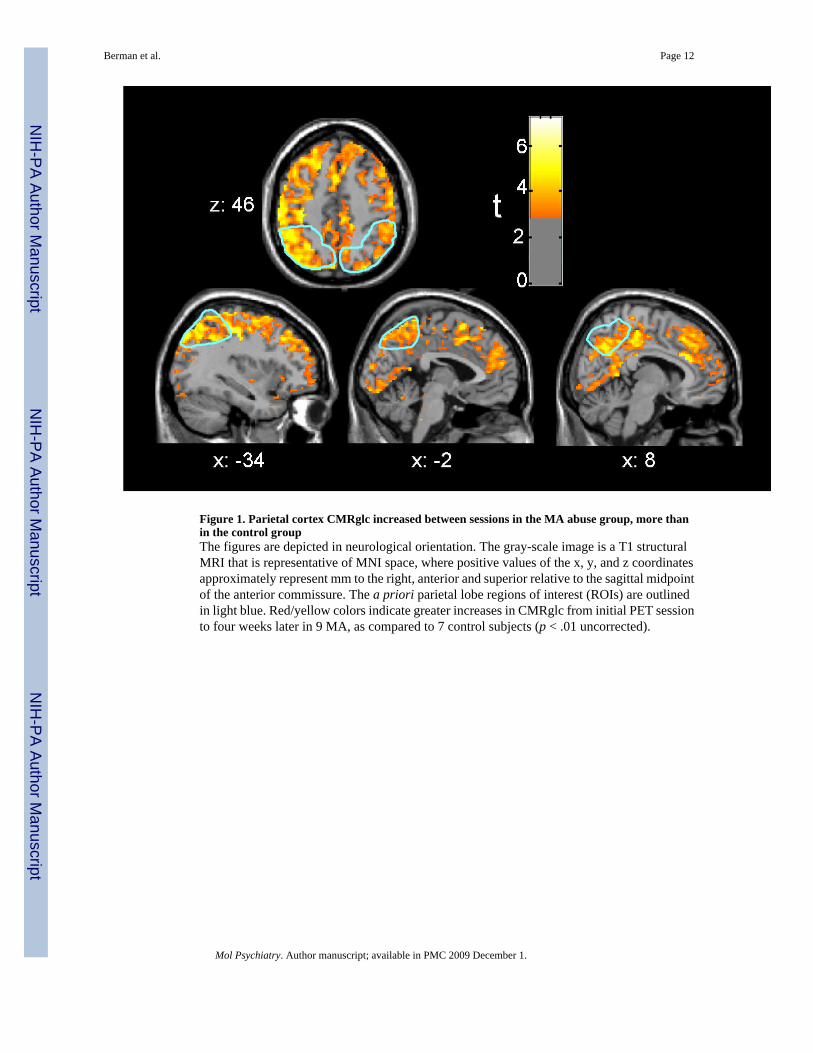
Figure 1. Parietal cortex CMRglc increased between sessions in the MA abuse group, more thanin the control groupThe figures are depicted in neurological orientation. The gray-scale image is a T1 structuralMRI that is representative of MNI space, where positive values of the x, y, and z coordinatesapproximately represent mm to the right, anterior and superior relative to the sagittal midpointof the anterior commissure. The a priori parietal lobe regions of interest (ROIs) are outlinedin light blue. Red/yellow colors indicate greater increases in CMRglc from initial PET sessionto four weeks later in 9 MA, as compared to 7 control subjects (p < .01 uncorrected).
Berman et al. Page 12
Mol Psychiatry. Author manuscript; available in PMC 2009 December 1.
NIH
-PA Author Manuscript
NIH
-PA Author Manuscript
NIH
-PA Author Manuscript

Figure 2. Between-session directional change in relative radioactivity of medial orbitofrontal cortexwas inversely correlated with vigilance reaction time (RT) in the MA abuse groupRed/yellow colors indicate areas where the amount of RT slowing from initial PET session tofour weeks later was associated with decreased regional relative radioactivity in 10 MAsubjects (p < .01 uncorrected).
Berman et al. Page 13
Mol Psychiatry. Author manuscript; available in PMC 2009 December 1.
NIH
-PA Author Manuscript
NIH
-PA Author Manuscript
NIH
-PA Author Manuscript
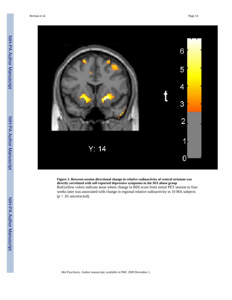
Figure 3. Between-session directional change in relative radioactivity of ventral striatum wasdirectly correlated with self-reported depressive symptoms in the MA abuse groupRed/yellow colors indicate areas where change in BDI score from initial PET session to fourweeks later was associated with change in regional relative radioactivity in 10 MA subjects(p < .01 uncorrected).
Berman et al. Page 14
Mol Psychiatry. Author manuscript; available in PMC 2009 December 1.
NIH
-PA Author Manuscript
NIH
-PA Author Manuscript
NIH
-PA Author Manuscript
NIH
-PA Author Manuscript
NIH
-PA Author Manuscript
NIH
-PA Author Manuscript
Berman et al. Page 15
Table 1
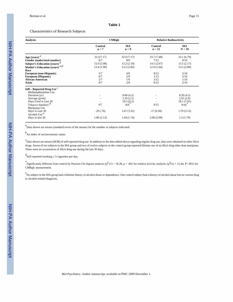
Characteristics of Research Subjects
Analysis: CMRglc Relative Radioactivity
Controln = 7
MAn = 9
Controln = 12
MAN = 10
Age (years) a 33.1(7.17) 32.9 (7.17) 33.7 (7.49) 33.1 (6.79)Gender (males/total number) 6/7 8/9 7/12 9/10Subject’s Education (years) a 15.0 (2.08) 13.2 (2.10) 14.5 (2.07) 13.5 (2.17)Mother’s Education (years) a,b 13.4 (1.90) 13.2 (2.82) 12.9 (1.64) 13.5 (2.80)RaceEuropean (non-Hispanic) 5/7 4/9 8/12 5/10European (Hispanic) 0/7 2/9 1/12 2/10African American 2/7 1/9 3/12 1/10Asian 0/7 2/9 0/12 2/10
Self – Reported Drug Use c Methamphetamine Use Duration (yr) - 8.89 (4.2) - 8.20 (4.5) Average (g/wk) - 2.10 (2.1) - 2.01 (2.0) Days Used in Last 30 - 19.3 (6.2) - 18.1 (7.01) Tobacco Smokers d 0/7 8/9* 0/12 9/10* Marijuana Use Days in Last 30 .29 (.76) 1.67 (3.32) .17 (0.58) 1.70 (3.13) Alcohol Use e Days in last 30 1.86 (2.12) 1.44 (1.74) 2.00 (2.09) 1.3 (1.70)
aData shown are means (standard errors of the means) for the number of subjects indicated.
bAn index of socioeconomic status
cData shown are means (SEM) of self-reported drug use. In addition to the data tabled above regarding regular drug use, data were obtained on other illicit
drugs. Seven of ten subjects in the MA group and two of twelve subjects in the control group reported lifetime use of an illicit drug other than marijuana.There were no occurrences of illicit drug use during the last 30 days.
dSelf-reported smoking ≥ 5 cigarettes per day.
*Significantly different from control by Pearson Chi-Square analysis (χ2 (1) = 18.28, p < .001 for relative activity analysis; (χ2(1) = 12.44, P<.001) for
CMRglc measurement.
eNo subject in the MA group had a lifetime history of alcohol abuse or dependence. One control subject had a history of alcohol abuse but no current drug
or alcohol-related diagnosis.
Mol Psychiatry. Author manuscript; available in PMC 2009 December 1.

NIH
-PA Author Manuscript
NIH
-PA Author Manuscript
NIH
-PA Author Manuscript
Berman et al. Page 16Ta
ble
2
Inte
ract
ion
of G
roup
with
Cha
nge
in A
bsol
ute
rCM
Rgl
cC
lust
er-le
vel
Peak
Vox
elIn
crea
se in
MA
gro
up a
nd /
or D
ecre
ase
in C
ontr
ol g
roup
Vol
ume
corr
ecte
dSp
atia
lE
xten
tp
Val
ue
Max
.C
lust
erSi
ze(v
oxel
s)
p<.0
1vo
xels
inT
otal
RO
I
% RO
I
Vol
ume
corr
ecte
dV
oxel
Hei
ght
p V
alue
Z-
Scor
eCoo
rdin
ates
(x, y
, z)
Parie
tal
Lobe
Left
<.00
05*
3328
3336
73%
.019
4.37
−30−5
464
Rig
ht<.
0005
*18
9419
2247
%.0
304.
2020
−82
34La
tera
lO
rbito
fron
tal
Left
.107
9219
916
%.0
353.
83−4
250
−6R
ight
.071
114
213
19%
.007
4.32
4422
−14
Insu
laLe
ft.3
2219
454%
.362
2.69
−42
18−2
Rig
ht.2
7624
292%
.028
3.78
4818
−6A
CC
sgLe
ft.0
1817
921
922
%.0
793.
29−2
4616
Rig
ht.0
3513
617
523
%.0
483.
498
2824
Thal
amus
Left
Rig
ht.0
6885
8515
%.0
403.
5912
−28
0W
ithin
Gro
up si
mpl
e ef
fect
s: In
crea
se in
MA
gro
upPa
rieta
lLo
beLe
ft<.
0005
* 453
9454
099%
<.00
05* 5
.30−
28−7
846
Rig
ht<.
0005
* 402
3402
398%
<.00
05* 5
.42
20−8
234
Late
ral
Orb
itofr
onta
lLe
ft<.
0005
*98
498
481
%.0
094.
25−4
250
−6R
ight
<.00
05*
872
872
78%
<.00
05* 5
.04
4422
−14
Med
ial
Orb
itofr
onta
lLe
ft.0
1919
525
527
%.0
094.
15−2
648
−14
Rig
ht.0
3414
922
226
%.1
193.
2422
44−1
6In
sula
Left
<.00
05*
746
752
64%
.034
3.73
−34
220
Rig
ht<.
0005
*60
968
155
%.0
01*
4.87
4818
−6A
CC
sgLe
ft<.
0005
*88
988
991
%.0
085.
84−6
3616
Rig
ht<.
0005
*72
372
396
%.0
064.
186
2624
AC
Cig
Left
.075
2938
20%
.064
2.90
−838
−14
Rig
ht.0
3955
5540
%.0
153.
414
42−8
Incl
uded
in ta
bles
2 a
nd 3
are
thos
e of
the
20 R
OIs
that
wer
e as
sess
ed (1
0 in
eac
h he
mis
pher
e) w
here
the
note
d ef
fect
atta
ined
vol
ume-
corr
ecte
d pr
obab
ility
<.0
5 at
the
peak
vox
el, a
nd fo
r com
para
tive
purp
oses
, the
hom
olog
ous R
OI i
n th
e op
posi
te h
emis
pher
e. T
he si
ze a
nd sp
atia
l-ext
ent p
roba
bilit
y as
soci
ated
with
the
larg
est c
lust
er, t
he to
tal p
ropo
rtion
of t
he R
OI c
onst
itute
d by
all
clus
ters
, and
the
peak
vox
el Z
-sco
re, a
ssoc
iate
d pr
obab
ility
, and
MN
I coo
rdin
ates
are
tabl
ed. A
CC
sg =
supr
agen
ual a
nter
ior c
ingu
late
cor
tex,
AC
Cig
= in
frag
enua
l ant
erio
r cin
gula
te c
orte
x
* Stat
istic
al si
gnifi
canc
e m
aint
aine
d af
ter a
pply
ing
the
Bon
ferr
oni c
orre
ctio
n fo
r 40
com
paris
ons (
.05
/ 40
= .0
012)
.
Mol Psychiatry. Author manuscript; available in PMC 2009 December 1.
NIH
-PA Author Manuscript
NIH
-PA Author Manuscript
NIH
-PA Author Manuscript
Berman et al. Page 17Ta
ble
3
MA
Gro
up: A
ssoc
iatio
n of
Cha
nge
in R
T to
Cha
nge
in R
elat
ive
Bra
in A
ctiv
ityC
lust
er-le
vel
Peak
Vox
elN
egat
ive
Cov
aria
tion
(One
mea
sure
incr
ease
s whe
n th
e ot
her
decr
ease
s)V
olum
eco
rrec
ted
Spat
ial
Ext
ent
p V
alue
Max
.C
lust
erSi
ze(v
oxel
s)
p<.0
1vo
xels
inT
otal
RO
I
% RO
I
Vol
ume
corr
ecte
dV
oxel
Hei
ght
p V
alue
Z-
Scor
eCoo
rdin
ates
(x, y
, z)
Late
ral
Orb
itofr
onta
lLe
ftR
ight
.025
367
381
20%
.042
3.46
4642
−10
Med
ial
Orb
itofr
onta
lLe
ft.0
1344
544
732
%.0
333.
45−1
022
−14
Rig
ht.0
01*
1188
1188
91%
.012
3.80
1046
−20
AC
Cig
Left
.029
129
129
59%
.006
3.58
−622
−10
Rig
ht.0
3111
311
350
%.0
312.
942
36−1
6D
orsa
lSt
riatu
mLe
ft.1
2492
928%
.046
3.31
−14
618
Rig
ht.1
4682
121
13%
.093
3.06
26−1
08
Ven
tral
Stria
tum
Left
.085
11
1%.0
862.
35−4
14−1
4R
ight
.049
4040
23%
.010
3.25
1014
−14
* Stat
istic
al si
gnifi
canc
e m
aint
aine
d af
ter a
pply
ing
the
Bon
ferr
oni c
orre
ctio
n fo
r 40
com
paris
ons (
.05
/ 40
= .0
012)
.
Mol Psychiatry. Author manuscript; available in PMC 2009 December 1.
NIH
-PA Author Manuscript
NIH
-PA Author Manuscript
NIH
-PA Author Manuscript
Berman et al. Page 18Ta
ble
4
Ass
ocia
tion
of C
hang
e in
BD
I to
Cha
nge
in R
elat
ive
Bra
in A
ctiv
ityC
lust
er-le
vel
Peak
Vox
elPo
sitiv
e C
ovar
iatio
n (B
oth
mea
sure
s dec
reas
e or
incr
ease
in p
aral
lel)
Vol
ume
corr
ecte
dSp
atia
lE
xten
tp
Val
ue
Max
.C
lust
erSi
ze(v
oxel
s)
p<.0
1vo
xels
inT
otal
RO
I
% RO
I
Vol
ume
corr
ecte
dV
oxel
Hei
ght
p V
alue
Z-
Scor
eCoo
rdin
ates
(x, y
, z)
Parie
tal
Lobe
Left
.364
1818
1%.2
892.
65−2
8−3
268
Rig
ht.0
4543
243
217
%.0
293.
6638
−32
68A
myg
dala
Left
Rig
ht.0
483
39%
.036
2.50
28−4
−16
Dor
sal
Stria
tum
Left
.079
176
176
16%
.012
3.69
−20
16−6
Rig
ht.0
7819
219
221
%.0
034.
1122
16−6
Ven
tral
Stria
tum
Left
.031
8585
57%
.002
3.72
−20
14−8
Rig
ht.0
4256
5633
%<.
0005
*4.
4320
14−1
0N
egat
ive
Cov
aria
tion
(One
mea
sure
incr
ease
s whe
n th
e ot
her
decr
ease
s)La
tera
lO
rbito
fron
tal
Left
.171
8989
5%.0
823.
13−3
440
−6R
ight
.099
242
242
13%
.008
3.92
5222
−8M
edia
lO
rbito
fron
tal
Left
.195
3237
3%.1
262.
81−1
844
−24
Rig
ht.0
4826
026
921
%.0
992.
9216
52−1
8In
sula
Left
.064
219
220
16%
.003
4.11
−34
816
Rig
ht.0
4129
829
822
%.0
103.
7736
−816
AC
Csg
Left
.128
6868
7%.0
553.
07−2
6014
Rig
ht.0
4327
227
227
%.0
483.
1512
38−4
AC
Cig
Left
.094
77
3%.0
772.
51−2
20−1
4R
ight
.032
126
126
56%
.014
3.17
1238
−6D
orsa
lSt
riatu
mLe
ft.0
9314
716
315
%.0
413.
27−1
816
14R
ight
.175
6180
9%.0
333.
3830
−212
* Stat
istic
al si
gnifi
canc
e m
aint
aine
d af
ter a
pply
ing
the
Bon
ferr
oni c
orre
ctio
n fo
r 40
com
paris
ons (
.05
/ 40
= .0
012)
.
Mol Psychiatry. Author manuscript; available in PMC 2009 December 1.
Related Documents