Plant Physiol. (1977) 59, 1088-1097 Changes in Biochemical Composition of the Cell Wail of the Cotton Fiber During Development1 Received for publication October 26, 1976 and in revised form February 1, 1977 MAUREEN C. MEINERT AND DEBORAH P. DELMER2 Michigan State University Energy Research and Development Administration Plant Research Laboratory, Department of Biochemistry, Michigan State University, East Lansing, Michigan 48824 ABSTRACT The composition of the cell wall of the cotton fiber (Gossypium hirsutum L. Acala SJ-1) has been studied from the early stages of elongation (5 days postaathesis) through the period of secondary wall formation, using cell wals derived both from fibers developing on the plant and from fibers obtained from excised, cultured ovules. The cell wall of the elongating cotton fiber was shown to be a dynamic structure. Expressed as a weight per cent of the total cell wall, cellulose, neutnl sugars (rhamnose, fucose, arabinose, mannose, galactose, and noncelOu- losic glucose), uronic acids, and total protein undergo marked changes in content during the elongation period. As a way of analyzing absolute changes in the walls with time, data have also been expressed as grams component per milimeter of fiber length. Expressed in this way for plant-grown fibers, the data show that the thickness of the ceUl wall is relatively constant until about 12 days postanthesis; after this time it markedly increases until secondary wall cellulose deposition is com- pleted. Between 12 and 16 days postanthesis increases in all components contribute to total wall increase per millimeter fiber length. The deposi- tion of secondary wall cellulose begins at about 16 days postanthesis (at least 5 days prior to the cessation of elongation) and continues until about 32 days postanthesis. At the time of the onset of secondary wall cellulose deposition, a sharp decline in protein and uronic acid content occurs. The content of some of the individual neutral sugars changes during development, the most prominent change being a large increase in noncellulosic glucose which occurs just prior to the onset of secondary waDl cellulose deposition. Methylation analyses indicate that this glucose, at least in part, is 3-linked. In contrast to the neutral sugars, no sdgnii- cant changes in cell wall amino add composition are observed during fiber development. Compositional analyses of cel waDls derived from culture-grown fibers indicate that these wals are remarkably similar to those derived from fibers grown on the plant, both in terms of composition and in terms of relative changes in composition during development. A comparison of our results on total cell wall composition and link- ages of sugars as determined by a preliminary methylation analysis of unfractionated fiber walls indicates that the pmary ceOl waDl of cotton fibers is similar to that of pmary cell wals of other dicotyledons and of gymnosperms as reported in the literature. While much information is now available on the composition and structure of plant cell walls (3, 6, 7, 36), we have very little ' Supported by Energy Research and Development Administration Contract No E(1 1-1)-1338. Much of this report is based on a portion of the thesis submitted by M. C. M. in partial fulfillment of the require- ments for the M.Sc degree in biochemistry Michigan State University. 2 To whom reprint requests should be addressed. data on the biochemical changes occurring in the cell wall of a single higher plant cell during the process of cell elongation and maturation. The most extensive studies to date on the structure of the primary cell wall of plant cells have been carried out in Albersheim's laboratory (2, 3, 8, 15, 22, 35, 50, 56). With one exception (35), these studies have been performed exclusively with plant cells grown in culture. Although such cells possess the distinct advantage of representing a relatively homogeneous cell type, they are not well suited for physiological studies which could correlate biochemical changes in the wall with cell elonga- tion and maturation. Nevertheless, two important concepts have evolved from this work: (a) that the primary cell wall is com- posed of only a limited number of distinct polymers; and (b) that the primary cell walls of cultured dicotyledonous cells, while quite similar to each other, are distinctly different from those of the cell walls derived from the monocotyledonous cell cultures which were examined (15). Although in earlier work (35), the elongating cell wall was referred to as a dynamic structure, implicit in all of this group's later work has been the concept that the primary cell wall is a relatively constant structure, and "the average chemical composition of the wall is not altered" (2) during wall growth. Some attempts have been made to determine changes in cell wall composition associated with the process of cell elongation, but these studies have almost invariably utilized tissues consist- ing of various cell types. Labavitch and Ray (26, 27) have recently shown that auxin treatment of pea epicotyls enhances the solubility of wall xyloglucan, and that this change may be correlated with growth. Increases in the level of the hydroxypro- line-rich wall protein (expressed as per cent of the fresh weight of tissue) toward the end of elongation have been observed, and the suggestion has been made that this component serves to stabilize the wall from further extension (16, 42). However, recent data of Klis (25) indicate that when hydroxyproline con- tent is expressed as per cent of the dry weight of cell wall, its content continually increases during cell elongation, but that there is no further increase once the cells have obtained their final length. Also of interest are studies -showing that a specific decrease in noncellulosic glucose, which is accompanied by a roughly compensating increase in cellulose, occurs in the cell wall of Avena coleoptiles elongating in response to auxin in the absence of an external carbon source (31, 39), and it has been suggested that changes in noncellulosic wall glucan may be involved in wall loosening (34). The developing cotton fiber represents a good experimental system for studying biochemical changes associated with cell elongation and/or maturation. Cotton fibers are single cells which elongate synchronously over a period of almost 3 weeks. Sufficient material can be readily obtained for biochemical anal- yses of the cell wall. Further potential advantages are that isolated unfertilized ovules with their associated fibers can be 1088 www.plantphysiol.org on June 10, 2018 - Published by Downloaded from Copyright © 1977 American Society of Plant Biologists. All rights reserved.

Welcome message from author
This document is posted to help you gain knowledge. Please leave a comment to let me know what you think about it! Share it to your friends and learn new things together.
Transcript

Plant Physiol. (1977) 59, 1088-1097
Changes in Biochemical Composition of the Cell Wail of theCotton Fiber During Development1
Received for publication October 26, 1976 and in revised form February 1, 1977
MAUREEN C. MEINERT AND DEBORAH P. DELMER2Michigan State University Energy Research and Development Administration Plant Research Laboratory,Department of Biochemistry, Michigan State University, East Lansing, Michigan 48824
ABSTRACT
The composition of the cell wall of the cotton fiber (Gossypiumhirsutum L. Acala SJ-1) has been studied from the early stages ofelongation (5 days postaathesis) through the period of secondary wallformation, using cell wals derived both from fibers developing on theplant and from fibers obtained from excised, cultured ovules. The cellwall of the elongating cotton fiber was shown to be a dynamic structure.Expressed as a weight per cent of the total cell wall, cellulose, neutnlsugars (rhamnose, fucose, arabinose, mannose, galactose, and noncelOu-losic glucose), uronic acids, and total protein undergo marked changes incontent during the elongation period. As a way of analyzing absolutechanges in the walls with time, data have also been expressed as gramscomponent per milimeter of fiber length. Expressed in this way forplant-grown fibers, the data show that the thickness of the ceUl wall isrelatively constant until about 12 days postanthesis; after this time itmarkedly increases until secondary wall cellulose deposition is com-pleted. Between 12 and 16 days postanthesis increases in all componentscontribute to total wall increase per millimeter fiber length. The deposi-tion of secondary wall cellulose begins at about 16 days postanthesis (atleast 5 days prior to the cessation of elongation) and continues untilabout 32 days postanthesis. At the time of the onset of secondary wallcellulose deposition, a sharp decline in protein and uronic acid contentoccurs. The content of some of the individual neutral sugars changesduring development, the most prominent change being a large increasein noncellulosic glucose which occurs just prior to the onset of secondarywaDl cellulose deposition. Methylation analyses indicate that this glucose,at least in part, is 3-linked. In contrast to the neutral sugars, no sdgnii-cant changes in cell wall amino add composition are observed duringfiber development.
Compositional analyses of cel waDls derived from culture-grown fibersindicate that these wals are remarkably similar to those derived fromfibers grown on the plant, both in terms of composition and in terms ofrelative changes in composition during development.A comparison of our results on total cell wall composition and link-
ages of sugars as determined by a preliminary methylation analysis ofunfractionated fiber walls indicates that the pmary ceOl waDl of cottonfibers is similar to that of pmary cell wals of other dicotyledons and ofgymnosperms as reported in the literature.
While much information is now available on the compositionand structure of plant cell walls (3, 6, 7, 36), we have very little
' Supported by Energy Research and Development AdministrationContract No E(1 1-1)-1338. Much of this report is based on a portion ofthe thesis submitted by M. C. M. in partial fulfillment of the require-ments for the M.Sc degree in biochemistry Michigan State University.
2 To whom reprint requests should be addressed.
data on the biochemical changes occurring in the cell wall of asingle higher plant cell during the process of cell elongation andmaturation. The most extensive studies to date on the structureof the primary cell wall of plant cells have been carried out inAlbersheim's laboratory (2, 3, 8, 15, 22, 35, 50, 56). With oneexception (35), these studies have been performed exclusivelywith plant cells grown in culture. Although such cells possess thedistinct advantage of representing a relatively homogeneous celltype, they are not well suited for physiological studies whichcould correlate biochemical changes in the wall with cell elonga-tion and maturation. Nevertheless, two important concepts haveevolved from this work: (a) that the primary cell wall is com-posed of only a limited number of distinct polymers; and (b) thatthe primary cell walls of cultured dicotyledonous cells, whilequite similar to each other, are distinctly different from those ofthe cell walls derived from the monocotyledonous cell cultureswhich were examined (15). Although in earlier work (35), theelongating cell wall was referred to as a dynamic structure,implicit in all of this group's later work has been the concept thatthe primary cell wall is a relatively constant structure, and "theaverage chemical composition of the wall is not altered" (2)during wall growth.Some attempts have been made to determine changes in cell
wall composition associated with the process of cell elongation,but these studies have almost invariably utilized tissues consist-ing of various cell types. Labavitch and Ray (26, 27) haverecently shown that auxin treatment of pea epicotyls enhancesthe solubility of wall xyloglucan, and that this change may becorrelated with growth. Increases in the level of the hydroxypro-line-rich wall protein (expressed as per cent of the fresh weightof tissue) toward the end of elongation have been observed, andthe suggestion has been made that this component serves tostabilize the wall from further extension (16, 42). However,recent data of Klis (25) indicate that when hydroxyproline con-tent is expressed as per cent of the dry weight of cell wall, itscontent continually increases during cell elongation, but thatthere is no further increase once the cells have obtained theirfinal length. Also of interest are studies -showing that a specificdecrease in noncellulosic glucose, which is accompanied by aroughly compensating increase in cellulose, occurs in the cellwall of Avena coleoptiles elongating in response to auxin in theabsence of an external carbon source (31, 39), and it has beensuggested that changes in noncellulosic wall glucan may beinvolved in wall loosening (34).The developing cotton fiber represents a good experimental
system for studying biochemical changes associated with cellelongation and/or maturation. Cotton fibers are single cellswhich elongate synchronously over a period of almost 3 weeks.Sufficient material can be readily obtained for biochemical anal-yses of the cell wall. Further potential advantages are thatisolated unfertilized ovules with their associated fibers can be
1088
www.plantphysiol.orgon June 10, 2018 - Published by Downloaded from Copyright © 1977 American Society of Plant Biologists. All rights reserved.

CELL WALL COMPOSITION OF COTTON FIBERS
cultured in vitro in a synthetic medium supplied with IAA andGA3 (10). Thus, the cell wall compositions of cell walls derivedfrom cells growing on the plant can be compared with those offibers grown in culture. It also seems (37, 41, 54 and Delmer,unpublished results) that the cotton fiber deposits cell wall mate-rial throughout its entire length during growth in a mannersimilar to that of most other plant cells (40, 45). It is stilluncertain whether elongation proceeds primarily by tip growthor over-all elongation, although recent unpublished results fromour laboratory strongly suggest the latter type of growth. Be-cause the cell wall composition of the elongating fiber can beeasily expressed as g/unit cell wall length, it is possible to exam-ine absolute rather than relative changes in composition occur-ring during primary cell wall elongation and maturation.
In view of the commercial importance of the cotton fiber, it issurprising that except for studies on cellulose, very little isknown about the composition of the primary wall of the cottonfiber. The most distinctive feature of the cotton fiber is that thesecondary wall is pure cellulose. As shown in the careful studiesby Marx-Figini (32, 33), the degree of polymerization of primarywall cellulose is low and heterogenous (DP from 2,000-6,000)while the onset of secondary wall deposition, which occurs to-ward the end of elongation, is characterized by an abrupt changein degree of polymerization, with the newly deposited secondarywall cellulose having a high and remarkably homogeneous de-gree of polymerization (DP = 14,000).The purpose of the work presented here has been 2-fold: (a)
to analyze the composition of the cell wall of the developingcotton fiber throughout the course of elongation and secondarywall formation; and (b) to compare the composition of walls offibers grown in the plant with those developing on ovules cul-tured in vitro. The results indicate that the cell wall of the cottonfiber is a dynamic structure, the composition of which is contin-ually changing throughout development, and that the cell wallsof cultured fibers are quite similar to those of plant-grown fibers.In addition, compositional and linkage analyses indicate that theprimary cell wall of the cotton fiber is similar to the reportedcompositions of primary cell walls from cultured cells of a gym-nosperm and a variety of dicotyledonous plants.
MATERIALS AND METHODS
Growth. Seeds of Gossypium hirsutum L. ('Acala SJ-1') wereobtained from Hubert Cooper, Jr., USDA/ARS U.S. CottonResearch Station, Shafter, Calif. They were germinated andgrown as described by Beasley and Ting (9) with the exceptionthat plants were maintained in growth chambers under the fol-lowing light and temperature regime: 11.5 hr light (10 hr,incandescent and fluorescent lamps, followed by 1.5 hr fromincandescent lamps only) at 33 C; 12.5 hr dark at 22 C. Plant-ings were made at 3-week intervals and the plants were dis-carded after 4 months. On the morning of anthesis, flowers werelabeled with tags and were fertilized by gently brushing theanthers to dislodge pollen. At any given time, no more thanthree bolls were allowed to develop on any one plant. Fiberlengths were measured as described by Schubert et al. (44).
Ovule Culture. Unfertilized ovules were taken on the day ofanthesis and cultured as described by Beasley and Ting (9, 10).Their basal medium was supplemented with 5 gM IAA and 0.5,LM GA3. The ovules were incubated in darkness at 34 C.
Cell Wall Preparation. Fibers, either plant- or culture-grown,of a known age were quantitatively removed from the ovule withforceps, immediately immersed in liquid N2, lyophilized, andthen finely ground, with dry ice to facilitate grinding, in a mortarand pestle. Cell walls were prepared from the fiber preparationessentially by the method of Talmadge et al. (50). The lyophi-lized fibers were thoroughly homogenized in 10 volumes (v/w) ofcold 100 mm K-phosphate (pH 7), centrifuged in a clinical
centrifuge for 15 min, and the supernatant discarded. In thismanner, the pellet was successively washed at least three timeswith 10 volumes of each of the following: 500 mm K-phosphatebuffer (pH 7), distilled H20, 1:1 chloroform-methanol and,finally, acetone. The resultant residue was frozen, lyophilized,and following drying to constant weight, the cotton fiber cell wallpreparations were stored over phosphorus pentoxide at roomtemperature in a vacuum desiccator. Cell walls of cultured syca-more maple cells, prepared by the procedure of Talmadge et al.(50), were the gift of D. Bauer and L. Cardemil de Balboa.
Extracellular Medium. Twenty-five ml of cell-free culture me-dium were lyophilized, resuspended in 20 ml of 70% ethanol,and stored 7 days at 4 C, at which time the supernatant wascarefully decanted. The remaining precipitate was resuspendedin 70% ethanol, stored at 4 C, and centrifuged at 3000g for 20min; the resulting pellet was lyophilized and analyzed for neutralsugar composition.Analyses for Starch. Cell walls of a variety of ages were tested
for the presence of starch by the iodine reaction (52). At no agewas a positive reaction observed. Cell wall preparations were nottreated with a-amylase prior to further analyses.Urea Extraction of Walls. Three mg of cell wall were sus-
pended in 4 ml of a solution as described by Allen and Neuber-ger (5) containing 8 M urea, 2% SDS, and 2.5% mercaptoetha-nol. The suspension was heated at 100 C for 5 min, then cooled,and centrifuged in a clinical centrifuge for 10 min. The superna-tant was decanted and saved for subsequent analyses. The pelletwas thoroughly washed with 70% ethanol and analyzed foramino acid content.
Preparation of Cotton Fiber Cellulose. Cell walls were freedof noncellulosic, organic constituents by digestion for varyinglengths of time with an acetic/nitric reagent according to Upde-graff (51).
Endopolygalacturonase Treatment. Cell walls (from 16 DPA,plant-grown fibers) were digested (by M. McNeil in the labora-tory of P. Albersheim) as described by Talmadge et al. (50) usingendopolygalacturonase purified from Colletotrichum lindemu-thianum.
Chemical Determinations. The cellulose content of the cellwalls was determined by the method described by Updegraff(51). Uronic acid concentrations we're determined by themethod of Blumenkrantz and Asboe-Hansen (13). Determina-tions of uronic acids were performed either directly on wallpreparations or using 2 N TFA3 hydrolysates (121 C, 75 min) ofthe cell walls. Within experimental error, the two methods gavecomparable results. Total neutral sugars were assayed usingsimilar 2 N TFA hydrolysates by the method of Park and John-son (38).Ash content and total N (Kjeldahl procedure) were analyzed
by Spang Micro-Analytical Laboratories, Ann Arbor, Mich.Amino acid analyses showed that the average mol wt of peptide-bound amino acid in the wall at all ages was approximately 112and, under the assumption that all N in the wall is present inamino acids, per cent by weight protein in the cell wall wascalculated accordingly. Amino acid analyses were performedusing a modified Technicon Autoanalyzer as described by Lam-port (29). The hydrolysates were prepared using 3 mg of cellwall which were added to microvial with redistilled constantboiling HCI. Hydroxyproline content was analyzed separately bya modification of the method of Kivirikko (24) as described byLamport and Miller (30).SDS mol wt determinations were performed by the method of
Weber and Osborn (53) as described by Heages (20). Purifiedvicilin, a storage protein isolated form Phaseolus aureus havingsubunit mol wt of 63,000, 50,000, 29,500, and 24,000, wasused as a standard for mol wt.
3 Abbreviations: TFA: trifluoroacetic acid; DPA: days postanthesis.
Plant Physiol. Vol. 59, 1977 1089
www.plantphysiol.orgon June 10, 2018 - Published by Downloaded from Copyright © 1977 American Society of Plant Biologists. All rights reserved.

MEINERT AND DELMER
Neutral Sugar Analyses by Gas Chromatography of AlditolAcetates. Alditol acetates of neutral sugars were prepared es-sentially by the method of Albersheim et al. (4). Gas chromato-graphic separations, when not combined with MS, were carriedout on a Varian 2100 gas chromatograph with helium as carriergas. Glass columns (180 cm x 0.2 cm i.d.) were packed with amixture of 0.2% poly(ethylene glycol adipate), 0.2 %poly(ethylene glycol succinate), and 0.4% silicone XE-1 150 onGas-chrom Q (80-100 mesh). Chromatography was performedwith temperature programing at 2 C/min from 130 to 180 C witha helium flow rate of 35 ml/min. Gas chromatographic peakswere integrated with a Hewlett-Packard 3370 A integrator,equipped with an internal timer. The peak areas and retentiontimes were expressed relative to the internal standard of inositoladded to each sample prior to TFA hydrolysis. Reagent gradestandard sugars, obtained commercially, were used without fur-ther purification. Standard sugar mixtures were run with each setof determinations.
Methylation Analyses. Methylation of unfractionated cellwalls was carried out by the method of Hakomori (19) as de-scribed by Sandford and Conrad (43). Following methylation, 1ml of chloroform-methanol (1:1, v/v) was added and this solu-tion dialyzed for 2 days against four changes of 2 liters each ofdistilled H20. The dialysate was evaporated under N2 at 30 C,resuspended in chloroform-methanol (1:1, v/v), and reevapor-ated. The dry residue was then extracted with chloroform andthe material solubilized was brought to dryness under N2 at 30 Cand hydrolyzed for 1 hr at 121 C with 0.5 ml of 2 N TFAcontaining internal inositol standard. The resulting partiallymethylated aldoses were reduced to their corresponding alditolswith 0.5 ml of 0.4% sodium borodeuteride (rather than sodiumborohydride) in 3 N NH40H to aid in identification of ambigu-ous mass spectral fragments. The presence of deuterium in theanomeric carbon provides a distinguishable difference betweenthe fragmentation patterns derived from stereochemically sym-metrical derivatives. These partially methylated alditols werethen converted to their corresponding alditol acetates (4). Gaschromatographic separations of the acetylated derivatives werecarried out as described for neutral sugar analyses except thatgas chromatography was performed with temperature program-ing at 1 degree/min from 110 to 190 C with a helium flow rate of30 ml/min. When combined with MS, separations were per-formed with the same column packing on an LKB 9000 com-bined GLC-mass spectrometer capable of repetitive scanning asdescribed by Laine et al. (28). A computer data system with aPDP-8/E minicomputer was used as described by Sweeley et al.(49) and modified by Laine et al. (28). Derivatized maltose,laminaribiose, and sycamore.maple cell walls were used as asource of standards for relative retention times. Final linkagedesignations were assigned using a combination of our own dataas well as published values on retention times and mass spectraldata for derivatized sugars.
RESULTS
Growth and Development of the Boll and Fiber. Although it iswell documented that cotton fibers develop synchronously in theboll (44), it was necessary to establish this for our growthconditions. For this purpose, we measured boll length, bollweight, fiber length, and dry weight of fibers per boll as afunction of time postanthesis. The courses of increase in bolllength and weight were found to be highly reproducible. Thus,these two easily measured parameters can be used in judgingwhether or not a boll has developed normally and will yieldcotton fibers characteristic of the physiological age (data notshown). The dry weight of fibers/boll (per 24 ovules in the caseof culture-grown fibers) is shown in Figure 1A. For plant-grownfibers, an increase in dry weight of fibers/boll is observed until
m01o¢. PLANT--a.-
.0 1.0
a05 /~~~~~~~~0.5Y
CULTURE
40
36 (oi)
32-
28. PLANT
E 24-
° 20i
16
.016 *1 0
CULTURE
12-
8-
4-
0 4 8 12 16 20 .24 28 32Days Post-Anthesis
FIG. 1. Basic parameters of fiber development. A: changes in dryweight during development. The dry weight of total fibers harvested isexpressed per boll in the case of plant-grown fibers, and in the case ofculture-grown fibers is expressed per 24 ovules. B: fiber length asfunction of time.
about 30 DPA, after which there is no further increase. The dryweight of culture-grown fibers also undergoes an increase withdevelopment although at a much slower rate. In order to deter-mine the rate of cell elongation, fiber lengths at various stages ofdevelopment were measured (Fig. 1B). Both the rate of elonga-tion and the final length of the fibers grown in culture are abouthalf that of fibers grown on the plant. Since the dry weight/bollequivalent of cultured fibers is considerably less than half that ofplant-grown fibers even though the final length is reduced byonly half, fiber number appears to be reduced in culture. Thecessation of elongation also occurs earlier in culture; for plant-grown fibers, elongation sharply declines at 22 DPA, comparedto about 18 DPA in culture. The maximal rate of fiber elonga-tion occurs between 6 and 12 DPA for plant-grown fibers.
1 090 Plant Physiol. Vol. 59, 1977
.h
www.plantphysiol.orgon June 10, 2018 - Published by Downloaded from Copyright © 1977 American Society of Plant Biologists. All rights reserved.

CELL WALL COMPOSITION OF COTTON FIBERS
/00
0
0
0
: X X
*-~~~0
tooF
go .
1400
.0C
J3
6000400 E,
S. a
600 o
400=200 -
28 32 36
FIG. 2. Cell wall yields during fiber development. Per cent cell wall isdefined as the per cent of the total dry weight of the fiber cells which isrecovered as cell wall material. (0): plant grown fiber; (0): culturedfibers. The procedure for the calculation of g/mm of cell wall (U) is givenin the text; data for this parameter are calculated only for plant-grownfibers.
CeDl Wall. Cell walls were prepared from lyophilized fibersand the per cent of the total dry weight of the fiber cell recoveredas cell wall material was calculated for fibers of various ages. Cellwall percentages from both plant- and culture-grown fibers (Fig.2) increase at similar rates with the developmental age of thefiber. The lower fiber production in culture-grown fibers has notaffected the rate at which cell wall material is being deposited/weight of fiber under these conditions.
In order to measure the quantity of cell wall/unit length offiber (plant-grown), the following calculation was made:
g cell wall g cell wall g fibermm fiber length g fibers boll
boll 1x x
no. of fibers mm fiber length
This calculation assumes that the number of fibers/boll is con-stant throughout development and equals 384,000 lint fibers4(C. A. Beasley, personal communication). The g cell wall/mmfiber length is relatively constant during the early phase of cellwall elongation (Fig. 2). The "thickness" of the wall (g/length)begins to increase at 12 DPA, several days prior to the onset ofsecondary wall cellulose synthesis.
Cellulose. The content of cellulose, expressed as a weight percent of the wall material analyzed, is given in Figure 3A for bothplant- and culture-grown fibers. The timing of what has beenconsidered to be the changeover from primary to secondary wallsynthesis (44) is very exact in the plant-grown fibers, as evi-denced by the abrupt increase in cellulose content between 16and 18 DPA, and the distinct appearance of birefringence in thefibers observed by polarized light microscopy between days 18
4 It should be noted that throughout this paper we have ignored anypossible contribution of fuzz fibers to our analyses. In Acala SJ-1, theper cent by dry weight of mature fiber yield which is accounted for bythese fuzz fibers is <15% (C. A. Beasley, personal communication).The calculations are also made with the assumption that wall material isdeposited uniformly throughout the fiber length.
gol
70
601.
501.
40
30
20
10
C- 800
._
EE 60010m2
0
x
o 400
10
m
200
CULTURE
PLANT
4 8 12 6 20
Doys Post-Anthesis24 28
CP
IS I
.0iLEEIscL
oo o00c3
2
0'o
50 0
7,
'a
Days Post-Anthesis
FIG. 3. Changes in cellulose content of the cell wall during fiberdevelopment. A: per cent of the weight of the cell wall that is cellulose isgiven for walls derived from both plant- and culture-grown fibers. Stan-dard deviations are given for multiple samples, representing multipleassays and in most cases multiple fiber harvests and multiple cell wallpreparations. B: solid line represents the g of cellulose/mm fiber lengthand was calculated by multiplying the g/unit length of total cell wall at aparticular fiber age by the per cent by weight of cellulose at that agetaken from the curves in A. The calculation for the apparent rate ofcellulose deposition is given in the text.
loor
90q
801
70
60
40[
30[
20t
10I0
0 4 8 12 16 20 24
Days Post-Anthesis
Plant Physiol. Vol. 59, 1977 1091
rplole50[
www.plantphysiol.orgon June 10, 2018 - Published by Downloaded from Copyright © 1977 American Society of Plant Biologists. All rights reserved.

MEINERT AND DELMER
and 20. Between 16 and 17 DPA, the per cent of the wall that iscellulose approximately doubles in 1 day. In culture-grown fibersthis transition occurs earlier and extends over a longer period oftime. In plant-grown fibers, there appear to be three distinctphases of cellulose deposition; two phases occur within theelongation phase when the primary wall is synthesized, and thethird phase occurs near the end of elongation when secondarywall synthesis takes place. It was surprising to find two apparentphases of cellulose deposition during primary wall synthesis.However, percentage changes in cellulose content are not neces-
sarily an indication of the rate at which cellulose is being depos-ited. Converting the per cent of the wall represented by celluloseto g cellulose/mm fiber length more clearly illustrates thechanges in cellulose content occurring in an elongating cottonfiber (Fig. 3B). Such a plot shows that the bulk of cellulosedeposition actually occurs many days later than would be indi-cated from the plot of per cent cellulose versus time. To under-stand this, it must be realized that at 16 DPA the cell wall is stillrelatively thin and small increases in cellulose deposition at thistime will cause large changes in the cellulose content expressedas per cent of the total wall, while as the cell wall begins tothicken with secondary wall cellulose, the percentage changesbecome smaller, but absolute deposition increases greatly.
This phenomenon is even more clearly illustrated by comput-ing the apparent rate of cellulose deposition/unit length of fiber.This computation was done by assuming no turnover of celluloseand calculating the changes in content/length of fiber/day withcorrections for increases in fiber length as follows:
rate = (G, x L) -(Gn-I x L,-,)Lavg
where G4 equals the g cellulose/mm fiber length at day n, Ln equals themm fiber length at day n, and
Lv Ln Ln-.Lave 2
Plotting the data in this manner shows that the rate of depositionis fairly constant throughout most of primary cell wall elonga-tion. At 14 DPA it begins to increase and continues to do so
through 16 DPA. Between 16 and 17 DPA there is an abrupt 4-fold increase in the rate of cellulose deposition. Surprisingly, therate of cellulose deposition reproducibly declines before un-
dergoing a second, even greater increase. This second rise coin-cides with the sharp decline in the rate of fiber elongation(compare Fig. 1). The maximal rate of cellulose depositionoccurs between 26 and 28 DPA after which the rate dropssharply.
Neutral Sugars. Purified cell walls from both plant- and cul-ture-grown fibers of various ages were hydrolyzed with 2 N TFAand the neutral sugars determined as their alditol acetates. Theneutral sugars obtained from cell walls by this procedure includerhamnose, fucose, arabinose, xylose, mannose, galactose, andglucose (noncellulosic). The quantity of each of the monosac-
charides released from the wall upon hydrolysis is expressed as a
mole per cent of the total amount of neutral sugars detected(Table I). The total weight of the wall which could be accountedfor by the sugars released in the TFA hydrolysis treatmentdecreased from 32% at 5 DPA to 6% at 29 DPA. The results ofassaying the TFA hydrolysate supernatant for total reducingsugars by a colorimetric assay are also presented and, for mostages,' are in agreement with the results obtained by GLC. Weconcluded from these data that the relative amounts of theindividual sugars present in the walls of plant-grown fibers are
variable with age, and that similar relative changes are observedin cell walls derived from plant- and from culture-grown fibers.The sugars that undergo the greatest changes when expressed as
a weight per cent are arabinose and noncellulosic glucose (Fig.4A). The changes in content of these neutral sugars from cellwalls of plant-grown fibers, expressed as g of neutral sugar/unitlength of fiber are shown in Figure 4B. These data also show thatas an absolute amount of the total wall, the noncellulosic glucosecontent undergoes a large increase during development which
LE I
Neutral Sugar Composition ot the Cell Walls of Plant- and Culture-grown Fibers of Various Ages
Purified cell walls were hydrolyzed and the neutral sugars were determined by GLC of the alditol acetate derivatives as described in Materials and Methods.Each sugar is expressed as a mole percent of the total neutral sugars detected. Each value represents an average of at least 4 akditol acetate preparations. andin many cases, of multiple cell wall preparations from multiple harvests. Standard deviations are given as superscripts. The total neutral sugar content wasdetermined independently by a colorimetric assay 06) as well as by GLC.
Age of Fibers ODPA)Sugar 5 8 10 12 14 15 16 18 20 22 24 25 29
Plant-Grown Fibers
Rhamnose 5.96 7.94 7.49 8.508 8.45 6.7°4 4.3 5.1 --- 2.3 - 3.1 1.71.3Fucose .93 1.201 1.2 0 1.402 1.2 1.007 0.4 0.50.6 0.40-1 0.2 0.3
Arabinose 24.4 26 31.83 32.84 6 22.326 22.3 2.1 13.60.2 7 0.6 8.61.3 __ 4.10.1 5.6 5.5 0.2Xylose 7.07 5 10.0004 10.214 10.81 2 10.30 4 9.00.3 6.6 0.4 8.50.6 4.01.0 3.7 3.50.
Mannose 3.508 390.1 3 0.3 4.20 3 39 3 3.606 1.7 0.5 1.90.6 2.51.6 1.3 1.2 0.1
Galactose 12.90 7 15.8° ;' 17. 1 9 16. 80 17.8 3 13.400 7 40 8 8.40.0 451.2 59 4 9g.
Glucose 45.02° 29.5- 28.06 36.2 36.2 52.7 71.7 67.1 --- 82.43.6 80.4 83.0
2.3 0~0 21 34 2 2.3 305 1.1 0.1o3GLC- 32.4 25.0 23.2 21. 7 21. 0 13. 8 13. 8 10.0 10.1 1 10.0 5.8
Total Percent of Cell Wall Coo 50 -- 2. 10 2. l0 -- --- --- --- 8.Color:---25. 0 24. 0 21.0 21.0 11.0-8.0
Culture-Grown FibersRhamnose --- 8.66 9.8 8.7 6.1 --- 4.606 3.005 2 0.4 2.7
o~~~~~co o'~~~~~~~~~~. 0.1 0.1 031Fucose --- 1.1 1.1' 1.2 0. 7 --- 0. 5 0.1 0.2 --- 0.2---
Arabinose --- 29.21 29.6 2 26.4 3 14.97 2 --_ 12.21 0 8.01 9 8.2 2.2 7.2 9---1
Xylose --- 12.314 17.4 3 16.7 17. 5 --- 12.512 16.28 9.106 975C.1 co,co1 005 0.3 0.3 0.5 0.6
Mannose --- 3.6 3. 5 3.0 2. 0 1. 3 1.2 1. 1 1.9
Galactose --- 18.6 17. 8 15.9 9. 2 --- 7.8 9 4.3 6 4.7 -- 5. 0.3Glucose --- 26.9 20.6 ' 27.92.6 49. 8 -3 61.13.5 67.2 ' 73.9 -6- 73.3
1. 40 2.1 31 11Total Percent of Cell Wall GLC: --- 27. 3 23. 3 21. 1 17. 3 --- 15.7 16.4 12.3 -- 11. 6
1 092 Plant Physiol. Vol. 59, 1977
www.plantphysiol.orgon June 10, 2018 - Published by Downloaded from Copyright © 1977 American Society of Plant Biologists. All rights reserved.
CELL WALL COMPOSITION OF COTTON FIBERS
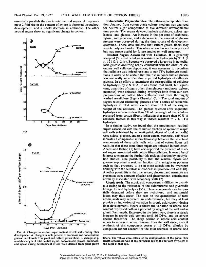
essentially parallels the rise in total neutral sugars. An approxi-mate 2-fold rise in the content of xylose is observed throughoutdevelopment, and a 2-fold decrease in arabinose. The otherneutral sugars show no significant change in content.
90
70
PLANT
5OF
301
lal0
1-
0
o I
0 1 7
C00 3
10
a.O* 90
70
50
30
10
CD
._
EE0
20.
co
0'C
-6.P-
0
zCP
GLUCOSE
ARABINOSE
CULTURE
ARABINOSE
4 8 12 16 20
Days Post Anthesis24 28
PLANT
TOTALNEUTRAL SUGARS -0
Days Post - Anthesis
FIG. 4. Changes in neutral sugar content of cell walls during fiberdevelopment. A: changes in mole per cent of arabinose and noncellulosicglucose in cell walls from plant and culture grown fibers. B: changes in g/mm fiber length of total neutral sugars, noncellulosic glucose, arabinose,and xylose during development of cell walls derived from plant-grown
Extraceliular Polysaccharides. The ethanol-precipitable frac-tion obtained from cotton ovule culture medium was analyzedfor neutral sugar composition at five different developmentaltime points. The sugars detected include arabinose, xylose, ga-lactose, and glucose. An increase in the per cent of arabinose,xylose, and galactose, and a decrease in the amount of glucosepresent were observed during the time course of developmentexamined. These data indicate that culture-grown fibers maysecrete polysaccharides. This observation has not been pursuedbut may prove useful for future studies on wall structure.
Residual Sugars Associated with Cellulose. It is generallyaccepted (50) that cellulose is resistant to hydrolysis by TFA (2N, 121 C, 1-2 hr). Because we observed a large rise in noncellu-losic glucose occurring nearly coincident with the onset of sec-ondary wall cellulose deposition, it was necessary to reconfirmthat cellulose was indeed resistant to our TFA hydrolysis condi-tions in order to be certain that the rise in noncellulosic glucosewas not really an artifact due to partial hydrolysis of cellulosicglucose. In an effort to quantitate the susceptibility of celluloseto hydrolysis by 2 N TFA, it was found that small, but signifi-cant, quantities of sugars other than glucose (arabinose, xylose,mannose) were released during hydrolysis both from our ownpreparations of cotton fiber cellulose and from thoroughlywashed a-cellulose (Sigma Chemical Co.). The total amount ofsugars released (including glucose) after a series of sequentialhydrolyses in TFA never exceed about 11% of the originalweight of the cellulose. The glucose released after sequentialhydrolyses represents less than 3% of the weight of the celluloseprepared from cotton fibers, indicating that more than 97% ofcellulose treated in this way is indeed resistant to 2 N TFAhydrolysis.
In a similar study, we found that the predominant residualsugars associated with the cellulosic fraction of sycamore maplecell walls (obtained by an acetic/nitric digest of total cell walls)were xylose, glucose, and to a lesser extent, mannose. This resultindicates a comparable interrelationship between the structuralcomponents of these cells walls with that of cotton fibers cellwalls, in that these same three sugars are released in both cases.Adams and Bishop (1) have also reported the presence of resid-ual sugars associated with cotton fiber.cellulose. It would be ofinterest to characterize further this residual fraction by methyla-tion studies. One possibility is that the residual xylose andglucose represent a residual fraction of a xyloglucan polymersuch as that proposed to be in close association by hydrogenbonding with the cellulose microfibrils in sycamore cell walls (8).Another possibility is that the xylose, glucose, and mannose arepresent as trace amounts of xylan and glucomannan, constituentsnormally associated with secondary walls (7).Uronc Adds. The uronic acid component is difficult to quanti-
tate owing to the resistance of the aldobiuronic acid glycosidiclinkage to acid hydrolysis (55). These compounds can be par-tially degraded before they are hydrolyzed, and substantiallosses may thus occur. The data on the quantitation of totaluronic acids may represent an underestimate, but they at leastprovide an indication of variation in uronic acid content duringfiber development. Figure 5 shows the variation in uronic acidcontent expressed both as a per cent by weight of the wall and asg/unit fiber length. Expressed in the latter way, there is a steadyincrease in uronic acid content until 16 DPA, and an abruptdecline thereafter. The sharp decline in uronic acid contentseems to represent actual removal from the wall since, even ifsynthesis of this component ceases at 16 DPA, dilution byelongation cannot account for the total decrease in uronic acid
fibers. The values were calculated by multiplication of the g/mm fiberlength of total cell wall at any particular age by the per cent by weight ofthe sugar at that age.
1093Plant Physiol. Vol. 59, 1977
www.plantphysiol.orgon June 10, 2018 - Published by Downloaded from Copyright © 1977 American Society of Plant Biologists. All rights reserved.

MEINERT AND DELMER
24
20j
<016
C
0
._
4
o=
- Jcr* Eo .0
C,CEEtoE
0
00
0
0 %
S '~PLANT
0% CULTURE
0 4 8 12 16 20 24
8 \
6 PLANT
4
2l
,) 4 8 12 16 20
Days Post-AnthesisFIG. 5. Uronic acid content in the fiber wall during development. A:
per cent by weight uronic acids i3 given for both plant- and culture-grownfibers. Each data point is an average of triplicate determinations on awall preparation. Where two points are given for the same age, theywere obtained by independent triplicate determinations. (-): plant-grown fiber cell walls; (0): culture-grown fiber cell walls. B: uronic acidcontent expressed as g/mm fiber length. Data points were calculated bymultiplication of the g/mm fiber length of total cell wall at any particularage by the per cent by weight uronic acids at that age taken from the linedrawn in (A) for plant-grown fibers.
content between 16 and 18 DPA. This can be seen from the factthat the absolute content drops by about 50% between 16 and18 DPA whereas the fibers only increase in length by about13%. Another possibility, which is difficult to rule out, is thatthe uronic acids become increasingly difficult to detect by thisassay procedure as the fiber wall changes composition during thisstage of development. The bulk of the uronic acids are probablypresent as polyuronides; this is indicated by a preliminary exper-iment which has shown that 60 to 70% of the total uronic acidsof the wall can be solubilized by digestion of cell walls withColletotrichum endopolygalacturonase.
Protein. Table II shows the amino acid composition of the cellwalls from plant- and culture-grown fibers as an average of all ofthe data for the various ages examined. The data for each age arenot presented since no significant change in composition withfiber age was found. This can be seen from the low standarddeviations obtained when all ages are considered together. Ap-proximately 50% of the amino acids are hydrophobic, and at anyage the composition shows no preponderance of any particularamino acid. No differences are observed in amino acid composi-tion for cell walls isolated from plant- and culture-grown fibers.The results of a separate assay for hydroxyproline are also shownin Table II. This amino acid comprises less than 0.6% of theweight of the wall at any age. Because these values are so lowand were repeated only once, we have not attempted to calculatechanges, if any, in the absoulte content of this amino acid duringdevelopment.The total protein content, measured by analyses for total N
and calculated on the assumption that all N is present as peptide-bound amino acids, is surprisingly high when expressed as weightper cent of the wall, especially at the youngest age examined, but
declines noticeably later in development (Fig. 6). However,when expressed as g/unit length of fiber, the content risessharply between 12 and 16 DPA, and subsequently declines. Asfor uronic acids, calculations show that this decrease in proteincontent indicates an absolute removal from the wall during thislater period.
In an attempt to ascertain whether or not the cell wall proteinis a true structural component of the wall, or whether at least aportion of it could have been adsorbed from the cytoplasmduring cell wall preparation, 14 DPA walls were extracted with asolution of SDS, urea, and mercaptoethanol for 5 min at 100 Cand the residue was analyzed for residual protein. This treatmentshould remove any protein not covalently bound to cell wallpolysaccharides. More than half of the protein of the walls wasindeed solubilized by this treatment, but the amino acid compo-sition of the protein remaining bound to the wall remained quitesimilar to that of unextracted walls.The supernatant of the SDS-urea-mercaptoethanol extraction
was dialyzed and analyzed by SDS gel electrophoresis to see towhat extent, if any, polypeptides had been released from thewall. The results (not illustrated) clearly showed at least eight
TABLE I I
Comparison of Amino Acid Composition of Cell Wall proteinfor Cell Walls D±rived from plant and Culture Grown Fibers.
Each value represents an average of the mole per cent of each aminoacid detected at a variety of ages for both plant and culture grownfibers. For plant-grown fibers, data were pooled from duplicate samplesobtained from fibers of 5, 9, 10, 11, 12, 14, 15, 16, 17, and 29 DPA; forculture-grown, from samples of fibers of 10, 12, 14 and 16 DPA.Standard deviations are given.
Plant CultureAmino acid Average Average
ASP 10.8 ± 0.74 9.6 ± 0.33THR 6.0 ± 0.36 5.5 ± 0.22SER 8.4 ± 1.21 8.8 ± 0.70
GLU 11.7 ± 0.98 10.5 ± 1.5GLY 11.8 ± 1.3 13.7 ± 1.6ALA 10.0 ± 1.7 8.8 ± 0.73
VAL 7.0 ± 0.68 7.0 ± 0.33MET 0.73± 0.98 0.75± 0.86ILE 4.9 ± 0.43 4.8 ± 0.05LEU 9.7 ± 0.58 9.5 ± 0.29
TYR 2.0 ± 0.66 1.9 ± 0.36PHE 4.5 ± 0.27 4.4 ± 0.16
LYS 6.0 ± 1.6 8.4 ± 0.34HIS 2.2 ± 0.56 2.5 ± 0.10ARG 4.2 ± 0.38 2.8 ± 1.9
HYP 0.23± 0.10 0.41± 0.10
c0
20a.
00
a.
40-
30[
I.I
.
II0 I
IIt -
1.20j
10
0 4 8 12 16
Days Post-Anthesis20
25
E20 E
0 .
IOc 0*n .0O11-0 L
5 aXI C
FiG. 6. Protein content in the cells wall during development of plant-grown fibers. Per cent by weight was calculated by multiplication of theper cent N by 8. Grams/unit length of fiber were calculated by multipli-cation of g/unit length of total cell wall at any particular age by the percent protein at that age.
Plant Physiol. Vol. 59, 19771 094
1[
4
www.plantphysiol.orgon June 10, 2018 - Published by Downloaded from Copyright © 1977 American Society of Plant Biologists. All rights reserved.

CELL WALL COMPOSITION OF COTTON FIBERS
discrete bands on SDS gels, and in addition, there appeared toexist high mol wt component(s) incapable of penetrating the gel.The mol wt of the bands observed were: 15,000, 21,000,33,000, 50,000, 63,000, 66,000, and 92,000. This supernatantwas also examined for carbohydrate content, and all neutralsugars found in unextracted 14 DPA walls were also found in theurea extract, although in different proportions from those inunextracted wails. This extraction procedure resulted in prefer-ential release of xylose and glucose and proportionally lessrhamnose and arabinose. With no correction for possible lossesduring the procedure, the per cent of total carbohydrate releasedby this treatment amounts to at least 7% of the total cell wallweight. This represents approximately one-third of the totalneutral sugars originally present in the cell wall, and may indi-cate that some covalent bonds have been broken by the proce-dure. Thus, information concerning how the proteins are boundto the cell wall was not clearly obtained, but may perhaps beobtained by a similar extraction at a lower temperature. Never-theless, the data do indicate that a variety of polypeptides ofdiscrete mol wt are present in the cell wall fraction, and thatsome of this protein can be removed by relatively mild treat-ments.
P!reliminary Linkage Analyses. Permethylated cell walls fromplant-grown fibers were separated into chloroform-soluble and-insoluble fractions, but only the chloroform-soluble fraction wasstudied further. Talmadge et al. (50) have shown for sycamorecell walls that the chloroform-insoluble polymers were primarilyincompletely methylated cellulose and methylated oligoarabino-sides attached to the hydroxyproline-rich protein. In the case ofcotton. fiber cell wails, it is not yet known what fraction of thewall remains insoluble under our methylation conditions. Com-parison of the methylated sugar derivatives obtained with thetotal neutral sugar analyses indicated that the fraction analyzedhere was in some respects different in composition. The presenceof low amounts of mono-methyl derivatives also indicates thatsome undermethylated derivatives are present even in the chlo-roform-soluble fraction. Therefore, only limited interpretationof the results is given.The gas chromatograms obtained for four ages of cell walls are
presented in Figure 7. Tentative identifications are listed in thefigure legend. Qualitatively, the derivatives obtained are similarfor all ages examined (5, 8, 12, 14, 16, and 20 DPA-datashown only for 8, 12, 16, and 20 DPA). Keeping in mind thelimitations stated above, several conclusions can be drawn. Thefirst is that in view of the large number of derivatives possible,the limited number of derivatives obtained indicates that thecarbohydrate polymers of the wall are of limited heterogeneity.Second, it appears that the area represented by some of thevarious peaks is age-dependent. Peak 5 (5-linked arabinose5) isprominent at all ages and represents the majority of the arabi-nose present, and since the total content of arabinose/unit lengthof fiber wall declines only slightly during development (see Fig.4B), we have chosen to compare the relative areas of the otherpeaks to this peak. Using this comparison, which is of coursesubject to a variety of limitations as stated above, one can seethat several derivatives vary in an age-dependent fashion; themost striking of these is the very large rise in the relative area ofpeak 9 (3-linked glucose6) which closely parallels the total rise in
5 Data are presented by indicating the deduced linkages by which eachglycosyl residue of the original sample was connected to other sugars,rather than by indicating the positions in the derivatives of the methoxyand acetyl groups. For example, 5-linked arabinose is equivalent to1,4,5-tri-0-acetyl-2,3-di-0-methylarabinitol. All arabinosyl residueshave been assigned the furanose ring form, and all other sugars thepyranose ring form.
6 Either 3-glucose or 3-mannose is consistent with relative retentiontime and mass spectra data, but the peak at later ages is too large relativeto total wall mannose content to be primarily 3-mannose.
noncellulosic glucose in the cell wails (see also Fig. 4B). Thearea of the 3-linked glucose peak relative to the 5-linked arabi-nose peak is 0.007, 0.001, 0.234, 0.353, 1.406, and 1.638 at 5,8, 12, 14, 16 and 20 DPA, respectively.
DISCUSSION
Table III summarizes the changes in the cell wall compositionof cotton fibers during their development, expressed both as aper cent by weight of the wall and in terms of g/unit length ofwall. A large per cent of the total weight of the wall has beenaccounted for by these analyses. Depending upon whether thecontent is expressed as a weight per cent or as g/unit length ofcell wall, the interpretation of the observed changes in cell wallcomposition during elongation may differ greatly. Because it ismore representative of actual changes in the wall, this discussionwill consider only the absolute changes (g/mm fiber length).These data permit us to present an over-all picture of thechanges in cell wall composition as the cotton fiber develops.The thickness of the cell wall remains relatively constant
during the early stages of cell elongation, indicating that wallsynthesis is keeping pace with fiber elongation. After 12 DPA
8 DPA 12 DPA5 6
6
2222
2 1
16~~~~~~~~~~~~~13130 2Q210 13 12150 1
FI.I . I .1 . I . I.696102030 40 SO 600102030 240 50DPA9 2
S14
22 1316DPAI
21
2~~~~~~~~~~~~~ 78 ~~14122 19
236142 4 I
110 130 150 170 110 350 150 1T0 *c0 10 20 30 40 50 60 0 10 20 30 40 50 60 70 MIN
FIG. 7. Gas chromatographic separations of the alditol acetate deriv-atives of permethylated plant-grown cotton fiber cell wall preparations ofvarious ages. Peak numbers refer to the following deduced derivatives: 1= T-arabinose; 2 = T-xylose*; 3 = 2-arabinose; 4 = T-glucose; 5 = 5-arabinose*; 6 and 7 = T-galactose*, 4-xylose*, 2-xylose; 8 = 2-hexose; 9= 3-glucose*; 10 = 3-galactose; 11 = 4-glucose*; 12 = 4- and/or 6-hexose; 13 = 6-galactose; 14 and 15 = unidentified dimethyl hexoses;16 = 4,6-glucose*; 17 = unidentified monomethyl hexose; 18 = 3,6-galactose; 19, 20, and 21 = unidentified monomethyl hexoses. Aster-isks(*) refer to sugars for which identification is supported by both massspectral data and relative retention times. The other identifications aresomewhat less conclusive and, in some cases, lack sufficient data fordefinitive identification.
1 095Plant Physiol. Vol. 59, 1977
www.plantphysiol.orgon June 10, 2018 - Published by Downloaded from Copyright © 1977 American Society of Plant Biologists. All rights reserved.

1096 MEINERT AND DELMER
TABLE III
Summary of Cotton Fiber Cell Wall Composition During Development
The first line of figures represents the percent by weight of that componentas taken from the best fit line of plotted data points, the second line(italics) of figures represents the g x 1010 of component per mm fiberlength. ND = not determined. Numbers in parentheses refer to the totaledpercent of analyzed components under conditions where not all componentswere determined. Data refer only to plant-grown fibers.
COMPONENT CELL WALL AGE (DPA)
5 8 10 12 14 16 18 20 22 25 29
Cellulose 9 10 23 24 25 31 74 82 87 90 943 3 6 9 14 34 131 201 292 484 1043
Neutral Sugars 33 25 25 20 17 14 12 10 9 7 411 7 7 7 9 16 21 24 30 37 44
Uronic Acids 10 16 22 21 16 9 3 2 2 ND ND4 5 6 8 9 10 v
Protein 40 24 22 22 25 21 3 3 ND ND ND14 7 6 8 14 23 c 7
Ash ND 3 3 2 11 11 ND 9 ND ND ND1 1 1 6 12 22
Total Cell Wall (91) 78 95 89 94 86 (92) 106 (98) (97) (98)34 29 28 36 54 711 177 244 335 v39 1110
the thickness of the wall begins to increase, and during theperiod from 12 to 16 DPA increases in essentially all compo-nents contribute to the increase in total wall thickness. After 16DPA, it is clear that deposition of cellulose (and to a much lesserextent, increase in noncellulosic glucose) are the only eventswhich contribute to the continuing, great increase in wall thick-ening.The data on cellulose deposition in the fiber wall point out two
curious facts. The first is evident from the plot of the rate ofcellulose deposition/unit length of fiber (Fig. 3B) which showsthat the rate sharply rises at the onset of secondary wall deposi-tion, declines, and then rises again. The second observation isthat fiber elongation continues with little change in rate for atleast 4 days after the first rise in the rate of cellulose deposition.This is curious because it seems that such an increase in deposi-tion should cause an abrupt cessation of elongation. However, asimilar observation, recently made by Benedict et al. (11) andSchubert et al. (44) for other varieties of cotton, revealed that itis not until the second rise in rate of cellulose deposition occursthat the fiber ceases to elongate. It is possible that the microfi-brils continue to be deposited in a random or transverse orienta-tion during this first phase of deposition and in this way do notlimit cell extension; if so, secondary wall deposition might bemore correctly defined as that deposition occurring during thetime of distinct increase in birefringence at approximately 20DPA. The other possibility is that the cells at this stage areelongating solely by tip growth and are not limited by depositionwhich occurs behind the tip region. There seem to be varietaldifferences in cotton regarding the capacity for elongation afterthe beginning of secondary wall deposition (11). In view of thecommercial value of cotton fibers and the desirability of long lintfibers, this problem deserves further study.The observation of a significant rise in noncellulosic glucan
(which the methylation analyses indicate is, at least in part, 3-linked) during the later stages of cell elongation is of interest inlight of the recent demonstration in our laboratory (17, 21) of ahighly active j8-(1---'3)-glucan synthetase in these fibers. Al-though it is premature to speculate about the functional and/orstructural role of this glucan, it is of interest that it increases incontent concomitant with the earliest stages of secondary wallcellulose deposition and toward the end of elongation. Three-linked glucans are not thought to be common components ofprimary cell walls of higher plants. It remains to be determinedwhether this glucan is a true structural component of the wall orwhether it serves as a reserve polymer, possibly for the biosyn-
Plant Physiol. Vol. 59, 1977
thesis of secondary wall cellulose. Callose is apparently mobi-lized as a reserve material in pollen (48), and there are otherreports in the literature which implicate turnover of noncellu-losic glucans of cell walls (14, 23, 31, 34). Callose has also beenobserved to be deposited behind the tip of growing pollen tubes(18), and it has been suggested that it serves to strengthen thetube wall (48), although the possibility that it turns over hasapparently not been explored.The question of turnover of all of the wall components of the
cotton fiber merits further study since calculations based on thedecrease in uronic acids and protein between 16 and 18 DPAlead to the conclusion that these components are partially re-moved from the wall during this time. It is clear that the patternsof cell wall synthesis (and probably also degradation) are un-dergoing great shifts at this period, and studies on the regulationof this transition period could help to provide a better generalunderstanding of cell wall development in plants.The results of both the compositional analyses and the prelim-
inary methylation analyses permit us to compare the cotton fibercell wall with some other primary cell walls as reported in theliterature. Inasmuch as the primary wall of the cotton fiber is nota static structure, such comparisons are to a certain extentdependent upon the age of the fiber. A compositional compari-son of the cotton fiber cell wall at 10 DPA (approximate mid-phase of elongation) with some other primary cell walls is shownin Table IV. All data used for comparison were obtained fromcell walls of cultured cells, and the monocotyledons representedare all grasses. Wheat and rye grass cell walls have been shownto be representative of monocotyledon (grass) walls (15,) all ofwhich are characterized by a high neutral sugar content, a lowcellulose content, a very low content of hydroxyproline, and byan arabinose to xylose ratio approximating unity. Rye grass(endosperm cells) are, however, somewhat different from othermonocot primary cell walls, in having higher levels of noncellu-losic glucan which has been shown to be a-(1 -* 3)-,f-(1 -- 4)-linked (46, 47). It is clear from Table IV that the cotton fiber cellwall at 10 DPA shows a greater similarity in composition to thewalls of the dicot sycamore maple (representative of other dicotprimary cell walls examined [56]) than to those of the monocots,but it resembles even more closely that of cell walls of culturedcells of the gymnosperm, Douglas fir.The results of the preliminary methylation analyses of unfrac-
tionated walls provide further support for the similarity of thecotton fiber cell wall to the cell walls of cultured cells of dicotsand the gymnosperm. Data of Burke et al. (15) indicate that themajor hemicellulosic component of primary walls of cells ofdicots and of Douglas fir is a (galacto)-xyloglucan, whereas thatof the cell walls of the grasses in culture is an arabinoxylan. Thecotton fiber walls contain significant amounts of 4-linked glu-cose, 4,6-linked glucose, terminal xylose, 2- or 4-linked xylose,and terminal galactose residues indicative of a (galacto)-xyloglu-
TABLE IV
Composition by Weight Percent of Cell Walls from Various Plant Sources
W SvcamorelWheatl Rye Grass maple Douglas fir Cotton fiber2
Total neutralsugars
RhamnoseFucoseArabinoseXy loseMannoseGalac toseGlucose(non-cellulosic)
CelluloseUronic acidsProteinHydroxvproline
63.1 73.7 49.8 37.2 25.3
0.6 0.6 3.] 2. 1 .90.1 0 1.3 1.0 0.320.7 20.3 21.0 12.7 8.224.8 19.8 7.6 5.0 2.61.0 0.1 0.3 0.5 1.0
12.0 5.9 12.8 7.6 4.33.9 27.0 3.7 7.9 7.0
14.0 13 '3 2 2'312 7 1 3 18 2211 7 10 18 220.14 '.05 2.0 0.4 0.3
ITaken from the data of Burke et al (15)From 10 DPA, plant grown fibers
www.plantphysiol.orgon June 10, 2018 - Published by Downloaded from Copyright © 1977 American Society of Plant Biologists. All rights reserved.

CELL WALL COMPOSITION OF COTTON FIBERS
can structure, and lack branched xylosyl residues indicative ofthe arabinoxylan characteristic of monocot cell walls. The pre-ponderance of 5-linked arabinose in the cotton fiber walls isanother similarity shared with the Douglas fir and may indicatethe presence of a polymer with an a-(1 -> 5)-arabinan backbonesimilar to those reported from a variety of plant tissues (6, 7).Although the amount of hydroxyproline is low in the cotton fibercell walls, the presence of some 2-linked arabinosyl residues inthese preparations could indicate such arabinosides (30). Addi-tional studies may show that the wall structure of cotton fiber cellwalls is indeed representative of dicots, and as such elongatingcotton fibers might be well suited to serve as a model system forelongation studies. A critical assessment of the mode of growthof these fibers will also be necessary before detailed elongationstudies can be properly interpreted; although it appears unlikely,if growth proceeds from the tip alone, then these observedchanges in composition may be more properly attributed tochanges associated with wall maturation rather than elongation.We stress that the changes in composition reported here shouldfor the present be interpreted only as changes correlated in timewith the elongation process. Much more detailed study will benecessary to determine whether any or all of these compositionalchanges are fundamental to the mechanism of cell wall growth inthis system.Our data show that cell walls from fibers cultured in vitro are
in many respects quite similar to those of fibers grown on theplant. The major similarities are the like compositions of the cellwalls and that changes in composition occur in the same develop-mental sequence on the plant and in culture. The entire develop-mental sequence seems to be compressed in time in culture, andthe final length of fibers is reduced by about half. Since an earlieronset of secondary wall deposition may limit fiber length, thismay account for the shorter final fiber length. The biochemicaland developmental similarities of walls in culture should makethis system useful for in vivo labeling studies designed to exam-ine turnover of wall components and perhaps also for studyingthe effects of hormones on the elongation process.
Acknowledgments -The authors wish to thank D. T. A. Lamport for his assistance with theamino acid analyses, C. C. Sweeley for use of and especially R. Thompson for technicalassistance with the mass spectrometer facility, L. C. de Balboa for advice on permethylationprocedures, and C. Kulow, E. Ryder, and L. McEnroe for technical assistance.
LITERATURE CffED
1. ADAMS GA, CT BISHOP 1955 Polysaccharides associated with alpha-cellulose. Tappi 38:672-676
2. ALBERSHEIM P 1975 The walls of growing plant cells. Sci Am 232: 81-943. ALBERSHEIM P, WD BAUER, K KEEGSTRA, KW TALMADGE 1973 The structure of the wall
of suspension-cultured sycamore cells. In F Loewus, ed. Biogenesis of Plant Cell WallPolysaccharides. Academic Press, New York pp 117-147
4. ALBERSHEIM P, DJ NEMNS, PD ENGLISH, A KARR 1967 A method for the analysis ofsugars in plant cell-wall polysaccharides by gas-liguid chromatography. Carbohydr Res 5:340-345
5. ALLEN AK, A NEUBERGER 1973 The purification and properties of the lectin from potatotubers, a hydroxyproline-containing glycoprotein. Biochem J 135: 307-314
6. ASPINALL GO 1970 Pectins, plant gums and other plant polysaccharides. In W Pigman, DHorton, eds, The Carbohydrates Vol IIB. Academic Press, New York pp 515-536
7. ASPINALL GO 1973 Carbohydrate polymers of plant cell walls. In F Loewus, ed. Biogenesisof Plant Cell Wall Polysaccharides. Academic Press, New York pp 95-115
8. BAUER WD, KW TALMADGE, K KEEGSTA, P ALBERSHEIM 1973 The structure of plant cellwalls. II. The hemicellulose of the walls of suspension-cultured sycamore cells. PlantPhysiol 51: 174-187
9. BEASLEY CA, IP TiNG 1973 The effects of plant growth substances on in vitro fiberdevelopment from fertilized cotton ovules. Am J Bot 60: 130-139
10. BEASLEY CA, IP TING 1974 Effects of plant growth substances on in vitro fiber developmentfrom unfertilized cotton ovules. Am J Bot 61: 188-194
11. BENEDicr CT, RH SaTn, R. KOHEL 1973 Incorporation of 14C-photosynthate into devel-oping cotton bolls, Gossypiun hirsutum L. Crop Sci 13: 88-91
12. BJ6RNDAHL H, CG HELLERQUIST, B LINDBERG, S SVENSON 1970 Gas liquid chromatogra-phy and mass spectrometry in methylation analysis of polysaccharides. Angew Chem IntEd Engl 9: 610-619
13. BLUMENKRANTz N, G AssoE-HANsEN 1973 New method for quantitative determination ofuronic acids. Anal Biochem 54: 484-489
14. BucHALA AJ, KCB WILIE 1971 The ratio of ,-(1 -. 3) to P-(1 -* 4) glucosidic linkages innon-endospermic hemicellulosic -glucans from oat plant (Avena saiva) tissues at differ-ent stages of maturity. Phytochemistry 10: 2287-2291
15. BURKE D, P KAuFMAN, M McNEIL, P ALBERSEiM 1974 The structure of plant cell walls.
VI. A survey of the walls of suspension-cultured monocots. Plant Physiol 54: 109-11516. CLELAND R. A KARLSNES 1967 A possible role for hydroxyproline-containing proteins in
the cessation of cell elongation. Plant Physiol 42: 669-67117. DELMER DP, U HEINIGER, C KuLow 1977 UDP-glucose: glucan synthetase in developing
cotton fibers. I. Kinetic and physiological properties. Plant Physiol. 59: 713-71818. GoRsKA-BRYLAsa A 1965 Cytomorphological studies of callose in pollen tubes. Acta Soc
Bot Pol 34: 757-76219. HAKOMORI S 1964 A rapid permethylation of glycolipid and polysaccharide catalyzed by
methylsulfinyl carbanion in dimethyl sulfoxide. J Biochem 55: 205-20820. HEAGES D 1973 Techniques for high resolution electrophoresis. SDS Molecular Weight
Determination. ORTEC Manual AN32A21. HEINIGER U, DP DELMER 1977 UDP-glucose: glucan synthetase in developing cotton
fibers. II. Structure of the reaction product. Plant Physiol 59: 719-72322. KEEGSTRA K. KW TALMADGE, WD BAUER, P ALBERSHEIM 1973 The structure of plant cell
walls. III. A model of the walls of suspension-cultured sycamore cells based on theinterconnections of the macromolecular components. Plant Physiol 51: 188-196
23. KIVILAAN A. RS BANDURsas, A SCHULYZ 1971 A partial characterization of an autolyti-cally solubilized cell wall glucan. Plant Physiol 48: 389-393
24. KvtRiKKo KI 1963 Hydroxyproline-containing fractions in normal and cortisone-treatedchick embryos. Acta Physiol Scand Suppl 219: 1-92
25. Kus F 1976 Glycosylated seryl residues in wall protein of elongating pea stems. PlantPhysiol 57: 224-226
26. LABAVrrCH J, P RAY 1974 Relationship between promotion of xyloglucan metabolism andinduction of elongation by indoleacetic acid. Plant Physiol 54: 499-502
27. LABAvrrcH J, P RAY 1974. Tumover of cell wall polysaccharides in elongating stemsegments. Plant Physiol 53: 669-673
28. LAINE RA, ND YOUNG, JN GERBER, CC SWEELEY 1974 Identification of 2-hydroxy fattyacids in complex mixtures of fatty acid methyl esters by mass chromatography. BiomedMass Spectr 1: 10-14
29. LAMPORT DTA 1969 The isolation and characterization of hydroxyproline-rich glycopep-tides obtained by enzymic degradation of primary cell walls. Biochemistry 8: 1155-1163
30. LAMPORT DTA, DH MILLER 1971 Hydroxyproline arabinosides in the plant kingdom. PlantPhysiol 48: 454-456
31. LOESCHER W, D NEvtNs 1972 Auxin-induced changes in Avena coleoptile cell wall compo-sition. Plant Physiol 50: 556-563
32. Mmmx-FiGiNi M 1966 Comparison of the biosynthesis of cellulose in vitro and in vivo incotton bolls. Nature 210: 754-755
33. MARX-FIGINI M, GV SCHULZ 1966 Uber die Kinetik und den Mechanismus der Biosyntheseder Cellulose in den hoheren Pflanzen (nach Versuchen an den Samenhaaren der Baum-wolle). Biochim Biophys Acta 112: 74-80
34. NEvItS D 1975 Effect of nojirimycin on plant growth and its implications concerning a rolefor exo-glucanases in auxin-induced cell expansion. Plant Cell Physiol 16: 707-717
35. NEvINs DJ, P ENGLISH, P ALBERSHEIM 1968 Changes in cell wall polysaccharides associ-ated with growth. Plant Physiol 43: 914-922
36. NORTHCOTE DH 1972 Chemistry of the plant cell wall. Annu Rev Plant Physiol 23: 113-13237. O'KELLEY JC 1953 The use of 14C in locating growth regions in the cell walls in elongating
cotton fibers. Plant Physiol 28: 281-28538. PARK JT, MJJOHNSON 1949 A sub-microdetermination of glucose. J Biol Chem 181: 14939. RAY PM 1963 Sugar composition of oat coleoptile cell walls. Biochem J 89: 144-15040. RAY PM 1967 Radioautographic study of cell wall deposition in growing plant cells. J Cell
Biol 35: 659-67441. ROELOPSON PA 1951 Orientation of cellulose fibrils in the cell wall of growing cotton hairs
and its bearing on the physiology of cell wall growth. Biochim Biophys Acta 7: 43-5342. SADAVA D, F WALKER, M CHRISPEELS 1973 Hydroxyproline-rich cell wall protein (exten-
sin): biosynthesis and accumulation in growing pea epicotyls. Dev Biol 30: 42-4843. SANDrORD PA, HE CONRAD 1966 The structure of Aerobacter aerogenes A3(S1) polysac-
charide. I. A reexamination using improved procedures for methylation analysis. Bio-chemistry 5: 1508-1517
44. SCHUBERT AM, CR BENEDicr, JD BERLIN, RJ KOHEL 1973 Cotton fiber development-kinetics of cell elongation and secondary wall thickening. Crop Sci 13: 704-709
45. SETTERFIELD G, ST BAYLEY 1959 Deposition of cell walls in oat coleoptiles. Can J Bot 37:861-865
46. SMIIm MM, BA STONE 1973 Chemical composition of the cell walls of Lolium multiflorumendosperm. Phytochemistry 12: 1361-1367
47. Smn MM, BA STONE 1973 Studies on Loluwn multiflorum endosperm in tissue culture. I.Nutrition. Aust J Biol Sci 26: 123-133
48. STANLEY RG, HF LINSKEN 1974 Pollen: Biology, Biochemistry, Management. Springer-Verlag, Berlin p 135
49. SWEELEY CC, BD RAY, WI WOOD, JF HOLLAND, MI KIcHEvsEv 1970 On-line digitalcomputer system for high-speed single focusing mass spectrometry. Anal Chem 42: 1505
50. TALMADGE K, K KEEGSTRA, WD BAUER, P ALBERSHEIM 1973 The structure of plant cellwalls. I. The macromolecular components of the walls of suspension-cultured sycamoreceDls with a detailed analysis of the pectic polysaccharides. Plant Physiol 51: 158-173
51. UPDEGRAFF DM 1969 Semi-micro determination of cellulose in biological materials. AnalBiochem 32: 420-424
52. VARNER JE, RM MENSE 1972 Characteristics of the process of enzyme release fromsecretory plant ceDls. Plant Physiol 49: 187-189
53. WEBER K, M OSBORN 1969 The reliability of molecular weight determinations by dodecylsulfate-polyacrylamide gel electrophoresis. J Biol Chem 244: 4406
54. WESTAFER JM, RM BROWN, JR. 1976 Electron microscopy of the cotton fibre: newobservations on ceDlwal formation. Cytobios 15: 111-138
55. WHIsTLER RL, EL RICHARDS 1970 Hemicelluloses. In W Pigman, D Horton, eds. TheCarbohydrates Vol HA. Academic Press, New York pp 447-469
56. WILDER BM, P ALBERSHEIM 1973 The structure of plant ceDl waDls. IV. A structuralcomparison of the wal hbemicelulose of cell suspension cultures of sycamore (Acerpseudoplatanus) and of red kidney bean (Phaseolus vulgaris). Plant Physiol 51: 889-893
Plant Physiol. Vol. 59, 1977 1 097
www.plantphysiol.orgon June 10, 2018 - Published by Downloaded from Copyright © 1977 American Society of Plant Biologists. All rights reserved.
Related Documents


