Running Head: CHANGE IN PA DURING ACTIVE TREATMENT 1 CHANGE IN PHYSICAL ACTIVITY DURING ACTIVE TREATMENT OF CARDIAC PATIENTS _______________________ A Thesis Presented to The Faculty of Health and Environmental Studies School of Sport and Recreation Auckland University of Technology Auckland, New Zealand ______________________ In Partial Fulfilment of the Requirements for the Degree Master of Health Science _______________________ by Leon W Tahana April, 2017

Welcome message from author
This document is posted to help you gain knowledge. Please leave a comment to let me know what you think about it! Share it to your friends and learn new things together.
Transcript

Running Head: CHANGE IN PA DURING ACTIVE TREATMENT 1
CHANGE IN PHYSICAL ACTIVITY DURING
ACTIVE TREATMENT OF
CARDIAC PATIENTS
_______________________
A Thesis
Presented to
The Faculty of Health and Environmental Studies
School of Sport and Recreation
Auckland University of Technology
Auckland, New Zealand
______________________
In Partial Fulfilment
of the Requirements for the Degree
Master of Health Science
_______________________
by
Leon W Tahana
April, 2017

CHANGE IN PA DURING ACTIVE TREATMENT 2
Attestation of Authorship
I hereby declare that this submission is my own work and that, to the
best of my knowledge and belief, it contains no material previously published or
written by another person (except where explicitly defined in the
acknowledgements), nor material which to a substantial extent has been
submitted for the award of any other degree or diploma of a university or other
institution of higher learning.
Signature: _____________________________
Date: 5 June 2017

CHANGE IN PA DURING ACTIVE TREATMENT 3
Abstract
Sedentary secular, domestic, and recreational behaviour is a major risk factor
(RF) for cardiovascular disease (CVD). This study examines the quality of at-
home physical activity (PA) and how it relates to physical fitness (PF) before
and during 12 weeks of supervised cardiac rehabilitation (CR) in a group of
medically referred cardiac patients. PA was measured with an accelerometer
(ActiGraph wGT3X-BT), pre and post-CR, to determine if patient at-home PA
behaviour changes during supervised CR. Cardiac patients’ (n=27),
haemodynamic and morphological measurements were taken. Direct
measurement of the volume of oxygen consumption (VO2peak) was done with
respiratory gas analysis during a submaximal cycle ergometer test to determine
PF. Analysis of covariance (ANCOVA) was used to assess whether exercise-
induced improvement in cardiovascular and muscular capacity (CVaM-capacity)
influences the relationships between stages of PF (pre vs. post-CR) and PA
behaviour. Pre-CR power output and CVaM-capacity correlated moderately with
overall at-home caloric expenditure per week (r=0.47 and 0.53). Calculated r2
values indicate that power output and peak oxygen consumption contribute
between 22.1% and 28.1% to the variance of weekly PA energy consumption.
At-home PA behaviour (volume and intensity) changed significantly (p≤0.001)
after 12 weeks of supervised CR, with moderate and vigorous PA increasing,
and sedentary, and light PA decreasing. Future CR research should consider
how at-home PA behaviour and other RF inter-associations affect a patient’s
cardiac health and CR effectiveness.
Keywords: cardiac rehabilitation, physical activity, behaviour, functional/
cardiovascular capacity, accelerometer

CHANGE IN PA DURING ACTIVE TREATMENT 4
Acknowledgements
I express sincere thanks to Assoc. Prof. Lukas Dreyer (Assoc. Prof of
Health Science, UCOL) for providing incisive guidance during the entire project.
I would also like to thank Assoc. Prof. Scott Duncan (Head of Research, School
of Sport and Recreation, AUT) for guidance in finalising the thesis.
I also thank the directorship and staff of the U-Kinetics Exercise &
Wellness Clinic (UCOL, Palmerston North) for providing clinical and technical
support and the utilisation of the necessary facilities to complete this project. I
also acknowledge Helen McDonald (Proofreading Solutions, Wellington) for
proofreading this work.
Finally, I acknowledge and thank my family for their perseverance and
understanding during this time.

CHANGE IN PA DURING ACTIVE TREATMENT 5
Table of Contents
Attestation of Authorship ....................................................................................... 2
Abstract .................................................................................................................. 3
Acknowledgements ................................................................................................ 4
List of Figures ....................................................................................................... 10
List of Tables......................................................................................................... 11
List of Abbreviations ............................................................................................. 12
Chapter 1 .............................................................................................................. 14
Chapter 2 – Literature Review ............................................................................. 17
2.1 Introduction ..................................................................................................... 18
2.2 The PA and PF Interrelationship for Cardiac Health and CR ....................... 19
2.3 Cardiac Rehabilitation .................................................................................... 21
2.3.1 CR defined. .......................................................................................... 21
2.3.2 CR barriers ........................................................................................... 23
2.3.3 CR: Recommended PA/exercises for cardiac health ......................... 25
2.3.4 CR: Intervention program considerations ........................................... 29
2.3.5 CR: Changing PA patterns/habits ....................................................... 32
2.4 Validity of Study Measurements .................................................................... 36
2.4.1 CVaM-capacity/PF considerations for CR .......................................... 36
2.4.1.1 Cardiorespiratory fitness ........................................................ 37
2.4.1.2 Resistance training ................................................................ 37
2.4.2 Measurement of at-home PA. ............................................................. 39

CHANGE IN PA DURING ACTIVE TREATMENT 6
2.4.2.1 Accelerometers ....................................................................... 40
2.4.2.1.1 Accelerometer wear site ........................................... 41
2.4.2.1.2 Number of days monitored ....................................... 42
2.4.2.2 The International Physical Activity Questionnaire ................. 43
2.4.2.3 Logbook record ....................................................................... 43
2.5 Summary......................................................................................................... 44
Chapter 3 – Method .............................................................................................. 44
3.1 Participants ..................................................................................................... 47
3.2 Entry Criteria ................................................................................................... 47
3.3 Sample Size.................................................................................................... 49
3.4 Data Collection ............................................................................................... 49
3.4.1 Resting haemodynamics ..................................................................... 50
3.4.2 Morphological measures...................................................................... 50
3.5 Functional Capacity ........................................................................................ 50
3.5.1 Cycle ergometry ................................................................................... 50
3.6 PA Assessment .............................................................................................. 52
3.6.1 ActiGraph wGT3X-BT Accelerometer ................................................. 52
3.6.2 ActiGraph wGT3X-BT Accelerometer wear site. ................................ 53
3.6.3 Number of days monitored .................................................................. 53
3.6.4 Pre-intervention physical assessment................................................. 53
3.6.5 During-intervention physical assessment ........................................... 54

CHANGE IN PA DURING ACTIVE TREATMENT 7
3.7 Intervention Program Protocols ..................................................................... 55
3.8 Sample Characteristics .................................................................................. 58
3.9 Statistical Procedures..................................................................................... 58
Chapter 4 – Results .............................................................................................. 60
4.1 Introduction ..................................................................................................... 60
4.2 Clinical Characteristics of the Participants’ ................................................... 60
4.3 Comparison of PF Measures with Accelerometer PA Data .......................... 62
4.3.1 PF and PA pattern measures. ............................................................. 62
4.4 CR Program Effect on Body Composition Variables..................................... 63
4.5 CR Program Effect on Haemodynamic Variables ......................................... 64
4.6 CR Program Effect on PF .............................................................................. 65
4.7 CR Program Effect on PA .............................................................................. 65
4.8 Relationship of Percentage (%) Change in PF with Pre-Intervention
Measures of PF .............................................................................................. 66
4.9 Relationship of Percentage (%) Change in CV Fitness with Pre and Post-
Training Measures of PA ............................................................................... 67
4.9.1 CV Fitness and PA pattern measures ................................................. 68
4.9.2 Relationship of CV-capacity stages with PA patterns before and after
exercise training in cardiac patients ............................................................. 68
Chapter 5 – Discussion ........................................................................................ 74
5.1 Introduction ..................................................................................................... 74
5.2 Relationship of Pre-CR CV-capacity with Pre-CR PA Patterns .................... 74

CHANGE IN PA DURING ACTIVE TREATMENT 8
5.2.1 Volume of daily and weekly caloric expenditure. ................................ 74
5.2.2 Intensity of at-home PA. ...................................................................... 76
5.3 The Effect of the CR Program on Clinical Variables ..................................... 77
5.3.1 Physical fitness/cardiovascular capacity/functional capacity ............. 77
5.3.2 Body composition ................................................................................. 78
5.3.3 Haemodynamics .................................................................................. 78
5.4 The Effect of the CR Program on PA Patterns .............................................. 79
5.5 Limitations ....................................................................................................... 82
5.5.1 Sample size .......................................................................................... 82
5.5.2 Accelerometry ...................................................................................... 82
5.5.3 Definition of Intensity ........................................................................... 83
5.5.4 PF Assessment Protocol ..................................................................... 84
5.5.5 Individual vs. general application ........................................................ 84
5.6 Future Research ............................................................................................. 85
5.7 Clinical Applications ....................................................................................... 86
5.8 Conclusion ...................................................................................................... 86
References ............................................................................................................ 88
Appendix A Flow diagram of PA assessment during CR ................................108
Appendix B Participant Information Sheet ........................................................111
Appendix C Consent Form ................................................................................116
Appendix D Health Screen ................................................................................117

CHANGE IN PA DURING ACTIVE TREATMENT 9
Appendix E PA Booklet Log ..............................................................................120
Appendix F Ethics Application........................................................................123
CHANGE IN PA DURING ACTIVE TREATMENT 10
List of Figures
Figure 2-01. Estimated Dose-response Curve for the Relative Risk of either
CHD or CVD by Sample Percentages of PA and PF (From Williams, 2001) . 19
Figure 4-01. Pre-CR Program Assessment - VO2peak Scores ............................ 63
Figure 4-02. Pre-CR Program Assessment - Participant Average Duration Spent
at Each Intensity (Accelerometry Data) ............................................................ 63
Figure 4-03. Post-CR Program Assessment-VO2peak Scores. ............................ 68

CHANGE IN PA DURING ACTIVE TREATMENT 11
List of Tables
Table 4-0 CR Participant Baseline Demographic and Clinical Characteristics. 61
Table 4-01 Comparison of PF Measures with PA Participation ......................... 62
Table 4-02 CR Program Effect on Body Composition Variables ....................... 64
Table 4-03 CR Program Effect on Haemodynamic Variables ............................ 64
Table 4-04 CR Program Effect on PF .................................................................. 65
Table 4-05 CR Program Effect on PA ................................................................. 65
Table 4-06 Comparison of VO2peak Change (%) with Pre-Training Measures
of PF ................................................................................................................... 66
Table 4-07 Correlation of VO2 Change (%) with Pre and Post-CR Measures
of PA ................................................................................................................... 67
Table 4-08 The F-ratio, p-values, ETA2 and Wilks Lambda Scores of Two
ANCOVAs Investigating the Relationship of CV-capacity Stages with PA
Patterns Before and After Exercise Training in Cardiac Patients ..................... 69
Table 4-09 Pre and Post-Data of the Participants who had a CV Fitness
< 5.9 METs at the Start and End of the Program .............................................. 70
Table 4-10 Pre and Post-Data of the Participants who had a CV Fitness of
between 6.0 – 6.5 METs at the Start and End of the Program ......................... 71
Table 4-11 Pre and Post Data of the Participant’s that had a CV Fitness
>6.5 METs at the Start and End of the Program .............................................. 73
Appendix A: Table 6-0 Flow diagram of PA assessment before and during CR
intervention .....................................................................................................108

CHANGE IN PA DURING ACTIVE TREATMENT 12
List of Abbreviations
AACVPR American Association of Cardiovascular and Pulmonary
Rehabilitation
ACSM American College of Sport Medicine
ADL Activities of daily living
AHA American Heart Association
ANCOVA Analysis of covariance
BMI Body mass index
CABG Coronary artery bypass grafting
CAD/CHD Coronary arterial disease/Coronary heart disease
CEP Clinical Exercise Physiologist
CR Cardiac rehabilitation
CVaM Cardiovascular and muscular
CVD Cardiovascular disease
DBP/SBP Diastolic/Systolic blood pressure
DLW Doubly labelled water
ECG Electrocardiogram
EE Energy expenditure
FITT Frequency, Intensity, Type, Time
GXT Graded exercise test

CHANGE IN PA DURING ACTIVE TREATMENT 13
HRR Heart rate reserve
IPAQ International Physical Activity Questionnaire
MC-DHB Mid Central – District Health Board
MET Metabolic equivalent of task
MI Myocardial Infarction
PA/PF Physical activity/Physical fitness
PTCA Percutaneous transluminal coronary angioplasty
RF Risk factor
RM Repetition maximum
RPE Rating of perceived exertion
RR Relative risk
RT Resistance training
VO2 Volume of oxygen consumed
CHANGE IN PA DURING ACTIVE TREATMENT 14
Chapter 1
CR (cardiac rehabilitation) is a multifaceted tool, empirically shown to
improve the physical condition, functional capacity, and quality of life of cardiac
patients. Given the high correlation between cardiovascular disease (CVD)
development and physical inactivity, an effective CR program must result in a
self-motivated change in physical activity (PA), specifically, a positive change in
at-home PA behaviour (Brubaker, 2014). Comprehensive research has been
committed to the construction and validation of CR programs that produce an
optimal benefit for the cardiac patient; however, gaps in knowledge still exist.
There is still a need to assess the impact of a supervised CR program on the at-
home PA patterns of cardiac patients.
A sedentary lifestyle is a major risk factor (RF) for CVD and the leading
cause of premature death, globally (World Health Organization (WHO), 2014).
The absence of PA is also a precursor to the formation and exacerbation of
most other modifiable RFs. Given the correlation between a sedentary lifestyle
and the development of CVD, any applied exercise intervention program must
include the assessment and monitoring of baseline and successive PA levels
(American College of Sports Medicine (ACSM), 2010; Fletcher et al., 1996).
The overriding goal of a CR program must be to change the behaviour
that led to the patient’s chronically sick state. Where PA assessment is
antecedent to constructing a program, subsequent rehabilitation based on that
program must incur improved PA behaviour. An immediate compliance to
changing exercise and PA behaviour is desirable since an acute effective
response to exercise is shown to predict future PA participation in sedentary
individuals (Williams et al., 2008).

CHANGE IN PA DURING ACTIVE TREATMENT 15
Logically there should be a correlation between the cardiovascular and
muscular capacity (CVaM-capacity) of cardiac patients and the volume and
quality of their PA, but it is unclear whether improved CVaM-capacity has an
impact on self-motivated at-home PA patterns. This lack of clarity is surprising
considering that the primary aim of a CR program is undoubtedly to improve the
CVaM-capacity of cardiac patients. It is assumed that improved CVaM-capacity
will positively influence the volume and intensity of PA, in the long term.
CVD patients participating in 12-week CR programs have shown positive
CVaM-capacity improvement from baseline, as indicated by pre to post-MET
values (Lavie, Milani, & Littman, 1993; Williams, Maresh, Esterbrooks,
Harbrecht, & Sketch, 1985). These studies show an estimated peak MET value
improvement of 34.0% (5.6 to 7.5 METs) and 53.0% (5.3 to 8.1 METs),
respectively. One study found that CV-capacity improvements were
maintainable at 1 year, post-exercise program, for peripheral vascular disease
patients (Guidon & McGee, 2012); however, no research is available that
associates these outcomes with at-home PA change. There seems to be a
need for research that investigates the effect of CR-induced changes in CVaM-
capacity on the quality of at-home PA patterns (volume and intensity) of CR
patients.
Ashworth, Chad, Harrison, Reeder, and Marshall (2005) outlined the
benefits of both at-home PA and supervised PA programs. The review reports
that for CVD patients, a short-term (12-week) supervised exercise program
produced superior improvements in cardiovascular fitness and long-term
exercise compliance compared with patients who exercised at home (Ashworth
et al., 2005). It is unclear what happens to the patient’s at-home activity patterns

CHANGE IN PA DURING ACTIVE TREATMENT 16
while they are engaged in the 12-week supervised program. One would expect
that Ashworth et al.’s (2005) findings would indicate a change in at-home
exercise behaviour. However, the envisioned outcome of improved exercise
participation, as an acute response to CR initiatives, remains unproven. If there
is little change of at-home PA from pre to post-CR program, but significant
change in physical fitness (PF), this may indicate an abdication of responsibility
for changing PA habits to the CR prescriber and a reliance on the CR program
for incurring exercise health benefits. The question is whether the patient will
assume responsibility for improving their at-home PA, at the conclusion of a
structured CR intervention.
Consideration must be given to expediting the patient’s transition from
outpatient to a clinically stable, independent, physically active individual, given
the short duration of regular CR programs (Giannuzzi et al., 2003). Cardiac
patients will likely have minimal understanding of the rehabilitation process and
will lack the ability to succinctly address adverse events occurring during
rehabilitation (Giannuzzi et al., 2003). Additionally, a fully effective CR program
will need to be safe, individualised, continually monitored, and adapted to the
patient’s response to exercise (King et al., 2005). Supervision and guidance of
the patient for the duration of CR is inferred here, especially for those
individuals who physically struggle due to post-event complications such as
angina, low ejection fraction, heart rhythm abnormalities, and orthostatic
intolerance issues. The higher the level of required supervision (extrinsic
motivation), the less likely patients are to engage in PA at home. Conversely,
the more autonomous the improvements patients make during the rehabilitation
program, the higher the likelihood of spontaneous at-home physical

CHANGE IN PA DURING ACTIVE TREATMENT 17
engagement (intrinsic motivation); (Teixeira, Carraca, Markland, Silva, & Ryan,
2012).
Medical supervision is advised during CR (New Zealand (NZ) Heart
Foundation, 2002), and can be described as a responsibility instrumental in
attaining optimal results for both the patient and program outcomes (King et al.,
2005). Fokkenrood et al. (2013) investigated peripheral arterial disease patient
responses to supervised compared to unsupervised, CR training. The author
found statistically significant improvement in maximal treadmill walking
distances for supervised compared to non-supervised exercise therapy
programs at 3 months (effect size (ES)=0.69) and 6 months (ES=0.48)
(Fokkenrood et al., 2013). However, this investigation did not extend to at-home
PA behaviour.
There is a general acceptance that to produce a health benefit, PA needs
to meet certain criteria of intensity, duration, frequency, and overall calorie
expenditure. Some studies suggest a minimum caloric expenditure of 1000
kilocalories per week (kcal.wk-1) is sufficient to produce health benefits, and an
acceptable level of cardiovascular fitness (Haskell et al., 2007; Pollock et al.,
1998; US Department of Health and Human Services, 1996).
The question that arises is whether supervised CR will increase at-home
PA to levels that can produce health benefits and whether fitness levels
influence that impact, if any. Therefore, the aims of this project are to determine:
a) whether pre-rehabilitation frequency, duration, and intensity of at-home PA
predict/correlate with the pre-rehabilitation CVaM-capacity of cardiac patients;
and b) whether the at-home PA patterns of cardiac patients change during
participation in a supervised CR program.

CHANGE IN PA DURING ACTIVE TREATMENT 18
Chapter 2 – Literature Review
2.1 Introduction
The benefits of PA and exercise for health and fitness are well purported;
however, many of the mechanisms by which these benefits are derived are still
unclear. Ambiguity persists with the PA/PF relationship, specifically, where and
how they fit within a CR paradigm. Do cardiac patient PF levels influence their
PA patterns/habits? Can we assume a cyclic relationship between PA and PF if
evidence suggests fitter patients are also more active? If the results show a
high correlation between fitness and PA patterns in cardiac patients can we
then also assume that the cardiac patient’s PF level is a relatively accurate,
although crude, reflection of PA habits? Also of interest, does attending a CR
program change the patient’s volitional inclination not only to be active but also
to engage in higher intensity exercise?
The interrelationship between PA and PF for healthy populations has
been comprehensively researched. Limited research exists that has
investigated this interrelationship with cardiac patients before, during, and after
CR. Additionally, is CR effective for changing risk behaviour? This review will
look at the PA/PF relationship, and the recommended exercise/PA behaviour
for cardiac health. The review will investigate what is known about the topic of
CR, and how CR fits within the spectrum of cardiac healthcare, with some focus
on existing barriers to CR implementation. CR intervention considerations are
identified, and literature that has investigated CR effectiveness for improving
risk behaviour (PA habits) is summarised. The CR effect on PA will include an
analysis of evidence, if any, of acute self-initiated at-home change in reaction to

CHANGE IN PA DURING ACTIVE TREATMENT 19
Figure 2.01. Estimated Dose-response Curve for the Relative Risk of either CHD or CVD by Sample Percentages of PA and PF (From Williams, 2001)
the rehabilitation intervention. Literature justification for the utilisation of
measuring instruments and protocols is included.
2.2 The PA and PF Interrelationship for Cardiac Health and CR
PA can be defined as any bodily movement produced by skeletal
muscles that results in energy expenditure (EE) (Casperson, Powell, &
Christenson, 1985). PF represents the ability/capacity of the individual to
perform PA (Sahn,
Lockwood, &
Scrimshaw, 1984;
Balady et al., 2010).
The benefits of PA
and PF for cardiac
health cannot be
understated, and in
a clinical setting it is
known that health-related PF and PA are interrelated (Seals, Hagberg, Hurley,
Ehsani, & Holloszy, 1984). Also known is the consequential improvement in PF
due to continued and adapted frequency, intensity, duration, and type of PA
(exercise); (Seals et al., 1984). Studies have also looked at the role PA plays in
improving cardiac RFs (Fletcher et al., 1996; Magalhaes et al., 2013; Perk et al.,
2012; Vuillemin et al., 2005). Most cardiac RFs respond positively to
intervention with PA, making it one of the most efficient and cost-effective CVD
treatment modalities. Although PA should not supersede or replace other
treatment methods (lifestyle change, medication, psychosocial counselling), it is

CHANGE IN PA DURING ACTIVE TREATMENT 20
widely considered to be an integral aspect of cardiac health maintenance and
rehabilitation.
Haskell, Montoye, and Ornstein (1985) suggest further areas of inquiry
that may improve our definitive understanding of the PA/PF inter-relationship:
Can variables of the FITT principle - exercise frequency, intensity, type,
and time (duration) be manipulated, individually or conjointly, within a
calculated paradigm to produce a specific outcome for the patient ’s
cardiac health and PF?
Considering the current general understanding of upper and lower PA
thresholds (caloric expenditure required to produce a benefit), will a
calculated approach for prescribing intervention based on individual
thresholds for each FITT variable be more effective than simply ‘doing
more’ for achieving greater benefit?
Is the dose-response paradigm different for each FITT variable, to
facilitate greater benefit?
Is the current FITT paradigm, or a refined version, translatable across all
population sectors? (Haskell et al., 1985).
The PA/PF interrelationship is one of the two investigation aims of this
study, and only contextual aspects regarding CR and cardiac health are
discussed. It is important to clarify that this investigation looks at the relationship
of PA and PF as factors in the CR equation and not as compared variables
More research is needed to fully understand the PA/PF interrelationship, and
the interplay of their component variables, for their effect on CVD through
structured CR (Fig. 2.01).

CHANGE IN PA DURING ACTIVE TREATMENT 21
2.3 Cardiac Rehabilitation
2.3.1 CR defined. On 10 December 1948, the General Assembly of the
United Nations (UN) issued a Universal Declaration of Human Rights (UDHR).
The declaration confirms the rights of each person, ‘to a standard of living
adequate for the health and well-being of himself and of his family, including
food, clothing, housing and medical care, and necessary social services’
(United Nations, 2015). The World Health Organization (WHO), in line with
Article 25 of the UDHR, defines rehabilitation as ‘the sum of activities required
to influence favourably the underlying cause of the disease’. The underlying
purpose of rehabilitation is to improve “physical, mental and social conditions,
so that the individual can, by their own efforts preserve or resume when lost, as
normal a place as possible in the community” (WHO, 1993, p.1 ). Widely
accepted and still relevant today, the WHO’s definition of medical rehabilitation
is a precursor to our current understanding of CR.
CR is a spectral process, initiated in a patient following an acute cardiac
event or operation. Phases are attributed to the CR spectrum of care, with
Phase 1 describing in hospital intervention (inpatient CR). Phase 2 describes
the acute post-discharge (out patient) CR who is typically supervised, with
Phase 3 preparing the patient for independence and Phase 4 being self-
directed long-term healthy lifestyle change (NZ Heart Foundation, 2002;
Scottish Intercollegiate Guidelines Network, 2002). CR cannot be regarded as
an isolated form or stage of therapy and must be integrated within other
secondary prevention services, of which it forms only one facet (WHO, 1993).
The current study focuses on Phase 2 outpatient rehabilitation. The best
practice procedure for a Phase 2 CR program involves: an initial interview

CHANGE IN PA DURING ACTIVE TREATMENT 22
(informed consent, health history, and current mental/ physical status); and pre-
program testing (muscle strength, body composition, flexibility,
cardiorespiratory). Data gained from pre-CR testing assists: program
construction, implementation and adaptation (exercise prescription, education,
counselling); and post-program retesting (ACSM, 2010).
The characteristics of each CR program differ according to the desired
outcomes and objectives sought. Given the diseased state of the patient,
immediate outcomes desired for the patient include improvement of
physiological functions to improve the performance of the activities of daily living
(ADLs) and reduce the adverse effects of a cardiac event. An additional
immediate aim of intervention programs is to control the symptoms of CVD and
lower the risk of re-infarction (NZ Heart Foundation, 2002; The Centre for
Medicare and Medicaid Services, 2006; Lear & Ignaszewski, 2001). Ultimately,
the goal is to impede or reverse atherosclerotic progression, bolster the
patient's psychological health, and instil a health-promoting lifestyle, maintained
over the patient’s lifetime (NZ Heart Foundation, 2002; The Centre for Medicare
and Medicaid Services, 2006; Lear & Ignaszewski, 2001). Physical exercise is
usually the cornerstone of a CR program. It is seen as the medium by which the
outcomes and goals of CR can be achieved and maintained. Benefits are
derived from the improved ability of the cardiopulmonary system to perfuse
skeletal muscle and the muscle's ability to utilise it (Baechle & Earle, 2008;
Marieb & Hoehn, 2010). Systemic proficiency extends to improved patient lipid
profile and weight management (Lavie et al., 1993). It is widely recommended
that dietary behaviour, education, pharmacological intervention, and counselling

CHANGE IN PA DURING ACTIVE TREATMENT 23
should be addressed concurrently with exercise training (NZ Heart Foundation,
2002; Perk et al., 2012).
2.3.2 CR barriers. An initial barrier to CR participation is evident with the
referral process. A prospective study investigating the CR referral rate of 906
patients with CVD indicated that only 30.0% were referred to a CR program.
Specific to New Zealand, emphasis must be given to the integration of
outpatient CR as a continuation of inpatient care, utilising examples, procedures
and outcomes from countries further progressed in CR provision. An example is
the use of an automated referral system in Canada, where results showed a
52.0% CR program enrolment rate with automatically referred, vs. 32.0% for
usually referred, patients. Not only were automatically referred patients more
likely to enrol (p<0.001), they were also referred in less time (p<0.001) (Grace
et al., 2007). Patient-orientated factors play a major role in non-participation.
Research highlights a diverse range of barriers affecting the patient's will and
ability to attend CR. Intrapersonal barriers typically include: psychosocial
outlook (depression, anxiety, low self-esteem, low self-efficacy) (Dunlay et al.,
2009; Higginson, 2008; Rogerson, Murphy, Bird, & Morris, 2012; Grace et al.,
2002); a lack of motivation (‘CR won’t work’, ‘other patients don’t attend’)
(Dunlay et al., 2009; Rogerson et al., 2012; Grace et al., 2009); and health
factors (cardiac condition, age, co-morbidities, pain, dyspnea, mobility/disability)
(Grace et al., 2009; Higginson, 2008; Dunlay et al., 2009).
Goal setting is an important tool for achieving behavioural change in
cardiac patients. The patient must be clear about the goals they want to
achieve. Patients may be given to ‘fatalistic’ thoughts and a feeling of
hopelessness. Strategies for dealing with negative internal impetus may include

CHANGE IN PA DURING ACTIVE TREATMENT 24
self-empowered goal setting, consideration of changes associated with a life
situation and how these changes will benefit them, and a sense of responsibility
for changing in consideration of family, friends, and work colleagues (NZ Heart
Foundation, 2002).
CR-provider orientated barriers also contribute to low CR participation
rates. The patient is not referred when healthcare providers (doctor,
cardiologist) disagree about referring, and where the provider is territorial, is
unaware of, misinterprets, or is distrusting of CR programs (Grace et al., 2006;
O’Connell, 2014). A lack of funding is also an issue, where there is limited if
any, public support due to institutional policy and health spending cuts (Grace et
al., 2006). Also, there may not be a Phase 2 CR facility in the patient’s
immediate location, as is the case in New Zealand, given the newness of the
concept and the lack of qualified clinical exercise physiologists (CEPs).
The concept and practice of CEP is new to New Zealand, with the
governing body (CEPNZ) newly established in 2012. The CEP is wholly
dedicated to the chronic disease rehabilitation of outpatients (Phase II patient
care). While currently other health professionals (such as nurses,
physiotherapists, dieticians, psychologists) perform certain aspects of CR, the
CEPs scope of practice is entirely dedicated to patient transition from inpatient
care to independent self-care. There is a unique opportunity in New Zealand to
structure and develop the initial phases of the CR continuum, learning from
others to minimise timeline and cost.
Empirical evidence indicates a need to review the CR referral process,
where currently barriers inhibit the CR attendance of two-thirds of eligible
cardiac patients. New Zealand, like many other countries, lacks the

CHANGE IN PA DURING ACTIVE TREATMENT 25
infrastructure and pathways to rehabilitate acute event cardiac outpatients. The
patient may be in a debilitated state, with a pessimistic psychological outlook
reflecting the disabling effects of the disease. A fear of exercise and
rehabilitation may prevent CR attendance due to a lack of exercise acumen,
minimal understanding of their specific CVD affliction, and a perceived
incapacity to exercise, especially post-cardiac event. Theoretically, a well-
structured CR program should belay the patient’s fear and uncertainty of
exercising by assessing for, educating and motivating the person to change at-
home PA behaviour, aiming at long-term compliance. However, it is uncertain
whether the patient’s baseline exercise capacity is related/attributable to base
activity levels, where an incorrect summation/interpretation might lessen
intervention effectiveness. Currently, there is no evidence that a cardiac event
affects the CVaM-capacity influence on at-home PA before CR intervention.
The evidence indicates that excluding a cardiac patient from attending a CR
program may affect their physiological and psychological health, quality of life,
their ability to conduct normal ADLs, and ability to cease or reverse disease
progression (Dunlay, et al., 2009; Marchionni, et al., 2002). While it is presumed
that CR-initiated lifestyle change (at-home PA patterns/habits) can positively
affect the patient's CVD condition, there is no clear evidence that a structured
CR program will modify their at-home exercise behaviour.
2.3.3 CR: Recommended PA/exercises for cardiac health. Large
population studies that investigated long-term behavioural trends (PA-patterns)
prior to the development of coronary heart disease (CHD) showed a linear
dose-response relationship between PA and risk of disease development
(Eaton, 1992; Eaton, 1992a) Dose requirements to elicit CHD health benefits,

CHANGE IN PA DURING ACTIVE TREATMENT 26
assessed in numerous studies, suggest a lower threshold for the caloric
expenditure of between ~150 to ~200 kilocalories per day (kcal.d-1), with greater
expenditure eliciting greater benefit to an upper threshold of 3500 kcal.wk-1,
beyond which additional benefits are negligible (Eaton, 1992). Eaton’s (1992)
analysis of epidemiological PA-studies highlights a large difference in the
relative risk of CHD when considering total PA. Total PA, inclusive of leisure
and secular activity, was compared with recreational and secular activity as
individual variables. The average CHD risk assessment for total activity
(RR=1.33) compared favourably to individual activity variables (RR=2.15). This
outcome suggests that total PA measure provides a more accurate assessment
of risk, and reinforces the statement that volume of activity is inversely
correlated with risk.
Investigations that assessed the amount of caloric expenditure
performed during a CR program, per session, day and week, highlighted the
inadequacy of CR programs for achieving recommended caloric expenditure.
The guidelines suggest at least 30 minutes of moderate intensity (3.0 to 5.9
METs) exercise 5 days per week, or at least 20 minutes of high intensity (>6.0
METs) activity for three weekdays, totalling 450 to 750 MET.min.wk-1 (1000 to
2000 kcal.wk-1) (Haskell et al., 2007; U.S. Department of Health and Human
Services, 2008). Hambrecht et al. (1993) suggest that a weekly caloric
expenditure of less than 1000 kcal.wk-1 is associated with coronary disease
progression. The study also suggests a higher caloric expenditure of 1400 to
1500 kcal.wk-1 is necessary to improve cardiorespiratory fitness and halt
disease progression, and that a caloric expenditure of ≥2200 kcal.wk-1 is
associated with disease regression (Hambrecht et al., 1993). As an implication,

CHANGE IN PA DURING ACTIVE TREATMENT 27
the recommended caloric expenditure value of 1000 kcal.wk-1 could potentially
have a negative impact on the patient’s long-term cardiac health; however, to
the best of my knowledge no follow-up study has confirmed the results of
Hambrecht et al.’s (1993) study outcomes, using a randomised, clinically
controlled study design.
Studies that investigated at-home PA during CR showed sufficient caloric
expenditure (>1500 kcal.wk-1) in total when combining both CR and at-home PA
(Ayabe et al., 2004; Schairer, Keteyian, Ehrman, Brawner, & Berkebile, 2003).
Patients undergoing CR should be encouraged to perform additional exercise
and develop a physically active lifestyle for both CR training days and non-
training days (Ayabe et al., 2004). Results from case studies of men who
recently underwent coronary artery bypass grafting (CABG) showed that
performing additional PA during a CR program may lead to improved cardiac
health (Sato, Makita, & Majima, 2005). Consideration of alternative training
programs that maximise caloric expenditure is recommended, with a focus on
incorporating caloric expenditure as an integral component of exercise
prescription to achieve maximal health benefits (Savage, Brochu, Scott, & Ades,
2000; Schairer et al., 1998; Schairer et al., 2003). Best practice CR guidelines
(ACSM, 2013; NZ Heart Foundation, 2002) emphasise the importance of
physical exercise as part of recovery after a cardiac event, due to the
demonstrable effect of CR on mortality, morbidity, recurrent events, and hospital
readmissions. Guidelines also state that a structured approach (with three
distinct phases), needs to be followed when introducing patients to physical
exercise. Exercise criterion for intensity, duration, frequency, and type of

CHANGE IN PA DURING ACTIVE TREATMENT 28
exercise needs to be tailored to the patient’s ability in each phase (ACSM,
2013).
The underpinning theory of the Health-Belief Model is that patients will
exhibit healthy behaviour if they value their health and expect their behaviour to
impact positively on their illness. The initiation in and adherence to health
behaviours have, according to this theory, four constructs including the
perceived seriousness of the health problem, the perceived benefits of and
barriers to taking action, the perceived risks, and cues to action. These aspects
interact, according to the model, with specific demographics such as
socioeconomic status, age, and gender (Champion & Skinner, 2008). The
Transtheoretical Model of Behaviour Change states that change normally
involves five distinct phases, namely; pre-contemplation, contemplation,
preparation, action, and maintenance (Prochaska, Redding, & Evers, 2008).
It is evident from the two models (Health-Belief Model and the
Transtheoretical Model) that a health scare such as a cardiac event, and
random health-related information (why the need to exercise), are not enough to
produce lifestyle change. The cardiac patient who has survived an acute event,
or suffers from chronic heart disease needs particular attention to restore quality
of life, improve CVaM-capacity and lead a productive life. Cardiac patients need
to develop safe and effective exercise habits, with individually tailored and
specific exercise and lifestyle education help to overcome fears, and an
understanding of the importance of exercise (NZ Heart Foundation, 2002). This
study is, consequently, designed around the following five general premises:
1. Exercise is essential for optimal recovery after a coronary arterial
disease (CAD) event (ACSM, 2013; NZ Heart Foundation, 2002).

CHANGE IN PA DURING ACTIVE TREATMENT 29
2. Exercise needs to meet certain criteria to be effective and safe
(ACSM, 2013).
3. Best practice CR guidelines suggest a structured process of at least
three phases, which will introduce the patient first to therapeutic
exercise in a supervised exercise environment, and then guide them
through support, monitoring and education, aiming at adopting regular
exercise as part of their lifestyle (NZ Heart Foundation, 2002).
4. The optimal goal of CR is to impact on the aspects that caused the
problem in the first place, and that involves helping the client to adopt
a health-promoting lifestyle, which should include regular physical
exercise/ activity (NZ Heart Foundation, 2002).
5. Personal and external factors like knowledge, fear, income, perceived
seriousness of the cardiac condition, exercise outcome expectancies,
and support systems will influence the successful adoption of long-
term exercise behaviour. The adoption of exercise behaviour is
therefore not an automatic process after a cardiac event (Balady et
al., 2007; NZ Heart Foundation, 2002). Carefully planned and
structured processes are needed to guide cardiac patients to regular,
safe and effective exercise habits.
2.3.4 CR: Intervention program considerations. Although there is no
one typical CR program, there is a general agreement about the basic structure
of a viable program (NZ Heart Foundation, 2002; Balady et al., 2007; Giannuzzi
et al., 2003; Lear & Ignaszewski, 2001; Mampuya, 2012). The spectrum of
health care for the cardiac patient starts as an in-patient, continuing through to

CHANGE IN PA DURING ACTIVE TREATMENT 30
self-reliance and independence from structured health care programs.
According to Giannuzzi et al. (2003) the program structure should consider the
requirements and the demographics of the participant, including social
circumstances and available resources. To be considered CR, a program must
address multiple issues related to the risk of disease progression where
exercise alone cannot be considered CR (Balady et al., 1994). CR core
components include: patient assessment (baseline and follow-up), exercise
training, counselling (nutrition, stress, PA and vocational), and RF/ psychosocial
intervention (Balady et al., 1994; Balady et al., 2007; Giannuzzi et al., 2003;
Lear & Ignaszewski, 2001; Mampuya, 2012; NZ Heart Foundation, 2002; Perk
et al., 2012). The long-term aim of CR is for the patient to have modified the risk
behaviour that promoted CVD progression and be able to maintain that new
‘behaviour’ independently.
It is hard to outline an optimal CR program format given the differences
in structure with regard to aspects of screening and assessment protocols, the
timing and duration of each program, and the exercise/ intervention regimes in
use at various well-known CR centres across the globe. There is limited
understanding of what CR program structure works best and what the optimal
period and duration of CR should be if the aim is to change the patient’s PA
habits (NZ Heart Foundation, 2002). Data shows mixed results for studies that
investigated cardiac patient PA habits post-CR intervention. Study results that
reported a significant improvement of PA habits (Giannuzzi et al., 2003;
Janssen, DeGucht, van Exel, & Maes, 2013) contrast with studies that showed
a substantial decline in PA habits from baseline (Lear et al., 2003; Oerkild et al.,
2010). Interventions applied to the experimental groups that significantly

CHANGE IN PA DURING ACTIVE TREATMENT 31
improved PA included supervised exercise, counselling, and group/social
support.
The response effectiveness of CR on PA behaviour is still uncertain.
Comparative studies have produced contradictory results, making identification
of an effective intervention difficult. Ornish et al. (1998) investigated the effects
of an intensive lifestyle intervention (vegetarian diet, exercise, stress
management, psychosocial support) on CR participant CHD. Patients with
proven moderate to severe atherosclerosis participated in the randomised 5-
year trial (Ornish et al., 1998). Results showed an average stenosis diameter
decrease for the intervention group compared to an increase in the usual care
(control) group at year 1, with greater atherosclerotic regression occurring at
year 5 (Ornish et al., 1998). Data also points to the control group having double
the amount of cardiac events (myocardial infarction (MI), percutaneous
transluminal coronary angioplasty (PTCA), CABG, hospitalisation, and death),
during the 5 years of follow-up. Haskell et al. (1994) similarly looked at the
effects of intensive risk reduction involving lifestyle change in patients with
atherosclerosis. Contrary to Ornish et al. (1998), their angiography data showed
atherosclerotic progression in both the intervention and control groups, although
the rate of progression was significantly slower with the intervention group
(Haskell et al., 1994). They also reported significantly less cardiac event
hospitalisations (n=45 vs. n=25; p=0.05) with the intervention group compared
to the control group over a 4-year follow-up period (Haskell et al., 1994). The
Giannuzzi et al. (2003) investigation of long-term (3-year) multifactorial
intervention showed improvement in multiple outcomes. They found a
significant reduction in CV mortality, higher MI survival, and reduced incidents

CHANGE IN PA DURING ACTIVE TREATMENT 32
of stroke. A similar investigation by Oldridge, Guyatt, Fischer, and Rimm (1988)
showed comparable results, highlighting greater risk reduction for longer
duration investigations, with statistical significance only reported in studies that
lasted at least 3 years. Until standardisation of intervention methods is seen,
CR study results will only apply to identical clinical intervention scenarios.
Data does not definitively prove CR effectiveness for improving patient
PA habits, due to mixed outcomes and low participant numbers in most
randomised trials (Hughes, Mutrie, & Macintyre, 2007; Oerkild et al., 2011; Pinto
et al., 2011). The strength of evidence for the effectiveness of structured CR
(centre-based) vs. no intervention, short term and long term, highlights the
disparity mentioned above (Ter Hoeve et al., 2015). Additionally, the duration of
intervention seems to be inconsequential to acute CR effect where (a) limited,
(b) no, and (c) conflicting evidence was found for short (1-3 months), medium
(4-11 months), and long duration (>12 months) interventions, respectively (Ter
Hoeve et al., 2015). The low participation numbers of women and the elderly
(≥65 years) suggest results may not apply to these and other excluded
subgroups. Is CR more effective for one subgroup/population compared to
another? A more robust investigation is needed that specifically looks at a
program design that will facilitate longer maintenance of desired behaviour and
improved PA (caloric expenditure). An understanding of how core components
of a CR program contribute, in tandem or in isolation, towards changing PA
habits will facilitate the design of an effective intervention.
2.3.5 CR: Changing PA patterns/habits. The American Heart
Association (AHA) and the American Association of Cardiovascular and
Pulmonary Rehabilitation (AACVPR) outline the core components of CR. A

CHANGE IN PA DURING ACTIVE TREATMENT 33
specific goal of CR is to reduce cardiovascular risk through lifestyle change by
promoting an active life, and the continual compliance of this behavioural
change (Balady et al., 2000). An investigation of the CR core components
outlines a detailed strategy for the promotion of exercise through a structured
counselling program. The study emphasises the determination of current at-
home PA patterns, the identification of barriers, and the availability of the social
support required for making a change (Balady et al., 2007). The counselling
process should start at the initial interview, continuing through the CR program,
aiming to educate the patient on how to exercise safely while accumulating at
least 30 minutes of exercise per day, most days of the week. PA should be
promoted as a lifestyle choice, avoiding calorie counting and rigid exercise
regimens for patients unaccustomed or disinclined to do so. To this end,
education on how to incorporate more PA into daily activities (such as taking the
stairs instead of the lift, walking to the shop instead of driving, being more
energetic with housework), will help to increase routine at-home PA (Balady et
al., 2007).
As discussed previously, a review of studies investigating if CR leads to
a change in PA habits showed conflicting results. The Ter Hoeve et al. (2015)
investigation to determine if CR after an acute cardiac syndrome leads to
changes in PA habits found ‘limited evidence’ that centre-based CR, and ‘no
evidence’ that home-based CR, changes exercise behaviour. The review
concluded that there was ‘no evidence’ that program type and/or program
duration alter the long-term PA habits of cardiac patients (Ter Hoeve et al.,
2015). The studies reviewed lacked congruence for CR program design, study
aims, and variables measured, which limited the commonality and comparison

CHANGE IN PA DURING ACTIVE TREATMENT 34
of data. In the United Kingdom, a multi-CR centre investigation assessed the
effectiveness of a comprehensive CR program for changing PA patterns/habits.
Significantly fewer patients in the rehabilitation group were exercising
(expending >100 kcal.d-1), compared to the control group, after a 12-month
follow-up (West, Jones, & Henderson, 2012). A second study, investigating the
impact of CR program duration and contact frequency on exercise habit, did
show improvements in daily PA over time (2-year follow-up) (Reid et al., 2005).
Reid et al. (2005) also assessed the benefits of extending a CR program (33
sessions) over a 12-month period compared with a 12-week program. No
greater benefit was seen for changing the exercise behaviour of cardiac
patients by extending the supervised training time or period, suggesting that
continued contact may not be a factor in predicting long-term exercise/PA
adherence post-CR program.
Studies that did show a significant effect on the exercise/PA adherence of
cardiac patients (comparing intervention with usual care groups) used
comparatively similar exercise intervention strategies, although follow-up and
lifestyle intervention procedures differed (Arrigo, Brunner-LaRocca, Lefkovits,
Pfisterer, & Hoffman, 2008; Janssen, De Gucht, van Exel, & Maes, 2012; Pinto
et al., 2011; Giannuzzi et al., 2008). Applied interventions ranged from
comprehensive training sessions consisting of supervised aerobic exercise,
lifestyle and RF counselling with one-to-one support, through to patients
receiving only telephone calls and reading material. The duration of investigation
ranged from 6 months to several years’ post-intervention follow-up. However,
some studies that found little to no effect and a non-significant difference in
exercise/PA behaviour also used similar care strategies (Lear et al., 2003,

CHANGE IN PA DURING ACTIVE TREATMENT 35
Mildestvedt, Meland, & Eide, 2008; Moore et al., 2006). Of note, data suggests
that CR-induced PA and functional capacity improvement peaks at 3 months,
where beyond that there is no greater improvement (Ter Hoeve, et al., 2015). In
summary, research underlines the lack of clear evidence that structured CR is
adequate to improve and maintain PA behaviour, and reinforces the difficulty of
disseminating, developing and applying effective CR interventions that positively
affect behavioural change.
The desired outcome for cardiac patients who have completed
outpatient (Phase 2) CR is to maintain the level (volume and intensity) of PA
achieved during CR (Giannuzzi et al., 2003; Lear & Ignaszewski, 2001).
Maintaining at-home PA post-CR is necessary to prevent cardiac event
recurrence, rehospitalisation, and death from cardiac causes (Giannuzzi et al.,
2003). This study defines at-home PA as self-motivated activity independent of
external impetus and direction. Aside from looking at the caloric expenditure of a
cardiac patient during CR attendance, this study also sought to identify whether
the CR program has an immediate effect on PA behaviour. How successful is
the CR program for producing an acute change in at-home PA volume and
intensity? The disparity of intent and purpose within the studies reviewed makes
finding successful interventions that improved PA habits difficult. Balady et al.
(1994) recommend the following strategies for improving CR-induced effect on
PA habits:
convenient patient scheduling;
individualised exercise prescription with periodic follow-up;
pre/post-assessment reports (to the patient and referring physician);
effective and varied exercise regimens;

CHANGE IN PA DURING ACTIVE TREATMENT 36
group camaraderie; and a
focus on the patients whose medical and social profiles predict
noncompliance (Balady et al., 1994).
2.4 Validity of Study Measurements
2.4.1 CVaM-capacity/PF considerations for CR. Physiological function
is a reflection of a person’s exercise ability and has a direct correlation with
cardiorespiratory fitness/CVaM-capacity (McArdle, Katch, & Katch, 2010). PF
testing measures the ability of the body, in particular the cardiorespiratory
system, to maintain or regulate internal homoeostasis to near resting values
when performing large muscle exercises and the patient's ability to recover
quickly from exercise (Sahn et al., 1984). An indication of low CV-capacity is
categorised as an independent risk factor for cardiovascular mortality (Williams,
2001). Care must be taken when deciding the structure of a CVaM-capacity test.
Patients entering a CR program will have diagnosed CVD, and will typically be
elderly given the chronic nature of the disease. No cardiac patient with relative
contraindications for exercise should be exercise tested before being cleared by
a physician, and those who present with absolute contraindications should not
be subjected to graded exercise testing (GXT) at all (ACSM, 2010). A maximal
test will present inherent dangers for the CR patient. Safer, submaximal test
protocols are recommended for non-diagnostic pre-exercise testing where the
primary aim is to prescribe a safe and effective exercise program. Additionally, a
maximal test will require a clinical setting and supervision by a qualified and
competent medical practitioner.
A summary of currently reviewed articles identifies the ‘exercise stress
test’ as the predominant measurement tool of participant CV-capacity.

CHANGE IN PA DURING ACTIVE TREATMENT 37
Symptom-limited GXT was employed by most studies, with few studies using
alternative protocols (Maximal GXT, 6-minute walk test), and subjective
assessment (SF-36 Physical Function Domain).
2.4.1.1 Cardiorespiratory fitness. The gold standard for measuring
cardiorespiratory fitness is a maximal exercise test and ventilator gas exchange
measurement (ACSM, 2010). The submaximal exercise test and ventilator gas
exchange measurement, while costly and equipment reliant, provides a safe
alternative, and achieves sufficient data for evaluating the patient’s health-
related PF (ACSM, 2010). Data are skewed due to the differing heart rate (HR)
vs. oxygen consumption relationships for maximal vs. submaximal exercise;
however, the error is usually small (Sahn et al., 1984). To limit further error,
measurements should be taken during work, not in recovery, and work must be
measurable but mechanically easy to maintain. Haemodynamic measurement
should be taken during a steady state HR (< 5 bpm difference between the ends
of stage minute-to-minute HR values). The work rate should not be too high
where motivation may influence output, nor should it be too low that
psychological factors can affect physiological function (Sahn et al., 1984).
Information regarding the number of test protocols available that relate to
different data requirements, and a comprehensive, peer-reviewed overview of
cardiorespiratory testing, is found elsewhere (Balady et al., 2010).
2.4.1.2 Resistance training. Resistance training (RT) is suitable for CR,
but the patient's condition, age and initial ability must be considered according to
the ACSM (2010). Special consideration should also be given to patients who
have a condition that affects PF, such as fibromyalgia or Crohn's disease.
Phase II CR may be the patient's first introduction to strength training since a

CHANGE IN PA DURING ACTIVE TREATMENT 38
short hospital stay may only allow for early mobilization and self care training,
and where training may be contraindicated for some cardiac inpatients
(Mampuya, 2012; Bjarnason-Wehrensa et al., 2004). Post-cardiac event CVaM-
capacity may have diminished due to prolonged bed stay or corticoid therapy,
compounded by pre-existing age-related muscle atrophy and habitual physical
inactivity (Bjarnason-Wehrensa et al., 2004). Commencement of RT (in research
studies normally) will have considered patient recovery and the continued
presence of any relative contraindications, with an initial emphasis on muscular
endurance (Wise & Patrick, 2011). The level of resistance should be gradually
increased in line with patient adaptation to the workload and may mean starting
the patient with movement only (body/limb weighted resistance against gravity),
incorporating the FITT principle for resistance loading (Bjarnason-Wehrensa et
al., 2004; Wise & Patrick, 2011).
Structured RT should be part of CR intervention to produce a beneficial
training effect. Benefits will include improved muscle strength and endurance,
muscle hypertrophy, coordination, and bone density (Bjarnason-Wehrensa et
al., 2004). The effect of these benefits may incur relative improvement for
functional independence and productivity, in turn improving the psychosocial
outlook, quality of life, depression, fatigue, and other components of health that
assist and mark cardiac recovery (Wise & Patrick, 2011). While RT is
acknowledged as an important part of CR, aerobic training promotes a greater
benefit for CV-capacity and CVD RFs (Wise & Patrick, 2011). A summary of CR
investigations reflected a greater emphasis on aerobic assessment and training,
where studies predominantly measured caloric expenditure, percentage (%) of
HR max, maximum or peak volume of oxygen consumed (VO2max/peak), METs,

CHANGE IN PA DURING ACTIVE TREATMENT 39
and intensity, frequency and duration of exercise. While it is assumed that these
measures represent the entire CR program exercise regime, the lack of RT
instruction/program construction methodology makes it difficult to extrapolate
proper loading for individualised intervention. Additionally, it is hard to
recommend RT exercise when the effect/stress of the exercise will be different
for each patient. This difficulty is especially relevant for high-risk cardiac
patients who present with differing CVD afflictions of differing severity and with
different co-conditions. It appears that only investigations that studied RT as an
outcome measure and PA guideline articles provided sufficient RT instruction to
enable the construction of an RT program unique to the individual patient. RT
recommendations for loading, repetitions, exercise selection, exercise order,
rest, progression, and exercise prescription for muscular flexibility are
comprehensively covered in other texts (Ratamess et al., 2009; Pollock et al.,
1998; Bjarnason-Wehrensa et al., 2004; Wise & Patrick, 2011).
2.4.2 Measurement of at-home PA. Measuring PA is important for
determining the effectiveness of an intervention program designed to improve
PA. Various PA questionnaires have been developed and tested for validity and
reliability over the years (Helmerhorst et al., 2012; Scott et al., 2013). The
majority correlate relatively poorly (r=0.27-0.56) with measures of
cardiovascular fitness (Williams, 2001; Warren et al., 2010). Most of these
questionnaires are absolute scales that calibrate the intensity of activity based
on effort required by healthy, young to middle-aged adults. The International
Physical Activity Questionnaire (IPAQ), generally considered the gold-standard
measuring tool, for instance, express PA in absolute terms as MET minutes per
week. The IPAQ calculates MET minutes per week by multiplying fixed MET-
values for walking (3.3 MET), moderate (4.0 MET) and vigorous activity (8.0

CHANGE IN PA DURING ACTIVE TREATMENT 40
MET) with minutes (duration), and days (frequency) of activity. This process
ignores the fact that relative intensity of effort required for the same activity
changes as one migrates across the PF spectrum. The consequence is that the
IPAQ adjusts negatively for speed. An unconditioned person can obtain a
higher IPAQ score because he/she perceives an activity to be hard and takes
longer to complete a set distance. For example a person running 2 km, 3 times
per week in 6 minutes (3 minute per kilometre pace) will get a lower IPAQ
activity score than a person who walks 6 km daily in 60 minutes (10 minutes per
kilometre pace). In terms of impact on cardiovascular fitness the 3 minute per
kilometre pace will undoubtedly produce a superior result even though the run is
performed 3 times per week. Helmerhorst et al., (2012) did a systematic review
of the reliability and objective criterion-related validity of PA questionnaires and
they concluded that their validity was moderate at best. They emphasise the
importance of an accurate assessment of intensity levels as part of improving
the validity of PA questionnaires.
Current knowledge of how the patient’s health condition affects PA
behaviour, and the level of PF, before and after rehabilitation, seems to be at a
rudimentary level. In addition, certain areas of inquiry in the current study have
yet to be investigated empirically. Intrinsic to understanding the underlying
contribution of physical activity/inactivity and PF to a patient’s CVD state is the
requirement for valid and reliable data (Warren et al., 2010). An explanation of
some of the more recent PA measurement approaches would consequently
contribute towards understanding aligned knowledge and the repeatability of
current study protocols.

CHANGE IN PA DURING ACTIVE TREATMENT 41
2.4.2.1 Accelerometers. Subjective methods for recounting PA
behaviour have been estimated to produce error margins between ~30.0% and
~60.0% (Ward, Evenson, Vaughn, & Rodgers, 2005; Maddison et al., 2007). To
minimise measurement margins of error, objective methods are preferred.
Given the many instruments available to the researcher, consideration must be
given to the appropriateness for and purpose of the study, environment of use,
and the ability to understand and analyse the data achieved. The type, level,
and amount of information required will also impact on choice, which may be
further affected by the requirement to wear multiple instruments and various
integral devices (HR monitors, global positioning). Finally, the device chosen
must be proven as a valid and reliable instrument.
The accelerometer is presented as a viable option for measuring PA. The
accelerometer is desirable over pedometers and step counters due to the
‘information-rich’ data produced. Information describing the concept and the
technology of accelerometers can be found elsewhere (Chen & Basset, 2005).
Newer accelerometer models are also fully integrated with HR monitoring and
position locating devices, allowing a more detailed analysis of PA patterns.
Combined with processing software, the accelerometry data can be sorted and
analysed according to user requirements (Chen & Basset, 2005). Multiplanar
movement can be measured as such with multi-axial devices, giving a 3D
perspective of movement as a unit of acceleration.
2.4.2.1.1 Accelerometer wear site. The trunk location is the most
favoured place for accelerometer wear. Additionally, little is known about
alternate wear sites (such as the wrist, ankle, and around the neck) and their
output interpretability compared to gold standard calorimetry (Ward et al.,

CHANGE IN PA DURING ACTIVE TREATMENT 42
2005). Boerema, Velsen, Schaake, Tonis, and Hermens (2014) have shown
that wearing the accelerometer on the hip at the optimal position achieves the
most reliable and least variable data output. Wear instruction to the participant
should include ensuring the device is firmly attached to the body using an
elastic belt or clips fitted as tightly as possible. The device may be worn over or
under clothing, to improve wearer comfort, and if free movement (arm swing) is
hampered, the device may be worn slightly forward at the hip where movement
is unhindered (Boerema et al., 2014). Once the participant is comfortable with
the instrument placement site, the wearing site during waking hours will not
change for the duration of the assessment.
2.4.2.1.2 Number of days monitored. Given that the accelerometers are
worn continuously, defining the amount of wear time (hours) that constitutes a
valid day of monitoring is not necessary. However, this does not preclude the
recording of spurious data that may affect hours of monitoring, resulting in
unreliable and invalid data. Empirically, there is little consensus for defining the
minimum hours of wear that constitute a valid day. Epidemiological studies
utilising accelerometry as part of their data collection define a valid day as a day
containing at least 10 valid hours. A valid hour is defined as an hour that does
not include a string of 30 or more zero counts using a 60-second epoch
(equivalent to 30 minutes) (Troiano et al., 2008; Matthews, 2005; Buman et al.,
2010; Kerr et al., 2013). Cut-off timings indicating a valid hour and day will only
be required if there is a need to define data validity per day (Evenson & Terry,
2009). The number of days monitored is dependent on the setting and purpose
of the study, the population to be assessed and the availability of resources
(accelerometers and accessories).

CHANGE IN PA DURING ACTIVE TREATMENT 43
PA objective measurement negates some self-reporting errors; however,
the propensity for producing error still exists. Most PA characteristics can be
measured by accelerometry; however, not all information is captured.
Accelerometers cannot determine the type of activity being performed and are
limited in determining activities that do not involve the movement of the wearing
site, such as an accelerometer worn at the hip while cycling on a static ergo
cycle or certain stationary resistance exercises (Matthews, 2005).
Accelerometry also involves a cost for the accelerometer devices, related
software, servicing and appropriate ancillaries that may be inhibitive for many
research projects. Gaps in knowledge concerning validation, statistical data
analysis, optimal wear site and duration, calibration for specific populations, and
the integration of current and emerging technologies need to be addressed
(Troiano, 2005).
2.4.2.2 The International Physical Activity Questionnaire. Some
questionnaires are available that measure PA. The IPAQ is considered a valid
instrument for assessing multiple domains of PA and is used by multiple
countries to collect subjective PA data (Craig et al., 2003). The suitability of the
IPAQ for this study is correlative, as multiple domain PA questionnaires are
shown to demonstrate a higher relationship to objective total PA measurement
compared to questionnaires that only assess leisure and structured PA
(Hagstromer, Oja, & Sjostrom, 2005). Of note is the validation of the IPAQ vs.
the DLW method for measuring caloric expenditure. DLW technical information
and protocols describing the process have been published elsewhere (Doubly-
labelled Water Resource Centre, (n.d.); Maddison et al., 2007).
CHANGE IN PA DURING ACTIVE TREATMENT 44
2.4.2.3 Logbook record. Another method of self-reporting caloric
expenditure is the use of logbooks to record PA patterns. Although this
approach may negate the requirement to recall, bias still exists with subject
interpretation of recording requirements, the desire to record activities
(participant burden, lack of motivation) and self-image bias. Despite these
drawbacks, the logbook has been shown as a valid instrument for PA pattern
measurement. Hagstromer et al. (2005) showed a significant correlation
between 7 days of logged activity and the IPAQ. Calculated as MET.hr.day-1,
the correlation coefficient showed a p-value of 0.67 (P <0.001) which was
slightly improved with the exclusion of outlying data (p=0.77, P <0.001). The
domain specific comparison showed correlation between the IPAQ and logbook
for PA at work (15.3 (MET.hr.day-1) SD (30.1) vs. 16.5 (4.2)), leisure time PA
(14.5 (12.8) vs. 15.4 (19.8)), and sedentary behaviour (sitting) (52.0 (16.0) vs.
44.9 (15.5)), respectively, although the PA during transport (5.1 (4.2) vs. 15.8
(14.3)) and PA at home (15.2 (12.6) vs. 7.4 (11.6)) showed a more divergent
relationship (Hagstromer et al., 2005). One study looking at whether PA
logbooks influence the validity of 7-day recall with PA questionnaires concluded
no apparent influence to either IPAQ long or short-form validity estimates,
suggesting better accuracy with recall if the log had a similar structure to the
IPAQ (Timperio, Salmon, Rosenburg, & Bull, 2004). To the best of this
researcher’s knowledge, no data is available that directly validates PA logbooks
with calorimetry method.
2.5 Summary
There is general agreement about the purveyance of CR. Studies
analysed here show agreement for the need and the importance of measuring

CHANGE IN PA DURING ACTIVE TREATMENT 45
PA and PF outcomes after CR. However, many studies do not outline method
and program structure adequately, impeding valid comparison. Many facets of
CR have been comprehensively researched; however, gaps in knowledge
remain. The aims of this study (whether at-home PA correlates with pre-
rehabilitation PF, and whether at-home PA patterns change during CR) sought
to address an identified gap.
An important goal of CR is to modify the lifestyle habits of cardiac
patients, specifically those habits that have negatively affected patient health, in
this case physical inactivity or a predominantly sedentary existence. The
question that seems to be inadequately addressed in scientific studies is
whether the patient learns how to change their lifestyle (at-home PA patterns),
over the course of a supervised CR program. No research was found that
addresses this issue directly. Also, to approach this question, it was important to
provide a reference point for change in the context of a CR program. Using PF
measurement, encompassing multiple physiological aspects was viable;
however, there was limited data that correlated or compared pre-CR PF with
pre-CR at-home PA patterns. Given the importance of PA and PF regarding
health and mortality, the current study aims to contribute to understanding the
effectiveness of CR programs as a tool to generate PA behaviour change.
That physical exercise and CR have a positive effect on the RF profile,
disease progression, morbidity, and/or mortality of cardiac patients seems to be
well established. Owing to the multifactorial nature of CVD and the design of
these studies, it is hard to proclaim an optimal CR strategy if the aim is to
produce long-term PA behaviour change. Review articles and meta-analyses
summarised in this chapter had pooled large participant numbers. However, the

CHANGE IN PA DURING ACTIVE TREATMENT 46
randomised trials reviewed were small and used dissimilar interventions.
Significant results were achieved for long-term (≥3 year) interventions regarding
cardiorespiratory fitness and in some cases long-term changes in PA habit;
however, this was at a financial cost that is outside the reach of many CR
centres and patients.

CHANGE IN PA DURING ACTIVE TREATMENT 47
Chapter 3 – Method
3.1 Participants
Patients referred by the Mid Central District Health Board (MC-DHB),
Cardiology Unit, Palmerston North to the U-Kinetics Wellness Clinic for 12
weeks of exercise rehabilitation participated in this study. U-Kinetics is an MC-
DHB funded physical exercise rehabilitation program located at the Universal
College of Learning, Palmerston North. Data of the medically referred cardiac
patients who were on an outpatient rehabilitation waiting list, and who were
assessed and inducted into the U-Kinetics CR program, was analysed in this
study.
3.2 Entry Criteria
The goal of CR is to restore the patient to an active and productive life.
Further to improved functionality, rehabilitation may slow or inhibit disease
progression and reduce the need for further medical and surgical intervention.
There is general agreement between medical associations, societies, and
foundations that there is no presupposed reason to inhibit a cardiac patient from
participating in CR (Thompson et al., 2003). Exclusions occur of necessity, in
circumstances where the cardiac patient is medically unstable, with a life-
threatening condition that contraindicates CR (Thomas et al., 2010).
The NZ Heart Foundation (2002) recommends CR for CVD patients with
acute coronary syndrome (acute MI, with stable angina, and following PTCA,
with or without stent implantation), CABG, and valvular heart disease (surgery).
The US Department of Health & Human Service, Centre for Medicare &

CHANGE IN PA DURING ACTIVE TREATMENT 48
Medicaid Services suggests the following timeline for the provision of outpatient
CR:
patients within 12 months of an acute MI;
patients having had CABG surgery - within 6 months of the procedure;
patients with current stable angina pectoris. A pre-entry stress test which
shows exercise-induced ischemia (a junction depression of 2 mm or
more with associated slowly rising ST segment, or 1 mm horizontal or
down-sloping ST-segment depression or by imaging studies) - within 6
months of starting CR;
patients having had a heart valve repair/replacement - within 6 months of
surgery;
patients having had PTCA - within 6 months of the procedure; and
patients having had a heart or heart-lung transplant - within 1 year of the
surgery (Centre for Medicare & Medicaid Services, 2010).
The decision for referral to the U-Kinetics CR program is made offsite by
a relevant physician, cardiac specialist physician or nurse practitioner as
approved by the MC-DHB. Given that the MC-DHB is the CR funding authority,
entry criteria into the U-Kinetics CR program is at their discretion. However, the
research team determined acceptance of the patients onto this research project.
The standard U-Kinetics CR program entry criteria of a stable exercise
response applied as a first step entry condition. The following additional criteria
apply:
documented history of diagnosed MI/ heart disease;

CHANGE IN PA DURING ACTIVE TREATMENT 49
patients having had CABG surgery;
patients with current stable angina;
patients having had PTCA or stent insertion; and
patients with no knee, lower back, or other musculoskeletal limitations
that inhibited/prevented their ability to exercise safely at home.
3.3 Sample Size
A power analysis using a correlation (bivariate normal) model for the first
aim indicated that a sample size of 45 participants would enable the detection of
a correlation coefficient of at least 0.4 (alpha=0.05, power=0.80) between the
PA and CVaM-capacity outcomes. Power analysis for the second aim, using the
paired-sample t-test model, indicated that a sample size of 45 participants
would enable the detection of a ‘small’ effect (dz) of 0.43 between baseline and
follow-up (two-tailed, alpha=0.05, power=0.80).
3.4 Data Collection
3.4.1 Resting haemodynamics. Resting HR and blood pressure (BP)
was taken after the client had been in a supine position for 5 minutes in a quiet
room. Resting HR was measured with a stethoscope and a stopwatch for 1
minute and compared with the values shown on the display screens of the cycle
ergometer and electrocardiography (ECG) unit. Resting BP was measured
manually three times with a stethoscope and aneroid sphygmomanometer, with
the lowest BP reading being recorded. Rate pressure product (RPP) and mean
arterial pressure (MAP) were mathematically calculated (RPP=Systolic BP
(SBP)*HR; MAP=[(2*Diastolic BP (DBP)) + SBP] / 3.
CHANGE IN PA DURING ACTIVE TREATMENT 50
3.4.2 Morphological measures. Stature was measured with shoes
removed on a stadiometer. Body mass was measured on a daily calibrated
electronic scale (Tanita, Model UM-051, Tokyo, Japan), with as much clothing
as was practically possible removed. Body mass index (BMI) was calculated
using height and weight data. Waist circumference was taken at the smallest
circumference above the umbilicus or navel and below the xiphoid process.
Percentage body fat was obtained using the six skinfold procedure at the
triceps, sub-scapula, supra-iliac, abdomen (umbilicus), thigh and medial calf
site, measured with Harpender callipers in accordance with the International
Society for the Advancement of Kinanthropometry guidelines (Stewart, Marfell-
Jones, Olds, & de Ridder, 2011). Body fat percentage was calculated using the
Montreal Olympic Games Anthropometric Project six-site formula for males
(sum of six skinfolds x 0.1051 + 2.59) and females (sum of six skinfolds x
0.1548 + 3.58) (Carter, 1982).
3.5 Functional Capacity
3.5.1 Cycle ergometry. All participants completed a modified version of
the YMCA submaximal cycle ergometer testing protocol (ACSM, 2013). Testing
was performed using a mechanically-braked cycle ergometer (Custo Med,
Model ec3000, Ottobrunn, Germany). The test comprised of an unloaded 2-
minute warm-up followed by three 4-minute stages. Participants started the test
at 25 watts for the first stage, with power output increased for each consecutive
stage depending on the participant’s rating of perceived exertion (RPE), HR,
BP, ECG and dyspnea responses. Power output was increased at the end of
the 4th minute of each stage if a steady state HR (within five beats per minute

CHANGE IN PA DURING ACTIVE TREATMENT 51
(bpm)) was achieved between the 3rd and 4th minute of each stage and if there
were no symptom limits.
The aim of testing is for the participant to reach either 70.0% of their age-
adjusted maximum HR or reach symptom maximum for RPE (15), SBP
(250mmHg), DBP (115mmHg), dyspnea rating (≥4), angina (≥3), ST-segment
change (>2mm) and/or onset of multifocal premature ventricular contractions.
ACSM (2013) guidelines for test termination criteria, for both absolute and
relative contraindications, were used to assess for and initiate early cessation of
the test. Resistance was incrementally adjusted, based on the professional
judgement of an experienced CEP, with the decision being based on HR
response, RPE scoring, physical signs, BP response, and ECG reading. HR
was manually recorded every minute of each stage using the ECG HR digital
display and compared with the polar HR monitor reading on the cycle
ergometer. In cases of disparity between the ECG and polar digital displays, the
HR was manually checked with a stethoscope over 1 minute. Exercise BP was
manually assessed during the last 30 seconds of each 4-minute stage.
Direct measurement of VO2peak was done with respiratory gas analysis
(Cortex Metalyzer 3B, Leipzig, Germany) used as a metabolic exercise testing
system for complete cardiopulmonary exercise testing. Respiratory gas using
breath by breath assessment was analysed in conjunction with 12-channel
ECG. The ECG was used to monitor heart waveform and rhythm during
exercise testing using modified electrode placement (McArdle, Katch, & Katch,
2010). Arm electrodes were positioned medial to the deltoid, 1-2 cm below the
right and left clavicle. Mason-Likar electrode placement was used during the
cycle ergometer test (Mason & Likar, 1966). Multistage equations were also

CHANGE IN PA DURING ACTIVE TREATMENT 52
used to calculate each participant's functional aerobic (predicted) and muscular
strength capacity. Patient VO2peak values were generated, in line with predicted
VO2max values, from ergometer test results.
3.6 PA Assessment
3.6.1 ActiGraph wGT3X-BT Accelerometer. The ActiGraph PA
monitoring device (ActiGraph wGT3X-BT, Florida, USA) is a tri-axial
accelerometer for vertical, horizontal, and perpendicular axis movement,
dimensioned at 4.6 cm x 3.3 cm x 1.5 cm and weighing 19 grams. The device
incorporates microelectromechanical technology and objectively measures PA
using accelerometry, ambient light, and a touch sensor as data collection
endpoints. The device samples movement at rates ranging from 30 Hz to 100
Hz (user selectable), with data stored directly in a non-volatile flash memory in a
raw, non-filtered/accumulated format as units of gravity (Dynamic Range +/-
8G). The accelerometer is highly sensitive and accurate to within ± 0.5% of the
data collected. Ambient light data is sampled and stored in memory at a rate of
1 Hz.
The ActiGraph does not filter or accumulate data into epochs. Raw data
is collected at the selected sample rate and is post-processed using data
analysis software (ActiLife v 6.8.1 and later). Because these devices gather
data from all sensors at all times users can generate native ActiLife, AgileGraph
data (*.agd) files containing any combination of parametric data at a later date.
Step counts and inclinometer data (available only when worn vertically on the
hip) can be derived from accelerometry data during archive file or raw data
(*.gt3x) decompression to *.agd format. The inclinometer helps users identify
the orientation of the device and when the device itself is removed.

CHANGE IN PA DURING ACTIVE TREATMENT 53
3.6.2 ActiGraph wGT3X-BT Accelerometer wear site. The optimal
placement site for the device is the most lateral point on the right hip seated on
the hipbone at a level approximately 2 cm to 5 cm below the umbilicus
(Boerema et al., 2014).
3.6.3 Number of days monitored. Only six accelerometers were
available to record PA. For this reason, the period of recording was staggered
during a typical week, considering the need to measure both the pre-CR PA (for
two patients inducted into the CR program) and post-CR PA (for two patients
about to complete the CR program) of study participants within a week.
Although accelerometer issue was staggered, all subjects were given the device
for a complete week, e.g. issued Wednesday 2 pm, after pre-program
consultation, and taken back 1 week later (Wednesday 1 pm). All participants
were required to wear the device during the weekend days within their period of
measure.
3.6.4 Pre-intervention physical assessment. Pre-intervention PA was
measured using accelerometry. The cardiac patients were interviewed on
inception to the CR program and informed of the program’s syllabus, protocols
and procedures, as well as inherent risks associated with program participation.
A subjective, detailed medical history profile was also obtained from the patient,
to ensure individualised, safe treatment was prescribed. Resting
haemodynamics were recorded as described during data collection. The
participants were instructed on the use and wearing of the device, but were only
given specific details (ethics considered) so as not to affect test outcomes.
The accelerometer was worn continuously for 6 days during waking
hours and when asleep. The only reason to remove the accelerometer was to
CHANGE IN PA DURING ACTIVE TREATMENT 54
protect the device from becoming waterlogged or damaged through rough
handling (bathing/showering, swimming, high-impact contact sports or pet
destruction). For this study, participants received an hour-by-hour activity
logbook into which they were asked to document all activities for the entire
period of wear. The patients were instructed to annotate reason, time, and
duration of accelerometer removal in the activity diary. They were also asked to
maintain normal activity patterns and routines, avoiding the tendency to over-
state activity levels just because they were documenting it and were wearing
the accelerometer and polar HR monitor. Post-monitoring data analysis was
compared to the diary time logs and notations.
The participants were asked to wear the accelerometer and polar HR
monitor, immediately before starting with their formal exercise program. They
were shown and received printed instructions on where and how to wear the
accelerometer, and where to reposition it when they slept. Included in the
analysis period were two weekend days. Data collected was processed as
previously indicated.
3.6.5 During-intervention physical assessment. During-intervention
PA was measured using the accelerometer and the daily feedback logbook. The
assessment process followed the same format and structure as the pre-
intervention physical assessment outlined above. Table 6.0 (Appendix A)
graphically illustrates the general protocol that was followed with each study
participant. As described, the study format for each participant involved pre-
intervention patient analysis, to gain an understanding of their health condition,
functionality, and physical ability before being prescribed targeted interventions.
Throughout the intervention period, the patients were monitored for physical
CHANGE IN PA DURING ACTIVE TREATMENT 55
response to prescribed exercise, with the exercise dose adapted accordingly.
As part of the CR process, the patient was continually informed and educated
about all aspects of their CR program according to their ability to understand.
Throughout the duration of the intervention, each participant was administered
as an individual case study (Appendix A: Table 6.0). On the final week of
intervention, the patient was reassessed as per the pre-intervention
assessment. Results and data outcomes are provided in the ‘Results’ section
(Chapter 4).
3.7 Intervention Program Protocols
Participants exercised three times per week for no more than 60 minutes
per session. The rest period was at least 48 hours between exercise sessions,
but clients were instructed to maintain normal at-home PA patterns, which could
include daily walks and engagement in group activities, such as playing bowls.
Regarding the structured exercise program, the participants followed an
individually prescribed exercise program that was designed in consideration of
exercise guidelines from ACSM for chronically sick and elderly populations
(ACSM, 2010; ACSM, 2003). These guidelines are the most appropriate for the
population assessed in this investigation and have been collaboratively used by
numerous international oversight councils, associations, and committees that
deal with CVD prevention and treatment (Merz et al., 2009; Thompson et al.,
2003). All participants were supervised/instructed during each exercise session
by a trained CEP. Participants performed exercises at sub-maximal intensities
based on the results of their fitness assessment administered at the beginning
of the study.

CHANGE IN PA DURING ACTIVE TREATMENT 56
ACSM exercise guidelines are based on the FITT principle, which
provides the framework for prescribing exercises to CVD patients (outpatient
CR). The design of the exercise intervention included aerobic (cardiovascular)
and resistance (strength) exercises, as well as flexibility training. Patients
performed aerobic exercise at every training session using an age-adjusted
maximum HR intensity threshold of 40.0-80.0% of HR reserve (HRR). A
subjective RPE scale was used to define exercise intensity (RPE of ≥11 to ≤16).
Patients also performed resistance exercises at every training session,
exercising subjects to an RPE scale score of 11 to 13 using loads that allowed
12 to 15 repetitions (ACSM, 2010; ACSM, 2003). Patients were required to
continue taking prescribed medication during the intervention, and to ensure
personal emergency medicine was immediately available while exercising.
Consideration was given to the medications effect on the patient’s physiological
response, and capacity to exercise, such as β-blockers attenuating effect on HR
(AACVPR, 2004; Schairer & Keteyian, 2006). Therefore, exercise prescription
was based on the pre-intervention test results of appropriately medicated
patients. Patients performed aerobic exercise at every training session using an
age-adjusted maximum HR intensity threshold of 40.0-80.0% of HR reserve
(HRR). A subjective RPE scale was used to define exercise intensity (RPE of
≥11 to ≤16). Patients also performed resistance exercises at every training
session, exercising subjects to an RPE scale score of 11 to 13 using loads that
allowed 12 to 15 repetitions (ACSM, 2010; ACSM, 2003). Exercise cadence
(timing) accentuated the eccentric phase of the movement during each
repetition (2 seconds concentric, 4 seconds eccentric), with rest periods
between each set, and between each exercise, varying according to the
patient’s needs (20 seconds to 1 minute).

CHANGE IN PA DURING ACTIVE TREATMENT 57
The format for each exercise session consisted of an initial check of
patient well-being, sleep patterns, and physical recovery from the previous
exercise session, before training, with haemodynamics monitored and recorded
before, during, and after the exercise session. Aerobic exercise was performed
by cycling on a stationary bike or arm ergometer, or by walking on a treadmill
for approximately 5-25 minutes, with a warm up and cool down included.
Resistance exercises were performed for approximately 15-30 minutes,
incorporating six to 12 resistance exercises targeting the major muscle groups.
Assigned resistance exercises for specified muscle groups included: chest
(presses, flyes, and push-ups), back (rows, extensions), core (crunches, twists),
arms (bicep curls, triceps extensions), and legs (presses, extensions, curls and
calf raises). Resistance exercises were performed using weight machines, free
weights (dumbbells, water-filled and medicine balls), elastic bands, and Swiss
exercise balls. Flexibility exercises were conducted for approximately 5-10
minutes, and included stretches for the chest (doorway, elbows back and wall
stretches), back (cat and camel and baby pose stretches), core (lateral twist
and cobra stretches), and legs (hamstring, quad and calf stretches).
Guidelines for progressing exercise intervention based on participant
adaptation to current exercise prescription is outlined by the ACSM, for
resistance training (Ratamess et al., 2009) and aerobic training (Pollock et al.,
1998). The ACSM (2013) also provides guidelines for safe practice and
supervision during exercise, contraindications to exercising, and special
considerations for cardiac patients.

CHANGE IN PA DURING ACTIVE TREATMENT 58
3.8 Sample Characteristics
A Shapiro-Wilks test (Shapiro & Wilks, 1965; Razali & Wah, 2011), and a
visual inspection of the histograms, normal Q-Q plots and box plots showed that
the pre and post-training VO2peak and EE scores were approximately normally
distributed (males and females). Pre-training VO2peak skewness scores were
0.454 (Standard Error (SE)=0.512) and 0.576 (SE=0.794) for males and
females, respectively. The EE pre-training skewness score for males was 0.481
(SE=0.512) and 0.711 (SE=0.794) for females. The pre-training VO2peak kurtosis
scores were 0.930 (SE=0.992) for males and 2.00 (SE=1.587) for females, and
the pre-training EE kurtosis scores were 2.377 (SE=1.587) for females and
1.467 (SE=0.992) for males. Post-training VO2peak and EE skewness and
kurtosis scores evidenced z-values within the -1.96 to +1.96 range, indicating
approximately normal data distribution in terms of skewness and kurtosis.
3.9 Statistical Procedures
Statistical analyses were performed using SPSS (Version 24).
Descriptive statistics are presented as means ± standard deviation. The data
set presented with a normal distribution. A total of eight statistical procedures
were conducted, including t-tests, correlations, and ANCOVAs.
Measures of functional capacity were correlated with accelerometer-
determined PA/EE as part of the first aim. T-tests were used to assess the
impact of the 12-week CR program on body composition, hemodynamic
measures, functional capacity, and at-home PA measures
The impact of CVaM-capacity on PA patterns was investigated with two
ANCOVAs, controlling for age and gender. The main aim with the ANCOVAs

CHANGE IN PA DURING ACTIVE TREATMENT 59
was to determine if exercise-induced improvements in CV-capacity influence
the relationships between stages of CV fitness (pre vs. post-CR) and
accelerometer-determined PA patterns. For the purpose of the ANCOVAs, the
participants were placed in three cardiovascular fitness groups based on the
initial cycle ergometer test results. Participants were placed into the low (< 5.9
METs), mid (6.0 – 6.5 METs), or high (>6.5 METs) range fitness group based
on the group distributions as determined by frequency tables. Those in the
upper 30.0% of the group distribution were classed into the high range fitness
group, while those in the bottom 30.0% were classified as the low fitness group.

CHANGE IN PA DURING ACTIVE TREATMENT 60
Chapter 4 – Results
4.1 Introduction
The results of the study are reported in this chapter. The structure for
presenting the results is related to the study aims. Baseline clinical
characteristics of the study participants are tabled as 4-0. Assessment results,
comparing the participants’ pre-CR CV and muscular fitness with their recorded
PA patterns, are listed in Table 4-01. The pre-intervention VO2peak score spread
and percentage of total recorded time spent at differing intensities are shown in
Figures 4-01 and 4-02, respectively.
Tables 4-02 to 4-05 report the change in the value of body composition,
haemodynamic, CV and muscular fitness, and PA variables, respectively,
before and after the CR intervention. Tables 4-06 and 4-07 show the pre to
post-intervention percentage change in VO2peak and power output, compared
with the participants’ initial capacity. VO2peak and power output is also compared
with the participants’ initial and post-intervention PA patterns, to assess
correlation. The post-intervention VO2peak score spread is shown in Figure 4-03,
and is inclusive of pre-program results for comparison. Analyses of the
relationship of VO2peak with PA patterns are provided in Tables 4-08 to 4-11.
4.2 Clinical Characteristics of the Participants
The anthropometric, haemodynamic and lipid profile data of the
participants’ are reported in Table 4-0. Female participants comprised 26.0% of
the total number of patients who completed the CR program. The participant
cardiac diagnosis data presented is inclusive of both male and female patients.
In total, three patients were diagnosed with ‘Other’ cardiac conditions. The

CHANGE IN PA DURING ACTIVE TREATMENT 61
conditions included: cardiomegaly (n=1); valve stenosis (n=1); and valve
replacement (n=1).
Table 4-0
CR Participant Baseline Demographic and Clinical Characteristics
Variable Male (n=20)
(SD)
Female (n=7)
(SD)
Age (yr) 60.45 (10.91) 61.86 (4.78)
Resting heart rate (bpm) 61.15 (11.41) 64.57 (10.40)
Resting blood pressure (mm.Hg-1
)
Systolic 135.85 (23.04) 137.00 (11.96)
Diastolic 80.90 (7.64) 79.14 (3.80)
Weight (kg) 84.89 (12.67) 76.97 (10.24)
BMI (kg.m-2
) 27.74 (2.73) 29.63 (5.16)
Cholesterol (mmol.l-1
)
Total 3.76 (1.12) 4.01 (1.04)
HDL 1.13 (0.23) 1.60 (046)
Triglycerides 1.31 (0.82) 1.51 (0.95)
Ratio (Tri:HDL) 3.41 (1.04) 2.73 (1.30)
Diagnosis; n (%)
MI(a)
13 (48)
PTCA(b)
5 (19)
CABG(c)
4 (15) x, (xx)
Pacemaker 2 (7)
Other 3 (11)
=group mean; SD=standard deviation; (a) myocardial infarction; (b)
percutaneous transluminal coronary angioplasty; (c) coronary artery bypass
grafting.

CHANGE IN PA DURING ACTIVE TREATMENT 62
4.3 Comparison of PF Measures with Accelerometer PA Data
The correlations between pre-CR CV and muscular fitness, and PA are
reported in Table 4-01. Power output and VO2peak correlate significantly
(p<0.05) with accelerometry recordings of kilocalorie expenditure per week
(r=0.47, and r=0.53). VO2peak correlates statistically significantly with
accelerometry determined moderate (r=0.48; p=0.005) and vigorous (r=0.59;
p=0.001) PA. Power output also correlates significantly with vigorous PA
(r=0.44; p=0.03), with a non-significant (p>0.05) negative correlation shown
between power output and sedentarism (r=-0.03).
Table 4-01
Comparison of PF Measures with PA Participation
Power Output (W) VO2peak ( ml.kg-1.min
-1)
Variable r(c)
p(d)
r p
kcal.wk-1(a)
0.47 0.007 0.53 0.002
Sedentary(b)
-0.03 0.433 0.13 0.266
Light(b)
0.14 0.240 0.32 0.051
Moderate(b)
0.28 0.076 0.48 0.005
Vigorous(b)
0.38 0.026 0.59 0.001
(a) kilocalories per week; (b) PA intensity; (c) correlation coefficient;
(d) level of statistical significance.
4.3.1 PF and PA pattern measures. The participants’ pre-CR VO2peak
scores and recorded PA patterns (intensities) are graphically reported in
Figures 4-01 and 4-02. respectively. Figure 4-01 outlines the spread of VO2peak
test scores (pre-CR range=10-29 ml.kg-1.min-1), and Figure 4-02 displays the
study participants’ total average volume of activity by categories of PA intensity.

CHANGE IN PA DURING ACTIVE TREATMENT 63
n = 9 n = 15 n = 30
10
20
30
40
50
60
10 - 16 17 - 22 23 - 29(%)
Stu
dy P
art
icip
an
ts
VO2peak (Ergo Test); (ml.kg-1.min-1)
The average time participants spent performing light to no activity
(accelerometrically measured) summated to 98.2% (pre) and 98.4% (post) of
the total recorded period (Fig. 4-02).
Figure 4-01. Pre-CR Program Assessment-VO2peak Scores
Figure 4-02. Pre-Post CR Program Assessment Comparison – Participant
Average Duration Spent at Each Intensity (Accelerometry Data)
4.4 CR Program Effect on Body Composition Variables
The effects of the CR program on measures of body composition are
reported in Table 4-02. There were no statistically significant changes over the
12 weeks relating to body weight, waist circumference and percentage of body
fat.
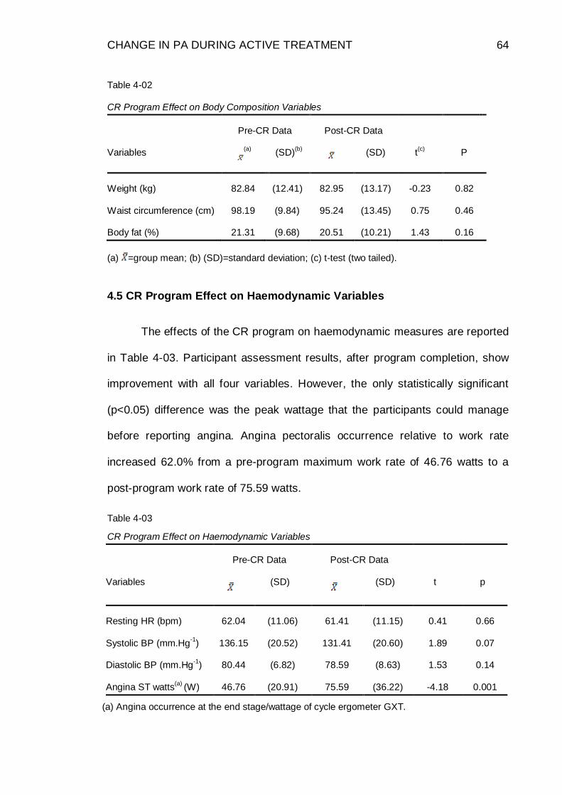
CHANGE IN PA DURING ACTIVE TREATMENT 64
Table 4-02
CR Program Effect on Body Composition Variables
Pre-CR Data Post-CR Data
Variables (a)
(SD)(b)
(SD) t(c)
P
Weight (kg) 82.84 (12.41) 82.95 (13.17) -0.23 0.82
Waist circumference (cm) 98.19 (9.84) 95.24 (13.45) 0.75 0.46
Body fat (%) 21.31 (9.68) 20.51 (10.21) 1.43 0.16
(a) =group mean; (b) (SD)=standard deviation; (c) t-test (two tailed).
4.5 CR Program Effect on Haemodynamic Variables
The effects of the CR program on haemodynamic measures are reported
in Table 4-03. Participant assessment results, after program completion, show
improvement with all four variables. However, the only statistically significant
(p<0.05) difference was the peak wattage that the participants could manage
before reporting angina. Angina pectoralis occurrence relative to work rate
increased 62.0% from a pre-program maximum work rate of 46.76 watts to a
post-program work rate of 75.59 watts.
Table.4-03
CR Program Effect on Haemodynamic Variables
Pre-CR Data Post-CR Data
Variables
(SD)
(SD) t
p
Resting HR (bpm) 62.04 (11.06) 61.41 (11.15) 0.41 0.66
Systolic BP (mm.Hg-1
) 136.15 (20.52) 131.41 (20.60) 1.89 0.07
Diastolic BP (mm.Hg-1
) 80.44 (6.82) 78.59 (8.63) 1.53 0.14
Angina ST watts(a)
(W) 46.76 (20.91) 75.59 (36.22) -4.18 0.001
(a) Angina occurrence at the end stage/wattage of cycle ergometer GXT.

CHANGE IN PA DURING ACTIVE TREATMENT 65
4.6 CR Program Effect on PF
The effects of the 3-month program on measures of CV and muscular
fitness are reported in Table 4-04. The 3-month physical exercise program had a
statistically significant (p≤0.05) effect on both measures of PF.
Table 4-04
CR Program Effect on PF
Pre-CR Data Post-CR Data
Variables
(SD)
(SD) t
P
VO2peak(ml.kg-1
.min-1
) 17.81 (4.62) 22.70 (5.60) -8.60 0.000
Power output (W) 84.63 (31.01) 122.04 (48.00) -7.10 0.000
4.7 CR Program Effect on PA
The effects of the CR program on PA variables are reported in Table 4-
05. The change in activity patterns (volume and intensity) from pre to post-
intervention, for all variables measured, is significant at the p≤0.001 level.
Table 4-05
CR Program Effect on PA
Pre-CR Data Post-CR Data
Variables
(SD)
(SD) t
p
kcal.wk-1
6457.40 (1690.90) 5621.30 (1325.30) -3.81 0.000
Sedentary 4528.20 (1259.98) 3590.44 (1077.69) 5.38 0.000
Light 1803.10 (596.43) 1871.30 (574.35) -0.66 0.001
Moderate 125.00 (121.54) 157.37 (140.73) -4.74 0.000
Vigorous 1.20 (3.41) 2.11 (3.97) -2.89 0.000

CHANGE IN PA DURING ACTIVE TREATMENT 66
The overall kilocalorie expenditure per week decreased statistically
significantly (p<0.05) at re-assessment; however, the pre to post t-test results
for calorie expenditure per week showed a ‘negative’ value. This disparity will
be discussed in more detail in Chapter 5. It is probable that this disparity
relates to the fact that participants did not wear the accelerometers for at least
1 hour, three times during re-assessment week, when training at the U-
Kinetics CR centre. The at-home light, moderate, and vigorous PA caloric
expenditure increased significantly (p<0.05) at re-assessment.
4.8 Relationship of Percentage (%) Change in PF with Pre-Intervention
Measures of PF
The correlation between the pre to post-CR program percentage (%)
change in CV fitness and pre-intervention PF variables are reported in Table
4-06. The data shows a negative correlation between percentage change
achieved in CV fitness and both pre-intervention measures of PF, although it is
not significant (p<0.05). The negative values mean that those with lower pre-
training CV and muscular fitness achieved better relative post-training
changes. In short, pre-training PF did not negatively influence CV fitness
improvements achieved.
Table 4-06
Comparison of VO2peak Change (%) with Pre-
Training Measures of PF
(%) Change
VO2peak
Variables r p
VO2peak (ml.kg-1.min
-1) -0.32 0.052
Power output (W) -0.10 0.31

CHANGE IN PA DURING ACTIVE TREATMENT 67
4.9 Relationship of Percentage (%) Change in CV Fitness with Pre and
Post-Training Measures of PA
The correlation between percentage (%) change in CV fitness and pre
and post-measures of PA are reported in Table 4-07. The data shows a larger
correlation coefficient between achieved change in CV fitness and post-
intervention PA patterns compared to pre-intervention patterns. Also, change in
fitness shows a negative correlation with pre-intervention PA intensities. All
correlations have a non-significant relationship (p>0.05). Those who made the
best improvement in cardiovascular fitness did not present with higher
participation in physical activity pre and post-intervention.
Table 4.07
Correlation of VO2 Change (%) with Pre and Post-
CR Measures of PA
Time (%) Change VO2peak
Variable r p
kcal.wk-1
T1
T2
0.21
0.31
0.14
0.06
Sedentary T1
T2
0.24
0.19
0.12
0.17
Light T1
T2
-0.03
0.04
0.45
0.43
Moderate T1
T2
-0.01
0.05
0.48
0.41
Vigorous T1
T2
-0.16
0.11
0.21
0.29
T1, pre-intervention assessment; T2, post-intervention assessment.

CHANGE IN PA DURING ACTIVE TREATMENT 68
n = 9 n = 14 n = 4n = 3 n = 14 n = 100
20
40
60
10 - 16.99 ≥17 - 22.99 ≥23
(%)
Stu
dy
Pa
rtic
ipa
nts
VO2peak (Ergo Test); (ml.kg-1.min-1)
Post CR
4.9.1 CV Fitness and PA pattern measures. The percentage of
participants with CV fitness scores below 5.0 METs (VO2peak ≤16.99), between
5.0 and 6.5 METs (VO2peak – 17.00 to 22.99), and above 6.5 METs (VO2peak
≥23) pre and post-training are graphically illustrated in Figure 4-03. Only three
participants presented with a VO2peak lower than 5.0 METs post-training
compared to nine pre-training. In contrast, 10 participants presented with a
VO2peak higher than 6.5 METs post-training compared to only four who were at
that level of pre-training.
Figure 4-03. Post-CR Program Assessment-VO2peak
Scores.
4.9.2 Relationship of CV-capacity stages with PA patterns before
and after exercise training in cardiac patients. The PF/PA pattern
relationship was investigated using two ANCOVAs, controlling for age and
gender. The aim was to determine the impact of CV fitness levels on PA
patterns. The main question was, did exercise-induced improvements in PF
strengthen the relationships between stages of CV-capacity (pre vs post-CR)
and PA patterns? The results of these ANCOVAs are presented in Table 4-08.
CV-stages became less predictive of mean VO2peak variance post-training
(68.0% (post) vs. 81.6% (pre)). Essentially, post-training CV capacity

CHANGE IN PA DURING ACTIVE TREATMENT 69
contributed less to the variance of peak oxygen uptake (VO2peak), probably due
to the migration of individual patients across fitness categories.
Concerning the PA patterns, ETA2 values indicate that post-training
CVaM-capacity contributed more to the variances of kilocalories expended per
week (25.7% vs. 9.6% (Table 4-08)) compared with pre-training CVaM-capacity.
Post-training CVaM-capacity also contributed more to the variances of
moderate PA (18.7% vs. 14.3% (Table 4-08)) and vigorous PA, EE (32.1% vs.
18.4% (Table 4-08)) than the pre-training CVaM-capacity stage.
Table 4-08
The F-ratio, p-values, ETA2
and Wilks Lambda Scores of Two ANCOVAs≠ Investigating the
Relationship of CV-capacity Stages with PA Patterns Before and After Exercise Training in
Cardiac Patients
Dependent variables ANCOVA groups F-ratio p-value ETA2
(%)
Wilks
Lambda
VO2peak (ml.kg-1.min
-1) CV-capacity stage (Pre)
CV-capacity stage (Post)
48.71
24.46
0.001
0.001
81.6
68.0
18.4
31.0
kcal.wk-1
CV-capacity stage (Pre)
CV-capacity stage (Post)
1.17
3.76
0.33
0.04
9.6
25.7
90.4
74.3
Sedentary CV-capacity stage (Pre)
CV-capacity stage (Post)
0.72
0.40
0.49
0.67
6.2
3.5
93.8
96.5
Light CV-capacity stage (Pre)
CV-capacity stage (Post)
2.67
0.09
0.13
0.91
17.1
0.9
82.9
99.1
Moderate CV-capacity stage (Pre)
CV-capacity stage (Post)
1.84
2.53
0.18
0.10
14.3
18.7
85.7
81.3
Vigorous CV-capacity stage (Pre)
CV-capacity stage (Post)
2.48
5.21
0.71
0.02
18.4
32.1
81.6
67.9
≠ - statistically controlled for sex and age.

CHANGE IN PA DURING ACTIVE TREATMENT 70
In contrast, post-training CV-capacity contributed less to the variances of
sedentary behaviour (3.5% vs. 6.2% (Table 4-08)) and light PA (0.9% vs. 17.1%
(Table 4-08)) caloric expenditure compared with pre-training CVaM-capacity.
Further dissemination of data presented in Tables 4-09 to 4-11 explains these
results.
Table 4-09
Pre and Post-Data of the Participants who had a CV Fitness < 5.9 METs at the Start and End of the Program
Variable
Time n T1 T2 P ES % change
(SD)
(SD)
VO2peak LowT1
LowT2
9
3
13.11
11.67
2.15
1.53
17.56
13.67
3.28
1.53
0.006
0.19
1.36
1.31
33.9%
17.1%
kcal.wk-1
LowT1
LowT2
9
3
6419.30
6246.80
1670.50
505.10
5754.60
5160.67
1719.10
107.80
0.31
0.001
0.39
2.15
-10.4%
-17.4%
Sedentary LowT1
LowT2
9
3
4564.90
4238.70
1164.20
715.00
3850.90
3435.00
1438.00
682.90
0.001
0.19
0.50
1.12
-15.6%
-18.9%
Light LowT1
LowT2
9
3
1805.40
1969.30
731.70
641.30
1820.60
1677.00
501.50
690.00
0.01
0.24
0.02
0.42
0.8%
-14.8%
Moderate LowT1
LowT2
9
3
49.00
38.67
54.00
47.90
83.10
48.67
44.70
43.70
0.007
0.04
0.63
0.21
69.6%
25.8%
Vigorous LowT1
LowT2
9
3
0.00
0.00
0.00
0.00
0.00
0.00
0.00
0.00
-
-
-
-
-
-
LowT1=individuals who had a VO2peak lower than 16.99 ml.kg.min-1
at the start of the program; LowT2=individuals who had a VO2peak lower than 16.99 ml.kg.min
-1 at the end of the program;
ES=Cohen’s effect size.
Sedentary behaviour decreased in all groups (Tables 4-09 to 4-11);
hence, the lower contribution of post-training CVaM-capacity (3.5% vs. 6.2%) to
the variance of sedentary behaviour in Table 4-08. Engagement in light PA
seems to have changed to more moderate PA, as evident with the re-
assessment groups performing less light, at-home PA (LowT2=14.8% decrease

CHANGE IN PA DURING ACTIVE TREATMENT 71
shown in Table 4-09, and VigT2=3.4% decrease in Table 4-11). Consequently,
the post CVaM-capacity stage contributed less to the variance of light PA than
the pre CVaM-capacity stage (0.9% vs. 17.1%), as seen in Table 4-08.
Table 4-10
Pre and Post-Data of the Participants who had a CV Fitness of between 6.0 – 6.5 METs at the Start and End of the Program
Variable
Time n T1 T2 P ES % change
(SD)
(SD)
VO2peak MidT1
MidT2
15
14
18.70
16.40
1.49
2.98
24.00
20.40
3.98
1.56
0.12
0.003
1.33
1.34
28.3%
24.4%
kcal.wk-1
MidT1
MidT2
15
14
6182.10
5840.00
1134.50
1636.90
5343.60
5447.40
1741.30
1763.60
0.12
0.001
0.48
0.22
-13.6%
-6.7%
Sedentary MidT1
MidT2
15
14
4368.60
4177.40
1427.70
1275.10
3403.50
3508.70
869.70
1306.70
0.02
0.001
0.68
0.51
-22.1%
-16.0%
Light MidT1
MidT2
15
14
1661.50
1586.10
465.30
596.10
1754.50
1829.70
500.00
469.40
0.08
0.003
0.19
0.41
5.6%
15.3%
Moderate MidT1
MidT2
15
14
151.20
76.40
125.20
59.50
183.70
108.10
157.50
66.90
0.001
0.001
0.21
0.47
21.5%
41.5%
Vigorous MidT1
MidT2
15
14
0.73
0.07
1.58
0.27
1.73
0.71
2.22
1.64
0.001
0.87
0.45
0.39
136.9%
914.3%
MidT1=individuals who had a VO2peak between 17 and 22.99 ml.kg.min-1
at the start of the program; MidT2=individuals who had a VO2peak between 17 and 22.99 ml.kg.min
-1 at the end of
the program.
Engagement in moderate at-home PA increased in all groups (Table 4-
09 to 4-11); hence, the larger contribution of post-CR CVaM-capacity to the
variance of moderate PA compared to pre-CR CVaM-capacity (18.7% vs.
14.3%). None of the participants with a VO2peak lower than 16.99 ml.kg.min-1 at
the start and end of the 12-week CR program (n=9 vs. n=3) engaged in PA
classified as vigorous. Participation in moderate PA did, however, improve
CHANGE IN PA DURING ACTIVE TREATMENT 72
markedly in those two groups (LowT1=69.6% and LowT2=25.8% (Table 4-09))
after the 12-week CR training program.
The data in Table 4-10 indicates that both the MidT1 group (n=15) who
were in the mid range fitness group at the start of the CR program, and the
MidT2 group (n=14) who were in the mid range fitness group, post-CR program,
did more vigorous PA after completing the 12-week CR program (MidT1, 1.73
kcal vs 0.73 kcal; MidT2, 0.71 kcal vs 0.07 kcal (Table 4-10)). The difference
was only statistically significant (p=0.001) with the MidT1 group. The patients in
the mid range fitness group at the end of the CR program (MidT2) were
individuals who started in the mid range group (n=8) and individuals who
migrated from the low range fitness group (n=6). This outcome influenced both
the start ( =0.07 kcal), and end ( =0.71 kcal) values, and produced a
comparatively high SD of 1.64 vs. a mean of 0.71 at T2 (Table 4-10).The result
was a statistically non-significant (p>0.05), but very dramatic percentage
change (914.3%), in the MidT2 group (Table 4-10). The resulting migration of
individuals into different groups might also explain the light PA anomaly with the
MidT2 group. The MidT2 group is the only re-assessment group that performed
greater light PA compared with pre-assessment values, as evident in Table 4-
10. In contrast, both the LowT2 and VigT2 groups performed less light PA
compared with pre-assessment values (Tables 4-09 and 4-11).
At the end of the 12-week CR training program, 10 participants were
placed in the vigorous (high range) fitness group (>6.5 METs) compared with
three participants at the start of the CR program (Table 4-11). This data
indicates that seven participants in the vigorously fit group (VigT2), at re-
assessment, migrated from the mid range fitness group. The individuals in the

CHANGE IN PA DURING ACTIVE TREATMENT 73
vigorous groups (VigT1 and VigT2) performed more at-home vigorous PA
before and after the 12-week CR training program than the participants in the
low and mid range fitness groups. This resulted in a comparatively small
percentage change compared with the two ‘mid’ groups (MidT1 and MidT2). In
summary, the data in Tables 4-08 to 4-11 indicate that at-home sedentary and
light PA kilocalorie expenditure decreased after the 12-week CR exercise
program, while moderate and vigorous PA increased.
Table 4-11
Pre and Post-Data of the Participants who had a CV Fitness >6.5 METs at the Start and End of the Program
Variable
Time n T1 T2 P ES % change
(SD)
(SD)
VO2peak VigT1
VigT2
3
10
27.30
21.60
1.53
4.12
31.70
28.70
1.16
3.57
0.88
0.03
2.88
1.70
16.1%
32.4%
kcal.wk-1
VigT1
VigT2
3
10
7948.30
7384.80
738.20
934.60
6607.30
6002.90
462.90
1465.50
0.60
0.64
1.82
0.95
-16.9%
-18.7%
Sedentary VigT1
VigT2
3
10
5216.00
5105.90
205.00
1328.50
3743.70
3751.50
964.40
858.90
0.06
0.009
1.53
1.02
-28.2%
-26.5%
Light VigT1
VigT2
3
10
2504.00
2057.00
282.90
518.60
2607.30
1987.80
777.50
706.10
0.21
0.24
0.13
0.10
4.1%
-3.4%
Moderate VigT1
VigT2
3
10
221.30
218.80
159.70
145.30
248.70
258.90
182.80
176.50
0.09
0.01
0.15
0.23
12.4%
18.3%
Vigorous VigT1
VigT2
3
10
7.00
3.10
8.66
5.19
7.67
4.70
10.00
5.46
0.13
0.001
0.07
0.29
9.6%
51.6%
VigT1=individuals who had a VO2peak higher than 23.00 ml.kg.min-1
at the start of the program; VigT2=individuals who had a VO2peak greater than 23.00 ml.kg.min
-1 at the end of the program.

CHANGE IN PA DURING ACTIVE TREATMENT 74
Chapter 5 – Discussion
5.1 Introduction
This chapter discusses the results of this investigation, and how the data
fits within current knowledge. The study aims of looking at the correlation
between pre-CR CVaM-capacity and PA EE, and what effect the CR program
intervention has on PA behaviour, are discussed separately. The limitations of
this study are discussed, and recommendations are made, for the further
development of research consequent to these findings. The relevant clinical
application of understandings gained in the research is outlined, and
conclusions given, in summation of the implications of this study.
In line with the study aims, the main findings of this investigation suggest
that cardiac patients’ CVaM-capacity is associated with their PA behaviour and
that PA behaviour changes favourably over the period of CR intervention.
These findings seem to support the hypothesis that CVaM-capacity is a
predictor of PA habits/patterns and that CR intervention facilitates a change in
at-home PA habits. A more detailed analysis of these findings is discussed in
Sections 5.2 and 5.3.
5.2 Relationship of Pre-CR CVaM-capacity with Pre-CR PA Patterns
5.2.1 Volume of daily and weekly caloric expenditure. The first aim of
this study was to observe whether the pre-rehabilitation functional ability of
cardiac patients correlated with their pre-rehabilitation volume and quality of at-
home PA. Does the fitter (VO2peak and power output) cardiac patient perform
more activity, and with greater intensity of effort, compared to the less fit patient
before joining a CR program? Results from this analysis indicate a significant

CHANGE IN PA DURING ACTIVE TREATMENT 75
relationship between CVaM-capacity and overall at-home PA behaviour (Table
4-01). Patient power output and peak oxygen consumption before the CR
program showed a moderate, positive correlation with overall at-home caloric
expenditure per week (r=0.47 and 0.53). The calculated r2 values indicate that
power output and VO2peak contribute, respectively, 22.1% and 28.1% of the
variance of weekly PA energy consumption.
No investigation was found that reported on the pre-CR CVaM-
capacity/PA behaviour relationship of cardiac patients. Eaton’s (1992; 1992a)
meta-analyses of the risk of CVD development, relative to PA and CVaM-
capacity, shows close similarities to this investigation. Similarities include: (a)
both studies investigated at-home PA (kilocalories expended per intensity
category), and CV-capacity (cardiorespiratory evaluation); and (b) both studies
looked at the PA/CVaM-capacity interrelationship. The differences between the
investigations were that: (a) Eaton analysed healthy individuals who had
developed CVD in longitudinal investigations, opposed to individuals who
already had CVD and (b) in Eaton’s analyses, multiple epidemiological studies
were assessed, which reflected larger subject numbers. Eaton’s (1992; 1992a)
investigations are somewhat relevant, considering the lack of direct subject
matter data. Further, a pre vs. post-CVD development comparison should
theoretically show a correlative association, whereas this study may be implied
as a continuance of Eaton’s investigations.
Both of the above-mentioned meta-analyses treated PA and CVaM-
capacity as variables independent of other RF confounders (Eaton, 1992;
Eaton, 1992a). As independently assessed variables, both PA and CVaM-
capacity exhibited a strong inverse relationship with CHD. These two studies
CHANGE IN PA DURING ACTIVE TREATMENT 76
(Eaton, 1992; Eaton, 1992a) essentially reported weak to moderate correlations
(r=0.02 to r=0.44) between PA intensity and CVaM-capacity across the
individual epidemiologic studies they analysed. This discrepancy of correlation
data may be attributable to individual study differences, given the inter-study
disparity of variables assessed and the incorrect classification of PA (Eaton,
1992a).
5.2.2 Intensity of at-home PA. Peak oxygen consumption correlates
statistically significantly (p<0.05) with moderate (r=0.48) and vigorous (r=0.59)
at-home caloric expenditure (Table 4-01). In contrast, VO2peak correlates weakly
with sedentary (r=0.13) and light PA (r=0.32), as indicated in Table 4-01. A
similar trend is seen with power output, although correlation coefficients ranged
from a weak to moderate positive (vigorous, r=0.38), to a weak negative
(sedentary, r=-0.03) correlation. This trend seems to indicate that the higher the
level of CV-fitness the more likely individuals are to engage in moderate and
vigorous at-home activity and that they are, compared to those with lower levels
of fitness, less sedentary and less engaged in light activity.
Eaton (1992a) suggests that CVaM-capacity change may be attributable
to genetic disposition, and/or disability (either disassociated with or due to
sedentarism), rather than PA correlated change (Eaton, 1992a). The patient’s
physical capacity to exercise or engage in PA may be inhibited, not only by
cardiovascular fragility (post-MI weakness or myocardial injury) but also by an
actual muscular-skeletal injury. PA is known to maintain muscle tone, inhibit
enervation, and facilitate joint mobility, all mechanisms that aid CVaM-capacity
maintenance/improvement (McArdle et al., 2010). As an implication, the longer
the patient stays physically inactive after a cardiac event, the higher the

CHANGE IN PA DURING ACTIVE TREATMENT 77
likelihood of a vicious cycle where chronic inactivity facilitates progressive
functional disability and vice versa. In the current study, maximum cycle power
output (which is influenced not only by physical inactivity but also by factors
such as leg strength, body weight, and cardiovascular discomforts, like
tiredness or angina) correlates slightly less with PA patterns than VO2peak. The
fact that VO2peak correlates slightly better with PA patterns than power output
suggests that PA patterns might be influenced more by cardiovascular ability
than muscular ability in this group of participants. Body weight is a confounding
variable that needs to be considered as well because it influences power output
positively and oxygen consumption negatively. It can be assumed that the
chronically inactive patient will be less able to perform activity due to functional
disability at the initial stages of CR intervention. However, there is sparse
research that investigates the contribution of musculoskeletal factors to
functional/training disability in cardiac patients. Improved understanding may
facilitate the development of guidelines that directly address the debilitating
PA/PF corrosion cycle and help formulate potentially superior treatment
strategies and recovery outcomes for cardiac patients.
5.3 The Effect of the CR Program on Clinical Variables
5.3.1 Physical fitness/cardiovascular capacity/functional capacity.
CR is shown to improve PF, with an incurred effect for CVD RFs, disease
progression/regression, and reduced mortality (Lear & Ignaszewski, 2001).
Figure 4-03 depicts the CVaM-capacity change occurring within the study
population over the course of the CR program. A comparison of pre to post-CR
peak oxygen uptake shows a significant improvement of VO2peak scores. The
mean VO2peak score increased from 17.81 to 22.70 ml.kg.min-1. The mean

CHANGE IN PA DURING ACTIVE TREATMENT 78
maximum wattage that patients could handle during the last stage of the GXT
increased from 84.6 watts to 122.0 watts. Patients with a VO2peak of 40
ml.kg.min-1 (~5% probability for all-cause, and ~2% for CV mortality) showed an
80% lower all-cause, and a 90% lower cardiovascular mortality risk, compared
to patients with a VO2peak of 12 ml.kg.min-1 (~25% probability for all-cause, and
~22% for CV mortality) (Vanhees, Fagard, Thijs, Staessen, & Amery, 1994).
5.3.2 Body composition. The CR intervention program had a small
effect on body composition variables. The changes were, however, not
statistically significant (p>0.05). During the 12-week intervention program,
dietary interventions were not enforced; therefore, no effect on body weight/
BMI was expected. Two clinical studies of similar duration (~3 months) that
measured patient weight loss after CR training showed an overall mean weight
loss of 0.6% (Brochu, Poehlman, Savage, Fragnoli-Munn, & Ades, 2000), and a
0.6% loss for non-obese patients (BMI=25.0–29.9 kg.m-2) compared to a 1.8%
loss for obese patients (BMI=30.0–39.9 kg.m-2) (Bader, Maguire, Spahn,
O’Malley, & Balady, 2001), respectively. In the current study cohort, only a small
number of subjects could be classified obese pre-CR. For instance, only four
respondents met the criteria for Stage 1 (30.0–34.9 kg.m-2) obesity, and only
two respondents could be classed as Stage 2 (35.0–39.9 kg.m-2) obese.
Additionally, the results in Table 4-02 are reflective of the data spread between
gender groups (weight – (male; (m)) = 84.89 kg-1 vs. (female; (f)) = 76.97 kg-
1; body fat (%) – (m) = 17.28% vs. (f) = 32.81%).
5.3.3 Haemodynamics. Patient haemodynamics changed positively but
mostly non-significantly (p>0.05) over the course of the 12-week CR program.
Results in Table 4-03 show a positive effect on resting HR and BP. Most

CHANGE IN PA DURING ACTIVE TREATMENT 79
importantly regarding the potential effect on exercise behaviour was the
statistically significant effect of the program on the wattage at which participants
reported angina, which increased from 46.80 watts to 75.60 watts.
What this data indicates is that patients were physically lighter, markedly
fitter, and had lowered susceptibility to angina (managing more exercise before
experiencing angina) during PA after the 12-week intervention program. It is
therefore not unrealistic to propose/expect that the above-mentioned physical
changes should impact positively on at-home exercise behaviour.
5.4 The Effect of a CR Program on PA Patterns
The second aim of this study was to observe whether the cardiac
patients at-home PA patterns changed during/after completion of the 12-week
supervised CR program. . Results measuring the change in PA patterns
(volume and intensity) showed significant statistical change at the p≤0.001 level
(Table 4-05). Light, moderate, and vigorous at-home PA increased by 101.48
kcal.wk-1 after the 12-week supervised CR program. In contrast, overall EE
decreased by 836.10 kcal.wk-1 (6457.40 – 5621.30=836.10 kcal.wk-1), with
sedentary EE decreasing by 937.76 kcal.wk-1 (4528.20 – 3590.44=937.76
kcal.wk-1 (Table 4-05)). A deficit is apparent when calculating the total decrease
in EE (pre to post-CR=836.10 kcal.wk-1). This deficit (836.10 kcal.wk-1) can be
explained by the exclusion of 3 hours of accelerometer recording during re-
assessment. The PA behaviour re-assessment (second accelerometer wearing)
occurred during the last week of the 12-week supervised CR intervention at U-
Kinetics. Participants were instructed not to wear the accelerometers during the
3 weekly training sessions at U-Kinetics because the aim was to only monitor
changes in at-home PA.

CHANGE IN PA DURING ACTIVE TREATMENT 80
The data presented in Tables 4-08 to 4-11 show that changes obtained
with both VO2peak and PA behaviour are not significantly negated by the
patients’ starting CVaM-capacity. Participants present with reduced sedentary
EE in all fitness categories at re-assessment, with the highly fit groups (VigT1
and VigT2) achieving higher percentage reductions (28.2% and 26.5% (Table 4-
11) vs 15.6% and 18.9% (Tables 4-09)). The three most fragile individuals (pre
and post-CR, VO2peak <17.00 ml.kg.min-1) presented with an 18.9% reduction in
sedentary EE and a 25.8% increase in moderate at-home PA (Table 4-09). The
corresponding sedentarism reduction and increased moderate PA indicates an
increased confidence and ability to engage in activity at home after a
supervised CR program, including those with the most restrictive CVaM-
capacity. It is also important to note that the 12-week CR program produced a
nearly identical percentage change in VO2peak for participants who had the
lowest (LowT2=17.1% (Table 4-09)), and highest (VigT1=16.1% (Table 4-11))
VO2peak values at the start of the program.
None of the participants who started in the low fitness group (VO2peak
<17.00ml.kg.min-1) engaged in vigorous PA during pre and post-assessment.
This lack of behaviour change indicates that the participants stayed within their
physical limitations when engaging in PA at home. This is a significant result
within the context of CR. Considering that the low fitness group engaged in
more moderate PA, but performed no vigorous PA, indicates that from a
psychological perspective the program positively affected participant confidence
to engage in PA and helped produce a better understanding of how to do so
safely. From a physiological perspective, it is important to note that 66.7% of
participants (n=6) who started in the low CV-capacity group (VO2peak <17.0

CHANGE IN PA DURING ACTIVE TREATMENT 81
ml.kg.min-1) migrated to the moderately fit group, presenting with a 33.9% mean
increase in CV-capacity at re-assessment. This group also achieved a 69.6%
increase in moderate PA, indicating that although physiologically restricted, they
made significant improvements with both CVaM-capacity and at-home PA
engagement.
Participants in the highly fit groups (VigT1 and VigT2) evidenced the
highest at-home engagement in vigorous PA before and after the 12-week
rehabilitation program. This data shows that the exercise capacity of a cardiac
patient does impact on the quality (intensity and volume) of PA that the patient
performs pre and post-supervised CR program. Overall the VO2peak percentage
change did not correlate significantly with re-assessment PA behaviour for the
entire study population (Table 4-07). The pre to post-change in VO2peak showed
a negative correlation with pre-intervention PA patterns. This negative
relationship indicates that ‘physiological trainability’ was associated with pre-
training PA patterns, but was not the primary determinant of post-CR at-home
PA-patterns.
Schairer et al. (1998) found that only 17.0% of CR patients maintained
exercise above levels that promote health (300 kilocalories per session).
Similarly, Savage et al. (2000) found caloric expenditure per CR session was
inadequate, and in both studies was reflective of insufficient weekly caloric
expenditure. The data presented in this thesis seems to suggest that it is not
principally EE that produces these outcomes, but the fact that the patients
learned how to challenge their CV systems safely with exercise. A 5-year follow-
up of the Schairer et al. (1998) study showed that 72.0% of participants
expended ~1000 kcal.wk-1, with 43.0% of participants performing over 1500

CHANGE IN PA DURING ACTIVE TREATMENT 82
kcal.wk-1 (Schairer et al., 2003). This long-term outcome suggests that patients
might over time increase their EE with exercise as their fitness and confidence
increases.
5.5 Limitations
5.5.1 Sample size. A small sample size is noted (n=27) and was
expected, due to the ‘real time’ rehabilitation management of the participants,
over the period of data collection. The overall goal of this study was to
investigate the effectiveness of a supervised CR program on CVaM-capacity
measures and at-home PA-patterns. In this context, data collected here will only
support a generalised interpretation.
5.5.2 Accelerometry. The methodology outlines the viability of
accelerometry PA measurement. One limitation of accelerometry is patient
adherence to wearing protocols. It is uncertain whether the patients wore the
accelerometer continuously over the period of assessment. Although a
minimum wear time was imposed, to be counted as a recorded day, this is not a
true reflection of habit. A logbook was issued, to improve patient compliance to
the wear protocols and to record hourly PA and periods of sleep. Good
compliance was seen with logbook recordings, although a small number of
recordings lacked sufficient detail to be included for analysis. Incidentally, it is
uncertain whether the initial recording period and CR program produced a
learning curve on the bias of the post-assessment wear procedure. Additionally,
in an attempt to mitigate for subjective recording error the patients were asked
to ‘’maintain normal activity patterns and routine, avoiding the tendency to over-
state activity levels just because they were documenting it and were wearing

CHANGE IN PA DURING ACTIVE TREATMENT 83
the accelerometer’. However, it is acknowledged that the instruction to ‘portray’
a behavior may have influenced the patient’s PA patterns.
The primary aim of this study was to investigate the effect of the
supervised CR program on at-home PA-patterns. Patients were asked not to
wear the accelerometer during training at the CR centre in the last week of the
12-week program when re-assessment was done. This produced 3 hours (3
training sessions per week, 1 hour per session) less wear time per week, and
affected the total amount of kilocalorie expenditure for the re-assessment week.
It is also possible that patients reduced their at-home vigorous activity in that
last week because they considered that they had already performed their
weekly high-quality training at the CR centre. In hindsight, it may have been
better to apply the accelerometer re-assessment in the first week after
cessation of the 12-week training program. The reason why the reassessment
was done in the last week of supervised training relates to preventing
equipment/data loss. Also, some of the poor CR responders traditionally get a
re-referral, meaning they do not stop but continue their training at the CR centre
for another 12 weeks. In that case, the poor respondent data would then have
been rejected as part of the final analysis. The poor respondent data provided
some of the best insights and, fortunately, was not lost to analysis.
5.5.3 Definition of Intensity. It is possible that patients classified with a
low range of fitness did not migrate to the vigorously fit group due to a
difference in objective and subjective definitions of intensity. Objective
parameters were absolutely defined, with intensity levels graduated within a
progressive and fixed range. This may not have reflected the perceived intensity

CHANGE IN PA DURING ACTIVE TREATMENT 84
of activity that the low fitness patient may be experiencing. This disparity will
also skew results, although the extent of bias has yet to be quantified.
5.5.4 PF Assessment Protocol. The only protocol available to this
project was cycle ergometry assessment which allowed the examiner to
measure the patient’s physiological response to exercise from a stable platform
without impeding correct biomechanical movement. Additionally, the burden of
body weight was minimised by sitting, and the patient could self-supported
using the handle bar. However, the more appropriate protocol to use is a
treadmill assessment, where the patient bears their full body weight, whilst
employing the biomechanical movement of walking, typically the preferred
mode of locomotion in daily life compared with cycling. Future assessment,
where possible, should employ measurement protocols specific to patient PA
behavior.
5.5.5 Individual vs. general application. A disparity between each
patient’s results for the measurement of PA habits relative to levels of PF
inhibits the generalisation of data. The nature of CR intervention within this
study was to treat each cardiac patient individually according to their particular
affliction and functional ability. Attenuation of disease types to a generalised
CVD categorisation, and identifying mean (SD) and correlation average,
allowed for a very general interpretation of results. To be able to apply learned
outcomes to the individual patient would require blinded clinical testing, with
compared groups pair matched to solve the problem of association. This task
will prove to be difficult given the numerous RF variable associations requiring
investigation. Currently, available research has only investigated major

CHANGE IN PA DURING ACTIVE TREATMENT 85
associations, where more detail that is unique to all RFs is necessary to be
applicable to the individual patient's intervention program.
5.6 Future Research
This study investigated measurement techniques for assessing PA
habits/patterns. No clear association (trend) is shown between participant
scores, which inhibits any ‘generalisation’ of results to the wider CVD
population. The task of forming generalised concepts requires the identification
of common occurrences that factor in the CVD cause and effect paradigm. A
comprehensive investigation of RF aligned assessment/testing protocols is
needed to identify the individual level of exposure per RF. While it is assumed
that our current understanding of CVD risk factors is comprehensive, these
factors only describe genetic, habitual and physiological associations. Other
variables should be included in successive investigations to assess levels of
influence. The encompassing model of care, Te Whare Tapawha, identifies a
holistic approach to wellbeing. While we assessed taha tinana (physical,
physiological) variables for intervention effect, taha wairua (spiritual), taha
hinengaro (mental and emotional), and taha whānau (support elements, family,
environment), aspects of health and wellbeing also require interventive
investigation.
The design of the CR program must align with the goals and outcomes
sought for the patient. A review of topical research attests to the benefits
attending a CR program has for the cardiac patient’s morbidity and mortality
risk; however, questions raised by our study require further investigation.
Although a strong to moderate relationship is shown between PA and PF at
high intensities (moderate to vigorous PA), the question remains, why is this not

CHANGE IN PA DURING ACTIVE TREATMENT 86
the case at the lower intensities (sedentary to low PA)? Given that greater
dose/response is seen with PF at these lower intensities (Fig. 2.01), what other
factors/variables are responsible aside from PF? The CEP cannot assume that
low PA will reflect relatively low CVaM-capacity. Current research lacks
sufficient information to facilitate a detailed investigation of program intervention
characteristics for treating each individual RF. The ability to identify how, or if,
these features contribute to the causality of achieved outcomes will facilitate the
construction of a highly effective CR program.
5.7 Clinical Applications
This study is an analysis of an operational CR clinic, with specific
intervention protocols applied to the CR patient. The results of this study are
directly applicable to the CR program and show that a peripheral investigation
of RFs may show a greater correlation with low CVaM-capacity, although these
RFs have yet to be identified. Therefore more appropriate assessment tools
may be required to identify the more appropriate RF. The results achieved here
are a progressive step toward a refined, accepted CR protocol that directly
addresses causality.
5.8 Conclusion
In summary, this study investigated whether the cardiac patient ’s CVaM-
capacity is associated with their PA behaviour before and during CR program
participation. The results of the investigation show a moderate to strong
association between the patient’s at-home volume of activity and their CVaM-
capacity. Also, the total time spent at the higher intensities of activity (moderate
to vigorous) seems to predicate CVaM-capacity. In contrast, the time spent at

CHANGE IN PA DURING ACTIVE TREATMENT 87
light to no activity was not predictive of CVaM-capacity. This seems to indicate
a causal relationship between CV-ability and the intensity of at-home PA in
cardiac patients. However, more research is required to clarify whether there is
indeed a causal relationship.
This study also investigated whether CR program participation has an
effect on changing a cardiac patient’s PA patterns. Investigation results show a
significant increase in PA habits and a significant decrease in sedentary
behaviour during involvement in a CR program. Unless the cardiac patient
presents with serious pathological trainability issues (abnormal physiological
response during light and moderate exercise), they should be encouraged to
perform moderately intense activity/exercise extra to CR to achieve greater
health benefits and improve CVaM-capacity.
The results of this study show encouraging health-related trends;
however, gaps in understanding still exist. Barriers to CR participation should be
minimised to expedite an epidemiological and multi-clinical investigation that
isolates individual RF impact on CVD. Data gained from multi-clinical research,
when applied, will improve our understanding of the disease and maximise
health benefits for the patient.

CHANGE IN PA DURING ACTIVE TREATMENT 88
References
Ainsworth, B., Haskell, W., Whitt, M., Irwin, M., Swartz, A., Strath, S., . . . Leon,
A. (2000). Compendium of physical activities: An update of activity codes
and MET intensities. Medicine and Science in Sports and Exercise, 32 (9
Suppl): S498-504.
American Association of Cardiovascular and Pulmonary Rehabilitation. (2004).
Guidelines for Pulmonary Rehabilitation and Secondary Prevention
Programs (4th ed.). Champaign, IL: Human Kinetics.
ACSM. (2003). ACSM's Exercise Management for Persons with Chronic
Diseases and Disabilities (2nd ed.). Champaign, IL: Human Kinetics.
ACSM. (2010). ACSM's Health-Related Physical Fitness Assessment Manual
(3rd ed.). Baltimore, MD: Lippincott, Williams & Wilkins.
ACSM. (2013). ACSM's Guidelines for Exercise Testing and Prescription (9th
ed.). Baltimore, MD: Lippincott Williams & Wilkins.
Arrigo, I., Brunner-LaRocca, H., Lefkovits, M., Pfisterer, M., & Hoffmann, A.
(2008). Comparative outcome one year after formal cardiac
rehabilitation: The effects of a randomised intervention to improve
exercise adherence. European Journal of Cardiovascular Prevention &
Rehabilitation, 15(3), 306-311.
Ashworth, N. L., Chad, K. E., Harrison, E. L., Reeder, B. A., & Marshall, S. C.
(2005). Home versus centre based physical activity programs in older
adults. Cochrane Database of Systematic Reviews.

CHANGE IN PA DURING ACTIVE TREATMENT 89
Ayabe, M., Brubaker, P. H., Dobrosielski, D., Miller, H. S., Ishi, K., Yahiro, T., …
Tanaka, H. (2004). The physical activity patterns of cardiac rehabilitation
program participants. Journal of Cardiopulmonary Rehabilitation, 24(2),
80-86.
Bader, D.S., Maguire, T.E., Spahn, C.M., O’Malley, C.J., & Balady, G.J. (2001).
Clinical profile and outcomes of obese patients in cardiac rehabilitation
stratified according to National Heart, Lung, and Blood Institute criteria.
Journal of Cardiopulmonary Rehabilitation, 21(4), 210-217.
Baechle, T. R., & Earle, R. W. (2008). Essentials of strength training and
conditioning (3rd ed.). Champaign, IL: Human Kinetics.
Balady, G. J., Ades, P. A., Comoss, P., Limacher, M., Pina, I. L., Southard, D.,
… Bazzarre, T. (2000). Core components of cardiac
rehabilitation/secondary prevention programs: A statement for healthcare
professionals from the American Heart Association and the American
Association of Cardiovascular and Pulmonary Rehabilitation Writing
Group. Circulation, 102(9), 1069-1073.
Balady, G. J., Arena, R., Sietsema, K., Myers, J., Coke, L., & Fletcher, G. F.
(2010). Clinician’s guide to cardiopulmonary exercise testing in adults: A
scientific statement from the American Heart Association. Circulation,
122(2), 191-225.
Balady, G.J., Fletcher, B., Froelicher, E., Hartley, H., Krauss, R., Oberman, A., .
. . Taylor, B. (1994). Cardiac rehabilitation programs. A statement for
healthcare professionals from the American Heart Association.
Circulation, 90(3), 1602-1610.

CHANGE IN PA DURING ACTIVE TREATMENT 90
Balady, G. J., Williams, M. A., Ades, P. A., Bittner, V., Comoss, P.,
Foody, J. M., … Southard, D. (2007). Core components of cardiac
rehabilitation/secondary prevention programs: 2007 Update: A scientific
statement from the American Heart Association Exercise, Cardiac
Rehabilitation, and Prevention Committee, the Council on Clinical
Cardiology; the Councils on Cardiovascular Nursing, Epidemiology and
Prevention, and Nutrition, Physical Activity, and Metabolism; and the
American Association of Cardiovascular and Pulmonary Rehabilitation.
Circulation, 115(20), 2675-2682.
Bjarnason-Wehrens, B., Mayer-Berger, W., Meister, E., Baum, K.,
Hambrecht, R., & Gielen, S. (2004). Recommendations for resistance
exercise in cardiac rehabilitation: Recommendations of the German
Federation for Cardiovascular Prevention and Rehabilitation. European
Journal of Cardiovascular Prevention & Rehabilitation, 11(4), 352-361.
Boerema, S., Van Velsen, L., Schaake, L., Tönis, T., & Hermens, H. (2014).
Optimal sensor placement for measuring physical activity with a 3D
accelerometer. Sensors, 14(2), 3188-3206.
Brochu, M., Poehlman, E.T., Savage, P., Fragnoli-Munn, K., Ross, S; & Ades,
P.A. (2000). Modest effects of exercise training alone on coronary risk
factors and body composition in coronary patients. Journal of
Cardiopulmonary Rehabilitation, 20(3),180-188.
Brubaker, P. (2014). Steps for improving physical activity orientation among
health-care providers of older cardiovascular patients. Current Geriatrics
Reports, 3(4), 291-298.

CHANGE IN PA DURING ACTIVE TREATMENT 91
Buman, M. P., Hekler, E. B., Haskell, W. L., Pruitt, L., Conway, T. L.,
Cain, K. L., … King, A. C. (2010). Objective light-intensity physical
activity associations with rated health in older adults. American Journal of
Epidemiology, 172(10), 1155-1165.
Carter, J. (1982). Physical structure of Olympic athletes, Part I: The Montreal
Olympic games anthropological project. Basel, Switzerland: Karger.
Casperson, C. J., Powell, K. E., & Christenson, G. M. (1985). Physical activity,
exercise, and physical fitness: Definitions and distinctions for health
related research. Public Health Reports, 100(2), 126–131. Retrieved
from https://www.ncbi.nlm.nih.gov/pmc/articles/PMC1424733/pdf/pub
healthrep00100-0016.pdf
Center for Medicare & Medicaid Services. (2010). Cardiac and intensive cardiac
rehabilitation: Medical policy article (a45888). Retrieved from http://apps.
ngsmedicare.com/sia/ARTICLE_A45888.htm
Champion, V., & Skinner, C. (2008). The health belief model. In K. Glanz, B.
Rimer, & K. Viswanath (EDs.), Behaviour and health education: Theory,
research, and practice (pp. 45-65). San Francisco, CA: Jossey-Bass.
Chen, K.Y., & Basset, D.R. (2005). The technology of accelerometry-based
activity monitors: Current and future. Medicine & Science in Sports &
Exercise, 37(Supplement), S490-S500.
Craig, C. L., Marshall, A. L., Sjostrom, M., Bauman, A. E., Booth, M. L.,
Ainsworth, B. E., … Oja, P. (2003). International Physical Activity
Questionnaire: 12-Country reliability and validity. Medicine & Science in
Sports & Exercise, 35(8), 1381-1395.

CHANGE IN PA DURING ACTIVE TREATMENT 92
Doubly-labelled Water Resource Centre (DLWRC). (n.d.). Measuring energy
expenditure using the doubly labelled water method. Retrieved from
https://www.abdn.ac.uk/energeticsresearch/documents/Technical_Paper
_905_DLW_Energy_Expend.pdf.
Dunlay, S. M., Witt, B. J., Allison, T. G., Hayes, S. N., Weston, S. A.,
Koepsell, E., & Roger, V. L. (2009). Barriers to participation in cardiac
rehabilitation. American Heart Journal, 158(5), 852-859.
Eaton, C. (1992). Relation of physical activity and cardiovascular fitness to
coronary heart disease, Part I: A meta-analysis of the independent
relation of physical activity and coronary heart disease. Journal of the
American Board of Family Practice, 5(1), 31-42.
Eaton, C. (1992a). Relation of physical activity and cardiovascular fitness to
coronary heart disease, Part II: Cardiovascular fitness and the safety and
efficacy of physical activity prescription. Journal of the American Board of
Family Practice, 5(2), 157-165.
Evenson, K. R., & Terry, J. W. (2009). Assessment of differing definitions of
accelerometer nonwear time. Research Quarterly for Exercise and Sport,
80(2), 355-362.
Fletcher, G. F., Balady, G., Blair, S. N., Blumenthal, J., Caspersen, C.,
Chaitman, B., … Pollock, M. L. (1996). Statement on exercise: Benefits
and recommendations for physical activity programs for all Americans: A
statement for health professionals by the Committee on Exercise and
Cardiac Rehabilitation of the Council on Clinical Cardiology, American
Heart Association. Circulation, 94(4), 857-862.

CHANGE IN PA DURING ACTIVE TREATMENT 93
Fokkenrood, H. J., Bendermacher, B. L., Lauret, G. J., Willigendael, E. M.,
Prins, M. H., & Teijink, J. A. (2013). Supervised exercise therapy versus
non-supervised exercise therapy for intermittent claudication. Cochrane
Database of Systematic Reviews.
Giannuzzi, P., Saner, H., Bjornstad, H., Fioretti, P., Mendes, M., Cohen-Solal,
A., . . . Veress, G. (2003). Secondary prevention through cardiac
rehabilitation position paper of the Working Group on Cardiac
Rehabilitation and Exercise Physiology of the European Society of
Cardiology. European Heart Journal, 24(13), 1273-1278.
Giannuzzi, P., Temporelli, L., Marchioli, R., Maggioni, A., Balestroni, G., Ceci,
V., . . . Vanuzzo, D. (2008). Global secondary prevention strategies to
limit event recurrence after myocardial infarction: Results of the GOSPEL
study, a multicenter, randomised controlled trial from the Italian Cardiac
Rehabilitation Network. Journal of the American Medical Association-
Internal Medicine, 168(20), 2194-2204.
Grace, S. L., Abbey, S. E., Shnek, Z. M., Irvine, J., Franche, R., &
Stewart, D. E. (2002). Cardiac rehabilitation II. General Hospital
Psychiatry, 24(3), 127-134.
Grace, S. L., Krepostman, S., Brooks, D., Jaglal, S., Abramson, B. L.,
Scholey, P., … Stewart, D. E. (2006). Referral to and discharge from
cardiac rehabilitation: Key informant views on continuity of care. Journal
of Evaluation in Clinical Practice, 12(2), 155-163.
Grace, S.L., Scholey, P., Suskin, N., Arthur, H., Brooks, D., Jaglal, S., …
Stewart, D. (2007). A prospective comparison of cardiac rehabilitation

CHANGE IN PA DURING ACTIVE TREATMENT 94
enrollment following automatic vs. usual referral. Journal of Rehabilitation
Medicine, 39(3), 239-245.
Grace, S. L., Shanmugasegaram, S., Gravely-Witte, S., Brual, J., Suskin, N., &
Stewart, D. E. (2009). Barriers to cardiac rehabilitation: Does age make a
difference? Journal of Cardiopulmonary Rehabilitation and Prevention,
29(3), 183-187.
Guidon, M., & McGee, H. (2012). One-year effect of a supervised exercise
program on functional capacity and quality of life in peripheral arterial
disease. Disability and Rehabilitation, 35(5), 397-404.
Hagströmer, M., Oja, P., & Sjöström, M. (2006). The International Physical
Activity Questionnaire (IPAQ): A study of concurrent and construct
validity. Public Health Nutrition, 9(6), 755-762.
Hambrecht, R., Niebauer, J., Marburger, C., Grunze, M., Kälberer, B.,
Hauer, K., … Schuler, G. (1993). Various intensities of leisure time
physical activity in patients with coronary artery disease: Effects on
cardiorespiratory fitness and progression of coronary atherosclerotic
lesions. Journal of the American College of Cardiology, 22(2), 468-477.
Haskell, W. L., Alderman, E. L., Fair, J. M., Maron, D. J., Mackey, S. F.,
Superko, H. R., … Krauss, R. M. (1994). Effects of intensive multiple risk
factor reduction on coronary atherosclerosis and clinical cardiac events
in men and women with coronary artery disease. The Stanford Coronary
Risk Intervention Project (SCRIP). Circulation, 89(3), 975-990.
Haskell, W.L., Lee, I., Pate, R.R., Powell, K., Blair, S.N., Franklin, B.A., . . .
Bauman, A. (2007). Physical activity and public health: Updated

CHANGE IN PA DURING ACTIVE TREATMENT 95
recommendation for adults from the American College of Sports
Medicine and the American Heart Association. Circulation, 116(9), 1081-
1093.
Haskell, W.L., Montoye, H.J., & Ornstein, D. (1985). Physical activity and
exercise to achieve health-related physical fitness components. Public
Health Reports, 100(2), 202-212. Retrieved from https://www.ncbi.nlm.
nih.gov/pmc/articles/PMC1424730/pdf/pubhealthrep00100-0092.pdf
Helmerhorst, H.J; Brage, S; Warren, J; Besson, H; & Ekelund; U. (2012). A
systematic review of reliability and objective criterion-related validity of
physical activity questionnaires. International Journal of Behavioural
Nutrition and Physical Activity, 9:103, 1-55.
Higginson, R. (2008). Women and cardiac rehab: Overcoming the barriers.
British Journal of Nursing, 17(22), 1380-1381.
Hughes, A. R., Mutrie, N., & MacIntyre, P. D. (2007). Effect of an exercise
consultation on maintenance of physical activity after completion of
phase III exercise-based cardiac rehabilitation. European Journal of
Cardiovascular Prevention & Rehabilitation, 14(1), 114-121.
Janssen, V., De Gucht, V., Van Exel, H., & Maes, S. (2012). Changes in illness
perceptions and quality of life during participation in cardiac
rehabilitation. International Journal of Behavioral Medicine, 20(4), 582-
589.
Janssen, V., De Gucht, V., Van Exel, H., & Maes, S. (2013). Beyond
resolutions? A randomised controlled trial of a self-regulation lifestyle

CHANGE IN PA DURING ACTIVE TREATMENT 96
program for post-cardiac rehabilitation patients. European Journal of
Preventive Cardiology, 20(3), 431-441.
Kerr, J., Sallis, J. F., Owen, N., De Bourdeaudhuij, I., Cerin, E., Sugiyama, T.,
… Bracy, N. (2013). Advancing science and policy through a coordinated
international study of physical activity and built environments: IPEN adult
methods. Journal of Physical Activity and Health, 10(4), 581-601.
King, M.L., Williams, M.A., Fletcher, G.F., Gordon, N.F., Gulanick, M., King,
C.N., . . . Wenger, N.K. (2005). Medical Director responsibilities for
outpatient cardiac rehabilitation/secondary prevention programs: A
scientific statement from the American Heart Association/American
Association for Cardiovascular and Pulmonary Rehabilitation. Circulation,
112(21), 3354-3360.
Lavie, C. J., Milani, R. V., & Littman, A. B. (1993). Benefits of cardiac
rehabilitation and exercise training in secondary coronary prevention in
the elderly. Journal of the American College of Cardiology, 22(3), 678-
683.
Lear, S. A., & Ignaszewski, A. (2001). Cardiac rehabilitation: A comprehensive
review. Current Controlled Trials in Cardiovascular Medicine, 2(5), 221-
232.
Lear, S.A., Ignaszewski, A., Linden, W., Brozic, A., Kiess, M., Spinelli, J.J., . . .
Frohlich, J.J. (2003). The Extensive Lifestyle Management Intervention
(ELMI) following cardiac rehabilitation trial. European Heart Journal,
24(21), 1920-1927.

CHANGE IN PA DURING ACTIVE TREATMENT 97
Maddison, R., Ni Mhurchu, C., Jiang, Y., Vander Hoorn, S., Rodgers, A.,
Lawes, C. M., & Rush, E. (2007). International Physical Activity
Questionnaire (IPAQ) and New Zealand Physical Activity Questionnaire
(NZPAQ): A doubly labelled water validation. International Journal of
Behavioral Nutrition and Physical Activity, 4(1), 62.
Magalhães, S., Miguel Ribeiro, M., Barreira, A., Fernandes, P., Torres, S.,
Lopes Gomes, J., & Viamonte, S. (2013). Long-term effects of a cardiac
rehabilitation program in the control of cardiovascular risk factors.
Revista Portuguesa de Cardiologia (English Edition), 32(3), 191-199.
Malliou, P., Fatouros, I., Beneka, A., Gioftsidou, A., Zissi, V., Godolias, G., &
Fotinakis, P. (2003). Different training programs for improving muscular
performance in healthy inactive elderly. Isokinetics and Exercise
Science, 11(4),189-195.
Mampuya, W. (2012). Cardiac rehabilitation past, present and future: an
overview. Cardiovascular Diagnosis and Therapy, 2(1), 38-49.
Marchionni, N., Fattirolli, F., Fumagalli, S., Oldridge, N., Del Lungo, F., Morosi,
L., . . . Masotti, G. (2003). Improved exercise tolerance and quality of life
with cardiac rehabilitation of older patients after myocardial infarction.
Results of a randomized, controlled trial. Circulation, (107), 2201-2206.
Marieb, E. N., & Hoehn, K. (2010). Human Anatomy and Physiology (8th ed.).
San Francisco, CA: Pearson Benjamin Cummings.
Mason, R. E., & Likar, I. (1966). A new system of multiple-lead exercise
electrocardiography. American Heart Journal, 71(2), 196-205.

CHANGE IN PA DURING ACTIVE TREATMENT 98
Matthews, C.E. (2005). Calibration of Accelerometer Output for Adults.
Medicine & Science in Sports & Exercise, 37(Supplement), S512-S522.
McArdle, W. K., Katch, F.I., Katch, V.L. (2010). Exercise Physiology: Nutrition,
Energy, and Human Performance (7th ed.). Baltimore, MD: Lippincott
Williams and Wilkins.
Merz, C. N., Alberts, M. J., Balady, G. J., Ballantyne, C. M., Berra, K.,
Black, H. R., … Weitz, H.H. (2009). ACCF/AHA/ACP 2009 Competence
and training statement: A curriculum on prevention of cardiovascular
disease. A report of the American College of Cardiology
Foundation/American Heart Association/American College of Physicians
Task Force on Competence and Training (Writing Committee to develop
a competence and training statement on prevention of cardiovascular
disease). Journal of the American College of Cardiology, 54(14), 1336-
1363.
Mildestvedt, T., Meland, E., & Eide, G. E. (2008). How important are individual
counselling, expectancy beliefs and autonomy for the maintenance of
exercise after cardiac rehabilitation? Scandinavian Journal of Public
Health, 36(8), 832-840.
Moore, S. M., Charvat, J. M., Gordon, N. H., Roberts, B. L., Pashkow, F.,
Ribisl, P., & Rocco, M. (2006). Effects of a CHANGE intervention to
increase exercise maintenance following cardiac events. Annals of
Behavioral Medicine, 31(1), 53-62.
NZ Heart Foundation. (2002). Cardiac Rehabilitation. Wellington, New Zealand:
NZ Guidelines Group.

CHANGE IN PA DURING ACTIVE TREATMENT 99
O'Connell, S. (2014). Cardiac rehabilitation 1: Barriers to attending cardiac
rehabilitation. Nursing times, 110(19), 15-17.
Oerkild, B., Frederiksen, M., Hansen, J. F., Simonsen, L., Skovgaard, L. T., &
Prescott, E. (2011). Home-based cardiac rehabilitation is as effective as
centre-based cardiac rehabilitation among elderly with coronary heart
disease: Results from a randomised clinical trial. Age and Ageing, 40(1),
78-85.
Oldridge, N.B., Guyatt, G.H., Fischer, M.E., & Rimm, A.A. (1988). Cardiac
rehabilitation after myocardial infarction. Combined experience of
randomised clinical trials. The Journal of the American Medical
Association, 260(7), 945-950.
Ornish, D., Scherwitz, L.W., Billings, J.H., Gould, L., Merritt, T., Sparler, S., . . .
Brand, R.J. (1998). Intensive lifestyle changes for reversal of coronary
heart disease. Journal of the American Medical Association, 280(23),
2001-2007.
Perk, J. B., De Backer, G., Gohlke, H., Graham, I., Reiner, Ž., Verschuren, M.,
... Zannad, F. (2012). European guidelines on cardiovascular disease
prevention in clinical practice (version 2012). The Fifth Joint Task Force
of the European Society of Cardiology and other societies on
cardiovascular disease prevention in clinical practice (constituted by
representatives of nine societies and by invited experts). European Heart
Journal, 33(13), 1635-1701.
Pinto, B. M., Goldstein, M. G., Papandonatos, G. D., Farrell, N., Tilkemeier, P.,
Marcus, B. H., & Todaro, J. F. (2011). Maintenance of exercise after

CHANGE IN PA DURING ACTIVE TREATMENT 100
phase II cardiac rehabilitation: A randomised controlled trial. American
Journal of Preventive Medicine, 41(3), 274-283.
Pollock, M. L., Gaesser, G. A., Butcher, J. D., Després, J-P., Dishman, R. K.,
Franklin, B. A., & Garber, C. E. (1998). ACSM position stand: The
recommended quantity and quality of exercise for developing and
maintaining cardiorespiratory and muscular fitness, and flexibility in
healthy adults. Medicine & Science in Sports & Exercise, 30(6), 975–991.
Prochaska, J., Redding, C., & Evers, K. (2008). The Transtheoretical Model and
Stages of Change. In K. Glanz, B. Rimer, & K. Viswanath, Health
Behavior and Health Education: Theory, Research, and Practice (4th ed.)
(pp. 97-121). San Francisco, CA: Jossey-Bass.
Ratamess, N.A., Alvar, B.A., Evetoch, T.K., Housh, T.J., Kibler, B., Kraemer,
W.J., & Triplett, T. (2009). Progression models in resistance training for
healthy adults. Medicine & Science in Sports & Exercise, 41(3), 687-708.
Razali, N.M., & Wah, Y.B. (2011). Power comparisons of Shapiro-Wilk,
Kolmogorov-Smirnov, Lilliefors and Anderson-Darling tests. Journal of
Statistical Modelling and Analytics, 2(1), 21-33.
Reid, R. D., Dafoe, W. A., Morrin, L., Mayhew, A., Papadakis, S., Beaton, L., …
Wells, G. A. (2005). Impact of program duration and contact frequency
on efficacy and cost of cardiac rehabilitation: Results of a randomised
trial. American Heart Journal, 149(5), 862-868.
Rogerson, M. C., Murphy, B. M., Bird, S., & Morris, T. (2012). “I don’t have the
heart”: A qualitative study of barriers to and facilitators of physical activity
for people with coronary heart disease and depressive symptoms.

CHANGE IN PA DURING ACTIVE TREATMENT 101
International Journal of Behavioral Nutrition and Physical Activity, 9(1),
140.
Sahn, D.E., Lockwood, R., & Scrimshaw, N.S. (Ed.). (1984). Methods for the
evaluation of the impact of food and nutrition programs. Tokyo, Japan:
United Nations University Press. Retrieved from United Nations
University (Old Site): http://archive.unu.edu/unupress/unupbooks/80473e
/80473E0f.htm
Sato, S., Makita, S., & Majima, M. (2005). Additional physical activity during
cardiac rehabilitation leads to an improved heart rate recovery in male
patients after coronary artery bypass grafting. Circulation Journal, 69(1),
69-71.
Savage, P. D., Brochu, M., Scott, P., & Ades, P. A. (2000). Low caloric
expenditure in cardiac rehabilitation. American Heart Journal, 140(3),
527-533.
Schairer, J. R., & Keteyian, S. J. (2006). Exercise training in patients with
cardiovascular disease. In ACSMs, Resource Manual for Guidelines for
Exercise Testing and Prescription (5th ed.), (pp. 439-451). Philadelphia,
PA: Lippincott Williams & Wilkins.
Schairer, J. R., Keteyian, S. J., Ehrman, J. K., Brawner, C. A., & Berkebile, N.
D. (2003). Leisure time physical activity of patients in maintenance
cardiac rehabilitation. Journal of Cardiopulmonary Rehabilitation, 23(4),
260-265.
CHANGE IN PA DURING ACTIVE TREATMENT 102
Schairer, J. R., Kostelnik, T., Proffitt, S. M., Faitel, K. I., Windeler, S., Rickman,
L. B, … Keteyian, S. J. (1998). Caloric expenditure during cardiac
rehabilitation. Journal of Cardiopulmonary Rehabilitation, 18(4), 290-294.
Scottish Intercollegiate Guidelines Network. (2002). Cardiac rehabilitation: A
national clinical guideline. Edinburgh, Scotland: SIGN. Retrieved from
http://www.scotphn.net/wp-content/uploads/2015/11/Cardiac_
Rehabilitation.pdf
Seals, D.R., Hagberg, J.M., Hurley, B.F., Ehsani, A.A., & Holloszy, J.Q. (1984).
Endurance training in older men and women. I. Cardiovascular
responses to exercise. Journal of Applied Physiology, 57(4),1024-1029.
Retrieved from http://jap.physiology.org/content/57/4/1024.long
Shapiro, S.S., & Wilk, M.B. (1965). An analysis variance test for normality
(complete samples). Biometrika, 52(3/4), 591-611.
Stewart, A., Marfell-Jones, M., Olds, T., & De Ridder, H. (2011). International
standards for anthropometric assessment. Lower Hutt, New Zealand:
International Society for the Advancement of Kinanthropometry.
Strath, S.J; Kaminsky, L.A; Ainsworth, B.E; Ekelund, U; Freedson, P.S; Gary,
R.A; ... Swartz, A.M. (2013). Guide to the assessment of physical activity:
Clinical and research applications: A scientific statement from the
American Heart Association. Circulation, 128(20), 2259-2279.
Ter Hoeve, N., Huisstede, B. M., Stam, H. J., Van Domburg, R. T.,
Sunamura, M., & Van den Berg-Emons, R. J. (2015). Does cardiac
rehabilitation after an acute cardiac syndrome lead to changes in

CHANGE IN PA DURING ACTIVE TREATMENT 103
physical activity habits? Systematic review. Physical Therapy, 95(2),
167-179.
Teixeira, P., Carraca, E., Markland, D., Silva, M., & Ryan, R. (2012). Exercise,
physical activity, and self-determination theory: A systematic review.
International Journal of Behavioral Nutrition and Physical Activity, (9), 78.
The Centers for Medicare and Medicaid Services (CMS). (2006, March 22).
Decision memo for cardiac rehabilitation programs (CAG-00089R).
Retrieved from https://www.cms.gov/medicare-coverage-database
/details /nca-decisionmemo
Thomas, R. J., King, M., Lui, K., Oldridge, N., Pina, I. L., & Spertus, J. (2010).
AACVPR/ACCF/AHA 2010 Update: Performance measures on cardiac
rehabilitation for referral to cardiac rehabilitation/secondary prevention
services: A report of the American Association of Cardiovascular and
Pulmonary Rehabilitation and the American College of Cardiology
Foundation/American Heart Association Task Force on Performance
Measures (Writing Committee to develop clinical performance measures
for cardiac rehabilitation). Circulation, 122(13), 1342-1350.
Thompson, P.D., Buchner, D., Piña, I.P., Balady, G.J., Williams, M.A., Marcus,
B.H., . . . Wenger, N.K. (2003). Exercise and physical activity in the
prevention and treatment of atherosclerotic cardiovascular disease: A
statement from the Council on Clinical Cardiology (Subcommittee on
Exercise, Rehabilitation, and Prevention) and the Council on Nutrition,
Physical Activity, and Metabolism (Subcommittee on Physical Activity).
Circulation, 107(24), 3109-3116.

CHANGE IN PA DURING ACTIVE TREATMENT 104
Timperio, A.D., Salmon, J., Rosenburg, M., & Bull, F.C. (2004). Do logbooks
influence recall of physical activity in validation studies? Medicine &
Science in Sports & Exercise, 36(7), 1181-1186.
Troiano, R. P. (2005). A timely meeting: Objective measurement of physical
activity. Medicine & Science in Sports & Exercise, 37(Supplement),
S487-S489.
Troiano, R. P., Berrigan, D., Dodd, K. W., Mâsse, L. C., Tilert, T., &
McDowell, M. (2008). Physical activity in the United States measured by
accelerometer. Medicine & Science in Sports & Exercise, 40(1), 181-188.
United Nations Universal Declaration of Human Rights. (1952). Nature,
169(4295), 310-311.
United Nations. (2015, June 28). The Universal Declaration of Human Rights.
Retrieved from http://www.un.org/en/udhrbook/pdf/udhr_booklet_en_web
U.S. Department of Health and Human Services. (1996). Physical activity and
health: A report of the Surgeon General. Atlanta, GA: US Department of
Health and Human Services, Centers for Disease Control and Prevention
and National Center for Chronic Disease Prevention and Health
Promotion. Retrieved from https://www.cdc.gov/nccdphp/sgr/pdf
/sgrfull.pdf
U.S. Department of Health and Human Services. (2008). 2008 Physical activity
guidelines for Americans. Washington, D.C: U.S. Department of Health
and Human Services. Retrieved from https://health.gov/paguidelines
/pdf/paguide.pdf

CHANGE IN PA DURING ACTIVE TREATMENT 105
Vanhees, L., Fagard, R., Thijs, L., Staessen, J., & Amery, A. (1994). Prognostic
significance of peak exercise capacity in patients with coronary artery
disease. Journal of the American College of Cardiology, 23(2), 358-363.
Vuillemin, A., Boini, S., Bertrais, S., Tessier, S., Oppert, J., Hercberg, S., …
Briançon, S. (2005). Leisure time physical activity and health-related
quality of life. Preventive Medicine, 41(2), 562-569.
Wannamethee, S. G., Shaper, A. G., & Walker, M. (1998). Changes in physical
activity, mortality, and incidence of coronary heart disease in older men.
The Lancet, 351(9116), 1603-1608.
Ward, D.S., Evenson, K.R., Vaughn, A., Rodgers, A.B., Troiano, R.P. (2005).
Accelerometer use in physical activity: Best practices and research
recommendations. Medicine & Science in Sports & Exercise, 37(Suppl),
S582-S588.
Warren, J. M., Ekelund, U., Besson, H., Mezzani, A., Geladas, N., &
Vanhees, L. (2010). Assessment of physical activity – a review of
methodologies with reference to epidemiological research: A report of the
exercise physiology section of the European Association of
Cardiovascular Prevention and Rehabilitation. European Journal of
Cardiovascular Prevention & Rehabilitation, 17(2), 127-139.
West, R. R., Jones, D. A., & Henderson, A. H. (2012). Rehabilitation after
myocardial infarction trial (RAMIT): Multicentre randomised controlled
trial of comprehensive cardiac rehabilitation in patients following acute
myocardial infarction. Heart, 98(8), 637-644.

CHANGE IN PA DURING ACTIVE TREATMENT 106
Williams, D. M., Dunsiger, S., Ciccolo, J. T., Lewis, B. A., Albrecht, A. E., &
Marcus, B. H. (2008). Acute affective response to a moderate-intensity
exercise stimulus predicts physical activity participation 6 and 12 months
later. Psychology of Sport and Exercise, 9(3), 231-245.
Williams, M. A., Maresh, C. M., Esterbrooks, D. J., Harbrecht, J. J., &
Sketch, M. H. (1985). Early exercise training in patients older than age
65 years compared with that in younger patients after acute myocardial
infarction or coronary artery bypass grafting. The American Journal of
Cardiology, 55(4), 263-266.
Williams, P. T. (2001). Physical fitness and activity as separate heart disease
risk factors: a meta-analysis. Medicine and Science in Sports and
Exercise, 33(5), 754-761.
Wise, F. M., & Patrick, J. M. (2011). Resistance exercise in cardiac
rehabilitation. Clinical Rehabilitation, 25(12), 1059-1065.
World Health Organisation (WHO). (1964). Rehabilitation of patients with
cardiovascular disease: Report of a WHO Technical Report Series No.
270. Geneva, Switzerland: WHO. Retrieved from http://apps.who.int/iris
/bitstream/10665/40577/1/WHO_TRS_270.pdf
WHO. (1993). Rehabilitation after cardiovascular diseases, with special
emphasis on developing countries: Report of a WHO Expert Committee.
Geneva, Switzerland: WHO. Retrieved from http://apps.who.int/iris/bit
stream /10665/ 38455/1/WHO_TRS_831.pdf

CHANGE IN PA DURING ACTIVE TREATMENT 107
WHO. (2014). World Health Statistics 2014: A Wealth of Information on Global
Public Health. Geneva, Switzerland: WHO Document Production
Services.
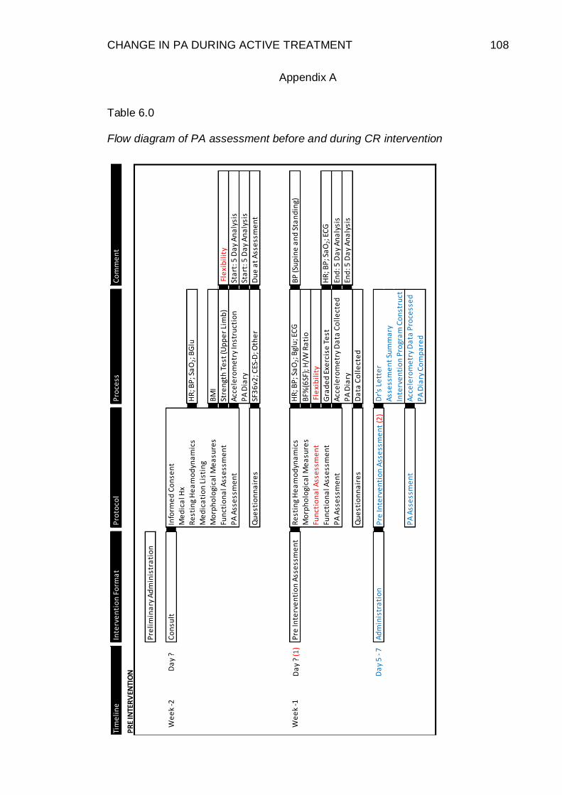
CHANGE IN PA DURING ACTIVE TREATMENT 108
Appendix A
Table 6.0
Flow diagram of PA assessment before and during CR intervention
Tim
eli
ne
Inte
rve
nti
on
Fo
rma
tP
roto
col
Pro
cess
Co
mm
en
t
Pre
lim
ina
ry A
dm
inis
tra
tio
n
We
ek
-2D
ay
?C
on
sult
Info
rme
d C
on
sen
t
Me
dic
al H
x
Re
stin
g H
ea
mo
dyn
am
ics
HR
; BP
; Sa
O2; B
Glu
Me
dic
ati
on
Lis
tin
g
Mo
rph
olo
gica
l Me
asu
res
BM
I
Fun
ctio
na
l Ass
ess
me
nt
Stre
ngt
h T
est
(Up
pe
r Li
mb
) F
lexi
bil
ity
PA
Ass
ess
me
nt
Acc
ele
rom
etr
y In
stru
ctio
nSt
art
: 5 D
ay
An
aly
sis
PA
Dia
rySt
art
: 5 D
ay
An
aly
sis
Qu
est
ion
na
ire
sSF
36
v2; C
ES-D
; Oth
er
Du
e a
t A
sse
ssm
en
t
We
ek
-1D
ay
? (1
)P
re In
terv
en
tio
n A
sse
ssm
en
tR
est
ing
He
am
od
yna
mic
sH
R; B
P; S
aO
2; B
glu
; EC
GB
P (S
up
ine
an
d S
tan
din
g)
Mo
rph
olo
gica
l Me
asu
res
BF%
(6SF
); H
/W R
ati
o
Fun
ctio
na
l Ass
ess
me
nt
Fle
xib
ilit
y
Fun
ctio
na
l Ass
ess
me
nt
Gra
de
d E
xerc
ise
Te
st
HR
; BP
; Sa
O2; E
CG
PA
Ass
ess
me
nt
Acc
ele
rom
etr
y D
ata
Co
lle
cte
dEn
d: 5
Da
y A
na
lysi
s
PA
Dia
ryEn
d: 5
Da
y A
na
lysi
s
Qu
est
ion
na
ire
sD
ata
Co
lle
cte
d
Da
y 5
- 7
Ad
min
istr
ati
on
Pre
Inte
rve
nti
on
Ass
ess
me
nt
(2)
Dr'
s Le
tte
r
Ass
ess
me
nt
Sum
ma
ry
Inte
rve
nti
on
Pro
gra
m C
on
stru
ct
PA
Ass
ess
me
nt
Acc
ele
rom
etr
y D
ata
Pro
cess
ed
PA
Dia
ry C
om
pa
red
PR
E IN
TER
VEN
TIO
N

CHANGE IN PA DURING ACTIVE TREATMENT 109
Table 6.0 Continued
Table 6.0 Continued
Sta
rt (W
ee
k 0
)D
ay
1In
du
ctio
n
Da
y 3
Fun
ctio
na
l Ass
ess
me
nt
Stre
ngt
h T
est
(Lo
we
r Li
mb
)H
UM
AC
No
rm IM
DA
sse
ssm
en
t R
eve
iw C
om
ple
ted
Ad
min
istr
ati
on
Inte
rve
nti
on
Ad
ap
tati
on
Edu
cati
on
We
ek
11
Da
y ?
PA
Ass
ess
me
nt
Acc
ele
rom
etr
y A
na
lysi
sSt
art
: 5 D
ay
An
aly
sis
PA
Dia
rySt
art
: 5 D
ay
An
aly
sis
We
ek
12
Da
y 1
Po
st In
terv
en
tio
n A
sse
ssm
en
tR
est
ing
He
am
od
yna
mic
sH
R; B
P; S
aO
2; B
glu
; EC
G
Mo
rph
olo
gica
l Me
asu
res
BM
I; B
F%(6
SF);
H/W
Ra
tio
Fun
ctio
na
l Ass
ess
me
nt
Gra
de
d E
xerc
ise
Te
st
Stre
ngt
h &
Fle
xib
ilit
y Te
sts
D
ay
?P
A A
sse
ssm
en
tA
cce
lero
me
try
An
aly
sis
End
: 5 D
ay
An
aly
sis
PA
Dia
ryEn
d: 5
Da
y A
na
lysi
s
Da
y 4
Ad
min
istr
ati
on
Po
st In
terv
en
tio
n A
sse
ssm
en
t D
r's
Lett
er
Ass
ess
me
nt
Sum
ma
ry
Ho
me
/ P
ost
Pro
gra
m C
on
stru
ct
PA
Ass
ess
me
nt
Acc
ele
rom
etr
y D
ata
Pro
cess
ed
PA
Dia
ry C
om
pa
red
Da
y 5
Fin
al D
ay
of I
nte
rve
nti
on
Po
st A
sse
ssm
en
t P
ati
en
t R
eve
iw
Ho
me
/ P
ost
Pro
gra
m In
stru
ct
INTE
RV
ENTI
ON

CHANGE IN PA DURING ACTIVE TREATMENT 110
Table 6.0 Continued
(1)
A
llo
w 4
8 h
ou
rs m
inim
um
re
cove
ry p
eri
od
pri
or
to In
du
ctio
n.
We
ek
20
Da
y ?
PA
Ass
ess
me
nt
Acc
ele
rom
etr
y A
na
lysi
sSt
art
: 5 D
ay
An
aly
sis
PA
Dia
ry?
Sta
rt: 5
Da
y A
na
lysi
s?
We
ek
21
Da
y ?
PA
Ass
ess
me
nt
Acc
ele
rom
etr
y A
na
lysi
sEn
d: 5
Da
y A
na
lysi
sP
A D
iary
?En
d: 5
Da
y A
na
lysi
s?
(2)
R
eve
iw r
ele
ase
de
laye
d t
o in
clu
de
low
er
lim
b s
tre
ngt
h t
est
.

CHANGE IN PA DURING ACTIVE TREATMENT 111
Appendix B
Participant Information
Sheet
Project title: Change in physical activity during active treatment of cardiac patients
Project Supervisor: A/Prof Lukas Dreyer
Researcher: Leon Tahana
An Invitation
Kia Ora.
My name is Leon Tahana and I am a Clinical Exercise Physiologist at U-Kinetics. Currently I am working towards my Masters qualification through Auckland University of Technology (AUT) and doing a research project that investigates the change in physical activity during active cardiac rehabilitation treatment of cardiac patients. Your participation in the research is entirely voluntary and you may withdraw at any time, even before all the data has been collected. If you withdraw, or choose not to participate, your referral to U-Kinetics will not be affected in any way. That is, you can still participate in the 12 week exercise training program that your doctor referred you to and you can still learn about (and personally experience) the benefits of regular supervised training. All people referred to U-Kinetics are allowed to attend the clinic three times a week (for about an hour each time) all fully funded
by UCOL and MidCentral DHB and each person participates on a voluntary basis.
My research needs volunteers living with cardiovascular disease (CVD), and who are able to train three times per week during the operational hours of the clinic (Monday, Wednesday
& Friday: 8am – 5pm; Tuesday & Thursday 8am -12noon).
What is the purpose of this research?
The purpose of this research project is to assess whether the functional ability (physical fitness) of cardiac patients before participating in the Ukinetics cardiac rehabilitation (CR) program, reflects the level of physical activity (PA) performed at home, at work, during sport or at play. The study will also look at whether the at home physical activity patterns of a
cardiac patient change during participation in U-kinetics CR program.
One component of the exercise that we use at U-kinetics is cycling on a stationary bike. The difficulty (also known as “intensity”) of the cycling is entirely based your fitness and will be designed to be at a comfortable level. Another component of the exercise is resistance training (gym machines) and this is also used by us to help people with CVD. Both these exercise components are done under the safe supervision of Clinical Exercise Physiologists
(CEPs) who will fully monitor you (e.g. Heart rate, blood pressure and blood sugar levels).
This study will essentially measure levels of PA before you enter the CR program. This will involve you wearing an accelerometer, similar to a pedometer; which measures your movement and interprets these movements as energy expended (caloric expenditure). We will also ask you how much activity you perform normally over the course of a week via a PA questionnaire (International PA Questionnaire (IPAQ)). This will give us data that we can compare with tests we will perform with you prior to you starting CR. These tests will

CHANGE IN PA DURING ACTIVE TREATMENT 112
evaluate your functional ability (flexibility, strength, balance) and physical fitness (a graded exercise test on an ergocycle (stationary bike)). These same tests and measurements will be performed at the end of the 12 week CR program. This data will give us information on
how effective our CR program is for producing necessary PA change.
As part of my qualification I will use the data obtained from your participation to write my thesis document, to write academic papers and oral presentations at scientific conferences. I will not include your name in any of these documents/presentations - only the results
achieved from the data we have collected.
How were you identified and why are you being invited to participate in this research?
From the notes that your doctor sent on your behalf to U-Kinetics (to get you enrolled on the funded program) we determined that you meet the entry requirements of my study (male or female having been recently diagnosed with CVD) and as such I want to invite you to attend a face-to-face meeting where we can meet each other and discuss the research further
(and answer any questions you may have).
In order to ensure safety this study will only include participants who do not have serious heart problems (low ejection fraction, uncontrolled angina, aneurysm etc.), muscle or joint concerns (e.g. A recent knee replacement). Remember that if you are not able to participate
in my research project you will still be able to train at U-Kinetics as per your referral.
What will happen in this research?
The picture below shows the timeline of my research. At the start and end of the CR program, there will be an assessment designed to see how you have progressed over the
12 weeks.
Pre Program: You will receive an appointment time during which you will be fully informed
about the tests and procedures that will be performed as part of the research project and the U-kinetics program. During this initial session you will be able to ask questions to clarify points and issues. Prior to acceptance onto the program you will be asked to sign a form
stating that you understand and consent to the processes involved in your CR program.
Following the informed consent process exercise testing will be conducted. All exercise
testing is based on exercise testing protocols that may include, but are not limited to:
Cardiovascular Exercise Testing: This will provide an estimation of your cardiorespiratory fitness. You will perform an easy-to-moderate exercise test on a stationary bicycle or other equipment determined by U-Kinetics. The exercise intensity will begin at a low level and will be advanced in stages depending on how your body responds to each stage of the test. Most clients will also have ECG stickers attached around their heart during this test so that accurate recordings of the cardiac rhythms can be viewed. A face mask will be worn to collect expired air data that will indicate aerobic fitness and a safe end point for testing. In

CHANGE IN PA DURING ACTIVE TREATMENT 113
some circumstances a finger-prick blood sample may be needed to update your glucose or
cholesterol readings.
Musculoskeletal Testing: This will provide information as to the functional capabilities of your muscles and the joints to which they attach. This may include range of motion (flexibility) tests and other general, and/or computer-aided, muscular strength and endurance testing, and balance testing.
Body Composition Assessment: This will provide data that will be used to help calculate baseline values in order to track program effect. These measurements include standing on a scale as well as the measuring of selected skinfold thicknesses and/or girths. During many of the tests it will be to your advantage to wear minimal clothing and/or loose fitting clothing so that accurate measurements can be recorded.
Physical Activity Assessment: This will provide an estimation of your caloric expenditure and physical activity habits. This will include wearing a waist strap fitted with a small accelerometer unit. Period of wear will be for 6 days; before starting, and at the end of the 12 week exercise program. A log book with self recorded activities performed over the same
periods will also be included in the assessment.
Other Measures of Health Status: Additional tests may be performed depending on your health status. These may include tests such as spirometry, peak flow, blood glucose and
blood lipids (e.g. Cholesterol).
As part of a research project, this data collected will aid in assessing and tracking the effectiveness of the U-Kinetics exercise program. This data will also be used to construct your exercise intervention program. The 12 week exercise program I will design for you is based on assessment results and will be in line with international recommendations for
people with cardiovascular disease and similar health conditions.
Intervention Period: For 12 weeks you will train 3 times per week for about an hour each
session. Here you will conduct aerobic exercise (stationary bike or treadmill), resistance training (weight machines, free weights and other equipment) and some flexibility exercises. The program will be progressed in intensity and or duration as you physically adapt to the prescribed exercise regime. During the 12 weeks ongoing information will be provided and discussed with you regarding your CVD condition and applicable/associated lifestyle. This will essentially target aspects that we think you may need to adapt, rethink and/or change because doing so will aid with your rehabilitation process and improve your personal outcomes. At week 11 you will again be asked to wear an accelerometer unit to measure your caloric expenditure and physical activity patterns.
Post Program: A post program testing battery -the same as your pre-program testing- will
be performed to assess for any changes after you have completed the 12-week exercise program. Reports will be constructed and issued to you and to your referring GP/ Specialist. If applicable you may be invited to attend a further 12 weeks of training to elicit further improvements to your health. You are welcome to continue training by yourself in your home or local gym and U-Kinetics staff will provide you with a home-based exercise program for that purpose. Up to this point the only aspect that will be different for you in comparison to individuals going through the normal U-kinetics program, is the wearing of
the accelerometer.
What are the discomforts and risks and how will they be alleviated?
The face-to-face meetings and assessment sessions take place in a private consultation room, but there may be a fourth-year student present to help with the note-taking. I will be asking you questions related to your medical conditions (reading what your doctor wrote in his referral letter), medications and activity levels - so that I get a better understanding of
how CVD is affecting you and your ability to exercise safely.

CHANGE IN PA DURING ACTIVE TREATMENT 114
Most of the assessments are done with you in a relaxed state (e.g. Height, weight, blood pressure, body fat). To obtain an accurate picture of your heart rhythm I may need to shave small areas of your chest (if very hairy) so that the electrodes stick better (and are not
painful when pulling them off afterwards).
However, there is one test that may place some strain on you. This test is necessary in order to determine your safe exercise limits and strength results. The first is a stationary cycling test which will start off very easy and where I will regularly monitor your heart rate and rhythm (via the ECG images) and blood pressure. Only if you are safe to continue to
the next level of the test, will I make it slightly more difficult.
I have been safely helping people to exercise for the past 3 years, but just in case, all staff members and students of U-Kinetics are trained regularly in first-aid and resuscitation, and there is an AED (portable defibrillator) and oxygen cylinder on site - which is checked daily.
What are the benefits?
The potential benefits to you include:
Enhanced understanding of CVD complications
Regular blood pressure checks
Regular body mass monitoring (to help with weight loss motivation)
Better understanding the effects of exercise (immediate and long-term) on CVD
Knowledge, experience and confidence to continue exercising by yourself This research will benefit me by providing me with:
Understanding of how exercise affects CVD
Data for the completion of my Masters qualification There is potential for a wider-community understanding to how exercise effects:
The CVD state
A reduction in future disease and death rates
A reduction in the associated health care expenses
Improved productivity through return to work
Additionally, all student helpers will be exposed to how research is conducted so that they may, in the future, build on this experience.
What compensation is available for injury or negligence?
In the unlikely event of a physical injury as a result of your participation in this study, rehabilitation and compensation for injury by accident may be available from the Accident Compensation Corporation, providing the incident details satisfy the requirements of the law
and the Corporation's regulations.
What are the costs of participating in this research?
During the entire program only travel and time costs will be your responsibility. All the assessments and supervised training is fully-funded by UCOL and the MidCentral DHB. The
car parks at, and around, U-Kinetics are free (60 minute time limit for on road parking).
Post program you may take your training program with you and you can continue to exercise regularly (if you wish) either at home or in your community, but this will not be funded.
What opportunity do I have to consider this invitation?
You will be given a week from our first face-to-face meeting to talk to your whanau/family
before needing to decide whether to accept the invitation (or not) .

CHANGE IN PA DURING ACTIVE TREATMENT 115
How do I agree to participate in this research?
At our first face-to-face meeting I will explain the process again, give you a tour of the assessment and training facilities and give you a consent form. Once you are satisfied that I have answered all your questions relating to the research you may consider signing the form.
Will I receive feedback on the results of this research?
After each assessment week you will receive feedback on your results. At the completion of
the research I will give a presentation at U-Kinetics to which all participants will be invited.
Data storage and future use:
All information is treated as confidential and information is recorded on documentation that will be secured in a file and locked away in a filing cabinet behind the reception desk. Electronic data will be stored in a secure data set on the UCOL network. Only people directly involved in the research will have access to the data. The data we record will/may also be published in
scientific journals or used in presentations, however you will not be identified.
What do I do if I have concerns about this research?
Any concerns regarding the nature of this project should be notified in the first instance to the Project Supervisor, Assoc. Prof. Lukas Dreyer, [email protected], (06) 592 7001
x70636
Concerns regarding the conduct of the research should be notified to the Executive Secretary of AUTEC, Kate O’Connor, [email protected] , 921 9999 ext 6038.
Whom do I contact for further information about this research?
Please feel welcome to contact me, or my supervisor, should you have any questions:
Researcher contact details: Project supervisor contact details:
Leon Tahana Assoc. Prof. Lukas Dreyer (06) 354 2866 UCOL
(06) 592 7001 x70636
Approved by the Auckland University of Technology Ethics Committee on type the date on which the final approval was granted AUTEC Reference number type the AUTEC reference number

CHANGE IN PA DURING ACTIVE TREATMENT 116
Appendix C
Consent Form
Project title: Change in physical activity during active treatment of cardiac patients
Project Supervisor: A/prof Lukas Dreyer
Researcher: Leon Tahana
I have read and understood the information provided about this research project in the Information Sheet dated 11 November 2014
I have had an opportunity to ask questions and to have them answered.
I understand that I may withdraw myself or any information that I have provided for this project at any time prior to completion of data collection, without being disadvantaged in any way.
I understand that notes will be taken (by Leon Tahana and/or fourth year students) during the interviews.
If I withdraw, I understand that all relevant information and transcripts, or parts thereof, will be destroyed.
I agree to take part in this research.
I wish to receive a copy of the report from the research (please tick one): Yes No
I understand that the data will be secured and only accessible to the research team
I understand that my test results will be sent to my GP
Participants signature: .....................................................……………………………
Participants Name: .....................................................……………………………
Participants contact details (if appropriate):
………………………………………………………………………………………..
Date:
Approved by the Auckland University of Technology Ethics Committee on type the date on which the final approval was granted AUTEC Reference number type the AUTEC reference number
Note: The Participant should retain a copy of this form.

CHANGE IN PA DURING ACTIVE TREATMENT 117
Appendix D
Health Screen
Medical Information Have you ever had a graded exercise stress test? YES NO
Date and Place of Test: ______________________________________________________ Have you ever had any cardiological tests (e.g. ECG)? YES NO
Date and Place of Test: ______________________________________________________
HOSPITALISATION: Details of your recent hospitalisations
Person to contact in case of an emergency: ____________________________________________ Phone: __________________________ Relationship: ______________________
PERSONAL HEALTH HISTORY
Have you ever had a heart attack? YES NO
If yes,
when?________________________________________________________________
Have you ever had a heart operation? YES NO
If yes, which procedure and where (and when) was it done?
Cardiac catheterization ______________________________________
Coronary angioplasty (PCI) ___________________________________
Year Location Reason _____________________________________________________________________________ _____________________________________________________________________________
Front Page Omitted

CHANGE IN PA DURING ACTIVE TREATMENT 118
Pacemaker _______________________________________________
Other ____________________________________________________
Have you been diagnosed with a heart condition? YES NO
If yes, which condition and when was it diagnosed?
Heart valve disease _________________________________________
Heart failure ______________________________________________
Arrhythmia of the heart______________________________________
Heart murmur _____________________________________________
Congenital heart disease _____________________________________
Other_____________________________________________________
While resting, do you experience pain/discomfort in the chest, neck, jaw or arms? YES NO
During physical exercise do you experience any chest discomfort? YES NO
Do you get strange sensations in your lower legs when walking short distances? YES NO
Do you experience unreasonable breathlessness (day or night)? YES NO
Do you ever experience any dizziness, fainting, or blackouts? YES NO
Do you ever get significant swelling of your feet (ankle oedema) YES NO
If yes to any of the above questions, please explain how often?
_______________________________________________________________________
_______________________________________________________________________
Have you been diagnosed with any of the following health conditions? YES NO
If yes, when first diagnosed and how is it currently managed?
Diabetes __________________________________________________
Asthma or other lung disease _______________________________
High Blood Pressure_________________________________________
High Cholesterol levels_______________________________________
Musculoskeletal problems that limit your physical activity
_________________________________________________________

CHANGE IN PA DURING ACTIVE TREATMENT 119
Other_____________________________________________________
CONSULTATION NOTES:
FAMILY HISTORY
Have any immediate family members had a heart attack, coronary revascularization (heart bypass) or suddenly died. YES NO Relationship and their age at time of incident: ___________________________________
Have any immediate family members been diagnosed as having:
o High Blood Pressure YES NO o High cholesterol levels YES NO o Gout YES NO o Obesity YES NO o Diabetes YES NO o Congenital heart defects YES NO o Stroke YES NO o Cancer YES NO o Lung diseases YES NO o Neurological diseases YES NO o Immune diseases YES NO o Other YES NO If you responded YES to any of the above conditions please elaborate:
_________________________________________________________________________
Approved by the Auckland University of Technology Ethics Committee on type the date on which the final approval was granted AUTEC Reference number type the AUTEC reference number
MEDICATION ------------------------------------------------------------------------------ ------------------------------------------------------------------------------
Dosage & times/day
Times taken Reason for taking

CHANGE IN PA DURING ACTIVE TREATMENT 120
Appendix E
Physical Activity
Booklet Log
Actigraph wGT3X-BT User Instructions
Wear Location: The device will be worn below the waist approximately 2 to 5cm below the umbilicus
(belly button), so that the device sits on your right hip bone, most lateral position (Figure. 1), using the
elastic belt/ adhesive belt clip to ensure
secure device fixation to the body. A small
black bump on one side of the device
(Insert) should always be facing upwards
when the belt is in the correct position.
During sleep the device may be moved to
the front of the hip, for a more
comfortable sleep. Reposition on waking.
Wearer Issues: Although manufacture
materials are not known to be irritating to
the skin, some irritation to individuals
with sensitive skin may occur. If irritation does occur consult your doctor for treatment, stop wearing
the device, and notify Ukinetics Clinic as soon as possible (contact details below).
Device Removal: Please remove the device when getting wet. Remove the device while taking a bath or
shower, or while swimming. Also remove when playing very rough contact sports or activities, i.e. for
karate or rugby. Please annotate reason, time and duration of removal in the log (as shown).
Device Handling:
1. Keep devices away from pets and other animals that may chew on, bite, urinate on, swallow,
and destroy the device.
2. Putting the device in a microwave will destroy various parts of the device and may completely
destroy the device.
3. Do not put the device in extreme cold or hot temperatures and do not apply any electrical
sources to the device.
Red LED (Fault Indicator):
No Flashing (LED Off) Normal operating condition
2 Flashes Low battery. The unit needs to be recharged.
3 Flashes Unexpected battery failure or battery has entered halt mode
You will wear the device for 5 days (3 week days and 2 weekend days), from 8pm on day 1 to 8am on
day 5. Please contact Ukinetics Clinic if the red LED is flashing as soon as possible:
Contact Ph: 12345678 (Substituted) Contact Cell: 12345678
Booklet Front Page Omitted
Figure. 2

CHANGE IN PA DURING ACTIVE TREATMENT 121
Date
Time Duration Time Duration
AM PM6:30 6 1hr30min10 1 hr 11:30
S Seated (Reading, Watching TV) 1 At Home
L Light Activity 2 During Work
M Moderate Activity 3 At the Gym
H Heavy Activity 4 During Recreation
Activity Key
Daily Physical Activity Log
Activity
AwakeL
Activity
S Sleep

CHANGE IN PA DURING ACTIVE TREATMENT 122
Signs and symptoms
Although some heart attacks are sudden the majority start slowly, with mild pain or discomfort. Confusion may lengthen the time taken to get help. Knowing the signs and symptoms will reduce time to intervene and may be lifesaving.
Signs vary as follows. The pain may:
Initially come and go Be in one or both arms (more commonly the left) Go into your neck, back, jaw, stomach and abdomen
and the chest may feel like:
squeezing pressing
tightness fullness pain
You may have one or more of the following symptoms with or without chest pain/discomfort:
sweating feeling faint feeling sick vomiting
being short of breath
Even if you're not sure it's a heart attack, have it checked out.
Calling 111 is almost always the fastest way to get
lifesaving treatment. Emergency medical services staff can
begin treatment when they arrive - up to an hour sooner
than you'd get treatment if you go to hospital by car.
NZ Heart Foundation
Signs and Symptoms of Cardiac Distress
Date:
Approved by the Auckland University of Technology Ethics Committee on type the date on which the final approval was granted AUTEC Reference number type the AUTEC reference number

CHANGE IN PA DURING ACTIVE TREATMENT 123
Appendix F
21 October 2015
Scott Duncan Faculty of Health and Environmental Sciences
Dear Scott
Re Ethics Application: 15/352 Change in physical activity during active treatment of cardiac patients.
Thank you for providing evidence as requested, which satisfies the points raised by the Auckland University of Technology Ethics Committee (AUTEC).
Your ethics application has been approved for three years until 21 October 2018.
As part of the ethics approval process, you are required to submit the following to AUTEC:
A brief annual progress report using form EA2, which is available online through http://www.aut.ac.nz/researchethics. When necessary this form may also be used to request an extension of the approval at least one month prior to its expiry on 21 October 2018;
A brief report on the status of the project using form EA3, which is available online through http://www.aut.ac.nz/researchethics. This report is to be submitted either when the approval expires on 21 October 2018 or on completion of the project.
It is a condition of approval that AUTEC is notified of any adverse events or if the research does not commence. AUTEC approval needs to be sought for any alteration to the research, including any alteration of or addition to any documents that are provided to participants. You are responsible for ensuring that research undertaken under this approval occurs within the parameters outlined in the approved application.
AUTEC grants ethical approval only. If you require management approval from an institution or organisation for your research, then you will need to obtain this. If your research is undertaken within a jurisdiction outside New Zealand, you will need to make the arrangements necessary to meet the legal and ethical requirements that apply there.
To enable us to provide you with efficient service, please use the application number and study title in all correspondence with us. If you have any enquiries about this application, or anything else, please do contact us at [email protected].
All the very best with your research,
Kate O’Connor Executive Secretary Auckland University of Technology Ethics Committee
Cc: Leon Tahana [email protected], [email protected]
Copy Only
Related Documents