Change in Apoplastic Aluminum during the Initial Growth Response to Aluminum by Roots of a Tolerant Maize Variety 1 Marı ´a Dolores Va ´zquez, Charlotte Poschenrieder, Isabel Corrales, and Juan Barcelo ´* Laboratorio de Fisiologı ´a Vegetal, Facultad de Ciencias, Universidad Auto ´ noma de Barcelona, E-08193 Bellaterra, Spain Root elongation, hematoxylin staining, and changes in the ultra- structure of root-tip cells of an Al-tolerant maize variety (Zea mays L. C 525 M) exposed to nutrient solutions with 20 mM Al (2.1 mM Al 31 activity) for 0, 4, and 24 h were investigated in relation to the subcellular distribution of Al using scanning transmission elec- tron microscopy and energy-dispersive x-ray microanalysis on sam- ples fixed by different methods. Inhibition of root-elongation rates, hematoxylin staining, cell wall thickening, and disturbance of the distribution of pyroantimoniate-stainable cations, mainly Ca, was observed only after 4 and not after 24 h of exposure to Al. The occurrence of these transient, toxic Al effects on root elongation and in cell walls was accompanied by the presence of solid Al-P deposits in the walls. Whereas no Al was detectable in cell walls after 24 h, an increase of vacuolar Al was observed after 4 h of exposure. After 24 h, a higher amount of electron-dense deposits containing Al and P or Si was observed in the vacuoles. These results indicate that in this tropical maize variety, tolerance mechanisms that cause a change in apoplastic Al must be active. Our data support the hypothesis that in Al-tolerant plants, Al can rapidly cross the plasma membrane; these data clearly contradict the former conclusions that Al mainly accumulates in the apoplast and enters the symplast only after severe cell damage has occurred. It is largely recognized that root tips are the primary site of Al-induced injury in plants (Ryan et al., 1993). The accumulation of Al in root tips has been found to be significantly correlated with root-growth inhibition in maize (Zea mays L.) varieties differing in Al tolerance (Llu- gany, 1994; Llugany et al., 1994). In Al-sensitive maize plants an inhibition of root elongation has been observed after only 30 min of exposure to Al (Llugany et al., 1995). Such a short response time, in addition to the common belief (Kochian, 1995) that Al accumulates mainly in the apoplast and crosses the plasma membrane slowly, has led to the hypothesis that Al-induced inhibition of root elon- gation may be caused by toxicity mechanisms that occur in the apoplast (Rengel, 1990, 1996; Horst, 1995) and that there is no need for Al to enter the symplast to cause primary toxicity effects (Rengel, 1992). However, investigations us- ing the highly Al-sensitive technique of secondary ion MS have shown that significant Al concentrations accumulate in the symplast of root-tip cells of soybean plants after only 30 min of exposure to Al (Lazof et al., 1994, 1996). Recent experiments on giant algae (Chara corallina) cells, where cell walls were separated from the cells by microsurgery, have also shown that Al uptake across the plasmalemma may be linear and occurs without delay (Rengel and Reid, 1997). These investigations support the view that symplastic phy- totoxicity mechanisms may also be responsible for Al- induced inhibition of root elongation after short exposure times (Kochian, 1995). More information on the subcellular distribution of Al in root tips would help to establish both the relative impor- tance of apoplastic and symplastic sites in the Al-toxicity syndrome and the role of Al compartmentation in Al re- sistance or tolerance. Unfortunately, ultrastructural inves- tigations under environmentally realistic growth condi- tions that relate the subcellular localization of Al in root tips to root growth in Al-tolerant varieties are scarce (Del- haize et al., 1993). Major difficulties for such an approach are the low sensitivity of electron probe x-ray microanaly- sis for Al determination (Lazof et al., 1994, 1997) and the poor visual distinction of subcellular structures in freeze- dried samples, in combination with the extremely low Al tissue concentrations, which have been shown to cause inhibition of root elongation (Lazof et al., 1994, 1996). Using a highly sensitive monitoring technique for root growth, we have previously shown that 20 mm Al (2.1 mm Al 31 activity) causes a significant decrease in the relative root-elongation rate in the Al-tolerant maize var C 525 M after 112 min of exposure, whereas after 24 h the relative elongation rate did not differ from that of the controls (Llugany et al., 1995). In this paper we report results on the changes in the subcellular distribution of Al in root tips during the initial root-growth response (0–24 h) of var C 525 M exposed to 20 mm Al (2.1 mm Al 31 activity). Hema- toxylin staining, ultrastructural observations, and EDXMA were performed on root tips after 0, 4, and 24 h of exposure of plants to control or Al-containing nutrient solutions to detect a possible relationship between changes in subcel- lular Al compartmentation and ultrastructural alterations, which may explain why, after a transient inhibition, the root-elongation rate recovers during the initial 24 h of 1 This work was supported by the Research Council of the European Union (contract nos. TS*CT922-0071 and ERBIC188CT- 0063) and by the Spanish National Research Council (contract no. DGICYT PB97-0163-C02-01). * Corresponding author; e-mail [email protected]; fax 34 –93– 581–2003. Abbreviations: EDXMA, energy-dispersive x-ray microanalysis; PA, pyroantimoniate; SEM, scanning electron microscopy; TEM, transmission electron microscopy. Plant Physiology, February 1999, Vol. 119, pp. 435–444, www.plantphysiol.org © 1999 American Society of Plant Physiologists 435 www.plant.org on July 20, 2015 - Published by www.plantphysiol.org Downloaded from Copyright © 1999 American Society of Plant Biologists. All rights reserved.

Welcome message from author
This document is posted to help you gain knowledge. Please leave a comment to let me know what you think about it! Share it to your friends and learn new things together.
Transcript

Change in Apoplastic Aluminum during the Initial GrowthResponse to Aluminum by Roots of a Tolerant Maize Variety1
Marıa Dolores Vazquez, Charlotte Poschenrieder, Isabel Corrales, and Juan Barcelo*
Laboratorio de Fisiologıa Vegetal, Facultad de Ciencias, Universidad Autonoma de Barcelona,E-08193 Bellaterra, Spain
Root elongation, hematoxylin staining, and changes in the ultra-structure of root-tip cells of an Al-tolerant maize variety (Zeamays L. C 525 M) exposed to nutrient solutions with 20 mM Al (2.1mM Al31 activity) for 0, 4, and 24 h were investigated in relation tothe subcellular distribution of Al using scanning transmission elec-tron microscopy and energy-dispersive x-ray microanalysis on sam-ples fixed by different methods. Inhibition of root-elongation rates,hematoxylin staining, cell wall thickening, and disturbance of thedistribution of pyroantimoniate-stainable cations, mainly Ca, wasobserved only after 4 and not after 24 h of exposure to Al. Theoccurrence of these transient, toxic Al effects on root elongationand in cell walls was accompanied by the presence of solid Al-Pdeposits in the walls. Whereas no Al was detectable in cell wallsafter 24 h, an increase of vacuolar Al was observed after 4 h ofexposure. After 24 h, a higher amount of electron-dense depositscontaining Al and P or Si was observed in the vacuoles. These resultsindicate that in this tropical maize variety, tolerance mechanismsthat cause a change in apoplastic Al must be active. Our datasupport the hypothesis that in Al-tolerant plants, Al can rapidlycross the plasma membrane; these data clearly contradict theformer conclusions that Al mainly accumulates in the apoplast andenters the symplast only after severe cell damage has occurred.
It is largely recognized that root tips are the primary siteof Al-induced injury in plants (Ryan et al., 1993). Theaccumulation of Al in root tips has been found to besignificantly correlated with root-growth inhibition inmaize (Zea mays L.) varieties differing in Al tolerance (Llu-gany, 1994; Llugany et al., 1994). In Al-sensitive maizeplants an inhibition of root elongation has been observedafter only 30 min of exposure to Al (Llugany et al., 1995).Such a short response time, in addition to the commonbelief (Kochian, 1995) that Al accumulates mainly in theapoplast and crosses the plasma membrane slowly, has ledto the hypothesis that Al-induced inhibition of root elon-gation may be caused by toxicity mechanisms that occur inthe apoplast (Rengel, 1990, 1996; Horst, 1995) and that thereis no need for Al to enter the symplast to cause primarytoxicity effects (Rengel, 1992). However, investigations us-ing the highly Al-sensitive technique of secondary ion MS
have shown that significant Al concentrations accumulatein the symplast of root-tip cells of soybean plants after only30 min of exposure to Al (Lazof et al., 1994, 1996). Recentexperiments on giant algae (Chara corallina) cells, where cellwalls were separated from the cells by microsurgery, havealso shown that Al uptake across the plasmalemma may belinear and occurs without delay (Rengel and Reid, 1997).These investigations support the view that symplastic phy-totoxicity mechanisms may also be responsible for Al-induced inhibition of root elongation after short exposuretimes (Kochian, 1995).
More information on the subcellular distribution of Al inroot tips would help to establish both the relative impor-tance of apoplastic and symplastic sites in the Al-toxicitysyndrome and the role of Al compartmentation in Al re-sistance or tolerance. Unfortunately, ultrastructural inves-tigations under environmentally realistic growth condi-tions that relate the subcellular localization of Al in roottips to root growth in Al-tolerant varieties are scarce (Del-haize et al., 1993). Major difficulties for such an approachare the low sensitivity of electron probe x-ray microanaly-sis for Al determination (Lazof et al., 1994, 1997) and thepoor visual distinction of subcellular structures in freeze-dried samples, in combination with the extremely low Altissue concentrations, which have been shown to causeinhibition of root elongation (Lazof et al., 1994, 1996).
Using a highly sensitive monitoring technique for rootgrowth, we have previously shown that 20 mm Al (2.1 mmAl31 activity) causes a significant decrease in the relativeroot-elongation rate in the Al-tolerant maize var C 525 Mafter 112 min of exposure, whereas after 24 h the relativeelongation rate did not differ from that of the controls(Llugany et al., 1995). In this paper we report results on thechanges in the subcellular distribution of Al in root tipsduring the initial root-growth response (0–24 h) of var C525 M exposed to 20 mm Al (2.1 mm Al31 activity). Hema-toxylin staining, ultrastructural observations, and EDXMAwere performed on root tips after 0, 4, and 24 h of exposureof plants to control or Al-containing nutrient solutions todetect a possible relationship between changes in subcel-lular Al compartmentation and ultrastructural alterations,which may explain why, after a transient inhibition, theroot-elongation rate recovers during the initial 24 h of
1 This work was supported by the Research Council of theEuropean Union (contract nos. TS*CT922-0071 and ERBIC188CT-0063) and by the Spanish National Research Council (contract no.DGICYT PB97-0163-C02-01).
* Corresponding author; e-mail [email protected]; fax 34 –93–581–2003.
Abbreviations: EDXMA, energy-dispersive x-ray microanalysis;PA, pyroantimoniate; SEM, scanning electron microscopy; TEM,transmission electron microscopy.
Plant Physiology, February 1999, Vol. 119, pp. 435–444, www.plantphysiol.org © 1999 American Society of Plant Physiologists
435 www.plant.org on July 20, 2015 - Published by www.plantphysiol.orgDownloaded from
Copyright © 1999 American Society of Plant Biologists. All rights reserved.
exposure to Al. EDXMA with scanning TEM onglutaraldehyde-fixed, PA-stained, and freeze-substitutedsamples were performed. Although these techniques onlyallow a semiquantitative estimation of mineral contents,the better visual resolution obtained results in more reli-able data on the subcellular localization than EDXMA withSEM on freeze-dried or frozen-hydrated bulk specimens(Van Steveninck and Van Steveninck, 1991).
MATERIALS AND METHODS
Plant Material and Growth Conditions
Al-tolerant maize (Zea mays L. var C 525 M, Embrapa,Siete Lagoas, Brazil) seeds were germinated in the dark at25°C on filter paper moistened with 1 mm CaSO4. After96 h, uniform seedlings with a radicle length of 13.7 6 0.9cm were transferred to plastic beakers (14-L capacity; 24plants per beaker) filled with continuously aerated nutrientsolution (pH 4.3) of the following composition (in mm): 500Ca(NO3)2, 395 K2SO4, 5 KH2PO4, 100 MgSO4, 200 NH4NO3,0.06 (NH4)6Mo7O24, 5 MnSO4, 0.38 ZnSO4, 0.16 CuSO4, 16H3BO3, and 10 FeEDTA. After 72 h, the plants were trans-ferred to treatment solutions of the same composition andvolume per plant. One-half of the plants received solutionsupplemented with 20 mm Al as AlCl3. The pH of thecontrol nutrient solutions remained constant throughoutthe experiment (4.31 6 0.01). In Al-supplemented solutionspH values were 4.34 6 0.02 and 4.13 6 0.02 after 4 and 24 h,respectively. According to the GEOCHEM speciation pro-gram (Parker et al., 1987), the activity of free Al31 in thetreatment solution was 2.1 mm and all Al was in solubleform. The concentrations of monomeric Al in the solution,analyzed by the short-term pyrocatechol method (Kervenet al., 1989), was 13 mm.
The seedlings were grown in an environmentally con-trolled growth chamber under the following conditions:16 h of light/8 h of darkness, day/night temperature 26°C/20°C, RH 70%, and PPFD 190 mmol m22 s21.
Root Growth and Hematoxylin Staining
Seedling seminal root length (n $ 24 per treatment andtime sample) was measured with a ruler before the transferof the plants to nutrient solution, after the 72-h pretreat-ment (0-h treatment), and after the 4- and 24-h treatmentswith solutions containing 0 (control) or 20 mm Al. Hema-toxylin staining of whole roots was performed on 10 plantsper treatment and time sample (Polle et al., 1978).
Sample Fixation for Electron Microscopy and EDXMA
For EM studies, after a short (10 s) rinse with distilledwater, the tips (0–2 mm and the following 2–5 mm) fromprimary roots were excised from control and Al-treatedseedlings after 0, 4, and 24 h of exposure to nutrient solu-tions. The samples were immediately fixed by the differentmethods described below.
Some samples were fixed in 2.5% (w/v) glutaraldehydein 0.1 m sodium cacodylate buffer (pH 7.2), but were not
postfixed with osmium. The fixed material was dehydratedin a graded alcohol series and embedded in Spurr’s resin(Spurr, 1969). Some of the non-osmified, thin, longitudinaltip sections (near root halves) were stained with saturatedaqueous uranyl acetate, followed by Reynolds lead citrate(Reynolds, 1963). Stained and unstained longitudinal serialsections, between 0 and 1.5 mm from apex, were studied byelectron microscopy (model H-7000, Hitachi, Tokyo, Ja-pan), and the elemental distribution in cell walls and vacu-oles was determined by EDXMA on unstained, dry-cutsections.
Other sampled tips were treated with PA to retain easilydiffusible cations (Mentre and Escaig, 1988; Mentre andHalpern, 1988). The composition of the fixation mixturewas 4% PA, 2% paraformaldehyde, and 1% phenol (pH7.8). After the treatment the specimens were rinsed withdistilled water, then dehydrated in graded alcohol andembedded in Spurr’s resin. Longitudinal tip sections werestudied by light microscopy (Optiphot, Nikon) and elec-tron microscopy. The elemental composition of PA precip-itates was identified by EDXMA.
A third group of root tips was processed by freeze-substitution, as previously described (Harvey, 1982;Vazquez et al., 1992). The samples were cryofixed in pen-tane cooled with liquid nitrogen. The freeze-substitutionwith acetone precooled with liquid nitrogen was allowedto proceed for 1 week at 280°C in a deep freezer. Thespecimens were gradually warmed to room temperaturefor 24 h and then infiltrated with Spurr’s resin. Transversesections at approximately 0.5 and 3 mm from the apex werestained for light and electron microscopy (photographs notshown) as for the glutaraldehyde-fixed sections describedabove. Corresponding unstained, dry-cut sections wereused for EDXMA.
EDXMA
Dry-cut sections of approximately 1.5 mm were mountedonto gold grids. The microanalytical determinations wereperformed on an electron microscope (model H-800, Hita-chi) operated at 100 kV in the scanning TEM mode using anenergy-dispersive detector (Kevex, Valencia, CA) and aDelta class 4460 analyzer (Kevex). The counts were madeover a 100-s period and spectra were recorded. Gaussiandeconvolution was applied to the results and, after back-
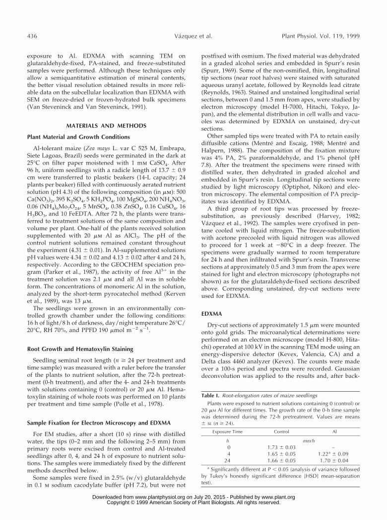
Table I. Root-elongation rates of maize seedlingsPlants were exposed to nutrient solutions containing 0 (control) or
20 mM Al for different times. The growth rate of the 0-h time samplewas determined during the 72-h pretreatment. Values are means6 SE (n $ 24).
Exposure Time Control Al
h mm/h0 1.73 6 0.03 –4 1.65 6 0.05 1.22a 6 0.09
24 1.66 6 0.05 1.70 6 0.04a Significantly different at P , 0.05 (analysis of variance followed
by Tukey’s honestly significant difference [HSD] mean-separationtest).
436 Vazquez et al. Plant Physiol. Vol. 119, 1999
www.plant.org on July 20, 2015 - Published by www.plantphysiol.orgDownloaded from Copyright © 1999 American Society of Plant Biologists. All rights reserved.

ground correction, the data were expressed as the counts tosecond ratio. A variable number of samples was used foreach treatment and fixation method. For glutaraldehyde-fixed samples or those treated with PA, n values were asfollows: control plants, n $ 4; Al-treated plants, n $ 9. Then values for freeze-substituted samples were 9 and 15 forcontrol and Al-treated plants, respectively. No Al signalswere detectable by EDXMA in electron-translucent cellareas and all data shown are from electron-dense deposits.As usual in EDXMA studies, the sd values of the resultswere high; therefore, the ranges were given in addition tomean values 6 sd. Blank resin was analyzed to check forcontaminants.
RESULTS
Maize seedlings exposed to Al for 4 h exhibited de-creased root-elongation rates, whereas after 24 h the rateshad recovered to the control values of plants before thestart of the Al treatment (Table I).
Seminal roots of seedlings stained with hematoxylin areshown in Fig. 1. After 4 h of exposure to Al-containingnutrient solution, root tips exhibited intense staining (Fig.1B), whereas after 24 h of Al exposure, no staining could beobserved (Fig. 1D) and the plants did not differ fromcontrols (Fig. 1, A and C).
Ultrastructural alterations in cell walls of root-tip (1.5-mm) cells were observed after 4 h of exposure to Al (Fig. 2).A thickening of tangential cell walls occurred in the inter-nal (third–sixth) cortex cells (Fig. 2, B and D). Sections fromglutaraldehyde-fixed specimens revealed cell walls withelectron-translucent areas and electron-dense zones nearthe plasmalemma (Fig. 2B). In the corresponding sectionsthat had been stained with PA for visualizing otherwisesoluble cations, a significantly higher accumulation of PA-stained deposits was found at the internal site of these cellwalls in Al-treated plants (Fig. 2D) than in controls (Fig.
2C). Al-treated plants also exhibited a higher amount ofPA-stained deposits inside of the cortex cells than controlplants.
The cortex cells exhibited numerous small vacuoles. Inglutaraldehyde-fixed samples from plants prior to thetransfer to treatment solutions (0 h) (Fig. 3A) and fromplants exposed to control (Fig. 3C) or Al-containing (Fig.3E) nutrient solution for 4 h, the small vacuoles wereelectron-lucent and only small, peripherical, electron-densedeposits were detected in some vacuoles of internal corticalcells (mainly the third to the sixth) of each section (Fig. 3, Cand E, arrows). In the corresponding PA-stained sectionsabundant electron-opaque precipitates in the central zoneof the vacuoles were found (Fig. 3, B, D, and F).
After 24 h of exposure to Al-containing nutrient solu-tions, thickened cell walls similar to those observed after4 h of exposure were not detected (Fig. 4, B and D). Root-tipvacuoles of plants exposed to Al for 24 h (Fig. 4D) exhibiteda considerably higher amount of electron-opaque aggre-gates than those from the 4-h Al treatment (Fig. 3E). After24 h, the vacuolar deposits in Al-treated plants (Fig. 4D)were also more abundant than in the corresponding controlsamples (Fig. 4C).
Figure 1. Seminal roots from maize plants stained with hematoxylin.A and C, Control (0 mM Al) plants after 4 and 24 h, respectively. B andD, Plants treated with 20 mM Al containing nutrient solution for 4 and24 h, respectively.
Figure 2. TEM images from longitudinal sections of root tips of maizeplants exposed for 4 h to control (0 mM Al) (A and C) or 20 mM Al (Band D) in nutrient solution. A and B, Non-osmified, glutaraldehyde-fixed samples. C and D, PA-stained samples. Note thickening of cellwalls (B) and higher amount of PA precipitates at the internal site ofcell walls (D) in samples from Al-treated plants. Scale bars represent0.5 mm in A and B, and 1.0 mm in C and D.
Localization of Al in Root Tips of Al-Tolerant Maize 437
www.plant.org on July 20, 2015 - Published by www.plantphysiol.orgDownloaded from Copyright © 1999 American Society of Plant Biologists. All rights reserved.

After 24 h of exposure to Al, but not after 4 h, abnormal,irregular divisions of internal cortex cells were detected inlongitudinal sections of PA-stained samples (Fig. 5, B andD), whereas in the samples of control plants all cell divi-sions occurred in regular planes (Fig. 5, A and C).
Tables II and III show relative contents (in counts persecond) of Al and selected mineral nutrients found byEDXMA in the electron-dense deposits of cell walls andvacuoles of control and Al-treated plants after 0, 4, and 24 hof exposure. In glutaraldehyde-fixed samples smallamounts of Al were detected in electron-dense deposits inthe walls of the roots treated with 20 mm Al after only 4 hof exposure. P, S, Ca, K, and Zn were also found in thesedeposits (Table II; Fig. 6B). However, Al was not detectablein electron-dense areas of the walls after 24 h of exposureto Al either in glutaraldehyde-fixed material or in samplesprepared by freeze-substitution or PA staining (Tables IIand IV). Al was not detected in the cell walls of controls atany sampling time (Table II; Fig. 6A).
Small amounts of Al were detected in electron-denseprecipitates found in the vacuoles of internal cortex cells of1.5-mm tips of Al-treated plants after the 4-h Al treatment(Table III; Fig. 6F). P, Zn, Ca, Mg, and S were also presentin these deposits. The few electron-dense deposits in thevacuoles of control plants after 0 and 4 h of exposurecontained neither Al nor S, but exhibited counts for P, Mg,
Ca, and Zn (Table III; Fig. 6E), that were similar to those ofAl-treated plants (Fig. 6F). After 24 h of exposure to Al,higher Al counts were found in the vacuoles (Table III; Fig.6, G and H). After 24 h in control solutions, small amountsof Al were also detected in some of the few vacuolardeposits of controls (Table III). These deposits contained P,Zn, Ca, S, and Mg.
Additional measurements on freeze-substituted sampleswere performed in the more differentiated cells at 3 mmfrom the tip after 24 h of treatment (Table IV). Al wasundetectable in electron-dense cell wall zones in bothcontrol and Al-treated plants (Table IV). In these cell wallzones, similar counts for P, S, K, and Ca were found incontrols and Al-treated plants. Counts for Si in walls werewithin the values of those in blank resin, where 4.8 to 11counts s21 Si were found. Traces of Mg were only detectedin some of the Al-treated samples. In a few of the scarce,electron-dense deposits of the vacuoles of controls, Al wasdetected (Table IV). In all deposits where the Al signal wassignificant, P, K, and Ca were found, whereas signals forMg and S were only occasionally detected. Two types ofelectron-dense deposits were found in the vacuoles ofcortex cells at approximately 3 mm from the tip in plantsexposed to Al for 24 h (Table IV; Fig. 7, A and B). Depositswith a relatively high P content, either without or with lowAl amounts (Table IV; Fig. 7B), and Si-rich deposits that
Figure 3. TEM images from longitudinal sec-tions of root tips of maize plants exposed for 0 (Aand B) or 4 h (C–F) to control (0 mM Al) (A–D) or20 mM Al (E and F) containing nutrient solution.A, C, and E, Glutaraldehyde-fixed samples. B,D, and F, PA-stained samples. Note the abun-dance of electron-translucent vacuoles withonly some peripheric electron-dense deposits (Cand E, arrows) in conventionally fixed samplesand the abundance of electron-dense precipi-tates in the central part of vacuoles from PA-stained samples (B, D, and F). All scale barsrepresent 10 mm, except B, where bar is 1 mm.
438 Vazquez et al. Plant Physiol. Vol. 119, 1999
www.plant.org on July 20, 2015 - Published by www.plantphysiol.orgDownloaded from Copyright © 1999 American Society of Plant Biologists. All rights reserved.

contained high Al amounts in addition to S, K, Ca, and Mg.These Si- and Al-rich deposits exhibited either low P countsor did not contain P (Table IV; Fig. 7A).
DISCUSSION
The tropical maize variety used in this study, C 525 M,has been found to be Al tolerant in both short-term (hours–days) nutrient solution studies (Llugany et al., 1995; Pos-chenrieder et al., 1995; Calba and Jaillard, 1997; Horst et al.,1997) and long-term (weeks) field experiments on acid soils(M.F. Guimaraes, C.H. The, C. Welcker, Final Report CE-project TS 3-CT 92–0071, unpublished). Nonetheless, aftershort-term exposure to nutrient solution with a low Al31
activity (2.1 mm), a decrease of root elongation has alreadybeen observed in a previous investigation (Llugany et al.,1995). However, as in the present study (Table I), thisinhibition was only transient, and after 24 h of exposure,the elongation rates recovered to those of controls. Asshown by the analysis of nutrient solutions after the exper-iment, the recovery of the root-elongation rate was neither
due to an increase of pH in the nutrient solution nor todepletion of Al during the experiment.
Ultrastructural observations performed in this study alsoshow that the Al-tolerant maize var C 525 M is highlyresponsive to low Al concentrations after 4 h of exposure,but is poorly affected after 24 h. Cell wall thickening anddisturbance of apoplastic and symplastic PA-stainable cat-ions occurred only during the initial period of the Altreatment (4 h), but not after longer (24 h) exposure times.The swelling of cell walls is an early Al-toxicity symptom(Eleftheriou et al., 1993), that seems to be related to adisplacement of Ca from the walls (Demarty et al., 1984). Inour study, alterations of the cation content in the cell wallsof cortex cells of plants exposed for 4 h to Al could beobserved in PA-stained samples. PA staining has beenshown to immobilize easily diffusible cations such as Caand Na (Mentre and Escaig, 1988; Mentre and Halpern,1988). After 4 h of exposure, considerably higher amountsof PA precipitates were found at the internal site of cellwalls and inside of the cytoplasm of cortex cells of Al-treated plants than in the controls. EDXMA showed thatthe PA deposits had a high K and Ca content. This resultsuggests that after short-term (4 h) exposure to Al, thecation homeostasis in the apoplast was severely disturbed.The transient character of Al-induced alterations of bothcell wall ultrastructure and cation homeostasis in our ex-perimental plants was chronologically related to thechange in root-elongation rate.
There also was a clear coincidence in time between theAl-induced inhibition of root-elongation rates and the de-tection of Al in the apoplast of root tips by two differenttechniques: hematoxylin staining and EDXMA. Hematox-ylin is considered to stain Al-P deposits in root tips thathave been damaged by Al exposure (Ownby, 1993). InAl-tolerant wheat a decrease of hematoxylin staining in-tensity after 6 to 24 h of Al exposure has been observed(Rincon and Gonzales, 1992). However, to our knowledge,this is the first time that such a change in apoplastic Al inan Al-tolerant variety has been confirmed by EDXMA. Thischange was related in time to a change in root-elongationrates and ultrastructure.
The facts that Al caused damage in the apoplast andcation homeostasis, decreasing root elongation only duringthe first hours of exposure, and that the recovery of theplants occurred in coincidence with a change in apoplasticAl, suggest that in maize var C 525 M, Al-tolerance or-resistance mechanisms must have been activated that in-duced a change in the speciation of apoplastic Al, makingit undetectable for hematoxylin and EDXMA. At present,there is considerable experimental evidence indicating thatincreased exudation of organic acids from root tips mayplay an important role in Al detoxification in Al-tolerantmaize and wheat (Pellet et al., 1995, 1997; Jorge and Ar-ruda, 1997). Ownby (1993) has shown that root tips of anAl-sensitive wheat variety that stain intensely with hema-toxylin exhibited no coloration when the roots were rinsedwith citrate before the staining procedure. We are currentlyinvestigating whether exudation of organic acids was thecause for the observed changes in apoplastic Al in the
Figure 4. TEM images from longitudinal sections of root tips of maizeplants exposed for 24 h to control (0 mM Al) (A and C) or 20 mM Al innutrient solution (B and D). A and B, PA-stained samples. C and D,Non-osmified, glutaraldehyde-fixed samples. Note the abundance ofelectron-dense vacuolar deposits (arrows) in conventionally fixedsamples from plants exposed to Al (D) in comparison with the scarcepresence of deposits in controls (C). All scale bars represent 10 mm.Is, Intercellular space.
Localization of Al in Root Tips of Al-Tolerant Maize 439
www.plant.org on July 20, 2015 - Published by www.plantphysiol.orgDownloaded from Copyright © 1999 American Society of Plant Biologists. All rights reserved.

maize variety used in this study. Our results on apoplasticAl, ultrastructural alterations in the apoplast, and root-elongation rates support the hypothesis that apoplastic Alcan be toxic to plants (Rengel, 1992; Horst 1995). However,because of the fact that after only 4 h of exposure Al wasdetectable in root-tip vacuoles, we cannot exclude thatsymplastic Al can also be responsible for the toxic effectsobserved after 4 h of exposure.
Our EDXMA data showing a significant increase of vac-uolar Al as soon as 4 h after the start of Al supply and onlya transient occurrence of insoluble Al deposits in the apo-
plast of root tips are in clear contrast to those from severalother studies. Marienfeld and Stelzer (1993) and Marien-feld et al. (1995) have observed a high accumulation of Alin root-tip cell walls and could not find Al inside cellsunless plants were exposed to Al for longer times. Lazof etal. (1997) indicated that in only 6 out of 17 electron probex-ray microanalysis studies was Al detected inside of plantcells, whereas in all studies Al was found mainly in cellwalls. This apparent contradiction with our results may beexplained, at least in part, by clear differences in the nu-trient solutions employed, the exposure time, and the fact
Table II. Relative contents of Al and selected mineral nutrients in cell walls of maize root tips (0–1.5 mm)Plants were exposed for different times to nutrient solutions containing 0 (control) or 20 mM Al. If not indicated otherwise, the values are from
glutaraldehyde-fixed samples. Values are given as means 6 SD; n $ 4 for control and n $ 9 for Al-treated plants.
ExposureTime
Al TreatmentRelative Contents
Al P S K Ca Zn
h mM counts s21
0 0 NDa 17 6 2 12 6 2 0.4 6 0.0 1.2 6 0.5 1.2 6 0.64 0 ND 22 6 4 11 6 1 ND 1.3 6 0.1 ND4 20 2.8 6 1.8 12 6 7 5.4 6 2.4 3.2 6 1.1 1.3 6 0.5 0.7 6 0.2
24 0 ND 18 6 3 8.1 6 2.9 ND 3.7 6 0.1 ND24 20 ND 7.1 6 4.2 9.2 6 4.2 ND ND ND24b 20 ND 13 6 6 7.5 6 3.8 23 6 8 2.5 6 1.0 ND24c 20 ND 16 6 10 11 6 6 105 6 52 6.0 6 2.8 ND
a ND, Not detected. b Samples prepared by freeze-substitution. c Samples prepared by pyroantimoniate staining.
Figure 5. Light-microscopy images from longi-tudinal sections (PA stained) of root tips ofmaize plants exposed for 24 h to control (0 mM
Al) (A and C) or 20 mM Al in nutrient solution (Band D). Controls (A and C) show well-organizedcortical cell lines with almost horizontal cell-division planes. Al-exposed plants (B and D)exhibit irregular cell-division planes in internalcortical cells. A and B, Scale bars represent 100mm; C and D, scale bars represent 10 mm.
440 Vazquez et al. Plant Physiol. Vol. 119, 1999
www.plant.org on July 20, 2015 - Published by www.plantphysiol.orgDownloaded from Copyright © 1999 American Society of Plant Biologists. All rights reserved.
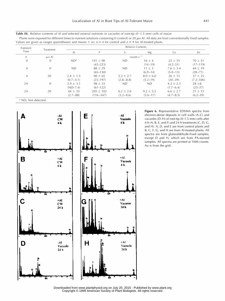
Table III. Relative contents of Al and selected mineral nutrients in vacuoles of root-tip (0–1.5 mm) cells of maizePlants were exposed for different times to nutrient solutions containing 0 (control) or 20 mM Al. All data are from conventionally fixed samples.
Values are given as ranges (parentheses) and means 6 SD; n $ 4 for control and n $ 9 for Al-treated plants.
ExposureTime
TreatmentRelative Contents
Al P S Mg Ca Zn
h mM Al counts s21
0 0 NDa 141 6 90 ND 16 6 4 22 6 15 70 6 51(43–223) (14–19) (4.2–31) (17–119)
4 0 ND 88 6 29 ND 11 6 3 7.6 6 3.4 44 6 19(65–130) (6.9–14) (5.0–13) (28–71)
4 20 2.4 6 1.5 90 6 65 5.3 6 2.7 8.0 6 6.0 26 6 13 37 6 35(0.7–4.1) (23–197) (2.8–8.8) (5.2–19) (20–39) (7.2–106)
24 0 3.9 6 3.1 98 6 33 ND ND 4.3 6 2.3 28 68(ND–7.4) (61–122) (1.7–6.4) (23–37)
24 20 44 6 35 205 6 103 6.2 6 2.0 9.2 6 5.5 6.6 6 2.7 21 6 15(2.7–88) (116–347) (3.2–8.6) (5.6–17) (4.7–8.5) (6.2–39)
a ND, Not detected.
Figure 6. Representative EDXMA spectra fromelectron-dense deposits in cell walls (A–C) andvacuoles (D–H) of root-tip (0–1.5 mm) cells after4-h (A, B, E, and F) and 24-h treatments (C, D, G,and H). A, D, and E are from control plants andB, C, F, G, and H are from Al-treated plants. Allspectra are from glutaraldehyde-fixed samples,except D and H, which are from PA-stainedsamples. All spectra are printed at 1000 counts.Au is from the grid.
Localization of Al in Root Tips of Al-Tolerant Maize 441
www.plant.org on July 20, 2015 - Published by www.plantphysiol.orgDownloaded from Copyright © 1999 American Society of Plant Biologists. All rights reserved.

that most of these studies were performed with Al-sensitive plants exposed to considerably higher Al concen-trations.
That Al may enter root cells after short-term exposure toAl has previously been demonstrated in investigations us-ing secondary ion MS (Lazof et al., 1994, 1996) or fluores-cence microscopy on morin-stained root tips (Tice et al.,1992). However, this is the first time to our knowledge thatAl has been shown to be present in root-tip vacuoles of anAl-tolerant variety after a few hours of exposure. Tice et al.(1992) also found most of the Al inside of the cells of roottips from Al-tolerant wheat. In contrast, they could notdetect Al in vacuoles. However, it is not likely that morinwould stain insoluble Al complexes in vacuoles.
The Al-rich vacuolar deposits, found in root-tip cellsbetween 0 and 1.5 mm from the apex of our experimentalplants contained P, Ca, Zn, and Mg (Table II; Fig. 6H), andtheir mineral composition was similar to that reported forphytate (Mikus et al., 1992). Phytate or polyphosphatedeposits in the vacuoles may be involved in vacuolar stor-age of Al in a way similar to that reported for Zn (VanSteveninck et al., 1987). Compartmentational analysis of32P elution in root cortical cells of intact roots of Loliumperenne exposed to nontoxic Al concentrations also sug-gests that considerable amounts of a condensed Pi formmay complex Al in root-cortex vacuoles (Macklon and Sim,1992).
According to our results, in the expanding cells at 3 mmfrom the apex a second storage form for considerable Alamounts occurs in this Al-tolerant maize variety. In thismore mature root zone most of the vacuolar Al of Al-treated plants was associated with high Si amounts (TableIII; Fig. 7A). The Al that was detectable occasionally in thecorresponding cells of control plants and that probablyderived from the seeds that had been formed on maternalplants growing on acid soils in Brazil was exclusivelyfound in P-rich deposits with a mineral composition simi-lar to phytate (Table III). There are several earlier reports
Table IV. Relative contents of Al and selected mineral nutrients in cell walls and vacuoles of maize root-tip cells (transverse sections 3 mmfrom apex)
Plants were exposed for 24 h to nutrient solutions with 0 (control) or 20 mM Al. All data are from freeze-substituted samples. Values are givenas ranges (parentheses) and means 6 SD; n $ 9 for control and n $ 15 for Al-treated plants.
TreatmentRelative Contents
Al Si P S K Ca Mg
mM Al counts s21
Cell Wall0 NDa 8.0 6 2.0
(6.6–13)19 6 12(7.8–26)
14 6 3(ND–19)
45 6 22(14–79)
5.4 6 1.5(3.3–8.2)
ND
20 ND 9.9 6 5.0(5.2–14)
29 6 10(12–50)
15 6 2(13–19)
55 6 21(15–82)
6.6 6 2.8(3.0–12)
1.0 6 0.4(ND–1.2)
Vacuole0 24 6 16
(ND–52)12 6 1
(ND–13)61 6 32
(ND–120)11 6 4(ND–16)
74 6 43(13–148)
9.9 6 8.8(4.4–27)
2.5 6 0.2(ND–1.2)
20b 6.9 6 1.8(ND–9.6)
7.5 6 1.9(ND–9.7)
68 6 65(23–130)
13 6 4.6(6.3–24)
52 6 19(27–77)
4.1 6 1.8(1.1–7.1)
6.5 6 5.0(ND–9.6)
20c 119 6 43(46–173)
193 6 55(120–229)
10 6 1(ND–11)
32 6 9(21–44)
73 6 18(46–97)
11 6 3(8.6–15)
6.4 6 3.4(0.6–12)
a ND, Not detected. b Element composition of P-rich vacuolar deposits. c Element composition of Si-rich vacuolar deposits.
Figure 7. Representative EDXMA spectra from electron-dense depos-its in the vacuoles of root-tip cortex cells (3 mm) of plants exposed toAl for 24 h. A, Al-containing deposit with high Si content. B, Al-containing deposit with P, but without Si. All samples were preparedby freeze-substitution. All spectra are printed at 1000 counts; Fe andCo are instrument contaminants; and Au is from the grid.
442 Vazquez et al. Plant Physiol. Vol. 119, 1999
www.plant.org on July 20, 2015 - Published by www.plantphysiol.orgDownloaded from Copyright © 1999 American Society of Plant Biologists. All rights reserved.

showing that the ameliorative effect of Si is not only due toAl-Si interactions at the substrate level, but that Al-Si in-teractions inside plants may play an important role in Altolerance (Barcelo et al., 1993; Hodson and Evans, 1995;Corrales et al., 1997). It is tentative to speculate that apreferential storage of Al in the form of Si-rich deposits inthe vacuoles of expanding cortex cells after 24 h of expo-sure to Al would reduce the toxic effects of Al and maycontribute to the enhancement of root elongation after aninitial transient growth reduction.
The relationship between vacuolar storage of Al in root-tip cells and Al tolerance, however, remains unclear. Ernst(1998) states that compartmentation into the vacuole is theprinciple of all hypotheses explaining intracellular metaltolerance in plants. Genotype differences in intracellulartolerance, however, cannot be explained per se by thepresence of metal deposits in vacuoles, but are probablyrelated to both the capacity of plants to form metal com-plexes that would be less toxic to cell components duringthe transport to the vacuole and the velocity of metaltransport across the tonoplast.
In spite of the fact that the accumulation of Al increasedin the small vacuoles of root-tip cells, a toxic effect on theplane of cell division occurred after 24 h of exposure to Al(Fig. 4D). This result suggests that under our experimentalconditions, not all Al was efficiently detoxified by chelationand vacuolar storage, and that there was sufficient Alremaining to interfere directly or indirectly, perhaps byan interaction with phosphatidylinositol bisphosphate(Kochian and Jones, 1997), with the direction of thecytoskeletal-directed plane of cell division.
In conclusion, the fact that after 4 h of exposure to Al,significant Al was detected in root-tip cell compartments,cell walls, and vacuoles does not allow us to clarify theproblem of the primary site of Al toxicity: the apoplast orsymplast. However, to our knowledge, this is the firstexperiment performed with an Al-tolerant maize varietythat provides analytical data supporting a relation in timebetween the lowering of insoluble, apoplastic Al, increasedAl accumulation in root-tip vacuoles, and a decrease ofAl-toxicity symptoms in the apoplast. Moreover, our re-sults provide evidence for the view that even in Al-tolerantmaize Al enters rapidly into the cells.
ACKNOWLEDGMENT
The supply of seeds of var C 525 M from Embrapa is gratefullyacknowledged.
Received June 25, 1998; accepted October 30, 1998.
LITERATURE CITED
Barcelo J, Guevara P, Poschenrieder CH (1993) Silicon ameliora-tion of aluminium toxicity in teosinte (Zea mays L. ssp. Mexi-cana). Plant Soil 54: 249–255
Calba H, Jaillard B (1997) Effect of aluminium on ion uptake andH1 release by maize. New Phytol 137: 607–616
Corrales I, Poschenrieder CH, Barcelo J (1997) Influence of siliconpretreatment on aluminium toxicity in maize roots. Plant Soil190: 203–209
Delhaize E, Craig S, Beaton CD, Bennet RJ, Jagadish VC, RandallPJ (1993) Aluminum tolerance in wheat (Triticum aestivum L.). I.Uptake and distribution of aluminum in root apices. PlantPhysiol 103: 685–693
Demarty M, Morvan C, Thellier M (1984) Calcium and the cellwall. Plant Cell Environ 7: 449–456
Eleftheriou EP, Moustakas M, Fragiskos N (1993) Aluminate-induced changes in morphology and ultrastructure of Thinopy-rum roots. J Exp Bot 44: 427–436
Ernst WHO (1998) Effects of heavy metals in plants at the cellularand organismic level. In G Schuurmann, B Markert, eds, Eco-toxicology. Wiley & Sons, New York, pp 587–620
Harvey DMR (1982) Freeze substitution. J Microsc 127: 209–221Hodson MJ, Evans DE (1995) Aluminium/silicon interactions in
higher plants. J Exp Bot 46: 161–171Horst WJ (1995) The role of the apoplast in aluminium toxicity and
resistance of higher plants: a review. Z Pflanzenernahr Bodenkd158: 419–428
Horst WJ, Puschel AK, Schmohl N (1997) Induction of calloseformation is a sensitive marker for genotypic aluminium sensi-tivity in maize. Plant Soil 192: 23–30
Jorge RA, Arruda P (1997) Aluminum-induced organic acids ex-udation by roots of an aluminum-tolerant tropical maize. Phy-tochemistry 45: 675–681
Kerven GL, Edwards DG, Asher CJ Hallman PS, Kokot S (1989)Aluminium determination in soil solution. II. Short-term color-imetric procedures for the measurement of inorganic aluminiumin the presence of organic acid ligands. Aust J Soil Res 27: 91–102
Kochian LV (1995) Cellular mechanisms of aluminum toxicity andresistance in plants. Annu Rev Plant Physiol Plant Mol Biol 46:237–260
Kochian LV, Jones DL (1997) Aluminum toxicity and resistance inplants. In RA Yokel, MS Golub, eds, Research Issues in Alumi-num Toxicity. Taylor and Francis Publishers, Washington, DC,pp 69–89
Lazof DB, Goldsmith JG, Linton RW (1997) The in situ analysis ofintracellular aluminium in plants. In HD Behnke, U Luttge, KEsser, JW Kadereit, M Runge, eds, Progress in Botany, Vol 58.Springer Verlag, Berlin, pp 112–149
Lazof DB, Goldsmith JG, Rufty TW, Linton RW (1994) Rapiduptake of aluminum into cells of intact soybean root tips. Amicroanalytical study using secondary ion mass spectrometry.Plant Physiol 106: 1107–1114
Lazof DB, Goldsmith JG, Rufty TW, Linton RW (1996) The earlyentry of Al into cells of intact soybean roots. A comparison ofthree developmental root regions using secondary ion massspectrometry imaging. Plant Physiol 112: 1289–1300
Llugany M (1994) Respuestas diferenciales de cultivares de Zeamays L. a la toxicidad por aluminio. PhD thesis, UniversidadAutonoma de Barcelona, Bellaterra, Spain
Llugany M, Massot N, Wissemeier A, Poschenrieder CH, BarceloJ (1994) Differences in aluminium tolerance between maize va-rieties as assesssed by callose formation and root elongation. ZPflanzenernahr Bodenkd 157: 447–451
Llugany M, Poschenrieder CH, Barcelo J (1995) Monitoring ofaluminium-induced inhibition of root elongation in four maizecultivars differing in tolerance to aluminium and proton toxic-ity. Physiol Plant 93: 265–271
Macklon AES, Sim A (1992) Modifying effects of a non-toxic levelof aluminium on phosphate fluxes and compartmentation inroot cortex cells of intact ryegrass seedlings. J Exp Bot 43:1483–1490
Marienfeld S, Lehmannn H, Stelzer R (1995) Ultrastructural in-vestigations and EDX-analyses of Al-treated oat (Avena sativa)roots. Plant Soil 171: 167–173
Marienfeld S, Stelzer R (1993) X-ray microanalyses in Al-treatedAvena sativa plants. J Plant Physiol 141: 569–573
Mentre P, Escaig F (1988) Localization of cations by pyroantimo-niate. I. Influence of fixation on distribution of calcium andsodium: an approach by analytical ion microscopy. J HistochemCytochem 36: 49–54
Localization of Al in Root Tips of Al-Tolerant Maize 443
www.plant.org on July 20, 2015 - Published by www.plantphysiol.orgDownloaded from Copyright © 1999 American Society of Plant Biologists. All rights reserved.

Mentre P, Halpern S (1988) Localization of cations by pyroanti-moniate. II. Electron probe microanalysis of calcium and sodiumin skeletal muscle of mouse. J Histochem Cytochem 36: 55–64
Mikus M, Bobak M, Lux A (1992) Structure of protein bodies andelemental composition of phytin from dry germ of maize (Zeamays L.). Bot Acta 105: 26–33
Ownby JD (1993) Mechanisms of reaction of hematoxylin withaluminium-treated wheat roots. Physiol Plant 87: 371–380
Parker DR, Zelazny LW, Kinraide TB (1987) Improvements to theprogram GEOCHEM. Soil Sci Soc Am J 51: 488–491
Pellet DM, Grunes DL, Kochian LV (1995) Organic acid exuda-tion as an aluminium tolerance mechanism in maize (Zea maysL). Planta 196: 788–95
Pellet DM, Papernik LA, Jones DL, Darrah PR, Grunes DL,Kochian LV (1997) Involvement of multiple aluminium exclu-sion mechanisms in aluminium tolerance in wheat. Plant Soil192: 63–68
Polle E, Konzak CF, Kittrick JA (1978) Visual detection of alumi-nium tolerance levels in wheat by hematoxylin staining of seed-ling roots. Crop Sci 18: 823–827
Poschenrieder CH, Llugany M, Barcelo J (1995) Short-term effectsof pH and aluminium on mineral nutrition in maize varietiesdiffering in proton and aluminium tolerance. J Plant Nutr 18:1495–1507
Rengel Z (1990) Competitive Al31 inhibition of net Mg21 uptakeby intact Lolium multiflorum roots. II. Plant age effects. PlantPhysiol 93: 1261–1267
Rengel Z (1992) Role of calcium in aluminium toxicity. NewPhytol 121: 499–513
Rengel Z (1996) Uptake of aluminium by plant cells. New Phytol134: 389–406
Rengel Z, Reid RJ (1997) Uptake of Al across the plasma mem-brane of plant cells. Plant Soil 192: 31–35
Reynolds ES (1963) The use of lead citrate at high pH as anelectron opaque stain in electron microscopy. J Cell Biol 17:208–210
Rincon M, Gonzales RA (1992) Aluminum partitioning in intactroots of aluminum-tolerant and aluminum-sensitive wheat(Triticum aestivum L.) cultivars. Plant Physiol 99: 1021–1028
Ryan PR, DiTomaso JM, Kochian LV (1993) Aluminium toxicityin roots: an investigation of spatial sensitivity and the role of theroot cap. J Exp Bot 44: 437–446
Spurr AR (1969) A low-viscosity epoxy resin embedding mediumfor electron microscopy. J Ultrastr Res 26: 31–43
Tice KR, Parker DR, DeMason DA (1992) Operationally definedapoplastic and symplastic aluminium fractions in root tips ofaluminum-intoxicated wheat. Plant Physiol 100: 309–318
Van Steveninck RFM, Van Steveninck ME, Fernando DR, HorstWJ, Marschner H (1987) Deposition of zinc phytate in globularbodies in roots of Deschampsia caespitosa ecotypes: a detoxifica-tion mechanism? J Plant Physiol 131: 247–257
Van Steveninck RFM, Van Steveninck ME (1991) Microanalysis.In JL Hall, C Hawes, eds, Electron Microscopy of Plant Cells.Academic Press, London, pp 415–455
Vazquez MD, Barcelo J, Poschenrieder CH, Madico J, Hatton P,Baker AJM, Cope GH (1992) Localization of zinc and cadmiumin Thlaspi caerulescens (Brassicaceae), a metallophyte that canhyperaccumulate both metals. J Plant Physiol 140: 350–355
444 Vazquez et al. Plant Physiol. Vol. 119, 1999
www.plant.org on July 20, 2015 - Published by www.plantphysiol.orgDownloaded from Copyright © 1999 American Society of Plant Biologists. All rights reserved.
Related Documents