Chandrakanta Nayak, Barada Prasanna Samal Department of Orthopaedics, All India Institute of Medical Sciences, Bhubaneswar, Odisha 751019, India. A Curious Case of Scrotal Claudication: A Rare Presentation of Prolapse Intervertebral Disc Abstract: A 58 year old male presented to our outpatient department with history of low back pain since last three years and recent onset pain in his scrotum. Pain in scrotum was typically exacerbated by walking and relieved on stooping or sitting. Urological abnormality was excluded. MRI showed severe canal stenosis at the level of L4/L5 and root compression. Surgical decompression relieved his scrotal and back pain. Cauda equina compression may have uncommon presentation which needs to be investigated by MRI. Key words: Back Pain, Cauda Equina, Decompression, Scrotum, Posture. JOURNAL OF CASE REPORTS 2015;5(2):390-392 Corresponding Author: Dr. Chandrakanta Nayak Email: [email protected] Received: May 29, 2015 | Accepted: August 24, 2015 | Published Online: September 10, 2015 This is an Open Access article distributed under the terms of the Creative Commons Attribution License (creativecommons.org/licenses/by/3.0) Conflict of interest: None declared | Source of funding: Nil | DOI: http://dx.doi.org/10.17659/01.2015.0100 Journal of Case Reports, Vol. 5, No. 2, July-December 2015 390 Introduction The Latin word “claudication” literally means “limping”. Symptoms of neurogenic claudication are described as pain, paraesthesia or cramping of one or both legs, brought on when walking and relieved in sitting [2]. Clinical symptoms are believed to result from stenotic changes, exacerbated by posture-related compression causing neural and microvascular compromise of the cauda equina and lumbosacral nerve roots [2,3]. Although, most patients have a previous history of back pain but there are limited reports of claudication [5]. Here we present an atypical site of claudication which responded well to spinal decompression surgery. Case Report A 58 year old male presented with a history of low back pain since last three years and a recent onset progressive pain in scrotum. He complained of pain and feeling of heaviness on his scrotum on walking and he could not walk more than ten minutes because of scrotal pain. He had to stoop forward to get relieved of pain and sitting used to make him comfortable. He had no history of claudication of lower limb. Good volume peripheral pulses were present. Neurological examination of lower limb showed absence of paraesthesia, normal muscle power, normal reflexes and no bladder and bowel dysfunction. However, scrotal numbness was present.

Welcome message from author
This document is posted to help you gain knowledge. Please leave a comment to let me know what you think about it! Share it to your friends and learn new things together.
Transcript
Chandrakanta Nayak, Barada Prasanna Samal Department of Orthopaedics, All India Institute of Medical Sciences, Bhubaneswar, Odisha 751019, India.
A Curious Case of Scrotal Claudication: A Rare Presentation of Prolapse Intervertebral Disc
Abstract:
A 58 year old male presented to our outpatient department with history of low back pain since last three years and recent onset pain in his scrotum. Pain in scrotum was typically exacerbated by walking and relieved on stooping or sitting. Urological abnormality was excluded. MRI showed severe canal stenosis at the level of L4/L5 and root compression. Surgical decompression relieved his scrotal and back pain. Cauda equina compression may have uncommon presentation which needs to be investigated by MRI.
Key words: Back Pain, Cauda Equina, Decompression, Scrotum, Posture.
JOURNAL OF CASE REPORTS 2015;5(2):390-392
Corresponding Author: Dr. Chandrakanta NayakEmail: [email protected] Received: May 29, 2015 | Accepted: August 24, 2015 | Published Online: September 10, 2015This is an Open Access article distributed under the terms of the Creative Commons Attribution License (creativecommons.org/licenses/by/3.0)Conflict of interest: None declared | Source of funding: Nil | DOI: http://dx.doi.org/10.17659/01.2015.0100
Journal of Case Reports, Vol. 5, No. 2, July-December 2015390
Introduction
The Latin word “claudication” literally means “limping”. Symptoms of neurogenic claudication are described as pain, paraesthesia or cramping of one or both legs, brought on when walking and relieved in sitting [2]. Clinical symptoms are believed to result from stenotic changes, exacerbated by posture-related compression causing neural and microvascular compromise of the cauda equina and lumbosacral nerve roots [2,3]. Although, most patients have a previous history of back pain but there are limited reports of claudication [5]. Here we present an atypical site of claudication which responded well to spinal decompression surgery.
Case Report
A 58 year old male presented with a history of low back pain since last three years and a recent onset progressive pain in scrotum. He complained of pain and feeling of heaviness on his scrotum on walking and he could not walk more than ten minutes because of scrotal pain. He had to stoop forward to get relieved of pain and sitting used to make him comfortable. He had no history of claudication of lower limb. Good volume peripheral pulses were present. Neurological examination of lower limb showed absence of paraesthesia, normal muscle power, normal reflexes and no bladder and bowel dysfunction. However, scrotal numbness was present.
391 Journal of Case Reports
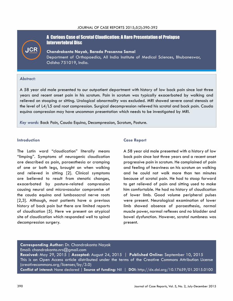
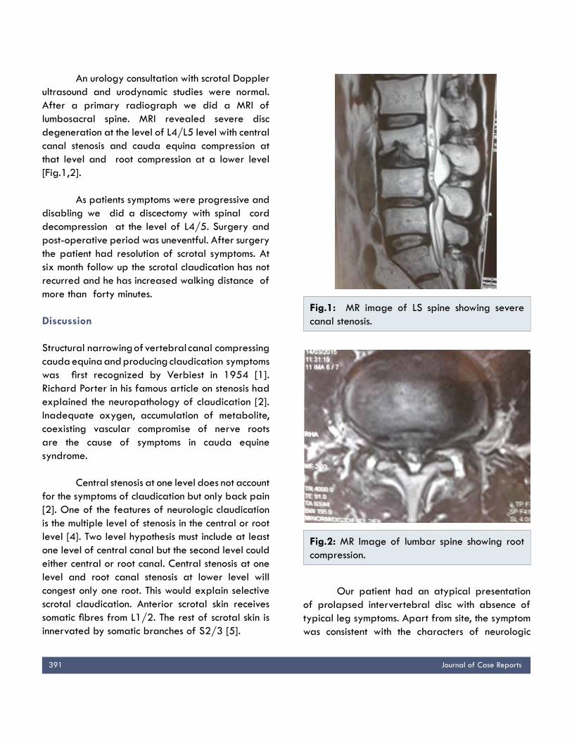
An urology consultation with scrotal Doppler ultrasound and urodynamic studies were normal. After a primary radiograph we did a MRI of lumbosacral spine. MRI revealed severe disc degeneration at the level of L4/L5 level with central canal stenosis and cauda equina compression at that level and root compression at a lower level [Fig.1,2].
As patients symptoms were progressive and disabling we did a discectomy with spinal cord decompression at the level of L4/5. Surgery and post-operative period was uneventful. After surgery the patient had resolution of scrotal symptoms. At six month follow up the scrotal claudication has not recurred and he has increased walking distance of more than forty minutes. Discussion
Structural narrowing of vertebral canal compressing cauda equina and producing claudication symptoms was first recognized by Verbiest in 1954 [1]. Richard Porter in his famous article on stenosis had explained the neuropathology of claudication [2]. Inadequate oxygen, accumulation of metabolite, coexisting vascular compromise of nerve roots are the cause of symptoms in cauda equine syndrome.
Central stenosis at one level does not account for the symptoms of claudication but only back pain [2]. One of the features of neurologic claudication is the multiple level of stenosis in the central or root level [4]. Two level hypothesis must include at least one level of central canal but the second level could either central or root canal. Central stenosis at one level and root canal stenosis at lower level will congest only one root. This would explain selective scrotal claudication. Anterior scrotal skin receives somatic fibres from L1/2. The rest of scrotal skin is innervated by somatic branches of S2/3 [5].
Our patient had an atypical presentation of prolapsed intervertebral disc with absence of typical leg symptoms. Apart from site, the symptom was consistent with the characters of neurologic
Fig.1: MR image of LS spine showing severe canal stenosis.
Fig.2: MR Image of lumbar spine showing root compression.
392 Journal of Case Reports
claudication like pain increasing on walking, standing and decreasing on squatting, sitting and stooping. MRI confirmed spinal canal stenosis at the level of L4/5 with additional root compression at a lower level.
Jacob et al. have described a similar case of scrotal and perianal claudication [6]. Fraser et al. [7] reported various presentations of cauda equina syndrome like bladder, bowel involvement, pain, sexual dysfunction, neurologic deficiency etc. Atypical presentations like intermittent priapism [8] and bilateral foot drop [9] has also been reported in the literature.
Dermatomal distribution should be considered before making such diagnoses. Due to corresponding dermatomal distribution to the stenosis level and resolution of symptoms after surgery we concluded that symptoms were part of cauda equina compression.
Conclusion
Atypical presentation of canal stenosis should be considered in case of scrotal claudication even in the absence of typical symptoms. MRI is the gold standard to confirm the diagnosis.
References
1. Verbiest H. A radicular syndrome from developmental narrowing of the lumbar
vertebral canal. J Bone Joint Surg Br. 1954;36:230-237.
2. Porter RW. Spinal stenosis and neurogenic claudication. Spine (Phila Pa 1976) 1996;21:2046-2052.
3. Inufusa A, An HS, Lim TH, Hasegawa T, Haughton VM, Nowicki BH. Anatomic changes of the spinal canal and intervertebral foramen associated with flexion-extension movement. Spine 1996; 21(21):2412-2420.
4. Porter RW, Ward D. Cauda equina dysfunction, the significance of multiple level pathology. Spine. 1992;17:9-15.
5. Morgan RJ, Parry JRW. Scrotal pain. Postgraduate Medical Journal. 1986;63:521-523.
6. Jacob YL Oh, Jun-Hao Tan. Spinal stenosis presenting with scrotal and perianal claudication Asian Spine J. 2015;9(1):103-105.
7. Fraser S, Roberts L, Murphy E. Cauda equina syndrome: a literature review of its definition and clinical presentation. Arch Phys Med Rehabil. 2009;90:1964-1968.
8. Baba H, Maezawa Y, Furusawa N, Kawahara N, Tomita K. Lumbar spinal stenosis causing intermittent priapism. Paraplegia. 1995;33:338-345.
9. Oluigbo CO, Qadri SR, Dardis R, Choksey MS. Lumbar canal stenosis presenting with acute bilateral foot drop. Br J Neurosurg. 2006;20:87-89.
Related Documents


![Redes sociales [Redes Sociales] [Carolina Barada] Contenidos Desarrollados Por Con Apoyo De.](https://static.cupdf.com/doc/110x72/54d712e34979596c658b58de/redes-sociales-redes-sociales-carolina-barada-contenidos-desarrollados-por-con-apoyo-de.jpg)