Ch. 7 The Skeletal System

Ch. 7 The Skeletal System. Composition It is a solid network of active tissues surrounded by deposits of minerals. Components: –Bone –Joints –Cartilage.
Dec 27, 2015
Welcome message from author
This document is posted to help you gain knowledge. Please leave a comment to let me know what you think about it! Share it to your friends and learn new things together.
Transcript

Ch. 7 The
Skeletal System

Composition
• It is a solid network of active tissues surrounded by deposits of minerals.
• Components:– Bone– Joints– Cartilage– Ligaments

Function of Bones
1. Protection- of soft tissues and organs. 2. Movement – muscles attached to bones=
function as levers.3. Storage – of minerals (Calcium and
Phosphorous) and lipids ( yellow marrow)4. Blood Cell Formation- hematopoiesis- occurs
within the marrow of the bone. 5. Support- for the entire body; Individual bones
provide a framework for attachment of soft tissues and organs.

Bones of the Human Body
• The adult skeleton has 206 bones
• Two basic types of bone tissue– Compact bone
• Homogeneous
– Spongy bone• Small needle-like
pieces of bone
• Many open spaces

Classification of Bones
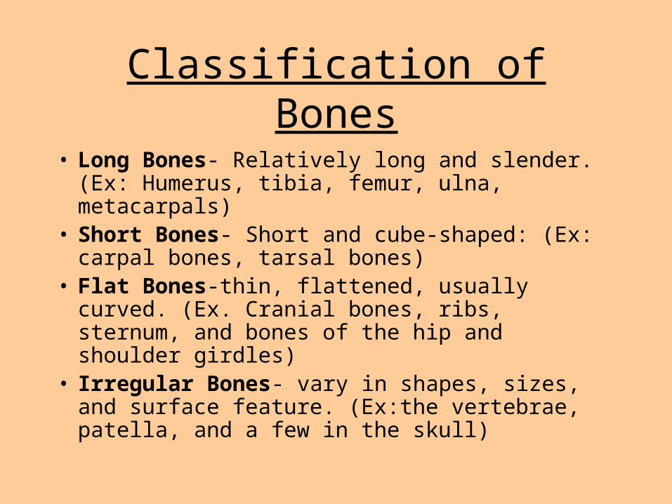
Classification of Bones
• Long Bones- Relatively long and slender. (Ex: Humerus, tibia, femur, ulna, metacarpals)
• Short Bones- Short and cube-shaped: (Ex: carpal bones, tarsal bones)
• Flat Bones-thin, flattened, usually curved. (Ex. Cranial bones, ribs, sternum, and bones of the hip and shoulder girdles)
• Irregular Bones- vary in shapes, sizes, and surface feature. (Ex:the vertebrae, patella, and a few in the skull)


Gross Anatomy of a Long Bone
• Diaphysis– Shaft
– Composed of compact bone
• Epiphysis – Ends of the bone
– Composed mostly of spongy bone
• Epiphyseal plate (growth plate)– a thin layer of cartilage
between the epiphysis
– a secondary bone-forming center
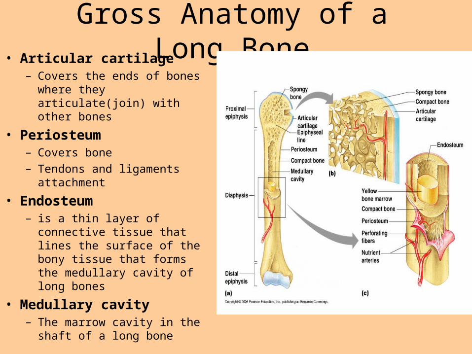
Gross Anatomy of a Long Bone• Articular cartilage
– Covers the ends of bones where they articulate(join) with other bones
• Periosteum– Covers bone
– Tendons and ligaments attachment
• Endosteum– is a thin layer of connective
tissue that lines the surface of the bony tissue that forms the medullary cavity of long bones
• Medullary cavity– The marrow cavity in the shaft
of a long bone
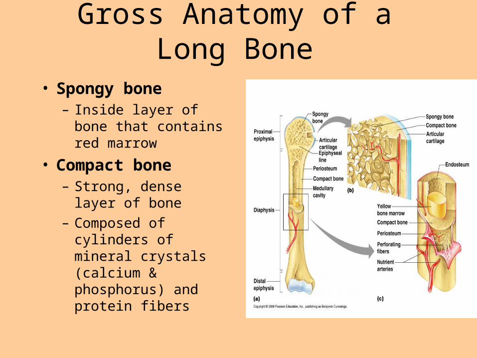
Gross Anatomy of a Long Bone
• Spongy bone– Inside layer of bone that
contains red marrow
• Compact bone– Strong, dense layer of
bone
– Composed of cylinders of mineral crystals (calcium & phosphorus) and protein fibers
Types of Bone Cells
• Osteocytes– Mature bone cells
• Osteoblasts– Bone-forming cells
• Osteoclasts– Bone-destroying cells– Break down bone matrix for remodeling and release of
calcium
• Bone remodeling is a process by both osteoblasts and osteoclasts

Bone Tissue
Microscopic Anatomy of Bone
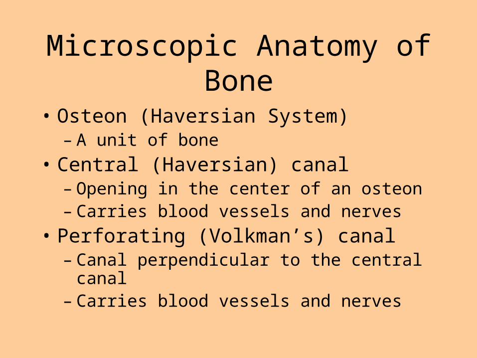
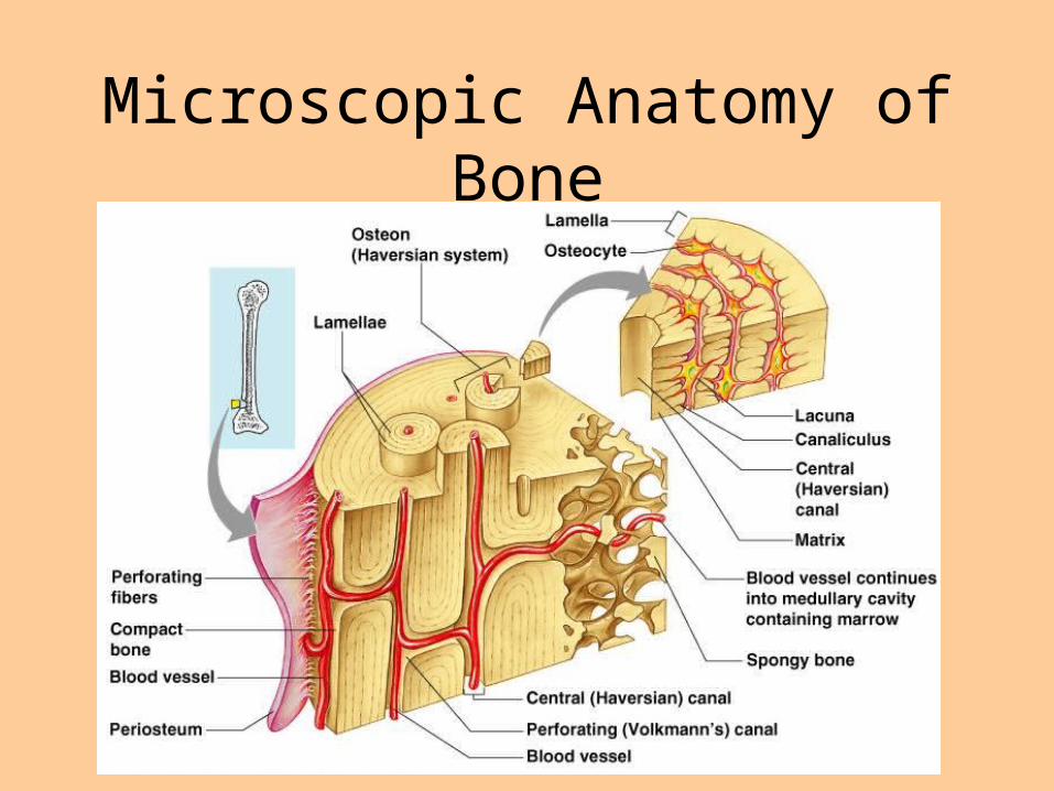
• Osteon (Haversian System)– A unit of bone
• Central (Haversian) canal– Opening in the center of an osteon– Carries blood vessels and nerves
• Perforating (Volkman’s) canal– Canal perpendicular to the central canal– Carries blood vessels and nerves
Microscopic Anatomy of Bone

Microscopic Anatomy of Bone
• Canaliculi – Tiny canals that
radiate from the central canal to lacunae
– Form a transport system

Microscopic Anatomy of Bone
• Lacunae– Cavities containing bone cells (osteocytes)
– Arranged in concentric rings
• Lamellae– Rings around the central canal
– Sites of lacunae

Bone Development and Growth

QuickTime™ and a decompressor
are needed to see this picture.

Bone Development and Growth
• Ossification– The process of producing bone from cartilage.
Bone Development and Growth
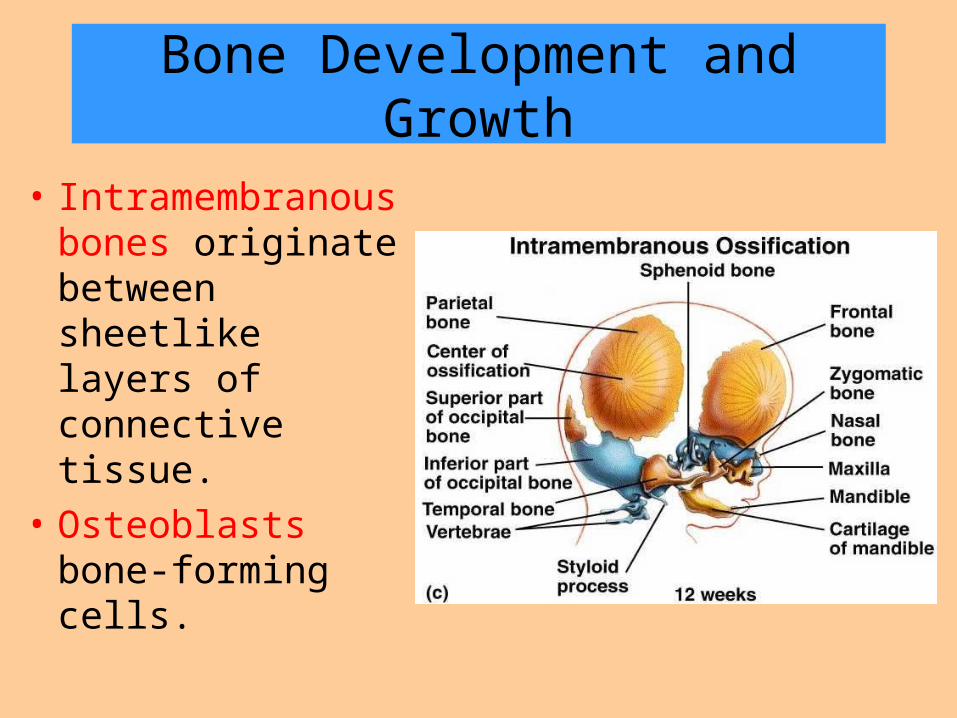
• Intramembranous bones originate between sheetlike layers of connective tissue.
• Osteoblasts bone-forming cells.

• Endochondral bones-Long Bones they develop from masses of cartilage shaped like future bony structures.

• Epiphyseal plate is the portion of bone where growth happens. Bones continue to grow until the plate closes.
• If an Epiphyseal plate is damaged before it ossifies, elongation of the long bone may cease prematurely, or growth maybe uneven.

• Babies are born with over 300 bones; many are composed almost entirely of cartilage.
• The fontanels of a baby’s skull will fuse around age 2, but growth of the skull continues until adulthood.

• Sutures develop and grow throughout childhood at the centers of ossification (growth plates)
• Between the ages 18 – 21, all of the cartilage of the epiphyseal plate is replaced by bone.
• This is called ossification and the bone lengthening process ends.

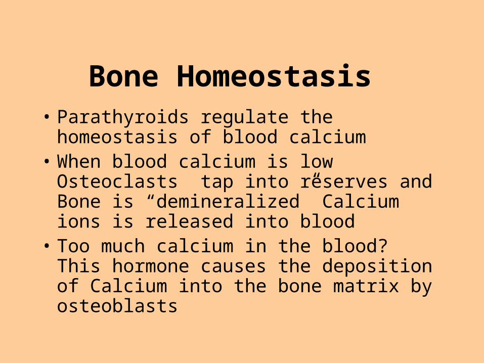
Bone Homeostasis• Parathyroids regulate the homeostasis of
blood calcium• When blood calcium is low Osteoclasts tap
into reserves and Bone is “demineralized” Calcium ions is released into blood
• Too much calcium in the blood? This hormone causes the deposition of Calcium into the bone matrix by osteoblasts
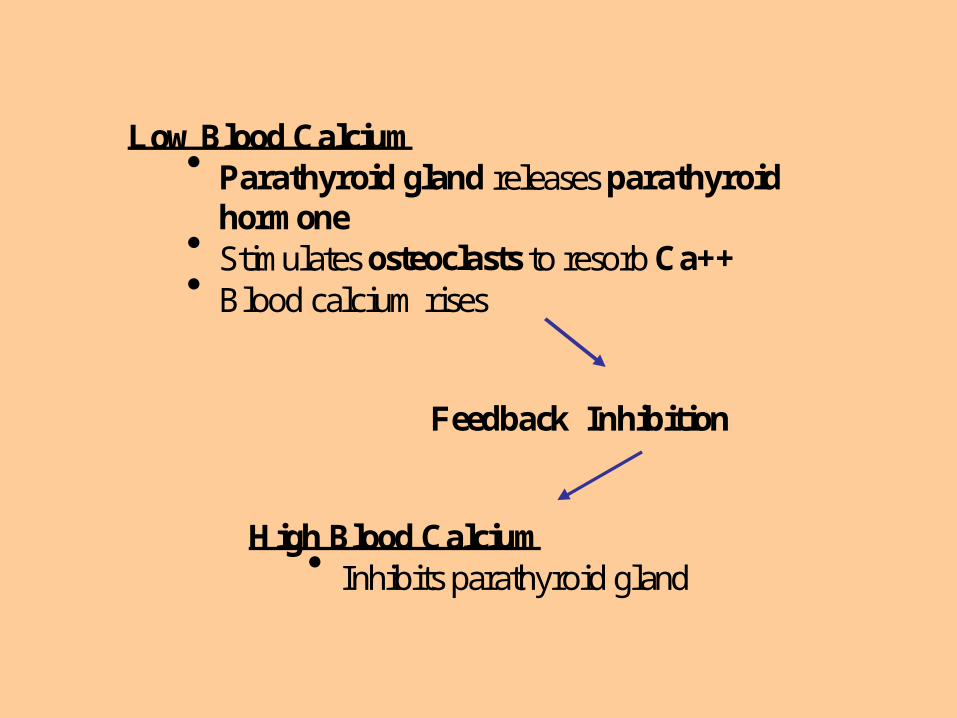
Low Blood Calcium Parathyroid gland releases parathyroid
hormone Stimulates osteoclasts to resorb Ca++ Blood calcium rises
Feedback Inhibition
High Blood Calcium Inhibits parathyroid gland

28
Factors Affecting Bone Development, Growth, and
Repair
• Deficiency of Vitamin A – retards bone development• Deficiency of Vitamin C – results in fragile bones • Deficiency of Vitamin D – rickets, osteomalacia• Insufficient Growth Hormone – dwarfism• Excessive Growth Hormone – gigantism, acromegaly • Insufficient Thyroid Hormone – delays bone growth• Sex Hormones – promote bone formation; stimulate ossification of epiphyseal plates•Insufficient Sex hormone – Osteoporosis • Physical Stress – stimulates bone growth

The Skeletal System
• Divided into two divisions– Axial skeleton– Appendicular skeleton
Skeletal Organization
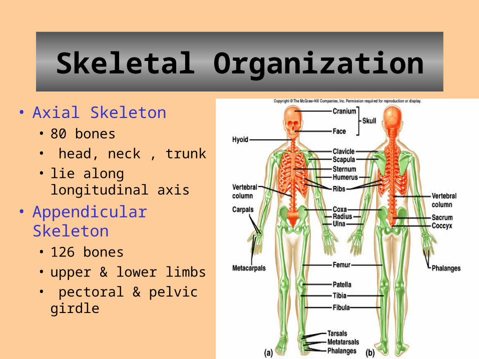
• Axial Skeleton• 80 bones
• head, neck , trunk
• lie along longitudinal axis
• Appendicular Skeleton• 126 bones
• upper & lower limbs
• pectoral & pelvic girdle
30
31
The Skull
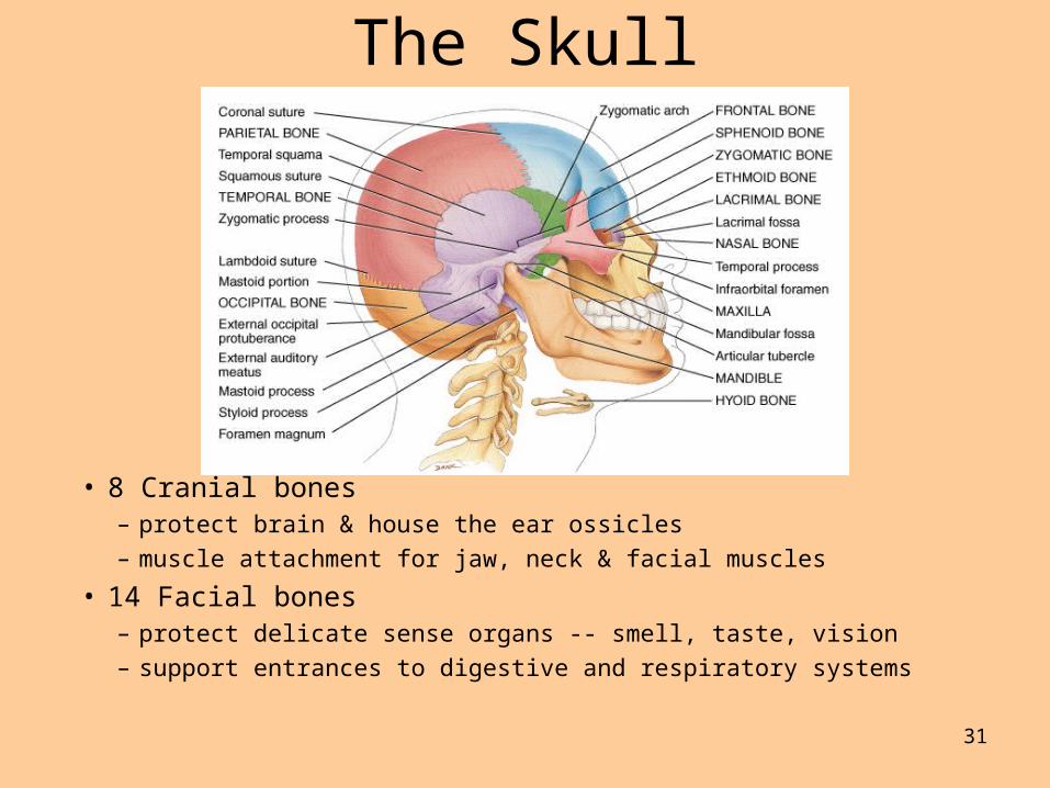
• 8 Cranial bones– protect brain & house the ear ossicles
– muscle attachment for jaw, neck & facial muscles
• 14 Facial bones– protect delicate sense organs -- smell, taste, vision
– support entrances to digestive and respiratory systems

Basic Cranial Bones
• Frontal, parietal, occipital, and temporal.– These make up the basic
brain case.
• Nasal, zygomatic, maxilla, and mandible.– The make up the front of
your face, and your jaw.

– The ears consist of 6 auditory ossicles– Floating in the throat is the Hyoid bone.
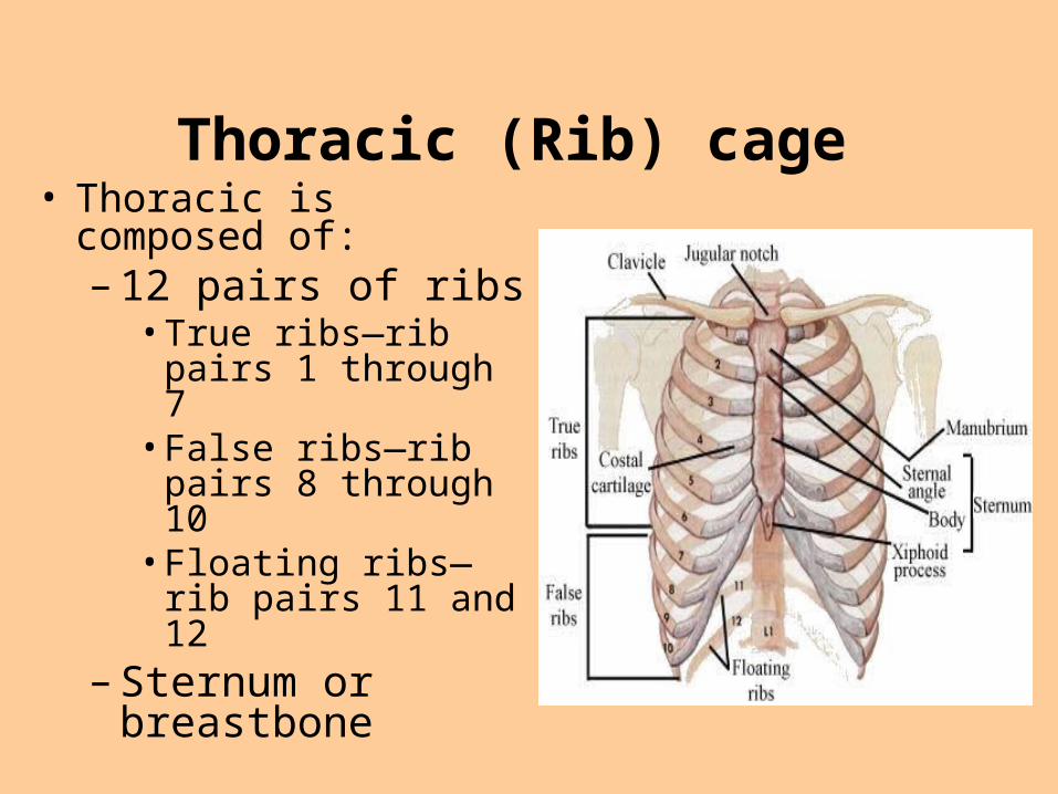
Thoracic (Rib) cage • Thoracic is composed of:
– 12 pairs of ribs• True ribs—rib pairs 1
through 7• False ribs—rib pairs 8
through 10• Floating ribs—rib
pairs 11 and 12– Sternum or breastbone

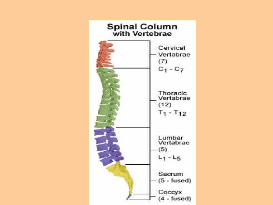
The vertebral column
• The vertebral column consists of
• Cervical vertebrae (7)– Your neck.
• Thoracic vertebrae (12)– Attaches to your ribs.
• Lumbar vertebrae (5)– Your lower back.
• Sacrum and coccyx– Part of your hip and tail bone.
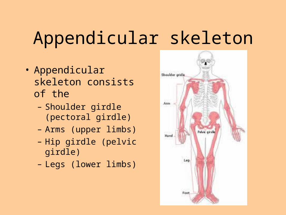
Appendicular skeleton
• Appendicular skeleton consists of the – Shoulder girdle
(pectoral girdle)
– Arms (upper limbs)
– Hip girdle (pelvic girdle)
– Legs (lower limbs)

38
Pectoral Girdle
• Also known as the shoulder girdle • Supports upper limbs•Clavicle (2)• Scapulae (2) Sternum
CostalcartilageRibScapula
Humerus
Ulna
Radius
Clavicle
(a)
Coracoidprocess
Head ofhumerus
Acromionprocess
Acromial endSternal end
Copyright © The McGraw-Hill Companies, Inc. Permission required for reproduction or display.
39
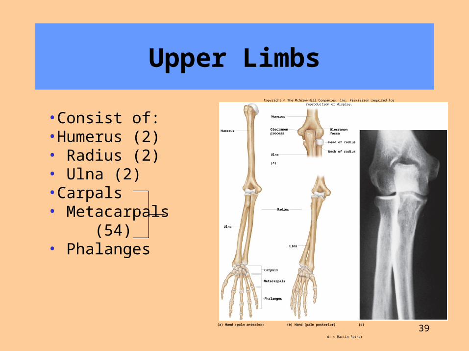
Upper Limbs
•Consist of:•Humerus (2)• Radius (2)• Ulna (2)•Carpals• Metacarpals (54)• Phalanges
Olecranonprocess
Head of radius
Neck of radiusUlna
Olecranonfossa
Carpals
Metacarpals
Phalanges
Humerus
Humerus
Ulna
Ulna
Radius
(c)
(d)(a) Hand (palm anterior) (b) Hand (palm posterior)
Copyright © The McGraw-Hill Companies, Inc. Permission required for reproduction or display.
d: © Martin Rotker
40
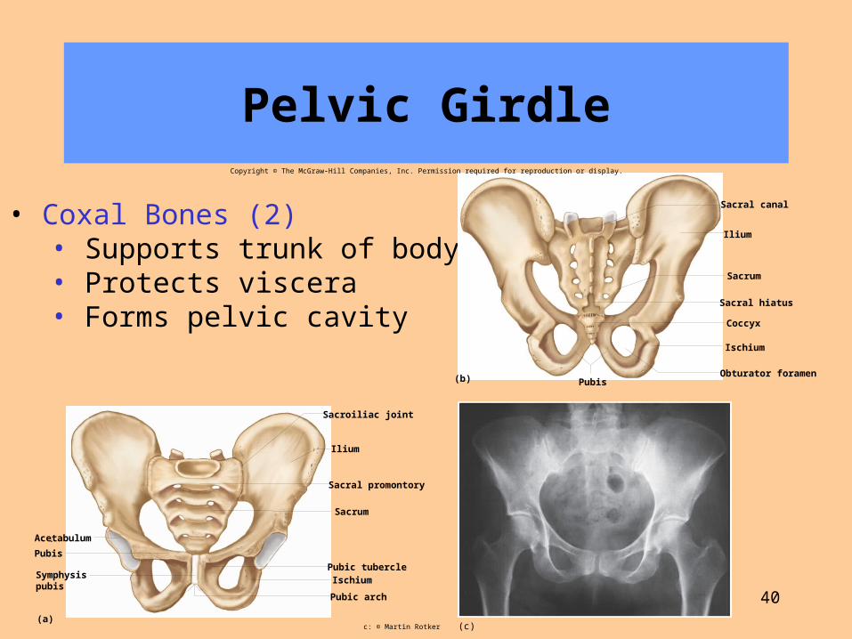
Pelvic Girdle
• Coxal Bones (2)• Supports trunk of body• Protects viscera• Forms pelvic cavity
Sacrum
Sacral promontory
Sacroiliac joint
Acetabulum
Pubis
Symphysispubis
(a)
Pubic arch
IschiumPubic tubercle
Ilium
Obturator foramen
Ischium
Coccyx
Sacral hiatus
Sacrum
(b)
Ilium
Sacral canal
Pubis
Copyright © The McGraw-Hill Companies, Inc. Permission required for reproduction or display.
(c)c: © Martin Rotker

41
Differences Between Male Female Pelves
• Female pelvis• Iliac bones more flared• Broader hips• Pubic arch angle greater•Lighter bones
Sacral promontory
Flared ilium
Pelvic brim
Symphysis pubis
Pubic arch
Pubic arch
(a) Female pelvis
(b) Male pelvis
Sacral promontory
Sacral curvature
Copyright © The McGraw-Hill Companies, Inc. Permission required for reproduction or display.

42
Lower Limb
• Femur (2)• Patella (2)• Tibia (2)• Fibula (2)• Tarsals• Metatarsals (52)• Phalanges
Metatarsals
Fibula
Tibia
Tibia
Patella
Femur
Fibula
(c) Lateral view
Fibula
Tibia
Lateralcondyle
(d) Posterior view
(b)
Medialcondyle
Femur
Tarsals
Phalanges
Femur
Patella
Copyright © The McGraw-Hill Companies, Inc. Permission required for reproduction or display.

43
Joint (Articulations)
A. Every bone except hyoid (which anchors the tongue) connects to at least one other bone
B. Joint types classified by degree of movement 1. Synarthrosis (no movement)—fibrous connective
tissue grows between articulating bones (e.g., sutures of skull)
2. Amphiarthrosis (slight movement)—cartilage connects articulating bones (e.g., symphysis pubis)

44
45
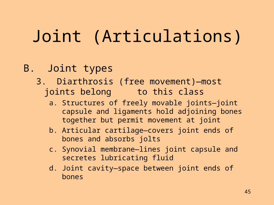
Joint (Articulations)
B. Joint types 3. Diarthrosis (free movement)—most joints belong to
this classa. Structures of freely movable joints—joint capsule and
ligaments hold adjoining bones together but permit movement at joint
b. Articular cartilage—covers joint ends of bones and absorbs jolts
c. Synovial membrane—lines joint capsule and secretes lubricating fluid
d. Joint cavity—space between joint ends of bones

46
Joint (Articulations)
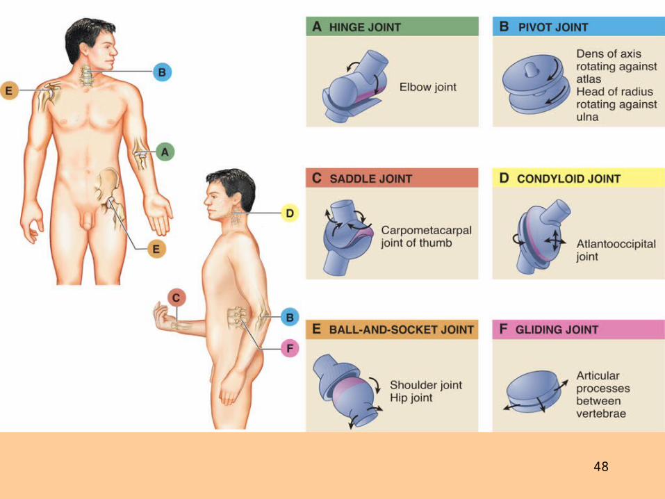
C. Freely movable joints – Ball-and-socket– Hinge– Pivot– Saddle– Gliding– Condyloid

47
48
49
Skeletal Disorders
A. Bone fractures 1. Open (compound) fractures pierce the skin
and closed (simple) fractures do not
2. Fracture types include complete and incomplete, linear, transverse and oblique

50

How does a broken bone heal?
1. Blood flow increases to the area of the break. This allows nutrients and oxygen to help
the healing process.
2. As bone becomes deposited, it grows
stronger, and eventually remodels itself.

1. Conversion of blood clot (hematoma) to a soft tissue procallus.
Takes 48 hours Loose network of fibers is laid down Inflammatory reaction = swelling =
edema Increased blood flow and infiltration
by white blood cells Macrophages phagocytize debris Fibroblasts begin repair process
by laying down connective tissue
53
Skeletal Disorders
B. Joint disorders1. Noninflammatory joint disorders—do not usually
involve inflammation of the synovial membrane; symptoms tend to be local and not systemic
a. Osteoarthritis, or degenerative joint disease (DJD) 1) Most common noninflammatory disorder of movable joints—
often called “wear and tear” arthritis2) Symptoms: joint pain, morning stiffness, Bouchard nodes (at
proximal interphalangeal joints), Heberden nodes (at distal interphalangeal joints) of the fingers
3) Most common cause for partial and total hip and knee replacements

54

55
Skeletal Disorders
• Joint disorders1. Noninflammatory joint disorders
b. Traumatic injury1) Dislocation or subluxation—articular surfaces of bones
in joint are no longer in proper contact
2) Sprain—acute injury to ligaments around joints (e.g., whiplash type injuries)
3) Strain—acute injury to any part of the “musculotendinous unit” (muscle, tendon, junction between the two, and attachments to bone)
56
Skeletal Disorders
• Joint disorders2. Inflammatory joint disorders
• Arthritis: general name for several inflammatory joint diseases that may be caused by infection, injury, genetic factors, and autoimmunity
• Inflammation of the synovial membrane occurs, often with systemic signs and symptoms

57
Skeletal Disorders– Inflammatory joint disorders: Arthritis
a. Rheumatoid arthritis Systemic autoimmune disease—chronic inflammation of synovial membrane with involvement of other tissues such as blood vessels, eyes, heart, and lungs
b. Gouty arthritissynovial inflammation caused by gout, a condition in which sodium urate crystals (URIC ACID) form in joints and other tissues
c. Infectious arthritisarthritis resulting from infection by a pathogen, as in Lyme arthritis and ehrlichiosis, caused by two different bacteria that are transmitted to humans by tick bites

58

59
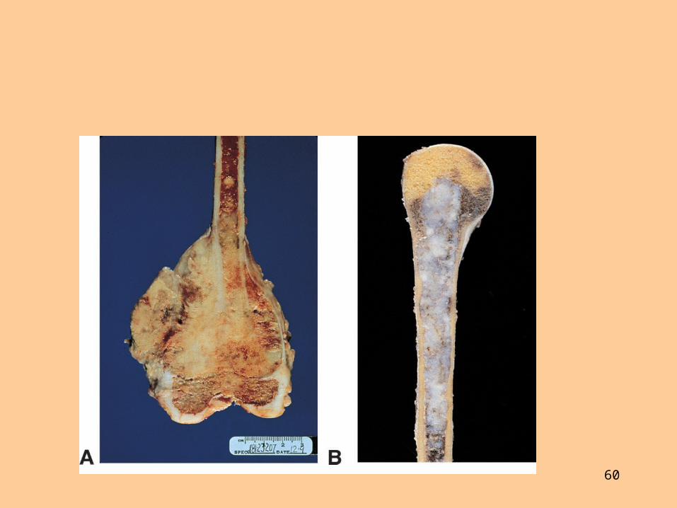
Skeletal Disorders
C. Tumors of bone and cartilage1. Osteosarcoma
a. Most common and serious malignant bone neoplasm
b. Frequent sites include distal femur and proximal tibia and humerus
2. Chondrosarcoma a. Cancer of skeletal hyaline cartilage
b. Second most common cancer of skeletal tissues
c. Frequent sites include medullary cavity of humerus, femur, ribs, and pelvic bones
60

61
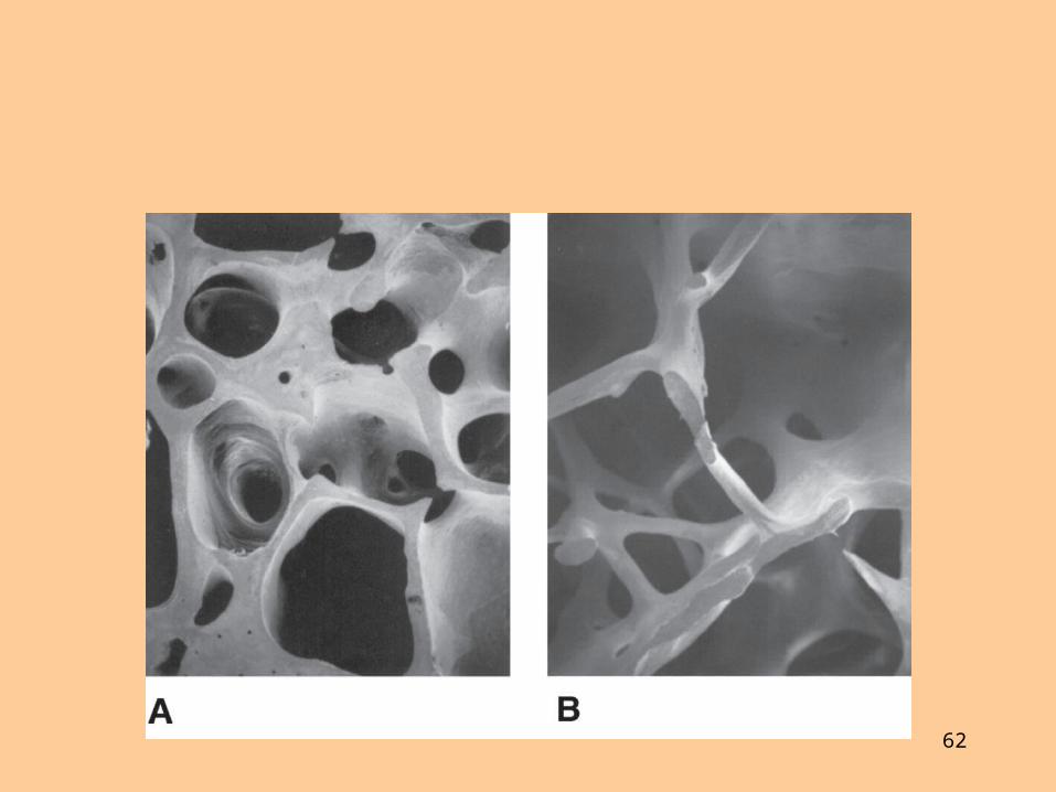
Skeletal Disorders
D. Metabolic bone diseases1. Osteoporosis
a. Characterized by loss of calcified bone matrix and reduction in number of trabeculae in spongy bone
b. Bones fracture easily, especially in wrists, hips, and vertebrae
c. Treatment includes drug therapy, exercise, and dietary supplements of calcium and vitamin D
62
63
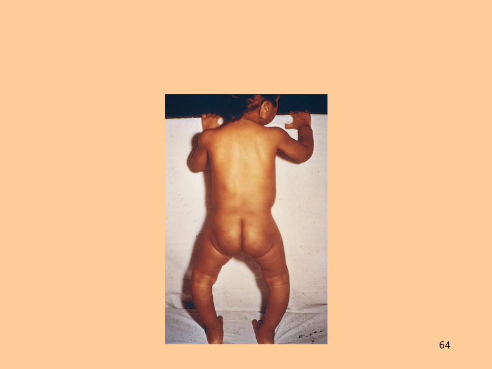
Skeletal Disorders
• Metabolic bone diseases2. Rickets and osteomalacia—both diseases
characterized by loss of bone minerals related to vitamins
a. Rickets – Loss of bone minerals occurs in infants and young
children before skeletal maturity
– Lack of bone rigidity causes gross skeletal changes (bowing of legs)
– Treated with vitamin D
64

65
Skeletal Disorders
• Metabolic bone diseases2. Rickets and osteomalacia
b. Osteomalacia– Mineral content is lost from bones that have already
matured
– Increases susceptibility to fractures
– Treated with vitamin D

66
Skeletal Disorders
• Metabolic bone diseases3. Paget disease (osteitis deformans)
• Faulty remodeling results in deformed bones that fracture easily
• Cause may be genetic or triggered by viral infections

67
68
Skeletal Disorders
• Metabolic bone diseases4. Osteogenesis imperfecta (also called brittle
bone disease) • Bones are brittle because of lack of organic matrix
• Treatment may include splinting to reduce fracture and drugs that decrease cell activity

69
70
Skeletal Disorders
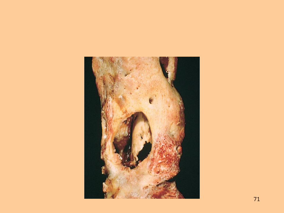
E. Bone infection1. Osteomyelitis
• General term for bacterial (usually staphylococcal) infection of bone
• Treatment may involve surgery, drainage of pus, and IV antibiotic treatment—often over prolonged periods
71

About Spinal Curvature
• At birth your spinal Column is shaped like a C
• When baby is about to crawl the cervical region curves towards posterior.
• As toddler begins to walk, another curve sets in the lumber area in the same direction…resulting in its characteristic S shape.
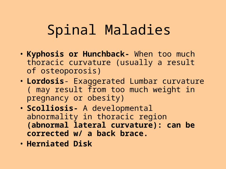
Spinal Maladies
• Kyphosis or Hunchback- When too much thoracic curvature (usually a result of osteoporosis)
• Lordosis- Exaggerated Lumbar curvature ( may result from too much weight in pregnancy or obesity)
• Scolliosis- A developmental abnormality in thoracic region (abnormal lateral curvature): can be corrected w/ a back brace.
• Herniated Disk


What do you think happens if you have a “blown disc”?
• Your cartilage is located between your vertebrae.
• When the tissue surrounding your disc ruptures, it allows your cartilage to move.– “Herniated disc”
• This movement can pinch surrounding nerves, causing back pain.

Rickets:
Vitamin D deficiency in growing children Unable to absorb calcium and phosphate from
gut Inorganic bone matrix (mineral salts) lacks
calcium- Bones deform
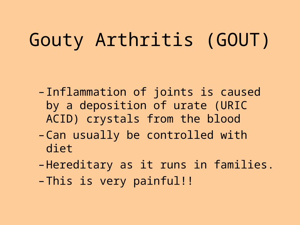
Gouty Arthritis (GOUT)
– Inflammation of joints is caused by a deposition of urate (URIC ACID) crystals from the blood
– Can usually be controlled with diet
– Hereditary as it runs in families.
– This is very painful!!

Cartilaginous Joints
• Bones connected by cartilage
• Examples– Pubic
symphysis– Intervertebral
joints
Figure 5.27d–e
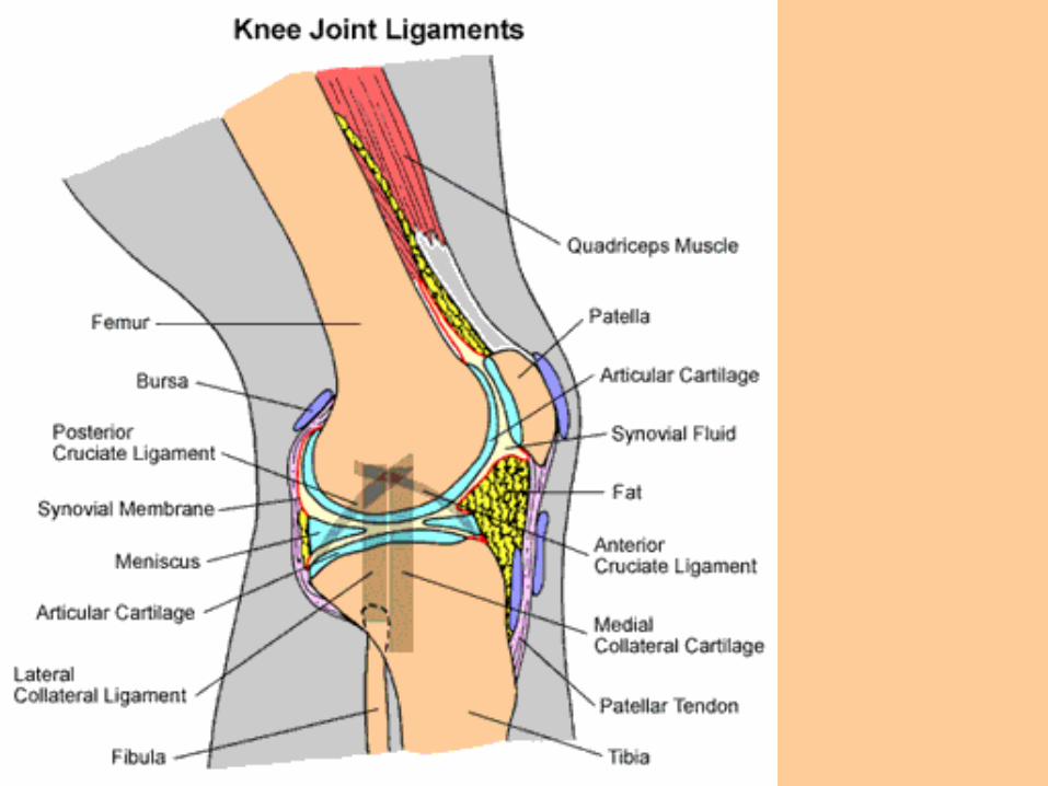

Synovial Joints
• Articulating bones are separated by a joint cavity
• Synovial fluid is found in the joint cavity
Figure 5.24f–h

The Synovial Joint
Figure 5.28
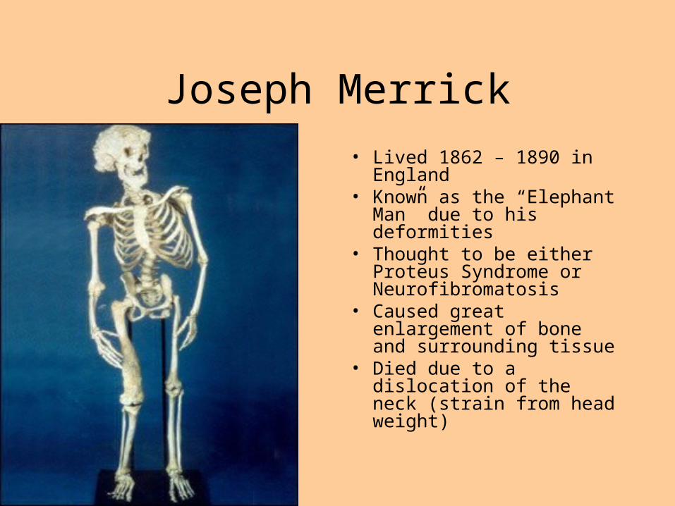
Joseph Merrick
• Lived 1862 – 1890 in England
• Known as the “Elephant Man” due to his deformities
• Thought to be either Proteus Syndrome or Neurofibromatosis
• Caused great enlargement of bone and surrounding tissue
• Died due to a dislocation of the neck (strain from head weight)
Related Documents