Copyright © 2009 Pearson Education, Inc.. Lectures by Gregory Ahearn University of North Florida Chapters 32,33 Circulation and Respiration

Welcome message from author
This document is posted to help you gain knowledge. Please leave a comment to let me know what you think about it! Share it to your friends and learn new things together.
Transcript

Copyright © 2009 Pearson Education, Inc..
Lectures by
Gregory AhearnUniversity of North Florida
Chapters 32,33
Circulation and Respiration
Copyright © 2009 Pearson Education Inc.
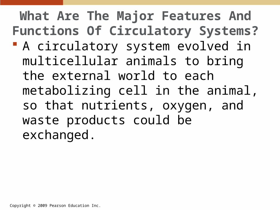
What Are The Major Features And Functions Of Circulatory Systems? A circulatory system evolved in multicellular
animals to bring the external world to each metabolizing cell in the animal, so that nutrients, oxygen, and waste products could be exchanged.

Copyright © 2009 Pearson Education Inc.
What Are The Major Features And Functions Of Circulatory Systems? Circulatory systems have three main
components:• Blood• Blood vessels• Heart
Animals have two types of circulatory systems:• Open circulatory system• Closed circulatory system
Copyright © 2009 Pearson Education Inc.
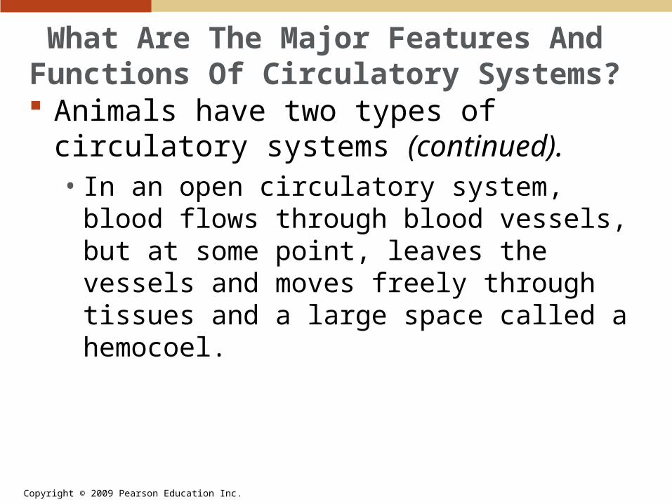
What Are The Major Features And Functions Of Circulatory Systems? Animals have two types of circulatory
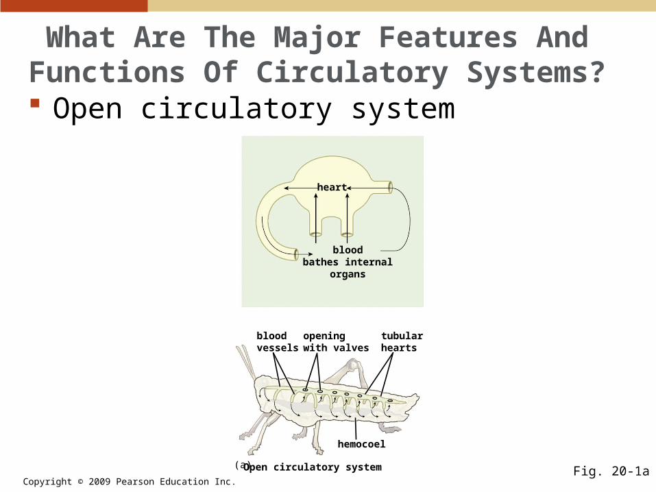
systems (continued).• In an open circulatory system, blood flows
through blood vessels, but at some point, leaves the vessels and moves freely through tissues and a large space called a hemocoel.
Copyright © 2009 Pearson Education Inc.
What Are The Major Features And Functions Of Circulatory Systems? Open circulatory system
Fig. 20-1a
heart
bloodbathes internal
organs
bloodvessels
hemocoel
openingwith valves
tubularhearts
Open circulatory system(a)

Copyright © 2009 Pearson Education Inc.
What Are The Major Features And Functions Of Circulatory Systems? Some invertebrates, including earthworms,
and all vertebrates have closed circulatory systems, where blood is confined to blood vessels and flows through them in a continuous circuit.

Copyright © 2009 Pearson Education Inc.
What Are The Major Features And Functions Of Circulatory Systems? Closed circulatory system
Fig. 20-1b
heart
extracellular fluid
vessels branch ineach organ
heartsvessel
small vessels
Closed circulatory system(b)

Copyright © 2009 Pearson Education Inc.
What Are The Major Features And Functions Of Circulatory Systems?
PLAYPLAY Animation—Open vs. Closed Circulatory Systems
Copyright © 2009 Pearson Education Inc.
What Are The Major Features And Functions Of Circulatory Systems? Animals have two types of circulatory
systems (continued).• In closed circulatory systems of vertebrates,
vessels that carry blood away from the heart are called arteries.
• Arteries carry blood to the smallest blood vessels, called capillaries, which are microscopic vessels that penetrate tissues.

Copyright © 2009 Pearson Education Inc.
What Are The Major Features And Functions Of Circulatory Systems? Animals have two types of circulatory
systems (continued).• Dissolved substances in the blood exchange
with those in the fluid surrounding capillaries, which is called extracellular fluid.
• After passing through capillaries, blood moves back toward the heart through vessels called veins.

Copyright © 2009 Pearson Education Inc.
What Are The Major Features And Functions Of Circulatory Systems? The vertebrate circulatory system transports
many substances.• Transports oxygen from the lungs or gills to
the tissues• Transports carbon dioxide from the tissues to
the lungs or gills• Transports nutrients from the digestive system
to the tissues• Transports waste products and toxic
substances to the liver and the kidney for excretion

Copyright © 2009 Pearson Education Inc.
What Are The Major Features And Functions Of Circulatory Systems? The vertebrate circulatory system transports
many substances (continued).• Transports hormones from the glands and
organs that produce them to the tissues on which they act
• Helps to regulate body temperature by adjusting blood flow
• Helps to protect the body from bacteria and viruses by circulating the cells and molecules of the immune system
Copyright © 2009 Pearson Education Inc.
How Does The Vertebrate Heart Work?
The vertebrate heart consists of muscular chambers.• In vertebrate hearts, muscular chambers,
called atria, collect blood. • Blood flows from the atria to the ventricles,
chambers whose contractions circulate blood through the body.
• The number of atria and ventricles differs among different classes of vertebrates.
Copyright © 2009 Pearson Education Inc.
How Does The Vertebrate Heart Work?
Fish hearts have two chambers, one atrium, and one ventricle; blood flows through the fish body in a single loop.

Copyright © 2009 Pearson Education Inc.
How Does The Vertebrate Heart Work?
Fish heart
Fig. 20-2aFish
gill capillaries
body capillaries
ventricle
atrium
(a)

Copyright © 2009 Pearson Education Inc.
How Does The Vertebrate Heart Work?
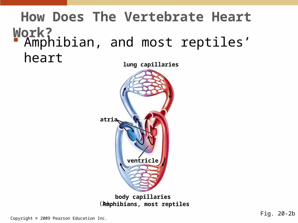
Amphibians and most reptiles have three-chambered hearts and two circulatory loops, one for the lungs and one for the rest of the body.
Blood from both circuits mixes in the heart; therefore, the two circuits are not completely separate.
Copyright © 2009 Pearson Education Inc.
How Does The Vertebrate Heart Work?
Amphibian, and most reptiles’ heart
Fig. 20-2b
Amphibians, most reptiles
lung capillaries
atria
ventricle
body capillaries(b)

Copyright © 2009 Pearson Education Inc.
How Does The Vertebrate Heart Work?
In alligators, crocodiles, birds, and mammals, the heart has four chambers, and the two circulatory loops are completely separate.

Copyright © 2009 Pearson Education Inc.
How Does The Vertebrate Heart Work?
Mammals and birds’ heart
Fig. 20-2cMammals, birds
lung capillaries
body capillaries
atria
ventricle
(c)

Copyright © 2009 Pearson Education Inc.
How Does The Vertebrate Heart Work?
Animation—Vertebrate HeartPLAYPLAY
Copyright © 2009 Pearson Education Inc.
How Does The Vertebrate Heart Work?
Four-chambered hearts, like the human heart, can be thought of as two separate pumps.• One pump, consisting of the right atrium and
right ventricle, pumps oxygen-depleted blood to the lungs.
• The other pump, consisting of the left atrium and left ventricle, moves oxygen-rich blood from the lungs and through the aorta to the rest of the body.

Copyright © 2009 Pearson Education Inc.
How Does The Vertebrate Heart Work?
The human heart and its valves and vessels
Fig. 20-3
aorta
left atrium
pulmonary artery(to left lung)
semilunar valves
pulmonary veins(from left lung)
atrioventricularvalve
left ventricle
thicker muscleof left ventricle
descending aorta(to lower body)right ventricle
inferior vena cava
atrioventricular valve
superiorvena cava
pulmonary artery(to right lung)
pulmonary veins(from right lung)
right atrium
Copyright © 2009 Pearson Education Inc.
How Does The Vertebrate Heart Work?
The atria and ventricles contract in a coordinated way.• The chambers of the heart alternatively
contract and relax.• The two atria contract at the same time,
emptying their contents into the ventricles.• A fraction of a second later, the two ventricles
contract, forcing blood into arteries that exit the heart.
• Both atria and ventricles then relax briefly, and the cycle repeats.
Copyright © 2009 Pearson Education Inc.
How Does The Vertebrate Heart Work?
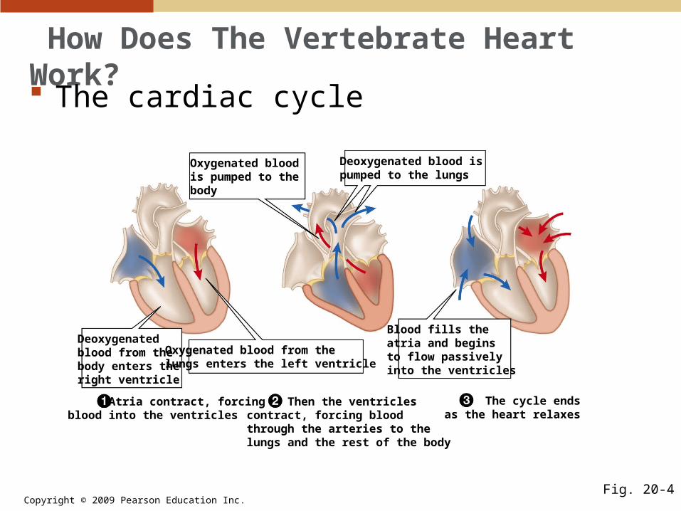
The cardiac cycle
Fig. 20-4
Atria contract, forcingblood into the ventricles
Then the ventriclescontract, forcing bloodthrough the arteries to thelungs and the rest of the body
The cycle endsas the heart relaxes
Deoxygenated blood ispumped to the lungs
Blood fills theatria and beginsto flow passivelyinto the ventricles
Deoxygenatedblood from thebody enters theright ventricle
Oxygenated blood from thelungs enters the left ventricle
Oxygenated bloodis pumped to thebody
Copyright © 2009 Pearson Education Inc.
How Does The Vertebrate Heart Work?
Animation—Function of the Human HeartPLAYPLAY

Copyright © 2009 Pearson Education Inc.
How Does The Vertebrate Heart Work?
The atria and ventricles contract in a coordinated way (continued).• At a normal resting heart rate, the cycle lasts
about 1 second.• Blood pressure changes as the cycle
proceeds.• Systolic pressure is measured during
ventricular contraction, and diastolic pressure is measured between contractions.

Copyright © 2009 Pearson Education Inc.
How Does The Vertebrate Heart Work?
Measuring blood pressure
Fig. 20-5
A stethoscopedetectspulse sounds
The cuff is inflated,putting pressureon the artery
cuff

Copyright © 2009 Pearson Education Inc.
How Does The Vertebrate Heart Work?
Animation—Measuring Blood PressurePLAYPLAY

Copyright © 2009 Pearson Education Inc.
How Does The Vertebrate Heart Work?
Valves prevent blood from moving in the wrong direction.• When the ventricles contract, blood must
move only out through the arteries and not back into the atria.
• Once blood has left the ventricles and entered the arteries, it must be prevented from flowing back as the heart relaxes.

Copyright © 2009 Pearson Education Inc.
How Does The Vertebrate Heart Work?
Valves prevent blood from moving in the wrong direction (continued).• These needs are met by four one-way valves.
• Atrioventricular valves separate the atria from the ventricles.
• Semilunar valves allow blood to enter the pulmonary artery and the aorta when the ventricles contract.

Copyright © 2009 Pearson Education Inc.
How Does The Vertebrate Heart Work?
Electrical impulses coordinate the sequence of contractions.• Contractions are coordinated by the
pacemaker, a cluster of specialized heart muscle cells that produce spontaneous electrical signals.
• The heart’s primary pacemaker is the sinoatrial (SA) node, located in the wall of the right atrium.

Copyright © 2009 Pearson Education Inc.
How Does The Vertebrate Heart Work?
Electrical impulses coordinate the sequence of contractions (continued).• From the SA node, an electrical impulse
creates a wave of muscular contraction that spreads through the right and left atria until it arrives at the unexcitable tissue between the atria and ventricles.
• There, the excitation is channeled through the atrioventricular (AV) node.

Copyright © 2009 Pearson Education Inc.
How Does The Vertebrate Heart Work?
Electrical impulses coordinate the sequence of contractions (continued).• From the AV node, the signal to contract
spreads along excitable fibers to the base of the two ventricles.
• This signal causes the ventricles to contract in unison.
• If the pacemaker fails, uncoordinated irregular contractions, called fibrillation, occurs, and blood cannot be pumped out of the heart.

Copyright © 2009 Pearson Education Inc.
How Does The Vertebrate Heart Work?
The heart’s pacemaker and its connections
Fig. 20-6
Unexcitable tissueseparates the atriaand ventricles
AV node
The sinoatrial nodeelectrical signal startsthe atrial contraction
The atrioventricularnode transmits thesignal to the ventricleswith a slight delay
The signal travels tothe base of the ventricles
Excitable fibers transmitthe signals to ventricularcardiac muscle, causingcontraction from the baseupwards
The signal spreads,causing the atria tocontract
excitablefibers
Copyright © 2009 Pearson Education Inc.
How Does The Vertebrate Heart Work?
The heart’s contractions result from movement of filaments in muscle cells.• The muscle tissue that makes up the heart
consists of cells known as muscle fibers.• Each muscle fiber contains many myofilbrils,
cylindrical structures that extend from one end of the fiber to the other.
• The myofibrils are composed of subunits called sarcomeres, which are aligned end to end long the length of the myofibril.

Copyright © 2009 Pearson Education Inc.
How Does The Vertebrate Heart Work?
A muscle fiber
Fig. 20-7a,b
myofibril
membrane
Cross section of fiber(a)
mu
scle
fib
er
sarcomere
myofibril
thin filament
Z lines
Myofibril and sarcomerethick filament
(b)

Copyright © 2009 Pearson Education Inc.
How Does The Vertebrate Heart Work?
Within each sarcomere is a precise arrangement of filaments of the proteins actin and myosin; actin forms the thin filaments, and myosin forms the thick filaments.
Fig. 20-7cThick and thin filaments
thin filament
thick filament(myosin)
cross-bridge
actin
accessoryproteins
(c)
Copyright © 2009 Pearson Education Inc.
How Does The Vertebrate Heart Work?
The heart’s contractions result from movement of filaments in muscle cells (continued).• The thin filaments are attached to fibrous
protein bands called Z lines, which separate adjacent sarcomeres.
• During contraction, the myosin filaments make contact with the actin filaments and, using ATP energy, pull the actin strand past the myosin strand, shortening the sarcomere and contracting the muscle.

Copyright © 2009 Pearson Education Inc.
How Does The Vertebrate Heart Work?
Muscle contraction
Fig. 20-8
Relaxedmuscle
Contractedmuscle
z line
sarcomere
A sarcomere shortensCross-bridge attachment and release
thick filament
binding sites
cross-bridge
ATP
thin filament
(b)(a)
When bindingsites are exposed,cross-bridgesattach to thebinding sites
The cross-bridges bend,moving thefilaments pastone another andshortening thesarcomere
Using energy fromATP, the cross-bridgesrelease, straighten, andreattach farther along
Copyright © 2009 Pearson Education Inc.
Muscle Contraction
Suggested Media Enhancement:
Muscle ContractionTo access this animation go to folder C_Animations_and_Video_Filesand open the BioFlix folder.

Copyright © 2009 Pearson Education Inc.
What Is Blood?
Blood transports dissolved nutrients, gases, hormones, and wastes through the body. • It has two major components:
• A fluid, called plasma• Cellular components—including red blood
cells, white blood cells, and platelets—which are suspended in the plasma
• The cellular components are produced in bone marrow and later move into the blood.

Copyright © 2009 Pearson Education Inc.
What Is Blood?
Blood cells
Fig. 20-9
platelets
megakaryocyte
neutrophil neutrophil
basophil
monocyte
eosinophil
lymphocyte
red blood cells
Erythrocytes White blood cells
Megakaryocyte forming platelets
(a) (b)
(c)

Copyright © 2009 Pearson Education Inc.
What Is Blood?
Plasma is primarily water and dissolved substances.• Plasma is 90% water.• Dissolved in the plasma are proteins,
hormones, nutrients, salts, and wastes, such as urea.

Copyright © 2009 Pearson Education Inc.
What Is Blood?
Red blood cells carry oxygen from the lungs to the tissues.• The most abundant cells in the blood are red
blood cells.• Red blood cells get their red color from
hemoglobin, an iron-containing protein that can bind up to four oxygen molecules.
• Hemoglobin picks up oxygen in the lungs, where oxygen is at high concentration, and releases it in other tissues of the body, where the oxygen concentration is low.

Copyright © 2009 Pearson Education Inc.
What Is Blood?
White blood cells help defend the body against disease.• White blood cells, or leukocytes, make up less
than 1% of blood cells but play a key role in the body’s resistance to disease.
• There are five types of white blood cells:• Neutrophils • Eosinophils • Basophils • Lymphocytes• Monocytes

Copyright © 2009 Pearson Education Inc.
What Is Blood?
Lymphocytes are responsible for the immune response against disease.• Neutrophils and monocytes engulf foreign
particles.
Fig. 20-10
Copyright © 2009 Pearson Education Inc.
What Is Blood?
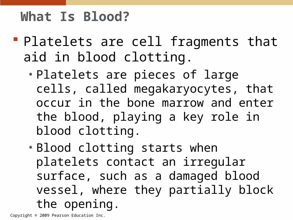
Platelets are cell fragments that aid in blood clotting.• Platelets are pieces of large cells, called
megakaryocytes, that occur in the bone marrow and enter the blood, playing a key role in blood clotting.
• Blood clotting starts when platelets contact an irregular surface, such as a damaged blood vessel, where they partially block the opening.

Copyright © 2009 Pearson Education Inc.
What Is Blood?
The platelets and injured tissue initiate a complex sequence of reactions among plasma proteins, which results in a fibrous network, called fibrin, that traps red blood cells and closes the wound.
Fig. 20-11
platelets white blood cell
red blood cell
fibrin strands

Copyright © 2009 Pearson Education Inc.
What Are The Types And Functions Of Blood Vessels? As it leaves the heart, blood travels from
arteries to arterioles to capillaries to venules to veins, and finally, it returns to the heart.
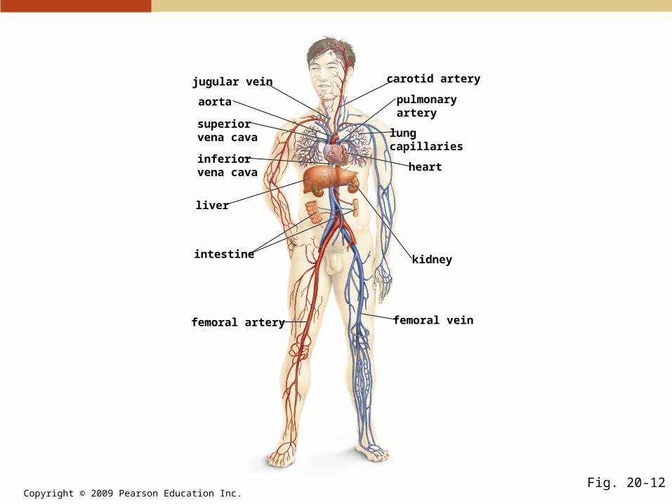
Copyright © 2009 Pearson Education Inc.Fig. 20-12
jugular vein
aorta
superiorvena cava
carotid artery
lungcapillaries
pulmonaryartery
heart
kidney
femoral vein
intestine
inferiorvena cava
liver
femoral artery

Copyright © 2009 Pearson Education Inc.
Arteries and arterioles carry blood away from the heart.• These vessels have thick walls embedded
with smooth muscle and elastic connective tissue.
• Arteries branch into vessels of small diameter called arterioles.
precapillarysphincters
arteriole
venule
artery vein
capillaryvalve
smooth muscleconnective tissue
smoothmuscle
crosssection
capillarynetwork
What Are The Types And Functions Of Blood Vessels?
Fig. 20-13
Copyright © 2009 Pearson Education Inc.
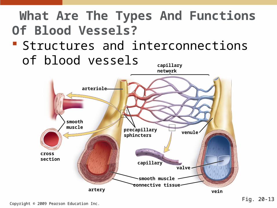
What Are The Types And Functions Of Blood Vessels? Structures and interconnections of blood
vessels
Fig. 20-13
precapillarysphincters
arteriole
venule
artery vein
capillaryvalve
smooth muscle
connective tissue
smoothmuscle
crosssection
capillarynetwork
Copyright © 2009 Pearson Education Inc.
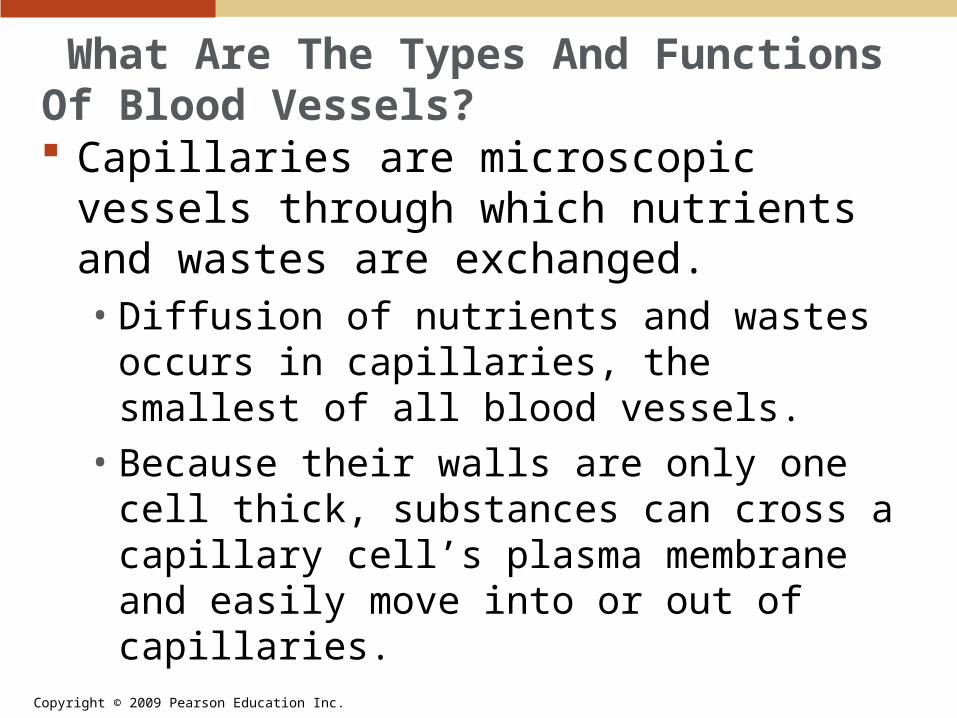
What Are The Types And Functions Of Blood Vessels? Capillaries are microscopic vessels through
which nutrients and wastes are exchanged.• Diffusion of nutrients and wastes occurs in
capillaries, the smallest of all blood vessels.• Because their walls are only one cell thick,
substances can cross a capillary cell’s plasma membrane and easily move into or out of capillaries.

Copyright © 2009 Pearson Education Inc.
What Are The Types And Functions Of Blood Vessels? Capillaries are microscopic vessels through
which nutrients and wastes are exchanged (continued).• Capillaries are so narrow that red blood cells
pass through them in single file.• The speed of blood flow drops very quickly as
it moves through this narrow capillary network.• The flow of blood in capillaires is regulated by
tiny rings of smooth muscle, called precapillary sphincters, which surround the junctions between arterioles and capillaries.

Copyright © 2009 Pearson Education Inc.
What Are The Types And Functions Of Blood Vessels? Red blood cells flow through a capillary.
Fig. 20-14
Red blood cells mustpass through capillariesin single file
Capillary walls are thinand permeable to gases,nutrients, and cellularwastes

Copyright © 2009 Pearson Education Inc.
What Are The Types And Functions Of Blood Vessels? Venules and veins carry blood back to the
heart.• After picking up carbon dioxide and other
cellular wastes from cells, capillary blood drains into larger vessels called venules, which empty into larger veins.
• To prevent blood from flowing away form the heart, veins are equipped with valves that allow blood to flow in only one direction.

Copyright © 2009 Pearson Education Inc.
What Are The Types And Functions Of Blood Vessels? Blood pressure is low in veins and
contraction of skeletal muscle during exercise helps return blood to the heart by squeezing the veins and forcing blood through them.

Copyright © 2009 Pearson Education Inc.
What Are The Types And Functions Of Blood Vessels? Valves direct the
flow of blood in veins.
Fig. 20-15
valveclosed
valveclosed
valveopen
relaxedmuscle
musclecontractioncompressesvein
Copyright © 2009 Pearson Education Inc.
How Does The Lymphatic System Work With The Circulatory System? The lymphatic system consists of:
• A network of lymph vessels that empty into the circulatory system
• Numerous small lymph nodes• The thymus• The spleen
Copyright © 2009 Pearson Education Inc.
How Does The Lymphatic System Work With The Circulatory System? The lymphatic system removes excess fluid
and dissolved substances that leak from the capillaries.• It transports fats from the small intestine to the
blood stream.• It defends the body by exposing bacteria and
viruses to white blood cells.

Copyright © 2009 Pearson Education Inc.Fig. 20-16
thoracic duct enters avein to the vena cava
thymus
heart
spleen
thoracic duct
valve preventsbackflow
lymph node
chamberspacked withwhite blood cells
superiorvena cava
lymphvessels
lymphnodes
Copyright © 2009 Pearson Education Inc.
How Does The Lymphatic System Work With The Circulatory System? Lymphatic vessels resemble the capillaries
and veins of the circulatory system.• The smallest lymph vessels are lymph
capillaries.• Lymph capillary walls have specialized
openings between the cells that act as one-way valves.
• These openings allow large particles to be carried into the lymph capillaries along with fluid.
Copyright © 2009 Pearson Education Inc.
How Does The Lymphatic System Work With The Circulatory System? Lymphatic vessels resemble the capillaries
and veins of the circulatory system (continued).• Materials collected in the lymph flow into
larger lymph vessels.• The direction of flow in lymph vessels is
regulated by one-way valves.
Copyright © 2009 Pearson Education Inc.
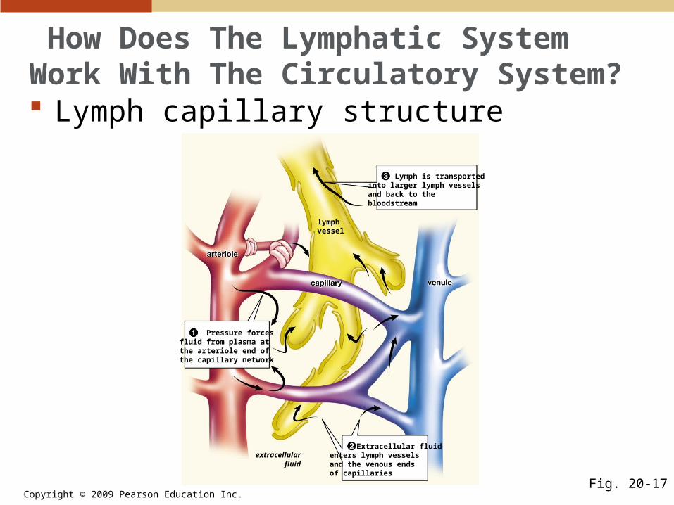
How Does The Lymphatic System Work With The Circulatory System? Lymph capillary structure
Fig. 20-17
extracellularfluid
lymphvessel
Pressure forcesfluid from plasma atthe arteriole end ofthe capillary network
Extracellular fluidenters lymph vesselsand the venous endsof capillaries
Lymph is transportedinto larger lymph vesselsand back to thebloodstream

Copyright © 2009 Pearson Education Inc.
How Does The Lymphatic System Work With The Circulatory System? The lymphatic system returns fluids to the
blood.• The lymphatic system collects excess fluid
that leaks out of the blood capillaries and returns it to the blood.
• As extracellular fluid accumulates, its pressure forces the fluid through the one-way opening in the lymph capillary walls.
• Once inside lymphatic vessels, the excess fluid taken up is now called lymph.
Copyright © 2009 Pearson Education Inc.
How Does The Lymphatic System Work With The Circulatory System? The lymphatic system transports fats from
the small intestine to the blood.• After a fatty meal, the cells of the small
intestine absorb globules of digested fat.• These globules are too large to diffuse into
blood capillaries, but they easily move into openings between lymph capillary cells.
• They are later deposited into the superior vena cava that enters the heart.

Copyright © 2009 Pearson Education Inc.
How Does The Lymphatic System Work With The Circulatory System? The lymphatic system helps defend the
body against disease.• In the lymph nodes, lymph is forced through
channels that are lined with masses of white blood cells that recognize and destroy foreign particles, such as bacteria or viruses.
• The thymus and spleen are considered part of the lymphatic system.

Copyright © 2009 Pearson Education Inc.
How Does The Lymphatic System Work With The Circulatory System? The lymphatic system helps defend the
body against disease (continued).• Certain types of white cells mature in the
thymus.• The spleen is another white blood cell-
producing organ, which also filters blood, exposing it to white blood cells that destroy foreign particles and aged red blood cells.

Copyright © 2009 Pearson Education Inc.
How Are Oxygen And Carbon Dioxide Exchanged In Animal Bodies? The process by which the body acquires
oxygen from the environment and delivers carbon dioxide back to it is called respiration.• Although animal respiration systems are
diverse, they all rely on diffusion of gases across a respiratory surface.
• Respiratory surfaces are large and moist; they must remain moist because gases must be dissolved in water in order to diffuse into or out of cells.

Copyright © 2009 Pearson Education Inc.
How Are Oxygen And Carbon Dioxide Exchanged In Animal Bodies? Aquatic animals may have gills.
• For some small aquatic animals, gases are exchanged across their body surface.
• Most larger animals have specialized respiratory structures; many have gills, which are elaborately folded to increase their surface area.

Copyright © 2009 Pearson Education Inc.
How Are Oxygen And Carbon Dioxide Exchanged In Animal Bodies? Aquatic animals may have gills (continued).
• Fish pump oxygen-rich water into the mouth and over the gills, ejecting it through an opening just behind the gills.
• Fish gills have a series of filaments that are covered with capillary-filled lamellae where gases are exchanged.

Copyright © 2009 Pearson Education Inc.
How Are Oxygen And Carbon Dioxide Exchanged In Animal Bodies? Gills exchange gases with water.
Fig. 20-19
Water flows over gills Gill structure Lamella
waterin
gill arch
water out
(protectiveflapremoved)
gill filament
gill archlamellae
lamella
water flow blood flow
capillaries
deoxygenatedblood
oxygenatedblood
(a) (b) (c)
Copyright © 2009 Pearson Education Inc.
How Are Oxygen And Carbon Dioxide Exchanged In Animal Bodies? Terrestrial animals have internal respiratory
structures.• In land animals, the respiratory structures are
lungs.• Lungs are chambers containing moist gas-
exchange membranes that are protected within the body, where water loss is minimized.

Copyright © 2009 Pearson Education Inc.
How Does The Human Respiratory System Work? There are two parts to the human
respiratory system:• The conducting portion• The gas exchange portion

Copyright © 2009 Pearson Education Inc.
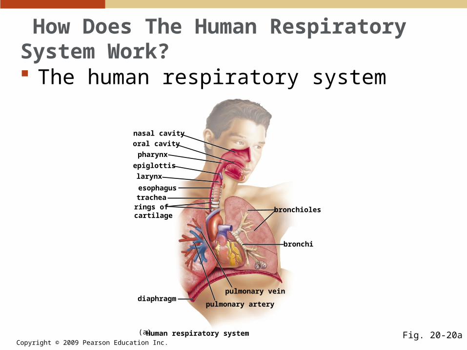
How Does The Human Respiratory System Work? The conducting portion brings air to the
lungs.• Air enters through the nose or mouth, passes
through the nasal cavity or oral cavity into the pharynx, then travels through the larynx.
• Both food and air pass through the pharynx; the epiglottis is a flap of tissue that covers the larynx when food is being swallowed, preventing it from going into the trachea and on to the lungs.
Copyright © 2009 Pearson Education Inc.
How Does The Human Respiratory System Work? The human respiratory system
Fig. 20-20aHuman respiratory system
diaphragm
nasal cavity
pharynx
oral cavity
epiglottis
larynx
esophagustrachearings ofcartilage
pulmonary vein
pulmonary artery
bronchi
bronchioles
(a)
Copyright © 2009 Pearson Education Inc.
How Does The Human Respiratory System Work? The conducting portion brings air to the
lungs (continued).• Inhaled air continues past the larynx into the
trachea, which is a flexible tube whose walls are reinforced with semicircular bands of stiff cartilage.
• The trachea splits into two bronchi, which continue to branch more and to get smaller with each branch until they become bronchioles.
• Bronchioles are connected to groups of small air sacs called alveoli.
Copyright © 2009 Pearson Education Inc.
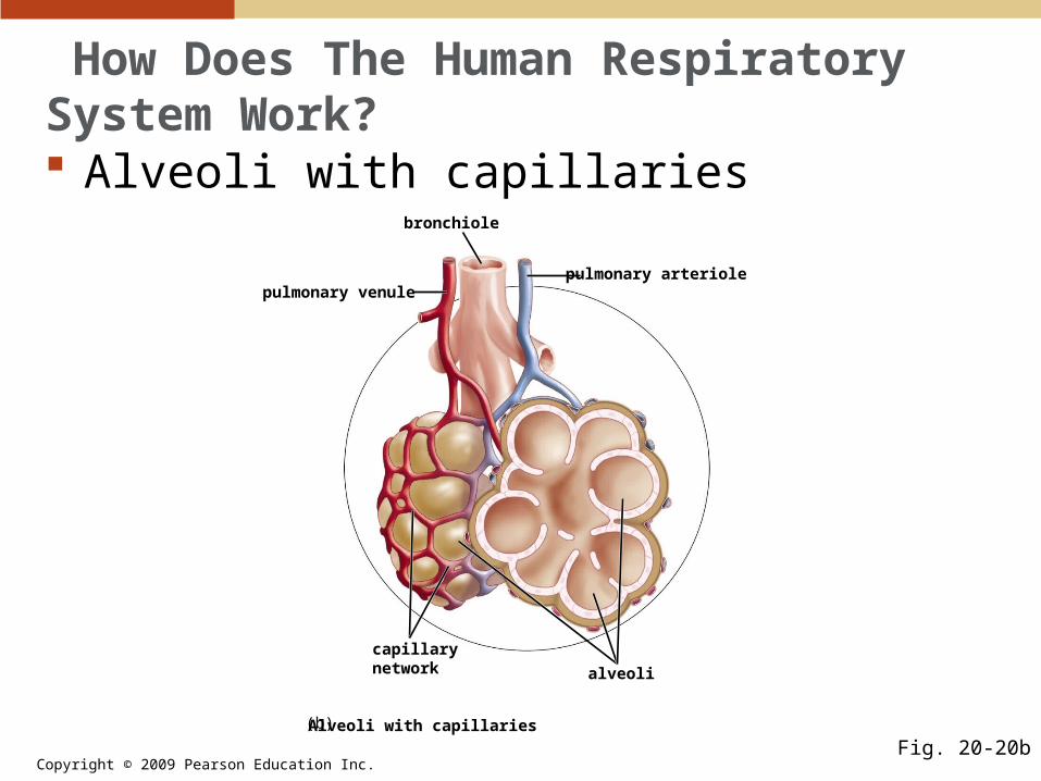
How Does The Human Respiratory System Work? Alveoli with capillaries
Fig. 20-20bAlveoli with capillaries
bronchiole
pulmonary venule
alveoli
capillarynetwork
pulmonary arteriole
(b)
Copyright © 2009 Pearson Education Inc.
How Does The Human Respiratory System Work? Gas exchange occurs in the alveoli.
• Both the alveolar wall and the adjacent capillary walls are only one cell thick, so the air in the lungs is extremely close to the blood in the capillaries.
• A thin layer of watery fluid lines each alveolus.
Copyright © 2009 Pearson Education Inc.
frompulmonaryartery
alveolarmembrane
respiratorymembrane
to pulmonary vein
(air) CO2
O2
capillary
fluid
Oxygen diffusesinto red blood cells
Carbon dioxidediffuses into alveolus
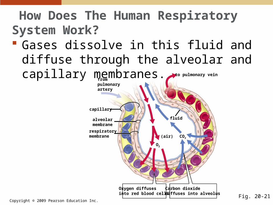
How Does The Human Respiratory System Work? Gases dissolve in this fluid and diffuse
through the alveolar and capillary membranes.
Fig. 20-21

Copyright © 2009 Pearson Education Inc.
How Does The Human Respiratory System Work? Oxygen diffuses from the air in the alveoli,
where its concentration is high, into the blood, where its concentration is low; the flow of carbon dioxide is opposite to that of oxygen.
Copyright © 2009 Pearson Education Inc.
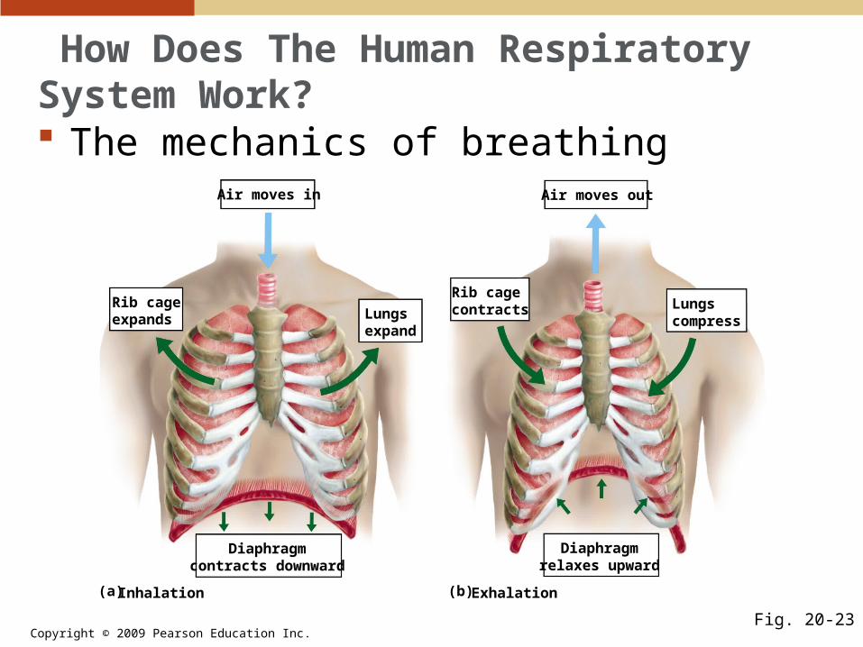
Air moves in Air moves out
Rib cagecontracts Lungs
compress
Diaphragmrelaxes upward
Diaphragmcontracts downward
Rib cageexpands Lungs
expand
Inhalation Exhalation(a) (b)
Fig. 20-23
How Does The Human Respiratory System Work? The mechanics of breathing
Copyright © 2009 Pearson Education Inc.
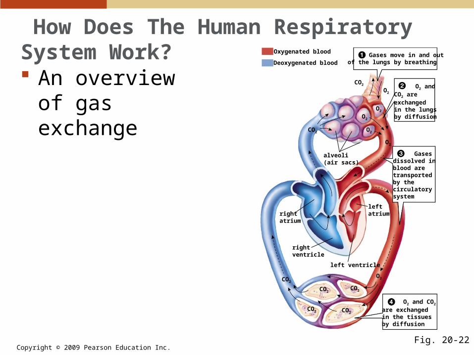
How Does The Human Respiratory System Work? An overview of
gas exchange
Fig. 20-22
CO2
alveoli(air sacs)
Oxygenated blood
Deoxygenated blood Gases move in and outof the lungs by breathing
O2 andCO2 areexchangedin the lungsby diffusion
Gasesdissolved inblood aretransportedby thecirculatorysystem
O2
left ventricle
O2 and CO2
are exchangedin the tissuesby diffusion
rightatrium
rightventricle
leftatrium
O2
O2
O2
CO2
O2
CO2
CO2
CO2
CO2 CO2
O2

Copyright © 2009 Pearson Education Inc.
How Does The Human Respiratory System Work?
Animation—Gas ExchangePLAYPLAY

Copyright © 2009 Pearson Education Inc.
How Does The Human Respiratory System Work? Carbon dioxide and oxygen are transported
in different ways.• Almost all of the oxygen transported in blood
is bound to hemoglobin molecules in red blood cells.
• Hemoglobin only carries about 20% of the carbon dioxide in the blood; the remainder is carried by two processes.• About 10% is dissolved in the plasma.• About 70% is converted to bicarbonate ions
(HCO3–) for transport.

Copyright © 2009 Pearson Education Inc.
How Does The Human Respiratory System Work? Carbon dioxide and oxygen are transported
in different ways (continued).• Bicarbonate is formed between water and
carbon dioxide in the presence of carbonic anhydrase, which is in the red blood cells:
CO2 + H2O H+ + HCO3–

Copyright © 2009 Pearson Education Inc.
How Does The Human Respiratory System Work? The lungs are protected in an airtight cavity.
• The chest cavity that surrounds the lungs is airtight.
• It is bounded by neck muscles and connective tissue on top, and by the dome-shaped muscular diaphragm on the bottom.
• Within the wall of the chest, the rib cage surrounds and protects the lungs.

Copyright © 2009 Pearson Education Inc.
How Does The Human Respiratory System Work? Air is inhaled actively and exhaled
passively.• We breathe in two stages:
• Inhalation, when air is actively drawn into the lungs
• Exhalation, when it is passively expelled from the lungs
Copyright © 2009 Pearson Education Inc.
How Does The Human Respiratory System Work? Inhalation is accomplished by enlarging the
chest cavity.• The diaphragm muscles contract, drawing the
diaphragm downward.• The rib muscles also contract, lifting the ribs
up an outward.• When the chest cavity expands, the lungs
expand with it and increase their volume, but lower the air pressure inside.
• The air then flows from high pressure outside to low pressure inside.

Copyright © 2009 Pearson Education Inc.
How Does The Human Respiratory System Work? Inhalation
Fig. 20-23a
Air moves in
Diaphragmcontracts downward
Rib cageexpands Lungs
expand
Inhalation(a)
Copyright © 2009 Pearson Education Inc.
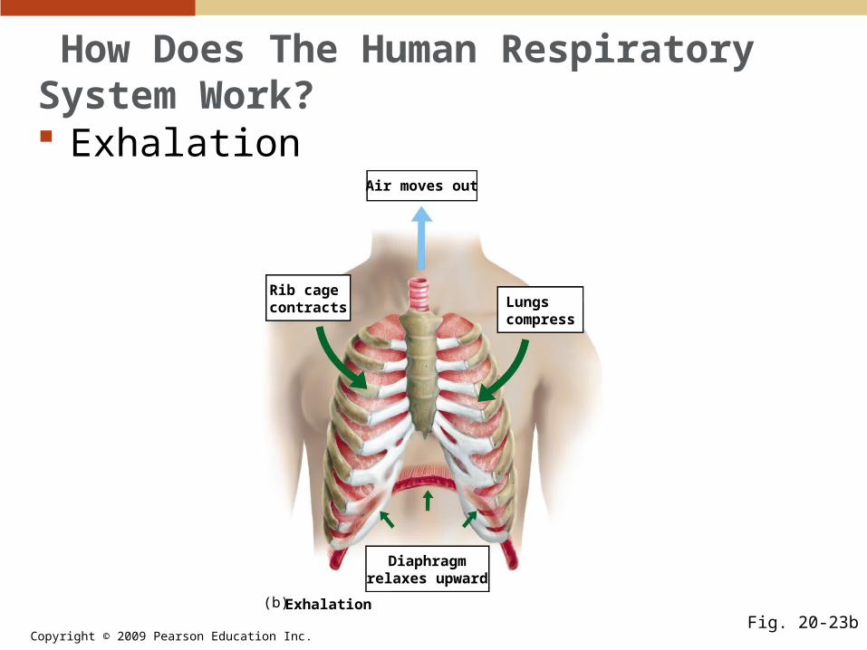
How Does The Human Respiratory System Work? Exhalation occurs automatically when the
muscles that cause inhalation are relaxed.• The relaxed diaphragm domes upward, and
the ribs move down and inward, decreasing the size of the chest cavity and forcing air out of the lungs.
Copyright © 2009 Pearson Education Inc.
How Does The Human Respiratory System Work? Exhalation
Fig. 20-23b
Air moves out
Rib cagecontracts Lungs
compress
Diaphragmrelaxes upward
Exhalation(b)
Copyright © 2009 Pearson Education Inc.
How Does The Human Respiratory System Work? Breathing rate is controlled by the
respiratory center of the brain.• Each contraction of the respiratory muscles is
stimulated by impulses from nerve cells.• The impulses originate in the respiratory
center of the brain, which is located in the medulla, the part of the brain just above the spinal cord.
Copyright © 2009 Pearson Education Inc.
How Does The Human Respiratory System Work? Breathing rate is controlled by the
respiratory center of the brain (continued).• Nerve cells in the respiratory center generate
cyclic bursts of impulses that cause alternating contraction and relaxation of the respiratory muscles.
• The respiratory rate is regulated to maintain a constant level of carbon dioxide in the blood, as monitored by carbon dioxide receptors in the medulla.
Related Documents