PowerPoint ® Lecture Slides prepared by Barbara Heard, Atlantic Cape Community College C H A P T E R © 2013 Pearson Education, Inc. © Annie Leibovitz/Contact Press Images 9 Muscles and Muscle Tissue:

Welcome message from author
This document is posted to help you gain knowledge. Please leave a comment to let me know what you think about it! Share it to your friends and learn new things together.
Transcript

PowerPoint® Lecture Slidesprepared byBarbara Heard,Atlantic Cape Community College
C H A P T E R
© 2013 Pearson Education, Inc.© Annie Leibovitz/Contact Press Images
9
Muscles and Muscle Tissue:
© 2013 Pearson Education, Inc.
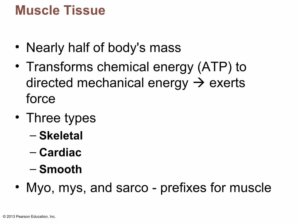
Muscle Tissue
• Nearly half of body's mass
• Transforms chemical energy (ATP) to directed mechanical energy exerts force
• Three types– Skeletal– Cardiac– Smooth
• Myo, mys, and sarco - prefixes for muscle

© 2013 Pearson Education, Inc.
Types of Muscle Tissue
• Skeletal muscles – Organs attached to bones and skin
– Elongated cells called muscle fibers– Striated (striped) – Voluntary (i.e., conscious control)– Contract rapidly; tire easily; powerful– Require nervous system stimulation
Table 9.3
© 2013 Pearson Education, Inc.
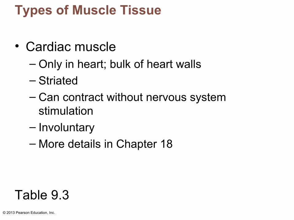
Types of Muscle Tissue
• Cardiac muscle– Only in heart; bulk of heart walls
– Striated– Can contract without nervous system
stimulation – Involuntary– More details in Chapter 18
Table 9.3
© 2013 Pearson Education, Inc.
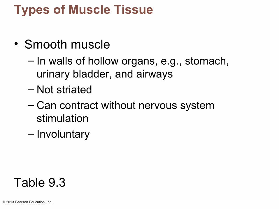
Types of Muscle Tissue
• Smooth muscle– In walls of hollow organs, e.g., stomach,
urinary bladder, and airways– Not striated– Can contract without nervous system
stimulation – Involuntary
Table 9.3

© 2013 Pearson Education, Inc.
Special Characteristics of Muscle Tissue
• Excitability (responsiveness): ability to receive and respond to stimuli
• Contractility: ability to shorten forcibly when stimulated
• Extensibility: ability to be stretched
• Elasticity: ability to recoil to resting length

© 2013 Pearson Education, Inc.
Muscle Functions
• Four important functions– Movement of bones or fluids (e.g., blood)
– Maintaining posture and body position – Stabilizing joints– Heat generation (especially skeletal muscle)
• Additional functions– Protects organs, forms valves, controls pupil
size, causes "goosebumps"

© 2013 Pearson Education, Inc.
Skeletal Muscle
• Each muscle served by one artery, one nerve, and one or more veins– Enter/exit near central part and branch
through connective tissue sheaths– Every skeletal muscle fiber supplied by nerve
ending that controls its activity– Huge nutrient and oxygen need; generates
large amount of waste

© 2013 Pearson Education, Inc.
Skeletal Muscle
• Connective tissue sheaths of skeletal muscle– Support cells; reinforce whole muscle– External to internal
• Epimysium: dense irregular connective tissue surrounding entire muscle; may blend with fascia
• Perimysium: fibrous connective tissue surrounding fascicles (groups of muscle fibers)
• Endomysium: fine areolar connective tissue surrounding each muscle fiber

© 2013 Pearson Education, Inc.
Figure 9.1 Connective tissue sheaths of skeletal muscle: epimysium, perimysium, and endomysium.
Bone
Tendon
Epimysium Epimysium
Perimysium
Endomysium
Muscle fiberin middle of a fascicle
Blood vessel
Perimysiumwrapping a fascicleEndomysium(between individualmuscle fibers)
Musclefiber
Perimysium
Fascicle

© 2013 Pearson Education, Inc.
Skeletal Muscle: Attachments
• Attach in at least two places– Insertion – movable bone
– Origin – immovable (less movable) bone
• Attachments direct or indirect– Direct—epimysium fused to periosteum of
bone or perichondrium of cartilage– Indirect—connective tissue wrappings extend
beyond muscle as ropelike tendon or sheetlike aponeurosis

© 2013 Pearson Education, Inc.
Microscopic Anatomy of a Skeletal Muscle Fiber
• Long, cylindrical cell – 10 to 100 µm in diameter; up to 30 cm long
• Multiple peripheral nuclei
• Sarcolemma = plasma membrane
• Sarcoplasm = cytoplasm– Glycosomes for glycogen storage,
myoglobin for O2 storage
• Modified structures: myofibrils, sarcoplasmic reticulum, and T tubules

© 2013 Pearson Education, Inc.
Myofibrils
• Densely packed, rodlike elements
• ~80% of cell volume
• Contain sarcomeres - contractile units – Sarcomeres contain myofilaments
• Exhibit striations - perfectly aligned repeating series of dark A bands and light I bands
© 2013 Pearson Education, Inc.
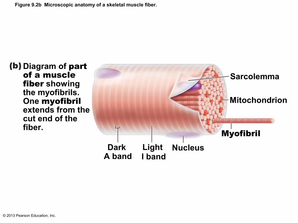
Figure 9.2b Microscopic anatomy of a skeletal muscle fiber.
Diagram of part of a muscle fiber showingthe myofibrils. One myofibril extends from the cut end of the fiber.
Sarcolemma
Mitochondrion
MyofibrilNucleusLight
I bandDark
A band
© 2013 Pearson Education, Inc.
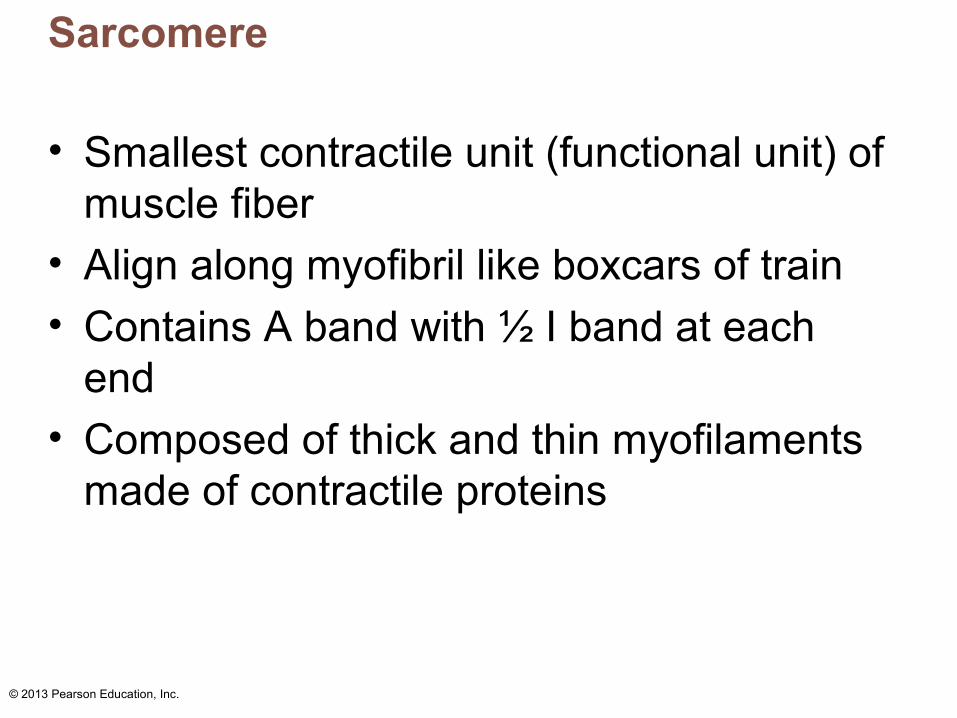
Sarcomere
• Smallest contractile unit (functional unit) of muscle fiber
• Align along myofibril like boxcars of train
• Contains A band with ½ I band at each end
• Composed of thick and thin myofilaments made of contractile proteins

© 2013 Pearson Education, Inc.
Figure 9.2c Microscopic anatomy of a skeletal muscle fiber.
Small part of onemyofibril enlarged to show the myofilamentsresponsible for thebanding pattern. Each sarcomere extends from one Z disc to the next.
Thin (actin)filament Z disc H zone Z disc
Thick (myosin)filament
I band A band I band M lineSarcomere
© 2013 Pearson Education, Inc.
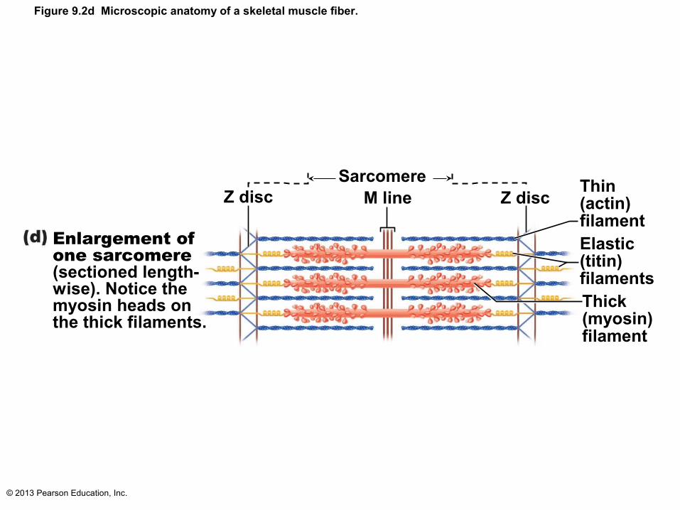
Figure 9.2d Microscopic anatomy of a skeletal muscle fiber.
Enlargement of one sarcomere (sectioned length-wise). Notice themyosin heads on the thick filaments.
Z discSarcomere
M line Z discThin (actin)filamentElastic (titin)filamentsThick(myosin)filament
© 2013 Pearson Education, Inc.
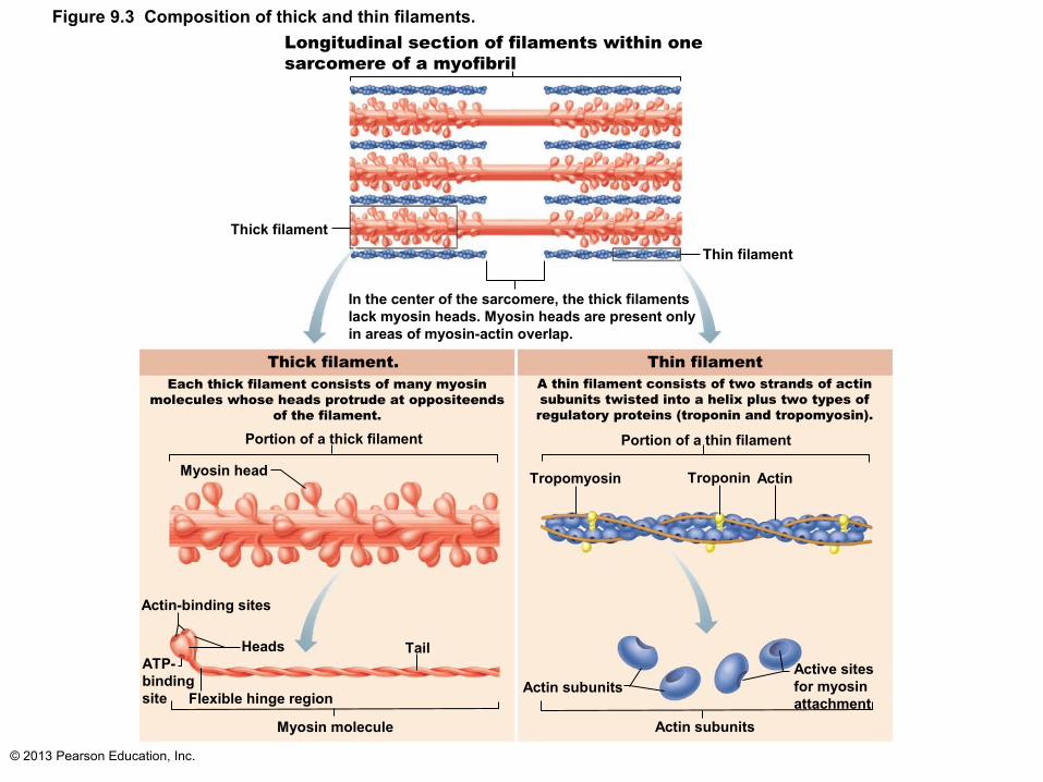
Longitudinal section of filaments within onesarcomere of a myofibril
Thick filament
Thin filament
In the center of the sarcomere, the thick filamentslack myosin heads. Myosin heads are present onlyin areas of myosin-actin overlap.
Thick filament. Thin filamentEach thick filament consists of many myosin
molecules whose heads protrude at oppositeends of the filament.
A thin filament consists of two strands of actinsubunits twisted into a helix plus two types ofregulatory proteins (troponin and tropomyosin).
Portion of a thick filament Portion of a thin filament
Myosin head Tropomyosin Troponin Actin
Actin-binding sites
ATP-bindingsite
Heads Tail
Flexible hinge region
Myosin molecule
Actin subunits
Actin subunits
Active sitesfor myosinattachment
Figure 9.3 Composition of thick and thin filaments.

© 2013 Pearson Education, Inc.
Figure 9.5 Relationship of the sarcoplasmic reticulum and T tubules to myofibrils of skeletal muscle.
Part of a skeletal muscle fiber (cell)
Myofibril
Sarcolemma
I band A band I band
Z disc H zone Z disc
Mline
Sarcolemma
Triad:• T tubule
• Terminal cisterns of the SR (2)
Tubules ofthe SRMyofibrils
Mitochondria

© 2013 Pearson Education, Inc.
Sliding Filament Model of Contraction
• Generation of force
• Does not necessarily cause shortening of fiber
• Shortening occurs when tension generated by cross bridges on thin filaments exceeds forces opposing shortening

© 2013 Pearson Education, Inc.
Sliding Filament Model of Contraction
• In relaxed state, thin and thick filaments overlap only at ends of A band
• Sliding filament model of contraction– During contraction, thin filaments slide past
thick filaments actin and myosin overlap more
– Occurs when myosin heads bind to actin cross bridges

© 2013 Pearson Education, Inc.
Sliding Filament Model of Contraction
• Myosin heads bind to actin; sliding begins
• Cross bridges form and break several times, ratcheting thin filaments toward center of sarcomere– Causes shortening of muscle fiber– Pulls Z discs toward M line
• I bands shorten; Z discs closer; H zones disappear; A bands move closer (length stays same)

© 2013 Pearson Education, Inc.
Figure 9.6 Sliding filament model of contraction. Slide 1Slide 1
Fully contracted sarcomere of a muscle fiber
1
2
Fully relaxed sarcomere of a muscle fiber
Z H Z
II A
Z Z
I IA

© 2013 Pearson Education, Inc.
Figure 9.6 Sliding filament model of contraction. Slide 2
1 Fully relaxed sarcomere of a muscle fiber
Z H Z
II A

© 2013 Pearson Education, Inc.
Figure 9.6 Sliding filament model of contraction. Slide 3
2 Fully contracted sarcomere of a muscle fiber
Z Z
II A

© 2013 Pearson Education, Inc.
Figure 9.6 Sliding filament model of contraction. Slide 4
Fully contracted sarcomere of a muscle fiber
1
2
Fully relaxed sarcomere of a muscle fiber
Z H Z
II A
Z Z
I IA

© 2013 Pearson Education, Inc.
The Nerve Stimulus and Events at the Neuromuscular Junction
• Skeletal muscles stimulated by somatic motor neurons
• Axons of motor neurons travel from central nervous system via nerves to skeletal muscle
• Each axon forms several branches as it enters muscle
• Each axon ending forms neuromuscular junction with single muscle fiber– Usually only one per muscle fiber

© 2013 Pearson Education, Inc.
Figure 9.8 When a nerve impulse reaches a neuromuscular junction, acetylcholine (ACh) is released. Slide 1
Actionpotential (AP)
Myelinated axonof motor neuron
Axon terminal of neuromuscular junction
Sarcolemma ofthe muscle fiber
Synaptic vesiclecontaining ACh
Synaptic cleft
Junctionalfolds of sarcolemma
Sarcoplasm ofmuscle fiber
Postsynaptic membraneion channel opens;ions pass.
Ion channel closes;ions cannot pass.
Action potential arrives at axon terminal of motor neuron.
Voltage-gated Ca2+ channels open. Ca2+ enters the axon terminal moving down its electochemical gradient.
Ca2+ entry causes ACh (aneurotransmitter) to be releasedby exocytosis.
ACh diffuses across the synaptic cleft and binds to its receptors on the sarcolemma.
ACh binding opens ionchannels in the receptors thatallow simultaneous passage ofNa+ into the muscle fiber and K+
out of the muscle fiber. More Na+
ions enter than K+ ions exit,which produces a local changein the membrane potential calledthe end plate potential.
ACh effects are terminated byits breakdown in the synapticcleft by acetylcholinesterase anddiffusion away from the junction.
Axon terminalof motor neuron
Fusing synaptic vesicles
Degraded AChACh
Acetylcho-linesterase
ACh
4
3
2
1
5
6

© 2013 Pearson Education, Inc.
Figure 9.8 When a nerve impulse reaches a neuromuscular junction, acetylcholine (ACh) is released. Slide 2
Synaptic vesiclecontaining ACh
Synaptic cleft
Axon terminalof motor neuron
Fusing synaptic vesiclesa
ACh Junctionalfolds of sarcolemma
Sarcoplasm ofmuscle fiber
Action potential arrives at axon terminal of motor neuron.1

© 2013 Pearson Education, Inc.
Figure 9.8 When a nerve impulse reaches a neuromuscular junction, acetylcholine (ACh) is released. Slide 3
Synaptic vesiclecontaining ACh
Synaptic cleft
Axon terminalof motor neuron
Fusing synaptic vesiclesa
ACh Junctionalfolds of sarcolemma
Sarcoplasm ofmuscle fiber
Action potential arrives at axon terminal of motor neuron.
Voltage-gated Ca2+ channels open. Ca2+ enters the axon terminal moving down its electochemical gradient.
1
2

© 2013 Pearson Education, Inc.
Figure 9.8 When a nerve impulse reaches a neuromuscular junction, acetylcholine (ACh) is released. Slide 4
Synaptic vesiclecontaining ACh
Synaptic cleft
Axon terminalof motor neuron
Fusing synaptic vesiclesa
ACh Junctionalfolds of sarcolemma
Sarcoplasm ofmuscle fiber
Action potential arrives at axon terminal of motor neuron.
Voltage-gated Ca2+ channels open. Ca2+ enters the axon terminal moving down its electochemical gradient.
Ca2+ entry causes ACh (aneurotransmitter) to be releasedby exocytosis.
1
2
3

© 2013 Pearson Education, Inc.
Figure 9.8 When a nerve impulse reaches a neuromuscular junction, acetylcholine (ACh) is released. Slide 5
Synaptic vesiclecontaining ACh
Synaptic cleft
Axon terminalof motor neuron
Fusing synaptic vesiclesa
ACh Junctionalfolds of sarcolemma
Sarcoplasm ofmuscle fiber
Action potential arrives at axon terminal of motor neuron.
Voltage-gated Ca2+ channels open. Ca2+ enters the axon terminal moving down its electochemical gradient.
Ca2+ entry causes ACh (aneurotransmitter) to be releasedby exocytosis.
ACh diffuses across the synaptic cleft and binds to its receptors onthe sarcolemma.
1
2
3
4
© 2013 Pearson Education, Inc.
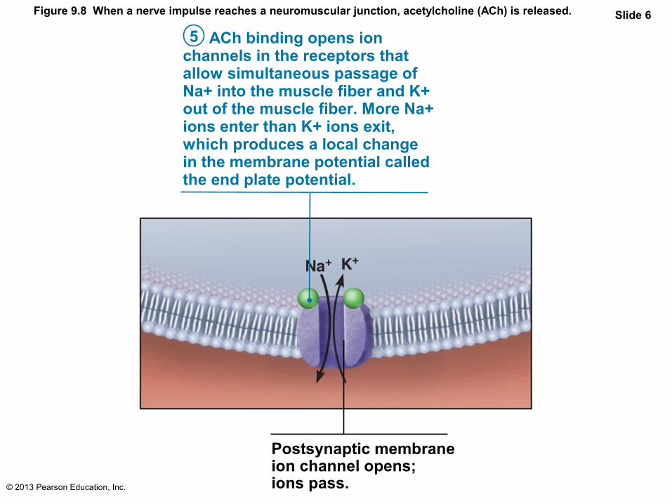
Figure 9.8 When a nerve impulse reaches a neuromuscular junction, acetylcholine (ACh) is released. Slide 6
Postsynaptic membraneion channel opens;ions pass.
ACh binding opens ionchannels in the receptors thatallow simultaneous passage ofNa+ into the muscle fiber and K+out of the muscle fiber. More Na+ions enter than K+ ions exit,which produces a local changein the membrane potential calledthe end plate potential.
5

© 2013 Pearson Education, Inc.
Figure 9.8 When a nerve impulse reaches a neuromuscular junction, acetylcholine (ACh) is released. Slide 7
ACh effects are terminated byits breakdown in the synapticcleft by acetylcholinesterase anddiffusion away from the junction.
6
Degraded AChACh
Acetylcholinesterase
Ion channel closes;ions cannot pass.

© 2013 Pearson Education, Inc.
Figure 9.8 When a nerve impulse reaches a neuromuscular junction, acetylcholine (ACh) is released. Slide 8
Actionpotential (AP)
Myelinated axonof motor neuron
Axon terminal of neuromuscular junction
Sarcolemma ofthe muscle fiber
Synaptic vesiclecontaining ACh
Synaptic cleft
Junctionalfolds of sarcolemma
Sarcoplasm ofmuscle fiber
Postsynaptic membraneion channel opens;ions pass.
Ion channel closes;ions cannot pass.
Action potential arrives at axon terminal of motor neuron.
Voltage-gated Ca2+ channels open. Ca2+ enters the axon terminal moving down its electochemical gradient.
Ca2+ entry causes ACh (aneurotransmitter) to be releasedby exocytosis.
ACh diffuses across the synaptic cleft and binds to its receptors on the sarcolemma.
ACh binding opens ionchannels in the receptors thatallow simultaneous passage ofNa+ into the muscle fiber and K+
out of the muscle fiber. More Na+
ions enter than K+ ions exit,which produces a local changein the membrane potential calledthe end plate potential.
ACh effects are terminated byits breakdown in the synapticcleft by acetylcholinesterase anddiffusion away from the junction.
Axon terminalof motor neuron
Fusing synaptic vesicles
Degraded AChACh
Acetylcho-linesterase
ACh
4
3
2
1
5
6

© 2013 Pearson Education, Inc.
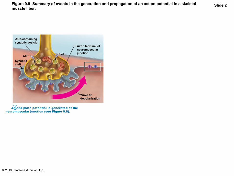
Figure 9.9 Summary of events in the generation and propagation of an action potential in a skeletalmuscle fiber.
Slide 1Open Na+
channel
Na+
Closed K+
channel
K+
Action potential
Axon terminal ofneuromuscularjunction
ACh-containingsynaptic vesicle
Ca2+
Ca2+
Synapticcleft
Wave ofdepolarization
An end plate potential is generated at theneuromuscular junction (see Figure 9.8).
Depolarization: Generating and propagating an actionpotential (AP). The local depolarization current spreads to adjacentareas of the sarcolemma. This opens voltage-gated sodium channelsthere, so Na+ enters following its electrochemical gradient and initiatesthe AP. The AP is propagated as its local depolarization wave spreads toadjacent areas of the sarcolemma, opening voltage-gated channels there.Again Na+ diffuses into the cell following its electrochemical gradient.
Repolarization: Restoring the sarcolemma to its initialpolarized state (negative inside, positive outside). Repolarizationoccurs as Na+ channels close (inactivate) and voltage-gated K+ channelsopen. Because K+ concentration is substantially higher inside the cellthan in the extracellular fluid, K+ diffuses rapidly out of the muscle fiber.
1
2
3
Closed Na+
channelOpen K+
channel
Na+
K+
– – – – – – – – – – – – – – – – – – – + + + +
– – – – + + + + + + + + + + + + + + + +
+ + + + + + + + + + + + + + + + + + + + + +
– – – – – – – – – – – – – – – – – – – – – –
© 2013 Pearson Education, Inc.
Figure 9.9 Summary of events in the generation and propagation of an action potential in a skeletalmuscle fiber.
Slide 2
Axon terminal ofneuromuscularjunction
ACh-containingsynaptic vesicle
Ca2+
Ca2+
Synapticcleft
Wave ofdepolarization
An end plate potential is generated at theneuromuscular junction (see Figure 9.8).
1

© 2013 Pearson Education, Inc.
Figure 9.9 Summary of events in the generation and propagation of an action potential in a skeletalmuscle fiber.
Slide 3Open Na+
channel
Na+
Closed K+
channel
K+
Action potential
Axon terminal ofneuromuscularjunction
ACh-containingsynaptic vesicle
Ca2+
Ca2+
Synapticcleft
Wave ofdepolarization
An end plate potential is generated at theneuromuscular junction (see Figure 9.8).
Depolarization: Generating and propagating an actionpotential (AP). The local depolarization current spreads to adjacentareas of the sarcolemma. This opens voltage-gated sodium channelsthere, so Na+ enters following its electrochemical gradient and initiatesthe AP. The AP is propagated as its local depolarization wave spreads toadjacent areas of the sarcolemma, opening voltage-gated channels there.Again Na+ diffuses into the cell following its electrochemical gradient.
1
2
+ + + +
+ + + + + + + + + + + + + + + +
– – – – – – – – – – – – – – – – – – –
– – – –

© 2013 Pearson Education, Inc.
Figure 9.9 Summary of events in the generation and propagation of an action potential in a skeletalmuscle fiber.
Slide 4Open Na+
channel
Na+
Closed K+
channel
K+
Action potential
Axon terminal ofneuromuscularjunction
ACh-containingsynaptic vesicle
Ca2+
Ca2+
Synapticcleft
Wave ofdepolarization
An end plate potential is generated at theneuromuscular junction (see Figure 9.8).
Depolarization: Generating and propagating an actionpotential (AP). The local depolarization current spreads to adjacentareas of the sarcolemma. This opens voltage-gated sodium channelsthere, so Na+ enters following its electrochemical gradient and initiatesthe AP. The AP is propagated as its local depolarization wave spreads toadjacent areas of the sarcolemma, opening voltage-gated channels there.Again Na+ diffuses into the cell following its electrochemical gradient.
Repolarization: Restoring the sarcolemma to its initialpolarized state (negative inside, positive outside). Repolarizationoccurs as Na+ channels close (inactivate) and voltage-gated K+ channelsopen. Because K+ concentration is substantially higher inside the cellthan in the extracellular fluid, K+ diffuses rapidly out of the muscle fiber.
1
2
3
Closed Na+
channelOpen K+
channel
Na+
K+
+ + + +
+ + + + + + + + + + + + + + + +
+ + + + + + + + + + + + + + + + + + + + + +
– – – – – – – – – – – – – – – – – – –
– – – –
– – – – – – – – – – – – – – – – – – – – – –

© 2013 Pearson Education, Inc.
Figure 9.10 Action potential tracing indicates changes in Na+ and K+ ion channels.
Mem
bran
e po
tent
ial (
mV
)
+30
0
–95
0 5 10 15 20
Depolarizationdue to Na+ entry
Na+ channelsclose, K+ channelsopen
Repolarizationdue to K+ exit
K+ channelsclosed
Na+
channelsopen
Time (ms)

© 2013 Pearson Education, Inc.
Excitation-Contraction (E-C) Coupling
• Events that transmit AP along sarcolemma lead to sliding of myofilaments
• AP brief; ends before contraction– Causes rise in intracellular Ca2+ which
contraction
• Latent period– Time when E-C coupling events occur– Time between AP initiation and beginning of
contraction
© 2013 Pearson Education, Inc.
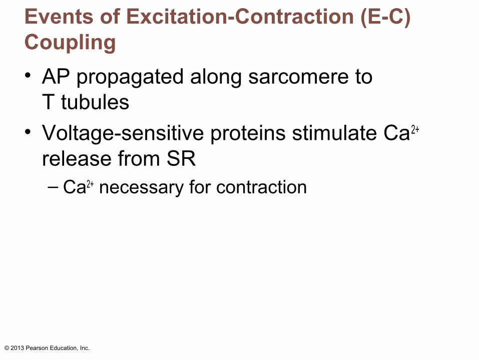
Events of Excitation-Contraction (E-C) Coupling
• AP propagated along sarcomere to T tubules
• Voltage-sensitive proteins stimulate Ca2+ release from SR – Ca2+ necessary for contraction

© 2013 Pearson Education, Inc.
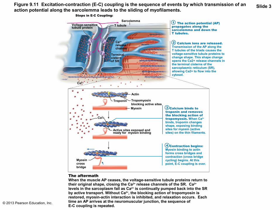
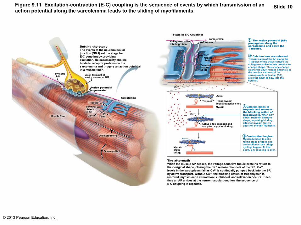
Figure 9.11 Excitation-contraction (E-C) coupling is the sequence of events by which transmission of anaction potential along the sarcolemma leads to the sliding of myofilaments.
Slide 1
Setting the stageThe events at the neuromuscular junction (NMJ) set the stage for E-C coupling by providing excitation. Released acetylcholine binds to receptor proteins on the sarcolemma and triggers an action potential in a muscle fiber.
Synapticcleft
Axon terminal ofmotor neuron at NMJ
Action potentialis generated
ACh
Muscle fiber
T tubule
Terminal cisternof SR
Triad
One sarcomere
One myofibril
Sarcolemma
The action potential (AP) propagates along the sarcolemma and down theT tubules.
Calcium ions are released. Transmission of the AP along the T tubules of the triads causes the voltage-sensitive tubule proteins to change shape. This shape change opens the Ca2+ release channels in the terminal cisterns of the sarcoplasmic reticulum (SR), allowing Ca2+ to flow into the cytosol.
Steps in E-C Coupling:
Terminal cisternof SR
Ca2+
releasechannel
Voltage-sensitivetubule protein
T tubuleSarcolemma 1
2
3
4
Calcium binds to troponin and removes the blocking action of tropomyosin. When Ca2+ binds, troponin changes shape, exposing binding sites for myosin (active sites) on the thin filaments.
Contraction begins: Myosin binding to actin forms cross bridges and contraction (cross bridge cycling) begins. At this point, E-C coupling is over.
The aftermathWhen the muscle AP ceases, the voltage-sensitive tubule proteins return to their original shape, closing the Ca2+ release channels of the SR. Ca2+ levels in the sarcoplasm fall as Ca2+ is continually pumped back into the SR by active transport. Without Ca2+, the blocking action of tropomyosin is restored, myosin-actin interaction is inhibited, and relaxation occurs. Each time an AP arrives at the neuromuscular junction, the sequence of E-C coupling is repeated.
Myosincross bridge
Active sites exposed and ready for myosin binding
Myosin
Tropomyosinblocking active sites
Actin
Troponin

© 2013 Pearson Education, Inc.
Figure 9.11 Excitation-contraction (E-C) coupling is the sequence of events by which transmission of anaction potential along the sarcolemma leads to the sliding of myofilaments.
Slide 2
Setting the stageThe events at the neuromuscular junction (NMJ) set the stage for E-C coupling by providing excitation. Released acetylcholine binds to receptor proteins on the sarcolemma and triggers an action potential in a muscle fiber.
Synapticcleft
Axon terminal ofmotor neuron at NMJ
Action poten-tial is generated
Muscle fiber
T tubuleTerminal cisternof SR
Triad
One sarcomere
One myofibril
SarcolemmaACh
© 2013 Pearson Education, Inc.
Figure 9.11 Excitation-contraction (E-C) coupling is the sequence of events by which transmission of anaction potential along the sarcolemma leads to the sliding of myofilaments.
Slide 3
The action potential (AP) propagates along the sarcolemma and down theT tubules.
Calcium ions are released. Transmission of the AP along the T tubules of the triads causes the voltage-sensitive tubule proteins to change shape. This shape change opens the Ca2+ release channels in the terminal cisterns of the sarcoplasmic reticulum (SR), allowing Ca2+ to flow into the cytosol.
Steps in E-C Coupling:
Terminal cisternof SR
Ca2+
releasechannel
Voltage-sensitivetubule protein
T tubule
Sarcolemma
Calcium binds to troponin and removes the blocking action of tropomyosin. When Ca2+ binds, troponin changes shape, exposing binding sites for myosin (active sites) on the thin filaments.
Contraction begins: Myosin binding to actin forms cross bridges and contraction (cross bridge cycling) begins. At this point, E-C coupling is over.
The aftermathWhen the muscle AP ceases, the voltage-sensitive tubule proteins return to their original shape, closing the Ca2+ release channels of the SR. Ca2+ levels in the sarcoplasm fall as Ca2+ is continually pumped back into the SR by active transport. Without Ca2+, the blocking action of tropomyosin is restored, myosin-actin interaction is inhibited, and relaxation occurs. Each time an AP arrives at the neuromuscular junction, the sequence of E-C coupling is repeated.
Myosincross bridge
Active sites exposed and ready for myosin binding
Myosin
Tropomyosinblocking active sites
Actin
Troponin
2
1
3
4

© 2013 Pearson Education, Inc.
Figure 9.11 Excitation-contraction (E-C) coupling is the sequence of events by which transmission of anaction potential along the sarcolemma leads to the sliding of myofilaments.
Slide 4
The action potential (AP) propagates along the sarcolemma and down theT tubules.
Steps in E-C Coupling:
Terminal cisternof SR
Ca2+ releasechannel
Voltage-sensitivetubule protein
T tubule
Sarcolemma 1
© 2013 Pearson Education, Inc.
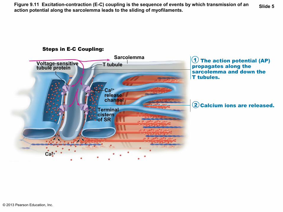
Figure 9.11 Excitation-contraction (E-C) coupling is the sequence of events by which transmission of anaction potential along the sarcolemma leads to the sliding of myofilaments.
Slide 5
The action potential (AP) propagates along the sarcolemma and down theT tubules.
Steps in E-C Coupling:
Terminal cisternof SR
Ca2+ releasechannel
Voltage-sensitivetubule protein
T tubule
Sarcolemma
Calcium ions are released.
1
2
© 2013 Pearson Education, Inc.
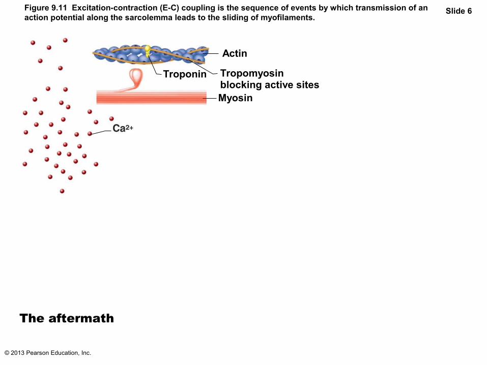
Figure 9.11 Excitation-contraction (E-C) coupling is the sequence of events by which transmission of anaction potential along the sarcolemma leads to the sliding of myofilaments.
Slide 6
Myosin
Tropomyosinblocking active sites
Actin
Troponin
The aftermath

© 2013 Pearson Education, Inc.
Figure 9.11 Excitation-contraction (E-C) coupling is the sequence of events by which transmission of anaction potential along the sarcolemma leads to the sliding of myofilaments.
Slide 7
Calcium binds to troponin and removes the blocking action of tropomyosin. When Ca2+ binds, troponin changes shape, exposing binding sites for myosin (active sites) on the thin filaments.
Active sites exposed and ready for myosin binding
Myosin
Tropomyosinblocking active sites
Actin
Troponin
The aftermath
3
© 2013 Pearson Education, Inc.
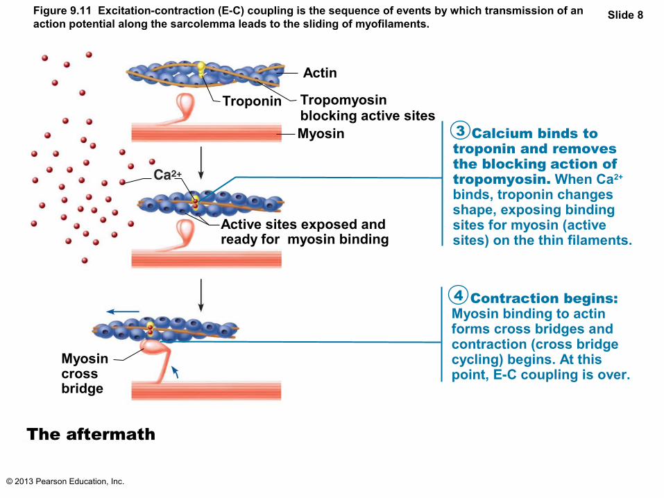
Figure 9.11 Excitation-contraction (E-C) coupling is the sequence of events by which transmission of anaction potential along the sarcolemma leads to the sliding of myofilaments.
Slide 8
Calcium binds to troponin and removes the blocking action of tropomyosin. When Ca2+ binds, troponin changes shape, exposing binding sites for myosin (active sites) on the thin filaments.
Contraction begins: Myosin binding to actin forms cross bridges and contraction (cross bridge cycling) begins. At this point, E-C coupling is over.
Myosincross bridge
Active sites exposed and ready for myosin binding
Myosin
Tropomyosinblocking active sites
Actin
Troponin
The aftermath
3
4

© 2013 Pearson Education, Inc.
Figure 9.11 Excitation-contraction (E-C) coupling is the sequence of events by which transmission of anaction potential along the sarcolemma leads to the sliding of myofilaments.
A&P Flix™: Excitation-contraction coupling.
PLAYPLAY
Slide 9
The action potential (AP) propagates along the sarcolemma and down theT tubules.
Calcium ions are released. Transmission of the AP along the T tubules of the triads causes the voltage-sensitive tubule proteins to change shape. This shape change opens the Ca2+ release channels in the terminal cisterns of the sarcoplasmic reticulum (SR), allowing Ca2+ to flow into the cytosol.
Steps in E-C Coupling:
Terminal cisternof SR
Ca2+
releasechannel
Voltage-sensitivetubule protein
T tubule
Sarcolemma
Calcium binds to troponin and removes the blocking action of tropomyosin. When Ca2+ binds, troponin changes shape, exposing binding sites for myosin (active sites) on the thin filaments.
Contraction begins: Myosin binding to actin forms cross bridges and contraction (cross bridge cycling) begins. At this point, E-C coupling is over.
The aftermathWhen the muscle AP ceases, the voltage-sensitive tubule proteins return to their original shape, closing the Ca2+ release channels of the SR. Ca2+ levels in the sarcoplasm fall as Ca2+ is continually pumped back into the SR by active transport. Without Ca2+, the blocking action of tropomyosin is restored, myosin-actin interaction is inhibited, and relaxation occurs. Each time an AP arrives at the neuromuscular junction, the sequence of E-C coupling is repeated.
Myosincross bridge
Active sites exposed and ready for myosin binding
Myosin
Tropomyosinblocking active sites
Actin
Troponin
2
1
3
4
© 2013 Pearson Education, Inc.
Figure 9.11 Excitation-contraction (E-C) coupling is the sequence of events by which transmission of anaction potential along the sarcolemma leads to the sliding of myofilaments.
Slide 10
Setting the stageThe events at the neuromuscular junction (NMJ) set the stage for E-C coupling by providing excitation. Released acetylcholine binds to receptor proteins on the sarcolemma and triggers an action potential in a muscle fiber.
Synapticcleft
Axon terminal ofmotor neuron at NMJ
Action potentialis generated
ACh
Muscle fiber
T tubule
Terminal cisternof SR
Triad
One sarcomere
One myofibril
Sarcolemma
The action potential (AP) propagates along the sarcolemma and down theT tubules.
Calcium ions are released. Transmission of the AP along the T tubules of the triads causes the voltage-sensitive tubule proteins to change shape. This shape change opens the Ca2+ release channels in the terminal cisterns of the sarcoplasmic reticulum (SR), allowing Ca2+ to flow into the cytosol.
Steps in E-C Coupling:
Terminal cisternof SR
Ca2+
releasechannel
Voltage-sensitivetubule protein
T tubuleSarcolemma 1
2
3
4
Calcium binds to troponin and removes the blocking action of tropomyosin. When Ca2+ binds, troponin changes shape, exposing binding sites for myosin (active sites) on the thin filaments.
Contraction begins: Myosin binding to actin forms cross bridges and contraction (cross bridge cycling) begins. At this point, E-C coupling is over.
The aftermathWhen the muscle AP ceases, the voltage-sensitive tubule proteins return to their original shape, closing the Ca2+ release channels of the SR. Ca2+ levels in the sarcoplasm fall as Ca2+ is continually pumped back into the SR by active transport. Without Ca2+, the blocking action of tropomyosin is restored, myosin-actin interaction is inhibited, and relaxation occurs. Each time an AP arrives at the neuromuscular junction, the sequence of E-C coupling is repeated.
Myosincross bridge
Active sites exposed and ready for myosin binding
Myosin
Tropomyosinblocking active sites
Actin
Troponin
© 2013 Pearson Education, Inc.
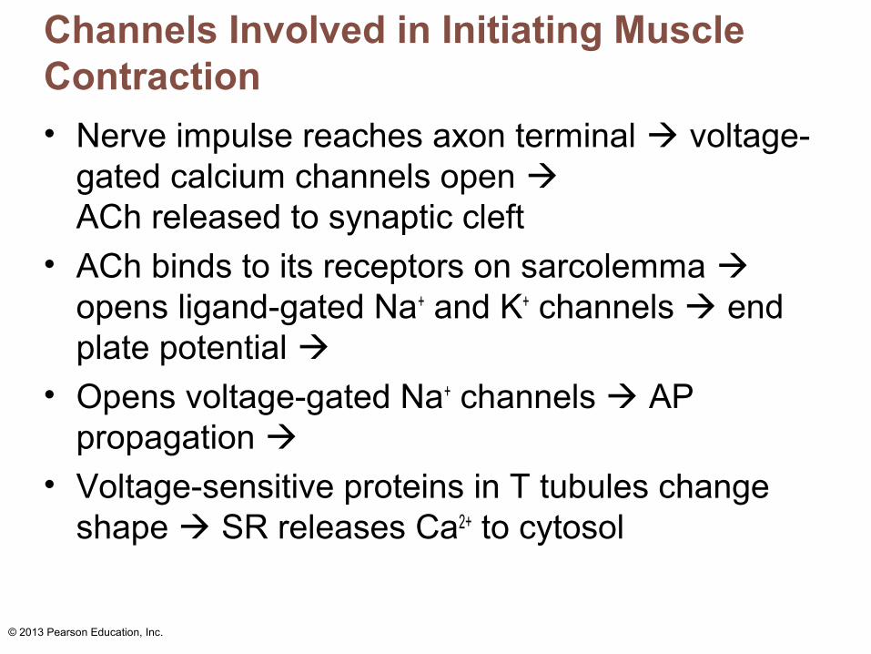
Channels Involved in Initiating Muscle Contraction
• Nerve impulse reaches axon terminal voltage-gated calcium channels open ACh released to synaptic cleft
• ACh binds to its receptors on sarcolemma opens ligand-gated Na+ and K+ channels end plate potential
• Opens voltage-gated Na+ channels AP propagation
• Voltage-sensitive proteins in T tubules change shape SR releases Ca2+ to cytosol

© 2013 Pearson Education, Inc.
Role of Calcium (Ca2+) in Contraction
• At low intracellular Ca2+ concentration– Tropomyosin blocks active sites on actin
– Myosin heads cannot attach to actin– Muscle fiber relaxed
© 2013 Pearson Education, Inc.
Role of Calcium (Ca2+) in Contraction
• At higher intracellular Ca2+ concentrations– Ca2+ binds to troponin
• Troponin changes shape and moves tropomyosin away from myosin-binding sites
• Myosin heads bind to actin, causing sarcomere shortening and muscle contraction
– When nervous stimulation ceases, Ca2+ pumped back into SR and contraction ends

© 2013 Pearson Education, Inc.
Cross Bridge Cycle
• Continues as long as Ca2+ signal and adequate ATP present
• Cross bridge formation—high-energy myosin head attaches to thin filament
• Working (power) stroke—myosin head pivots and pulls thin filament toward M line

© 2013 Pearson Education, Inc.
Cross Bridge Cycle
• Cross bridge detachment—ATP attaches to myosin head and cross bridge detaches
• "Cocking" of myosin head—energy from hydrolysis of ATP cocks myosin head into high-energy state

© 2013 Pearson Education, Inc.
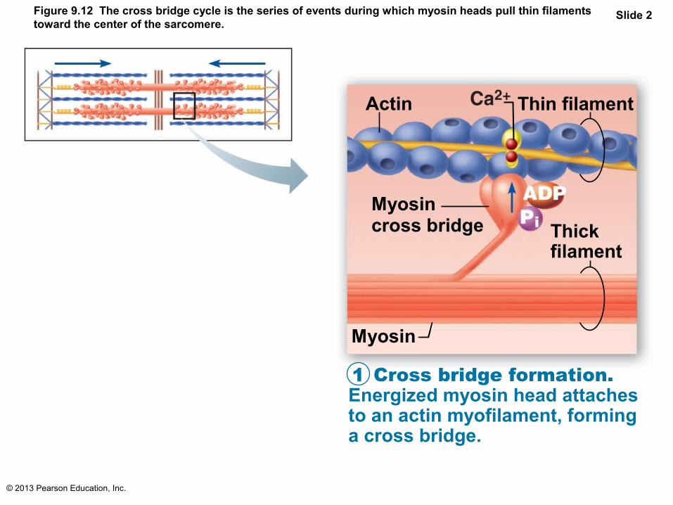
Figure 9.12 The cross bridge cycle is the series of events during which myosin heads pull thin filamentstoward the center of the sarcomere.
Slide 1
Actin Ca2+ Thin filament
Myosincross bridge Thick
filament
Myosin
ATPhydrolysis
In the absence of ATP, myosin heads will not detach, causing rigor mortis.
*This cycle will continue as long
as ATP is available and Ca2+ is
bound to troponin.
Cross bridge formation. Energized myosin head attaches to an actin myofilament, forming a cross bridge.
Cocking of the myosin head. As ATP is hydrolyzed to ADP and Pi, the myosin head returns to its prestroke high-energy, or “cocked,” position. *
Cross bridge detachment. After ATP attaches to myosin, the link between myosin and actin weakens, and the myosin head detaches (the cross bridge “breaks”).
The power (working) stroke. ADP and Pi are released and the myosin head pivots and bends, changing to its bent low-energy state. As a result it pulls the actin filament toward the M line.
1
2
3
4
© 2013 Pearson Education, Inc.
Figure 9.12 The cross bridge cycle is the series of events during which myosin heads pull thin filamentstoward the center of the sarcomere.
Slide 2
Actin Thin filament
Myosincross bridge Thick
filament
Myosin
Cross bridge formation. Energized myosin head attaches to an actin myofilament, forming a cross bridge.
1

© 2013 Pearson Education, Inc.
Figure 9.12 The cross bridge cycle is the series of events during which myosin heads pull thin filamentstoward the center of the sarcomere.
Slide 3
The power (working) stroke. ADP and Pi are released and the myosin head pivots and bends, changing to its bent low-energy state. As a result it pulls the actin filament toward the M line.
2

© 2013 Pearson Education, Inc.
Figure 9.12 The cross bridge cycle is the series of events during which myosin heads pull thin filamentstoward the center of the sarcomere.
Slide 4
Cross bridge detachment. After ATP attaches to myosin, the link between myosin and actin weakens, and the myosin head detaches (the cross bridge “breaks”).
3

© 2013 Pearson Education, Inc.
Figure 9.12 The cross bridge cycle is the series of events during which myosin heads pull thin filamentstoward the center of the sarcomere.
Slide 5
Cocking of the myosin head. As ATP is hydrolyzed to ADP and Pi, the myosin head returns to its prestroke high-energy, or “cocked,” position. *
4
ATPhydrolysis
*This cycle will continue as long
as ATP is available and Ca2+ is
bound to troponin.

© 2013 Pearson Education, Inc.
Figure 9.12 The cross bridge cycle is the series of events during which myosin heads pull thin filamentstoward the center of the sarcomere.
Slide 6
A&P Flix™: The Cross Bridge Cycle
PLAYPLAY
Actin Ca2+ Thin filament
Myosincross bridge Thick
filament
Myosin
ATPhydrolysis
In the absence of ATP, myosin heads will not detach, causing rigor mortis.
*This cycle will continue as long
as ATP is available and Ca2+ is
bound to troponin.
Cross bridge formation. Energized myosin head attaches to an actin myofilament, forming a cross bridge.
Cocking of the myosin head. As ATP is hydrolyzed to ADP and Pi, the myosin head returns to its prestroke high-energy, or “cocked,” position. *
Cross bridge detachment. After ATP attaches to myosin, the link between myosin and actin weakens, and the myosin head detaches (the cross bridge “breaks”).
The power (working) stroke. ADP and Pi are released and the myosin head pivots and bends, changing to its bent low-energy state. As a result it pulls the actin filament toward the M line.
1
2
3
4
© 2013 Pearson Education, Inc.
Homeostatic Imbalance
• Rigor mortis– Cross bridge detachment requires ATP
– 3–4 hours after death muscles begin to stiffen with weak rigidity at 12 hours post mortem
• Dying cells take in calcium cross bridge formation
• No ATP generated to break cross bridges
© 2013 Pearson Education, Inc.
Review Principles of Muscle Mechanics
• Same principles apply to contraction of single fiber and whole muscle
• Contraction produces muscle tension, force exerted on load or object to be moved

© 2013 Pearson Education, Inc.
Review Principles of Muscle Mechanics
• Contraction may/may not shorten muscle– Isometric contraction: no shortening; muscle
tension increases but does not exceed load – Isotonic contraction: muscle shortens
because muscle tension exceeds load
• Force and duration of contraction vary in response to stimuli of different frequencies and intensities

© 2013 Pearson Education, Inc.
Motor Unit: The Nerve-Muscle Functional Unit
• Each muscle served by at least one motor nerve– Motor nerve contains axons of up to hundreds
of motor neurons– Axons branch into terminals, each of which
NMJ with single muscle fiber
• Motor unit = motor neuron and all (four to several hundred) muscle fibers it supplies– Smaller number = fine control
© 2013 Pearson Education, Inc.
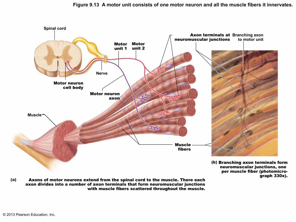
Figure 9.13 A motor unit consists of one motor neuron and all the muscle fibers it innervates.
Spinal cord
Motorunit 1
Motorunit 2
Axon terminals atneuromuscular junctions
Branching axonto motor unit
Motor neuroncell body
Motor neuronaxon
Muscle
Musclefibers
Nerve
Branching axon terminals form neuromuscular junctions, one per muscle fiber (photomicro-
graph 330x). Axons of motor neurons extend from the spinal cord to the muscle. There each
axon divides into a number of axon terminals that form neuromuscular junctions with muscle fibers scattered throughout the muscle.

© 2013 Pearson Education, Inc.
Motor Unit
• Muscle fibers from motor unit spread throughout muscle so single motor unit causes weak contraction of entire muscle
• Motor units in muscle usually contract asynchronously; helps prevent fatigue

© 2013 Pearson Education, Inc.
Muscle Twitch
• Motor unit's response to single action potential of its motor neuron
• Simplest contraction observable in lab (recorded as myogram)
© 2013 Pearson Education, Inc.
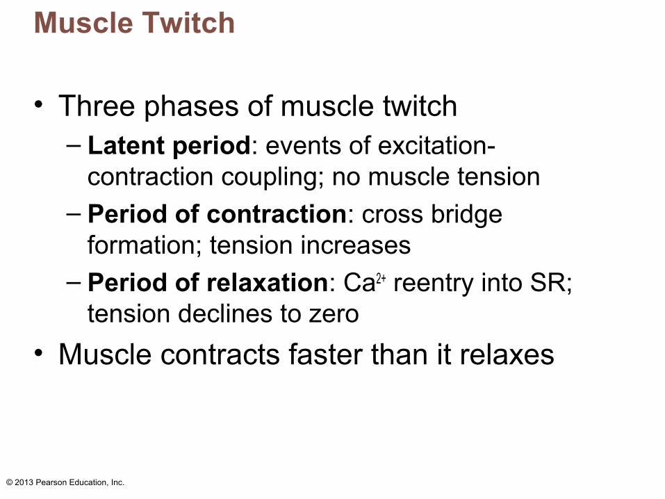
Muscle Twitch
• Three phases of muscle twitch– Latent period: events of excitation-
contraction coupling; no muscle tension– Period of contraction: cross bridge
formation; tension increases– Period of relaxation: Ca2+ reentry into SR;
tension declines to zero
• Muscle contracts faster than it relaxes

© 2013 Pearson Education, Inc.
Figure 9.14a The muscle twitch.
Latentperiod
Period ofcontraction
Period ofrelaxation
Per
cent
age
ofm
axim
um t
ensi
on
Singlestimulus
Time (ms)140120100806040200
Myogram showing the three phases of an isometric twitch

© 2013 Pearson Education, Inc.
Muscle Twitch Comparisons
• Different strength and duration of twitches due to variations in metabolic properties and enzymes between muscles
• Muscle twitch only in lab or neuromuscular problems; normal muscle contraction smooth

© 2013 Pearson Education, Inc.
Figure 9.14b The muscle twitch.
Latent period
Extraocular muscle (lateral rectus)
Gastrocnemius
Soleus
Per
cent
age
ofm
axim
um t
ensi
on
Singlestimulus
Comparison of the relative duration of twitch responses of three muscles
160 200Time (ms)
12080400
© 2013 Pearson Education, Inc.
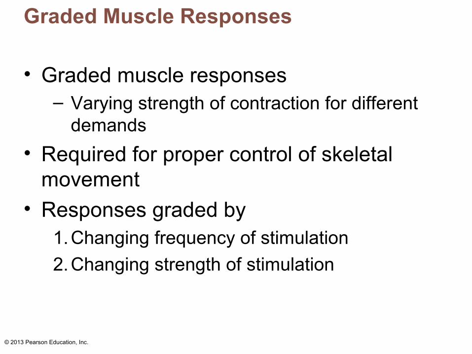
Graded Muscle Responses
• Graded muscle responses– Varying strength of contraction for different
demands
• Required for proper control of skeletal movement
• Responses graded by1.Changing frequency of stimulation
2.Changing strength of stimulation

© 2013 Pearson Education, Inc.
Response to Change in Stimulus Frequency
• Single stimulus results in single contractile response—muscle twitch

© 2013 Pearson Education, Inc.
Figure 9.15a A muscle's response to changes in stimulation frequency.
Single stimulus single twitch
Contraction Maximal tension of a single twitch
Relaxation
Stimulus
300200100Time (ms)
A single stimulus is delivered. The muscle contracts and relaxes.
Ten
sion
0

© 2013 Pearson Education, Inc.
Response to Change in Stimulus Frequency
• Wave (temporal) summation– Increased stimulus frequency (muscle does
not completely relax between stimuli) second contraction of greater force
• Additional Ca2+ release with second stimulus stimulates more shortening
• Produces smooth, continuous contractions
• Further increase in stimulus frequency → unfused (incomplete) tetanus

© 2013 Pearson Education, Inc.
Figure 9.15b A muscle's response to changes in stimulation frequency.
Low stimulation frequency
unfused (incomplete) tetanus
Partial relaxation
Time (ms)
If another stimulus is applied before the muscle relaxescompletely, then more tension results. This is wave (or
temporal) summation and results in unfused (or incomplete) tetanus.
Ten
sion
300200100
0 Stimuli
© 2013 Pearson Education, Inc.
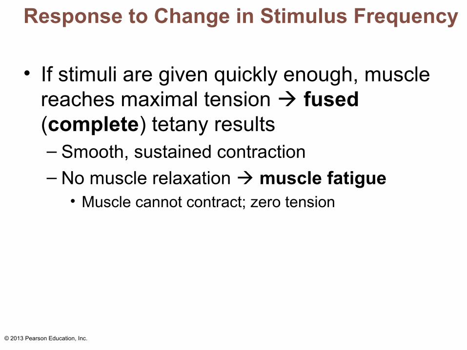
Response to Change in Stimulus Frequency
• If stimuli are given quickly enough, muscle reaches maximal tension fused (complete) tetany results– Smooth, sustained contraction– No muscle relaxation muscle fatigue
• Muscle cannot contract; zero tension
© 2013 Pearson Education, Inc.
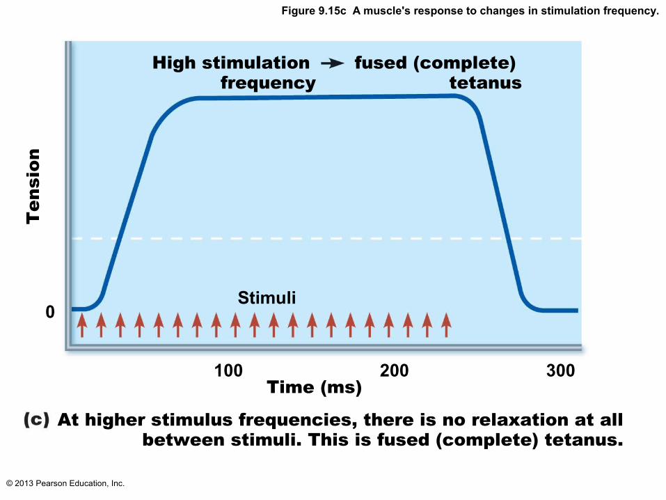
Figure 9.15c A muscle's response to changes in stimulation frequency.
High stimulation frequency
fused (complete) tetanus
Stimuli
At higher stimulus frequencies, there is no relaxation at allbetween stimuli. This is fused (complete) tetanus.
Ten
sion
Time (ms)
0
300200100

© 2013 Pearson Education, Inc.
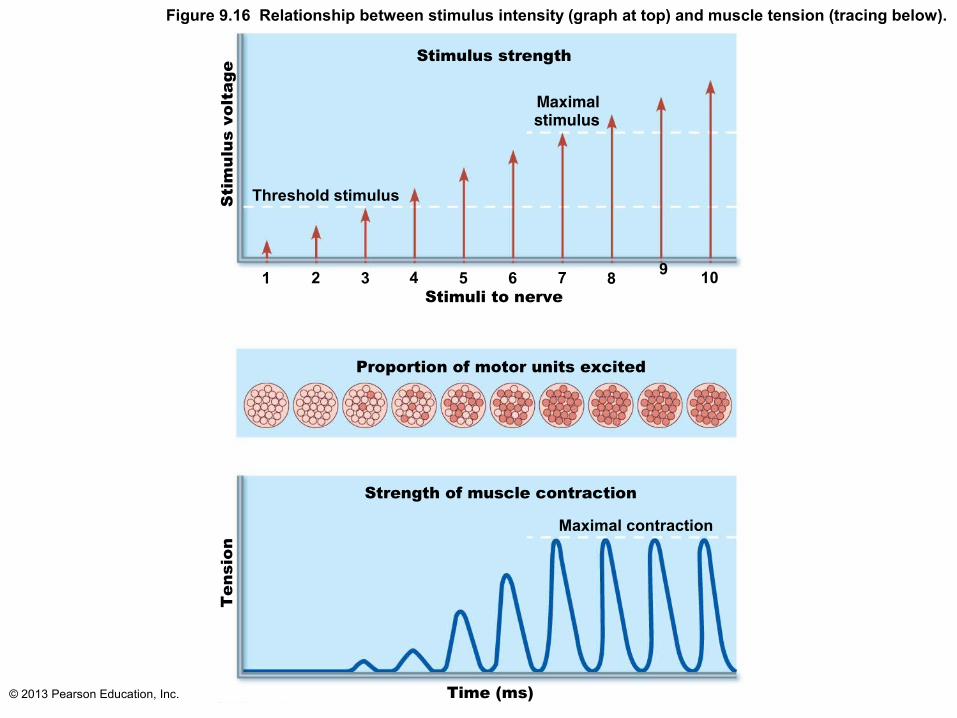
Response to Change in Stimulus Strength
• Recruitment (multiple motor unit summation) controls force of contraction
• Subthreshold stimuli – no observable contractions
• Threshold stimulus: stimulus strength causing first observable muscle contraction
• Maximal stimulus – strongest stimulus that increases contractile force

© 2013 Pearson Education, Inc.
Response to Change in Stimulus Strength
• Muscle contracts more vigorously as stimulus strength increases above threshold
• Contraction force precisely controlled by recruitment – activates more and more muscle fibers
• Beyond maximal stimulus no increase in force of contraction
© 2013 Pearson Education, Inc.
Figure 9.16 Relationship between stimulus intensity (graph at top) and muscle tension (tracing below).
Stimulus strength
Sti
mul
us v
olta
ge
Threshold stimulus
Maximalstimulus
109
87654321
Proportion of motor units excited
Strength of muscle contraction
Maximal contraction
Time (ms)
Ten
sion
Stimuli to nerve

© 2013 Pearson Education, Inc.
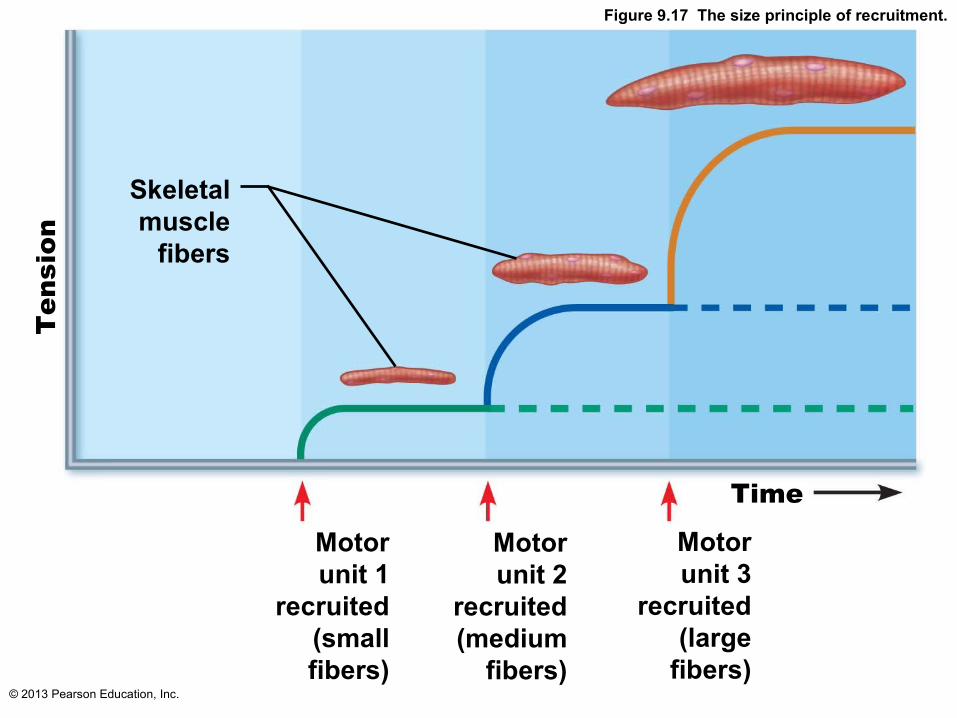
Response to Change in Stimulus Strength
• Recruitment works on size principle– Motor units with smallest muscle fibers
recruited first – Motor units with larger and larger fibers
recruited as stimulus intensity increases– Largest motor units activated only for most
powerful contractions
© 2013 Pearson Education, Inc.
Figure 9.17 The size principle of recruitment.
Skeletalmuscle
fibers
Ten
sion
Motorunit 1
recruited(smallfibers)
Motorunit 2
recruited(medium
fibers)
Motorunit 3
recruited(large
fibers)
Time

© 2013 Pearson Education, Inc.
Isotonic Contractions
• Muscle changes in length and moves load– Thin filaments slide
• Isotonic contractions either concentric or eccentric:– Concentric contractions—muscle shortens
and does work– Eccentric contractions—muscle generates
force as it lengthens

© 2013 Pearson Education, Inc.
Isometric Contractions
• Load greater than tension muscle can develop
• Tension increases to muscle's capacity, but muscle neither shortens nor lengthens– Cross bridges generate force but do not move
actin filaments

© 2013 Pearson Education, Inc.
Muscle Tone
• Constant, slightly contracted state of all muscles
• Due to spinal reflexes – Groups of motor units alternately activated in
response to input from stretch receptors in muscles
• Keeps muscles firm, healthy, and ready to respond
© 2013 Pearson Education, Inc.
Muscle Metabolism: Energy for Contraction
• ATP only source used directly for contractile activities– Move and detach cross bridges, calcium
pumps in SR, return of Na+ & K+ after excitation-contraction coupling
• Available stores of ATP depleted in 4–6 seconds

© 2013 Pearson Education, Inc.
Muscle Metabolism: Energy for Contraction
• ATP regenerated by:– Direct phosphorylation of ADP by creatine
phosphate (CP) – Anaerobic pathway (glycolysis lactic acid) – Aerobic respiration
© 2013 Pearson Education, Inc.
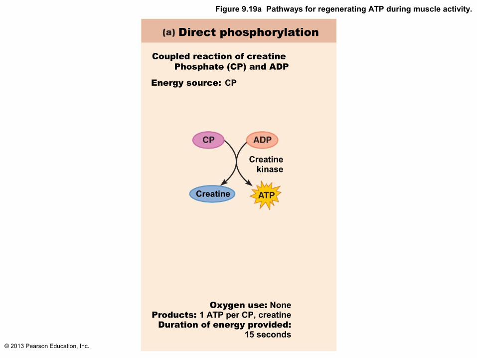
Figure 9.19a Pathways for regenerating ATP during muscle activity.
Direct phosphorylation
Coupled reaction of creatine Phosphate (CP) and ADP
Energy source: CP
Oxygen use: NoneProducts: 1 ATP per CP, creatine
Duration of energy provided:15 seconds
Creatinekinase
Creatine

© 2013 Pearson Education, Inc.
Anaerobic Pathway
• Glycolysis – does not require oxygen– Glucose degraded to 2 pyruvic acid molecules
• Normally enter mitochondria aerobic respiration
• At 70% of maximum contractile activity– Bulging muscles compress blood vessels;
oxygen delivery impaired– Pyruvic acid converted to lactic acid

© 2013 Pearson Education, Inc.
Anaerobic Pathway
• Lactic acid– Diffuses into bloodstream
– Used as fuel by liver, kidneys, and heart– Converted back into pyruvic acid or glucose
by liver– Anaerobic respiration yields only 5% as much
ATP as aerobic respiration, but produces ATP 2½ times faster

© 2013 Pearson Education, Inc.
Figure 9.19b Pathways for regenerating ATP during muscle activity.
Anaerobic pathway
Glycolysis and lactic acid formation
Energy source: glucose
Glucose (fromglycogen breakdown ordelivered from blood)
Glycolysisin cytosol
Pyruvic acidnet gain
Releasedto blood
Lactic acid
Oxygen use: NoneProducts: 2 ATP per glucose, lactic acid
Duration of energy provided: 30-40 seconds, or slightly more
2

© 2013 Pearson Education, Inc.
Aerobic Pathway
• Produces 95% of ATP during rest and light to moderate exercise; slow
• Series of chemical reactions that require oxygen; occur in mitochondria– Breaks glucose into CO2, H2O, and large
amount ATP
• Fuels - stored glycogen, then bloodborne glucose, pyruvic acid from glycolysis, and free fatty acids

© 2013 Pearson Education, Inc.
Figure 9.19c Pathways for regenerating ATP during muscle activity.
Aerobic pathway
Aerobic cellular respiration
Energy source: glucose; pyruvic acid; freefatty acids from adipose tissue; amino
acids from protein catabolism
Glucose (fromglycogen breakdown ordelivered from blood)
Pyruvic acidFattyacids
Aminoacids
net gain perglucose
Oxygen use: RequiredProducts: 32 ATP per glucose, CO2, H2O
Duration of energy provided: Hours
Aerobic respirationin mitochondria
Aerobic respirationin mitochondria
32

© 2013 Pearson Education, Inc.
Energy Systems Used During Sports
• Aerobic endurance– Length of time muscle contracts using aerobic
pathways
• Anaerobic threshold– Point at which muscle metabolism converts to
anaerobic
© 2013 Pearson Education, Inc.
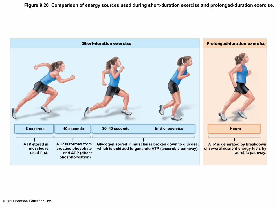
Figure 9.20 Comparison of energy sources used during short-duration exercise and prolonged-duration exercise.
Short-duration exercise
6 seconds
ATP stored inmuscles isused first.
10 seconds
ATP is formed fromcreatine phosphate
and ADP (directphosphorylation).
30–40 seconds
Glycogen stored in muscles is broken down to glucose,which is oxidized to generate ATP (anaerobic pathway).
End of exercise
Prolonged-duration exercise
Hours
ATP is generated by breakdownof several nutrient energy fuels by
aerobic pathway.

© 2013 Pearson Education, Inc.
Muscle Fatigue
• Physiological inability to contract despite continued stimulation
• Occurs when– Ionic imbalances (K+, Ca2+, Pi) interfere with
E‑C coupling– Prolonged exercise may damage SR and
interferes with Ca2+ regulation and release
• Total lack of ATP occurs rarely, during states of continuous contraction, and causes contractures (continuous contractions)

© 2013 Pearson Education, Inc.
Excess Postexercise Oxygen Consumption
• To return muscle to resting state– Oxygen reserves replenished
– Lactic acid converted to pyruvic acid– Glycogen stores replaced– ATP and creatine phosphate reserves
replenished
• All require extra oxygen; occur post exercise

© 2013 Pearson Education, Inc.
Heat Production During Muscle Activity
• ~40% of energy released in muscle activity useful as work
• Remaining energy (60%) given off as heat
• Dangerous heat levels prevented by radiation of heat from skin and sweating
• Shivering - result of muscle contractions to generate heat when cold

© 2013 Pearson Education, Inc.
Force of Muscle Contraction
• Force of contraction depends on number of cross bridges attached, which is affected by
• Number of muscle fibers stimulated (recruitment)
• Relative size of fibers—hypertrophy of cells increases strength
• Frequency of stimulation• Degree of muscle stretch
© 2013 Pearson Education, Inc.
Force of Muscle Contraction
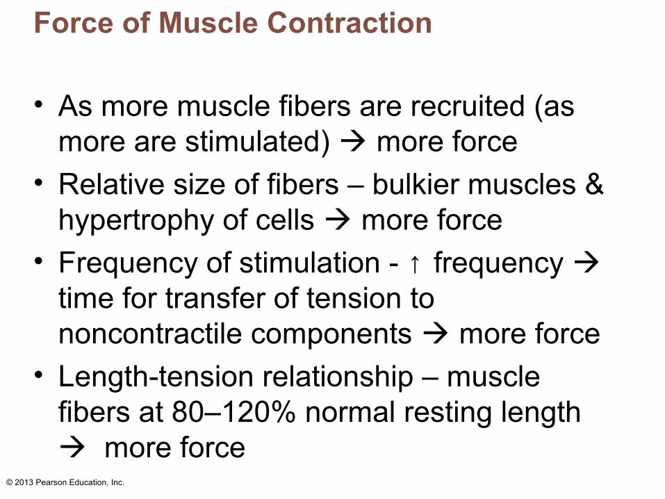
• As more muscle fibers are recruited (as more are stimulated) more force
• Relative size of fibers – bulkier muscles & hypertrophy of cells more force
• Frequency of stimulation - ↑ frequency time for transfer of tension to noncontractile components more force
• Length-tension relationship – muscle fibers at 80–120% normal resting length more force

© 2013 Pearson Education, Inc.
Figure 9.21 Factors that increase the force of skeletal muscle contraction.
Largenumber of
musclefibers
recruited
Largemusclefibers
Highfrequency ofstimulation
(wavesummation
and tetanus)
Muscle andsarcomerestretched to
slightly over 100%of resting length
Contractile force (more cross bridges attached)

© 2013 Pearson Education, Inc.
Velocity and Duration of Contraction
• Influenced by:– Muscle fiber type
– Load– Recruitment
© 2013 Pearson Education, Inc.
Muscle Fiber Type
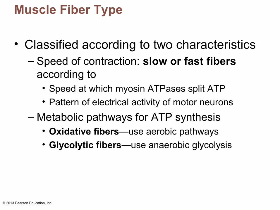
• Classified according to two characteristics– Speed of contraction: slow or fast fibers
according to• Speed at which myosin ATPases split ATP
• Pattern of electrical activity of motor neurons
– Metabolic pathways for ATP synthesis• Oxidative fibers—use aerobic pathways
• Glycolytic fibers—use anaerobic glycolysis
© 2013 Pearson Education, Inc.
Muscle Fiber Type
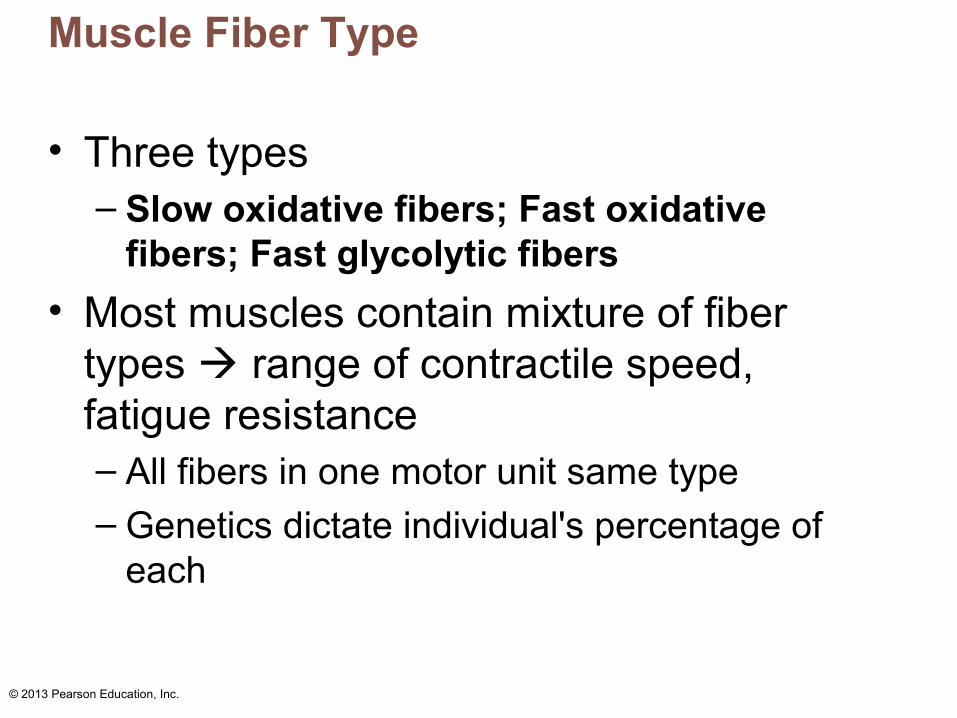
• Three types – Slow oxidative fibers; Fast oxidative
fibers; Fast glycolytic fibers
• Most muscles contain mixture of fiber types range of contractile speed, fatigue resistance– All fibers in one motor unit same type– Genetics dictate individual's percentage of
each

© 2013 Pearson Education, Inc.
Table 9.2 Structural and Functional Characteristics of the Three Types of Skeletal Muscle Fibers

© 2013 Pearson Education, Inc.
Adaptations to Exercise
• Aerobic (endurance) exercise– Leads to increased
• Muscle capillaries
• Number of mitochondria
• Myoglobin synthesis
– Results in greater endurance, strength, and resistance to fatigue
– May convert fast glycolytic fibers into fast oxidative fibers
© 2013 Pearson Education, Inc.
Effects of Resistance Exercise
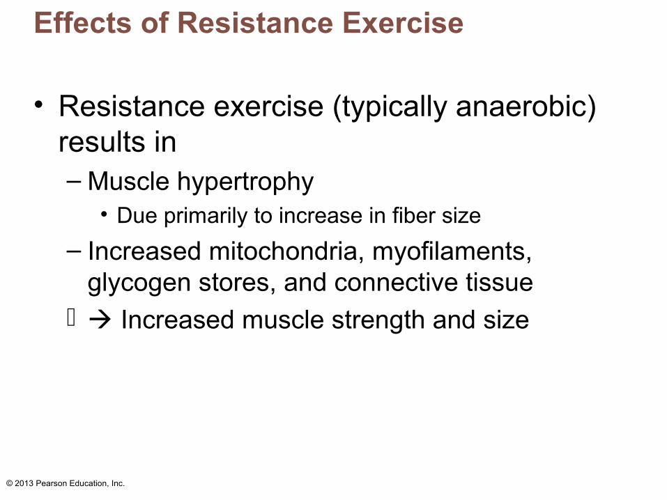
• Resistance exercise (typically anaerobic) results in– Muscle hypertrophy
• Due primarily to increase in fiber size
– Increased mitochondria, myofilaments, glycogen stores, and connective tissue
Increased muscle strength and size

© 2013 Pearson Education, Inc.
A Balanced Exercise Program
• Overload principle– Forcing muscle to work hard promotes
increased muscle strength and endurance– Muscles adapt to increased demands– Muscles must be overloaded to produce
further gains– Overuse injuries may result from lack of rest– Best programs alternate aerobic and
anaerobic activities

© 2013 Pearson Education, Inc.
Homeostatic Imbalance
• Disuse atrophy– Result of immobilization
– Muscle strength declines 5% per day
• Without neural stimulation muscles atrophy to ¼ initial size– Fibrous connective tissue replaces lost
muscle tissue rehabilitation impossible

© 2013 Pearson Education, Inc.
Smooth Muscle
• Found in walls of most hollow organs(except heart)
• Usually in two layers (longitudinal and circular)
© 2013 Pearson Education, Inc.
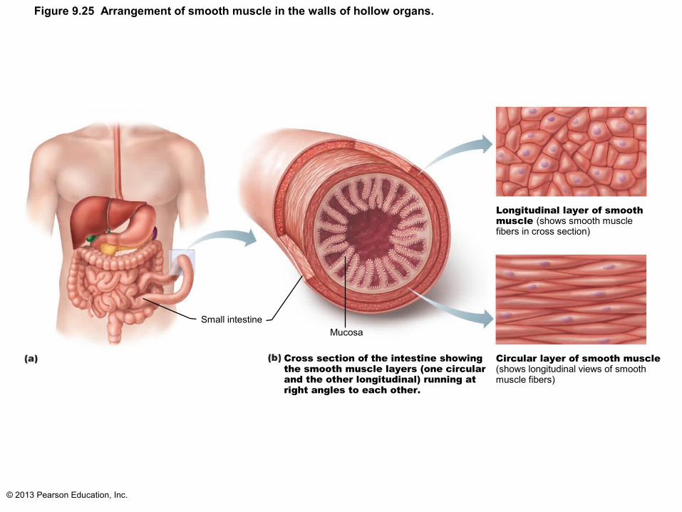
Figure 9.25 Arrangement of smooth muscle in the walls of hollow organs.
Small intestineMucosa
Cross section of the intestine showing the smooth muscle layers (one circular and the other longitudinal) running at right angles to each other.
Circular layer of smooth muscle(shows longitudinal views of smoothmuscle fibers)
Longitudinal layer of smooth muscle (shows smooth musclefibers in cross section)

© 2013 Pearson Education, Inc.
Microscopic Structure
• Spindle-shaped fibers - thin and short compared with skeletal muscle fibers; only one nucleus; no striations
• Lacks connective tissue sheaths; endomysium only
• SR - less developed than in skeletal muscle • Pouchlike infoldings (caveolae) of sarcolemma
sequester Ca2+ - most calcium influx from outside cell; rapid
• No sarcomeres, myofibrils, or T tubules

© 2013 Pearson Education, Inc.
Microscopic Structure of Smooth Muscle Fibers
• Longitudinal layer– Fibers parallel to long axis of organ; contraction
dilates and shortened
• Circular layer– Fibers in circumference of organ; contraction
constricts lumen, elongates organ
• Allows peristalsis - Alternating contractions and relaxations of smooth muscle layers that mix and squeeze substances through lumen of hollow organs

© 2013 Pearson Education, Inc.
Table 9.3 Comparison of Skeletal, Cardiac, and Smooth Muscle (1 of 4)

© 2013 Pearson Education, Inc.
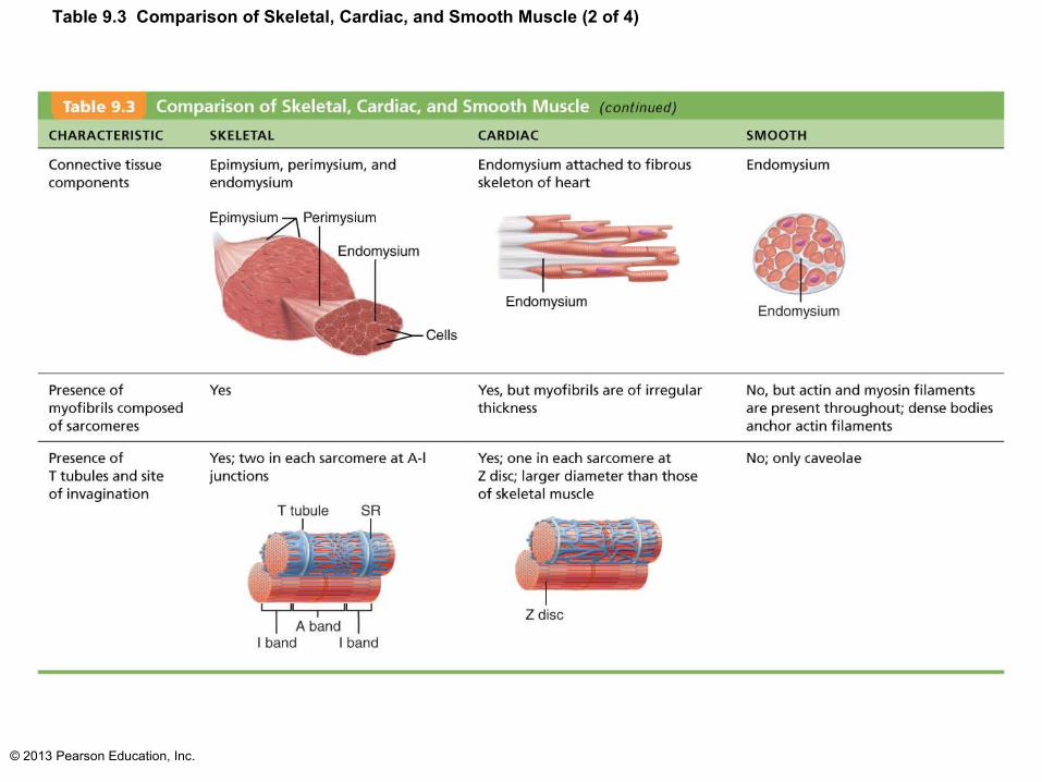
Table 9.3 Comparison of Skeletal, Cardiac, and Smooth Muscle (2 of 4)

© 2013 Pearson Education, Inc.
Innervation of Smooth Muscle
• No NMJ as in skeletal muscle
• Autonomic nerve fibers innervate smooth muscle at diffuse junctions
• Varicosities (bulbous swellings) of nerve fibers store and release neurotransmitters into diffuse junctions

© 2013 Pearson Education, Inc.
Figure 9.26 Innervation of smooth muscle.
Varicosities
Autonomicnerve fibersinnervatemost smoothmuscle fibers.
Smoothmusclecell
Synapticvesicles
Mitochondrion Varicosities releasetheir neurotransmittersinto a wide synaptic cleft (a diffuse junction).

© 2013 Pearson Education, Inc.
Myofilaments in Smooth Muscle
• Ratio of thick to thin filaments (1:13) is much lower than in skeletal muscle (1:2)
• Thick filaments have heads along entire length
• No troponin complex; protein calmodulin binds Ca2+

© 2013 Pearson Education, Inc.
Myofilaments in Smooth Muscle
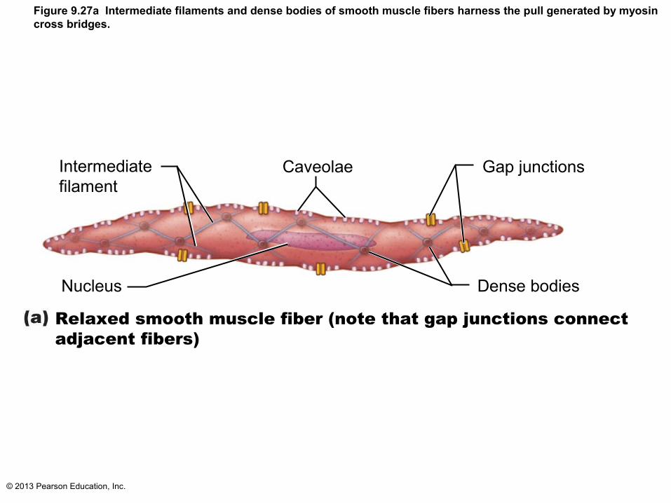
• Myofilaments are spirally arranged, causing smooth muscle to contract in corkscrew manner
• Dense bodies– Proteins that anchor noncontractile
intermediate filaments to sarcolemma at regular intervals
– Correspond to Z discs of skeletal muscle
© 2013 Pearson Education, Inc.
Intermediatefilament
Caveolae Gap junctions
Nucleus Dense bodies
Relaxed smooth muscle fiber (note that gap junctions connect adjacent fibers)
Figure 9.27a Intermediate filaments and dense bodies of smooth muscle fibers harness the pull generated by myosincross bridges.

© 2013 Pearson Education, Inc.
NucleusDense bodies
Contracted smooth muscle fiber
Figure 9.27b Intermediate filaments and dense bodies of smooth muscle fibers harness the pull generated by myosincross bridges.
© 2013 Pearson Education, Inc.
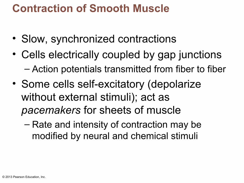
Contraction of Smooth Muscle
• Slow, synchronized contractions
• Cells electrically coupled by gap junctions– Action potentials transmitted from fiber to fiber
• Some cells self-excitatory (depolarize without external stimuli); act as pacemakers for sheets of muscle – Rate and intensity of contraction may be
modified by neural and chemical stimuli
© 2013 Pearson Education, Inc.
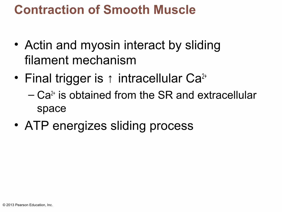
Contraction of Smooth Muscle
• Actin and myosin interact by sliding filament mechanism
• Final trigger is ↑ intracellular Ca2+
– Ca2+ is obtained from the SR and extracellular space
• ATP energizes sliding process
© 2013 Pearson Education, Inc.
Table 9.3 Comparison of Skeletal, Cardiac, and Smooth Muscle (2 of 4)
© 2013 Pearson Education, Inc.
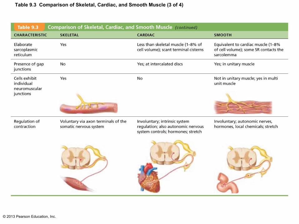
Table 9.3 Comparison of Skeletal, Cardiac, and Smooth Muscle (3 of 4)

© 2013 Pearson Education, Inc.
Contraction of Smooth Muscle
• Slow to contract and relax but maintains for prolonged periods with little energy cost– Slow ATPases– Myofilaments may latch together to save
energy
• Relaxation requires– Ca2+ detachment from calmodulin; active
transport of Ca2+ into SR and ECF; dephosphorylation of myosin to reduce myosin ATPase activity

© 2013 Pearson Education, Inc.
Regulation of Contraction
• By nerves, hormones, or local chemical changes
• Neural regulation– Neurotransmitter binding → ↑ [Ca2+] in
sarcoplasm; either graded (local) potential or action potential
– Response depends on neurotransmitter released and type of receptor molecules

© 2013 Pearson Education, Inc.
Regulation of Contraction
• Hormones and local chemicals– Some smooth muscle cells have no nerve
supply• Depolarize spontaneously or in response to
chemical stimuli that bind to G protein–linked receptors
– Some respond to both neural and chemical stimuli
– Chemical factors include hormones, CO2, pH
© 2013 Pearson Education, Inc.
Special Features of Smooth Muscle Contraction
• Stress-relaxation response – Responds to stretch only briefly, then adapts
to new length– Retains ability to contract on demand– Enables organs such as stomach and bladder
to temporarily store contents
• Length and tension changes– Can contract when between half and twice its
resting length
© 2013 Pearson Education, Inc.
Special Features of Smooth Muscle Contraction
• Hyperplasia– Smooth muscle cells can divide and increase
numbers– Example
• Estrogen effects on uterus at puberty and during pregnancy

© 2013 Pearson Education, Inc.
Table 9.3 Comparison of Skeletal, Cardiac, and Smooth Muscle (4 of 4)

© 2013 Pearson Education, Inc.
Types of Smooth Muscle
• Smooth muscle varies in different organs– Fiber arrangement and organization
– Innervation– Responsiveness to various stimuli
• Categorized as unitary and multi unit

© 2013 Pearson Education, Inc.
Types of Smooth Muscle
• Unitary (visceral) smooth muscle– In all hollow organs except heart
– Arranged in opposing sheets– Innervated by varicosities – Often exhibit spontaneous action potentials– Electrically coupled by gap junctions– Respond to various chemical stimuli

© 2013 Pearson Education, Inc.
Types of Smooth Muscle: Multiunit
• Multiunit smooth muscle– Located in large airways, large arteries,
arrector pili muscles, and iris of eye– Gap junctions; spontaneous depolarization
rare– Independent muscle fibers; innervated by
autonomic NS; graded contractions occur in response to neural stimuli
– Has motor units; responds to hormones

© 2013 Pearson Education, Inc.
Developmental Aspects
• All muscle tissues develop from embryonic myoblasts
• Multinucleated skeletal muscle cells form by fusion
• Growth factor agrin stimulates clustering of ACh receptors at neuromuscular junctions
• Cardiac and smooth muscle myoblasts develop gap junctions
© 2013 Pearson Education, Inc.
Developmental Aspects
• ~ All muscle tissue develops from myoblasts• Cardiac and skeletal muscle become amitotic,
but can lengthen and thicken in growing child
• Myoblast-like skeletal muscle satellite cells have limited regenerative ability
• Cardiomyocytes can divide at modest rate, but injured heart muscle mostly replaced by connective tissue
• Smooth muscle regenerates throughout life

© 2013 Pearson Education, Inc.
Developmental Aspects
• Muscular development reflects neuromuscular coordination
• Development occurs head to toe, and proximal to distal
• Peak natural neural control occurs by midadolescence
• Athletics and training can improve neuromuscular control

© 2013 Pearson Education, Inc.
Developmental Aspects
• Female skeletal muscle makes up 36% of body mass
• Male skeletal muscle makes up 42% of body mass, primarily due to testosterone
• Body strength per unit muscle mass same in both sexes

© 2013 Pearson Education, Inc.
Developmental Aspects
• With age, connective tissue increases and muscle fibers decrease
• By age 30, loss of muscle mass (sarcopenia) begins
• Regular exercise reverses sarcopenia
• Atherosclerosis may block distal arteries, leading to intermittent claudication and severe pain in leg muscles
© 2013 Pearson Education, Inc.
Muscular Dystrophy
• Group of inherited muscle-destroying diseases; generally appear in childhood
• Muscles enlarge due to fat and connective tissue deposits
• Muscle fibers atrophy and degenerate
© 2013 Pearson Education, Inc.
Muscular Dystrophy
• Duchenne muscular dystrophy (DMD):– Most common and severe type
– Inherited, sex-linked, carried by females and expressed in males (1/3500) as lack of dystrophin
• Cytoplasmic protein that stabilizes sarcolemma• Fragile sarcolemma tears Ca2+ entry
damaged contractile fibers inflammatory cells muscle mass drops
– Victims become clumsy and fall frequently; usually die of respiratory failure in 20s

© 2013 Pearson Education, Inc.
Muscular Dystrophy
– No cure– Prednisone improves muscle strength and
function
– Myoblast transfer therapy disappointing– Coaxing dystrophic muscles to produce more
utrophin (protein similar to dystrophin) successful in mice
– Viral gene therapy and infusion of stem cells with correct dystrophin genes show promise
Related Documents