Current Neuropharmacology, 2011, 9, 715-727 715 1570-159X/11 $58.00+.00 ©2011 Bentham Science Publishers cGMP Signaling, Phosphodiesterases and Major Depressive Disorder Gillian W. Reierson 1 , Shuyu Guo 2 , Claudio Mastronardi 2 , Julio Licinio 2 and Ma-Li Wong 2, * 1 University of Miami Miller School of Medicine, Miami, FL, USA; 2 Department of Translational Medicine, John Curtin School of Medical Research, Australian National University, Acton, ACT, Australia Abstract: Deficits in neuroplasticity are hypothesized to underlie the pathophysiology of major depressive disorder (MDD): the effectiveness of antidepressants is thought to be related to the normalization of disrupted synaptic trans- mission and neurogenesis. The cyclic adenosine monophosphate (cAMP) signaling cascade has received considerable attention for its role in neuroplasticity and MDD. However components of a closely related pathway, the cyclic guanosine monophosphate (cGMP) have been studied with much lower intensity, even though this signaling transduction cascade is also expressed in the brain and the activity of this pathway has been implicated in learning and memory processes. Cyclic GMP acts as a second messenger; it amplifies signals received at postsynaptic receptors and activates downstream effector molecules resulting in gene expression changes and neuronal responses. Phosphodiesterase (PDE) enzymes degrade cGMP into 5’GMP and therefore they are involved in the regulation of intracellular levels of cGMP. Here we review a growing body of evidence suggesting that the cGMP signaling cascade warrants further investigation for its involvement in MDD and antidepressant action. Keywords: Major Depression, cyclic guanosine monophosphate, neurogenesis, neuroplasticity, phophodiesterases, cyclases, antidepressants, pharmacology. 1. INTRODUCTION Major depressive disorder (MDD) is a chronic and highly disabling condition. It is the most common mood disorder: up to 17% of the population suffers from this condition at some point during their lifetimes. While antidepressants have been shown to be effective in treating the symptoms of MDD in 60-70% of patients, 30-40% of patients do not respond to pharmacotherapy [1, 2]. Furthermore, antidepressants must be taken chronically for several weeks before treatment re- sponse is achieved and it is still unclear what are the mecha- nisms by which these drugs relieve the clinical symptoms of MDD. Evidence from clinical and preclinical research accu- mulated in recent years support the concept that aberrant neuroplasticity, including functional synaptic plasticity and structural plasticity, in the hippocampus and prefrontal cortex, might underlie MDD [3, 4]. Intracellular cyclic adenosine monophosphate (cAMP) signaling activated by brain-derived neurotrophic factor (BDNF) has been shown to be a pivotal player in neuroplasticity and MDD; moreover, studies have demonstrated that treatment with antidepressants can alter the expression of components of this signaling pathway in rodents [5-7]. Unfortunately, earlier findings have been inconsistently replicated making it premature to conceptualize translational strategies for new antidepressant pharmacotherapy targeting the cAMP signaling. The cyclic guanosine monophosphate (cGMP) signaling pathway is an intracellular nucleotide cascade that is also involved in neuroplasticity. It is closely related to cAMP signaling, but it *Address correspondence to this author at Building 131, Garran Road, John Curtin School of Medical Research, The Australian National University Canberra, ACT, Australia; Tel: +61 2 6125 8557; Fax: +61 2 6125 2337; E-mail: [email protected] has received little attention in the biology of MDD and antidepressant action. The cGMP signaling cascade consists of a number of different elements that can regulate cGMP levels and influence downstream cellular responses. Fig. (1) depicts the presynaptic and postsynaptic elements of the cGMP signaling pathway in the central nervous system; these elements are expressed in the hippocampus, a brain region that has been implicated in MDD [8, 9]. Pharma- cotherapy targeting different elements of the cGMP pathway is currently available; this class of drugs includes phosphodi- esterase inhibitors, which increase cGMP signaling. In this review, we summarize the evidence that supports the role of cGMP signaling in neuroplasticity and in the pathophysiology of MDD and antidepressant treatment response, with a specific focus on the role of phosphodiesterases (PDEs). 2. cGMP SIGNALING COMPONENTS AND THEIR DISTRIBUTION IN THE BRAIN 2.1. cGMP Synthesis: pGC and sGC The synthetic enzyme guanylate/guanylyl cyclase (GC) converts guanosine triphosphate (GTP) to 3’-5’-cyclic guanosine monophosphate to produce the second messenger molecule known as cGMP. The production of cGMP by GC can occur in two different ways: 1) following activation of membrane bound guanylate cyclase (particulate GC or pGC) [10, 11] and 2) following activation of soluble guanylate cyclase (sGC, also called NO receptors) in the cytoplasm by the diffusible second messenger molecule, nitric oxide (NO) [12, 13]. Particulate GC is composed of two subfamilies: the pep- tide hormone receptor subfamily, which binds natriuretic peptides, and the Ca 2+ -modulated ROS-GC (rod outer

Welcome message from author
This document is posted to help you gain knowledge. Please leave a comment to let me know what you think about it! Share it to your friends and learn new things together.
Transcript
Current Neuropharmacology, 2011, 9, 715-727 715
1570-159X/11 $58.00+.00 ©2011 Bentham Science Publishers
cGMP Signaling, Phosphodiesterases and Major Depressive Disorder
Gillian W. Reierson1, Shuyu Guo
2, Claudio Mastronardi
2, Julio Licinio
2 and
Ma-Li Wong
2,*
1University of Miami Miller School of Medicine, Miami, FL, USA;
2Department of Translational Medicine, John Curtin
School of Medical Research, Australian National University, Acton, ACT, Australia
Abstract: Deficits in neuroplasticity are hypothesized to underlie the pathophysiology of major depressive disorder
(MDD): the effectiveness of antidepressants is thought to be related to the normalization of disrupted synaptic trans-
mission and neurogenesis. The cyclic adenosine monophosphate (cAMP) signaling cascade has received considerable
attention for its role in neuroplasticity and MDD. However components of a closely related pathway, the cyclic guanosine
monophosphate (cGMP) have been studied with much lower intensity, even though this signaling transduction cascade is
also expressed in the brain and the activity of this pathway has been implicated in learning and memory processes. Cyclic
GMP acts as a second messenger; it amplifies signals received at postsynaptic receptors and activates downstream effector
molecules resulting in gene expression changes and neuronal responses. Phosphodiesterase (PDE) enzymes degrade
cGMP into 5’GMP and therefore they are involved in the regulation of intracellular levels of cGMP. Here we review a
growing body of evidence suggesting that the cGMP signaling cascade warrants further investigation for its involvement
in MDD and antidepressant action.
Keywords: Major Depression, cyclic guanosine monophosphate, neurogenesis, neuroplasticity, phophodiesterases, cyclases,
antidepressants, pharmacology.
1. INTRODUCTION
Major depressive disorder (MDD) is a chronic and highly
disabling condition. It is the most common mood disorder:
up to 17% of the population suffers from this condition at
some point during their lifetimes. While antidepressants have
been shown to be effective in treating the symptoms of MDD
in 60-70% of patients, 30-40% of patients do not respond to
pharmacotherapy [1, 2]. Furthermore, antidepressants must
be taken chronically for several weeks before treatment re-
sponse is achieved and it is still unclear what are the mecha-
nisms by which these drugs relieve the clinical symptoms of
MDD. Evidence from clinical and preclinical research accu-
mulated in recent years support the concept that aberrant
neuroplasticity, including functional synaptic plasticity and
structural plasticity, in the hippocampus and prefrontal cortex,
might underlie MDD [3, 4]. Intracellular cyclic adenosine
monophosphate (cAMP) signaling activated by brain-derived
neurotrophic factor (BDNF) has been shown to be a pivotal
player in neuroplasticity and MDD; moreover, studies
have demonstrated that treatment with antidepressants
can alter the expression of components of this signaling
pathway in rodents [5-7]. Unfortunately, earlier findings
have been inconsistently replicated making it premature to
conceptualize translational strategies for new antidepressant
pharmacotherapy targeting the cAMP signaling. The cyclic
guanosine monophosphate (cGMP) signaling pathway is
an intracellular nucleotide cascade that is also involved in
neuroplasticity. It is closely related to cAMP signaling, but it
*Address correspondence to this author at Building 131, Garran Road, John
Curtin School of Medical Research, The Australian National University
Canberra, ACT, Australia; Tel: +61 2 6125 8557; Fax: +61 2 6125 2337;
E-mail: [email protected]
has received little attention in the biology of MDD and
antidepressant action. The cGMP signaling cascade consists
of a number of different elements that can regulate cGMP
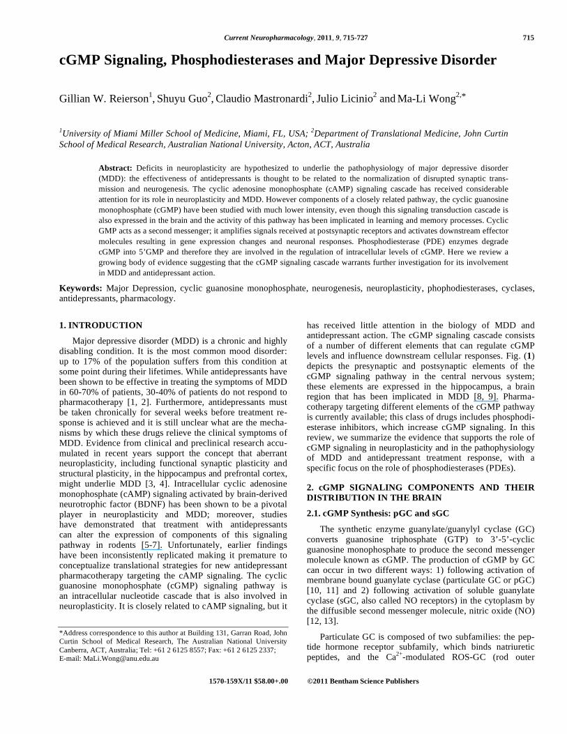
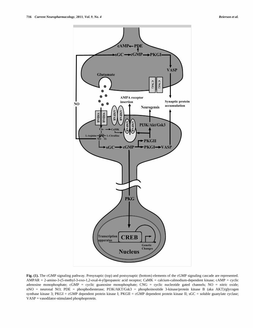
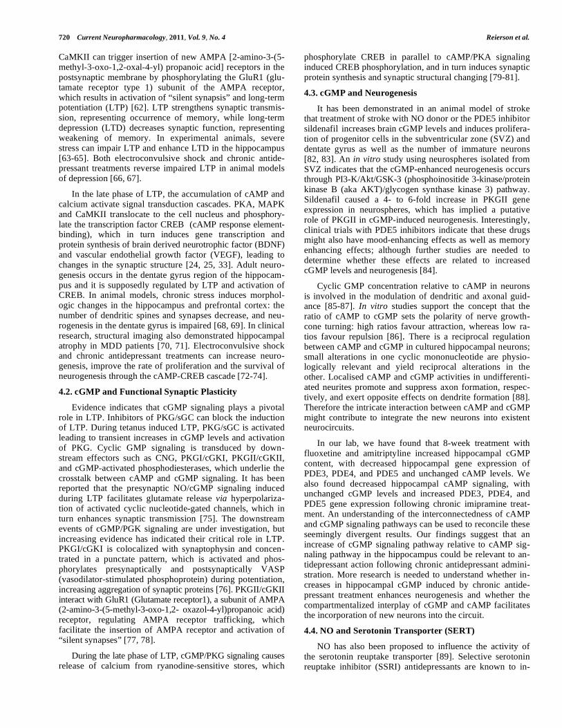
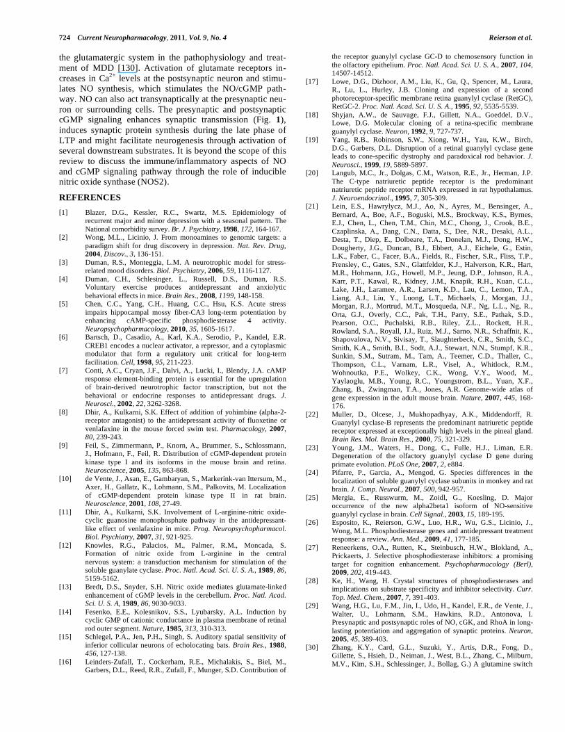
levels and influence downstream cellular responses. Fig. (1)
depicts the presynaptic and postsynaptic elements of the
cGMP signaling pathway in the central nervous system;
these elements are expressed in the hippocampus, a brain
region that has been implicated in MDD [8, 9]. Pharma-
cotherapy targeting different elements of the cGMP pathway
is currently available; this class of drugs includes phosphodi-
esterase inhibitors, which increase cGMP signaling. In this
review, we summarize the evidence that supports the role of
cGMP signaling in neuroplasticity and in the pathophysiology
of MDD and antidepressant treatment response, with a
specific focus on the role of phosphodiesterases (PDEs).
2. cGMP SIGNALING COMPONENTS AND THEIR
DISTRIBUTION IN THE BRAIN
2.1. cGMP Synthesis: pGC and sGC
The synthetic enzyme guanylate/guanylyl cyclase (GC)
converts guanosine triphosphate (GTP) to 3’-5’-cyclic
guanosine monophosphate to produce the second messenger
molecule known as cGMP. The production of cGMP by GC
can occur in two different ways: 1) following activation of
membrane bound guanylate cyclase (particulate GC or pGC)
[10, 11] and 2) following activation of soluble guanylate
cyclase (sGC, also called NO receptors) in the cytoplasm by
the diffusible second messenger molecule, nitric oxide (NO)
[12, 13].
Particulate GC is composed of two subfamilies: the pep-
tide hormone receptor subfamily, which binds natriuretic
peptides, and the Ca2+
-modulated ROS-GC (rod outer
716 Current Neuropharmacology, 2011, Vol. 9, No. 4 Reierson et al.
Fig. (1). The cGMP signaling pathway. Presynaptic (top) and postsynaptic (bottom) elements of the cGMP signaling cascade are represented.
AMPAR = 2-amino-3-(5-methyl-3-oxo-1,2-oxal-4-yl)propanoic acid receptor; CaMK = calcium-calmodium-dependent kinase; cAMP = cyclic
adenosine monophosphate; cGMP = cyclic guanosine monophosphate; CNG = cyclic nucleotide gated channels; NO = nitric oxide;
nNO = neuronal NO; PDE = phosphodiesterase; PI3K/AKT/Gsk3 = phosphoinositide 3-kinase/protein kinase B (aka AKT)/glycogen
synthase kinase 3; PKGI = cGMP dependent protein kinase I; PKGII = cGMP dependent protein kinase II; sGC = soluble guanylate cyclase;
VASP = vasodilator-stimulated phosphoprotein.
CN
G
NMDAR
CN
G
NMDAR
AMPAR
AMPAR
AMPAR
AMPAR
CREBGeneticChanges
Nucleus
Transcriptionapparatus
PKG
cGMP PKGIPKGII
VASP
VASP
PI3K/Akt/Gsk3
Neurogensis
AMPA receptorinsertion Synaptic protein
accumulation
sGC
NO O2
Ca2+
nNOsL-Arginine L-Citrulline
CaMK
NO
Glutamate
sGC cGMP PKGI
PDEcAMP
cGMP Signaling, Phosphodiesterases and Major Depression Disorder Current Neuropharmacology, 2011, Vol. 9, No. 4 717
segment-derived guanylate cyclase) subfamily, which is
linked with the senses of vision, olfaction and taste, and
light-regulated pinealocytes [1, 14-16]. The isoforms of pGC
expressed in the brain include guanylate cyclase A/natriuretic
peptide receptor 1 (GC-A/Npr1), guanylate cyclase B/natriuretic
peptide receptor 2 (GC-B/Npr2), guanylate cyclase 2D (GC-
D/GUCY2D); while two eye-specific GC have been identi-
fied in mammals (designed GC-E and GC-F or RetGC-1 and
RetGC-2 in humans) [17-19]. GC-A/Npr1 is activated by
atrial natriuretic peptide (ANP) and brain natriuretic peptide
(BNP) and the mRNA is expressed in the subfornical organ,
medial habenula, cerebellum and olfactory bulb [20, 21].
GC-B/Npr2 is activated by natriuretic peptide type C (CNP)
and the mRNA is expressed in the pineal gland, cerebellum,
cortex, hippocampus, hypothalamus, olfactory bulb, neural
lobe of the pituitary gland, and thalamus [21, 22]. GC-D
is expressed in the olfactory bulb in rodents, but it is a
pseudogene in humans [23].
Soluble GC exists as a heterodimer composed of differ-
ent combinations of subunits ( 1 or 2) and subunits
( 1 or 2); all sGC subunits mRNAs are expressed in the
brain with the exception of the 2 subunit mRNAs [24].
Both 1 and 2 sGC subunits mRNAs are expressed in the
cerebral cortex and olfactory bulb; 1 is also expressed in
striatum and pallidum and 2 is also expressed in hippocam-
pus and medulla [21]. The 1 sGC subunit mRNA is
expressed throughout the brain, with predominant expression
in the cerebral cortex, hippocampus, lateral septal complex,
olfactory bulb, and striatum [21, 24]. In the hippocampus,
the sGC heterodimer is composed of 2 and 1 subunits
[25].
2.2. cGMP Degradation: PDEs
Several different enzymes of the phosphodiesterase
(PDE) family are capable of cleaving cGMP into 5’-GMP.
PDEs are classified as cAMP specific (PDE4, PDE7, and
PDE8), cGMP specific (PDE5, PDE6, and PDE9), or dual
substrate (PDE1, PDE2, PDE3, PDE10, and PDE11) [26,
27]. Dual substrate PDEs are able to hydrolyze both cAMP
and cGMP. The specificity of a dual substrate PDE for each
cyclic nucleotide substrate is determined by the orientation
of an invariant glutamine in the binding pocket of the cata-
lytic domain of the PDE, a concept that is termed “glutamine
switch” [28-30]. The orientation of the glutamine is thought
to be determined by the presence of neighboring residues
that constrain the rotation of the glutamine to only allow the
purine ring of one or the other cyclic nucleotides to bind via
H bond formation; free rotation of the glutamine allows both
substrates to bind [30, 31]. The binding affinities (Km val-
ues), the catalytic hydrolyzing activities (Vmax, Kcat) and the
presence of specific domains in the N-terminal region of
these genes reveal much information about how these PDEs
might be uniquely suited to regulate cyclic nucleotide cross-
talk. The presence of N-terminal domains is especially
influential, as activity in these domains can cause conforma-
tional changes in the catalytic domain of the PDE, changing
the Km and Vmax of the enzyme toward cyclic nucleotide
substrates [30, 32].
In the following two paragraphs, we will summarize the
CNS expression of cGMP specific PDEs (PDE5, PDE6,
PDE9) and dual substrate PDEs (PDE1, PDE2, PDE3,
PDE10, and PDE11).
All cGMP specific PDEs are expressed in the brain. In
the rodent brain, PDE5A mRNA expression has been re-
ported in the purkinje cells of the cerebellum; strong staining
has also been observed in scattered cells in the hippocampus,
including pyramidal cells of CA1, CA2 and CA3, as well as
in the dentate gyrus [33]. PDE6 was initially thought to be
limited to the retina; however, PDE6B mRNA expression
has also been reported in mouse hippocampus [34]. CNS
expression of the PDE9A mRNA in the rodent brain has
been reported in the purkinje cells and granule cells of the
cerebellum, olfactory bulb and tubercle, caudate putamen,
and CA1 and dentate gyrus areas of the hippocampus [35-
37]. In the human brain, PDE9 mRNA expression has been
reported in the insular and visual cortices as well as in the
CA1, CA2 and CA3 subfields, and dentate gyrus of the hip-
pocampal formation [38].
All dual substrate cGMP are also expressed in the brain.
In rodents, in situ hybridization and immunohistochemistry
studies demonstrated that the PDE1A isoform is expressed in
the following brain areas: cerebral cortex, pyramidal cells of
the hippocampus, and striatum [39, 40]. PDE1B is also ex-
pressed in several brain areas including the caudate-putamen,
nucleus accumbens, dentate gyrus of hippocampus, olfactory
tubercle, medial thalamic nuclei, and brainstem [39, 40].
PDE1C mRNA is expressed in the granule cells of the cere-
bellum, caudate-putamen, olfactory tubercle, and brainstem
of the rodent brain [41]. In the human brain, hippocampal
PDE1B expression has been reported in the granule cells of
the dentate gyrus and in pyramidal cells [42]. PDE2 mRNA
is expressed in the rodent medial habenula, olfactory bulb
and tubercle, cortex, amygdala, striatum, and hippocampus
[33]. Within the rodent hippocampus, PDE2 protein is
expressed in the pyramidal cells of CA1 to CA3 subfields and
in the granule cells of the dentate gyrus [37]. In the human
brain, PDE2 mRNA expression has been found in the insular
and visual cortices as well as in the hippocampal formation
[38]. In a systematic immunohistochemistry study, PDE2A
protein was expressed in the limbic system, including hippo-
campus, basal ganglia, amygdala, isocortex, habenula, and
interpeduncular nucleus [43]. The mRNAs of both PDE3A
and PDE3B isoforms are expressed in the rodent hippocam-
pus, with PDE3A also displaying expression in the striatum
and PDE3B displaying expression in the cerebellum [44].
According to immunohistochemistry studies, PDE10A is
expressed in the pyramidal cells and dentate gyrus of the
hippocampus, cortex, granule cells of the cerebellum, and is
especially enriched in the striatum [45-47]. The mRNA and
protein of PDE11A are expressed in the trigeminal ganglion,
neocortex, spinal trigeminal nucleus, and purkinje cells of
the cerebellum of rats [48]. In the human brain, PDE11A4
protein is expressed in the pituitary [49].
2.3. cGMP Downstream Effectors: PKG/cGK
The downstream effectors of cGMP include protein
kinases, cyclic nucleotide gated channels (CNG), and cAMP
specific PDEs. The major downstream effector of cGMP
is the protein kinase G (PKG), which is alternatively referred
to as cyclic GMP dependent protein kinase (cGK). PKGs
718 Current Neuropharmacology, 2011, Vol. 9, No. 4 Reierson et al.
belong to a family of serine/threonine kinases because they
phosphorylate target protein substrates at specific structural
motifs. There are two different PKG genes, PRKG1 and
PRKG2 that encode cytosolic PKGI/cGKI and membrane-
bound PKGII/cGKII, respectively. PKGI can be alternatively
spliced at the N-terminal into two isoforms, PKGI
and PKGI , varying in tissue distribution, subcellular
localization, and substrate interaction [50].
PKG is comprised of three different functional domains:
N-terminal domain containing regulatory sites, a regulatory
domain containing cGMP binding sites, and a catalytic do-
main in the C-terminal region. The regulatory sites of the N-
terminal domain of PKG include domains that influence di-
merization, autoinhibitory, autophosphorylation, affinity and
cooperative behavior, and intracellular localization. Upon
binding of two molecules of cGMP to the two binding sites
present in the regulatory domain of PKG, the autoinhibitory
site that normally inhibits the catalytic domain of PKG is
removed, thus activating the protein. The catalytic domain of
PKG is located at the C-terminal end and contains binding
sites for Mg2+
, ATP, and the target protein. Once activated,
PKG phosphorylates target proteins at RKRKXST structural
motifs [51].
Although PKGI was previously reported to display lim-
ited expression restricted to the Purkinje cells of the cerebel-
lum, neurons in the nigrostriatal pathway, and dorsomedial
nucleus of the hypothalamus, further studies now demon-
strate that PKGI is expressed in additional brain areas such
as the hippocampus, olfactory bulb and amygdala [21, 52,
53]. PKGI expression has been found in the following mouse
brain structures: cerebellum, hippocampus, dorsomedial hy-
pothalamus, medulla, subcommisural organ, cerebral cortex,
amygdala, habenula, hypothalamus, olfactory bulb, pituitary,
and retina, with isoform specific PKGI expression in the
cerebellum and medulla and PKGI expression in the cortex,
hippocampus, hypothalamus, and olfactory bulb [9]. PKGII
is widely expressed throughout the rat brain, in structures
including the cerebral cortex, cerebellum, and brainstem
with greater expression in the neuropil as compared to
cell bodies [10]. PKGII expression has been also reported in
thalamus, outer layers of cerebral cortex, septum, amygdala,
and olfactory bulb [21, 53].
3. REGULATION OF cGMP SIGNALING
3.1. Compartmentalization and Crosstalk: Two Important
Concepts
A variety of extracellular cues are able to initiate com-
mon intracellular signaling cascades, such as the cGMP sig-
nal transduction pathway, to produce distinct biological ef-
fects. It is unclear how exactly different ligands are capable
of producing distinct messages through intracellular signal-
ing cascades resulting in unique cellular responses despite
acting through cGMP, which is their common second mes-
senger intermediate. The concept of “compartmentalization”
of cyclic nucleotide signaling in time and space has been
proposed to account for this discrepancy.
Through the use of fluorescence energy resonance trans-
fer (FRET) techniques in which specially engineered fluo-
rescent probes are used to detect cyclic nucleotide levels in
cells, much information has been gained regarding the
propagation of the cyclic nucleotide signal in time and space.
For example, observations of cAMP signaling in live cells
using FRET have demonstrated that the accumulation of
this small second messenger occurs in local “cAMP pools”
[54]. This observation has led to the idea of “compart-
mentalization” or the presence of confined compartments/
microdomains of cAMP signaling resulting from physical
barriers in the cell that restrict the diffusion of cAMP, form-
ing gradients [55]. Macromolecular complexes comprising
this physical barrier are composed of the A-Kinase anchor-
ing proteins (AKAPs), which create a scaffold or platform
for interactions between regulatory and effector proteins
relevant to cAMP signaling also present in the complex [34].
One of the target effectors of cGMP is PKG. PKG is ac-
tivated when two molecules of cGMP bind to the regulatory
subunits of this protein, releasing the autoinhibitory domain
of the regulatory subunit from the catalytic subunit. Acti-
vated PKG then can go on to phosphorylate any number of
different downstream targets to result in distinct cellular re-
sponses. Interestingly, PKGs participate in the negative
feedback of cGMP signaling by phosphorylating and activat-
ing PDEs, the enzymes that degrade cyclic nucleotides, to
decrease the levels of cGMP [56]. By regulating the levels of
cyclic nucleotides, PDEs are thought to play a critical regula-
tory role in fine-tuning the cGMP message in both time and
space. As PDEs are expressed in specific subcellular loca-
tions, it has been proposed that PDEs are strategically placed
in cells and act as “sinks” to increase the turnover of cyclic
nucleotides and thereby dampen the cyclic nucleotide mes-
sage [54]. PDEs recruited to macromolecular complexes
through their activation by PKG phosphorylation could po-
tentially decrease the amount of cGMP in a distinct subcellu-
lar microdomain. Pharmacological PDE inhibitors are hy-
pothesized to result in abnormal cyclic nucleotide signaling
by disrupting local cyclic nucleotide pools so that they “spill
over” from their confined compartments to inappropriately
activate target effectors previously not accessible [55].
The presence of cGMP pools located in specific cellular
regions suggests that different biological effects could be
produced through the action of different combinations of
PKGs and PDEs present in macromolecular complexes at
these specialized cyclic nucleotide microdomains [54].
While the details of “compartmentalization” of different
cGMP messages need to be clarified, the concept provides an
intriguing explanation to the question of how different ex-
tracellular cues working through the same intracellular mes-
senger can produce different biological outcomes. Therefore,
it is likely that the cGMP message is subject to compartmen-
talization, with macromolecular complexes composed of
AKAPs, PKG, and PDEs confining cGMP into specialized
microdomains or localized pools of cGMP [55]. Further
studies are needed to determine which AKAPs coordinate
cGMP hydrolyzing PDEs and PKG to regulate the amount of
cGMP.
The concept of “crosstalk” in intracellular signaling
transduction cascades refers to the ability of components
from one signaling pathway to interact with components of
another signaling pathway. There is already ample evidence
of crosstalk between cAMP and cGMP signaling pathways,
cGMP Signaling, Phosphodiesterases and Major Depression Disorder Current Neuropharmacology, 2011, Vol. 9, No. 4 719
with PDEs specifically hypothesized to act as points of inter-
section between these two pathways [57, 58]. Given the evi-
dence that PDEs are targeted to distinct subcellular locations
where they potentially regulate the levels of cyclic nucleo-
tides by acting as “sinks” within “compartmentalized”
anchored signaling complexes to define microdomains of
cyclic nucleotides within cells, it is plausible that PDEs
could also operate as points of crosstalk control in the cell
between cAMP and cGMP signaling networks. Two dual
substrate PDEs, namely PDE2 and PDE3, have emerged
as likely candidates to coordinate the interaction between
intracellular cyclic nucleotide signaling pathways [57].
3.2. Synthesis Regulation: NO/sGC/cGMP
Nitric oxide (NO) is a free radical and a small molecule
gas that is capable of diffusing across membranes where it
can act as a second messenger in signaling cascades. Specifi-
cally, NO acts as the intracellular ligand for the soluble
guanylate cyclase (sGC or NO receptors), which are cGMP
synthetic enzyme present in the cytosol. Upon binding of NO
to sGC, cGMP is synthesized from GTP. Therefore, NO is
an important regulator of cGMP levels because of its activat-
ing effects on sGC. In the nervous system, NO/cGMP signal-
ing mediates long-term potentiation (LTP) and NO was con-
sidered as a retrograde messenger [59]. Specifically, activa-
tion of NMDA (N-methyl-d-aspartate) receptors have been
found to increase postsynaptic synthesis of NO by Ca2+
/
calmodulin dependent neuronal NOS (NO synthase, NOS-
1/nNOS). The diffusible NO can then activate sGC that re-
sult in the presynaptic production of cGMP and neurotrans-
mitter release. Soluble GCs occurs as two isoforms: NO-
GC1 and NO-GC2. Interestingly, it has been reported that
both NO-GCs are required for LTP and that NMDA-induced
NO increases cGMP only in the presence of NO-GC1, which
have implied different synaptic localizations for NO-GC1
and NO-GC2. This evidence indicates that the compartmental
regulation of cGMP signaling is involved in synaptic plastic-
ity and that sGC may play an important role in the precise
spatial control of cGMP synthesis.
3.3. Degradation Regulation: PDE2, PDE3 and PDE5.
PDE2, 3 and 5 are relevant regulatory PDE’s because
they may be activated or inhibited by cGMP and they effect
changes in the levels of cGMP or cAMP.
PDE2
The binding affinities of PDE2 for cAMP (Km=30 M)
and cGMP (Km=10 M) reveal that PDE2 can bind both cy-
clic nucleotides equally and is capable of hydrolyzing both
cAMP and cGMP [57]. However, PDE2 is referred to as the
“cGMP-stimulated cAMP PDE” because cGMP binding to
GAF (cGMP-biding PDE, Anabaena adenylyl cyclases,
Eschericihia coli FhlAs) domains present in the N-terminal
region allosterically stimulates the cAMP hydrolyzing activ-
ity of PDE2, resulting in decreased cAMP levels. Cyclic
GMP binding to the GAF domain of PDE2 results in a con-
formational change to the catalytic domain of PDE2, chang-
ing the kinetics (decreasing Km and increasing Vmax values)
of PDE2 towards the cAMP substrate, and increasing the
hydrolyzing activity of PDE2 specifically towards the break-
down of cAMP into 5’AMP. Interestingly, PDE2A has three
splice variants, among which, PDE2A3 is entirely membrane
associated in the mouse brain. Moreover, in cultures of hip-
pocampal neurons, PDE2A co-localized with the synaptic
marker synaptophysin, in a punctate pattern. Therefore,
PDE2A represents a key point of compartmentalized cross-
talk between cAMP and cGMP signaling.
PDE3
Cyclic AMP and cGMP act as mutually competitive sub-
strates for PDE3, and the binding affinities of PDE3 for
cAMP (Km=0.08 M) and cGMP (Km=0.02 M) demon-
strate that PDE3 is indeed a dual substrate PDE capable of
binding each cyclic nucleotide substrate with similar affinity
[57]. However, as PDE3 demonstrates a higher rate of ca-
talysis for cAMP (10- fold higher Vmax) versus cGMP, it is
referred to as “cAMP preferring.” PDE3 has also been called
the “cGMP-inhibited cAMP PDE” because of in vitro ex-
perimental evidence that cGMP can inhibit the cAMP hydro-
lyzing activity of PDE3 to increase the levels of cAMP [60].
Evidence of a putative PKG phosphorylation site in the N-
terminal domain of PDE3 indicates that PDE3 activity can
be indirectly influenced by cGMP via downstream kinase
effects of PKG to phosphorylate PDE3 [57]. PDE3 therefore
represents a point of crosstalk control, another example of a
cAMP hydrolyzing PDE that might be a target of regulation
by cGMP-mediated signaling.
PDE5
PDE5 is referred to as the “cGMP-stimulated cGMP
PDE,” as the N-terminal region of the PDE5 gene contains a
GAF domain where cGMP can bind, as well as a site for
phosphorylation by PKG. Cyclic GMP allosteric binding to
the GAF domain of PDE5 stimulates the enzyme, decreasing
the Km to increase the affinity of the binding site for cGMP
as well increasing the catalytic activity of PDE5 to hydrolyse
cGMP. By binding to the GAF domain of PDE5, cGMP is
thought to increase the accessibility of the Ser102 residue of
PDE5 to phosphorylation by PKG, thereby activating the
enzyme. There is evidence that PKA might also phosphory-
late PDE5, an intriguing example of possible crosstalk
regulation between cAMP and cGMP signaling pathways
[61].
4. THE ROLE OF cGMP SIGNALING IN BRAIN
PLASTICITY AND MDD
4.1. The Neuroplasticity Hypothesis of MDD
Although the currently accepted mechanism of action of
available antidepressants suggests the involvement of the
monoamine system in MDD, increasing evidence has sup-
ported that neuroplasticity is disturbed in this disorder. Neu-
roplasticity includes functional synaptic plasticity (regulation
of synaptic transmission) and structural plasticity (such as
adult neurogenesis and permanent morphological changes of
synaptic structure).
Functional synaptic plasticity is mediated by glutamate
signaling. Glutamate binds to postsynaptic NMDA receptor,
increasing synaptic calcium flux, which induces the activa-
tion of kinases that is critical for the early phase of LTP,
such as calcium-calmodulin-dependent kinase II (CaMKII).
720 Current Neuropharmacology, 2011, Vol. 9, No. 4 Reierson et al.
CaMKII can trigger insertion of new AMPA [2-amino-3-(5-
methyl-3-oxo-1,2-oxal-4-yl) propanoic acid] receptors in the
postsynaptic membrane by phosphorylating the GluR1 (glu-
tamate receptor type 1) subunit of the AMPA receptor,
which results in activation of “silent synapsis” and long-term
potentiation (LTP) [62]. LTP strengthens synaptic transmis-
sion, representing occurrence of memory, while long-term
depression (LTD) decreases synaptic function, representing
weakening of memory. In experimental animals, severe
stress can impair LTP and enhance LTD in the hippocampus
[63-65]. Both electroconvulsive shock and chronic antide-
pressant treatments reverse impaired LTP in animal models
of depression [66, 67].
In the late phase of LTP, the accumulation of cAMP and
calcium activate signal transduction cascades. PKA, MAPK
and CaMKII translocate to the cell nucleus and phosphory-
late the transcription factor CREB (cAMP response element-
binding), which in turn induces gene transcription and
protein synthesis of brain derived neurotrophic factor (BDNF)
and vascular endothelial growth factor (VEGF), leading to
changes in the synaptic structure [24, 25, 33]. Adult neuro-
genesis occurs in the dentate gyrus region of the hippocam-
pus and it is supposedly regulated by LTP and activation of
CREB. In animal models, chronic stress induces morphol-
ogic changes in the hippocampus and prefrontal cortex: the
number of dendritic spines and synapses decrease, and neu-
rogenesis in the dentate gyrus is impaired [68, 69]. In clinical
research, structural imaging also demonstrated hippocampal
atrophy in MDD patients [70, 71]. Electroconvulsive shock
and chronic antidepressant treatments can increase neuro-
genesis, improve the rate of proliferation and the survival of
neurogenesis through the cAMP-CREB cascade [72-74].
4.2. cGMP and Functional Synaptic Plasticity
Evidence indicates that cGMP signaling plays a pivotal
role in LTP. Inhibitors of PKG/sGC can block the induction
of LTP. During tetanus induced LTP, PKG/sGC is activated
leading to transient increases in cGMP levels and activation
of PKG. Cyclic GMP signaling is transduced by down-
stream effectors such as CNG, PKGI/cGKI, PKGII/cGKII,
and cGMP-activated phosphodiesterases, which underlie the
crosstalk between cAMP and cGMP signaling. It has been
reported that the presynaptic NO/cGMP signaling induced
during LTP facilitates glutamate release via hyperpolariza-
tion of activated cyclic nucleotide-gated channels, which in
turn enhances synaptic transmission [75]. The downstream
events of cGMP/PGK signaling are under investigation, but
increasing evidence has indicated their critical role in LTP.
PKGI/cGKI is colocalized with synaptophysin and concen-
trated in a punctate pattern, which is activated and phos-
phorylates presynaptically and postsynaptically VASP
(vasodilator-stimulated phosphoprotein) during potentiation,
increasing aggregation of synaptic proteins [76]. PKGII/cGKII
interact with GluR1 (Glutamate receptor1), a subunit of AMPA
(2-amino-3-(5-methyl-3-oxo-1,2- oxazol-4-yl)propanoic acid)
receptor, regulating AMPA receptor trafficking, which
facilitate the insertion of AMPA receptor and activation of
“silent synapses” [77, 78].
During the late phase of LTP, cGMP/PKG signaling causes
release of calcium from ryanodine-sensitive stores, which
phosphorylate CREB in parallel to cAMP/PKA signaling
induced CREB phosphorylation, and in turn induces synaptic
protein synthesis and synaptic structural changing [79-81].
4.3. cGMP and Neurogenesis
It has been demonstrated in an animal model of stroke
that treatment of stroke with NO donor or the PDE5 inhibitor
sildenafil increases brain cGMP levels and induces prolifera-
tion of progenitor cells in the subventricular zone (SVZ) and
dentate gyrus as well as the number of immature neurons
[82, 83]. An in vitro study using neurospheres isolated from
SVZ indicates that the cGMP-enhanced neurogenesis occurs
through PI3-K/Akt/GSK-3 (phosphoinositide 3-kinase/protein
kinase B (aka AKT)/glycogen synthase kinase 3) pathway.
Sildenafil caused a 4- to 6-fold increase in PKGII gene
expression in neurospheres, which has implied a putative
role of PKGII in cGMP-induced neurogenesis. Interestingly,
clinical trials with PDE5 inhibitors indicate that these drugs
might also have mood-enhancing effects as well as memory
enhancing effects; although further studies are needed to
determine whether these effects are related to increased
cGMP levels and neurogenesis [84].
Cyclic GMP concentration relative to cAMP in neurons
is involved in the modulation of dendritic and axonal guid-
ance [85-87]. In vitro studies support the concept that the
ratio of cAMP to cGMP sets the polarity of nerve growth-
cone turning: high ratios favour attraction, whereas low ra-
tios favour repulsion [86]. There is a reciprocal regulation
between cAMP and cGMP in cultured hippocampal neurons;
small alterations in one cyclic mononucleotide are physio-
logically relevant and yield reciprocal alterations in the
other. Localised cAMP and cGMP activities in undifferenti-
ated neurites promote and suppress axon formation, respec-
tively, and exert opposite effects on dendrite formation [88].
Therefore the intricate interaction between cAMP and cGMP
might contribute to integrate the new neurons into existent
neurocircuits.
In our lab, we have found that 8-week treatment with
fluoxetine and amitriptyline increased hippocampal cGMP
content, with decreased hippocampal gene expression of
PDE3, PDE4, and PDE5 and unchanged cAMP levels. We
also found decreased hippocampal cAMP signaling, with
unchanged cGMP levels and increased PDE3, PDE4, and
PDE5 gene expression following chronic imipramine treat-
ment. An understanding of the interconnectedness of cAMP
and cGMP signaling pathways can be used to reconcile these
seemingly divergent results. Our findings suggest that an
increase of cGMP signaling pathway relative to cAMP sig-
naling pathway in the hippocampus could be relevant to an-
tidepressant action following chronic antidepressant admini-
stration. More research is needed to understand whether in-
creases in hippocampal cGMP induced by chronic antide-
pressant treatment enhances neurogenesis and whether the
compartmentalized interplay of cGMP and cAMP facilitates
the incorporation of new neurons into the circuit.
4.4. NO and Serotonin Transporter (SERT)
NO has also been proposed to influence the activity of
the serotonin reuptake transporter [89]. Selective serotonin
reuptake inhibitor (SSRI) antidepressants are known to in-
cGMP Signaling, Phosphodiesterases and Major Depression Disorder Current Neuropharmacology, 2011, Vol. 9, No. 4 721
crease the amount of serotonin at the synapse by inhibiting
serotonin transporter (SERT); therefore, SERT has been hy-
pothesized as an important player in the pathophysiology of
MDD. A recent proteomics based approach identified NOS1
as one of the proteins that physically interacts with the PSD-
95/DISC large/ZO-1 (PDZ) domain of SERT [90]. HEK-293
cells transfected with NOS1 and YFP (yellow fluorescent
protein)-tagged SERT were used to demonstrate that a
physical interaction between these two proteins can be
recapitulated in an in vitro cell culture model. Further, the
interaction of NOS1 with SERT was found to decrease 5-HT
uptake, indicating that SERT activity was decreased upon
physical interaction with NOS1. In light of these results,
NOS1 has been hypothesized as a putative endogenous anti-
depressant by decreasing the activity of SERT to increase
extracellular 5-HT [59].
4.5. cGMP Signaling and MDD
4.5.1. Genotyping SNPs in cGMP Signaling Components
(NOS1 and PDEs)
Only one study has investigated the relationship between
a specific NOS1 genetic variant (C2767) and MDD. The
authors chose to study this genetic variant as a previous ge-
netic study had found an association of this single nucleotide
polymorphism (SNP) and schizophrenia [91]. In this study of
114 MDD patients treated with fluoxetine for 4 weeks, the
NOS1 C2767 polymorphism was not significantly associated
with susceptibility to MDD or to antidepressant treatment
response when compared to 82 controls. However, the
authors of that study have noted that 4 weeks fluoxetine
treatment might not have been sufficiently long enough to
achieve antidepressant treatment response and therefore they
could not exclude the possibility that a longer duration of
treatment could have yielded different results. Another ge-
netic association analysis performed in a sample of Japanese
patients with MDD and bipolar disorder failed to detect an
association of 8 weeks fluvoxamine treatment with the
rs41279104 or ex1c SNP in the NOS1 gene in MDD pa-
tients. The researchers acknowledged that using linkage
disequilibrium and a larger sample size could yield different
results [92].
Our lab has previously found that several SNPs in cGMP
degrading PDE genes are related to the susceptibility to
MDD and to antidepressant treatment response [93]. In that
study, Mexican-American men and women MDD patients
and controls were treated for 8 weeks with desipramine or
fluoxetine and blood samples were taken for genotyping.
Two SNPs in a cGMP specific PDE gene (rs729861 in
PDE9A) and a dual substrate PDE gene (rs3770018 in
PDE11A) were significantly associated with MDD at a Bon-
ferroni corrected significance level of <0.0006 comparing
control and depressed groups. Within the depressed group,
two SNPs in dual substrate PDE genes (rs1549870 in
PDE1A and rs1880916 in PDE11A) were associated with
attained remitter and non-remitter status, indicative of anti-
depressant treatment response. Remission is defined as
HAM-D (Hamilton Depression Rating Scale) score of less
than 8. Although our findings indicating associations of
SNPs in PDE1A, PDE9A, and PDE11A with MDD and an-
tidepressant treatment response failed to reproduce in an
independent sample of MDD patients versus controls (the
sequenced treatment alternatives to relieve depression/
STAR*D sample) [94, 95]. Recent post-mortem data support
the disruption of PDE signaling system in the cerebellum of
subjects with MDD, specifically, PRKG1 (protein kinase,
cGMP dependent regulatory type 1) was upregulated [96].
The concept that genetic variations in cGMP-related PDEs
could be relevant to susceptibility to MDD and to anti-
depressant treatment response provides a novel target for anti-
depressant drug development in the cGMP signaling system.
4.5.2. Behavioral Phenotype of cGMP Signaling Pathway
knockout Mice
NOS1-/-, GCs-/-
NOS1 knockout mice display aggressive and impulsive
behaviors [97, 98]. Other roles for NOS1 in the behavioral
sensitization to cocaine [99], the behavioral response to in-
flammatory pain [100], and the development of social mem-
ory [101] have been proposed from behavioral assessments
of NOS1 knockout mice. Knocking out either of the two
subunits of the GC enzyme, 1 or 2, in mice leads to ab-
sent LTP in the visual cortex, but the application of a cGMP
analog combined with theta burst stimulation was capable
restoring LTP [102].
PDE1B-/-, PDE10A-/-, PDE11A-/-
Mice deficient in the dual substrate PDE1B gene are re-
ported to display hyperactivity when compared to wild-type
mice in baseline measures of locomotor activity, with exag-
gerated hyperactivity in response to amphetamine and meth-
amphetamine [103-105]. One study found additional defects
in hippocampal spatial learning in PDE1B mutants, with an
increase in path length to find a hidden platform in the Mor-
ris Water Maze [104]. Another study reported no significant
differences in behavioral tests of anxiety-like behaviors
(elevated plus maze), depressive-like behaviors (forced
swim test), nociception (hot plate), or models of cognition
(passive avoidance test and acquisition of conditioned
avoidance response) in PDE1B knockout mice [103].
Behavioral phenotype assessment for mice deficient in
the dual substrate PDE10A gene reveal decreased baseline
locomotor activity, with decreased locomotor response to
PCP and MK-801 as well as defects in the acquisition of the
conditioned avoidance response [106, 107]. No significant
differences were reported in behavioral tests of anxiety, de-
pression, and nociception or in the passive avoidance and
Morris Water Maze tasks of cognition [107].
Deletion of PDE11A results in a series of phenotypes
associated with dysfunction of ventral hippocampus. The
PDE11A-/- mice exhibit hyperactive locomotor behavior and
increased sensitivity to NMDA receptor inhibitor MK 801.
However, they tested normal in hippocampus-associated
memory and anxiety/ depressive related behavior evaluations.
PKG/cGKII-/-
PKG/cGKII knockout animals showed enhanced anxiety-
like behavior in the light-dark box test and elevated O-maze
test. They were hyposensitive to the hypnotic effects of
alcohol and consumed more alcohol [108].
722 Current Neuropharmacology, 2011, Vol. 9, No. 4 Reierson et al.
4.5.3. Behavioral Phenotypes with cGMP Signaling
Pharmacotherapy
NOS Inhibitor
Several studies have investigated the effects of NOS1
inhibition on behavioral tests of anxiety and depression. In
these studies, NOS1 inhibition is achieved either through
pharmacological means, using NOS1 inhibitors or via silenc-
ing the NOS1 gene. Results from these studies have led to
the hypothesis that NOS1 inhibition has an antidepressant
effect. Intra-hippocampal administration of the NOS1 inhibi-
tor 7-nitroindazole decreases immobility time of rodents on
the forced swim test [109]. Another NOS1 inhibitor, 1-(2-
trifluoromethylphenyl)-imidazole (TRIM) can augment the
effects of imipramine, citalopram, fluoxetine, and tianeptine
on the forced swim test [110].
Male mice deficient in NOS1 were shown to display ag-
gressive and impulsive behavior, thought to be related to
decreased 5-HT receptor function and reuptake transporter
activity [98]. Although chronic, 28 day treatment with the
SSRI fluoxetine could not mitigate the anxiogenic phenotype
of NOS1 knockout mice, hippocampal NOS1 expression was
decreased in wild-type mice following chronic treatment
with fluoxetine [97]. The chronic mild stress model of de-
pression used in mice was shown to increase hippocampal
NOS1 expression both acutely after 4 days and chronically
after 21 or 56 days, with a coincident finding of decreased
hippocampal neurogenesis in wild-type mice. In contrast
NOS1 knockout mice were resistant to the effects of chronic
mild stress on depressive-like behaviors and did not display
decreased hippocampal neurogenesis [111]. In another study,
chronic stress was once again shown to increase NOS1 and
NOS2 expression in neocortex and hippocampus, with the
administration of the non-specific NOS inhibitor N(omega)-
nitro-L-arginine methyl ester (L-NAME) (10 mg/kg) increas-
ing depressive-like behaviors [112].
PDE Inhibitors (PDEI)
Mice treated acutely with sildenafil displayed increased
anxiety-like behaviors in the elevated plus-maze test, and
this effect was reversed by pretreatment with the guanylyl
cyclase (GC) inhibitor, methylene blue [92]. In contrast, an-
other study in mice found that sildenafil decreased anxiety-
like behaviors in the elevated plus-maze and open field tests
of anxiety only when combined with high dose of L-arginine,
and when sildenafil was given alone, it did not produce anxi-
ety-like behaviors [113]. Sildenafil given chronically for
seven days to rats in combination with atropine, a muscarinic
receptor blocker displayed an antidepressant-like effect in
the FST and combination treatment was as effective as
chronic fluoxetine on decreasing immobility in the FST [8].
One study has reported an increase in aggressive behavior in
mice one week following discontinuation of a chronic silde-
nafil treatment regimen (4 weeks, 10mg/kg sildenafil) [114].
Pretreatment with acute sildenafil has effects on behavioral
tests of depression, preventing the decrease in immobility
that normally occurs with the administration of acute adeno-
sine [115], venlafaxine [11], and bupropion [116], but not of
fluoxetine [117]. The results from studies on the effects of
sildenafil in behavioral tests of depression and anxiety are
complicated by differences in methodology including dosing
regimens and behavioral tests. Different from the other
cGMP signaling targeting drugs, the PDE2 inhibitors exhibit
a univocal anxiolytic effect. It has been reported that PDE2
inhibitor Bay 60-7550 and ND7001 can induce anxiolytic
effect in both stress and non-stressed mice, which can be
antagonized by GCs inhibitor ODQ [118]. The oxidative-
induced anxiety can also be reversed by Bay 60-7550 [119].
5. PHARMACOTHERAPY TARGETING cGMP SIGNALING (TABLE 1)
5.1. NO Donors
Although inhaled NO gas is used clinically to relieve
pulmonary hypertension, NO gas is unfortunately highly
reactive and can quickly oxidize to nitrogen dioxide. NO
donor drugs resolve this issue by acting as carriers for NO,
providing stability until appropriate release of the NO mole-
cule is desired. There are three major classes of NO donor
drugs: organic nitrates, diazeniumdiolates and S-nitrosothiols.
The organic nitrates are the only NO donor drugs that are
currently approved for clinical use and include the heart
medications nitroglycerin and sodium nitroprusside (SN).
Nitroglycerin, also known as glyceryl trinitrate (GTN), is
used to acutely relieve angina pectoris (chest pain) and SN
is used in the setting of a hypertensive crisis to quickly
decrease blood pressure. The diazeniumdiolates, also known
as NONOates, are composed of a diolate group bound via a
nitrogen atom to a nucleophile adduct. The diazeniumdio-
lates are at present used experimentally in models of cardio-
vascular disease and include diethylamine NONOate (DEA/
NO), spermine NONOate (SPER/NO), PROLI/NO, V-
PYRRO/NO, and JS-K/NO. S-nitrothiols, also known as
thionitrites, are composed of a thiol connected to a NO moi-
ety by a single chemical bond. The S-nitrothiols are also
currently used experimentally in models of cardiovascular
disease include S-nitroso-glutathione (GSNO), S-nitroso-
N-acetylpenicillamine (SNAP) and S-nitroso-N-valeryl-
penicillamine (SNVP) [120].
5.2. sGC/NO Receptor Agonists
Several stimulators or activators of the soluble GC en-
zyme (sGC), developed by Bayer Healthcare (Leverkusen,
Germany), are currently being tested in experimental and
clinical settings. The sGC stimulator drugs include BAY 41-
2272, BAY 41-8543 and BAY 63-2521 (riociguat). Rio-
ciguat is currently being tested in clinical trials for the treat-
ment of chronic thromboembolic pulmonary hypertension as
well as in pulmonary arterial hypertension [121]. The sGC
activator drug BAY 58-2667 (cinaciguat) is currently being
tested in clinical trials for its use in the treatment of acute
decompensated heart failure [122].
5.3. PDE Inhibitors (PDEI)
There is growing interest in the clinical utility of PDE
inhibitor (PDEI) drugs as these pharmacologic agents
can increase levels of cyclic nucleotides to influence cell
responses [123, 124]. Some PDEI drugs demonstrating
selective inhibition of PDE3 or PDE5 enzymes are approved
for clinical use, and while inhibitors for other PDEs can be
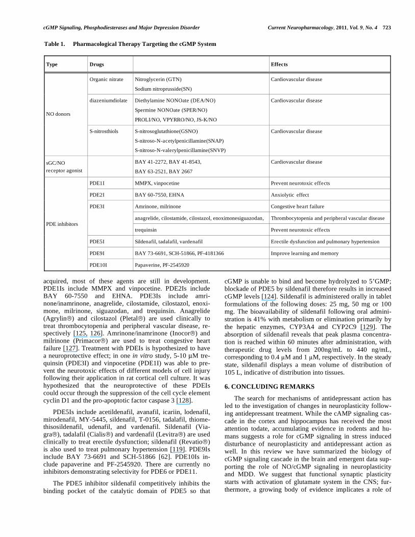
cGMP Signaling, Phosphodiesterases and Major Depression Disorder Current Neuropharmacology, 2011, Vol. 9, No. 4 723
acquired, most of these agents are still in development.
PDE1Is include MMPX and vinpocetine. PDE2Is include
BAY 60-7550 and EHNA. PDE3Is include amri-
none/inamrinone, anagrelide, cilostamide, cilostazol, enoxi-
mone, milrinone, siguazodan, and trequinsin. Anagrelide
(Agrylin ) and cilostazol (Pletal ) are used clinically to
treat thrombocytopenia and peripheral vascular disease, re-
spectively [125, 126]. Amrinone/inamrinone (Inocor ) and
milrinone (Primacor ) are used to treat congestive heart
failure [127]. Treatment with PDEIs is hypothesized to have
a neuroprotective effect; in one in vitro study, 5-10 μM tre-
quinsin (PDE3I) and vinpocetine (PDE1I) was able to pre-
vent the neurotoxic effects of different models of cell injury
following their application in rat cortical cell culture. It was
hypothesized that the neuroprotective of these PDEIs
could occur through the suppression of the cell cycle element
cyclin D1 and the pro-apoptotic factor caspase 3 [128].
PDE5Is include acetildenafil, avanafil, icariin, lodenafil,
mirodenafil, MY-5445, sildenafil, T-0156, tadalafil, thiome-
thisosildenafil, udenafil, and vardenafil. Sildenafil (Via-
gra ), tadalafil (Cialis ) and vardenafil (Levitra ) are used
clinically to treat erectile dysfunction; sildenafil (Revatio )
is also used to treat pulmonary hypertension [119]. PDE9Is
include BAY 73-6691 and SCH-51866 [62]. PDE10Is in-
clude papaverine and PF-2545920. There are currently no
inhibitors demonstrating selectivity for PDE6 or PDE11.
The PDE5 inhibitor sildenafil competitively inhibits the
binding pocket of the catalytic domain of PDE5 so that
cGMP is unable to bind and become hydrolyzed to 5’GMP;
blockade of PDE5 by sildenafil therefore results in increased
cGMP levels [124]. Sildenafil is administered orally in tablet
formulations of the following doses: 25 mg, 50 mg or 100
mg. The bioavailability of sildenafil following oral admini-
stration is 41% with metabolism or elimination primarily by
the hepatic enzymes, CYP3A4 and CYP2C9 [129]. The
absorption of sildenafil reveals that peak plasma concentra-
tion is reached within 60 minutes after administration, with
therapeutic drug levels from 200ng/mL to 440 ng/mL,
corresponding to 0.4 M and 1 M, respectively. In the steady
state, sildenafil displays a mean volume of distribution of
105 L, indicative of distribution into tissues.
6. CONCLUDING REMARKS
The search for mechanisms of antidepressant action has
led to the investigation of changes in neuroplasticity follow-
ing antidepressant treatment. While the cAMP signaling cas-
cade in the cortex and hippocampus has received the most
attention todate, accumulating evidence in rodents and hu-
mans suggests a role for cGMP signaling in stress induced
disturbance of neuroplasticity and antidepressant action as
well. In this review we have summarized the biology of
cGMP signaling cascade in the brain and emergent data sup-
porting the role of NO/cGMP signaling in neuroplasticity
and MDD. We suggest that functional synaptic plasticity
starts with activation of glutamate system in the CNS; fur-
thermore, a growing body of evidence implicates a role of
Table 1. Pharmacological Therapy Targeting the cGMP System
Type Drugs Effects
Organic nitrate Nitroglycerin (GTN)
Sodium nitroprusside(SN)
Cardiovascular disease
diazeniumdiolate Diethylamine NONOate (DEA/NO)
Spermine NONOate (SPER/NO)
PROLI/NO, VPYRRO/NO, JS-K/NO
Cardiovascular disease
NO donors
S-nitrosthiols S-nitrosoglutathione(GSNO)
S-nitroso-N-acetylpenicillamine(SNAP)
S-nitroso-N-valerylpenicillamine(SNVP)
Cardiovascular disease
sGC/NO
receptor agonist
BAY 41-2272, BAY 41-8543,
BAY 63-2521, BAY 2667
Cardiovascular disease
PDE1I MMPX, vinpocetine Prevent neurotoxic effects
PDE2I BAY 60-7550, EHNA Anxiolytic effect
Amrinone, milrinone Congestive heart failure
anagrelide, cilostamide, cilostazol, enoximonesiguazodan, Thrombocytopenia and peripheral vascular disease
PDE3I
trequinsin Prevent neurotoxic effects
PDE5I Sildenafil, tadalafil, vardenafil Erectile dysfunction and pulmonary hypertension
PDE9I BAY 73-6691, SCH-51866, PF-4181366 Improve learning and memory
PDE inhibitors
PDE10I Papaverine, PF-2545920
724 Current Neuropharmacology, 2011, Vol. 9, No. 4 Reierson et al.
the glutamatergic system in the pathophysiology and treat-
ment of MDD [130]. Activation of glutamate receptors in-
creases in Ca2+
levels at the postsynaptic neuron and stimu-
lates NO synthesis, which stimulates the NO/cGMP path-
way. NO can also act transynaptically at the presynaptic neu-
ron or surrounding cells. The presynaptic and postsynaptic
cGMP signaling enhances synaptic transmission (Fig. 1),
induces synaptic protein synthesis during the late phase of
LTP and might facilitate neurogenesis through activation of
several downstream substrates. It is beyond the scope of this
review to discuss the immune/inflammatory aspects of NO
and cGMP signaling pathway through the role of inducible
nitric oxide synthase (NOS2).
REFERENCES
[1] Blazer, D.G., Kessler, R.C., Swartz, M.S. Epidemiology of
recurrent major and minor depression with a seasonal pattern. The
National comorbidity survey. Br. J. Psychiatry, 1998, 172, 164-167.
[2] Wong, M.L., Licinio, J. From monoamines to genomic targets: a
paradigm shift for drug discovery in depression. Nat. Rev. Drug, 2004, Discov., 3, 136-151.
[3] Duman, R.S., Monteggia, L.M. A neurotrophic model for stress-
related mood disorders. Biol. Psychiatry, 2006, 59, 1116-1127.
[4] Duman, C.H., Schlesinger, L., Russell, D.S., Duman, R.S.
Voluntary exercise produces antidepressant and anxiolytic
behavioral effects in mice. Brain Res., 2008, 1199, 148-158.
[5] Chen, C.C., Yang, C.H., Huang, C.C., Hsu, K.S. Acute stress
impairs hippocampal mossy fiber-CA3 long-term potentiation by
enhancing cAMP-specific phosphodiesterase 4 activity.
Neuropsychopharmacology, 2010, 35, 1605-1617.
[6] Bartsch, D., Casadio, A., Karl, K.A., Serodio, P., Kandel, E.R.
CREB1 encodes a nuclear activator, a repressor, and a cytoplasmic
modulator that form a regulatory unit critical for long-term
facilitation. Cell, 1998, 95, 211-223.
[7] Conti, A.C., Cryan, J.F., Dalvi, A., Lucki, I., Blendy, J.A. cAMP
response element-binding protein is essential for the upregulation
of brain-derived neurotrophic factor transcription, but not the
behavioral or endocrine responses to antidepressant drugs. J. Neurosci., 2002, 22, 3262-3268.
[8] Dhir, A., Kulkarni, S.K. Effect of addition of yohimbine (alpha-2-
receptor antagonist) to the antidepressant activity of fluoxetine or
venlafaxine in the mouse forced swim test. Pharmacology, 2007,
80, 239-243.
[9] Feil, S., Zimmermann, P., Knorn, A., Brummer, S., Schlossmann,
J., Hofmann, F., Feil, R. Distribution of cGMP-dependent protein
kinase type I and its isoforms in the mouse brain and retina.
Neuroscience, 2005, 135, 863-868.
[10] de Vente, J., Asan, E., Gambaryan, S., Markerink-van Ittersum, M.,
Axer, H., Gallatz, K., Lohmann, S.M., Palkovits, M. Localization
of cGMP-dependent protein kinase type II in rat brain.
Neuroscience, 2001, 108, 27-49.
[11] Dhir, A., Kulkarni, S.K. Involvement of L-arginine-nitric oxide-
cyclic guanosine monophosphate pathway in the antidepressant-
like effect of venlafaxine in mice. Prog. Neuropsychopharmacol. Biol. Psychiatry, 2007, 31, 921-925.
[12] Knowles, R.G., Palacios, M., Palmer, R.M., Moncada, S.
Formation of nitric oxide from L-arginine in the central
nervous system: a transduction mechanism for stimulation of the
soluble guanylate cyclase. Proc. Natl. Acad. Sci. U. S. A., 1989, 86,
5159-5162.
[13] Bredt, D.S., Snyder, S.H. Nitric oxide mediates glutamate-linked
enhancement of cGMP levels in the cerebellum. Proc. Natl. Acad. Sci. U. S. A, 1989, 86, 9030-9033.
[14] Fesenko, E.E., Kolesnikov, S.S., Lyubarsky, A.L. Induction by
cyclic GMP of cationic conductance in plasma membrane of retinal
rod outer segment. Nature, 1985, 313, 310-313.
[15] Schlegel, P.A., Jen, P.H., Singh, S. Auditory spatial sensitivity of
inferior collicular neurons of echolocating bats. Brain Res., 1988,
456, 127-138.
[16] Leinders-Zufall, T., Cockerham, R.E., Michalakis, S., Biel, M.,
Garbers, D.L., Reed, R.R., Zufall, F., Munger, S.D. Contribution of
the receptor guanylyl cyclase GC-D to chemosensory function in
the olfactory epithelium. Proc. Natl. Acad. Sci. U. S. A., 2007, 104, 14507-14512.
[17] Lowe, D.G., Dizhoor, A.M., Liu, K., Gu, Q., Spencer, M., Laura,
R., Lu, L., Hurley, J.B. Cloning and expression of a second
photoreceptor-specific membrane retina guanylyl cyclase (RetGC),
RetGC-2. Proc. Natl. Acad. Sci. U. S. A., 1995, 92, 5535-5539.
[18] Shyjan, A.W., de Sauvage, F.J., Gillett, N.A., Goeddel, D.V.,
Lowe, D.G. Molecular cloning of a retina-specific membrane
guanylyl cyclase. Neuron, 1992, 9, 727-737.
[19] Yang, R.B., Robinson, S.W., Xiong, W.H., Yau, K.W., Birch,
D.G., Garbers, D.L. Disruption of a retinal guanylyl cyclase gene
leads to cone-specific dystrophy and paradoxical rod behavior. J.
Neurosci., 1999, 19, 5889-5897.
[20] Langub, M.C., Jr., Dolgas, C.M., Watson, R.E., Jr., Herman, J.P.
The C-type natriuretic peptide receptor is the predominant
natriuretic peptide receptor mRNA expressed in rat hypothalamus.
J. Neuroendocrinol., 1995, 7, 305-309.
[21] Lein, E.S., Hawrylycz, M.J., Ao, N., Ayres, M., Bensinger, A.,
Bernard, A., Boe, A.F., Boguski, M.S., Brockway, K.S., Byrnes,
E.J., Chen, L., Chen, T.M., Chin, M.C., Chong, J., Crook, B.E.,
Czaplinska, A., Dang, C.N., Datta, S., Dee, N.R., Desaki, A.L.,
Desta, T., Diep, E., Dolbeare, T.A., Donelan, M.J., Dong, H.W.,
Dougherty, J.G., Duncan, B.J., Ebbert, A.J., Eichele, G., Estin,
L.K., Faber, C., Facer, B.A., Fields, R., Fischer, S.R., Fliss, T.P.,
Frensley, C., Gates, S.N., Glattfelder, K.J., Halverson, K.R., Hart,
M.R., Hohmann, J.G., Howell, M.P., Jeung, D.P., Johnson, R.A.,
Karr, P.T., Kawal, R., Kidney, J.M., Knapik, R.H., Kuan, C.L.,
Lake, J.H., Laramee, A.R., Larsen, K.D., Lau, C., Lemon, T.A.,
Liang, A.J., Liu, Y., Luong, L.T., Michaels, J., Morgan, J.J.,
Morgan, R.J., Mortrud, M.T., Mosqueda, N.F., Ng, L.L., Ng, R.,
Orta, G.J., Overly, C.C., Pak, T.H., Parry, S.E., Pathak, S.D.,
Pearson, O.C., Puchalski, R.B., Riley, Z.L., Rockett, H.R.,
Rowland, S.A., Royall, J.J., Ruiz, M.J., Sarno, N.R., Schaffnit, K.,
Shapovalova, N.V., Sivisay, T., Slaughterbeck, C.R., Smith, S.C.,
Smith, K.A., Smith, B.I., Sodt, A.J., Stewart, N.N., Stumpf, K.R.,
Sunkin, S.M., Sutram, M., Tam, A., Teemer, C.D., Thaller, C.,
Thompson, C.L., Varnam, L.R., Visel, A., Whitlock, R.M.,
Wohnoutka, P.E., Wolkey, C.K., Wong, V.Y., Wood, M.,
Yaylaoglu, M.B., Young, R.C., Youngstrom, B.L., Yuan, X.F.,
Zhang, B., Zwingman, T.A., Jones, A.R. Genome-wide atlas of
gene expression in the adult mouse brain. Nature, 2007, 445, 168-
176.
[22] Muller, D., Olcese, J., Mukhopadhyay, A.K., Middendorff, R.
Guanylyl cyclase-B represents the predominant natriuretic peptide
receptor expressed at exceptionally high levels in the pineal gland.
Brain Res. Mol. Brain Res., 2000, 75, 321-329.
[23] Young, J.M., Waters, H., Dong, C., Fulle, H.J., Liman, E.R.
Degeneration of the olfactory guanylyl cyclase D gene during
primate evolution. PLoS One, 2007, 2, e884.
[24] Pifarre, P., Garcia, A., Mengod, G. Species differences in the
localization of soluble guanylyl cyclase subunits in monkey and rat
brain. J. Comp. Neurol., 2007, 500, 942-957.
[25] Mergia, E., Russwurm, M., Zoidl, G., Koesling, D. Major
occurrence of the new alpha2beta1 isoform of NO-sensitive
guanylyl cyclase in brain. Cell Signal., 2003, 15, 189-195.
[26] Esposito, K., Reierson, G.W., Luo, H.R., Wu, G.S., Licinio, J.,
Wong, M.L. Phosphodiesterase genes and antidepressant treatment
response: a review. Ann. Med., 2009, 41, 177-185.
[27] Reneerkens, O.A., Rutten, K., Steinbusch, H.W., Blokland, A.,
Prickaerts, J. Selective phosphodiesterase inhibitors: a promising
target for cognition enhancement. Psychopharmacology (Berl), 2009, 202, 419-443.
[28] Ke, H., Wang, H. Crystal structures of phosphodiesterases and
implications on substrate specificity and inhibitor selectivity. Curr.
Top. Med. Chem., 2007, 7, 391-403.
[29] Wang, H.G., Lu, F.M., Jin, I., Udo, H., Kandel, E.R., de Vente, J.,
Walter, U., Lohmann, S.M., Hawkins, R.D., Antonova, I.
Presynaptic and postsynaptic roles of NO, cGK, and RhoA in long-
lasting potentiation and aggregation of synaptic proteins. Neuron,
2005, 45, 389-403.
[30] Zhang, K.Y., Card, G.L., Suzuki, Y., Artis, D.R., Fong, D.,
Gillette, S., Hsieh, D., Neiman, J., West, B.L., Zhang, C., Milburn,
M.V., Kim, S.H., Schlessinger, J., Bollag, G.) A glutamine switch
cGMP Signaling, Phosphodiesterases and Major Depression Disorder Current Neuropharmacology, 2011, Vol. 9, No. 4 725
mechanism for nucleotide selectivity by phosphodiesterases. Mol.
Cell, 2004, 15, 279-286.
[31] Zoraghi, R., Bessay, E.P., Corbin, J.D., Francis, S.H. Structural and
functional features in human PDE5A1 regulatory domain that
provide for allosteric cGMP binding, dimerization, and regulation.
J. Biol. Chem., 2005, 280, 12051-12063.
[32] Zhan, C.G., Zheng, F. First computational evidence for a catalytic
bridging hydroxide ion in a phosphodiesterase active site. J. Am. Chem. Soc., 2001, 123, 2835-2838.
[33] Van Staveren, W.C., Steinbusch, H.W., Markerink-Van Ittersum,
M., Repaske, D.R., Goy, M.F., Kotera, J., Omori, K., Beavo, J.A.,
De Vente, J. mRNA expression patterns of the cGMP-hydrolyzing
phosphodiesterases types 2, 5, and 9 during development of the rat
brain. J. Comp. Neurol., 2003, 467, 566-580.
[34] Jarnaess, E., Tasken, K. Spatiotemporal control of cAMP signalling
processes by anchored signalling complexes. Biochem. Soc. Trans., 2007, 35, 931-937.
[35] Andreeva, S.G., Dikkes, P., Epstein, P.M., Rosenberg, P.A.
Expression of cGMP-specific phosphodiesterase 9A mRNA in the
rat brain. J. Neurosci., 2001, 21, 9068-9076.
[36] van Staveren, W.C., Glick, J., Markerink-van Ittersum, M.,
Shimizu, M., Beavo, J.A., Steinbusch, H.W., de Vente, J. Cloning
and localization of the cGMP-specific phosphodiesterase type 9 in
the rat brain. J. Neurocytol., 2002, 31, 729-741.
[37] van Staveren, W.C., Steinbusch, H.W., Markerink-van Ittersum,
M., Behrends, S., de Vente, J. Species differences in the
localization of cGMP-producing and NO-responsive elements in
the mouse and rat hippocampus using cGMP immunocyto-
chemistry. Eur. J. Neurosci., 2004, 19, 2155-2168.
[38] Reyes-Irisarri, E., Markerink-Van Ittersum, M., Mengod, G., de
Vente, J. Expression of the cGMP-specific phosphodiesterases 2
and 9 in normal and Alzheimer's disease human brains. Eur. J. Neurosci., 2007, 25, 3332-3338.
[39] Polli, J.W., Kincaid, R.L. Expression of a calmodulin-dependent
phosphodiesterase isoform (PDE1B1) correlates with brain regions
having extensive dopaminergic innervation. J. Neurosci., 1994, 14, 1251-1261.
[40] Yan, C., Bentley, J.K., Sonnenburg, W.K., Beavo, J.A. Differential
expression of the 61 kDa and 63 kDa calmodulin-dependent
phosphodiesterases in the mouse brain. J. Neurosci., 1994, 14, 973-
984.
[41] Yan, C., Zhao, A.Z., Bentley, J.K., Beavo, J.A. The calmodulin-
dependent phosphodiesterase gene PDE1C encodes several
functionally different splice variants in a tissue-specific manner. J. Biol. Chem., 1996, 271, 25699-25706.
[42] Yu, J., Wolda, S.L., Frazier, A.L., Florio, V.A., Martins, T.J.,
Snyder, P.B., Harris, E.A., McCaw, K.N., Farrell, C.A., Steiner, B.,
Bentley, J.K., Beavo, J.A., Ferguson, K., Gelinas, R. Identification
and characterisation of a human calmodulin-stimulated
phosphodiesterase PDE1B1. Cell Signal., 1997, 9, 519-529.
[43] Stephenson, D.T., Coskran, T.M., Wilhelms, M.B., Adamowicz,
W.O., O'Donnell, M.M., Muravnick, K.B., Menniti, F.S.,
Kleiman, R.J., Morton, D. Immunohistochemical localization of
phosphodiesterase 2A in multiple mammalian species. J. Histochem. Cytochem., 2009, 57, 933-949.
[44] Reinhardt, R.R., Bondy, C.A. Differential cellular pattern of gene
expression for two distinct cGMP-inhibited cyclic nucleotide
phosphodiesterases in developing and mature rat brain.
Neuroscience, 1996, 72, 567-578.
[45] Coskran, T.M., Morton, D., Menniti, F.S., Adamowicz, W.O.,
Kleiman, R.J., Ryan, A.M., Strick, C.A., Schmidt, C.J., Stephenson,
D.T. Immunohistochemical localization of phosphodiesterase 10A in
multiple mammalian species. J. Histochem. Cytochem., 2006, 54,
1205-1213.
[46] Hebb, A.L., Robertson, H.A., Denovan-Wright, E.M. Striatal
phosphodiesterase mRNA and protein levels are reduced in
Huntington's disease transgenic mice prior to the onset of motor
symptoms. Neuroscience, 2004, 123, 967-981.
[47] Seeger, T.F., Bartlett, B., Coskran, T.M., Culp, J.S., James, L.C.,
Krull, D.L., Lanfear, J., Ryan, A.M., Schmidt, C.J., Strick, C.A.,
Varghese, A.H., Williams, R.D., Wylie, P.G., Menniti, F.S.
Immunohistochemical localization of PDE10A in the rat brain.
Brain Res., 2003, 985, 113-126.
[48] Kruse, L.S., Moller, M., Tibaek, M., Gammeltoft, S., Olesen, J.,
Kruuse, C. PDE9A, PDE10A, and PDE11A expression in rat
trigeminovascular pain signalling system. Brain Res., 2009, 1281,
25-34.
[49] Loughney, K., Taylor, J., Florio, V.A. 3', 5'-cyclic nucleotide
phosphodiesterase 11A: localization in human tissues. Int. J. Impot. Res., 2005, 17, 320-325.
[50] Hofmann, F., Ammendola, A., Schlossmann, J. Rising behind NO:
cGMP-dependent protein kinases. J. Cell Sci., 2000, 113 ( Pt 10),
1671-1676.
[51] Pfeifer, A., Ruth, P., Dostmann, W., Sausbier, M., Klatt, P.,
Hofmann, F. Structure and function of cGMP-dependent
protein kinases. Rev. Physiol. Biochem. Pharmacol., 1999, 135,
105-149.
[52] Wang, X., Robinson, P.J. Cyclic GMP-dependent protein kinase
and cellular signaling in the nervous system. J. Neurochem., 1997,
68, 443-456.
[53] Domek-Lopacinska, K., Strosznajder, J.B. Cyclic GMP metabolism
and its role in brain physiology. J. Physiol. Pharmacol., 2005, 56
Suppl 2, 15-34.
[54] Houslay, M.D. Compartmentalization of cyclic AMP phos-
phodiesterases, signalling 'crosstalk', desensitization and the
phosphorylation of Gi-2 add cell specific personalization to the
control of the levels of the second messenger cyclic AMP. Adv. Enzyme Regul., 1995, 35, 303-338.
[55] Conti, M., Beavo, J. Biochemistry and physiology of cyclic
nucleotide phosphodiesterases: essential components in cyclic
nucleotide signaling. Annu. Rev. Biochem., 2007, 76, 481-511.
[56] Corbin, J.D., Turko, I.V., Beasley, A., Francis, S.H. Phosphoryla-
tion of phosphodiesterase-5 by cyclic nucleotide-dependent protein
kinase alters its catalytic and allosteric cGMP-binding activities.
Eur. J. Biochem., 2000, 267, 2760-2767.
[57] Zaccolo, M., Movsesian, M.A. cAMP and cGMP signaling cross-
talk: role of phosphodiesterases and implications for cardiac
pathophysiology. Circ. Res., 2007, 100, 1569-1578.
[58] Pelligrino, D.A., Wang, Q. Cyclic nucleotide crosstalk and
the regulation of cerebral vasodilation. Prog. Neurobiol., 1998, 56,
1-18.
[59] Garthwaite, J. Neuronal nitric oxide synthase and the serotonin
transporter get harmonious. Proc. Natl. Acad. Sci. U. S. A., 2007,
104, 7739-7740.
[60] Maurice, D.H. Cyclic nucleotide phosphodiesterase-mediated
integration of cGMP and cAMP signaling in cells of the
cardiovascular system. Front. Biosci., 2005, 10, 1221-1228.
[61] Murthy, K.S. Activation of phosphodiesterase 5 and inhibition of
guanylate cyclase by cGMP-dependent protein kinase in smooth
muscle. Biochem. J., 2001, 360, 199-208.
[62] Malenka, R.C., Nicoll, R.A. Learning and memory. Never fear,
LTP is hear. Nature, 1997, 390, 552-553.
[63] Shors, T.J., Seib, T.B., Levine, S., Thompson, R.F. Inescapable
versus escapable shock modulates long-term potentiation in the rat
hippocampus. Science, 1989, 244, 224-226.
[64] Yang, C.H., Huang, C.C., Hsu, K.S. Behavioral stress modifies
hippocampal synaptic plasticity through corticosterone-induced
sustained extracellular signal-regulated kinase/mitogen-activated
protein kinase activation. J. Neurosci., 2004, 24, 11029-11034.
[65] Ahmed, T., Frey, J.U., Korz, V. Long-term effects of brief acute
stress on cellular signaling and hippocampal LTP. J. Neurosci., 2006, 26, 3951-3958.
[66] Levkovitz, Y., Grisaru, N., Segal, M. Transcranial magnetic
stimulation and antidepressive drugs share similar cellular
effects in rat hippocampus. Neuropsychopharmacology, 2001, 24, 608-616.
[67] Stewart, C.A., Reid, I.C. Repeated ECS and fluoxetine
administration have equivalent effects on hippocampal synaptic
plasticity. Psychopharmacology (Berl), 2000, 148, 217-223.
[68] Dranovsky, A., Hen, R. Hippocampal neurogenesis: regulation by
stress and antidepressants. Biol. Psychiatry, 2006, 59, 1136-1143.
[69] Duman, R.S. Depression: a case of neuronal life and death? Biol.
Psychiatry, 2004, 56, 140-145.
[70] Sheline, Y.I., Wang, P.W., Gado, M.H., Csernansky, J.G., Vannier,
M.W. Hippocampal atrophy in recurrent major depression. Proc. Natl. Acad. Sci. U. S. A., 1996, 93, 3908-3913.
[71] MacQueen, G.M., Campbell, S., McEwen, B.S., Macdonald,
K., Amano, S., Joffe, R.T., Nahmias, C., Young, L.T. Course of
illness, hippocampal function, and hippocampal volume in major
depression. Proc. Natl. Acad. Sci. U. S. A., 2003, 100, 1387-1392.
726 Current Neuropharmacology, 2011, Vol. 9, No. 4 Reierson et al.
[72] Warner-Schmidt, J.L., Duman, R.S. Hippocampal neurogenesis:
opposing effects of stress and antidepressant treatment.
Hippocampus, 2006, 16, 239-249.
[73] Nakagawa, S., Kim, J.E., Lee, R., Malberg, J.E., Chen, J., Steffen,
C., Zhang, Y.J., Nestler, E.J., Duman, R.S. Regulation of
neurogenesis in adult mouse hippocampus by cAMP and the
cAMP response element-binding protein. J. Neurosci., 2002, 22,
3673-3682.
[74] Malberg, J.E., Eisch, A.J., Nestler, E.J., Duman, R.S. Chronic
antidepressant treatment increases neurogenesis in adult rat
hippocampus. J. Neurosci., 2000, 20, 9104-9110.
[75] Neitz, A., Mergia, E., Eysel, U.T., Koesling, D., Mittmann, T.
Presynaptic nitric oxide/cGMP facilitates glutamate release via
hyperpolarization-activated cyclic nucleotide-gated channels in the
hippocampus. Eur. J. Neurosci., 2011, 33, 1611-1621.
[76] Wang, H., Robinson, H., Ke, H. The molecular basis for different
recognition of substrates by phosphodiesterase families 4 and 10.
J.Mol. Biol., 2007, 371, 302-307.
[77] Serulle, Y., Arancio, O., Ziff, E.B. A role for cGMP-dependent
protein kinase II in AMPA receptor trafficking and synaptic
plasticity. Channels (Austin), 2008, 2, 230-232.
[78] Serulle, Y., Zhang, S., Ninan, I., Puzzo, D., McCarthy, M., Khatri,
L., Arancio, O., Ziff, E.B. A GluR1-cGKII interaction regulates
AMPA receptor trafficking. Neuron, 2007, 56, 670-688.
[79] Chen, Y., Zhuang, S., Cassenaer, S., Casteel, D.E., Gudi, T., Boss,
G.R., Pilz, R.B. Synergism between calcium and cyclic GMP in
cyclic AMP response element-dependent transcriptional regulation
requires cooperation between CREB and C/EBP-beta. Mol. Cell Biol., 2003, 23, 4066-4082.
[80] Lu, Y.F., Kandel, E.R., Hawkins, R.D. Nitric oxide signaling
contributes to late-phase LTP and CREB phosphorylation in the
hippocampus. J. Neurosci., 1999, 19, 10250-10261.
[81] Lu, Y.F., Hawkins, R.D. Ryanodine receptors contribute to cGMP-
induced late-phase LTP and CREB phosphorylation in the
hippocampus. J. Neurophysiol., 2002, 88, 1270-1278.
[82] Zhang, R., Wang, Y., Zhang, L., Zhang, Z., Tsang, W., Lu, M.,
Chopp, M. Sildenafil (Viagra) induces neurogenesis and promotes
functional recovery after stroke in rats. Stroke, 2002, 33, 2675-
2680.
[83] Zhang, R., Zhang, L., Zhang, Z., Wang, Y., Lu, M., Lapointe,
M., Chopp, M. A nitric oxide donor induces neurogenesis and
reduces functional deficits after stroke in rats. Ann. Neurol., 2001,
50, 602-611.
[84] Wang, L., Gang Zhang, Z., Lan Zhang, R., Chopp, M. Activation
of the PI3-K/Akt pathway mediates cGMP enhanced-neurogenesis
in the adult progenitor cells derived from the subventricular zone.
J. Cereb. Blood Flow Metab., 2005, 25, 1150-1158.
[85] Tojima, T., Itofusa, R., Kamiguchi, H. The nitric oxide-cGMP
pathway controls the directional polarity of growth cone guidance
via modulating cytosolic Ca2+ signals. J. Neurosci., 2009, 29,
7886-7897.
[86] Nishiyama, M., Hoshino, A., Tsai, L., Henley, J.R., Goshima, Y.,
Tessier-Lavigne, M., Poo, M.M., Hong, K. Cyclic AMP/GMP-
dependent modulation of Ca2+ channels sets the polarity of nerve
growth-cone turning. Nature, 2003, 423, 990-995.
[87] Murray, A.J., Tucker, S.J., Shewan, D.A. cAMP-dependent axon
guidance is distinctly regulated by Epac and protein kinase A. J.
Neurosci., 2009, 29, 15434-15444.
[88] Shelly, M., Lim, B.K., Cancedda, L., Heilshorn, S.C., Gao, H., Poo,
M.M. Local and long-range reciprocal regulation of cAMP and
cGMP in axon/dendrite formation. Science, 2010, 327, 547-552.
[89] Zhu, C.B., Hewlett, W.A., Feoktistov, I., Biaggioni, I., Blakely,
R.D. Adenosine receptor, protein kinase G, and p38 mitogen-
activated protein kinase-dependent up-regulation of serotonin
transporters involves both transporter trafficking and activation.
Mol. Pharmacol., 2004, 65, 1462-1474.
[90] Chanrion, B., Mannoury la Cour, C., Bertaso, F., Lerner-Natoli,
M., Freissmuth, M., Millan, M.J., Bockaert, J., Marin, P. Physical
interaction between the serotonin transporter and neuronal nitric
oxide synthase underlies reciprocal modulation of their activity.
Proc. Natl. Acad. Sci. U. S. A., 2007, 104, 8119-8124.
[91] Shinkai, T., Ohmori, O., Hori, H., Nakamura, J. Allelic association
of the neuronal nitric oxide synthase (NOS1) gene with
schizophrenia. Mol. Psychiatry, 2002, 7, 560-563.
[92] Okumura, T., Kishi, T., Okochi, T., Ikeda, M., Kitajima, T.,
Yamanouchi, Y., Kinoshita, Y., Kawashima, K., Tsunoka, T.,
Inada, T., Ozaki, N., Iwata, N. Genetic association analysis
of functional polymorphisms in neuronal nitric oxide synthase 1
gene (NOS1) and mood disorders and fluvoxamine response
in major depressive disorder in the Japanese population.
Neuropsychobiology, 2010, 61, 57-63.
[93] Wong, M.L., Whelan, F., Deloukas, P., Whittaker, P., Delgado, M.,
Cantor, R.M., McCann, S.M., Licinio, J. Phosphodiesterase genes
are associated with susceptibility to major depression and
antidepressant treatment response. Proc. Natl. Acad. Sci. U. S. A.,
2006, 103, 15124-15129.
[94] Teranishi, K.S., Slager, S.L., Garriock, H., Kraft, J.B., Peters, E.J.,
Reinalda, M.S., Jenkins, G.D., McGrath, P.J., Hamilton, S.P.
Variants in PDE11A and PDE1A are not associated with
citalopram response. Mol. Psychiatry, 2007, 12, 1061-1063.
[95] Cabanero, M., Laje, G., Detera-Wadleigh, S., McMahon, F.J.
Association study of phosphodiesterase genes in the Sequenced
Treatment Alternatives to Relieve Depression sample. Pharmacogenet.
Genom., 2009, 19, 235-238.
[96] Fatemi, S.H., Folsom, T.D., Reutiman, T.J., Vazquez, G.
Phosphodiesterase signaling system is disrupted in the cerebella of
subjects with schizophrenia, bipolar disorder, and major
depression. Schizophr. Res., 2010, 119, 266-267.
[97] Zhang, J., Huang, X.Y., Ye, M.L., Luo, C.X., Wu, H.Y., Hu, Y.,
Zhou, Q.G., Wu, D.L., Zhu, L.J., Zhu, D.Y. Neuronal nitric
oxide synthase alteration accounts for the role of 5-HT1A receptor
in modulating anxiety-related behaviors. J. Neurosci., 2010, 30,
2433-2441.
[98] Chiavegatto, S., Dawson, V.L., Mamounas, L.A., Koliatsos, V.E.,
Dawson, T.M., Nelson, R.J. Brain serotonin dysfunction accounts
for aggression in male mice lacking neuronal nitric oxide synthase.
Proc. Natl. Acad. Sci. U. S. A., 2001, 98, 1277-1281.
[99] Balda, M.A., Anderson, K.L., Itzhak, Y. Development and
persistence of long-lasting behavioral sensitization to cocaine in
female mice: role of the nNOS gene. Neuropharmacology, 2009,
56, 709-715.
[100] Boettger, M.K., Uceyler, N., Zelenka, M., Schmitt, A., Reif, A.,
Chen, Y., Sommer, C. Differences in inflammatory pain in nNOS-,
iNOS- and eNOS-deficient mice. Eur. J. Pain, 2007, 11, 810-818.
[101] Juch, M., Smalla, K.H., Kahne, T., Lubec, G., Tischmeyer,
W., Gundelfinger, E.D., Engelmann, M. Congenital lack of
nNOS impairs long-term social recognition memory and alters
the olfactory bulb proteome. Neurobiol. Learn Mem., 2009, 92,
469-484.
[102] Haghikia, A., Mergia, E., Friebe, A., Eysel, U.T., Koesling, D.,
Mittmann, T. Long-term potentiation in the visual cortex requires
both nitric oxide receptor guanylyl cyclases. J. Neurosci., 2007, 27,
818-823.
[103] Siuciak, J.A., McCarthy, S.A., Chapin, D.S., Reed, T.M., Vorhees,
C.V., Repaske, D.R. Behavioral and neurochemical characteri-
zation of mice deficient in the phosphodiesterase-1B (PDE1B)
enzyme. Neuropharmacology, 2007, 53, 113-124.
[104] Reed, T.M., Repaske, D.R., Snyder, G.L., Greengard, P., Vorhees,
C.V. Phosphodiesterase 1B knock-out mice exhibit exaggerated
locomotor hyperactivity and DARPP-32 phosphorylation in
response to dopamine agonists and display impaired spatial
learning. J. Neurosci., 2002, 22, 5188-5197.
[105] Ehrman, L.A., Williams, M.T., Schaefer, T.L., Gudelsky, G.A.,
Reed, T.M., Fienberg, A.A., Greengard, P., Vorhees, C.V.
Phosphodiesterase 1B differentially modulates the effects of
methamphetamine on locomotor activity and spatial learning
through DARPP32-dependent pathways: evidence from PDE1B-
DARPP32 double-knockout mice. Genes Brain Behav., 2006, 5, 540-551.
[106] Siuciak, J.A., McCarthy, S.A., Chapin, D.S., Martin, A.N., Harms,
J.F., Schmidt, C.J. Behavioral characterization of mice deficient in
the phosphodiesterase-10A (PDE10A) enzyme on a C57/Bl6N
congenic background. Neuropharmacology, 2008, 54, 417-427.
[107] Siuciak, J.A., McCarthy, S.A., Chapin, D.S., Fujiwara, R.A.,
James, L.C., Williams, R.D., Stock, J.L., McNeish, J.D., Strick,
C.A., Menniti, F.S., Schmidt, C.J. Genetic deletion of the striatum-
enriched phosphodiesterase PDE10A: evidence for altered striatal
function. Neuropharmacology, 2006, 51, 374-385.
cGMP Signaling, Phosphodiesterases and Major Depression Disorder Current Neuropharmacology, 2011, Vol. 9, No. 4 727
[108] Werner, C., Raivich, G., Cowen, M., Strekalova, T., Sillaber, I.,
Buters, J.T., Spanagel, R., Hofmann, F. Importance of NO/cGMP
signalling via cGMP-dependent protein kinase II for controlling
emotionality and neurobehavioural effects of alcohol. Eur. J. Neurosci., 2004, 20, 3498-3506.
[109] Joca, S.R., Guimaraes, F.S. Inhibition of neuronal nitric oxide
synthase in the rat hippocampus induces antidepressant-like effects.
Psychopharmacology (Berl), 2006, 185, 298-305.
[110] Ulak, G., Mutlu, O., Akar, F.Y., Komsuoglu, F.I., Tanyeri, P.,
Erden, B.F. Neuronal NOS inhibitor 1-(2-trifluoromethylphenyl)-
imidazole augment the effects of antidepressants acting via
serotonergic system in the forced swimming test in rats.
Pharmacol. Biochem. Behav., 2008, 90, 563-568.
[111] Zhou, Q.G., Hu, Y., Hua, Y., Hu, M., Luo, C.X., Han, X.,
Zhu, X.J., Wang, B., Xu, J.S., Zhu, D.Y. Neuronal nitric oxide
synthase contributes to chronic stress-induced depression by
suppressing hippocampal neurogenesis. J. Neurochem., 2007, 103,
1843-1854.
[112] Khovryakov, A.V., Podrezova, E.P., Kruglyakov, P.P., Shikhanov,
N.P., Balykova, M.N., Semibratova, N.V., Sosunov, A.A.,
McKhann, G., 2nd, Airapetyants, M.G. Involvement of the NO
synthase system in stress-mediated brain reactions. Neurosci. Behav. Physiol., 2010, 40, 333-337.
[113] Volke, V., Wegener, G., Vasar, E. Augmentation of the NO-cGMP
cascade induces anxiogenic-like effect in mice. J. Physiol.
Pharmacol., 2003, 54, 653-660.
[114] Hotchkiss, A.K., Pyter, L.M., Gatien, M.L., Wen, J.C., Milman,
H.A., Nelson, R.J. Aggressive behavior increases after termination
of chronic sildenafil treatment in mice. Physiol. Behav., 2005, 83,
683-688.
[115] Kaster, M.P., Rosa, A.O., Santos, A.R., Rodrigues, A.L.
Involvement of nitric oxide-cGMP pathway in the antidepressant-
like effects of adenosine in the forced swimming test. Int. J.
Neuropsychopharmacol., 2005, 8, 601-606.
[116] Dhir, A., Kulkarni, S.K. Involvement of nitric oxide (NO) signaling
pathway in the antidepressant action of bupropion, a dopamine
reuptake inhibitor. Eur. J. Pharmacol., 2007, 568, 177-185.
[117] Brink, C.B., Clapton, J.D., Eagar, B.E., Harvey, B.H. Appearance
of antidepressant-like effect by sildenafil in rats after central
muscarinic receptor blockade: evidence from behavioural and
neuro-receptor studies. J. Neural. Transm., 2008, 115, 117-125.
[118] Masood, A., Huang, Y., Hajjhussein, H., Xiao, L., Li, H., Wang,
W., Hamza, A., Zhan, C.G., O'Donnell, J.M. Anxiolytic effects of
phosphodiesterase-2 inhibitors associated with increased cGMP
signaling. J. Pharmacol. Exp. Ther., 2009, 331, 690-699.
[119] Masood, A., Nadeem, A., Mustafa, S.J., O'Donnell, J.M.
Reversal of oxidative stress-induced anxiety by inhibition of
phosphodiesterase-2 in mice. J. Pharmacol. Exp. Ther., 2008, 326,
369-379.
[120] Miller, M.R., Megson, I.L. Recent developments in nitric oxide
donor drugs. Br. J. Pharmacol., 2007, 151, 305-321.
[121] Belik, J. Riociguat, an oral soluble guanylate cyclase stimulator for
the treatment of pulmonary hypertension. Curr. Opin. Investig. Drugs, 2009, 10, 971-979.
[122] Mitrovic, V., Hernandez, A.F., Meyer, M., Gheorghiade, M. Role
of guanylate cyclase modulators in decompensated heart failure.
Heart Fail. Rev., 2009, 14, 309-319.
[123] Lugnier, C. Cyclic nucleotide phosphodiesterase (PDE)
superfamily: a new target for the development of specific
therapeutic agents. Pharmacol. Ther., 2006, 109, 366-398.
[124] Bender, A.T., Beavo, J.A. Cyclic nucleotide phosphodiesterases:
molecular regulation to clinical use. Pharmacol. Rev., 2006, 58,
488-520.
[125] Spencer, C.M., Brogden, R.N. Anagrelide. A review of its
pharmacodynamic and pharmacokinetic properties, and therapeutic
potential in the treatment of thrombocythaemia. Drugs, 1994, 47,
809-822.
[126] Kambayashi, J., Liu, Y., Sun, B., Shakur, Y., Yoshitake, M.,
Czerwiec, F. Cilostazol as a unique antithrombotic agent. Curr. Pharm. Des., 2003, 9, 2289-2302.
[127] Sharma, M., Teerlink, J.R. A rational approach for the treatment of
acute heart failure: current strategies and future options. Curr.
Opin. Cardiol., 2004, 19, 254-263.
[128] Chen, R.W., Williams, A.J., Liao, Z., Yao, C., Tortella, F.C., Dave,
J.R. Broad spectrum neuroprotection profile of phosphodiesterase
inhibitors as related to modulation of cell-cycle elements and
caspase-3 activation. Neurosci. Lett., 2007, 418, 165-169.
[129] Goldenberg, M.M. Safety and efficacy of sildenafil citrate in the
treatment of male erectile dysfunction. Clin. Ther., 1998, 20, 1033-
1048.
[130] Paul, I.A., Skolnick, P. Glutamate and depression: clinical and
preclinical studies. Ann. N. Y. Acad. Sci., 2003, 1003, 250-272.
Received: April 12, 2010 Revised: September 09, 2010 Accepted: September 24, 2010
Related Documents