Cervicothoracic Multisegmental Transpinal Evoked Potentials in Humans Jonathan Einhorn 1 , Alan Li 1 , Royi Hazan 1 , Maria Knikou 1,2,3* 1 The Graduate Center, City University of New York, New York, New York, United States of America, 2 Sensory Motor Performance Program, Rehabilitation Institute of Chicago, Chicago, Illinois, United States of America, 3 Department of Physical Medicine and Rehabilitation, Northwestern University Feinberg School of Medicine, Chicago, Illinois, United States of America Abstract The objectives of this study were to establish the neurophysiological properties of the transpinal evoked potentials (TEPs) following transcutaneous electric stimulation of the spine (tsESS) over the cervicothoracic region, changes in the amplitude of the TEPs preceded by median nerve stimulation at group I threshold, and the effects of tsESS on the flexor carpi radialis (FCR) H-reflex in thirteen healthy human subjects while seated. Two re-usable self-adhering electrodes, connected to function as one electrode (cathode), were placed bilaterally on the clavicles. A re-usable electrode (anode) was placed on the cervicothoracic region covering from Cervical 4 – Thoracic 2 and held under constant pressure throughout the experiment. TEPs were recorded bilaterally from major arm muscles with subjects seated at stimulation frequencies of 1.0, 0.5, 0.33, 0.2, 0.125, and 0.1 Hz, and upon double tsESS pulses delivered at an inter-stimulus interval of 40 ms. TEPs from the arm muscles were also recorded following median nerve stimulation at the conditioning-test (C-T) intervals of 2, 3, 5, 8, and 10 ms. The FCR H-reflex was evoked and recorded according to conventional methods following double median nerve pulses at 40 ms, and was also conditioned by tsESS at C-T intervals that ranged from −10 to +50 ms. The arm TEPs amplitude was not decreased at low-stimulation frequencies and upon double tsESS pulses in all but one subject. Ipsilateral and contralateral arm TEPs were facilitated following ipsilateral median nerve stimulation, while the FCR H-reflex was depressed by double pulses and following tsESS at short and long C-T intervals. Non-invasive transpinal stimulation can be used as a therapeutic modality to decrease spinal reflex hyper-excitability in neurological disorders and when combined with peripheral nerve stimulation to potentiate spinal output. Citation: Einhorn J, Li A, Hazan R, Knikou M (2013) Cervicothoracic Multisegmental Transpinal Evoked Potentials in Humans. PLoS ONE 8(10): e76940. doi:10.1371/journal.pone.0076940 Editor: William Phillips, University of Sydney, Australia Received July 1, 2013; Accepted August 30, 2013; Published October 7, 2013 Copyright: © 2013 Einhorn et al. This is an open-access article distributed under the terms of the Creative Commons Attribution License, which permits unrestricted use, distribution, and reproduction in any medium, provided the original author and source are credited. Funding: This work was supported by the Graduate Center of the City University of New York. The funders had no role in study design, data collection and analysis, decision to publish, or preparation of the manuscript. Competing interests: The authors have declared that no competing interests exist. * E-mail: [email protected] Introduction Epidermal or implanted electro-magnetic stimulation of the spine produces multisegmental compound muscle action potentials, termed here transpinal evoked potentials (TEPs), in distal and proximal muscles of upper and lower limbs in animals and humans [1-13]. TEPs, recorded from the soleus muscle, are facilitated following voluntary contraction of the homonymous muscle and depressed upon Achilles tendon vibration [2], suggesting that TEPs are modulated by spinal interneuronal circuits that mediate presynaptic inhibition. Further, the TEPs of the leg muscles are not susceptible to homosynaptic (or post-activation) depression [12,13], a spinal inhibitory mechanism documented for the H-reflex in both animals and humans and ascribed to presynaptic inhibition related to the amount of released neurotransmitters by the previously activated Ia afferents [14-19]. The time course of the homosynaptic depression of the soleus H-reflex is similar to that observed in the flexor carpi radialis (FCR) H-reflex [20], suggesting absent homosynaptic depression asymmetry in human upper and lower limbs. TEPs recorded from leg muscles are not susceptible to homosynaptic depression because they are largely due to antidromic activation of afferents and orthodromic activation of motor axons [12,13], in opposition to the H-reflexes, which reflect the efficacy of monosynaptic activation of alpha motoneurons by primary spindle Ia afferents [21]. This neurophysiological property makes transcutaneous electric stimulation of the spine (tsESS) and associated TEPs suitable for diagnostic and/or therapeutic purposes in central nervous system neurological disorders, since TEPs bypass the pathological excitability state of alpha motoneurons. However, for this to be possible, a better understanding of the neurophysiological properties of TEPs as well as the neuronal PLOS ONE | www.plosone.org 1 October 2013 | Volume 8 | Issue 10 | e76940

Welcome message from author
This document is posted to help you gain knowledge. Please leave a comment to let me know what you think about it! Share it to your friends and learn new things together.
Transcript

Cervicothoracic Multisegmental Transpinal EvokedPotentials in HumansJonathan Einhorn1, Alan Li1, Royi Hazan1, Maria Knikou1,2,3*
1 The Graduate Center, City University of New York, New York, New York, United States of America, 2 Sensory Motor Performance Program, RehabilitationInstitute of Chicago, Chicago, Illinois, United States of America, 3 Department of Physical Medicine and Rehabilitation, Northwestern University Feinberg Schoolof Medicine, Chicago, Illinois, United States of America
Abstract
The objectives of this study were to establish the neurophysiological properties of the transpinal evoked potentials(TEPs) following transcutaneous electric stimulation of the spine (tsESS) over the cervicothoracic region, changes inthe amplitude of the TEPs preceded by median nerve stimulation at group I threshold, and the effects of tsESS onthe flexor carpi radialis (FCR) H-reflex in thirteen healthy human subjects while seated. Two re-usable self-adheringelectrodes, connected to function as one electrode (cathode), were placed bilaterally on the clavicles. A re-usableelectrode (anode) was placed on the cervicothoracic region covering from Cervical 4 – Thoracic 2 and held underconstant pressure throughout the experiment. TEPs were recorded bilaterally from major arm muscles with subjectsseated at stimulation frequencies of 1.0, 0.5, 0.33, 0.2, 0.125, and 0.1 Hz, and upon double tsESS pulses deliveredat an inter-stimulus interval of 40 ms. TEPs from the arm muscles were also recorded following median nervestimulation at the conditioning-test (C-T) intervals of 2, 3, 5, 8, and 10 ms. The FCR H-reflex was evoked andrecorded according to conventional methods following double median nerve pulses at 40 ms, and was alsoconditioned by tsESS at C-T intervals that ranged from −10 to +50 ms. The arm TEPs amplitude was not decreasedat low-stimulation frequencies and upon double tsESS pulses in all but one subject. Ipsilateral and contralateral armTEPs were facilitated following ipsilateral median nerve stimulation, while the FCR H-reflex was depressed by doublepulses and following tsESS at short and long C-T intervals. Non-invasive transpinal stimulation can be used as atherapeutic modality to decrease spinal reflex hyper-excitability in neurological disorders and when combined withperipheral nerve stimulation to potentiate spinal output.
Citation: Einhorn J, Li A, Hazan R, Knikou M (2013) Cervicothoracic Multisegmental Transpinal Evoked Potentials in Humans. PLoS ONE 8(10): e76940.doi:10.1371/journal.pone.0076940
Editor: William Phillips, University of Sydney, Australia
Received July 1, 2013; Accepted August 30, 2013; Published October 7, 2013
Copyright: © 2013 Einhorn et al. This is an open-access article distributed under the terms of the Creative Commons Attribution License, which permitsunrestricted use, distribution, and reproduction in any medium, provided the original author and source are credited.
Funding: This work was supported by the Graduate Center of the City University of New York. The funders had no role in study design, data collection andanalysis, decision to publish, or preparation of the manuscript.
Competing interests: The authors have declared that no competing interests exist.
* E-mail: [email protected]
Introduction
Epidermal or implanted electro-magnetic stimulation of thespine produces multisegmental compound muscle actionpotentials, termed here transpinal evoked potentials (TEPs), indistal and proximal muscles of upper and lower limbs inanimals and humans [1-13]. TEPs, recorded from the soleusmuscle, are facilitated following voluntary contraction of thehomonymous muscle and depressed upon Achilles tendonvibration [2], suggesting that TEPs are modulated by spinalinterneuronal circuits that mediate presynaptic inhibition.Further, the TEPs of the leg muscles are not susceptible tohomosynaptic (or post-activation) depression [12,13], a spinalinhibitory mechanism documented for the H-reflex in bothanimals and humans and ascribed to presynaptic inhibitionrelated to the amount of released neurotransmitters by thepreviously activated Ia afferents [14-19]. The time course of the
homosynaptic depression of the soleus H-reflex is similar tothat observed in the flexor carpi radialis (FCR) H-reflex [20],suggesting absent homosynaptic depression asymmetry inhuman upper and lower limbs.
TEPs recorded from leg muscles are not susceptible tohomosynaptic depression because they are largely due toantidromic activation of afferents and orthodromic activation ofmotor axons [12,13], in opposition to the H-reflexes, whichreflect the efficacy of monosynaptic activation of alphamotoneurons by primary spindle Ia afferents [21]. Thisneurophysiological property makes transcutaneous electricstimulation of the spine (tsESS) and associated TEPs suitablefor diagnostic and/or therapeutic purposes in central nervoussystem neurological disorders, since TEPs bypass thepathological excitability state of alpha motoneurons. However,for this to be possible, a better understanding of theneurophysiological properties of TEPs as well as the neuronal
PLOS ONE | www.plosone.org 1 October 2013 | Volume 8 | Issue 10 | e76940

pathways that tsESS is channeled in the human spinal cord isneeded.
We have recently shown that transcutaneous magnetic orelectric stimulation of the thoracolumbar spine attenuatessignificantly the soleus H-reflex, while the TEPs recorded fromankle and thigh muscles are not susceptible to homosynapticdepression in seated healthy subjects [12,13]. In this study, weexamined whether similar effects are produced followingtranscutaneous electric stimulation of the cervicothoracicregion. Accordingly, we established the susceptibility of TEPsrecorded simultaneously from arm muscles to homosynapticdepression, the relationship of their latency and shape to FCRH-reflexes, the interaction of TEPs with peripheral (median)nerve stimulation, and the effects of tsESS on the FCR H-reflexin healthy people while seated.
Materials and Methods
SubjectsThe experimental protocol was approved by the City
University of New York (NY, USA) Institutional Review Board(IRB) committee (IRB No. 295205-4) and was conducted incompliance with the Declaration of Helsinki. Each subjectsigned an informed consent form before participating to thestudy. Thirteen adults (8 male, 5 female) free of anyneuromuscular or orthopaedic disorders and between the agesof 23-42 years (26.54 ± 4.94; mean ± SD) participated in thestudy. In each subject, the blood pressure was monitoredperiodically during testing. No changes in blood pressure wereobserved during and after the experiment and no adverseeffects were reported by the subjects.
Electromyographic (EMG) recordingsFollowing standard skin preparation, single differential
bipolar surface EMG electrodes (Bagnoli 8 system, Delsys,Boston, MA, USA) were placed bilaterally on the FCR, extensorcarpi radials (ECR), biceps brachii (BIC), and triceps brachii(TRIC) muscles, and were secured with 3M Tegadermtransparent film (3M, St. Paul, MN, USA). All EMG signals werefiltered with a cut-off frequency of 20 - 1000 Hz (1401 plusrunning Spike 2; Cambridge Electronic Design, Cambridge,UK) and were sampled at 2000 Hz.
Transcutaneous electric stimulation of thecervicothoracic spine
Two re-usable self-adhering electrodes of 10.16 × 5.08 cm(cathode; model EP84169, Uni-Patch, MN, USA), connected tofunction as one electrode, were placed on the left and rightclavicles. The Cervical 4 vertebra was identified throughpalpation, and a monopolar stainless-steel circular handheldelectrode (anode) was used to establish the most optimalstimulation site. This site corresponded to the one that TEPs inmost arm muscles could be evoked at low stimulationintensities. The monopolar hand-held stainless steel electrodewas then replaced by a self-adhering electrode of 10.16 × 5.08cm (model EP84169, Uni-Patch, MN, USA), and held underconstant pressure throughout the experiment via an athletic
wrap. The anode electrode was placed on top of the spinebeing equally on the left and right sides. Because of its size,the stimulating electrode covered from Cervical 4 to Thoracic 2vertebrae levels. The anode and cathode electrodes wereconnected to a stimulator (DS7A, Digitimer, Welwyn GardenCity, Hertfordshire, UK), that was triggered by an analog-to-digital acquisition system with customized scripts written inSpike 2 with single pulses of 1-ms duration. The stimulationintensity during which TEPs in the arm muscles were first notedon the oscilloscope (TDS 2014, Tektronix, Beaverton, OR,USA) was termed as TEP threshold. Stimulation intensities atTEP threshold ranged from 77 to 228 (159.15 ± 54.69; mean ±SD) mA across subjects, while stimulation during testing wasdelivered at 1.2 (± 0.58) times the TEP threshold acrosssubjects. At these stimulation intensities, subjects reported nopain or discomfort, while even at higher stimulation intensitiesno pain or discomfort was reported while a synchronouscontraction of paraspinal and diaphragm muscles was evident.
Median nerve stimulation (FCR H-reflex)The FCR H-reflex was evoked by percutaneous stimulation
of the right median nerve with rectangular shocks of 1-msduration at 0.2 Hz delivered by a constant current stimulator(DS7A, Digitimer, Hertfordshire, UK). A hand-held bipolarstainless steel electrode was placed medial to the brachialartery on the cubital fossa, and used as a probe to determinethe most optimal stimulation site [21]. This site corresponded tothe one during which at low stimulation intensities Ia afferentscould selectively be excited with absent activation of motoraxons (M-wave), and the shape of the M-wave was similar tothat of the H-reflex at both low and high stimulation intensities.When the optimal stimulation site was identified, the bipolarelectrode was maintained in place via an athletic wrap. Themedian nerve was stimulated at intensities that ranged from 28to 331 (122.62 ± 74.49) mA across subjects.
Experimental protocolWith the subjects seated and having established the most
optimal stimulation sites, TEPs in the arm muscles followingtsESS were recorded randomly at 1.0, 0.5, 0.33, 0.2, 0.125,and 0.1 Hz in order to establish their susceptibility tohomosynaptic depression. Then, a double tsESS pulse at aninter-stimulus interval of 40 ms, repeated once every 5 s, wasdelivered in order to establish whether the TEPs evoked by thesecond tsESS pulse are depressed compared to those evokedby the first tsESS pulse. At each stimulation frequency (from1.0 to 0.1 Hz) and in the double tsESS pulses paradigm, 10TEPs were recorded.
Then, the maximal M-wave following median nervestimulation was evoked and measured as peak-to-peakamplitude on the digital oscilloscope and saved for offlineanalysis. The stimulation intensity was adjusted to evoke FCRH-reflexes on the ascending limb of the recruitment curve thatranged from 15 to 25% of the maximal M-wave or equivalent to50% of the maximal FCR H-reflex. A double pulse at an inter-stimulus interval of 40 ms, repeated once every 5 s, wasdelivered to the median nerve in order to establish whether theFCR H-reflexes evoked by the second pulse are depressed
Transpinal Evoked Potentials in Humans
PLOS ONE | www.plosone.org 2 October 2013 | Volume 8 | Issue 10 | e76940
compared to those evoked by the first pulse. In order toestablish the effects of peripheral nerve stimulation on theTEPs amplitude, tsESS was preceded by median nervestimulation at the conditioning-test (C-T) intervals of 0, 2, 3, 5,8, and 10 ms. Further, in order to establish the effects of tsESSon spinal reflex excitability, the FCR H-reflex was conditionedby tsESS at C-T intervals that ranged from -10 (-10, -8, -3, and-2) to 50 ms (0, 2, 3, 5, 8, 10, 20 and 50 ms) with the subjectsseated. Negative C-T intervals denote that tsESS wasdelivered after median nerve stimulation. Conditioned andcontrol H-reflexes were randomly recorded, while for each set20 H-reflexes at 0.2 Hz were recorded in all subjects.
Data analysisOffline data analysis started with identification of the
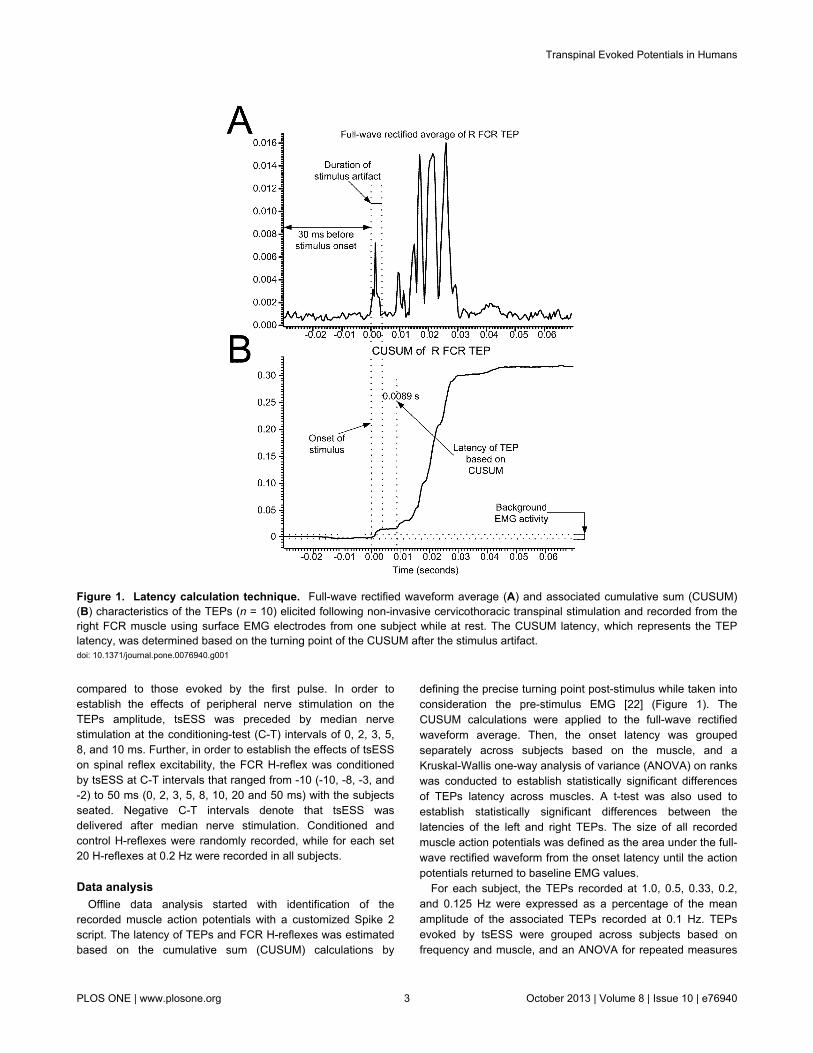
recorded muscle action potentials with a customized Spike 2script. The latency of TEPs and FCR H-reflexes was estimatedbased on the cumulative sum (CUSUM) calculations by
defining the precise turning point post-stimulus while taken intoconsideration the pre-stimulus EMG [22] (Figure 1). TheCUSUM calculations were applied to the full-wave rectifiedwaveform average. Then, the onset latency was groupedseparately across subjects based on the muscle, and aKruskal-Wallis one-way analysis of variance (ANOVA) on rankswas conducted to establish statistically significant differencesof TEPs latency across muscles. A t-test was also used toestablish statistically significant differences between thelatencies of the left and right TEPs. The size of all recordedmuscle action potentials was defined as the area under the full-wave rectified waveform from the onset latency until the actionpotentials returned to baseline EMG values.
For each subject, the TEPs recorded at 1.0, 0.5, 0.33, 0.2,and 0.125 Hz were expressed as a percentage of the meanamplitude of the associated TEPs recorded at 0.1 Hz. TEPsevoked by tsESS were grouped across subjects based onfrequency and muscle, and an ANOVA for repeated measures
Figure 1. Latency calculation technique. Full-wave rectified waveform average (A) and associated cumulative sum (CUSUM)(B) characteristics of the TEPs (n = 10) elicited following non-invasive cervicothoracic transpinal stimulation and recorded from theright FCR muscle using surface EMG electrodes from one subject while at rest. The CUSUM latency, which represents the TEPlatency, was determined based on the turning point of the CUSUM after the stimulus artifact.doi: 10.1371/journal.pone.0076940.g001
Transpinal Evoked Potentials in Humans
PLOS ONE | www.plosone.org 3 October 2013 | Volume 8 | Issue 10 | e76940

at 5 × 8 levels (5: frequency, 8: muscles) was conducted toestablish statistically significant differences across stimulationfrequencies and muscles.
The FCR H-reflex conditioned by tsESS was expressed as apercentage of the mean amplitude of the control FCR H-reflex.The mean amplitude of the conditioned FCR H-reflex fromeach subject was grouped based on the C-T interval and aone-way ANOVA was conducted to establish statisticallysignificant differences. This analysis was conducted also forthe M-waves which were expressed as a percentage of themaximal M-wave.
In the double stimuli paradigm, the right and left FCR andECR TEPs evoked by the second tsESS pulse were expressedas a percentage of the associated TEP evoked by the firsttsESS pulse. The same analysis was done for the FCR M-wave and FCR H-reflex upon double stimuli delivered to themedian nerve. The mean amplitude of each response fromeach subject was grouped based on the type of the responseand an ANOVA for repeated measures was conducted. Allstatistical tests were conducted at 95 % of confidence interval.Mean amplitudes are reported along with the standard error ofmeans (SEM), unless otherwise stated.
Results
In Figure 2, non-rectified waveform averages (n = 10 evokedat 0.2 Hz) of TEPs recorded from three subjects while seatedfollowing tsESS of the cervicothoracic region are indicated.Note that the shape of TEPs for the right and left muscles isdifferent for each subject and across subjects, consistent to theshape of the TEPs we recently reported for the leg muscles[12,13]. TEPs of arm muscles were manifested mostly with atriphasic waveform, but a polyphasic waveform was also
present (see left ECR in subject 1 and right ECR in subject 11in Figure 2).
In Figure 3A and 3B, non-rectified waveform averages of theright FCR H-reflex and right FCR TEP from two subjects areillustrated. In subjects 2 and 7, the latency of the FCR H-reflexwas 18.1 ms and 16.94 ms, respectively while the latency ofthe R FCR TEP was 11.4 ms and 10.2 ms, respectively. Themean latencies of the right FCR H-reflex and TEPs recordedfrom all muscles and subjects are indicated in Figure 3C. TheFCR H-reflex latency (20.86 ± 2.5; mean ± SD) was 1.98 timesthe R FCR TEP latency (10.52 ± 1.06; mean ± SD), but theTEPs recorded from the ipsilateral antagonistic ECR andcontralateral FCR and ECR muscles were prolonged by 0.43,0.72, and 0.3 ms compared to the half latency of the R FCR H-reflex, respectively. A Kruskal-Wallis one-way ANOVA on ranksshowed that the latency of the TEPs was statistically significantdifferent from the FCR H-reflex latency (F (8)= 74.53, P <0.001), while a t-test conducted separately for the pairs of theR and L FCR, ECR, TRIC, and BIC TEPs showed that thelatencies were not statistically significant different (for all P >0.05).
The overall amplitude of the TEPs evoked by tsESS andrecorded from the right and left FCR, ECR, BIC, and TRICmuscles at 1.0, 0.5, 0.33, 0.2, and 0.125 Hz is indicated inFigure 4. TEPs are presented as a percentage of the meanamplitude of the associated TEP recorded at 0.1 Hz. TEPsamplitude at different stimulation frequencies are groupedtogether for subjects 1-12 (Figure 4A), while data from subject13 are shown separately (Figure 4B) because the effectsobserved in this subject were different from the other subjectsand if grouped together these effects would have beenmasked. For data from subjects 1-12, the right FCR TEPs didnot vary at different stimulation frequencies (F (5) = 2.94, P =
Figure 2. Waveforms of transpinal evoked potentials (TEPs). Representative examples of non-rectified waveform averages (n= 10, elicited at 0.2 Hz) of TEPs recorded following transcutaneous electric stimulation of the cervicothoracic region from the flexorcarpi radialis (FCR), biceps brachii (BIC), extensor carpi radialis (ECR), and triceps brachii (TRIC) muscles in both arms.doi: 10.1371/journal.pone.0076940.g002
Transpinal Evoked Potentials in Humans
PLOS ONE | www.plosone.org 4 October 2013 | Volume 8 | Issue 10 | e76940

0.708; one-way ANOVA) (Figure 4A). The same result wasalso found for TEPs recorded from the other arm muscles. AnANOVA for repeated measures at 5 × 8 levels (5: frequencies,8: muscles) showed that the amplitude of the normalized TEPsdid not vary across muscles (F (7) = 1.74, P = 0.097) andacross stimulation frequencies (F (4) = 0.59, P = 0.67), while astatistically significant interaction between muscles andstimulation frequencies was not found (F (28) = 0.38, P = 0.99).In contrast to these findings, in subject 13, the R FCR TEPsamplitude varied significantly across stimulation frequencies (F(5) = 44.97, P < 0.001; Kruskal-Wallis one-way ANOVA onranks along with Dunn’s multiple comparisons), an effect thatwas observed in the L FCR, L ECR, R BIC, and R TRIC TEPs(Figure 4B).
In order to further delineate the susceptibility of TEPs tohomosynaptic depression, TEPs and FCR H-reflexes were
recorded following a double tsESS and/or median nervestimulation pulse at an inter-stimulus interval of 40 ms. InFigure 5A and 5B, the effects of the double pulse on the rightFCR H-reflex and FCR TEP are indicated. In subject 2, theFCR H-reflex was completely abolished following a secondstimulus to the median nerve (Figure 5A, left graph), while theFCR TEP was not affected following a second tsESS stimulus(Figure 5A, right graph). In contrast, in subject 13, the FCRTEP was depressed following a tsESS pulse at 40 ms inter-stimulus interval (Figure 5B, right graph) similar to thatobserved for the FCR H-reflex (Figure 5B, left graph). Theoverall amplitude of the right FCR H-reflex, right FCR M-wave,and TEPs recorded from the right and left FCR and ECRmuscles for subjects 1-12, elicited by a second stimulus andnormalized to the mean amplitude of the associated actionpotential evoked by the first stimulus is indicated in Figure 5C.
Figure 3. Latency of transpinal evoked potentials (TEPs). A, B: Non-rectified waveform averages (evoked at 0.2 Hz) of flexorcarpi radialis (FCR) H-reflexes and FCR TEPs from two subjects. Note that in these subjects that the FCR TEP appeared nearly ata half latency compared to the FCR H-reflex latency. C: Overall mean latency of the FCR H-reflex and TEPs recorded from the leftand right arm muscles following transcutaneous electric stimulation of the spine over the cervicothoracic region. Error barsrepresent the SD.doi: 10.1371/journal.pone.0076940.g003
Transpinal Evoked Potentials in Humans
PLOS ONE | www.plosone.org 5 October 2013 | Volume 8 | Issue 10 | e76940

The FCR H-reflex amplitude was statistically significantdifferent compared to the TEPs amplitudes (F (5) = 14.07, P =0.015; one-way ANOVA) (Figure 5C). The mean amplitude ofthe right FCR H-reflex, right FCR M-wave, and TEPs recordedfrom the right and left FCR and ECR muscles in subject 13 areindicated in Figure 5D, which were all statistically significantdifferent from the associated control values (P < 0.05). A one-way ANOVA showed that there was a statistically significantdifference between the different types of responses (F (5) =46.48, P < 0.001).
The overall effects of median nerve stimulation on the TEPsamplitude recorded from the right and left ECR, BIC, TRICmuscles and from the left FCR are indicated in Figure 6. Notethat TEPs for the R FCR muscle are not indicated becauseTEPs were contaminated by the stimulus artifact followingmedian nerve stimulation and could not be measuredaccurately. Low-threshold median nerve stimulation increasedsignificantly the TEPs amplitude recorded from the right ECRmuscle when compared to control values at C-T intervals of 2,3, 5, 8, and 10 ms (F (5) = 17.51, P = 0.004; Kruskal-Wallisone-way ANOVA on ranks along with Tukey’s test multiplecomparisons) (Figure 6A). The same result was also found forTEPs recorded from the right TRIC (Figure 6B), right and leftBIC (Figure 6C), and left FCR muscles (Figure 6D).
While median nerve stimulation facilitated the ipsilateral andcontralateral TEPs, tsESS of the cervicothoracic spinedepressed the FCR H-reflex. The effects of tsESS at the C-Tinterval of 2 ms on the average soleus H-reflex recorded fromtwo subjects while seated is shown in Figure 7A. In bothexamples, the magnitude of the conditioned FCR H-reflex wassignificantly reduced when compared to control reflex values,and the FCR H-reflex depression occurred with stable FCR M-waves under control conditions and during tsESS. Theamplitude of the conditioned FCR H-reflex from all subjects andC-T intervals tested is shown in Figure 7B. The FCR H-reflexvaried significantly across the C-T intervals tested (F (12, 65) =2.09, P = 0.03), while the FCR H-reflexes at the C-T intervalsranging from 2 to +50 ms were statistically significant differentfrom the other C-T intervals and from control reflex values (P <0.05). The FCR H-reflex depression coincided with stable M-waves (F (12) = 2.4, P = 0.992; one-way ANOVA) (Figure 7C).
Discussion
Transcutaneous electric stimulation of the spine over thecervicothoracic region attenuated significantly FCR H-reflexexcitability in seated healthy human subjects, while TEPsrecorded from arm muscles were not susceptible to
Figure 4. Susceptibility of transpinal evoked potentials (TEPs) to homosynaptic depression. The overall mean amplitude ofthe TEPs recorded bilaterally from the right (R) and left (L) flexor carpi radialis (FCR), extensor carpi radialis (ECR), biceps brachii(BIC), and triceps brachii (TRIC) muscles for subjects 1-12 (A) and for subject 13 (B). TEPs recorded at 1.0, 0.5, 0.33, 0.2, and0.125 Hz are presented as a percentage of the mean amplitude of the associated TEPs recorded at 0.1 Hz. Asterisks indicatestatistically significant differences from control TEPs values. Error bars represent the SEM.doi: 10.1371/journal.pone.0076940.g004
Transpinal Evoked Potentials in Humans
PLOS ONE | www.plosone.org 6 October 2013 | Volume 8 | Issue 10 | e76940

homosynaptic depression in all but one subject, and werefacilitated following peripheral nerve stimulation. These findingsare consistent to those we have recently reported for TEPsrecorded from the leg muscles following electric or magneticstimulation of the thoracolumbar region [12,13], supporting thatneuronal pathways associated with TEPs and tsESS-mediatedreflex effects in human upper and lower limbs are similar. Theneurophysiological characteristics of the TEPs and the use oftsESS as a therapeutic modality are discussed in the followingsections.
TEPs were present at similar latencies in left and right armmuscles with shorter latencies observed for the biceps/tricepsmuscles (L BIC: 6.8 ± 1.29 ms) compared to the more distalwrist flexors/extensors (R FCR: 10.5 ± 1.06 ms) (Figure 3B),
consistent to that reported elsewhere [11,23]. It should benoted that the R ECR, L FCR, and L ECR TEPs latency wasprolonged by ~0.5 ms when compared to the half latency of theR FCR H-reflex, supporting for synaptic events betweenantagonistic muscles and interlimb neuronal circuits uponmanifestation of TEPs. Across the studies reported in theliterature, the latency of the FCR TEPs ranges from 7.17 to8.46 ms [11,23], while TEPs recorded from thenar musclesfollowing transcutaneous magnetic stimulation over thecervicothoracic spine had an average latency of ~13.5 ms [24].Differences of latencies reported in the literature may berelated to the placement and size of the stimulating electrode,anthropometric characteristics, as well as to the thickness of
Figure 5. Transpinal evoked potentials (TEPs) and FCR H-reflexes upon double stimuli. A, B: Non-rectified waveformaverages of the FCR H-reflex and the FCR TEPs when a double pulse at an inter-stimulus interval of 40 ms was delivered to thecervicothoracic region or to the median nerve, respectively for two subjects. C: Overall average amplitude of the FCR H-reflex, FCRM-wave, and TEPs for subjects 1-12 evoked by a second pulse delivered to the median nerve or to the cervicothoracic region. D:Overall average amplitude of the FCR H-reflex, FCR M-wave, and TEPs evoked by a second pulse delivered to the median nerve orto the cervicothoracic region for subject 13. In both C and D graphs, the FCR H-reflex, FCR M-wave and TEPs were normalized tothe associated potentials evoked by the first pulse. Asterisks indicate statistically significant differences from control values. Errorbars indicate the SEM.doi: 10.1371/journal.pone.0076940.g005
Transpinal Evoked Potentials in Humans
PLOS ONE | www.plosone.org 7 October 2013 | Volume 8 | Issue 10 | e76940
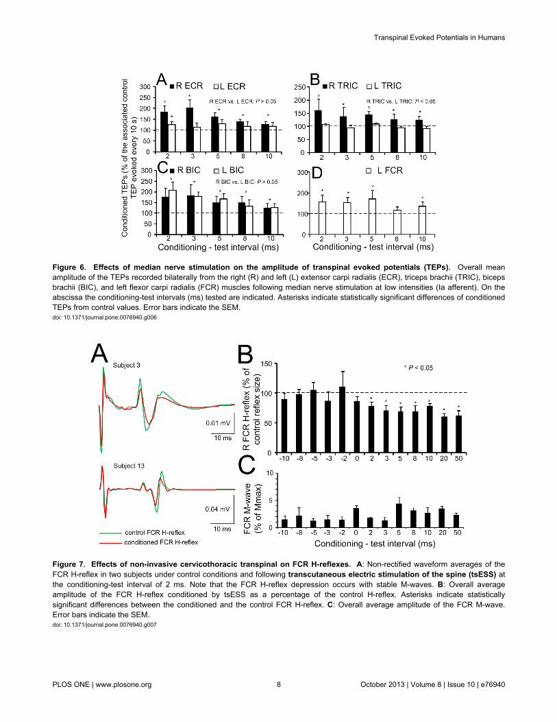
Figure 6. Effects of median nerve stimulation on the amplitude of transpinal evoked potentials (TEPs). Overall meanamplitude of the TEPs recorded bilaterally from the right (R) and left (L) extensor carpi radialis (ECR), triceps brachii (TRIC), bicepsbrachii (BIC), and left flexor carpi radialis (FCR) muscles following median nerve stimulation at low intensities (Ia afferent). On theabscissa the conditioning-test intervals (ms) tested are indicated. Asterisks indicate statistically significant differences of conditionedTEPs from control values. Error bars indicate the SEM.doi: 10.1371/journal.pone.0076940.g006
Figure 7. Effects of non-invasive cervicothoracic transpinal on FCR H-reflexes. A: Non-rectified waveform averages of theFCR H-reflex in two subjects under control conditions and following transcutaneous electric stimulation of the spine (tsESS) atthe conditioning-test interval of 2 ms. Note that the FCR H-reflex depression occurs with stable M-waves. B: Overall averageamplitude of the FCR H-reflex conditioned by tsESS as a percentage of the control H-reflex. Asterisks indicate statisticallysignificant differences between the conditioned and the control FCR H-reflex. C: Overall average amplitude of the FCR M-wave.Error bars indicate the SEM.doi: 10.1371/journal.pone.0076940.g007
Transpinal Evoked Potentials in Humans
PLOS ONE | www.plosone.org 8 October 2013 | Volume 8 | Issue 10 | e76940

the underlying tissue and the methodology utilized to evokeand measure the latency of these potentials [2,25,26].
The shape of the TEPs following tsESS of thecervicothoracic region was largely triphasic in both wrist andelbow flexor/extensor muscles (Figure 2), a shape apparent forthe FCR H-reflex (Figure 2, Figure 3A, Figure 5A, B). Further,the shape of the TEPs was asymmetrical across muscles forthe same subject throughout the whole duration of anexperiment (Figure 2). The different shape of TEPs acrossmuscles within a single subject suggests that TEPs recordedfrom different muscles might be due to different synaptic eventsand thus have different origin, in a similar manner to the spinalsegmental potentials recorded following stimulation of theposterior tibial nerve [27]. TEPs may be due to concomitantexcitation of different types of afferents and motor axons, andthus their waveform may contain a direct motor response (i.e.,M-wave) as well as reflex-mediated action potentials. If TEPsencompass an M-wave, then they should not be influenced byrepetitive discharges, while their amplitude should decreasewhen reflex-mediated synaptic events are present upon theirmanifestation. This constitutes a clear distinction between amotor response and a reflexly-afferent mediated response,since the FCR H-reflexes are depressed after a precedinghomonymous reflex discharge occurring at 1-2 s [20]. In thisstudy, TEPs recorded from arm flexor/extensor muscles werenot affected when stimulation was delivered at low-frequenciesin all but one subject (Figure 4), supporting that they are notsusceptible to homosynaptic depression and likely do notrepresent reflexly-mediated action potentials. This is consistentto our recent findings reported for TEPs of the leg muscles inhealthy subjects [12,13]. In order to elucidate further thisneurophysiological property, we recorded FCR H-reflexes andTEPs upon double stimuli delivered at an inter-stimulus intervalof 40 ms. The FCR H-reflexes were clearly depressed upondouble stimuli reaching an amplitude half of the responsesobtained with the first stimuli (Figure 5C, D), while theamplitude of the TEPs was not significantly affected (Figure5C). However, in subject 13, the right and left FCR and ECRTEPs were depressed upon double stimuli, consistent to thedepression of TEPs observed in this subject following tsESS atlow frequencies (Figures 4B, 5D). The depression of the TEPsacross muscles upon double stimuli and at low stimulationfrequencies in this subject suggests that these potentials wereaffected by homosynaptic depression and thus we can proposethat in this subject TEPs were partly reflexly-mediated evokedpotentials involving activation of group I afferents. While wecannot know why TEPs in this subject were channeled into thespinal cord through reflex pathways, it is possible that thiseffect may be related to a different orientation of the stimulatingelectrode relative to the cervical roots and trunks of the brachialplexus compared to the other subjects, because the paraspinalmuscles of this subject were not hypertrophic as his life stylewas sedentary. Because F-waves are depressed upon doublestimuli due to refractoriness of motor axons by the precededafferent volley [28], one may consider that TEPs in subject 13are F-waves, which are produced at high stimulation intensitiesof a mixed peripheral nerve and are recurrent discharges ofantidromically activated motoneurons [29]. However, TEPs
cannot be considered to reflect F-waves because the TEPslatency of thenar muscles is shorter by 1.4 ms when comparedto F+M-1/2 latency [30], and the latency of TEPs in this subjectwas similar to that observed in the other subjects. Inconclusion, TEPs of arm muscles following stimulation of thespine at the cervicothoracic region represent compositeexcitatory potentials of motor nerve fibers excitedorthodromically and different types of afferents excitedantidromically in the majority of subjects, while in somesubjects TEPs may represent excitation of spinal reflexpathways similar to that known for the H-reflexes in humans.
Most notable findings of this study are the FCR H-reflexdepression following tsESS (Figure 7B) and the facilitation ofTEPs recorded from left and right arm muscles followingpercutaneous stimulation of the right median nerve (Figure 6).These findings have never been documented in humans andhave great clinical significance. The FCR H-reflex depression isin line with the soleus H-reflex depression we reportedfollowing transcutaneous electric or magnetic stimulation of thethoracolumbar spine [12,13]. FCR H-reflex depression mighthave been mediated at a presynaptic or postsynaptic level orsimultaneously at both synaptic levels. Potentiation of Iaafferents hyperpolarization by antidromic excitation of group Iaafferents in the dorsal columns [5], repetitive volleys producedby activation of fast conducting afferents acting at apostsynaptic level [31], polysynaptic spinal reflex pathwaysaffecting alpha motoneurons [23], and supraspinal activitymodulated by the orthodromic transmission of impulses indorsal column fibers following tsESS may all account for theFCR H-reflex depression at short and long C-T intervals.Because in anaesthetized rats, spinal direct current stimulationmodulates in a polarity-specific manner the supraspinal activityof the somatosensory system [32], and spinal cord neuronsexcitability [33], both supraspinal and spinal neuronal circuitsmight have mediated the FCR H-reflex depression observed inthis study.
While the FCR H-reflex was depressed following tsESS,TEPs recorded from the left and right arm muscles werefacilitated at C-T intervals ranging from 2 to 10 ms followingstimulation of the right median nerve (Figure 6). Theaforementioned mechanisms for FCR H-reflex depressioncannot account for the facilitation of TEPs. Stimulation of themedian nerve produced action potentials that run dorsally andperpendicular to the surface of the cord representing activity ofdorsal roots travelling rostrally and ventral roots travellingcaudally [34]. These spinal potentials could have modulatedactivity of interneurons that are involved in interlimbcoordination, supported by the fact that facilitation of TEPs wasobserved on both arms. However, changes in the excitability ofthe stimulated axons cannot be disregarded. Based on thecurrent experimental protocol, the exact neuronal pathwaycannot be determined, and it is apparent that further researchoutlining facilitatory and inhibitory synaptic events, including butnot limited to motor unit recordings and peristimulusfrequencygrams [35], associated with TEPs is needed.
Transpinal Evoked Potentials in Humans
PLOS ONE | www.plosone.org 9 October 2013 | Volume 8 | Issue 10 | e76940

Clinical application of findingsInvasive dorsal column stimulation in two people with
multiple sclerosis improved their motor, reflex, and bladderfunction [36], while continuous epidural stimulation enabled aperson with motor complete paraplegia to achieve full-weightbearing and locomotor-like EMG activity [37], consistent to themodulation of spinal locomotor networks of adult spinal rats[38]. These results were obtained with stimulation administeredinvasively, limiting thus their possibility of application to a largernumber of patients. This study showed for the first time thatnon-invasive transpinal stimulation of the cervicothoracic regiondepressed FCR H-reflex excitability, and when cervicothoracictranspinal stimulation was combined with peripheral nerve
stimulation the spinal output - manifested as multisegmentalTEPs - was facilitated. Based on our current and publishedfindings [12,13], tsESS can be utilized in upper motor neuronlesions to normalize the reflex hyper-excitability of upper andlower limbs, and to potentiate spinal output.
Author Contributions
Conceived and designed the experiments: MK. Performed theexperiments: JE AL RH MK. Analyzed the data: JE AL RH MK.Contributed reagents/materials/analysis tools: MK. Wrote themanuscript: MK.
References
1. Mills KR, Murray NMF (1986) Electrical stimulation over the humanvertebral column: which neural elements are excited?Electroencephalogr Clin Neurophysiol 63: 582-589. doi:10.1016/0013-4694(86)90145-8. PubMed: 2422007.
2. Maertens de Noordhout AM, Rothwell JC, Thompson PD, Day BL,Marsden CD (1988) Percutaneous electrical stimulation of lumbosacralroots in man. J Neurol Neurosurg Psychiatry 51: 174-181. doi:10.1136/jnnp.51.2.174. PubMed: 2831302.
3. Cros D, Chiappa KH, Gominak S, Fang J, Santamaria J, et al. (1990)Cervical magnetic stimulation. Neurology 40: 1751-1756. doi:10.1212/WNL.40.11.1751. PubMed: 2234432.
4. Chokroverty S, Picone MA, Chokroverty M (1991) Percutaneousmagnetic coil stimulation of human cervical vertebral column: site ofstimulation and clinical application. Electroencephalogr ClinNeurophysiol 81: 359-365. doi:10.1016/0168-5597(91)90025-S.PubMed: 1718722.
5. Hunter JP, Ashby P (1994) Segmental effects of epidural spinal cordstimulation in humans. J Physiol Lond 474: 407-419. PubMed:8014902.
6. Gerasimenko YP, Lavrov IA, Courtine G, Ichiyama RM, Dy CJ, et al.(2006) Spinal cord reflexes induced by epidural spinal cord stimulationin normal awake rats. J Neurosci Methods 157: 253-263. doi:10.1016/j.jneumeth.2006.05.004. PubMed: 16764937.
7. Courtine G, Harkema SJ, Dy CJ, Gerasimenko YP, Dyhre-Poulsen P(2007) Modulation of multisegmental monosynaptic responses in avariety of leg muscles during walking and running in humans. J PhysiolLond 582: 1125-1139. doi:10.1113/jphysiol.2007.128447. PubMed:17446226.
8. Minassian K, Persy I, Rattay F, Dimitrijevic MR, Hofer C, et al. (2007)Posterior root-muscle reflexes elicited by transcutaneous stimulation ofthe human lumbosacral cord. Muscle Nerve 35: 327-336. doi:10.1002/mus.20700. PubMed: 17117411.
9. Hofstoetter US, Minassian K, Hofer C, Mayr W, Rattay F, et al. (2008)Modification of reflex responses to lumbar posterior root stimulation bymotor tasks in healthy subjects. Artif Organs 32: 644-648. doi:10.1111/j.1525-1594.2008.00616.x. PubMed: 18782137.
10. Sabbahi MA, Sengul YS (2011) Thoracolumbar multisegmental motorresponses in the upper and lower limbs in healthy subjects. Spinal Cord49: 741-748. doi:10.1038/sc.2010.165. PubMed: 21173778.
11. Sabbahi MA, Sengul YS (2012) Cervical multisegmental motorresponses in healthy subjects. Spinal Cord 50: 432-439. doi:10.1038/sc.2011.166. PubMed: 22249322.
12. Knikou M (2013) Neurophysiological characteristics of human legmuscle action potentials evoked by transcutaneous magneticstimulation of the spine. Bioelectromagnetics 34: 200-210. doi:10.1002/bem.21768. PubMed: 23192827.
13. Knikou M (2013) Neurophysiological characterization of transpinalevoked potentials in human leg muscles. Bioelectromagnetics. doi:10.1002/bem.21808 [in press]
14. Magladery JW, Teasdall RD, Park AM, Languth HW (1952)Electrophysiological studies of reflex activity in patients with lesions ofthe nervous system. I. A comparison of spinal motoneurone excitabilityfollowing afferent nerve volleys in normal persons and patients withupper motor neurone lesions. Bull Johns Hopkins Hosp 91: 219-244.PubMed: 12987919.
15. Lloyd DP, Wilson VJ (1957) Reflex depression in rhythmically activemonosynaptic reflex pathways. J Gen Physiol 40: 409-426. doi:10.1085/jgp.40.3.409. PubMed: 13398572.
16. Paillard J (1955) Electrophysiologic analysis and comparison in man ofHoffmann’s reflex and myotatic reflex. Pflugers Arch 260: 448-479. doi:10.1007/BF00363666. PubMed: 13245374.
17. Hultborn H, Illert M, Nielsen J, Paul A, Ballegaard M, et al. (1996) Onthe mechanism of the post-activation depression of the H-reflex inhuman subjects. Exp Brain Res 108: 450-462. PubMed: 8801125.
18. Kohn AF, Floeter MK, Hallett M (1997) Presynaptic inhibition comparedwith homosynaptic depression as an explanation for soleus H-reflexdepression in humans. Exp Brain Res 116: 375-380. doi:10.1007/PL00005765. PubMed: 9348136.
19. Jessop T, DePaola A, Casaletto L, Englard C, Knikou M (2013) Short-term plasticity of human spinal inhibitory circuits after isometric andisotonic ankle training. Eur J Appl Physiol 113: 273-284. doi:10.1007/s00421-012-2438-1. PubMed: 22684372.
20. Aymard C, Katz R, Lafitte C, Lo E, Pénicaud A, et al. (2000)Presynaptic inhibition and homosynaptic depression: a comparisonbetween lower and upper limbs in normal human subjects and patientswith hemiplegia. Brain 123: 1688-1702. doi:10.1093/brain/123.8.1688.PubMed: 10908198.
21. Knikou M (2008) The H-reflex as a probe: pathways and pitfalls. JNeurosci Methods 171: 1-12. doi:10.1016/j.jneumeth.2008.02.012.PubMed: 18394711.
22. Brinkworth RSA, Türker KS (2003) A method for quantifying reflexresponses from intra-muscular and surface electromyogram. JNeurosci Methods 122: 179-193. doi:10.1016/S0165-0270(02)00321-7.PubMed: 12573477.
23. Vucic S, Cairns KD, Black KR, Chong PS, Cros D (2006) Cervicalnerve root stimulation. Part I: technical aspects and normal data. ClinNeurophysiol 117: 392-397. doi:10.1016/j.clinph.2005.10.011. PubMed:16403485.
24. Ugawa Y, Genba-Shimizu K, Kanazawa I (1995) Electrical stimulationof the human descending motor tracts at several levels. Can J NeurolSci 22: 36-42. PubMed: 7750071.
25. Butson CR, McIntyre CC (2005) Tissue and electrode capacitancereduce neural activation volumes during deep brain stimulation. ClinNeurophysiol 116: 2490-2500. doi:10.1016/j.clinph.2005.06.023.PubMed: 16125463.
26. Kuhn A, Keller T, Lawrence M, Morari M (2009) A model fortranscutaneous current simulation: simulations and experiments. MedBiol Eng Comput 47: 279-289. doi:10.1007/s11517-008-0422-z.PubMed: 19005714.
27. Ertekin C (1976) Studies on the human evoked electrospinogram. I.The origin of the segmental evoked potentials. Acta Neurol Scand 53:3-20. doi:10.1111/j.1600-0404.1976.tb04321.x. PubMed: 1251678.
28. Mastaglia FL, Carroll WM (1985) The effects of conditioning stimuli onthe F-response. J Neurol Neurosurg Psychiatry 48: 182-184. doi:10.1136/jnnp.48.2.182. PubMed: 3981180.
29. Magladery JW, McDougal DD (1950) Electrophysiological studies ofnerve and reflex activity in normal man. Identification of certain reflexesin the electromyogram and the conduction velocity of peripheral nervefibres. Bull Johns Hopkins Hosp 86: 265-290. PubMed: 15414383.
30. Ugawa Y, Rothwell JC, Day BL, Thompson PD, Marsden CD (1989)Magnetic stimulation over the spinal enlargements. J Neurol Neurosurg
Transpinal Evoked Potentials in Humans
PLOS ONE | www.plosone.org 10 October 2013 | Volume 8 | Issue 10 | e76940

Psychiatry 52: 1025-1032. doi:10.1136/jnnp.52.9.1025. PubMed:2795071.
31. Halonen JP, Jones SJ, Edgar MA, Ransford AO (1989) Conductionproperties of epidurally recorded spinal cord potentials following lowerlimb stimulation in man. Electroencephalogr Clin Neurophysiol 74:161-174. doi:10.1016/0013-4694(89)90002-3. PubMed: 2470572.
32. Aguilar J, Pulecchi F, Dilena R, Oliviero A, Priori A, et al. (2011) Spinaldirect current stimulation modulates the activity of gracile nucleus andprimary somatosensory cortex in anaesthetized rats. J Physiol Lond589: 4981-4996. PubMed: 21825031.
33. Ahmed Z, Wieraszko A (2012) Trans-spinal direct current enhancescorticospinal output and stimulation-evoked release of glutamateanalog, D-2,3-³H-aspartic acid. J Appl Physiol 112: 1576-1592. doi:10.1152/japplphysiol.00967.2011. PubMed: 22362399.
34. Yiannikas C, Shahani BT (1988) The origins of lumbosacral spinalevoked potentials in humans using a surface electrode recording
technique. J Neurol Neurosurg Psychiatry 51: 499-508. doi:10.1136/jnnp.51.4.499. PubMed: 3379422.
35. Türker KS, Powers RK (2005) Black box revisited: a technique forestimating postsynaptic potentials in neurons. Trends Neurosci 28:379-386. doi:10.1016/j.tins.2005.05.007. PubMed: 15927277.
36. Illis LS, Sedwick EM, Oygar AE, Sabbahi MA (1976) Dorsal-columnstimulation in the rehabilitation of patients with multiple sclerosis.Lancet 26: 1383-1386. PubMed: 59019.
37. Harkema S, Gerasimenko Y, Hodes J, Burdick J, Angeli C, et al. (2011)Effect of epidural stimulation of the lumbosacral spinal cord onvoluntary movement, standing, and assisted stepping after motorcomplete paraplegia: a case study. Lancet 377: 1938-1947. doi:10.1016/S0140-6736(11)60547-3. PubMed: 21601270.
38. Lavrov I, Dy CJ, Fong AJ, Gerasimenko Y, Courtine G, et al. (2008)Epidural stimulation induced modulation of spinal locomotor networks inadult spinal rats. J Neurosci 28: 6022-6029. doi:10.1523/JNEUROSCI.0080-08.2008. PubMed: 18524907.
Transpinal Evoked Potentials in Humans
PLOS ONE | www.plosone.org 11 October 2013 | Volume 8 | Issue 10 | e76940
Related Documents