J Korean Soc Spine Surg. 2018 November;25(S2):S137-198. S137 http://dx.doi.org/10.4184/jkss.2018.25.S2.S137 © Copyright 2018 Korean Society of Spine Surgery Journal of Korean Society of Spine Surgery. www.krspine.org. pISSN 2093-4378 eISSN 2093-4386 This is an Open Access article distributed under the terms of the Creative Commons Attribution Non-Commercial License (http://creativecommons.org/licenses/by-nc/4.0/) which permits unrestricted non-commercial use, distribution, and reproduction in any medium, provided the original work is properly cited. Cervical Spine C5 Palsy After Aterior Cervical Surgeries Jae-Hyung Eoh, Bo-Gun Suh, Ji-Hun Shin Department of Orthopedic Surgery, Po-Hang Semyeong Christianity Hospital, Po-Hang, Korea Backgrounds and Introduction: Postoperative C5 palsy is well known as common complication after decompression surgery at the cervical spine. However, the incidence, prognosis, and etiology of C5 Palsy after anterior cervical decompressive surgeries have not yet been fully established. In the current review article, we discuss the incidence, prognosis and possible mechanism of C5 palsy after anterior cervical decompressive surgeries. Main Body: C5 palsy is defined as a new onset of deltoid or biceps muscle paralysis after cervical decompressive surgeries. Shorter C5 ventral rootlets and ventral root might easily became taut and overstretched because of dural expansion after anterior decompression in the treatment of chronic compression disease. The average reported incidence of C5 Palsy after anterior decompressive surgeries is 6.7% (1.6-9.1%) with 92% of the C5 palsy occurring unilaterally. Majority of the motor symptoms occur with in first week after the operation and are expected to resolve by 6mons. 50% of C5 palsy patients experienced full recovery within 6mons and 91.7% with in 2 years. The exact etiology remains uncertains, possible mechanism of C5 palsy include direct injury, nerve ischemia, reperfusion injury, tethering effect caused by spinal cord drift, and preoperative cord rotation. Conclusion: C5 Palsy may developed after either anterior or posterior surgery. The average reported incidence of C5 Palsy after anterior decompressive surgeries is 6.7% and the exact etiology remains uncertains. Keywords: Cervical, Anterior surgeries, C5 Palsy 경추 전방 수술 이후에 발생한 제 5경추 신경마비에 대한 고찰 어재형, 서보건, 신지훈 포항 세명기독병원 정형외과 서론: 수술후 제 5경추 신경 마비는 경추 감압수술 이후에 발생 하는 잘 알려진 일반적인 합병증이다. 하지만 경추 전방 감압수 술 이후에 발생한 제 5경추 신경마비의 발생률, 예후, 병인등은 아직 충분히 밝혀지지 않았다. 문헌 고찰을 통하여 경추 전방 감 압수술 이후에 발생한 제 5경추 신경마비의 발생률, 예후 및 그 발생기전을 알아 보고자 한다. 본론: 경추 제 5신경 마비는 경추 감압수술후 새롭게 발생한 삼 각근 혹은 이두박근의 마비로 알려져 있다. 해부학적으로 짧은 배측 제 5신경근은, 만성 압박성 질환의 전방감압시 경막이 팽 창 되면서 쉽게 팽팽해 지고, 당겨지기 쉽다. 전방 감압수술 이 후에 발생한 제 5경추 신경마비에 대해 보고된 평균 발생률은 6.7%(1.6-9.1%)이며, 92%가 편측으로 발생한다. 대부분이 수술 후 1주이내에 발생하며, 6개월이내에 회복되는 경우가 많으며, 6개월 이내에 50%, 2년이내에 91.7%가 완전 회복 된다고 보고 된다. 정확한 발생기전은 아직 불명확 하며, 직접손상, 신경허 혈, 척수이동에 따른 신경근 당김, 수술전 척수의 회전등이 원인 으로 알려져 있다. 결론: 경추 제 5신경 마비는 전방 혹은 후방수술 이후에 모두 발 생 할 수 있다. 전방 감압수술 이후에 발생한 제 5경추 신경마비 에 대해 보고된 평균 발생률은 6.7%이며, 그 발생 기전은 아직 정확하지 않다. 색인 단어: 경추, 전방수술, 경추 제 5신경 마비 C5 Palsy After Posterior Cervical Surgeries Jae Jun Yang Department of Orthopedic Surgery, Dongguk University Ilsan Hospital, Goyang, Korea Backgrounds and Introduction: C5 palsy after posterior cervical surgeries is a major complication causing muscle weakness, brachialgia, numbness, and most of all, dissatisfaction on the surgery. Therefore, effort to prevent this complication based on the knowledge including the incidence, risk factors, and prevention methods is necessary for spine surgeons. The objective of this article is to review the nature and how to prevent the complication . Main Body: The incidence of C5 palsy after cervical spine surgeries has been reported as 6.3%. C5 palsy occurred more frequently after posterior surgeries than anterior surgeries (6.2% vs 5%) and laminectomy caused C5 palsy more frequently than laminoplasty in comparison among posterior surgeries. The pathomechanism of postoperative C5 palsy might be multifactorial including direct root damage, tethering of the nerve root, segmental spinal cord disorder,

Welcome message from author
This document is posted to help you gain knowledge. Please leave a comment to let me know what you think about it! Share it to your friends and learn new things together.
Transcript

J Korean Soc Spine Surg. 2018 November;25(S2):S137-198.
S137
http://dx.doi.org/10.4184/jkss.2018.25.S2.S137
© Copyright 2018 Korean Society of Spine Surgery Journal of Korean Society of Spine Surgery. www.krspine.org. pISSN 2093-4378 eISSN 2093-4386 This is an Open Access article distributed under the terms of the Creative Commons Attribution Non-Commercial License (http://creativecommons.org/licenses/by-nc/4.0/) which permits unrestricted non-commercial use, distribution, and reproduction in any medium, provided the original work is properly cited.
Cervical Spine
C5 Palsy After Aterior Cervical Surgeries
Jae-Hyung Eoh, Bo-Gun Suh, Ji-Hun ShinDepartment of Orthopedic Surgery, Po-Hang Semyeong Christianity Hospital, Po-Hang, Korea
Backgrounds and Introduction: Postoperative C5 palsy is well known as common complication after decompression surgery at the cervical spine. However, the incidence, prognosis, and etiology of C5 Palsy after anterior cervical decompressive surgeries have not yet been fully established. In the current review article, we discuss the incidence, prognosis and possible mechanism of C5 palsy after anterior cervical decompressive surgeries.Main Body: C5 palsy is defined as a new onset of deltoid or biceps muscle paralysis after cervical decompressive surgeries. Shorter C5 ventral rootlets and ventral root might easily became taut and overstretched because of dural expansion after anterior decompression in the treatment of chronic compression disease. The average reported incidence of C5 Palsy after anterior decompressive surgeries is 6.7% (1.6-9.1%) with 92% of the C5 palsy occurring unilaterally. Majority of the motor symptoms occur with in first week after the operation and are expected to resolve by 6mons. 50% of C5 palsy patients experienced full recovery within 6mons and 91.7% with in 2 years. The exact etiology remains uncertains, possible mechanism of C5 palsy include direct injury, nerve ischemia, reperfusion injury, tethering effect caused by spinal cord drift, and preoperative cord rotation.Conclusion: C5 Palsy may developed after either anterior or posterior surgery. The average reported incidence of C5 Palsy after anterior decompressive surgeries is 6.7% and the exact etiology remains uncertains.Keywords: Cervical, Anterior surgeries, C5 Palsy
경추 전방 수술 이후에 발생한 제 5경추 신경마비에 대한
고찰
어재형, 서보건, 신지훈
포항 세명기독병원 정형외과
서론: 수술후 제 5경추 신경 마비는 경추 감압수술 이후에 발생
하는 잘 알려진 일반적인 합병증이다. 하지만 경추 전방 감압수
술 이후에 발생한 제 5경추 신경마비의 발생률, 예후, 병인등은
아직 충분히 밝혀지지 않았다. 문헌 고찰을 통하여 경추 전방 감
압수술 이후에 발생한 제 5경추 신경마비의 발생률, 예후 및 그
발생기전을 알아 보고자 한다.
본론: 경추 제 5신경 마비는 경추 감압수술후 새롭게 발생한 삼
각근 혹은 이두박근의 마비로 알려져 있다. 해부학적으로 짧은
배측 제 5신경근은, 만성 압박성 질환의 전방감압시 경막이 팽
창 되면서 쉽게 팽팽해 지고, 당겨지기 쉽다. 전방 감압수술 이
후에 발생한 제 5경추 신경마비에 대해 보고된 평균 발생률은
6.7%(1.6-9.1%)이며, 92%가 편측으로 발생한다. 대부분이 수술
후 1주이내에 발생하며, 6개월이내에 회복되는 경우가 많으며,
6개월 이내에 50%, 2년이내에 91.7%가 완전 회복 된다고 보고
된다. 정확한 발생기전은 아직 불명확 하며, 직접손상, 신경허
혈, 척수이동에 따른 신경근 당김, 수술전 척수의 회전등이 원인
으로 알려져 있다.
결론: 경추 제 5신경 마비는 전방 혹은 후방수술 이후에 모두 발
생 할 수 있다. 전방 감압수술 이후에 발생한 제 5경추 신경마비
에 대해 보고된 평균 발생률은 6.7%이며, 그 발생 기전은 아직
정확하지 않다.
색인 단어: 경추, 전방수술, 경추 제 5신경 마비
C5 Palsy After Posterior Cervical Surgeries
Jae Jun YangDepartment of Orthopedic Surgery, Dongguk University Ilsan Hospital, Goyang, Korea
Backgrounds and Introduction: C5 palsy after posterior cervical surgeries is a major complication causing muscle weakness, brachialgia, numbness, and most of all, dissatisfaction on the surgery. Therefore, effort to prevent this complication based on the knowledge including the incidence, risk factors, and prevention methods is necessary for spine surgeons. The objective of this article is to review the nature and how to prevent the complication .Main Body: The incidence of C5 palsy after cervical spine surgeries has been reported as 6.3%. C5 palsy occurred more frequently after posterior surgeries than anterior surgeries (6.2% vs 5%) and laminectomy caused C5 palsy more frequently than laminoplasty in comparison among posterior surgeries. The pathomechanism of postoperative C5 palsy might be multifactorial including direct root damage, tethering of the nerve root, segmental spinal cord disorder,
Volume 25 • Number S2 • November 16 2018
www.krspine.orgS138
spinal cord ischemia, and reperfusion injury. The risk factors of C5 palsy after posterior cervical surgeries are ossified posterior longitudinal ligament (OPLL), foraminal stenosis in C4-5, large posterior shift of the spinal cord, large anterior protrusion of the C5 superior articular process, open-door laminoplasty among laminoplasty techniques, large extent of opening and the location of hinge-side gutter lateral to the medial margin of the facet joint, occupying ratio more than 60% with straight or kyphotic alignment in OPLL cases, and wide laminectomy trough width. To prevent this complication, prophylactic C4-5 foraminotomy for patients with the risk factors and intraoperative neuromonitoring to detect abnormal motor-evoked potential might be effective. Conclusion: C5 palsy after posterior cervical surgeries is a major complication causing new symptoms as well as dissatisfaction on the surgery. Minimization of the complication risk by analyzing the risk factors before and during the surgeries and prevention maneuvers including prophylactic C4-5 foraminotomy and intraoperative neuromonitoring might be helpful to prevent occurrence of the complication. Keywords: Cervical spine, C5 palsy, Posterior surgery, Laminoplasty, Laminectomy
후방 경추 수술 후 발생하는 제 5경추 신경근 마비
양재준
동국대학교 일산병원 정형외과학교실
서론: 후방 경추 수술 후 발생하는 제 5경추 신경근 마비는 근력
약화, 상지 통증, 감각저하 등의 증상과 함께 환자의 수술 후 만
족도를 저해시키는 주요 합병증이다. 따라서 그 유병률, 위험인
자, 예방법에 대한 이해를 바탕으로 후방 경추 수술전 및 수술중
상기 합병증 발생의 예방을 위한 노력이 필수적이다. 이에 본 논
문에서는 상기 합병증의 위험인자 및 예방법에 대해 살펴 보고
자 한다.
본론: 경추 수술 후 제 5신경근 마비의 유병율은 6.3%로 보고된
바 있으며, 후방 수술시 전방 수술보다 더 흔히 발생하고(6.2%
vs 5%), 후방 수술 중에는 laminectomy 시 lamioplasty보다 더
흔히 발생하는 것으로 알려져 있다. 발생기전은 신경근의 직접
적 손상, 신경근의 당김 현상, 척수의 분절 질환, 척수의 허혈,
재관류 손상 등이 복합적으로 관여하는 것으로 제시된 바 있다.
후방 경추 수술 후 제 5신경근 마비의 위험 인자로는 ossified
posterior longitudinal ligament (OPLL), 제 4-5경추 추간공 협
착증, 수술 후 척수의 후방 전위가 큰 경우, 제 5경추 상부 관절
돌기의 전방 돌출이 큰 경우, 후궁 절제술, 후궁 성형술 중 open-
door technique, open- door 후궁 성형술에서 opening 정도
가 크거나 hinge side의 gutter 위치가 후관절 내측 경계 보다 외
측에 있는 경우, OPLL 환자에서 occupying ratio가 60% 이상
이면서 시상면상 직선 또는 후만곡이 동반된 경우, 후궁 절제술
시 trough 간격이 넓은 경우 등이 제시된 바 있다. 상기 합병증
을 예방하기 위해 위험 인자가 있는 환자에서 예방적 제 4-5경추
추간공 절제술을 시행하거나, 수술중 신경감시(intraoperative
neuromonitoring)로 운동유발 전위(motor evoked potential)
의 변화를 확인하는 방법 등이 보고된 바 있다.
결론: 후방 경추 수술 후 제 5신경근 마비는 수술 후 새로운 증
상 발생과 함께 수술에 대한 만족도를 저해하는 중요 합병증으
로 수술 전 및 수술 중 위험인자에 대해 분석하고 위험인자가 있
는 환자의 경우 위험인자를 최소화 하려는 노력과 함께 예방적
제 4-5경추 추간공 절제술 또는 수술 중 신경감시를 시행하는
방법이 그 발생 예방에 도움이 될 수 있다.
색인 단어: 경추, 제 5경추 신경근 마비, 후방 수술, 후궁 성형술,
후궁 절제술
Electro-diagnostic Study
Suk-Joong LeeDepartment of Orthopaedic Surgery, Kyemyung University, Dongsan Medical Center, Daegu, Korea
Backgrounds and Introduction: The diagnosis of spinal disease is based on clinical history and physical examination. However, further evaluation using imaging studies and electrodiagnostic study can help diagnose spine disease. In 1771, Galvani showed that electrical stimulation of muscular tissue produces contraction and force. The first modern EMG machine was constructed by Jasper in 1942 at McGill University, Montreal, Canada.Main Body: Electrodiagnostic study is an extension of the physical examination. Medical history and physical examinations are important early steps in confirming differential diagnosis. A focused neuromuscular test that assesses the strength, reflexes, and sensations of the affected limbs and the opposite leg provides a framework for electrical diagnostic assessment. Electrodiagnostic studies can be divided into nerve conduction studies (NCS), electromyography (EMG) test and evoked potential test. Evoked potential test can be divided into somatosensory
Journal of Korean Society of Spine Surgery Specials
www.krspine.org S139
evoked potentials (SEPs) and motor evoked potentials (MEPs). The American Association of Neuromuscular and Electrodiagnostic Medicine guidelines recommend that, for an optimal evaluation of a patient with suspected radiculopathy, a needle EMG screen of a sufficient number of muscles and at least 1 motor and 1 sensory nerve conduction study should be performed in the involved limb. An electrodiagnostic studies are used to confirm a radiculopathy. If the abnormality is found in two or more muscles operated by the same nerve in the electrodiagnostic test, but normal in the muscles dominated by the other peripheral nerve or the inflated nerve root, it can be confirmed that there is no polyneuropathy. Evoked potential testing can be used not only for intraoperative monitoring, but also for the identification of myelopathy.Conclusion: Electrophysiologic testing is an extension of the history and physical examination, and is tailored to the clinical scenario.Keywords: Electro-diagnostic Study, Physical examination, Nerve conduction study, Electromyography (EMG) test, Evoked potential test
전기 진단 검사
이석중
계명대학교 의과대학 정형외과학교실
서론: 척추 질환의 진단은 임상 병력 청취 및 이학적 검사를 기
반으로 하며, 영상 검사 및 전기 진단 검사를 정확한 진단을 위
해 수행할 수 있다. 1771년 Galvani가 근 조직의 전기자극이 수
축과 힘을 일으킨다는 것을 발표한 이래, 1942 년 캐나다 몬트
리올 맥길 대학에서 Jasper 에 의해 최초의 근전도 기계가 만들
어졌다.
본론: 전기 진단 검사는 이학적 검사의 연장으로, 병력 및 신체
검사가 환자의 증상을 유발할 수 있는 진환을 결정하고 감별 진
단을 확인하는 중요한 검사이다. 환측과 건측의 힘, 반사 및 감
각을 평가하는 신경근 검사가 전기 진단 평가의 기본 틀을 제공
한다. 또한, 전기진단검사는 이미지에 대한 연구가 아니다. 전
기 진단 검사는 크게 신경전도 및 침 (needle) 근전도 검사와 유
발전위검사로 나뉠 수 있으며, 유발전위검사는 체성감각유발전
위(somatosensory evoked potentials, SEPs)와 운동유발전위
(Motor Evoked potentials, MEPs)로 나눌 수 있다. 미국 신경
근 및 전기 진단협회의 지침에서는 신경근 병증이 의심되는 환
자를 평가하기 위해 충분한 수의 근육에 대한 침근전도와, 최소
1 개의 운동신경과 1 개의 감각신경전도(NCS)를 관련된 사지
에서 선별 검사로 수행하는 것을 권장하고 있다. 전기진단 검사
는 신경근증에 대한 확인을 위해 사용된다. 전기진단 검사상 이
상이 같은 신경에 의해 작용하는 2개 이상의 근육에서 발견되나
다른 말초신경이나 인접한 신경근에 의해 지배되는 근육에서
정상이라면, 다발성 신경병증이 없음을 확인할 수 있다. 유발전
위검사는 수술 중 감시뿐 아니라 척수증의 확인을 위해서도 사
용 가능하다.
결론: 전기 진단 검사는 척추 질환의 감별 진단에 도움을 주는
검사이나, 이는 이학적 검사의 연장으로 생각하여 임상 상황에
맞게 판단해야 한다.
색인 단어: 전기 진단 검사, 이학적 검사, 신경 전도 검사, 침 근
전도 검사, 유발전위검사
Kyphosis Correction in Cervical Spine
Kyung-Soo SukDepartment of Orthopedic Surgery, Gangnam Severance Hospital, College of Medicine, Yonsei University, Seoul, Korea
Backgrounds and Introduction: Cervical kyphosis can be classified as flexible and rigid depending on flexibility. The kyphosis can be classified as dynamic kyphosis with hypolordosis, reverse C-shape kyphosis, sigmoid shape kyphosis, and acute angular kyphosis by shape of the curve. The kyphosis also can be classified as idiopathic, congenital, degenerative, sequale of Tb spine, postlaminectomy, infectious, tumor, and neuromuscular by cause. Main Body: Advantages of anterior approach are anterior release, anterior support and good root decompression. Disadvantages of anterior approach are relatively poor stability and hard to get multilevel cord decompression. Advantages of posterior approach are relatively stable fixation and easy to get multilevel cord decompression. Disadvantages of posterior approach are no anterior release, no anterior support, and high risk of iatrogenic foraminal stenosis. Advantages of AP combined approach are anterior release, anterior support, good root decompression, good stability, easy to get multilevel cord decompression, less risk of iatrogenic foraminal stenosis. Disadvantages of AP combined approach are two stage surgery and expenses.Conclusion: Surgical treatment of the cervical kyphosis can be performed by anterior, posterior or AP combined approach. Each approach has its own advantages and disadvantages.

Volume 25 • Number S2 • November 16 2018
www.krspine.orgS140
The author prefers AP combined approach because of better surgical outcome.Keywords: Cervical spine, Kyphosis, Surgery, Correction
경추 후만 변형의 교정
석경수
연세대학교 의과대학 정형외과학교실
서론: 경추 후만변형은 유연성 여부에 따라 유연형과 강직형으
로 분류할 수 있다. 후만변형은 형태에 따라 저전만증을 동반한
동적 후만증, 역 C자형 후만증, S자형 후만증, 예각 후만증으로
분류할 수 있다. 후만변형은 발생 원인에 따라 특발성, 선천성,
퇴행성, 결핵성, 추궁판 절제술후, 감염성, 종양, 신경근육성(뇌
성마비 등) 등이 있겠다.
본론: 경추 후만증의 수술적 치료는 전방도달법, 후방도달법, 또
는 전후방 도달법이 있다. 각각의 장단점은 다음과 같다. 전방도
달법의 장점은 전방유리 및 지지, 신경근 감압이라 할 수 있으
며 단점은 상대적으로 불량한 안정성, 다분절 척수감압의 어려
움이라 할 수 있다. 후방도달법의 장점은 안정적인 고정, 다분
절 척수감암의 용이함이며 단점은 전방 유리술을 하지 않고 전
방 지주재건을 하지 않으므로 술후 추간공 협착증의 발생으로
인한 신경근 마비의 위험성이 증가한다고 하겠다. 전후방 동시
도달법의 장점은 전방도달법과 후방도달법의 장점을 모두 취할
수 있으며 그 장점으로는 전방유리, 전방지주 재건, 신경근 감압
및 술후 추간공 협착의 위험성 감소, 다분절 척수감압술에 용이
한 점 등이다. 단점은 수술을 2단계로 시행한다는 점과 비용이
라 하겠다.
결론: 경추후만증의 치료는 전방, 후방, 또는 전후방 도달법으로
치료할 수 있으며 각각의 도달법은 각각의 장단점이 있으며 저자
의 경우 전후방 병행 수술 시 가장 좋은 결과를 얻을 수 있었다.
색인 단어: 경추, 후만, 변형, 교정
Fig. 1. Cervical kyphosis was corrected by AP combined approach.
Miniscrew Fixation for Posterior Bone Graft
Jin Sup YeomDepartment of Orthopedic Surgery, Seoul National University Bundang Hospital, Seongnam, Korea
Backgrounds and Introduction: Matchsticks and bone chips are usually used for posterior fusion of the cervical spine. In these cases, it is not certain whether high fusion rate can be acquired, and determination of solid fusion may be difficult on CT scans.Main Body: If corticocancellous plates are used for bone graft, more rigid fixation of the bone graft to the laminae may be acquired and determination of solid fusion using CT scans may become easier. For fixation of the corticocancellous plates to the lamina, miniscrew fixation is most commonly used by the presenter. In this presentation, he will talk about the technique of miniscrew fixation, pitfalls of this technique, and how to overcome them, with cases and surgical videos.Conclusion: Miniscrew fixation of corticocancellous plates using meticulous surgical techniques may improve the results of posterior fusion of the cervical spine.Keywords: Cervical spine, Posterior fusion, Bone graft
Miniscrew를 이용한 후방 이식골의 고정
염진섭
분당서울대학교병원 정형외과
서론: 경추의 후방유합술 시에 일반적으로는 후방 나사고정술
후에 matchsticks와 bone chips를 이용한 골이식술을 시행한
다. 이 경우, 높은 골유합률을 얻을 수 있을지도 불확실하고, 수
술 후 CT를 촬영한다고 해도 유합 여부를 명확히 밝히기 힘든
경우가 있다.
본론: Matchsticks보다는 corticocancellous plate 형태의
bone graft를 이용한다면 보다 든든한 유합을 얻을 수 있고,
CT를 이용한 유합 여부의 판단에도 유리할 것이다. 이 때 이
corticocancellous plate를 후궁에 든든하게 고정할 필요가 있
는데, 발표자가 이용하는 몇 가지 고정 방법 중에서 가장 많이
사용하는 방법은 miniscrew를 이용하는 것이다. 본 발표에서는
miniscrew를 이용한 이식골 고정의 술식과 증례 및 문제점과
그 극복 방법을 수술 video와 함께 발표하고자 한다.
결론: 섬세한 술식을 이용한 corticocancellous plate의
miniscrew 고정은 경추의 후방 유합술의 결과를 향상시킬 수
있을 것이다.
색인 단어: 경추, 후방 유합술, 골이식술
Journal of Korean Society of Spine Surgery Specials
www.krspine.org S141
Case Presentation
Woo-Kie MinDepartment of Orthopaedics, Kyungpook National University Hospital
증례발표
민우기
경북대학교병원 정형외과
Surgery for Multi-level Myelopathy Combined with Cervical Spondylolisthesis - Fusion Surgery -
Byung-Wan ChoiDepartment of Orthopedic Surgery, Inje University, Haeundae Paik Hospital, Busan, Korea
Backgrounds and Introduction: Cervical laminoplasty is one of the most popular procedures in non-fusion surgery for treatment of cervical myelopathy. Basically it is an indirect decompression by anticipating spinal cord posterior shifting after operation; it can be done in selected lordotic aligned patients. And instability, preoperative neck pain, or kyphotic cases considered as relatively contraindication for laminoplasty. The objective of this article is to clarify the effectiveness of fusion surgery for the treatment in cervical myelopathy with spondylolisthesis by review the published articles. Main Body: Some authors as Shigematsu et al and Suzuki et al reported that there was no clinical difference and no progression of slip when we do cervical laminoplasty in cervical spondylolisthesis patient. Miyazaki et al showed there was no correlation with C2-7 ROM and surgical outcome, but preoperative segmental ROM was negatively correlated with surgical outcomes. Oichi et al also reported anterolisthesis is a significant risk factor for and predictor of poor neurological outcomes after cervical laminoplasty. Several systemic reviews comparing between laminoplasty and laminectomy and fusion for the treatment of cervical myelopathy, the clinical outcome was no difference even though there are some differences in operation time, bleeding, complications (Yoon et al, Liu et al, Phan et al). When we consider sagittal alignment, there showed some limitation
in laminoplasty. Lee et al reported cervical laminoplasty increased the probability of cervical kyphotic alignment and lead to severe neck pain. Sakai et al also showed SVA and advanced age were detected as preoperative risk factors using multivariate analysis for the development of postoperative kyphosis. Kato et al reported patents with a C2-7 SVA of 35 mm experienced severe postoperative neck pain. And C2-7 SVA is a parameter worth considering because it can lead to poor QOL and axial neck pain after laminoplasty. Conclusion: Cervical spondylolisthesis itself can cause neurologic symptom by pincer mechanism. Cervical laminoplasty in spondylolisthesis patients lead to postoperative instability and poor clinical outcomes. Many articles fail to conclude which technique is better between laminectomy and fusion. When we consider sagittal alignment, laminoplasty can result in postoperative kyphosis, severe neck pain and neurological deterioration. Fusion surgery will be better for the surgical treatment of cervical myelopathy when combined with spondylolisthesis, instability, increased SVA. Keywords: Cervical myelopathy, Spondylolisthesis, Laminoplasty, Fusion
척추 전위증을 동반한 다발성 경추 척수증의 수술적 치료
- 유합술 -
최병완
인제의대 해운대백병원 정형외과학교실
서론: 경추 후방 수술 중 대표적인 비유합술인 후궁성형술은 근
본적으로 척수의 후방 전이를 통한 간접적인 방법으로 경추의
전만이 유지된 환자에서만 시행할 수 있는 단점이 있다. 이 외
수술 전 불안정성을 보이거나, 수술 후 발생하거나 악화되는 목
통증과 경추 후만의 진행에 대한 우려가 있다. 저자는 척추 전위
증을 동반한 경추 척수증 환자에서 유합술의 유용성에 대하여
기존 문헌들을 검토하고, 일반적인 후궁성형술의 제한점에 대
하여 최근 대두되고 강조되고 있는 시상면 정렬의 개념을 추가
로 그 한계점을 확인해 보고자 한다.
본론: Shigematsu 등과 Suzuki 등은 경추 척수증 환자의 후
궁 성형술 결과 전위증이 없는 환자와 비교하여 임상적 결과
에 차이가 없고 전위증의 진행도 없었다고 하였다. 하지만 최근
Miyazaki 등은 C2-7 ROM은 결과에 영향이 없었으나 수술 전
segmental ROM은 수술 결과에 나쁜 결과와 연관이 있다고 하
였고, Oichi 등은 수술 전 retrolisthesis를 보이는 경우 괜찮았
으나 anterolisthesis를 보이는 경우 후궁 성형술 후 불량한 신경
Volume 25 • Number S2 • November 16 2018
www.krspine.orgS142
학적 결과를 보여 유합술을 보다 유용한 방법으로 제시하였다.
Yoon 등, Liu 등, Phan 등이 시행한 경추 척수증에서 유합술과
후궁 성형술을 비교한 체계적 검토 논문에서 수술 시간, 출혈량,
합병증 등에서 약간의 차이를 보였으나 임상적 결과의 차이는
없었다. 하지만 Lee 등은 수술 전 SVA가 40 mm 이상으로 증가
된 경우 수술 후 후만 변형이 발생하며 심한 목통증이 발생하였
고, Sakai 등도 SVA가 증가된 고령의 경우 후만 변형과 신경증
상의 악화가 발생한다고 하였다.
결론: 경추 전위증 자체가 신경 압박을 유발하여 신경 증상이 나
타날 수 있고 전위증이 동반된 경우 후궁성형술은 수술 후 불안
정성 증가와 신경 증상 악화를 초래할 수 있다. 기존의 많은 논
문에서 후궁 성형술과 후방 유합술 간 임상적 차이가 없다고 보
고하였으나 시상면 정렬을 고려하였을 때 후궁 성형술은 수술
후 후만 진행과 목 통증 악화로 불량한 예후를 보였다. 결국 척
추 전위증이나 불안정성, SVA가 증가된 경우 유합술이 비유합
수술보다 유용한 술식으로 사료된다.
색인 단어: 경추 척수증, 전위증, 유합술, 후궁성형술
Surgery for Multi-level Myelopathy Combined with Cervical Spondylolisthesis: Non-fusion Surgery
Moon Soo ParkDepartment of Orthopaedic Surgery Hallym University Sacred Heart Hospital Medical College of Hallym University, Anyang, Korea
Backgrounds and Introduction: Since Lee et al. first reported the differences of the radiographic findings between traumatic and degenerative slippages of the cervical spine in 1986, cervical degenerative spondylolisthesis has been reported to have the overall prevalence of 5.2% to 11%. Surgical treatment was commonly necessary to old patients. Arthrodesis was therefore considered to be the appropriate treatment for cervical spondylolisthesis since there was a concern of deformity progression and/or instability. However, cervical laminoplasty for cervical myelopathy in patients with degenerative spondylolisthesis did not result in further instability or negatively influence surgical results. There have been controversies over the best treatment options for cervical myelopathy with degenerative spondylolisthesis. We can determine the best treatment option if we understand the natural history of this process. Hence the purpose of this study is to investigate whether cervical degenerative spondylolisthesis progresses or not.
Main Body: In a retrospective radiographic study of 36 patients with cervical degenerative spondylolisthesis, none of the anterolistheses or retrolistheses had progressed at the final visit during the follow-up of mean 34 months ( range, 24-92). At the final visit, 11 out of 14 patients with instability had no change. In a following study of 218 patients with and without cervical spondylolisthesis at greater than five-year follow-up (mean 6.4 years, range, 5-9.4), progression of cervical spondylolisthesis is not related to the presence of spondylolisthesis at baseline. Conclusion: Cervical laminoplasty for cervical myelopathy in patients with degenerative spondylolisthesis could get the good clinical outcome without the further progress of spondylolisthesis. Keywords: Degenerative cervical spondylolisthesis, Laminoplasty, Non-fusion surgery, Natural history
경추 전위증과 동반된 다분절 척수증의 수술적 치료 : 운동
분절 유지 수술법
박문수
한림대학교 성심병원 정형외과
서론: 1986년 Lee 등이 최초로 경추에서의 외상성 전위증
(traumatic spondylolisthesis)과 퇴행성 전위증(degenerative
spondylolisthesis)의 방사선 소견을 보고한 이후, 퇴행성 경
추 전위증의 유병율은 문헌에 따라 5.2%에서 11%까지 보고되
고 있으며, 수술적 치료는 고령의 환자에서 흔하게 요구된다.
구체적 수술적 치료 방법에 대하여 퇴행성 경추 전위증이 진행
(progression)할 수 있어서 골유합술이 요구된다는 의견도 있으
나, 경추성형술(cervical laminoplasty) 후에도 퇴행성 경추 전위
증의 진행 없이 좋은 임상결과를 보고하는 경우도 있어서, 수술
적 치료 방법에 대한 이견이 있어 왔다. 퇴행성 경추 전위증의 진
행이 발생하는 지 여부를 알 수 있다면 수술적 치료 방법에 대한
이견을 해소할 수 있을 것으로 생각하여 본 한림대학교 정형외과
교실에서는 퇴행성 경추 전위증의 진행 여부를 조사하여 보았다.
본론: 퇴행성 경추 전방전위증 또는 후방전위증이 있는 36예를
대상으로 평균 34개월(12-92개월)간 추적 관찰한 결과 전방 및
후방전위증는 진행하지 않았으며 불안정성은 초기에 발견된 14
예 중 11예에서 동일하게 유지되었다. 후속 연구로써 평균 34개
월의 짧은 추시 기간을 보완하기 위하여 퇴행성 경추 전위증을
수술을 시행하지 받지 않고 5년 이상 추시된 환자 218예를 대상
으로 평균 6.4년(5-9.4년)간 추시한 결과 최초 내원시의 경추 전
위증이 진행하지 않았다.
결론: 골유합술 없이 경추성형술으로도 경추 전위증의 진행 없
Journal of Korean Society of Spine Surgery Specials
www.krspine.org S143
이 좋은 임상 결과를 얻을 수 있을 것으로 사료된다.
색인 단어: 퇴행성 경추 전위증, 경추성형술, 운동분절 유지 수
술법, 자연경과
Deformity
Why Should We Consider Sagittal Alignment in Short Lumbar Fusion? Impact of Short Lumbar Fusion on Sagittal Alignment
Kyu-Jung Cho, Yeop NaDepartment of Orthopedic Surgery, Inha University Hospital, Incheon, Korea
Backgrounds and Introduction: It is critical to make sufficient correction of lumbar lordosis in order to achieve restoration of sagittal imbalance in degenerative lumbar spinal disease or adult spinal deformity. However, surgeons do not pay attention to create higher correction of lumbar lordosis in short segment lumbar fusion. Main Body: Loss of lumbar lordosis after spinal fusion can be developed not only in long-segmental fusion, but also in short-segmental fusion. In short-segment lumbar fusion, compensatory hyperextension at the adjacent unfused segments can prevent global sagittal imbalance. In the long-term follow-up, with the disc degeneration, the effect of compensation at the unfused segments is reduced, resulting in global sagittal imbalance. Accordingly, it is important to restore lumbar lordosis even in the short segment fusion. Each segmental lordosis increases as it goes down to the lower lumbar spine, 15 to 20 degree at L4-5 and L5-S1. Theoretically, higher degree lordotic angle cage is better than smaller angle of case in order to restore lumbar lordosis especially at the lower lumbar spine. The authors conducted a study whether correction of lumbar lordosis affect restoration of sagittal imbalance in 1-3 segment short lumbar fusion. The study turned out the correlations between lumbar lordosis and SVA was statistically significant after surgery in the short segment lumbar fusion surgery. And correction of lumbar lordosis in the short segment lumbar fusion also improved sagittal vertical axis. Conclusion: Although the surgical restoration of sagittal imbalance in degenerative spine of elderly patients gradually decreased over time, it is advantageous to obtain more correction of lumbar lordosis even in short segment lumbar fusion.
Keywords: Lumbar fusion, Sagittal imbalance, Short segment fusion, Lumbar lordosis
단분절 요추 유합술에서 요추 전만각 회복이 시상면 불균
형 회복에 미치는 영향
조규정, 나엽
인하대학교병원 정형외과학교실
서론: 퇴행성 요추 질환 또는 성인 척추 변형에서 시상 불균형의
회복을 달성하기 위해서는 요추 전만증을 충분히 교정하는 것
이 중요하다. 하지만 짧은 분절 요추 유합에서 요추 전만의 회복
에 대해서는 주의를 덜 기울이고 있다.
본론: 척추 유합술 후 요추 전만의 소실은 긴 분절 유합 뿐만 아
니라 짧은 분절 유합에서도 발생할 수 있다. 짧은 구간 요추 유
합에서 인접 분절에서 유합되지 않은 부분에서의 보상 작용으
로 인해 전체 시상면의 불균형을 예방할 수 있다. 하지만 장기
긴 추시하게 되면 추간판의 퇴행성 변화로 인해 추간판에서 얻
어지는 보상 효과가 감소되어 전체 시상면 불균형을 초래하게
된다. 따라서 짧은 부분 유합에서도 요추 전만증을 회복하는 것
이 중요하다고 하겠다. 요추 분절 특히 하부 요추 분절인 제 4-5
요추와 5요추-1천추 분절에서 요추 전만각은 15-20도를 보인다.
이론적으로 전만각이 큰 케이지를 사용하면 전만각이 작은 케
이지보다 요추 전만의 회복에 유리하다. 저자들은 1-3분절의 짧
은 요추 유합술에서도 요추 전만각 교정이 시상 불균형의 회복
에 영향을 미치는지에 관한 연구를 수행했다. 이 연구에서 요추
전만과 시상축 회복의 상관 관계가 통계적으로 유의미한 것으
로 나타났고, 짧은 부분 요추 유합술에서도 요추부 전만각의 교
정은 시상축을 향상시켰다.
결론: 퇴행성 요추 질환에서 짧은 분절의 요추 유합술에서도 요
추 전만각의 교정을 최대한 많이 얻는 것이 시상 불균형의 회복
에 도움이 되었다.
색인 단어: 요추 유합술, 시상면 불균형, 짧은 분절 유합, 요추
전만각
Why Should We Consider Sagittal Alignment in Short Lumbar Fusion? - Pelvic Incidence in Short Lumbar Fusion -
Ki-Ho Na Department of Orthopedic Surgery, Seoul St. Mary’s Hospital, College of Medicine, The Catholic University of Korea, Seoul, Korea
Backgrounds and Introduction: Fusion of the spinal segment
Volume 25 • Number S2 • November 16 2018
www.krspine.orgS144
was firstly performed by Hibbs (1911) in order to stop progression of kyphosis in patient with spinal tuberculosis. The distraction power by Harrington rod (1958) in deformity surgery dramatically corrected coronal deformity, however necessarily resulted in flat back deformity by decreasing lumbar lordosis (LL). After the use of pedicle screw (1984) and cage (1996), which enabled three dimensional correction, the incidence of short segmental fusion for the degenerative lumbar diseases abruptly increased, and finally adjacent segment disease (AjSD) developed, called ‘fusion disease’. Short fusion done in young age caused 2nd or 3rd revision surgery due to AjSD and a new kind of postsurgical flat back deformity arose from the degenerative disc disease. After the introduction of sagittal balance concept (1990’s), AjSD after short fusion has been thought to be a local problem due to stress concentration after fusion, and not to be related with sagittal imbalance because sagittal plane would compensate. Recently, it was recognized that AjSD after short fusion was related with sagittal imbalance, and there is a debate about the necessity of considering sagittal balance in short lumbar fusion. Main Body: Successful fusion (local problem) following decompression has been a main theme in degenerative lumbar spinal surgery before the understanding of sagittal plane concept (global problem). Spinal fusion changed the segmental load distribution and resulted in early adjacent segment degeneration, and this phenomenon should be exaggerated in patient with high pelvic incidence (PI). Postoperative LL approximating PI (PI-LL) is more important than the absolute value of postoperative LL. Lumbar short fusion in order to solve the local problem without considering global problem would require revision surgery, and the revision fusion aiming only at local problems would result in 1 to 2 level longer fusion later. The final result would be flat back deformity requiring much longer fusion. The above would be a typical process of postsurgical flat back deformity. Sometimes obtaining sufficient LL in short fusion is technically demanding, however the followings are requisite in order to enhance the LL. ① Anterior release and anterior column support with high lordotic angled cages: The biggest stumbling block of segmental lordosis restoration is anterior longitudinal ligament (ALL). Release of ALL through anterior approach and insertion of highly lordotic angled cages may be the most
effective in restoring segmental lordosis, however anterior approach is not easily performable as a routine procedure except for L5-S1 segment. Cage surgery through posterior approach has advantages of increasing fusion rate by inter-body bone graft and indirect foraminal decompression by increasing disc height. However, less segmental lordosis has been obtained by the cage surgery with the previous 0, 4, or 8 degree lordotic angled cages, esp., in a relatively stable segment, when compared with posterolateral fusion. Insertion of highly lordotic angled cages would be a critical factor of obtaining more LL in short fusion. Technically, in order to release ALL through posterior approach, it is important to remove disc sufficiently and expand disc space repeatedly using a device such as Cobb retractor. Use of 15 or 18 degree cage would make a considerable segmental angle. Recently, a new technique of anterior column realignment (ACR) through lateral approach made it possible to get a considerable segmental angle. ② Firm fixation of pedicle screws: Various fixation techniques of pedicle screws in elderly spine has been introduced including large diameter screw, bicortical fixation, pretapping, insertion with convergence angle, bone cement augmentation, and expandable screws. Rod over-bending without sufficient anterior release would induce loosening of screws and should be avoided. ③ Revision surgery in a decisive manner: Revision surgey focusing again on the local problem without analysis of sagittal balance would result in cause chronic pain and disability due to postoperative sagittal imbalance, and would require further revision surgery. Revision surgery should be performed in a decisive manner. Postsurgical LL should be approximated to PI using combination of high degree cages, ACR or osteotomy.Conclusion: Sagittal plane evaluation is an essential prerequisite in every lumbar spinal surgery. Decompression only, if possible, is desirable in patients with large PI. Even in short level fusion surgery, the surgern should exert every effort to maximize the postsurgical LL, near to PI. In revision surgery, the surgern should make a careful and bold decision about the use of osteotomies. Keywords: Lumbar, postsurgical flat back deformity, Spinal fusion
Journal of Korean Society of Spine Surgery Specials
www.krspine.org S145
우리는 왜 단 분절 요추부 유합시 시상면 정렬을 고려하여
야 하는가? - 단 분절 요추부 유합시 골반 입사각 -
나기호
가톨릭대학교 서울성모병원 정형외과학교실
서론: 척추 분절간 유합술의 기원은 1911년 Hibbs가 척추결
핵의 진행을 차단하려는 시도로 시작되었다. Harrington 강봉
(1958년)의 사용은 신연력으로 인하여 요추부 전만(LL, Lumbar
Lordosis)을 감소시키는 편평 배부 변형(flat back deformity)
을 발생시켰다. 분절간 고정으로 3차원적 교정을 가능케 하는
척추경 나사못(1984년)과 케이지(1996년)의 도입으로 퇴행성
요추부 질환에 대한 단분절 유합술이 폭발적으로 늘어나게 되
면서, 인접분절질환(adjacent segment disease, AjSD) 이라는
일명 유합병이 발생하게 되었다. 젊어서 시행된 단분절 유합술
은 시간이 지나면서 AjSD로 인하여 2차, 3차의 재수술을 하면
서 장분절 유합이 되는 새로운 유형의 수술후 편평 배부 변형
(Postsurgical flat back deformity) 을 발생시켰다. 1990년대 시
상면 균형의 개념이 도입된 이후에도 단분절 유합술후 AjSD은
스트레스 증가로 인한 국소적 문제이고 시상면은 보상하므로
문제시 되지 않는다고 생각되어 왔으나, 근자에 AjSD의 원인 인
자로 시상면 불균형이 인식되면서, 단분절 요추부 유합술을 시
행하는 경우에도 시상면 정렬을 고려하여야 하는지에 대하여
논란이 있다.
본론: 시상면 개념(전체 요소)이 알려지기 전까지 퇴행성 요추
부 질환의 수술은 주로 감압 후 유합의 성공여부(국소 요소)에
만 관심을 두어온 것이 사실이다. 유합은 분절간 부하 배분을
변화시켜 인접분절의 조기퇴행을 유발하며, 이러한 현상은 골
반 입사각(pelvic incidence, PI)이 클수록 두드러진다. 수술후
LL 의 절대값보다는 PI 에 근접하게 회복시키는 것(PI-LL)이 중
요하다. 환자의 주 증상인 국소 문제에만 치중하여 시상면에 대
한 고려 없이 시행된 단분절 유합은 AjSD로 재수술을 야기하
며, 재수술시에도 국소문제에만 치중하여 한 두 분절 추가적인
유합을 하면 LL가 충분히 복원되지 아니하여 수술후 편평 배부
변형이 발생하게 되는 전형적인 과정을 밟는다. 단분절 유합에
서 충분한 LL 를 얻기 어려울 때가 많으나, LL의 회복을 위하여
다음의 항목이 요구된다. ① 전방 유리 및 충분한 전만각 케이
지로 전방지지: 요추부 분절간 전만 회복의 최대 장애물은 전종
인대(ALL, Anterior Longitudinal Ligament)이다. 전방 접근하
여 ALL을 제거하고 충분한 각도의 케이지를 삽입하는 것이 가
장 효과적이기는 하나 제 5요추-1천추간 이외에는 전방 접근이
일반적으로 시행되기는 어렵다. 후방접근으로 추체간 유합술
은 후외방 유합술에 비하여 유합율 증가와, 케이지 사용으로 추
간공 감압이라는 장점은 있으나, 기존의 0도, 4도, 혹은 8도 전
만각의 케이지로는, 특히 안정적 분절에서는, 후외방 유합술보
다 추체간 유합술에서 분절 전만각이 덜 만들어지는 경우가 많
았다. 술후 분절 전만각은 케이지의 전만 각도를 따라가므로 충
분한 전만 각도의 케이지를 사용하는 것이 요추부 전만을 회복
하는데 결정적이다. 후방도달 술기상 최대한 분절 전만각을 이
루기 위하여는 추간판을 충분히 제거하고, 콥스 등의 기구를 사
용하여 추간판 간격을 반복적으로 넓히는 조작을 통하여 어느
정도는 전방인대의 유리를 유도한 후, 근자에 개발되어 시판중
인 15도, 혹은 18도 케이지를 사용하면 상당한 정도의 분절 각
도를 얻을 수 있을 것이다. 또한 근자에 새로운 수술방법인 측방
도달 전방주 복원(anterior column realignment, ACR)으로도
상당한 정도의 분절 전만각을 획득할 수 있게 되었다. ② 척추경
나사못의 견고한 고정: 노인에서 골다공증이 동반된 경우가 대
부분이므로 척추경 나사못의 견고한 고정으로 나사못의 이완과
케이지의 침강을 최소화하여야 한다. 이를 위하여 굵은 나사못
의 사용, 이중피질고정, pre-tapping, 수렴각 조절, 골시멘트 보
강과 확장성 나사못 등의 방법들이 있다. 전방인대의 유리 없이
강봉을 과도하게 구부리는 것은 술중 나사못 이완을 유발하므
로 피하여야 한다. ③ 재수술은 신중하고 과감하게 시행: 재수술
시도 시상면 균형에 대한 분석 없이 국소적 문제만 해결하면 재
수술후 시상면 불균형으로 인하여 만성적인 통증과 장애를 호
소하는 경우를 흔히 보며, 차후에 다시 재수술이 필요하게 된다.
따라서 재수술시에는 술전에 충분한 시상면 분석을 통하여 단
호한 계획을 수립하여, 케이지 사용, ACR, 그리고 과감한 절골
술 등의 방법을 을 조합하여 PI 에 근접하게 LL 를 회복시켜야
한다.
결론: 요추부 유합후 편평 배부 변형을 예방하기 위하여 술전 시
상면 평가는 반드시 선행되어야 하며, PI 가 큰 경우에는 가능하
면 감압술만을 시행하는 것도 바람직하며, 단분절 유합시에도
LL 를 PI 에 근접하게 회복하는데 최선을 다하여야 하며, 재수술
시에는 절골술 시행여부를 신중하고 과감하게 판단하여야 한다.
색인 단어: 요추부, 수술후 편평 배부 변형, 척추 유합술
How to Prevent Postsurgical Flat Back Deformity During Short Lumbar Fusion? - Single Posterior Surgery -
Ho-Joong Kim, Feng Shen, Bong-Soong Chang*, Choon-Ki Lee*, Jin S. YeomDepartment of Orthopedic Surgery, Seoul National University Bundang Hospital, Seangnam, Korea *Department of Orthopedic Surgery, Seoul National University Hospital, Seoul, Korea
Backgrounds and Introduction: Iatrogenic flat back is one
Volume 25 • Number S2 • November 16 2018
www.krspine.orgS146
of chronic complications following lumbar fusion surgery. This would cause progressive worsening sagittal imbalance, which negatively impact on the quality of life and disability in the patients with previous fusion surgery. Therefore, the restoration of proper segmental lordosis would be a critical point for successful lumbar fusion surgery. Main Body: Preoperative segmental rigidity is a reason of iatrogenic flat back. In this case, the inserted lordotic cage would not make the segmental lordosis. If the tight ligament and annulus are not sufficiently released, the cage would be inserted with endplate damage. Therefore, the below are prerequisite for successful restoration of segmental lordosis during lumbar fusion surgery. First, the release of ligament and annulus should be the first step at the index segment. Second, the damage of end plate should be minimized. Third, the cage with proper lordotic angle should be used. Lastly, the anterior placement of cage would be good for making lordosis. Conclusion: Using several steps and technique, single posterior fusion can make enough segmental lordosis during short lumbar fusion.Keywords: Flat back deformity, Lordosis, Posterior approach, Lumbar fusion
척추 유합술에서 수술 후 편평 척추를 예방하기 위한 방법
- 후방 단독 수술 -
김호중, 심봉, 장봉순*, 이춘기*, 염진섭
분당서울대학교병원 정형외과교실 *서울대학교병원 정형외과학교실
서론: 척추 유합술을 시행할 때, 환자가 가지고 있는 적절한 정
도의 전만을 회복시키는 것이 바람직하다. 이러한 과정이 적절
히 이루어지지 않았을 때, 수술 후 편평 척추가 초래되게 되고,
이것은 좋지 않은 장기 추시 결과를 나타낸다. 후방 단독 수술을
할 때 이를 예방하기 위한 방법을 소개하고자 한다.
본론: 요추 후방 유합술을 시행할 때, 적절한 전만을 회복시키지
못하는 데에는 다양한 원인이 있을 수 있지만, 대표적으로 해당
척추 분절이 유연하지 못한 경우(rigid), 척추 내에 삽입되는 케
이지의 전만각을 살리지 못하게 된다. 즉, 해당 분절의 tight 한
인대와 섬유륜을 충분히 release하지 않으면, 추체 사이의 케이
지가 종판을 손상시키면서 삽입되게 되고, 이는 해당 분절의 전
만 소실의 원인이 된다. 따라서, 수술 후 적절한 전만을 유지하
기 위해서는 첫째로, 해당 분절의 적절한 이완이 필수적이고, 둘
째, 케이지의 삽입 시 종판의 손상을 최소화 해야하며, 세째, 적
절한 전만각을 가지고 있는 케이지를 사용해야하며, 네째, 경우
에 따라 케이지가 앞쪽에 위치하는 것이 전만을 회복하는데 유
용하다.
결론: 후방 요추 유합술만을 이용하여, 적절한 정도의 전만각을
회복할 수 있고, 편평 척추를 예방할 수 있다.
색인 단어: 편평 척추, 전만각, 요추 유합술, 후방 접근법
How to Prevent Postsurgical Flat Back Deformity During Short Lumbar Fusion? - Anterior and Posterior Surgery -
Dong-Gune Chang, Se-Il Suk, Jin-Hyok Kim*, Dong-Joo LimDepartment of Orthopedic Surgery, Sanggye Paik Hospital, College of Medicine, Inje University, Seoul, Korea *Department of Orthopedic Surgery, CM Hospital, Seoul, Korea
Backgrounds and Introduction: There are various factors in the occurrence of postsurgical flat back deformity. Especially, it is important to achieve sagittal balance for sufficient correction of lumbar lordosis for the prevention of postsurgical flat back deformity in the treatment of short lumbar fusion.Main Body: Proper surgical methods should be performed to restore sagittal alignment through precise radiologic analysis because the postoperative sagittal imbalance would affect postoperative poor surgical outcomes. In the early postoperative period, lumbar lordosis may be maintained due to the compensatory mechanism of unfused segments. However, as the degenerative changes gradually progress to unfused segments, the intervertebral disc space become narrower, and lordotic angles decrease, the decrease of lumbar lordosis could result in a sagittal imbalance. Particularly in short lumbar fusion, it is often difficult to obtain sufficient lumbar lordosis because it is not easy to recover sufficient lumbar lordosis by simply using a cage with a large lordotic angle and posterior instrumentation. Therefore, it is important to prevent postsurgical flat back deformity and regain sagittal balance for sufficient restoration of lumbar lordosis and complete decompression of stenosis using adequate anterior release and high degree of lordotic angled cage through both anterior and posterior approach.Conclusion: Thorough sagittal plane evaluation before
Journal of Korean Society of Spine Surgery Specials
www.krspine.org S147
surgery and proper selection of the correction method is critical to achieve sagittal balance for sufficient correction of lumbar lordosis in the treatment of short lumbar fusion.Keywords: Short lumbar fusion, Postsurgical flat back deformity, Lumbar lordosis, Sagittal balance, Anterior and posterior approach
단분절 요추 수술후 편평배부변형: 어떻게 예방할 것인가?
- 전방 및 후방 수술 -
장동균, 석세일, 김진혁*, 임동주
인제대학교 상계백병원 정형외과학교실, *CM병원 정형외과
서론: 척추 수술후 발생하는 편평배부변형의 발생에는 많은 다
양한 원인들이 작용하고 있으며, 특히 충분한 요추 전만의 교정
을 통한 시상면 균형의 회복이 중요하다.
본론: 척추 수술에서 시상면 불균형은 수술 치료 결과에 큰 영향
을 미치기 때문에 수술전 정확한 방사전학적 조사를 통해 시상
면 불균형을 회복시키는 수술 방법을 시행해야 한다. 수술 후 초
기 단계에서는 요추 전만의 회복이 충분하지 않더라도 유합되
지 않은 분절의 보상 기전으로 요추 전만이 유지될 수 있으나,
점차적으로 유합되지 않은 분절에 퇴행성 변화가 진행되면서
추간판 간격이 협소해지고, 전만각이 감소되면서 요추 전만이
감소하여 시상면 불균형을 초래 할 수 있다. 특히 단분절 요추
유합술에서 충분한 요추 전만을 얻기는 어려울때가 많으며 이
는 단순히 후방 기기 고정술과 각도가 큰 케이지를 사용하는 것
만으로는 요추 전만각의 충분한 회복은 쉽지 않기 때문이다. 따
라서 적절한 요추 전만을 교정하기 위해서는 전방 접근을 통한
추체간 유리술과 함께 충분한 전만각 케이지를 이용해서 추간
공의 감압뿐만 아니라 충분한 요추 전만을 교정하는 것이 시상
면 균형에 중요하고 이는 수술후 발생할 수 있는 편평배부변형
의 발생 예방에 매우 중요한 요소이다.
결론: 단분절 요추 유합후 편평배부변형을 예방하기 위해서는
수술전 철저한 방사선학적 평가를 통한 시상면 평가를 시행하
는 것이 중요하며, 수술 방법의 선택에 있어서 충분한 요부 전만
의 교정을 통한 시상면 균형을 회복할 수 있는 방법을 선택하는
것이 필수적이다.
색인 단어: 단분절 요추 유합술, 수술후 편평배부변형, 요추 전
만, 시상면 균형, 전방 및 후방 수술
Revision Surgery for Adult Spinal Deformity Following Short Lumbar Fusion - Anterior and Posterior Surgery -
Chang Ju HwangDepartment of Orthopedic Surgery, Asan Medical Center, University of Ulsan College of Medicine, Seoul, Korea
Backgrounds and Introduction: Prevention of iatrogenic flatback is critical, and surgeons should consider the regional and global alignment consequences for single and/or multilevel lumbar fusions. Every attempt must be made to maximize segmental lordosis when performing a lumbar fusion. Adult spinal deformity (ASD) may develop after lumbar fusion due to a variety of causes, and revision surgery is planned when conservative treatment modalities do not work. Anterior and/or posterior surgery is considered according to the conditions that the patient has. Main Body: Development of adjacent segment disease and continuous degeneration of lumbar extensor muscles may deteriorate ASD even after short lumbar fusion. Short fusions may have been performed only to relieve stenotic symptoms, or ASD may have been overlooked. Various surgical options have been reported for the treatment of ASD, and the techniques must be carefully selected based upon the patient’s medical and surgical factors. The least invasive procedure is preferentially considered according to the planned amount of correction, and more invasive one is secondarily performed. Pedicle subtraction osteotomy or vertebral column resection may expose patients to a high incidence of complications, overcorrection, and different patterns of mechanical failures. These aggressive procedures may be used when the patients has few mobile lumbar segments due to previous long fusion or high pelvic incidence. Lateral lumbar interbody fusion using minimally invasive technique has gained popularity, making correction of ASD easier without osteotomies. Anterior fusion is superior to posterior one because of the larger surface area available between the vertebral bodies, which facilitates the use of wider cages that can rest on a strong peripheral cortical bone on either side, thus minimizing the risk of cage subsidence. High cases can be placed with minimally invasive technique even in L5-S1. Satisfactory deformity correction can be obtained through anterior cage placement and additional posterior correction including Ponte osteotomy in patients with ASD after short
Volume 25 • Number S2 • November 16 2018
www.krspine.orgS148
lumbar fusion because they have several mobile segments. Conclusion: In revision surgery for ASD following short lumbar fusion, satisfactory deformity correction is usually accomplished through anterior cage placement and additional posterior correction unless the pelvic incidence is very high because the patients have several mobile segments. Keywords: Short lumbar fusion, Adult spinal deformity, Revision surgery
단분절 요추 유합술 후 발생한 성인 척추 변형의 재수술
- 전방 및 후방 수술 -
황창주
울산대학교 의과대학 서울아산병원 정형외과학교실
서론: 의인성 척추 변형을 예방하기 위해 항상 수술적 치료 전에
국소 및 전체 척추의 정렬을 염두에두어야 하며 특히 유합술 시
분절 전만을 최대화하기 위해 노력하여야 한다. 요추 유합술 후
다양한 원인에 의해 척추 변형이 발생 또는 진행할 수 있고 보존
적 치료에 반응이 없는 경우 수술적 치료를 고려할 수 있는데,
전방 및 후방 수술법을 상황에 따라 선택 또는 병용할 수 있다.
본론: 단분절 유합술 후에도 인접 분절 질환의 발생 및 진행, 요
추 기립근의 지속적인 약화, 퇴행성 변화의 진행 등으로 척추 변
형이 발생할 수 있으며, 증상 및 변형이 심한 경우 재수술을 시
행하게 된다. 최초 수술 시 단지 신경 압박에 의한 협착증 증상
만을 해결하기 위해 또는 척추 변형을 간과하여 단분절 유합술
만 시행되었을 수도 있다. 척추 변형 교정을 위한 다양한 방법들
이 보고되고 있는데, 환자의 제반 조건들을 신중히 고려하여 어
떤 술식을 사용할 것인지 선택하여야 한다. 교정 목표 설정 후
최대한 덜 침습적인 수술을 계획하고, 여의치 않을 경우 보다 침
습적인 수술 방법을 고려한다. 척추경 제거 절골술이나 척추 절
제술은 교정력은 매우 우수하나 합병증의 빈도 및 위험성이 높
은 것으로 보고되므로 이전수술 시 여러 분절을 유합하여 남아
있는 가동성 분절이 거의 없거나 골반 경사가 매우 커서 많은 교
정이 필요할 경우 고려할 수 있다. 최근 최소 침습 척추 수술의
일환으로 외측 요추체간 유합술이 많이 사용되면서 복잡한 절
골술 없이도 척추 변형의 교정이 용이해졌다. 전방 유합술은 넓
은 케이지를 종판 주위의 강력한 피질골 사이에 위치시킬 수 있
기 때문에 후방 유합술과 비교하여 유리한 점이 많다. 제 5요추-
제1 천추간에도 최소 침습 기술로 높은 케이지를 삽입할 수 있
다. 단분절 유합술 후에는 남아 있는 가동성 분절이 많기 때문에
이러한 전방 케이지를 삽입하고 후방 고정 및 교정을 추가로 시
행하여 만족스러운 결과를 얻을 수 있는 경우가 많다.
결론: 단분절 요추 유합술 후 발생한 성인 척추 변형의 재수술
시에는 남아 있는 가동 분절이 많기 때문에, 골반 경사가 매우
커서 고도의 교정이 필요하지 않는 이상 전방으로 케이지를 삽
입하고 후방 고정 및 교정술을 추가로 시행하면 대부분 만족스
러운 결과를 얻을 수 있다.
색인 단어: 단분절 요추 유합술, 성인 척추 변형, 재수술
Revision Surgery for Adult Spinal Deformity Following Short Lumbar Fusion - Single Posterior Surgery by PSO -
Kee-Yong HaDepartment of Orthopedic Surgery, Seoul St. Mary’s Hospital, College of Medicine, The Catholic University of Korea, Seoul, Korea
Backgrounds and Introduction: Failure to maintain or obtain adequate lumbar lordosis (LL) during lumbar fusion surgery can lead to iatrogenic flat back and sagittal malalignment. Postoperative sagittal malalignment results in pain and disability and negativelyaffects health-related quality-of-life (HRQOL) outcomes. To restore sagittal balance, surgical correction is necessary to regain sufficient LL. Among various reconstructive surgical procedures, pedicle subtraction osteotomy (PSO) is a very powerful tool to correct postoperative flat back deformity.Main Body: It has been reported that low-level PSO and small pelvic incidence(PI) were associated with high influence on the final pelvic tilt (PT) not LL.In my study, however, PSO level (L3 vs. L4) did not affect pelvic orientation as well as sagittal alignment. Meanwhile, segmental correction angle was associated with pelvic orientation and sagittal alignments. Therefore, closing-opening wedge osteotomycan show greater improvement in segmental and global sagittal alignment than PSO achieving 30 degrees of correction .PT has been also demonstrated to be closely correlated with HRQOL. Ha et al. has reported that iliac screw fixation (ILF) could effectively achieve better correction of pelvic orientation. Ha et al. has recently reported that there are two types of postoperative sagittal deformities: proximal junctional kyphosis (PJK) and global stooped posture, so-called jack-knife posture.Unlike PJK, jack-knife posture can result mainly from hip and paravertebral extensor muscle weakness, long fusion stopping at L5, and insufficient correction. However, PJK is considered to have different etiologies. There is a concern that greater sagittal

Journal of Korean Society of Spine Surgery Specials
www.krspine.org S149
correction, including PSO, can result in PJK. Overcorrection may increase proximal junctional stress, leading to PJK. Perioperative complications are common when PSO is performed in postoperative flat back patients. The overall complication rate has been reported to be as high as 40%. However, complication rates have been reported to be similar between primary and revisional PSO in previous study as well as in my series. One of common major complications was neurologic deficits with the incidence of 6-17%. To avoid this, intraoperative neuromonitoring may be helpful, especially at the time of osteotomy closure.Conclusion: Reestablishing harmonious spinopelvic alignment is associated with significant improvement in HRQOL outcome measures and patient satisfaction. Postoperative fixed sagittal deformities often require major reconstructive procedures, including osteotomies, to adequately restore acceptable sagittal plane alignment. Despite good clinical and radiographic outcomes, these procedures are fraught with possible complications. Spine surgeons should assess these patients preoperatively and establish surgical plan to obtain better outcomes.Keywords: Postoperative, Fixed, Sagittal, Deformity, Pedicle
Subtraction osteotomy
단분절 요추고정후 발생한 성인 척추 변형을 위한 재수술
- 단순후방척추경 절골술 -
하기용
가톨릭대학교 서울성모병원 정형외과
서론: 요추 유합술 시에 충분한 요추 전만을 얻지 못하면 시상
면부정정렬을 유발할 수 있다.이렇게 발생한 시상면부정정렬은
통증과 장해를 유발하여 삶의 질을 떨어뜨린다.시상면 정렬을
회복시키기 위해서는 충분한 요추 전만을 다시 얻어야 하는데,
수술 방법에는 여러가지 방법이 있으나, 가장 효과적인 척추경
절골술을 소개하고자 한다.
본론: 척추경 절골술을 시행하는 분절과 골반 입사각은 수술 후
골반 경사 및 요추 전만에 영향을 미친다고 알려져 있다.하지만
저자의 연구에서는 척추경 절골술 후 결과가 절골술 분절에 따
른 차이는 없었으나,교정각도는 영향을 미치는 것으로 나타났
다. 간혹 충분한 척추경 골절술로 충분한 교정각을 얻지 못하는
경우에는 closing-opening 절골술을 이용 할 수있으며, 일반적
으로 30도 이상의 교정각도는 얻기 힘든 척추경 절골술보다 시
상면 정렬 회복에 더 효과적이라고 할 수 있다. 골반 경사 또한
수술 후 삶의 질과 유의한 연관성이 있으며,본 저자는 골반 경
사를 교정하기 위해서 장골 나사의 필요성을 보고한 바 있다. 본
저자는 최근에 수술 후 발생하는 시상면상 척추 변형의 새로운
형태를 발표하였다. 여러가지 위험 인자들이 알려져 있는 근위
분절 후만 변형과 달리, Jack-knife 변형은 골반 및 척추 주변 근
육의 약화와 불충분한 척추 교정 후에 나타날 수 있는 시상면 척
추 변형이다. 척추경 절골술은 재수술 환자에서 시행하면 합병
증 발생율이 높아질 수 있으며,일부 문헌에서는 40%까지 보고
하기도 하였다.가장 흔한 합병증은 신경학적 결손인데, 6-17%
로 발생한다고 알려져 있다.이를 줄이기 위해서는 수술 중 신경
감시 장비가 도움이 될 수 있다.
결론: 조화로운 척추 정렬을 다시 얻는 것은 삶의 질을 회복하고
환자의 만족도를 높이기 위해 매우 중요하다.수술 후 발생한 고
정형 시상면상 변형은 충분한 시상 정렬을 얻기 위해 절골술과
같은 침습적 수술이 필요하다.이러한 환자에서 절골술은 좋은
임상적,영상의학적 결과를 얻을 수 있지만,종종 합병증이 발생
하기도 한다.따라서 척추외과의는 고정형 시상면상 변형을 갖
고 있는 환자에서 절골술을 시행하기 전에 환자에게 충분한 설
명을 하고 철저한 준비를 통해 합병증을 줄이고 좋은 결과를 얻
기 위해 노력해야 하겠다.
색인 단어: 고정형, 시상면, 척추 변형, 척추경절골술
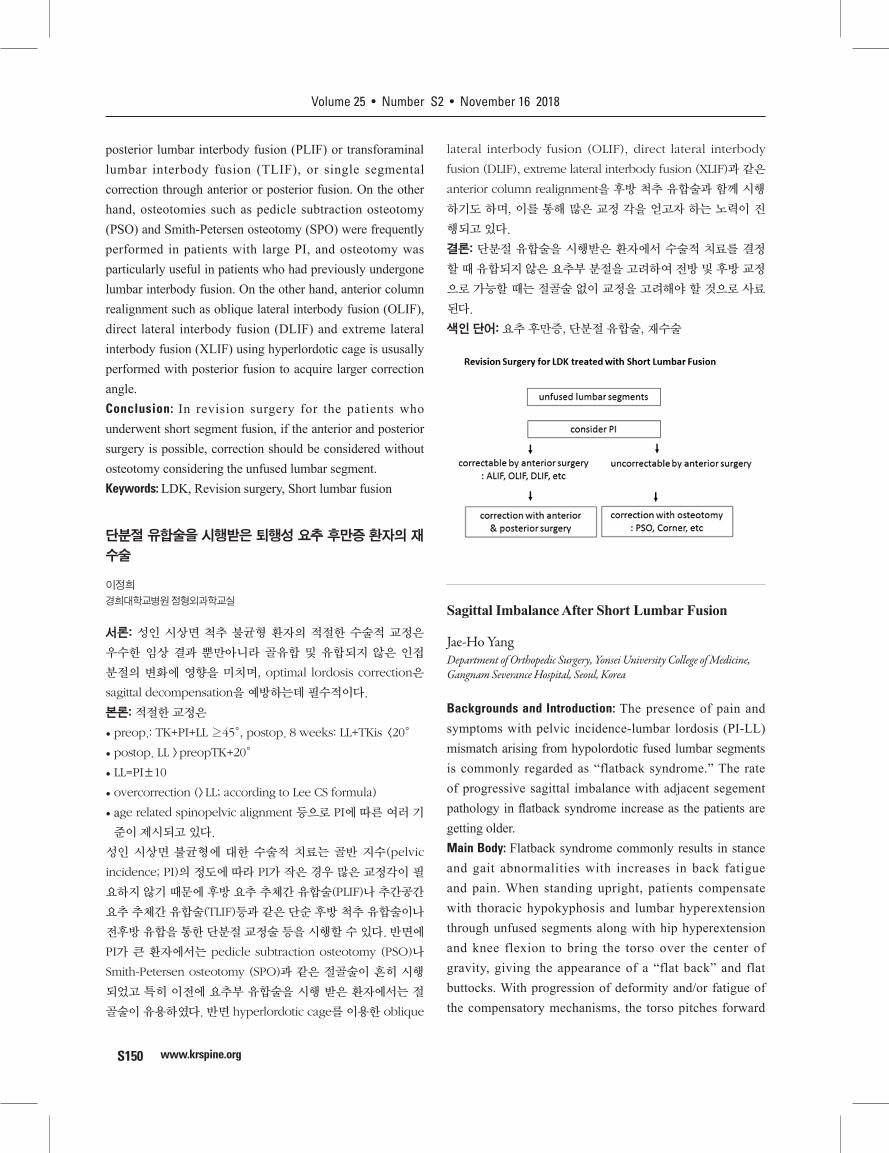
Revision Surgery for LDK Treated with Short Lumbar Fusion
Jung-Hee LeeDepartment of Orthopedic Surgery, Kyung Hee University, Hospital, Seoul, Korea
Backgrounds and Introduction: Appropriate surgical correction of adult spinal sagittal imbalance patients affects not only good clinical results but also bone union and changes in adjacent unfused segment, and optimal lordosis correction is essential to prevent sagittal decompensation.Main Body: Appropriate surgical correction• preop.: TK+PI+LL ≥ 45°, postop. 8 weeks: LL+TKis <20°• Postop. LL> preopTK+20°• LL=PI±10• overcorrection (>LL according to Lee CS formula)• age related spinopelvic alignment and other criteria have
been proposed for PI.Surgical treatment of adult spinal sagittal imbalance is based on the degree of pelvic incidence (PI), and because small amount of PI does not requires large correction angles,
Volume 25 • Number S2 • November 16 2018
www.krspine.orgS150
posterior lumbar interbody fusion (PLIF) or transforaminal lumbar interbody fusion (TLIF), or single segmental correction through anterior or posterior fusion. On the other hand, osteotomies such as pedicle subtraction osteotomy (PSO) and Smith-Petersen osteotomy (SPO) were frequently performed in patients with large PI, and osteotomy was particularly useful in patients who had previously undergone lumbar interbody fusion. On the other hand, anterior column realignment such as oblique lateral interbody fusion (OLIF), direct lateral interbody fusion (DLIF) and extreme lateral interbody fusion (XLIF) using hyperlordotic cage is ususally performed with posterior fusion to acquire larger correction angle.Conclusion: In revision surgery for the patients who underwent short segment fusion, if the anterior and posterior surgery is possible, correction should be considered without osteotomy considering the unfused lumbar segment.Keywords: LDK, Revision surgery, Short lumbar fusion
단분절 유합술을 시행받은 퇴행성 요추 후만증 환자의 재
수술
이정희
경희대학교병원 정형외과학교실
서론: 성인 시상면 척추 불균형 환자의 적절한 수술적 교정은
우수한 임상 결과 뿐만아니라 골유합 및 유합되지 않은 인접
분절의 변화에 영향을 미치며, optimal lordosis correction은
sagittal decompensation을 예방하는데 필수적이다.
본론: 적절한 교정은
• preop.: TK+PI+LL ≥45°, postop. 8 weeks: LL+TKis <20°
• postop. LL >preopTK+20°
• LL=PI±10
• overcorrection (>LL; according to Lee CS formula)
• age related spinopelvic alignment 등으로 PI에 따른 여러 기
준이 제시되고 있다.
성인 시상면 불균형에 대한 수술적 치료는 골반 지수(pelvic
incidence; PI)의 정도에 따라 PI가 작은 경우 많은 교정각이 필
요하지 않기 때문에 후방 요추 추체간 유합술(PLIF)나 추간공간
요추 추체간 유합술(TLIF)등과 같은 단순 후방 척추 유합술이나
전후방 유합을 통한 단분절 교정술 등을 시행할 수 있다. 반면에
PI가 큰 환자에서는 pedicle subtraction osteotomy (PSO)나
Smith-Petersen osteotomy (SPO)과 같은 절골술이 흔히 시행
되었고 특히 이전에 요추부 유합술을 시행 받은 환자에서는 절
골술이 유용하였다. 반면 hyperlordotic cage를 이용한 oblique
lateral interbody fusion (OLIF), direct lateral interbody
fusion (DLIF), extreme lateral interbody fusion (XLIF)과 같은
anterior column realignment을 후방 척추 유합술과 함께 시행
하기도 하며, 이를 통해 많은 교정 각을 얻고자 하는 노력이 진
행되고 있다.
결론: 단분절 유합술을 시행받은 환자에서 수술적 치료를 결정
할 때 유합되지 않은 요추부 분절을 고려하여 전방 및 후방 교정
으로 가능할 때는 절골술 없이 교정을 고려해야 할 것으로 사료
된다.
색인 단어: 요추 후만증, 단분절 유합술, 재수술
Sagittal Imbalance After Short Lumbar Fusion
Jae-Ho YangDepartment of Orthopedic Surgery, Yonsei University College of Medicine, Gangnam Severance Hospital, Seoul, Korea
Backgrounds and Introduction: The presence of pain and symptoms with pelvic incidence-lumbar lordosis (PI-LL) mismatch arising from hypolordotic fused lumbar segments is commonly regarded as “flatback syndrome.” The rate of progressive sagittal imbalance with adjacent segement pathology in flatback syndrome increase as the patients are getting older. Main Body: Flatback syndrome commonly results in stance and gait abnormalities with increases in back fatigue and pain. When standing upright, patients compensate with thoracic hypokyphosis and lumbar hyperextension through unfused segments along with hip hyperextension and knee flexion to bring the torso over the center of gravity, giving the appearance of a “flat back” and flat buttocks. With progression of deformity and/or fatigue of the compensatory mechanisms, the torso pitches forward
Journal of Korean Society of Spine Surgery Specials
www.krspine.org S151
resulting in worsening back pain and fatigue. Prevention of iatrogenic flatback is critical, and surgeons should consider the regional and global alignment consequences for single and/or multilevel lumbar fusions. Every attempt should be made to maximize segmental lordosis when performing a lumbar fusion. Adequate preoperative imaging and planning is essential for treatment of iatrogenic flatback with adjacent segment pathology. Nonoperative management for sagittally imbalanced (SVA>5 cm) flatback syndrome is frequently unsuccessful. Despite significant complication rates, surgical management to recreate LL using interbody fusions and/or osteotomies can significantly improve quality of life. Conclusion: Proper correction surgery with decompression in iatrogenic flatback with adjacent segment pathology is important for preventing repetative surgery and improving patients’ clincal outcome. Keywords: Degenerative lumbar disease, Sagittal imbalance, Adjacent segment pathology, Lumbar fusion surgery
요추의 단분절 유합술 후 시상면 불균형
양재호
연세대학교 강남세브란스병원 정형외과학교실
서론: 요추 단분절 유합 수술 후 유합된 요추 분절의 전만증 감소
로 인한 골반지수-요추전만 불일치(PI-LL mismatch) 소견과 함
께 통증과 증상이 있는 경우 이를 흔히 Flatback syndrome 으로
언급하여 왔다. 수술 후 시간이 경과함에 따라 인접분절병변과
함께 시상면 불균형이 초래되는 경우가 점차 증가하고 있다.
본론: Flatback syndrome은 허리의 피로도와 통증을 증가시
켜 흔히 서있는 자세와 보행의 장애를 초래한다. 이 경우 똑바
로 선 자세에서, 환자는 흉추의 후만이 감소하고 고관절의 과신
전과 슬관절 굴곡과 함께 유합되지 않은 요추 분절이 과신전되
면서 몸통의 무게 중심을 맞추어 보상한다. 그러나 시간이 경과
함에 따라 이러한 보상기전이 더이상 작용하지 못할 경우 변형
은 진행하게 되어 몸이 앞으로 구부러지게 되며 이는 환자의 통
증을 증가 시키게 된다. 따라서 많은 연구자들이 요추의 단분절
유합술을 시행할 경우 전반적인 척추의 정렬에 대해 반드시 고
려하고, 각 유합분절에서의 lordosis를 최대한 만들려는 노력
을 기울일 것을 권고하고 있다. 또한 인접분절 병변이 발생한
경우, 시상면 불균형에 대한 평가가 수술전 반드시 이루어져야
한다. 여러 문헌에서 시상면 불균형이 동반되어 있는 flatback
defromity (SVA>5 cm) 환자에서 비수술적 치료는 효과적이
지 않은 것으로 보고되고 있다. 비록 수술 후 발생하는 합병증
이 많음에도 불구하고 척추체간 유합술이나 절골술을 이용하여
lumbar lordosis 를 다시 만들어 주는 변형 교정술이 환자의 삶
의 질을 향상 시키는 것으로 사료된다.
결론: 요추의 단분절 유합술 후 인접분절병변으로 인한 협착증
으로 재수술이 필요한 환자에서 시상면 불균형이 초래되는 경
우 후방감압술 및 유합술의 연장과 동시에 시상면 불균형을 교
정하는 것이 추가적인 재수술을 막고 장기적인 환자의 예후와
임상증상을 호전시키는데 중요하다.
색인 단어: 퇴행성척추질환, 시상면 불균형, 인접분절병변, 척추
유합술
Nonunion in Adult Deformity Surgery
Young-Hoon Kim, Kee-Yong Ha, Ki-Ho Na, Sang-Il Kim, Hyung-Youl Park, Hyung-Ki MinDepartment of Orthopedic Surgery, Seoul St. Mary’s Hospital, College of Medicine, The Catholic University of Korea, Seoul, Korea
Backgrounds and Introduction: Since the introduction of spinal arthrodesis by Hibbs and Albee in 1911, the failure to achieve a solid fusion has been a major concern in spinal surgery. Especially, pseudarthrosis remains a prevalent complication following adult deformity surgery. Because corrective surgery spans multiple vertebral levels, can be performed in multiple stages, and may require osteotomies with extensive removal of posterior elements. Here, we present several cases successfully treated for pseudarthrosis after long-segment fusion in adult spinal deformity.Main Body: Recently, meta-analysis calculated an incidence of 6.3% (95% CI 4.3–8.2%) for pseudarthrosis in adult spinal deformity. Suggested risk factors for pseudarthrosis include age over 55, construct length greater than 12 segments, smoking, thoracolumbar kyphosis greater than 20°, and fusion to the sacrum. Mechanisms for pseudarthrosis can be largely divided into two categories: inadequate fixation and inadequate bone formation. Instrumentation provides structural support and immobilization until fusion can be achieved, while insufficient bone formation can be attributed to inadequate grafting material or technique, or decreased biological activity due to patient characteristics. Regarding the clinical and radiological outcomes, pseudarthrosis can lead to reduced quality of life, loss of correction, and progression of deformity. Revision surgery is recommended for the patients with symptomatic pseudarthrosis because

Volume 25 • Number S2 • November 16 2018
www.krspine.orgS152
symptoms can be improved significantly after achieving solid union. Conclusion: Although the pseudarthrosis following long arthrodesis is inevitable, we should consider the characteristics of pseudarthosis and perform appropriate managements to achieve solid fusion in adult deformity surgery. The clinical outcomes of pseudarthrosis are known to be worse compared with solid union. Therefore, revision surgery is recommended for the patients with symptomatic pseudarthrosis.Keywords: Spinal curvatures, Pseudarthrosis, Outcomes assessment, Reoperation
성인 척추 변형 수술에서 불유합
김영훈, 하기용, 나기호, 김상일, 박형열, 민형기
가톨릭대학교 서울성모병원 정형외과학교실
서론: Hibbs와 Albee가 1911년 척추 유합술을 소개한 이후, 견
고한 척추 유합을 얻지 못하고 불유합이 발생하는 것이 척추 수
술의 주요한 우려였다. 성인 척추 변형에서 불유합은 특히 문제
가 되는데 이는 다 분절 유합술, 광범위한 절골술이 필요한 변형
수술의 특징과 고령, 골다공증과 같은 환자 요인이 원인이 된다
고 할 수 있다. 본 연구에서 저자들은 성인 척추 변형에 대해 장
분절 유합술 이후 발생한 척추 불유합을 효과적으로 치료한 증
례를 발표하고자 한다.
본론: 최근 발표된 메타 분석 논문은 성인 척추 변형 수술에서
불유합의 발생률을 6.3%로 보고하였다. 불유합의 위험 인자로
는 55세 이상의 연령, 12 분절 이상, 흡연, 20도 이상의 흉요추
후만각, 천골까지 유합술을 시행한 경우가 제시되고 있다. 불유
합이 발생하는 기전은 크게 두 가지로, 적절한 고정이 이루어지
지 않거나 불충분한 골 형성이다. 기구가 골 유합이 이루어질 때
까지 충분한 구조적인 지지와 고정을 제공하는데 실패하거나,
골 유합 재료나 수술 술기, 생물학적인 환자 특성으로 인한 불
충분한 골 형성이 이루어지지 않으면 불유합이 발생하는 것이
다. 불유합의 임상적인 결과와 관련해서 많은 문헌에서 불량한
것으로 보고 하고 있으며, 삶의 질이 감소하고, 척추 교정이 소
실되거나 변형이 더 진행할 수 있다. 따라서 증상이 있는 불유합
환자의 경우에는 견고한 골유합을 목표로 재수술이 권장된다.
결론: 성인 척추 변형에서 장분절 유합술 후 불유합은 피하기 힘
든 합병증이지만, 견고한 유합을 얻기 위해 척추외과 의사는 불
유합의 특징을 잘 이해해야 하며, 이에 대한 적절한 치료를 시
행할 수 있어야 한다. 성인 척추 변형 수술에서 불유합의 임상적
결과는 불량하기 때문에 증상이 있는 환자에 대해서는 재수술
을 고려할 필요가 있다.
색인 단어: 불유합, 성인 척추 변형, 재수술
Basic Science
Application of 3-D Printing to Spine Surgery
Hyun-Jin ParkDepartment of Orthopaedic Surgery, Hallym University, Kangnam Sacred Heart Hospital, Seoul, Korea
Backgrounds and Introduction: 3D printing has attracted attention as one of the technologies that will lead the “Fourth industrial revolution”, with many applications in various fields. As 3D printing can better produce three-dimensional constructs with more complicated inner/outer structures than conventional manufacturing methods, its virtue is invaluable in the biomedical industry. Especially regarding spine surgery, 3D printing is exceptionally useful considering the complexity of the anatomy of the spine and its surrounding structures.Main Body: 3D printing has various applications in medicine, mainly in surgical fields such as orthopaedic surgery, spinal surgery, oral- maxillofacial surgery and cardiovascular surgery. 3D printers can create patient-specific models for preoperative planning in such subspecialties, and promising results are published reporting less operative time, radiation exposure, and blood loss. The models are also effective in resident training and patient education as well. Such models are also beneficial in the more advanced usage in preparing implants before the actual surgery. For example, in pelvic fractures, the pelvic anatomy and the bony contour shows significant variance from person to person. Therefore, it is necessary to dissect the fracture site intraoperatively and contour the metal plate accordingly. It is not only time consuming, but also technically difficult as exposure is limited. Methods to shorten the operative time by pre-bending the metal plate to the 3D printed contralateral intact pelvic bone before surgery have been published. Advanced methods of direct 3D printing of joint replacement prosthetics or supports directly with metal have also been developed. In addition, the technology of bio-printing is being researched, utilizing collagen scaffolds and lamination of cultured chondrocytes. Further possible advancements
Journal of Korean Society of Spine Surgery Specials
www.krspine.org S153
include artificial tissues and organs for transplantation. The use of 3D printing as an area of spinal surgery is further described.(1) Preoperative planning and simulationCompared to other structures, the vertebrae have many anatomical variations and often do not have normal anatomical structures due to deformity. 3D modeling especially beneficial in the surgery of C1-2, lumbar revisions, and scoliosis patients. A thorough three-dimensional understanding of such anatomically complex structures can shorten operative time and blood loss. It is also invaluable in resident training, patent education and satisfaction.(2) Patient-specific implants and surgical guide systemThe use of 3D printed metal substitutes for the bone defect after tumor removal have been published with good results. When commercially available cages are inadequate to produce proper lumbar lordosis, patient-specific designs are reported to be promising. Patient-specific surgical guides are also developed. Such drill/screw guides of the precise angle and screw length can be used when operating on deformed or anatomically complex spine. However, as it can facilitate easy screw insertion, a thorough dissection of the soft tissues is needed, and so is the increase in operative time and bleeding.Conclusion: 3D printing will be more commonly used in medicine, especially in spine surgery. Future applications of ongoing research of bio-printing can be promising for the regenerative treatments in spinal cord injury and degenerative disc disease. The currently reported patient-specific implants have significant potential in “personalized” medicine, but commercialization is yet underway. We as spine surgeons should all prepare to embrace this new technology and value creation.Keywords: 3D printing, Preoperative simulation, Patient-specific implants, Bioprinting
척추 수술에서 3-D프린팅의 적용
박현진
한림대학교 강남성심병원 정형외과학교실
서론: 3D 프린팅은 최근 여러 산업현장에서 관심을 가지고 있는
기술로서 4차혁명을 이끌 기술 중 하나로 주목받고 있다. 3D 프
린팅 기술은 기존 생산 제조 기술에 비해 내/외부 구조가 좀 더
복잡한 3차원 형상을 비교적 간단하게 제작할 수 있다는 장점을
가지고 있고 이러한 제조 기술의 특성상 바이오 메디컬 분야에
서 그 활용가치가 높게 평가되고 있다. 특히 척추 분야에서는 척
추의 해부학적 구조의 복잡성 뿐만 아니라 주변 구조물들의 미
세한 위치 관계 등을 고려 할 때 3D 프린팅의 여러 장점이 더욱
유용하게 활용될 것이라 기대된다.
본론: 3D 프린팅 기술은 현재 의료분야에서 다양하게 활용되
고 있는데 정형외과 수술, 척추수술, 구강악안면 수술, 심장 혈
관 수술 등 주로 수술분야에서 활용되고 있다. 3D 프린터를 통
해 환자의 수술전 모델을 만들어 수술전 계획을 세우는 데 도움
이 될 수 있다. 심혈관 수술, 치과수술, 안면수술, 정형외과 수술
등에서 3D 프린팅 된 모델을 통해 수술전 계획을 세우고 이를
통해 수술시간의 절감, 방사선 노출 시간의 절감, 출혈량의 감
소 등으로 인해 수술 후 결과가 향상되었다는 연구들이 발표되
고 있다. 또한 수술전 미리 제작된 모델을 이용하면 전공의의 교
육 자료로서 유용할 뿐만 아니라 환자에게 수술전 환자 상태를
설명 할 때 더욱 용이하게 사용할 수 있다. 이보다 조금 더 발전
한 단계는 수술전에 수술에 사용할 기구를 환자 맞춤형으로 만
드는 것이다. 예를 들어 골반 골절의 경우 사람마다 골반의 모양
이 다양하고 골절되는 양상과 부위가 각기 달라 환자에게 맞는
금속판을 제작하기 어렵다. 따라서 수술장에서 골절 부위를 열
고 이에 맞게 금속판을 변형하여 적용하게 되는데 시간이 소요
될 뿐만 아니라 노출 부위가 제한적이여서 정확히 맞추기 어려
운 경우가 많다. 이를 미리 3D 프린팅으로 골반골을 복원후 금
속판을 맞추어 수술시간을 단축하는 방법들이 발표되었다. 이
보다 더 발전하여 환자에게 맞는 인공관절 치환물이나 지지체
로서의 금속을 직접 3D 프린팅 하는 방법도 발표되고 있다. 또
한 3D프린터의 잉크재료로서 세포를 이용하는 바이오 프린팅
기술을 이용해 collagen 등으로 scaffold를 만들고 배양된 연골
세포를 적층하는 방법등이 연구 개발 되고 있으며 더 나아가 인
공 조직, 인공 장기를 만들어 생체이식을 시도하는 기술도 연구
되고 있다. 이와 같은 3D 프린팅의 활용을 척추수술의 영역으로
적용하여 현재까지의 연구결과들을 소개하면 다음과 같다.
(1) 수술 전 디자인 및 수술 전 시뮬레이션
타 영역에 비교하여 척추는 해부학적으로 많은 복잡성을 가지
고 있고 척추변형으로 인해 정상적인 해부학적 구조를 가지지
않은 경우가 많다. 특히 경추1-2번 수술이나 요추부의 재수술,
척추 측만증 환자 등 복잡한 해부학 구조를 가진 부위의 수술에
서 3D 프린팅으로 수술전 모델을 만들어 미리 환자의 3차원 구
조를 충분히 숙지하고 수술한 경우 수술시간과 출혈량의 감소
라는 장점들이 있었다고 보고 하였다. 또한 함께 참여하는 전공
의들의 수술 이해도의 증가와 교육적인 측면에서 도움이 될 수
있으며 환자에게 직접 3D 프린팅된 모델을 보여주어 설명을 하
였을 때 환자의 이해도와 만족도가 더욱 증가 하였다는 보고도
있다.
Volume 25 • Number S2 • November 16 2018
www.krspine.orgS154
(2) 환자 맞춤형 임플란트 , 수술 가이드 시스템
척추종양의 수술시 심한 골결손이 예상되는 수술에서 3D 프린
터를 이용해 환자의 골결손 부위에 맞는 적절한 모양의 금속 치
환물을 설계하고 제거된 골결손 부위에 삽입 고정하여 만족할
만한 결과를 보였다고 보고되고 있다. 또한 하부 요추의 일반적
인 유합수술에서 전방 골결손이나 변형등으로 상용화된 케이지
의 사용으로는 적절한 요추 전만각을 만들기 어려운 경우 환자
맞춤형 케이지를 이용하여 만족할 만한 수술결과를 얻었다는
보고도 있다. 환자 맞춤형 수술가이드 system도 보고 되고 있다.
앞에서 언급한 변형이 심하거나 복잡한 해부학적 구조를 가지
는 척추수술에서 미리 정확한 나사못의 삽입 각도와 길이 등을
고려한 screw insertion guide를 만들어 수술에 적용하는 방법
이다. 비록 스크류 삽입은 용이하게 할 수 있으나 이를 적용하기
위해 연부조직을 깨끗이 제거 해야 하고 그로 인한 출혈량 증가
와 수술시간의 증가는 단점으로 우려되고 있다.
결론: 3D 프린터는 추후 미래를 이끌어 갈 산업으로 바이오 메
디컬 분야 , 특히 척추분야에서의 활용도가 높을 것이며 향후 바
이오 프린팅 기술을 척추분야에 적용하여 척수손상 환자의 재
생치료, 퇴행성 추간판 질환의 재생치료 등에 이용가능 할 것으
로 보이며 현재 연구중인 기술도 있다. 현재 보고되고 있는 환자
맞춤 임플란트 기술은 개인 맞춤형 의료 시장에서 상당한 가능
성을 가지고 있을 것으로 보나 아직 그에 대한 상용화 기술이나
여건은 부족한 상태이다.
3D 프린팅은 아직 많은 가능성을 가지고 있는 기술로서 새로운
가치 창출을 위한 준비를 지금부터라도 철저히 해야 될 것이다.
색인 단어: 3D 프린팅, 수술 전 시뮬레이션, 환자 맞춤 임플란
트, 바이오 프린팅
AR VR in Spine Surgery
Young-Yul KimDepartment of Orthopedic Surgery, Daejeon St. Mary’s Hospital, College of Medicine, The Catholic University of Korea, Duejeon, Korea
Backgrounds and Introduction: AR (Augmented Reality) and VR (Virtual Reality) have started from the initial simple experiential elements and have expanded their indications to various fields recently. VR is expressed as a virtual reality, which is rapidly evolving due to the recent development of motion sensing devices such as haptic function, 3D graphic technology, 360-degree camera, AR is expressed as augmented reality, it’s use and possibilities are emerging.
The purpose of this study is to investigate the development of these patients and their role in the field of spinal surgery.Main Body: In the case of VR, devices and programs have been developed as surgical training methods such as laparoscopic surgery in the present surgical field. Devices for raising the accuracy of the surgical procedure in the virtual reality monitor by tracking the movement of the laparoscopic surgeon have been developed and commercialized. Similar training equipment has been introduced in the field of arthroscopy, which is likely to lead to the development of devices that help train endoscopic surgery in spinal surgery. In the AR area, laparoscopic anatomical structures have been developed to be displayed in the laparoscopic monitor by contrasting with existing registered CT and MRI structures, which is expected to be applicable to spine surgery in the future.Conclusion: At present, the development of devices due to AR and VR is inadequate in the spine field, but there are many technologies that can be commercialized sufficiently if the demand of the device is lowered and the accurate surgical guide is increased. It was judged to be a field in which suffering could have a great influence on the area of surgery.Keywords: Virtual reality, Augmented reality, Spine surgery
척추수술에서의 가상. 증강 현실
김영율
가톨릭대학교 대전성모병원 정형외과학교실
서론: AR (Agumented Reality)와 VR (Virtural Reality)는 초기
단순 체험적 요소에서 시작해서 최근 그 적응증을 여러 분야로
확대하고 있는 영역이다. VR은 가상 현실이라 표현되며 이는 최
근에 발달되고 있는 motion sensing device인 haptic 기능과 3
차원 그래픽 기술, 360도 촬영 카메라 등의 발달로 급속한 발전
을 보이고 있으며 AR은 증강현실로 표현되며 최근 수술의 영역
에서도 그 사용과 가능성이 대두되고 있다. 이에 현재 이들의 발
달과 앞으로 척추 수술의 분야에서의 역할에 대하여 알아보고
자 한다.
본론: VR의 경우 현재 외과적 영역에서 복강경 등의 수술
training의 방법으로 기기와 프로그램이 개발되어 있으며, 복
강경을 잡은 술자의 위치 움직임을 추적하여 가상 현실 모니터
에서의 수술 과정의 정확도를 올리는 기기가 개발되어 상용화
되어 있다. 또한 관절경 분야에서도 이와 유사한 training장비
가 도입되어 있으며, 이는 척추 수술에서의 내시경적 수술의
training을 도와주는 기기의 개발로 연결될 수 있는 가능성이 있
Journal of Korean Society of Spine Surgery Specials
www.krspine.org S155
다. 또한 AR영역에서는 복강경의 해부학적 구조물들을 기존의
등록된 CT, MRI 구조물과 대조하여 복강경 모니터내에 표시가
될 수 있는 장비들이 개발되어 있으며, 이는 추 후 척추 수술 분
야에서도 적용 가능할 것으로 예상되고 있다.
결론: 현재 척추 분야에서는 AR, VR로 인한 기기의 개발은 미흡
한 상태이나 기기의 가격의 하락과 정확한 수술적 가이드에 따
른 수요가 증가하면 충분히 상용화할 수 있는 기술들이 많이 존
재하며 수술자의 술기를 초기의 training에서부터 고난이 수술
의 영역에까지 큰 영향을 미칠 수 있는 분야로 판단되었다.
색인 단어: 가상현실, 증강현실, 척추 수술.
Robotics in Spine Surgery
Sungmin KimDepartment of Medical Assistant Robot, Korea Institute of Machinery and Materials, Daegu, Korea
Backgrounds and Introduction: The interest of surgical robotic system has been increasing due to the success of da Vinci Surgical Robotic System. Because it’s verified that the surgical robotic system can provide more benefits comparing to the conventional method, lots of researcher and surgeon have interest in developing surgical robotic system and applying it to clinical environment.Main Body: The main technologies that can be applied to surgical robot system are surgical navigation using medical images and robotic arm using stable and precise control. Surgical navigation technology can be used to accurately identify the target area and the adjacency and to plan the path of the screw insertion, and to monitor the correct insertion of the screw. If bone resection is needed, such as laminectomy, medical image data can be used to plan the site of bone resection and monitor whether resection is being performed as planned during bone resection. Then, using a stable and precise robot arm, the screw can be inserted based on the surgical planning data, or bone resection can be performed. Particularly, the robot system does not perform this process automatically, and the surgeon can directly perform the operation using the robotic surgical tool. When the robotic surgical tool reaches the boundary of the resecting site, the virtual wall based on haptic technique makes help to the robotic surgical tool be kept in the resection area.Conclusion: It is expected that the surgical robot system
will enable more stable and precise spinal surgery. If the collaboration is carried out more actively between engineers to develop surgical robotic system and clinicians to apply it and to provide feedback to improve it, various types of spinal surgery robotic systems have appeared, and it would make the spinal surgery to be improved. Keywords: Spine surgery robot, Pedicle screw insertion, Laminectomy, Laminaplasty
척추 수술을 위한 수술 로봇 시스템
김성민
한국기계연구원, 의료지원로봇연구실
서론: 최근 수술에 있어 신기술의 활용을 통한 미세 침습 수술
등이 수술 시간의 단축, 환자의 고통 경감, 그리고 수술 예후의
향상으로 이어지며 큰 관심을 끌고 있다. 수술 로봇 시스템 또한
신기술의 한 축으로 다빈치 수술 로봇 시스템의 큰 성공으로 인
해 여러 임상 분야에서 수술 로봇 시스템 기술의 개발과 적용에
큰 관심을 가지기 시작하였다.
본론: 수술 로봇에 활용 가능한 기술로는 크게 의료 영상 등을
활용한 수술 네비게이션 기술과 로봇의 정교하고 정밀한 제어
기술을 활용한 로봇 암 기술이 있다. 수술 네비게이션 기술을 활
용하여 환부의 주변을 정확히 확인할 수 있고, 나사못 삽입 등의
경로를 계획하고 나사못의 삽입이 정확하게 이루어지고 있는지
를 모니터링 할 수 있다. 또한 후궁 절제술 등과 같이 뼈의 절제
가 필요한 경우, 의료 영상 데이터를 활용하여 뼈의 절제 부위
를 계획하고, 뼈의 절제가 이루어지는 동안 계획대로 절제가 이
루어지고 있는지 모니터링 할 수 있다. 그리고, 정교하고 정밀
한 로봇 암을 이용하여 수술 계획 데이터를 기반으로 나사못을
삽입한다거나, 뼈의 절제를 수행할 수 있다. 특히 이러한 과정
을 로봇 시스템이 자동으로 수행하지 않고, 의사가 직접 수술 도
구를 활용하여 수술을 수행할 수 있고, 가상 벽을 통하여 계획된
경로 혹은 절삭 부위의 경계에 도달하였을 경우 햅틱 피드백 등
을 통하여 계획 범위를 벗어날 수 있도록 유도할 수 있다.
결론: 위에서 언급된 수술 로봇 기술의 도입을 통하여 척추 수술
을 더욱 정교하게 수행할 수 있을 것으로 기대하며, 기술 개발과
임상적 적용과 피드백이 활발하게 이루어진다면, 오래지 않아
다양한 분야의 척추 수술 로봇 시스템이 등장하여 수술에 많은
도움을 줄 것으로 기대한다.
색인 단어: 척추 수술 로봇, 척추경 나사못 삽입술, 척추 후궁 절
제술, 척추 후궁 성형술
Volume 25 • Number S2 • November 16 2018
www.krspine.orgS156
Spine Pain
Current Update of Spinal Instability
Young Bae KimDepartment of Orthopedic Surgery, Veterans Healthcare Service System, Seoul, Korea
Backgrounds and Introduction: White and Panjabi defined clinical instability of the spine as the loss of the spine’s ability to maintain its patterns of displacement under physiologic loads so there is no initial or additional neurologic deficit, no major deformity, and no incapacitating pain. However it is controversial and not well understood.Main Body: In this symposium, we focused only on the degenerative lesions of the functional spinal unit (FSU). The spinal stabilizing system can be thought of as consisting of three subsystems: spinal column; muscles surrounding the spine; and motor control unit. The lumbar spine is known to be unstable when subjected to compressive loads of 100 N. However it supported a load of up to 1200 N without damage or instability when the load path was tangent to the spinal curve, or the follower load. An abnormal increase of motion in FSU can be found through analysis of angular and translational motion. The concept of a neutral and an elastic zone has offered many novel ideas including the posterior dynamic fixation of the lumbar spine. Historically, many methods have been used to identify the instability through radiographical examinations, but it has been difficult to show its dynamic characteristics. Recently several MR images including those from functional MRI have been reported and used.Conclusion: Although our current knowledge of instability is still far from satisfactory, studying biomechanical data and imaging features will shed more light on the concept of spinal instability.Keywords: Spine, Biomechanics, Degeneration, Instability
요추 불안정성의 이해
김영배
중앙보훈병원 정형외과학교실
서론: 척추의 불안정성은 ‘척추가 정상적인 생리적 부하에서 구
조적 손상이나 신경 또는 척수자극 없이 정상적인 기능을 수행
할 수 있는 능력을 상실한 경우’로 정의할 수 있다. 그러나 여러
저자들과 광범위한 대상 질환들로 인하여 불안정성의 정의와
기전에 대하여는 다양한 견해가 있다.
본론: 이 심포지엄에서는 요추에서 Functional spinal unit(이
하 FSU)의 퇴행성 병변으로 제한하여 논의하고자 한다. 요추의
안정성은 추체와 주변 근육 그리고 신경의 조절 등의 세 요소에
의하여 유지된다. 단일 요추는 100N의 수직 압축력에도 파괴되
지만, 전체 요추는 주변 근육의 동적 안정성과, 하중경로를 요
추의 만곡의 접선 경로를 따라 조절하는 종동력(follower load)
을 통하여 이론상 3000-5000N까지 견딜 수 있다. FSU의 기능
적 불안정성에 대하여는, 각운동(angular motion)과 전위 운동
(translational amotion)에 분석을 통하여 비정상적인 증가를 찾
을 수 있다. 운동 범위 내에서 중립 지역(neutral zone)과 탄성
지역의 존재는 이 후 요추의 후방 연성 고정술의 이론적 근거가
되었다. 영상의학적 검사를 통하여 불안정성을 확인하기 위하
여 역사적으로 많은 방법들이 이용되어왔으나, 동적인 질환의
특징을 파악하기에는 어려움이 있었다. 이를 위하여 단순 방사
선 검사를 포함하여 기능적 MRI imaging 등의 기법이 개발되어
사용되고 있다.
결론: 불안정성에 대한 연구가 진행됨에 따라 이들을 바탕으로
향 후 임상에서 치료와 연구에 도움이 되는 자료들이 될 것으로
사료된다.
색인 단어: 척추, 생체역학, 퇴행, 불안정
Instability with Symptoms
Tae-Keun AhnDepartment of Orthopedic Surgery, CHA Bundang Medical Center, CHA University, Seongnam, Korea
Backgrounds and Introduction: The spinal instability is a state in which the spine lose their normal mechanical stiffness and have abnormal movement beyond the normal range of motion. If the physical characteristics of the vertebral segments are lost, the range of motion is increased or abnormal motion pattern is caused by external force within the physiological range or minor environmental changes, resulting in pain and neurological impairment . Main Body: According to Kirkaldy-Willis cascade, the spine undergoes a series of stages of dysfunction, instability, and restabilization as degenerative changes progress. In this continuous process, degeneration of the disc and degenerative changes of the posterior facet joint made the spine to enter the instability and cause spondylolisthesis.
Journal of Korean Society of Spine Surgery Specials
www.krspine.org S157
However, after the stage of instability, the spine goes through a restorative period and most of them do not require surgical fixation because most symptomatic stenosis occurs at this time. However, many patients who come to the clinic have symptoms associated with the stage of instability and do not respond to conservative treatment, so surgical treatment with fusion is frequently required. Until now, there has been no standard for diagnosing instability reflecting the physical characteristics of the individual spine with various pain and neurological symptoms. The widespread radiologic criteria about instability includes a translation of more than 3 mm or a angulation of more than 10 degrees on a dynamic simple radiograph of the sagittal plane, but it can judge only radiologically instability without consideration of clinical symptoms. Since this radiological criteria is less relevant to the symptoms, other findings or factors suggesting the instability of the spine associated with the symptoms should be considered. Through the literature review, other findings related to an instability with clinical symptoms are as follows, in which the disc height of spondylolisthesis has more than 6.5 mm, the articular angle of facet joint has more than 50 degrees, the facet joint has an increased articular fluid, or the endplate demonstrates Modic type 1change in MRI. Conclusion: Unstable spine can cause damage to the spine itself and surrounding tissue through abnormal and unbalanced movements, which are associated with symptoms. In particular, patients with clinical symptoms at the time of injury of anatomical structures during degenerative cascade are considered to have a poor prognosis if they are associated with imaging findings suggesting instability. In those patients, more aggressive treatment is recommended like surgery.Keywords: Degeneration, Instability, Symptom
증상을 동반한 불안정성 척추
안태근
차의과학대학교 분당차병원 정형외과학교실
서론: 척추에서 정상적인 기계적 강성도를 소실하여 정상 운동
의 범위 이상 운동하는 상태를 척추의 불안정성 이라고 한다. 척
추 분절의 강성도 등의 물리적 특성이 소실되면 생리적 범위내
의 외력 혹은 사소한 외적 환경의 변화에 의하여서도 운동범위
가 증가되거나 비정상적 운동 양상을 초래하게 되고 이로 인해
통증이 유발되고 신경학적 손상이 진행되는 상태가 발생할 수
있게 된다.
본론: Kirkaldy-Willis의 cascade에 따르면 척추는 퇴행성 변화
가 진행됨에 따라 기능 부전기, 불안정기 및 재안정기의 연속적
인 단계를 거치게 된다고 주장하였다. 이러한 연속적인 과정에
서 디스크의 퇴행 및 후관절의 퇴행성 변화는 척추를 불안정기
에 진입하게 만들고 전방 전위증을 유발한다고 보고하였다. 하
지만 이 시기가 지나면 척추는 재안정기를 거치게 되고 증상을
유발하는 협착증은 이 시기에 발생하기 때문에 대부분에서는
수술적 고정을 요하지 않는다고 하였다. 하지만 임상에서 접하
게 되는 환자들은 불안정기에 증상이 심하게 동반되어 보존적
치료에도 반응하지 않아 유합술을 동반한 수술적 치료를 요하
는 경우가 빈번하다. 통증 및 신경학적 증상이 동반되어 있는 다
양한 척추 중에서 환자 개개인의 신체적 특성을 반영하여 불안
정성을 진단하는 기준은 아직 존재하지 않으며 통상적으로 시
상면의 역동적 단순 방사선 사진상 3 mm 이상의 전위 혹은 10
도 이상의 각형성이 있을 때 방사선학적으로 불안정하다라고
판정을 하고 있다. 하지만 이러한 방사선학적 기준은 증상과의
연관성이 떨어지기 때문에 실제 임상에서는 증상과 연관이 있
는 척추의 불안정성을 시사하는 다른 소견들을 함께 고려해야
할 것이다. 문헌 고찰상 증상과 연관이 있는 불안정성 척추를 시
사하는 소견에는 6.5 mm 이상의 추간판 높이 혹은 50도 이상
의 후관절 각도를 가진 척추 전방 전위증, 비대칭 후관절 및 후
관절의 관절액 증가, 시상면 방사선 사진상 3 mm 이상의 전위,
Modic type 1에 해당하는 추제의 변화 등이 보고 되어 있다.
결론: 불안정한 척추는 비정상적이고 불균형적인 움직임을 통
하여 척추 및 주변 조직에 손상을 초래할 수 있고 이는 증상과
연관된다. 특히 손상이 초래되는 시점에서 환자들은 임상 증상
을 호소하게 되고 이러한 증상이 불안정성을 시사하는 영상학
적 소견과 연관이 있는 경우에는 예후가 불량할 수 있어 적극적
인 치료가 필요할 것으로 사료된다.
색인 단어: 퇴행, 불안정성, 증상
Instability Without Symptoms
Jin-Man ChoiDepartment of Orthopedic Surgery & Spine Center Allbarun Seoul Hospital, Seoul, Korea
Backgrounds and Introduction: Many cases of spinal pathology associated with instability shows mild complaints compared with diagnostic imaging. In recent years, due to insurance problems, simple decompressive procedures without fusion
Volume 25 • Number S2 • November 16 2018
www.krspine.orgS158
are widely performed. However, instability rarely arises if appropriate patient group are selected. Main Body: The stability of the spine is achieved through the harmonization of passive, active, and neural control systems. Even if a problem arises in one of the system, it can prevent the problem of instability through compensation of other systems. There are reports that state that when activating the muscles the neutral zone is not affected when the passive system is damaged. The neuromotor system is activated and the recruitment and force of the muscles around the spine are increased. In Kirklandy-Willis Farfan’s model, the stiffness of spine increases with time. Instability of the disc and facet joint is increased in the initial stage, but when the degeneration increases beyond a certain level, the spinal segment motion decreases. In addition, there are reports that the problem of instability does not occur when the appropriate patient group is not combined with the fusion procedure. Pchelmann reported the results of total faceectectomy of the intervertebral foramen stenosis in the 2-year follow-up; revision fusion was performed in only 6.95% of 222 patients. Inui et al. also conclude that simple decompression without fusion has not caused instability in the group with a displacement of more than 5 mm.Conclusion: New evaluation criteria should be prepared given that the instability and its effects are exaggerated, and the evaluation of instability is limited only by the current simple radiological imaging.Keywords: Spinal instability, Postoperative instability, Decompression
무증상적 척추불안정성
최진만
올바른서울병원 정형외과/척추센터
서론: 불안정성을 동반한 척추 질환의 많은 경우에서 영상학적
소견에서 보이는 것과 달리 경미한 증상만 호소하는 경우를 많
이 경험하게 된다. 최근에는 보험 삭감 등의 이유로 인해 유합술
없이 단순감압술만 시행하는 경우가 있는데, 적절한 환자군 선
택시 술후 불안정성의 문제가 발생되는 경우가 많지 않음을 보
는 등 불안정성과 증상을 직접적으로 연계시키기 어려운 경우
가 많다.
본론: 척추의 안정성은 수동적, 능동적, 신경운동 시스템의 조
화를 통해 이뤄지며 정적 구조물인 수동적 시스템의 문제가 초
래되어도 여타의 시스템의 보상을 통해 불안정성의 문제를 방
지할 수 있다. 실제로 수동적 시스템에 손상을 가해 인위적인 불
안정성을 유발한 후 척추 주변 근육을 활성화할 경우 중립 지역
(neutral zone)의 범위가 손상전후 차이가 없다는 보고들이 존
재하며 수동적 시스템의 손상시 신경운동 시스템이 활성화 되
어 척추 주변 근육의 recruitment 및 force가 증가됨 또한 밝혀
져 있다. 이뿐 아니라, Kirkaldy-Willis Farfan 모델에서 보듯 불
안정성이 발생하였어도 시간이 흐름에 따라 척추 주변 조직의
stiffness가 증가되고 척추의 안정성이 증가됨 또한 주지의 사실
이다. 추간판 및 후관절의 퇴행이 진행할 경우 초기에는 불안정
성이 증가되나 일정 수준 이상으로 퇴행이 증가할 경우 오히려
척추 분절 운동성과 불안정성이 감소되는 양상이 이를 뒤받침
한다. 또한 적절한 환자군을 선택하여 감압술을 시행하였을 경
우 유합술을 병행치 않았어도 술후 불안정성의 문제가 발생되
지 않았다는 보고들도 다수 존재한다. Pchelmann은 추간공의
협착을 전후관절절제술(total facectectomy)를 시행하여 치료
한 결과를 보고하였는데 2년 추시 중 재차 유합술을 시행하였던
경우는 총 222예 중 6.95% 불과하다 하였으며 Inui 등은 퇴행성
전방전위증 환자군을 후방구조물에 유의하면서 단순감압술을
시행하였을 때 5 mm 이상의 전위를 보이는 군에서도 불안정성
에 관한 문제가 유발되지 않았음을 보고하였다
결론: 불안정성 및 이로 인한 영향은 과장된 측면이 많으며 현재
의 단순방사선학적 영상 진단만으로는 불안정성의 평가는 제한
적일수 밖에 없으며 이에 대한 새로운 평가 및 치료기준이 마련
되어야 할 것으로 보인다.
색인 단어: 척추 불안정성, 술후 불안정성, 단순감압술
Axial Back Pain After Uneventful Decompression Spine Surgery
Chang-Su KimDepartment of Orthopedic Surgery, Kosin Gospel Hospital, College of Medicine, The Kosin University, Busan, Korea
Backgrounds and Introduction: Axial back is term to describe the pain occuring around the vertebral and paravertebral area without radiating pain. It is a pain syndrome, combining both nociceptive and neuropathic pain component simultaneously. It is considered as uneventable surgery when decompression was successfully performed without mistake at appropriate location and range which surgeons had planned. Operators can be disappointed when patients complain an axial back-pain after uneventable surgery. Several studies have reported that patients with axial back pain might have a worse
Journal of Korean Society of Spine Surgery Specials
www.krspine.org S159
prognosis compared to radiating pain. Here, we present the probable causes of axial back pain after decompression.Main Body: Discectomy is the treatment of choice for patients with disc herniation and decompression is for patients with spinal stenosis. Disc herniation occurs in young age. Realtively few patients have an instability on affected level with disc herniation. Appropriate technique is to resect the herniated nucleus after laminectomy and to irrigate on affected level depending on operators. Most patients with spinal stenosis are old ages and have various underlying diseases. It is difficult to find the cause of postoperative pain due to multiple degenerative lesions accompanied by the spine. It was reported that decompression was better than non-surgical methods in pain relief and function outcome. The failure rate was about 35-36.2% for 4-12 years. Ebeling et al reported that, 43% of patients with a pain after discectomy had recurrent disc herniation, 23% had a new disc herniation at a different level. However, the radiating pain was the most common symptom and relatively easy to diagnose in post-operative MRI. Spondylitis or discitis could be diagnosed through laboratory studies, physical exams, and wound status. Post-operative pain due to epidural fibrosis is controversial. Epidural fibrosis could occur due to operator’s manipulation, bleeding of a surgical site, dural tear and chronic stimulation caused by instability of a surgical site. Thus, it is considered that a fusion might have better outcomes than a revision surgery for neurolysis. Facet joint was reported the main cause of chronic back pain. Facet joint also causes postoperative back pain. Decompression is performed by resecting the medial portion of the facet joint process and it leads to partial instability that induce the pain after surgery. In addition, back pain is induced by a biomechanical change which affects the facet joint in the lumbar portion. Local anesthetic injection is the most accurate diagnostic procedure and intra-articular steroid injection, medial joint block, rhizotomy ablation and fusion surgery could be performed for the treatment. After decompression, instability could occur when more than 50% of the facet joint is resected. In most cases, radiating pain could occur, but in some cases patients complain only axial back pain. It occurs when patients walked, standed for a long time and performed Valsalva maneuver in such cases of coughing and sneezing. In flexion-extension view, and progressing segmental arthritis in corresponding segments
can be observed in spine with instability. In physical exam, patients complain tenderness and pain when doing heel gait or propulsion. It can also be diagnosed when pain is relieved after application of orthosis. Fusion surgery could solve this problem. In addition, psychiatric disorders and secondary gain could be causes of postoperative pain. When there is far gap between clinical symptoms and various examinations, it could be considered. Accurate diagnosis and treatment are required before operation through sufficient history taking and examinations.Conclusion: Axial back pain after decompression is difficult to diagnose since there are many factors involved. Unexpected results make surgeons difficult to treat patients. Therefore, it is considered that multifactoral understanding, appropriate examination, exact diagnosis, and treatment may result in good outcomes.Keywords: Axial back pain, Lumbar decompression surgery, Discogenic back pain, Facet joint, Sacro-iliac joint
척추 수술에서의 적절한 감압술 이후의 축성 요통
김창수
고신대학교병원 정형외과
서론: Axial back pain이란 허리 주변 부위의 통증이 존재하
지만 전형적인 하지 방사통은 없는 상태를 표현하는 상태로,
nociceptive pain component와 neuropathic pain component
를 동시에 가지고 있는 syndrome으로 표현하고 있다. 수술자
의 입장에서 수술 전 계획했던 부위(수술이 필요한 level), 감
압 시 적절한 범위, 수술 중 부적절한 조작 없이 만족하는 수술
을 uneventable surgery로 생각해 볼 수 있다. 제목처럼 수술자
가 만족하는 수술을 시행한 후에도 axial back pain을 호소하는
환자가 있다면 수술자의 입장에서 암담하다. 특히 여러 논문에
서 술 후 통증의 양상이 radiating pan 보다도 axial back pain
인 경우에 예후가 좋지 않음을 보고하고 있다. 술 후 발생하는
axial back pain 의 원인에 대해서 고려해보자.
본론: Disc herniation 환자에서 discectomy와 spinal stenosis
환자에서 decompression이 대표적인 decompression
operation 이다. Disc herniation은 대게 젊은 환자에서 발생
하며, 해당분절의 불안정성은 비교적 적다. 적절한 수술 방법
은 laminectomy 시행 후 돌출된 수핵을 절제하는 것이며, 수
술자에 따라서 추간판 돌출 부위의 세척을 위해 섬유륜 내 수
핵의 절제 및 세척을 시행하는 경우도 있다. Spinal stenosis
는 대부분의 환자가 고령이며, 다양한 동반이환질환을 가지
고 있다. 여러 퇴행성 병변이 척추에 동반되어 있기 때문에
Volume 25 • Number S2 • November 16 2018
www.krspine.orgS160
술 후 통증의 원인을 구분하기에 어려운 경향이 있다. 하지만
decompression surgery 는 비수술적 방법에 비해 통증이나 기
능적인 측면에서 결과가 좋은 것으로 되어있다. 4-12년 추시 상,
약 35-36.2% 까지는 failure rate 를 보인다고 보고하고 있다.
Ebeling 등은 discectomy 후 통증 환자 중, 43%에서 recurrent
disc herniation 이 발생했으며, different level 에 new disc
herniation는 23%에서 발생하였다. 그러나 비교적 방사통이 주
증상인 경우가 많으며, 술 후 MRI 등으로 비교적 진단이 용이
하다. spondylitis 혹은 discitis 는 술 후 혈액검사나, 이학적 검
사, 수술 상처의 상태로 진단 할 수 있다. Epidural fibrosis는 그
여부가 과연 통증의 원인이 되는 가에 대해서 많은 이견이 존재
한다. Fibrosis의 원인으로 술자의 manipulation, 수술부위의
bleeding 혹은 dural tears 뿐 아니라, 수술 부위의 불안정성에
의한 만성적인 자극 또한 원인으로 보고하기에 neurolysis 시행
하기 위해 revision surgery 를 시행하는 것보다 fusion surgery
를 시행하는 것이 비교적 좋은 결과를 기대할 수 있다고 한
다. Facet joint는 만성요통의 주요 원인으로 지목되고 있다. 이
는 수술 후 허리 통증의 원인도 될 수 있는데, decompression
수술의 대부분이 facet joint process의 medial portion을
resection 하는 것인데 술 후 joint에 partial instability를 유발
하여 통증의 원인이 될 수 있다. 이 뿐만 아니라 수술로서 요추
부의 biomechanics의 변화가 수술 주변 후관절에 영향을 미치
면서 허리 통증의 원인이 될 수 있다. 이를 위한 가장 정확한 진
단법은 diagnostic local anesthetic injection이며, 치료 목적
으로 intraarticular steroid injection이나 Medial joint block,
rhizotomy ablation, fusion surgery가 있다. Decompression
시, facet joint의 50% 이상을 resection 할 때 instability가 발생
할 수 있다. 대부분에서 방사통이 발생되는 경우가 있으나, 축
성 통증만 호소하는 경우도 많다. 오래 걷거나, 서있을 때, 기침
이나 재채기 같은 Valsalva maneuvers와 같은 상황에서 통증이
발생하는 것이 특징이며, 굴곡-신전 검사에서 retrolisthesis 를
보이거나, 해당 분절의 segemental arthritis 가 진행되는 소견이
관찰된다. 이학적 검사상에서 압통이 있거나 발끝으로 서있다
가 뒷꿈치를 땅에 댈 때, 통증이 발생하기도 한다. 또한 orthosis
을 적용했을 때 통증호전이 있는 것으로도 진단할 수 있으며,
fusion surgery 가 해결책이다. 이 외에도 Psychiatric disorders
와 Secondary gain은 수술 후 통증의 원인 중 하나이다. 임상증
상과 여러 검사상에서 차이가 많을 경우에 고려할 수 있으며, 수
술 전 충분한 대화와 검사를 통해, 적절한 진단 및 처치가 필요
하다.
결론: Decompression 수술 이후 발생할 수 있는 axial back
pain의 원인은 여러 요소가 복합적으로 작용할 수 있기에 진단
적 어려움이 있으며, 술자로서 예상과 다른 결과는 올바른 접근
을 어렵게 한다. 따라서, 여러 요소에 대한 이해와 이에 대한 적
절한 검사, 명확한 진단, 정확한 처치가 좋은 결과를 얻을 수 있
을 것으로 사료된다.
색인 단어: 축성 요통, 감압술, 추간판성 통증, 후관절, 천장관절
Axial Pain Following Uneventful Lumbar Spinal Surgery - Decompression and Fusion -
Seung-Hwan Lee, Byeong-Mun Park, Kyung-Sub Song, Su-Keon Lee, Bong-Seok YangDepartment of Orthopedic Surgery, Gwangmyeong Sungae Hospital, Gwangmyeong, Korea
Backgrounds and Introduction: Lumbar spinal fusion (LSF) with decompression surgery aims to stabilize the lumbar spine in various degenerative disorders such as spinal stenosis, spondylolisthesis, disc herniation, and discogenic low back pain. Even if after successful lumbar spinal fusion, many patients suffered from remained axial back pain. The purpose of this article is to access the etiology of axial pain after uneventful LSF and to review the pathophysiology.Main Body: In systemic review, Koenders et al. reported that there is a substantial improvement in pain and disability after first-time lumbar spinal fusion for degenerative disorders. However, long-term outcomes indicate that leg pain might be more reduced and for a longer period of time than axial back pain and disability. Pain generators after LSF can be divided into pain from fusion segment, pain from adjacent segment and pain from paraspinal muscle and soft tissue. Pain from segment can come from painful disc within posterolateral fusion, spinal instrument and epidural adhesion. After LSF, due to the stiffness of fusion segment, stress transfer to adjacent segment occurs. These increase intradiscal pressure and induce adjacent segmental degeneration. After lumbosacral fusion sacroiliac joint becomes caudal adjacent joint and degeneration occurs also. Other sources of axial pain after LSF are damage and degeneration of paraspinal muscle. In LSF, it is unavoidable to dissect paraspinal muscles. This leads to damages to them, amyotrophy and pain. The extensors of paraspinal muscles are vulnerable to atrophy. In addition, paraspinal muscles are also vulnerable to ischemia and denervation due to the dissection of them from the process and intraoperative prolonged retraction of them. This eventually leads to degenerative changes and pain in paraspinal muscles.
Journal of Korean Society of Spine Surgery Specials
www.krspine.org S161
Conclusion: There are multiple factors contribute to axial pain after LSF. Therefore, outcomes of revision surgery are poor. Treatment of axial pain after LSF should be done with multimodal methods.Keywords: Lumbar spine, Decompression and fusion, Axial pain, Etiology.
요추부 수술 후에 발생하는 요통 - 감압술 및 유합술 -
이승환, 박병문, 송경섭, 이수건, 양봉석
광명성애병원 정형외과
서론: 요추 감압술 및 유합술은 척추 협착, 전방 전위증, 디스크
탈출증 등의 여러 퇴행성 질환에서 요추부를 안정화시키기 위
한 술식이다. 그러나 성공적인 요추 유합술 후에도 많은 환자들
이 남아있는 요통으로 고통받는 경우가 많다. 요추 유합술 후에
발생하는 요통의 원인과 병리 기전에 대해 알아보고자 하였다.
본론: 처음 요추 유합술을 받고 난 후에는 하지 통증은 효과적으
로 호전되지만, 요통은 어느 정도 남아 지속되는 경우가 많다.
이러한 척추 유합술 후에 발생하는 통증의 발생 부위는 해부학
적으로 유합부 자체에서 발생하는 통증, 유합부 위아래의 인접
부위에서 발생하는 통증, 그리고 근육 및 연부 조직에서 발생하
는 통증으로 분류할 수 있다. 유합 부 자체에서 발생하는 통증
은 후외방 유합술 시에 남아있는 추간판에 의한 통증, 금속 삽입
물에 의해 발생하는 통증, 감압술 부위에 경막 유착에 의해 발생
하는 통증 등을 들 수 있다. 유합 인접 부위에서 발생한는 통증
은, 유합분절의 강직에 의해 인접 부위 추간판과 후관절의 압력
을 증가시켜 퇴행 변화를 가속화하여 인접분절 질환을 발생하
게 한다. 요천추부 유합술 시에는 천장 관절이 인접 분절이 되어
퇴행 변화에 의한 요통의 원인이 될 수 있다. 근육에서 발생하는
통증은 후방 접근시에 근육 손상 및 후방 근육으로 가는 신경 분
지의 손상에 의해 탈신경화가 발생하고 이로 인한 근육의 지방
변성 및 위축 등의 수술 후 요통이 원인이 될 수 있다.
결론: 요추 유합술 후에 발생하는 요통이 여러 가지 원인들이 함
께 작용하여 발생한다고 볼 수 있다. 어느 한가지 원인만을 교정
하려는 수술적 치료 보다는 다양한 치료 방법을 함께 사용하여
치료하는 것이 좋다.
색인 단어: 요추, 감압술 및 유합술, 요통, 원인
Why Postoperative Axial Pain Develop, Despite Uneventful Lumbar Spinal Surgery? - Correction of Sagittal Imbalance -
Tae Hoon Kim, Suk Ha Lee, In Seok SonDepartment of Orthopedic Surgery, Konkuk University Medical Center, Seoul, Korea
Backgrounds and Introduction: The sagittal imbalance has been emphasized since it can cause walking disability, a propensity of falling, respiratory symptoms, back extensor weakness, and poorer balance. However, even though uneventful lumbar spinal surgery can adequately correct the imbalance of the sagittal plane, many patients actually suffer from postoperative axial pain. The purpose of this study is to investigate the cause and pathogenesis of axial pain after uneventful correction of sagittal imbalance.Main Body: Three major goals of surgery include (1) ensuring that the pelvic tilt is less than 20 degrees, (2) keeping the lumbar lordosis within 9 degrees of the pelvic incidence, and (3) correction of the sagittal vertical axis to an inside of 5cm of neutral. Considering these indicators, even if corrected sufficiently, postoperative axial pain may occur. The pain can be divided into the pain caused by the anatomical location itself and the pain caused by the change of the adjacent joint which is not related to the surgery site. The pain at the fusion site after correction of the sagittal imbalance can be considered as pain due to multiple screw fixations and hip joint pain due to the iliac screw during long segment fusion. Also, the pain caused by changes in adjacent joints, which are not related to the surgery site, may result in biomechanical changes of the hip and sacroiliac joints after a sufficient correction of the sagittal imbalance. This can lead to pain in the surrounding joints and muscles, and in severe cases can accelerate the degenerative changes of the hip and sacroiliac joints, leading to the development of adjacent arthritis. Conclusion: Postoperative axial pain after correction of sagittal imbalance may be caused by multiple factors. It should not be overlooked that even if sufficient correction of sagittal imbalance has been made, there may be axial pain due to various factors. In addition, a thorough evaluation of adjacent joints and hip joints that are not included in the union site during correction of the sagittal plane should be performed before and after the operation.
Volume 25 • Number S2 • November 16 2018
www.krspine.orgS162
Keywords: Lumbar spine, Sagittal imbalance, Axial pain, etiology
특별한 문제없는 요추수술 후에 왜 수술 후 축성통증이 발
생하는가? - 시상 면 불균형의 교정 측면에서 -
김태훈, 이석하, 손인석
건국대학교병원 정형외과학교실
서론: 시상 면의 불균형은 보행 장애, 낙상 사고, 호흡기 증상,
척추 기립근의 약화 및 불균형을 유발할 수 있어 이전부터 그 중
요성이 강조되어 왔다. 그러나 성공적인 장분절 유합술로 시상
면의 불균형이 충분히 교정되었더라도, 실제로 많은 환자들이
수술 후 축성 통증으로 고통받고 있다. 시상 면 불균형의 교정
이후에 발생하는 요통의 원인과 발생기전에 대해 알아보고자
한다.
본론: 시상 면의 불균형을 교정하는 수술적 치료의 유용한 지
표로는 (1) 골반 경사 20도 미만으로 유지, (2) 골반 입사각 9도
이내에서 요추 전만각을 유지, (3) 시상면 수직축을 중립에서
50mm 이내로 교정하는 것 등이다. 그러나, 이러한 지표를 고려
하여 충분히 교정하였다 하더라도 수술 후 통증이 발생할 수 있
다. 이 통증은 해부학적 위치에 따라 유합 부위 자체에서 발생하
는 통증과 유합 부위와 상관없는 인접관절의 변화로 인해 발생
하는 통증으로 크게 분류할 수 있다. 척추 시상 면의 교정 이후
유합 부위 자체에서 발생하는 통증은 장분절 유합술 시 다발성
나사못 고정에 의한 통증과 장골 나사못에 의한 둔부 통증 등을
고려해 볼 수 있다. 또한 유합 부위와 상관없는 인접관절의 변화
로 인해 발생하는 통증은 척추의 시상 면의 충분한 교정 이후에
고관절 및 천장관절의 생역학적 변화로 인하여 발생할 수 있다.
이는 주위 관절 및 근육의 통증을 유발시킬 수 있으며 심한 경우
고관절 및 천장관절의 퇴행성 변화를 가속화하여 인접 관절의
질환을 발생시킬 수 있다.
결론: 시상 면의 교정 이후 발생하는 축성 통증은 여러 원인들
이 복합적으로 작용하여 발생한다고 볼 수 있다. 충분한 시상 면
의 교정이 이루어졌다 하더라고 다양한 원인에 의한 축성 통증
이 발생할 수 있다는 점을 간과해서는 안 된다. 또한 수술 전후
에 시상 면의 교정 시 유합 부위에 포함되지 않는 인접 관절 및
고관절에 대한 철저한 평가가 이루어져야 할 것으로 사료된다.
색인 단어: 요추, 시상 면의 불균형, 축성통증, 원인
Lumbar Spine
Definition and Diagnosis of Spondyloarthropathy
Hong-Moon SohnDepartment of Orthopaedic Surgery, Chosun University Hospital, Gwangju, Korea
Backgrounds and Introduction: Spondyloarthritis (or spondyl-oarthropathy) is the name for a family of inflammatory rheumatic diseases that cause arthritis. It differs from other types of arthritis, because it involves the sites are where ligaments and tendons attach to bones called “entheses.” The disease usually starts in the third decade of life, and involves mainly axial skeleton, and causes low chronic back pain and joint pain.Main Body: Family of spondyloarthritis includes ankylosing spondylitis, psoriatic arthritis, arthritis associated with inflammatory bowel disease, and reactive arthritis. Common clinical figures of these disease are low back pain, inflammation of entheses, positive family history, and relation to the HLA-B27. Recently, ASAS (Assesment in Spondylo-Arthriits international Society) published diagnostic criteria for axial and peripheral spondyloarthritis and which is widely accepted. Main characteristics of ASAS criteria is to evaluate sacroiliitis not only radiologically but MRI. To diagnose axial spondyloarthropathy, history taking, blood work which includes CRP and HLA-B27, X-ray and MRI should be performed. And the result should be meet the diagnostic criteria. Conclusion: Patients with chronic low back pain over 3 months period and ages under 45 could be axial spondyloarthritis. So, radiologic evaluation of SI joint and hematologic evaluation including HLA B-27 should be performed to diagnose axial spondyloarthritis.Keywords: Spondyloarthritis, Ankylosing spondylitis, ASAS criteria.
척추관절병증의 정의 및 진단
손홍문
조선대학교병원 정형외과학교실
서론: 척추염(spondyloarthritis, SpA)은 관절염을 유발하는 염
증성 류마티스질환군의 일종으로 일반적인 관절염과 다른 점은
인대나 건의 뼈 부착부(entheses)를 침범한다는 것이다. 20대부
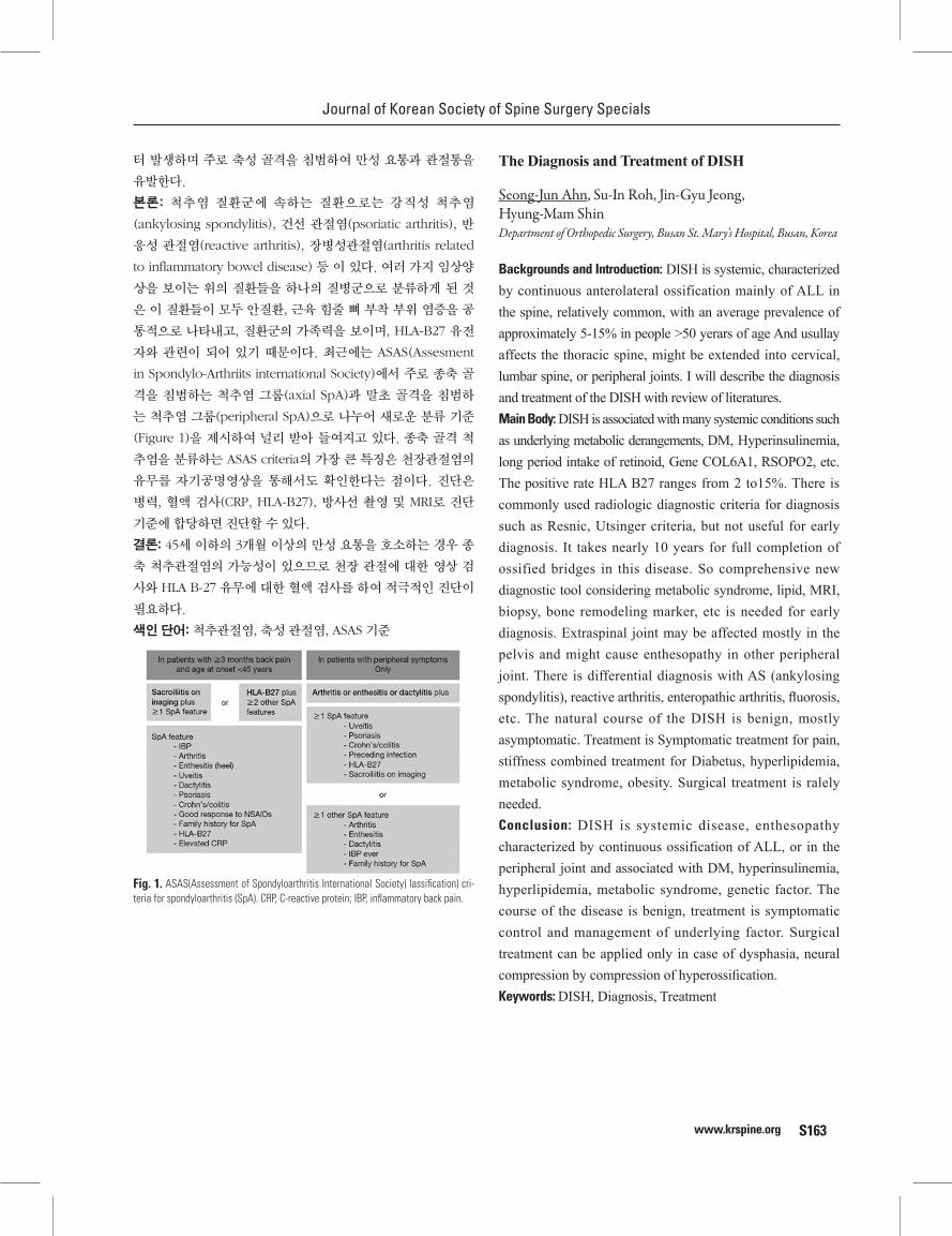
Journal of Korean Society of Spine Surgery Specials
www.krspine.org S163
터 발생하며 주로 축성 골격을 침범하여 만성 요통과 관절통을
유발한다.
본론: 척추염 질환군에 속하는 질환으로는 강직성 척추염
(ankylosing spondylitis), 건선 관절염(psoriatic arthritis), 반
응성 관절염(reactive arthritis), 장병성관절염(arthritis related
to inflammatory bowel disease) 등 이 있다. 여러 가지 임상양
상을 보이는 위의 질환들을 하나의 질병군으로 분류하게 된 것
은 이 질환들이 모두 안질환, 근육 힘줄 뼈 부착 부위 염증을 공
통적으로 나타내고, 질환군의 가족력을 보이며, HLA-B27 유전
자와 관련이 되어 있기 때문이다. 최근에는 ASAS(Assesment
in Spondylo-Arthriits international Society)에서 주로 종축 골
격을 침범하는 척추염 그룹(axial SpA)과 말초 골격을 침범하
는 척추염 그룹(peripheral SpA)으로 나누어 새로운 분류 기준
(Figure 1)을 제시하여 널리 받아 들여지고 있다. 종축 골격 척
추염을 분류하는 ASAS criteria의 가장 큰 특징은 천장관절염의
유무를 자기공명영상을 통해서도 확인한다는 점이다. 진단은
병력, 혈액 검사(CRP, HLA-B27), 방사선 촬영 및 MRI로 진단
기준에 합당하면 진단할 수 있다.
결론: 45세 이하의 3개월 이상의 만성 요통을 호소하는 경우 종
축 척추관절염의 가능성이 있으므로 천장 관절에 대한 영상 검
사와 HLA B-27 유무에 대한 혈액 검사를 하여 적극적인 진단이
필요하다.
색인 단어: 척추관절염, 축성 관절염, ASAS 기준
Fig. 1. ASAS(Assessment of Spondyloarthritis International Society) lassification) cri-teria for spondyloarthritis (SpA). CRP, C-reactive protein; IBP, inflammatory back pain.
The Diagnosis and Treatment of DISH
Seong-Jun Ahn, Su-In Roh, Jin-Gyu Jeong, Hyung-Mam ShinDepartment of Orthopedic Surgery, Busan St. Mary’s Hospital, Busan, Korea
Backgrounds and Introduction: DISH is systemic, characterized by continuous anterolateral ossification mainly of ALL in the spine, relatively common, with an average prevalence of approximately 5-15% in people >50 yerars of age And usullay affects the thoracic spine, might be extended into cervical, lumbar spine, or peripheral joints. I will describe the diagnosis and treatment of the DISH with review of literatures.Main Body: DISH is associated with many systemic conditions such as underlying metabolic derangements, DM, Hyperinsulinemia, long period intake of retinoid, Gene COL6A1, RSOPO2, etc. The positive rate HLA B27 ranges from 2 to15%. There is commonly used radiologic diagnostic criteria for diagnosis such as Resnic, Utsinger criteria, but not useful for early diagnosis. It takes nearly 10 years for full completion of ossified bridges in this disease. So comprehensive new diagnostic tool considering metabolic syndrome, lipid, MRI, biopsy, bone remodeling marker, etc is needed for early diagnosis. Extraspinal joint may be affected mostly in the pelvis and might cause enthesopathy in other peripheral joint. There is differential diagnosis with AS (ankylosing spondylitis), reactive arthritis, enteropathic arthritis, fluorosis, etc. The natural course of the DISH is benign, mostly asymptomatic. Treatment is Symptomatic treatment for pain, stiffness combined treatment for Diabetus, hyperlipidemia, metabolic syndrome, obesity. Surgical treatment is ralely needed.Conclusion: DISH is systemic disease, enthesopathy characterized by continuous ossification of ALL, or in the peripheral joint and associated with DM, hyperinsulinemia, hyperlipidemia, metabolic syndrome, genetic factor. The course of the disease is benign, treatment is symptomatic control and management of underlying factor. Surgical treatment can be applied only in case of dysphasia, neural compression by compression of hyperossification. Keywords: DISH, Diagnosis, Treatment

Volume 25 • Number S2 • November 16 2018
www.krspine.orgS164
미만성 특발성 근골격계 과형성증의 진단과 치료
안성준, 노수인, 정진규, 심형남
부산성모병원 정형외과
서론: 미만성 특발성 근골격계 과골화증(DISH)은 척추 종인대
전외측부 골화를 특징으로 하는 주로 50-60대에 발생하는 전신
질환이며, 유병률은 5-15%이며 주로 흉추에 발생하며, 경추나
요추에 이환 될 수 있고, 통증, 운동장애를 유발할 수 있으며, 척
추외 상, 하지 근골격계 인대부착부위 염증을 유발할 수 있다.
이질환의 진단과 치료에 대해 문헌고찰과 함께 알아보았다.
본론: 위험인자로 당뇨병, 고인술린혈증, 비만, 고지질혈증, 대
사증후군 등이 있으며 retinoid장기복용이나, 최근 유전자 연
구에서 COL6A1, RSOPO2와 연관되어 있다고 한다. 환자의
약 2-30%에서 HLA B27 양성이며, 방사선학적 진단기준으로
Resnick, Utsinger criteria가 있지만 이는 질환의 조기발견에는
유용하지 못하다. 골화가 완성되는데 약 10년간 소요되는 점을
고려할때, 조기진단을 위해 대사증후군, 지질, MRI, 조직검사,
Bone remodeling marker 등을 고려한 포괄적인 진단방법이
필요하다. 척추외 주로 골반골 주위나 상 하지 골격에 골부착부
병증이 생길 수 있으며, 감별진단으로 강직성 척추염, 반응성 관
절염, Chron씨병 등으로 인한 장병성관절염, 불소증 등이 있다.
치료는 질환의 경과가 대부분 심하지 않아 대증 요법을 하며, 수
술이 필요한 경우는 매우 드물고 비만, insulin, 당뇨, 등의 관리
가 필요하다.
결론: DISH는 당뇨, 지질대사, 비만, 유전자 이상 등으로 인한
골대사 이상으로 전방 종인대의 전외측부, 인대부착부위에 발
생하는 골부착부병증이며, 질환의 경과는 양성적으로, 대개의
경우 심한 임상증상을 유발하지 않으며, 대증요법으로 치료한
다, 현재의 진단방법은 병이 진행된 이후에만 가능하여, 조기진
단을 위해 포괄적인 새로운 진단방법이 필요하다. 수술은 과골
격형성으로 인한 연하곤란이나, 신경압박증상, 골절 등의 경우
에 한해 매우 드물게 시행될 수 있다.
색인 단어: 미만성 특발성 근골격계 과골화증, 진단, 치료.
Current Issues in Medical Treatment of Ankylosing Spondylitis
Hun-Kyu ShinDepartment of Orthopedic Surgery, Kangbuk Samsung Hospital, Sungkyunkwan University School of Medicine, Seoul, Korea
Backgrounds and Introduction: Ankylosing spondylitis is inflammatory rheumatologic disease that causes various
clinical manifestation. Diagnosis, classification and treatment of ankylosing spondylitis has been controversy up to date. In particular, treatment of ankylosing spondylitis is on progress with the development of new pharmaceuticals. In 2016, ASAS/EULAR (Assessment of SpondyloArthritis international Society/European League Against Rhematism) reported Management Recommendation for Axial Spondyloarthritis.Main Body: 2016 ASAS/EULAR Management Recommendation reported 5 Overarching principles and 13 Recommendations. Treatment goal of axial spondyloarthritis, including ankylosing spondylitis, is to maximize quality of life, control of symptoms and inflammation, prevention of structural damage, and it should be managed multidisciplinary way about diverse manifestations. Also, exercise and smoking cessation should be educated along with medial treatment. The first-line medical treatment is NSAIDs. Though, anecdotal initial study reported that indomethacin is most effective among the NSAIDs, follow researches mentioned that other NSAIDs exhibit similar effect. Taking high dose NSAIDs rather than lower dose and Taking continuous use of NSAIDs rather than on demand use, can help prevent radiologic progression of disease. In case of failure or contraindication of NSAIDs treatment, other analgesics can be considered, such as paracetamol, opioid, etc. They should only be used in the lowest dose and for the shortest time necessary. Local steroid injection can be considered directed to local musculoskeletal inflammation. Systemic corticosteroids are not considered in axial spondyloarthritis. cs (conventional synthetic) DMARDs, that can be used in peripheral arthritis, such as sulfasalazine, methotrexate, normally not be considered to treat axial symptoms. For severe disease activity that lasts for prior treatment, b (biological) DMARDs, including TNF-α inhibitors and IL-17 inhibitors, can be used. In ASAS/EULAR recommendation, bDMARDs can be administered if the symptoms, with a 2.1 or more ADAS score and a 4 or more BASDAI score, persist for more than 4 weeks treatment with 2 or more NSAIDs. In Korea, the use of bDMARDs is approved for patients who have been treated with two or more NSAIDs or other DMARDs for more than 3 months, but the BASDAI score continues 4 or more. In particular, TNF-α inhibitors are recommended at first, and if these DMARDs treatment show no effect, it should be substituted another TNF-α inhibitors
Journal of Korean Society of Spine Surgery Specials
www.krspine.org S165
or IL-17 inhibitors. ASAS/EULAR recommends to maintain DMARDs treatment in case that ASDAS score improved 1.1 or more, BASDAI score improved 2 or more, after at least 12 weeks use of DMARDs. In Korea, maintenance of golimumab(Simponi) can be approved when BASDAI score improve 2 or 50% on 3 months, 6 months after golimumab start. If a patient is in sustained remission, tapering of a bDMARD can be considered.Conclusion: As the incidence of ankylosing spondylitis is increasing, clinical significance of ankylosing spondylitis has been becoming important. Moreover, development of diagnostic technique and new pharmaceuticals made medical treatment of ankylosing spondylitis effective. Based on proper diagnosis and classification, it is important to treat patients according to their symptoms. And it is necessary to understand mainstream of medical treatment, which acts primary role among the treatment of ankylosing spondylitis.Keywords: Ankylosing spondylitis, Medical treatment, DMARDs
강직성 척추염의 약물 치료에 대한 최신 지견
신헌규
성균관대학교 의과대학 강북삼성병원 정형외과학교실
서론: 강직성 척추염은 다양한 임상 양상을 보이는 염증성 류
마티스성 질환이다. 강직성 척추염의 분류, 진단 및 치료는 최
근까지도 논란이 되고 있으며, 특히 그 치료에 대해서는 신약
의 발전과 함께 지침이 변화하는 추세이다. 2016년, ASAS/
EULAR(Assessment of SpondyloArthritis international
Society/European League Against Rhematism)에서는
Management Recommendation for Axial Spondyloarthritis를
제시하였다.
본론: 2016 ASAS/EULAR Management Recommendation에
서는 5개의 중요한 원칙과 13개의 권고 사항을 제시한다. 강직
성 척추염을 포함한 중심성 척추관절증 환자의 치료 목표는 환
자의 삶의 질 증진, 증상과 염증의 관리, 구조적 손상의 예방,
기능의 보존이며, 나타날 수 있는 여러 증상에 따라 다학제 관
리가 필요하다. 또한, 약물 치료와 더불어 운동 치료 및 금연에
대한 충분한 교육이 필요하다. 최우선의 약물 치료는 NSAIDs
이다. 입증 되지 않은 초기의 연구에서는 indomethacin이 가
장 효과적이라는 보고가 있었으나, 이후 시행된 연구를 통해 다
른 NSAIDs 또한 비슷한 효과를 보인다는 것을 확인할 수 있었
다. 선택적 COX-2 억제제와 비선택적 NSAIDs 간 효과의 차이
는 보이지 않았다. 증상이 있을 때에만 NSAIDs를 복용하는 것
보다는 지속적으로 복용하는 것이, 저용량 NSAIDs 보다는 고
용량을 복용하는 것이 질병의 방사선학적 진행을 막는데 도움
이 되는 것으로 알려져 있다. NSAIDs 치료에 실패하거나, 금기
증인 경우 paracetamol, opioid 등 다른 진통제를 고려해볼 수
있다. 이러한 진통제는 저용량으로, 필요한 짧은 기간 동안 복용
하도록 권유된다. 국소적인 근골격계 염증이 있는 경우 국소 스
테로이드 주사를 고려해볼 수 있으나, 중심성 척추관절증에 대
해 장기간의 스테로이드의 전신적 사용은 권유되지 않는다. 말
초 관절염의 치료에 이용되는 sulfasalazine, methotrexate 등의
합성 항류마티스제제는, 척추관절증이 중심성의 양상만을 보일
경우에는 고려되지 않는다. 앞선 치료에도 심한 증상이 지속되
는 경우, 종양 괴사 인자 알파 억제제 및 인터류킨 17 억제제 등
의 생물학적 항류마티스제제의 복용을 고려할 수 있다. ASAS/
EULAR 권고 사항에서는 4주 이상, 2가지 이상의 NSAIDs 복용
에도 ASDAS 점수 2.1 이상, BASDAI 점수 4 이상으로 증상이
지속되는 경우 생물학적 항류마티스제제를 복용할 것을 권유한
다. 한국의 보험 급여 기준에서는 2가지 종류 이상의 NSAIDs 및
항류마티스제제로 3개월 이상 치료를 하였음에도 BASDAI 점
수가 4 이상으로 지속되는 경우 항류마티스제제의 사용을 인정
하고 있다. 약물 치료에도 이 경우 종양 괴사 인자 알파 억제제
의 사용이 우선적으로 권유되며, 치료에 실패할 경우 다른 종양
괴사 인자 알파 억제제 및 인터류킨 17 억제제로 교체되어야 한
다. ASAS/EULAR 권고 사항에서는 12주 이상 생물학적 항류마
티스제제를 복용 후 ASDAS 점수의 호전이 1.1 이상, BASDAI
점수의 호전이 2 이상일 경우 항류마티스 제제를 지속 복용하도
록 권유한다. Golimumab(Simponi) 투여에 대한 국내 보험 급
여 기준에서는 투여 후 3개월, 6개월 째 BASDAI 점수가 2점 혹
은 50% 이상 감소하는 경우 복용 유지에 대한 보험 급여를 인정
하고 있다. 임상 양상이 호전될 경우 생물학적 항류마티스제제
의 중단을 고려할 수 있다.
결론: 강직성 척추염의 유병률이 증가함에 따라 그 중요성은 높
아지고 있다. 또한 진단 기술 및 신약의 발달로 강직성 척추염의
약물 치료 또한 다양하고 더욱 효과적으로 발전하고 있다. 정확
한 진단 및 분류를 기반으로 환자의 증상에 맞춘 치료를 고려하
는 것이 중요하겠으며, 치료 방법 중 가장 중요한 약물적 치료에
대해 흐름을 숙지하고 환자에 적용하는 것이 필요하다.
색인 단어: 강직성 척추염, 약물 치료, 항류마티스제제
Volume 25 • Number S2 • November 16 2018
www.krspine.orgS166
Surgical Treatment for Spondyloarthropathy
Yong-Chan KimDepartment of Orthopedic Surgery, Kyung Hee University Hospital at Gangdong College of Medicine, Kyung Hee University, Seoul, Korea
Backgrounds and Introduction: The spinal surgical treatment for spondyloarthropathy (rheumatoid arthritis; RA, ankylosing spondylitis; AS) patients pose several intraoperative or perioperative challenges that may lead to significant complications, if not recognized and efficiently managed. Surgical intervention for AS is necessary in cases of traumatic instability and significant deformity. These 2 categories have unique characteristics among patients with AS.Main Body: In a systemic inflammatory disease such as RA, there is a high rate of nonunion after fusion surgery, due to the remodeling process which is disturbed by an imbalance between the resorption and formation of bone owing to high levels of proinflammatory cytokines. Some authors have reported that the time to union in patients corresponding to Lenke Grades A and B, was clearly delayed in those with RA. Patients with RA develop osteoporosis owing to many causes, including circulating cytokines, the activity and duration of RA, and reduced physical activity, as well as the pharmacological treatment of RA. Implant-related problems including vertebral body collapse and screw pull-out more commonly occurred in the patients with RA, mostly at the most proximal or distal level included in the fusion. Also some authors have reported that there is also a high incidence of infection after orthopaedic surgery in patients with RA and the risk of infection after arthroplasty is two- to three-times higher in these patients than osteoarthritis. The severely rigid yet brittle nature of the spine in patients with AS must not be underestimated when managing their care in the operating room. An increased rate of neurological damage exists in these patients due to excessive rotation and angulation at fracture sites, a phenomenon exacerbated by preexisting deformity and associated corrective maneuvers. The spinal reconstructive deformity surgery for AS has the potential to cause severe neurological damage because of the complex dynamic forces involved in deformity correction acting on a both rigid and brittle spine.Conclusion: When doing spinal surgical treatment for RA, we should perform autogenous bone grafts of sufficient volume osteoporotic bone. Moreover we need to adjust the peri-
operative management of anti-rheumatic drugs. To avoid infection, it may be wise to use additional antibiotics in a different regimen, or to supplement with another modality. Early recognition of potential pitfalls and complications associated with the treatment of patients with AS will help to avoid preventable or iatrogenic injuries.Keywords: Lumbar spine, Spondyloarthropathy, Surgical treatment
척추관절증을 위한 수술적 치료
김용찬
강동경희대학교병원 정형외과
서론: 척추관절증(류마티스관절염, 강직성척추염)에 대한 척추
수술 시 인지하지 못하거나 적절하게 대처하지 못할 경우 술 중
또는 수술 전후의 심각한 합병증을 야기할 수 있다. 강직성 척추
염 환자는 외상성 불안정성과 심각한 변형으로 수술적인 처치
를 요한다. 이들 두 상태는 강직성 척추염 환자에만 해당되는 독
특한 특성을 가지고 있다.
본론: 류마티스 관절염과 같은 전신적인 염증성 질환에서 유합
술을 시행할 경우 불유합의 비율이 높다. 이는 proinflammatory
cytokine이 높아 골흡수와 골형성 간의 불균형이 심하여 리모
델링 과정이 방해를 받기 때문이다. 몇 저자들은 Lenke grade
A와 B에 해당하는 환자들의 골유합에 걸리는 시간이 류마티스
관절염 환자에서 저명하게 지연되었다고 보고하였다. 류마티스
관절염 환자에서 골다공증을 유발하는 원인은 여러 가지가 있
으며 류마티스 관절염 환자에서 약물치료에 사용되는 약제 뿐
만 아니라 circulating cytokine, 류마티스 관절염의 활성정도
와 질병 이환기간 그리고 환자의 활동 정도의 감소 등이 해당된
다. 류마티스 관절염 환자에서 척추체의 붕괴와 나사의 pull out
을 포함한 내고정물과 관련된 문제점들이 더 자주 생기며, 이들
은 특히 유합 분절 중 가장 근위부 또는 원위부 분절에서 흔하
다. 또한 몇 저자들은 류마티스 관절염 환자의 정형외과 수술 시
행 후 감염의 빈도가 높게 나타났고 관절 치환술 시행 후 감염의
위험성이 골관절염 환자에 비해 2배에서 3배까지 높았다고 보
고하였다. 강직성 척추염 환자의 척추는 아주 rigid하나 brittle
한 특성을 갖고 있어 수술시행 시 이를 간과해서는 안 된다. 이
런 환자들의 신경학적인 손상은 골절부위의 과도한 회전, 각변
형, 수술 전부터 존재하였던 변형이 악화되거나 관련된 변형 교
정 술기로 인해 생길 수 있다. Rigid하면서도 brittle한 척추에 변
형 교정술을 시행할 때 복잡한 역학적 힘을 주게 되면서 강직성
척추염 환자의 척추 변형 교정 수술은 심각한 신경학적인 이상
을 일으킬 수 있는 잠재성이 있다.
결론: 류마티스 관절염 환자에서 척추의 수술적인 처치를 시행
Journal of Korean Society of Spine Surgery Specials
www.krspine.org S167
할 때 골다공증성 뼈에 대해 충분한 자가 뼈를 이용한 골유합을
시행해야 한다. 더 나아가 수술 전후 anti-rheumatic 약제를 조
정하여 적절한 처치를 시행해야 한다. 또한 감염을 예방하기 위
하여 서로 다른 regimen을 이용해서 항생제를 추가하거나 다른
modality를 추가하는 것이 필요할 수 있다. 강직성 척추염 환자
의 치료와 관련된 잠재적인 위험과 합병증을 일찍 발견하는 것이
예방 가능한 또는 의인성 손상을 피하는데 도움을 줄 수 있다.
색인 단어: 요추, 척추관절증, 수술적인 처치
Pathophysiology of Back Muscle Degeneration
Tae-Hwan KimSpine Center, Department of Orthopedics, Hallym University Sacred Heart Hospital, Hallym University College of Medicine, Anyang, Korea
Backgrounds and Introduction: Most studies on the pathoph-ysiology of skeletal muscle have been done about two decades ago. They demonstrated the mechanisms of the three-step process including muscle injury, degeneration, and regeneration. Recently, the studies on the pathophysiology of skeletal muscle have been done in association with sarcopenia. Main Body: The studies about the morphology of muscle fiber should be preceded to understand the pathophysiology of skeletal muscle degeneration. During development, somite divides into ventral sclerotome and dorsal dermomyotome. Back muscle develops from the medial portion of the dermomyotome. Differentiation to the myogenic lineage requires the upregulation of MyoD and Myf5, basic helix-loop-helix transcriptional activators of the myogenic regulatory factor family (MRF). Proliferating myoblasts become terminally differentiated myocytes that express the MRFs, Myogenin and MRF4, and subsequently muscle-specific genes such as myosin heavy chain (MHC) and muscle creatine kinase (MCK). Mononucleated myocytes fuse to form multinucleated syncytium, which eventually mature into muscle fiber. A distinct subpopulation of myoblasts fail to differentiate, and remain on the surface of the developing muscle fiber as quiescent muscle satellite cells. Muscle fiber is covered by plasma membrane, sarcolemma, which keeps concentration of calcium ion and protein within muscle fiber. Muscle injury always accompanies degeneration and regeneration regardless of
type of injury. Muscle degeneration is generally triggered by disruption of the myofiber sarcolemma, which increases myofiber permeability. Disruption of the myofiber sarcolemma increases calcium influx, which causes a loss of calcium homeostasis and increases calcium-dependent proteolysis. In addition, unregulated influx of cytosolic calcium corresponds to the magnitude of subsequent leakage of cytosolic proteins into the extracellular space. Such disruption of homeostasis results in focal or total autolysis of muscle fiber. The degeneration of injured muscle could be accelerated by 1) activation of inflammatory cell, 2) phospholipase activation, 3) disruption of vascular perfusion, and resultant activation of free radical. The disruption of muscle cell membrane produces tremendous hazardous effect on normal homeostasis, and restoration of integrity in normal membrane is prerequisite for muscle regeneration. Dysferlin was identified to have an important role in the membrane sealing. Satellite cell and migration of myogenic cell contributes to muscle regeneration. Recent studies about the pathophysiology of muscle degeneration have been focused on the age-related loss of muscle mass, sarcopenia. The increase in proinflammatory status is a major characteristic of the aging process, which was referred to as inflammaging by Franceschi et al. Several inflammatory markers such as IL-1 and 6, CRP, and TNF-α were identified to be associated with the inflammation, which could induce decrease in muscle mass via Ubiquitine-Protease system.Conclusion: The pathophysiology of back muscle degeneration can be explained by the three-step process including injury, degeneration, regeneration and by age-related muscle loss resulting from inflammaging. Keywords: Back muscle, Degeneration, Pathophysiology, Regeneration, Sarcopenia
요부 근육 퇴행의 병태생리
김태환
한림대학교 성심병원 정형외과
서론: 골격근의 병태 생리(Pathophysiology)에 대한 연구는
2000년대 이전에 대부분이 진행되었고, 골격근의 손상(injury),
퇴행(degeneration), 재생(regeneration)에 이르는 3단계의 유
기적인 과정이 있음이 밝혀졌다. 최근에는 이러한 이전 연구를
바탕으로, 근감소증(sarcopenia)에 초점을 두면서 이를 임상적

Volume 25 • Number S2 • November 16 2018
www.krspine.orgS168
인 부분과 연결 시켜보려고 하고 있다.
본론: 골격근의 퇴행(degeneration)과 관련된 병태 생리
를 이해하기 위해서는 먼저 근섬유(Muscle fiber)의 형태
(morphology)에 대한 이해가 필요하다. 골격근 중 요부
근육(back muscle)은 발생 과정상, 체절(somite)이 복측
(ventral) 뼈분절(sclerotome)과 배측(dorsal) 피부근육분절
(dermomyotome)로 나누어지는데, 이중 피부근육분절의 내
측에서 발생한다. 이렇게 근원성 계통(myogenic lineage)
으로 분화되는 과정에는 Myf5와 MyoD gene의 상향조절
(upregulation)이 필요하다. 이후 근원 세포(myoblast)는 최
종적으로는 근세포(myocyte)로 분화되어 myogenin, MRF4,
myogin heavy chain 및 muscle creatine kinase를 형성하
는 유전자를 표현하게 된다. 최종 분화 형태의 단핵 근세포
(mononucleated myocyte)는 서로 합쳐져서 다핵 근섬유
(multinucleated myofiber)를 형성하고, 이 과정에서 분화가
안된 근원 세포가 남아 satellite cell이 되어 다핵 근섬유의 표
면에 붙게 된다. 근섬유는 sarcolemma라는 원형질막(plasma
membrane)으로 덮혀 있어, 바깥과 칼슘 이온 및 단백질의 농
도차가 유지된다. 근손상이 발생하면 손상의 종류와 관계 없이
이후의 과정은 항상 퇴행과 재생을 동반한다. 근손상로 인한 해
부학적 변화는 sarcolemma에서 시작하는 것으로 알려져 있다.
sarcolemma의 손상은 칼슘 이온의 유입을 통해 널리 알려진
calpain과 같은 calcium dependent protease를 활성화시키고,
근섬유내 존재하는 단백질을 밖으로 유출시켜서 세포 항상성
(homeostasis)을 파괴한다. 이 과정에서 근섬유에 부분적 혹은
전체적으로 자가분해(autolysis)가 일어난다. 이 후 근섬유의 퇴
행이 1) 염증세포의 활성(activation of inflammatory cells), 2)
인지질 가수분해 효소 활성 (phospholipase activation) 및 3)
혈관 관류 손상(disruption of vascular perfusion)을 통해 일어
나고 이것들은 또한 유리기(Free radical)를 활성화시켜 퇴행을
가속화 한다. 근섬유막의 파괴는 세포 항상성 유지에 큰 문제를
일으키기 때문에, 이후에 재생 과정에서 가장 중요한 부분은 세
포막을 복원하는 것이다. 여기에 dysferin이라는 protein이 큰
역할을 하며, 이후 satellite cell과 근원성 세포(myogenic cell)
의 이동(migration)으로 인한 근재생이 본격적으로 시작된다.
최근 골격근의 병태생리 연구는 노화로 인한 근육량의 감소,
근감소증에 초점을 두고 진행되었다. 노화와 더불어 증가되
는 염증성 경향으로 근육 퇴화를 유발하는 기전들이 점차로 강
하게 활성화되며, 이것이 결과적으로 조직손상을 유발하게 된
다는 가설을 세웠는데 Franceschi 등은 이를 inflammaging
이라는 용어를 사용하여 명명하였다. 다양한 연구들을 통해
inflammaging에 다양한 면역세포 변화 및, IL-1, IL-6, CRP,
TNF-α 등과 같은 inflammatory marker가 관여함이 밝혀졌고
이는 Ubiquitine-Protease system을 통해 근육량의 감소에 기
여하는 것으로 생각하고 있다.
결론: 요부 근육의 병태생리는 손상(injury), 퇴행(degeneration),
재생(regeneration)에 이르는 3단계의 과정에, 노화에 따른 요부
근육의 퇴행을 설명하는 inflammaging의 개념을 이용하여 설명
할 수 있다.
색인 단어: 요부 근육, 퇴행, 병태생리, 재생, 근감소증
Clinical Relevance of Back Muscle Degeneration
Jung-Hoon KimDepartment of Orthopedic Surgery, Ilsan Paik Hospital, College of Medicine, Inje University, Goyang, Korea
Backgrounds and Introduction: The paraspinal muscles have an important role in trunk movement and spinal stability. Paraspinal muscle degeneration could result in spinal instability which may predispose to further injury. Three principal signs of muscle degeneration were detected on imaging: decreased muscle size, decreased radiographic density, and increased fat deposits. These changes associated with muscular degeneration can be evaluated by CT or MRI and may manifest as changes in MR signal intensity, CT density or cross sectional area. Many studies have used these imaging parameters to evaluate the paraspinal muscle degeneration. And, several studies have investigated to recognize the association between clinical manifestation and back muscle degeneration. Main Body: CSA and the quality of paraspinal muscles decrease with age, most probably as an expression of age-related sarcopenia. Paraspinal muscle CSA and density are higher in men than in women. Men show lower fatty infiltration in the paraspinal muscles than women. BMI was shown not to influence the presence of fat in the lumbar multifidus. Numerous studies have shown that an association exists between paraspinal muscle degeneration and LBP. Fatty infiltration in the lumbar multifidus is strongly associated with LBP in adults and chronic LBP was also found to be associated with reductions in muscle fiber density. In patients with lumbar disc herniation, dysfunction of the back muscles is common. There is evidence explaining degenerative changes in the lumbar multifidus after disc herniation, that is, a decrease in the multifidus CSA (especially on the lesion side), a decrease of muscle density

Journal of Korean Society of Spine Surgery Specials
www.krspine.org S169
and a decrease in the size of Type I and Type II/MHC-2X fibers and intersititial fibrosis. Facet joint osteoarthritis was significantly associated with low density and higher grades of fatty infiltration in the multifidus and erector spinae. Some studies reported that patients with spondylolisthesis (isthmic or degenerative) suffer from segmental atrophy of the multifidus muscle and the presence of erector spinae hypertrophy could be a mechanism to compensate for this instability. The results of one study presented convincing evidence that multifidus fatty infiltration was more representative of a neural injury than the degree of lumbar stenosis. It also suggests that multifidus fatty infiltration can be used as a prognostic factor of functional performance in spinal stenosis patients instead of the severity of spinal canal stenosis. According to most studies, the percentage of fat tissue within the back muscle was increased significantly by the fat infiltration and the muscle mass of the back muscle compartment was smaller in the degenerative lumbar flat back patients compared with that in the normal control. Conclusion: Accurate identification of the origin of LBP can potentially provide a more rational approach to patient management. So, there is a strong need for establishing uniform methods of evaluating degenerative changes of the paraspinal muscles. Additional studies are needed to improve the understanding of the association and causal relationships between LBP, spinal degeneration, and changes in the paraspinal muscle. Accurate identification of the origin of LBP can potentially provide a more rational approach to patient management. Especially, the implications for clinical research could be immense as the precise quantification of specific muscle abnormalities associated with LBP would allow for better evaluation of interventional treatments.Keywords: Paraspinal muscle, Multifidus, Erector spinae, Muscle degeneration, Lower back pain
등 근육 퇴행의 임상적 연관성
김정훈
인제대학교 일산백병원 정형외과학교실
서론: 척추 주위 근육은 몸체의 운동과 척추 안정화에 중요한 역
할을 하므로 척추 주위 근육의 퇴행성 변화는 척추 불안정성을
초래할 수 있으며 이로 인해 추가 손상이 발생할 수 있다. 근육
퇴행성 변화의 3가지 주요 징후인 근육 크기 감소, 근육 밀도 감
소 및 지방 축적 증가를 영상에서 확인할 수 있는데 특히, 근육
의 퇴행에 의한 이러한 변화를 CT 또는 MRI로 평가할 수 있으
며 MRI의 신호 강도와 CT 밀도 또는 단면적의 변화로 나타날
수 있다. 많은 연구들에서 이러한 영상 척도를 사용하여 척추 주
위 근육의 퇴행성 변화를 평가하였으며 또한, 등 근육 퇴행과 임
상 증상과의 연관성을 알아보기 위한 몇몇 연구들이 시행되고
있다.
본론: 척추 주위 근육의 단면적과 질은 나이에 따라 감소하게 되
는데 이는 노화와 연관된 근감소증의 현상이며 척추 주위 근육
의 단면적과 밀도는 여성보다 남성에서 높게 나타난다. 또한 남
성에서 여성 보다 척추 주위 근육에 낮은 지방 침윤을 보여 주
며 BMI는 요추 다열근의 지방 침윤에 영향을 미치지 않는 것으
로 나타났 다. 많은 연구에서 척추 주위 근육 퇴행성 변화와 요
통 사이에 연관성이 있음을 보고하였다. 요추 다열근의 지방 침
윤은 성인의 요통과 강하게 연관되어 있으며, 만성 요통 또한 근
섬유 밀도의 감소와 관련이 있는 것으로 밝혀져 있다. 요추 추간
판 탈출증 환자에서 등 근육의 기능 장애가 흔하게 나타나는데
추간판 탈출증 후에 다열근 단면적의 감소(특히 병변 쪽), 근육
밀도의 감소, Type I 및 Type II / MHC-2X 섬유의 크기 감소 및
간질성 섬유화 등과 같은 요추 다열근의 퇴행성 변화를 시사하
는 소견들이 나타난다. 후관절 관절염은 다열근과 척추 기립근
의 저밀도 및 많은 지방 침윤과 유의한 연관성이 있었으며 일부
연구에 따르면 척추 전방 전위증 (협부형 혹은 퇴행성) 환자에
서 다열근의 분절 위축이 발생하는데 척추 기립근의 비대는 이
러한 불안정성을 보완하는 기전에 의한 것이라고 보고하고 있
다. 한 연구의 결과 다열근의 지방 침윤이 척추관 협착 정도보다
신경 손상을 더 잘 대표한다는 설득력 있는 증거를 제시했으며
또한, 다열근의 지방 침윤은 척추관 협착증의 중증도 대신에 척
추관 협착증 환자의 기능 수행의 예후 인자로 사용될 수 있다고
하고 있다. 대부분의 연구에 따르면, 퇴행성 요추 후만증 환자에
서 정상군의 근육과 비교하여 지방 침윤에 의해 등 근육 내의 지
방 조직의 비율이 현저하게 증가하였고, 등 근육 구획의 근육량
이 더 작았음을 보고하고 있다.
결론: 요통에 대한 정확한 원인 파악은 환자 관리에 보다 합리적
으로 접근하게 하므로 척추 주위 근육의 퇴행성 변화를 평가하
는 일정한 방법을 확립할 필요가 있으며 또한 요통 및 척추 퇴행
성 변화와 척추 주위 근육의 퇴행성 변화 사이의 연관성뿐만 아
니라 인과 관계에 대한 이해를 향상시키기 위한 추가적인 연구
가 필요하리라 사료된다. 특히, 요통과 관련된 특정 근육의 이상
에 대해 정확하게 정량화 하는 것은 중재적 치료에 대한 더 나은
평가를 가능하게 하므로 임상 연구에 상당한 영향을 미칠 것으
로 사료된다.
색인 단어: 척추 주위 근육, 다열근, 척추 기립근, 근육 퇴행, 요
통

Volume 25 • Number S2 • November 16 2018
www.krspine.orgS170
Back Muscle Exercise
Soo-An ParkDepartment of Orthopedic Surgery, Parkweonwook Hospital, Busan, Korea
Backgrounds and Introduction: Physical fitness refers to ability to perform occupational, recreational, and daily activities without becoming unduly fatigued. Exercises for physical fitness includes (1) cardiorespiratory exercise, (2) muscle resistance exercise, (3) weight management and body composition exercises, (4) flexibility and stretching exercises, and (5) stress managing exercise. Exercise therapy for the spinal muscle in spinal pain condition is the core stabilizing exercise using motor control method. In addition, whole-body vibration has been recently applied in treatment of spinal symptoms. Vibration exercise may increase muscle power and muscle blood volume by stimulating stretch reflex of the muscle, and therefore improve muscular performance in healthy subjects and reduce chronic back pain and pain-related disability in patients with chronic spinal pain. Scoliosis-specific exercise (SSE) has been performed in patients with adolescent idiopathic scoliosis (AIS) for slowing progression and corrections of curve and body balance. Objectives of this study are to introduce the knowledge of sport science for the muscle exercise and to establish the proper exercise for the spinal muscle that can be applied to the spinal patients, and to discuss about the specific therapeutic exercises currently applied to patients with spinal pain, senile sarcopenia, and adolescent idiopathic scoliosis in practice. Main Body: Cardiorespiratory exercise is enhancing endurance of the heart, lungs, and circulatory system to supply oxygen and nutrients to working muscles efficiently, and various aerobic exercises are in this category. Muscle strengthening is increasing maximal force or tension level that can be produced by a muscle group. Muscle endurance exercise is for improving the ability of muscle to maintain submaximal force levels for extended periods. Static, dynamic, and isokinetic exercises are in the muscle resistance training, and all are effective for developing strength and muscle endurance. The exercise program is designed to produce a weight loss by increasing the caloric expenditure. To maximize the total energy expenditure, a mode of activity (aerobic exercise) that can be performed at a low-to-moderate intensity for a long duration should be selected.
For weight gain, a high-volume resistance training is the best approach to maximize the development of muscle size. For individuals who wish to improve their body composition without changing their body weight, exercise programs to either decrease body fat, increase fat-free mass, or both should be selected. Three types of stretching techniques are ballistic stretching, slow and static stretching, and proprioceptive neuromuscular facilitation (PNF) stretching. All three types of stretching are effective in increasing the ROM. However, better improvement in the flexibility of some regions were reported for subjects who trained using the PNF technique compared to ballistic stretching. Aerobic exercise and the progressive relaxation technique are two methods that produce relaxation through the physical mode. Regular, aerobic exercise is effective for reducing psychological stress and increasing the body’s resistance to many types of stressors. In patients with chronic back pain, cross-sectional area of the paraspinal muscle decreased and fatty infiltration increased especially in the multifidus muscle. Therefore, chronic back pain patients can develop paraspinal muscle dysfunction. In patients of elite athlete or laborer can develop spinal instability or spinal pain by the repetitive rotational spine movement. For these patients with chronic back pain, core stabilizing exercise using motor control method can be applied. Core stabilizing exercise is expected to improve spinal symptoms by recovering spinal muscle function through activating deep spinal muscles, such as transversus abdominis and multifidus muscles. Likewise, therapeutic exercise for treating spinal pain refers to flexibility/stretching exercise in terms of the physical fitness. In addition to the voluntary muscle exercise, vibration has been applied recently to the patients as another modality of therapeutic exercise. Whole-body vibration exercise is currently performed in sports, geriatrics, and rehabilitation. Vibration exercise is thought to elicit muscular activity via stretch reflexes. Whole-body vibration exercise is helpful in patients with nonspecific chronic lower back pain and in postmenopausal osteoporotic women by relaxing the back muscles. Treatment options for prevention of scoliosis progression are SSE, bracing and surgical correction, even it is controversial whether the use of exercise for the treatment of AIS is useful. However, there are numerous studies that reported SSE combined with/without vibration exercise counteracted the progression of scoliosis in patients with AIS.
Journal of Korean Society of Spine Surgery Specials
www.krspine.org S171
Conclusions: Back muscle exercise is currently accepted as a part of treatments for the spinal patients. However, lacking organization in terms of the medical terminology and concept and lacking customization by the individual spinal disease should be limitations that make spine patients and physicians feel difficult to understand. Therefore, spinal exercise should be systemized with introducing the knowledge of sport science for the physical fitness. In addition to the voluntary muscle exercise, vibration exercise to the whole-body is recently performed in wide range of physiotherapy for aiming spinal muscles. Moreover, spine physician should understand spinal diseases that can be treated by the various spinal exercises, such as chronic back pain, senile sarcopenia, and AIS as well. Keywords: Back muscle exercise, Physical fitness, Spinal pain, Core stabilizing exercise, Vibration exercise
배부 근육 운동
박수안
박원욱병원 정형외과
서론: 근육 운동을 이해하기 위해서는 육체 운동으로 해석되
는 육체적 적합성 (physical fitness)에 대한 설명이 선행되어
야 한다. 육체적 적합성이라 함은 직업적, 오락적, 그리고 일
상 생활에 있어 과도하게 피로해지지 않고 활동을 수행할 수 있
는 능력을 말한다. 개인의 육체적 접합성을 위한 육체 운동에
는 (1) 심폐 운동 (cardiorespiratory exercise), (2) 근 저항 운동
(resistance exercise), (3) 체중 조절 및 신체 조성 운동 (weight
management and body composition exercises), (4) 유연성
및 신전 운동 (flexibility and stretching exercise), (5) 스트레
스 해소 운동 (stress managing exercise) 이 포함된다. Spinal
pain condition에서 spinal muscle을 대상으로 하는 운동 치료
는 motor control 기법을 사용한 중추 근육 안정화 운동 (core
stabilizing exercise)를 활용하고 있다. 일정한 속도의 vibration
으로 근육의 stretch reflex를 유발하고 근육 내 metabolic
power를 증가시켜 근육 활동성을 증가시킴으로써, 근육을 이
완시키고 혈류량을 증가시켜서 척추 증상을 개선시키는데 활용
하기도 한다. 더불어 수술적 교정술이 필요하지 않은 정도의 청
소년 특발성 척추측만증 환자를 대상으로 만곡의 진행을 늦추
고 신체 불균형을 교정할 목적으로 근육 운동을 기반으로 하는
운동 치료가 시행되기도 한다. 본 연구에서는 근육 운동에 대한
운동과학적 지식을 소개하고, 척추 의사 관점의 의학적 수정을
가하여 척추 환자에게 적용 가능한 척추 근육 운동을 확립하고
자 한다. 또한, 척추 근육 운동의 대상이 되는 척추 통증, 골다공
증 및 근 감소증, 그리고 청소년 특발성 척추측만증에서 실제로
시행되고 있는 근육 운동에 대하여 고찰하고자 한다.
본론: 육체 운동 중 심폐 운동은 활동중인 근육에 혈액 및 영양
을 효과적으로 전달하는 순환기 및 호흡기의 능력을 증진시키
고자 하는 심폐 지구력 운동으로, 다양한 종류의 유산소 운동이
이에 해당된다. 근력 운동은 같은 기능을 하는 근육군의 최대 힘
혹은 신전 능력을 증진시키는 운동이다. 근 지구력 운동은 장시
간 동안 근육의 submaximal force level을 유지하는 능력을 기
르는 운동이다. 근 저항 운동은 근력과 근 지구력을 향상시킬 뿐
만 아니라 muscle power와 근육 크기를 늘리는 역할을 하기 때
문에 bodybuilder, power lifter 뿐만 아니라 각 분야의 전문 운
동 선수들도 목적에 맞는 근 저항 운동을 시행한다. 체중 감소
를 목적으로 하는 운동은 칼로리 소모를 증가시키는 유산소 운
동을 충분한 강도와 시간으로 지속하는 것이다. 신체 조성 운동
은 체지방을 줄이고 지방 제거 체중(fat free mass)을 늘릴 목적
으로 시행하는 유산소 운동과 고강도의 근 저항운동이다. 유연
성 및 신전 운동에는 ballistic stretching, slow static stretching,
그리고 proprioceptive neuromuscular facilitation (PNF)
stretching 이 포함된다. 스트레스 해소 운동에는 유산소 운동
과 점진적인 근육 이완 방법이 적용된다. Spinal Pain Condition
중 만성 요통에서 paraspinal muscle 특히 multifidus의 cross-
sectional area가 감소하고 fatty infiltration이 증가하는 것으
로 알려져 있다. 더불어 만성 요통의 경우 paraspinal muscle
dysfunction이 발생할 수 있다. 운동선수나 작업자 중에서 반
복적인 rotational spinal movement로 인해서 spinal muscle의
microtrauma가 유발되고 spinal instability나 spinal pain이 발
생할 수 있다. 이런 환자를 대상으로 motor control 기법을 사용
한 core stabilizing exercise를 시행하기도 한다. 이 방법은 deep
spinal muscle, 즉 transversus abdominis, multifidus muscle
을 활성화시켜서 spinal muscle의 기능 회복을 유도하는 방법
이다. 즉, 운동과학적 관점에서 척추 통증을 치료하기 위한 운동
은 육체운동 중 유연성 및 신전 운동에 해당한다. 치료 목적의 근
육 운동에는 자의적 골격 움직임으로 근육의 수축, 이완을 유발
하는 운동과 더불어 vibration exercise가 활용되기도 한다. 이러
한 진동 운동은 이미 스포츠의학, 노인의학, 재활의학 분야에서
활용되고 있는 비교적 새로운 운동 치료 방법이다. 진동 운동은
stretch reflex를 통해서 근육을 활성화시킨다. 전신에 대한 진동
운동을 하는 동안 metabolic power가 증가하는데, 이러한 진동
에 의한 metabolic power는 어깨에 일정한 무게를 가한 이후에
더 증가했다는 보고가 있다. 즉, 진동 운동 중 근육에 가해지는
부가적인 무게는 근육 활동을 증가시키는 것으로 알려져 있다.
이렇게 진동 운동으로 근육 활동을 증가시키는 것 만으로도 요추
신전근의 회전력이 증가하고 자의적 요추근 신전 운동과 차이 없
는 만성 요통 개선 효과를 보인다. 이러한 Vibration exercise는
Volume 25 • Number S2 • November 16 2018
www.krspine.orgS172
골밀도를 개선시키고, 골다공증이 동반된 고령 여성의 요통을 개
선시키는 방법으로도 알려져 있다. 청소년 특발성 척추측만증에
대하여 시행하는 대표적인 보존적 치료는 보조기와 더불어 척추
측만증 대상 운동(scoliosis specific exercise)을 하는 것이다. 척
추측만증 대상 운동 중 가장 보편적 방법으로 받아들여지고 있는
Schroth 운동은 척추 근육의 근력을 강화하고 만곡의 concave
side에서 짧아진 척추 근육을 늘리는 것을 목표로 한다. 더불어
전신 진동 운동을 병행한 주만곡 30도 이하 환자에서 치료 6개월
이후 만곡의 개선이 관찰되었다는 보고도 있다.
결론: 배부 근육 운동은 척추 환자에 대한 치료의 일 부분으로
받아들여지고 있지만, 의학적 범위 내에서 정리가 부실하고, 척
추 질환 별 맞춤화에 대한 체계가 갖추어 지지 않았다. 이러한
치료 목적의 척추 근육 운동에 운동 과학적 지식을 도입하여 이
해하기 쉽고 척추 환자에게 적용 가능한 방법으로 체계화 할 필
요가 있다. 척추 근육 운동에는 자의적 근육 운동과 더불어 전신
진동 운동이 척추 근육을 대상으로 하는 효과적인 운동 치료 방
법으로 받아들여 지고 있다. 더불어, 척추 운동의 대상이 되는
만성 요통, 고령환자의 근감소증 및 청소년 특발성 척추측만증
에 대한 개별화된 운동 치료에 대한 관심이 필요하다.
색인 단어: 배부 근육 운동, 육체적 적합성, 척추 통증, 중추 근
육 안정화 운동, 진동 운동
Minimally Invasive Spinal Surgery (MISS)
Tubular Microscopic Decompression: What are the Best and Worst Indications of MIS Decompression for the Successful Treatment of Degenerative Lumbar Stenosis?
Jae Hyup LeeDepartment of Orthopedic Surgery, Seoul National University, College of Medicine, SMG-SNU Boramae Medical Center, Seoul, Korea
Backgrounds and Introduction: Foley and Smith introduced minimally invasive laminectomy uses smaller incisions with tubular retractor to minimize invasiveness in 1997 with the hope of reducing blood loss, reducing pain, and shortening hospitalization compared with conventional laminectomy procedures. I want to identify the advantages and disadvantages of MIS decompression with tubular retractor and find an appropriate indication of MIS decompression with tubular retractor.
Main Body: The potential advantages of MIS decompression with tubular retractor are less muscle damage caused by muscle splitting approach and less spinal instability by preservation of supra and interspinous ligaments and contralateral structure. Phan and Mobbs performed meta-analysis and reported that Satisfaction rates were significantly higher after unilateral laminectomy and bilateral decompression (ULBD) compared with open laminectomy, postoperative VAS scores were significantly lower in the ULBD compared with the open approach, the operation duration was significantly longer in the ULBD cohort by 11.1 minutes, significantly lower number of reoperations in the ULBD compared with open laminectomy patients. Hatta et al reported a microscopic posterior decompression procedure, muscle-preserving interlaminar decompression (MILD) in 2009. MILD technique, performs decompression through the midline approach, can minimized invasiveness to paravertebral muscles and preserve the facet joint in cases with narrow lamina is expected. Arai et al reported their prospective comparative study between ULBD and MILD in 2014. The clinical and radiological results between two groups were not different in single level surgery. However, ULBD was superior to MILD in terms of improvement of low back pain and lumbar function at the 2-year time point in multilevel decompression surgery. Moreover, the postoperative sagittal translation at L4-L5 was significantly greater in the MILD group than in the ULBD group. The indications of MIS decompression with tubular retractor in spinal stenosis are almost the same as open laminectomy such as claudication caused by any compressive lesion on the thecal sac or nerve root. However, there are bad indications of MIS decompression with tubular retractor in spinal stenosis. First, instability such as spondylolisthesis greater than Meyerding grade 2. Second, degenerative scoliosis. Third, radiculopathy caused by extra or intraforaminal stenosis. Lastly, previous lumbar surgery or injury. Conclusion: MIS decompression with tubular retractor in spinal stenosis has many advantages over open laminectomy. The indication of MIS decompression with tubular retractor is basically the same as open laminectomy, although it depends on the surgeon’s preference. However, this procedure has limitations particularly the radiculopathy by extra or intraforaminal stenosis. Therefore, it is important to perform MIS decompression with tubular retractor
Journal of Korean Society of Spine Surgery Specials
www.krspine.org S173
considering surgeon proficiency and lesion characteristics.Keywords: Spinal stenosis, Decompression, Minimal invasive surgery, Indication
원통형 견인기를 이용한 현미경 감압술:
퇴행성 척추관 협착증의 성공적인 치료를 위해 최적의 적
응증과 최악의 적응증
이재협
서울대학교 의과대학 보라매병원 정형외과
서론: 척추관 협착증에 대한 통상적인 수술방법은 관혈적 후궁
절제술이나 척추근육손상을 유발하고 요통을 유발할 수 있으며
신경손상을 유발할 수 있다는 단점이 있다. Foley와 Smith는 원
통형 견인기를 이용한 최소침습적 후궁절제술을 소개한 이후
관혈적 후궁절제술에 비해 출혈이 적고 통증을 줄일 수 있으며
재원일수를 줄일 수 있다는 장점 때문에 점차 시행이 늘고 있다.
이에, 원통형 견인기를 이용한 최소침습적 후궁절제술의 장점
과 단점을 확인하고 적절한 적응증을 알아보고자 한다.
본론: 원통형 견인기를 이용한 최소침습적 후궁절제술의 잠재
적인 장점은 근육을 박리하지 않고 절개함으로써 근육 손상을
줄일 수 있고 극상인대와 극간인대를 보존하기 때문에 척추의
불안정증을 줄여주며, 반대쪽 구조물을 보존할 수 있다는 것이
다. 메타분석에서 원통형 견인기를 이용한 최소침습적 후궁절
제술을 이용해 편측 후궁절제술과 양측 감압술을 시행한 군이
통상적인 후궁절제술을 시행한 군에 비해 환자의 만족도도 높
고 수술 후 통증 점수도 낮았으며 재수술의 빈도도 낮았으나 수
술 시간은 11.1분정도 길었다고 보고되었다. 또한, Hatta 등은
정중앙 접근법을 이용한 추궁판간 감압술을 소개하였는데, 척
추주변근육 손상을 최소화 하고 후관절을 보존할 수 있는 장점
이 있다. Arai 등은 편측 후궁절제술과 양측 감압술을 시행한 군
과 정중앙 접근법을 이용한 추궁판간 감압술군을 비교한 결과
임상결과와 방사선학적 결과에는 차이가 없다고 보고하였다.
그러나, 다분절 감압술을 시행할 경우 2년 추시에서 요통과 요
추부 기능회복 정도가 정중앙 접근법을 이용한 추궁판간 감압
술군에 비해 우수하였다고 보고하였다. 따라서, 원통형 견인기
를 이용한 최소침습적 후궁절제술의 적응증은 통상적인 관혈적
후궁절제술의 적응증과 거의 동일하다. 그러나, 전방전위증이 2
등급 이상의 불안정증이 있는 경우, 퇴행성 측만증이 동반된 경
우, 추간공내 협착증이나 추간공외 협착증이 증상의 주된 원인
인 경우, 이전에 동일 분절에 수술을 시행받은 경우는 원통형 견
인기를 이용한 최소침습적 후궁절제술에 좋지 않은 적응증이라
고 판단된다.
결론: 척추관 협착증에서 원통형 견인기를 이용한 최소침습적
후궁절제술은 관혈적 후궁절제술에 비해 많은 장점이 있다. 원
통형 견인기를 이용한 최소침습적 후궁절제술의 적응증은 술자
의 선호도에 따라 차이가 있으나 관혈적 후궁절제술과 거의 유
사하지만, 병변이 추간공내 협착증이나 추간공외 협착증인 경
우는 한계가 있다. 따라서, 원통형 견인기를 이용한 최소침습적
후궁절제술은 술자의 숙련도와 병변의 특성에 맞게 사용될 때
가장 좋은 결과가 기대되는 술식이다.
색인 단어: 척추관 협착증, 감압술, 최소침습적 수술, 적응증
Uniportal Endoscopic Decompression: What are the Best and Worst Indication of MIS Decompression for the Successful Treatement of Degenerative Lumbar Stenosis
Youk-Sang Kwon, Seung-Hyun Lee, June-Kyu LeeDepartment of Orthopaedic Surgery , Daejeon Centum Hospital, Daejeon, Korea
Backgrounds and Introduction: Objective is to analyze the surgical strategy, safety, and clinical results of percutaneous endoscopic decompression of lumbar disc herniation with spinal stenosis, and then to contemplate about indications of uniportal endoscopic decompressionMain Body: Thirty-six patients satisfied the inclusion criteria during the period from august 2015 to September 2017. There are another many trials for endoscopic decompression, but if operations are not completed by endoscopic procedures, and then those cases are excluded. Postoperatively ODI and VAS of low back and leg pain were evaluated by periods. Male twenty and Female sixteen. Average age was 52.5 years and mean follow up period was 14.2 months. Patients had symptoms for 3 weeks to 12 months (2.2 months on average). The location of disease was L3/4 (2 cases), L4/5 (19 cases), L5S1 (15 cases in total, 13 cases on paracentral, 2 cases on extraforaminal stenosis). Inclusion criteria were as follows: (1) obvious leg radiating pain by stenotic lesion with or without disc herniation, (2) no signs of improvement despite of conservative treatment such as block procedures and medicine, (3) MRI revealing a single segment of lumbar spine. Exclusion criteria were as follows: (1) central type huge disc herniation, (2) MRI evaluation suggesting mass, infection, deformity, and spinal instability, (3) coagulopathy. All cases are applied by interlaminar aporoach. 1 case of L5-S1 extraforaminal HNP with foraminal stenosis were applied
Volume 25 • Number S2 • November 16 2018
www.krspine.orgS174
by vertical trasforaminal approach (intertransverse approach). The preoperative VAS score was 8.5±1.5 and ODI score was 48.5±6.6. Six months after operation, the VAS score was 1.9±0.2 and ODI score was 13.4±3.3. The effectiveness was evaluated using the Macnab criteria after 12 months : 23 cases were excellent (63.8%), 4 cases were good (11%), 5 cases were fair (13.8%), and 4 cases were poor (11%). 5 cases of fair result have 2 severe causalgia and 3 dysesthesia and more older than average patients. They were treated with intermittent root block and analgesics medication. 4 cases of poor results were 3 cases of infection (1 meningitis with dural injury and wound infection, 1 spondylodiscitis, 1 delayed epidural abscess).Conclusion: there are several cases of causalgia, dysesthesia, their average age was more older than other patients, then surgical procedures such as dissection of neural structure, distraction are more difficult. The cause of postoperative causalgia, dysesthesia were technical problems and bulky instrument (4.5 mm Kerrison, 4.0 mm Rongeur). So, severe lateral recess stenosis requiring excessive dissection and distraction of neural structure will be difficult to consider endoscopic surgery. Preoperative precise evaluation about each patient is prerequisite.Keywords: Interlaminar, Lumbar stenosis, Causalgia, Dysesthesia, Endoscopic decompression
단일공 경피적 내시경 감압술: 퇴행성 요추 협착증에서 성
공적인 수술을 위한 최소침습 감압술의 적응증 권육상, 이승현, 이준규
대전센텀병원 정형외과
서론: 내시경적 척추수술에 있어서 광학기술의 발전, 접근방법,
술기의 발전과 함께 그 적응증이 확장되고 있다. 요추 협착증에
서 내시경적 감압술의 수술전략, 안정성, 임상 결과를 비교하고
적응증의 범위에 대해 고찰해 보고자 한다.
본론: 2015년 8월부터 2017년 9월까지 36예의 내시경적 수술
을 분석하였다. 내시경적 수술로 끝난 환자에 한하였다. 남자
20, 여자 16예 였으며, 평균연령 52.5세, 평균 추시기간 14.2개
월이었다. 3-4요추간 2예, 4-5요추간 19예, 5요추-1천추간 15
예 였다. 내시경은 VERTEBRIS stenosis system (Richard Wolf,
Germany) -5.6 mm working channel, outer diameter 9.3×7.4
mm, working sleeve 10.5 mm, 20도 discoscope를 사용하였
다. 극외측 수핵탈출증을 동반한 제 5요추-1천추간공 협착증 1
예를 제외한 모든 경우에서 추궁판간 접근을 이용하였다. 수술
전 VAS 8.5±1.5 ODI score 48.5±6.6. 수술후 6개월에 the VAS
s 1.9±0.2 ODI score 13.4±3.3. Macnab criteria에 기준한 수술
후 유효성은 23예 excellent(63.8%), 4예 were good(11%), 5
예 fair(13.8%), 4예 poor(11%). 5예 fair: 2 severe causalgia,
3 dysesthesia. 4예 poor results: 3예의 감염(1 meningitis
with dural injury and wound infection, 1 spondylodiscitis, 1
delayed epidural abscess )이었다.
결론: 수술후 결과와 유효성의 분석에서 약 25%의 합병증이 나
타났다. 이는 특히 작열통, 이상감각증등은 과도한 신경구조물
의 견인과 수술미숙, 그리고 적응증의 선택의 오류로 해석되었
으며, 이에 대한 예방을 위해 수술전 척추변형의 유무, 혈액응고
장애, 반복적인 신경차단술, 고령, 수술기법의 숙련도등에 대한
고려가 필요할 것으로 보인다.
색인단어: 추궁판간, 요추협착증, 작열통, 이상감각, 내시경적
감압술
Biportal Arthroscopic Spinal Surgery for Lumbar Spinal Stenosis
Si Young ParkDepartment of Orthopedic Surgery, Korea University College of Medicine, Seoul, Korea
Backgrounds and Introduction: Lumbar decompressive surgery is a standard surgical technique for lumbar spinal stenosis. Many new surgical techniques have been introduced, ranging from open surgery to minimally invasive procedures. Minimally invasive surgical techniques are preferred because patients experience less postoperative pain and shorter hospital stays. However, the success rate of minimally invasive techniques have been controversial. Main Body: Biportal arthroscopic spine surgery is an emerging technique in the field of minimally invasive spinal surgery. The biportal arthroscopic technique combines the advantages of conventional open surgery and uniportal endoscopic surgery. It allows for a good field of vision of the contralateral, sublaminar, and foraminal areas with high magnification. It uses ordinary arthroscopic and spine instruments without the need for special endoscopic instruments. This allows free movement and handling as well as angulation of the surgical instruments and the arthroscope, independent of each other, as they are not restricted in a single portal. It also reduces irritation to the nerve roots,
Journal of Korean Society of Spine Surgery Specials
www.krspine.org S175
and technical flexibility by both sufficient bony and soft tissue work is as possible as much as in conventional open surgery. Finally, continuous saline irrigation can control epidural and bone bleeding, as well as infection. Several studies showed satisfactory results in lumbar disc herniations with biportal arthroscopic discectomy. Comparing spinal decompressive surgery using biportal arthroscopy compared with microscopic surgery in lumbar spinal stenosis patients, shorter operation time, less hemovac drain output, less opioid usage and shorter hospital stay were shown in arthroscopic group. The arthroscopic group experienced no significant differences in clinical outcomes compared with microscopic group. Favorable clinical outcomes were shown at 6 months after surgery in both groups.Conclusion: Lumbar decompressive surgery using arthroscopy showed favorable clinical outcomes, less pain and a shorter hospital stay compared to microscopic surgery in patients with lumbar spinal stenosis.Keywords: Lumbar spinal stenosis, Biportal endoscopy, Minimal invasive surgery, Decompression
요추 척추관 협착증 환자의 양방향 내시경을 이용한 척추
수술
박시영
고려대학교 안암병원 정형외과학교실
서론: 요추 척추관 협착증에서 감압술은 가장 기본적인 수술법
이다. 기존의 고식적인 척추관 감압술의 여러 합병증을 최소화
하기 위하여 최소 침습적 척추 수술이 최근 선호되고 있다. 내시
경을 이용하는 감압술에 대해 많은 논란이 있다. 특히 단방향 내
시경을 이용한 감압술은 구조적으로 한계가 있어 그 사용이 제
한적이며 임상적인 경과도 불확실 한 것으로 보고 되고 있다. 최
근 내시경 혹은 관절경을 이용한 양방향 내시경을 이용한 척추
수술이 시행되고 있으며, 그 결과에 대해 알아보고자 한다.
본론: 양방향 내시경 수술을 시행하는 경우, 내시경 안에 다른
기구가 없고 내시경을 자유롭게 움직일 수 있기 때문에 렌즈
를 병변 바로 앞에 위치시킬 수 있으며, 근육과 인대를 보호하
면서 반대편까지 관찰할 수 있다. 일반적인 관절경 기구와 기
존의 척추 수술 기구들을 자유롭게 사용할 수 있으며, 이를 통
해 고식적인 수술법만큼 충분한 감압술을 시행할 수 있다. 두개
의 삽입구를 이용하여 내시경 삽입구와 기구 삽입구가 각각 다
른 피부 절개를 통해 후궁에 접근하게 되어 내시경과 수술 기구
들이 서로 충돌하지 않게 되어 기술적인 어려움을 해결하였으
며, 한 쪽 삽입구를 통해 신경 조직을 젖히고 다른 삽입구를 통
해 쉽고 안전하게 조작을 할 수 있다. 또한 지속적으로 생리 식
염수가 흐르면서 경막 외 정수압과 추간판 내 압력의 증가를 막
을 수 있고, 열 손상을 예방할 수 있으며, 시야를 좋게 하고 지혈
기능도 할 수 있다. 요추 척추관 협착증에서 양방향 내시경을 이
용한 감압술의 유용성을 현미경을 이용한 감압술과 비교하였을
때 양방향 내시경 군에서 현미경 군보다 더 짧은 수술 시간, 적
은 hemovac 양, 짧은 입원 기간, 적은 마약성 진통제 사용을 보
였다. ODI와 VAS를 이용한 임상 결과는 두 군간에 유의한 차이
를 보이지 않았으며, 두 군 모두에서 수술 후에 좋은 결과를 보
였다.
결론: 요추 척추관 협착증에서 양방향 내시경을 이용한 감압술
은 안전하게 충분한 감압술을 시행할 수 있으며 좋은 임상 경과
를 예측할 수 있을 것으로 사료된다.
색인 단어: 요추 척추관 협착증, 양방향 내시경 수술, 최소 침습
적 수술, 감압술
How MIS Works to Achieve Successful Lumbar Fusion for Degenerative Spondylolisthesis? Slip Reduction; Bony Fusion; Repeat Surgery Minimally Invasive Transforaminal Lumbar Interbody Fusion (TLIF)
Yung ParkDepartment of Orthopedic Surgery, National Health Insurance Service Ilsan Hospital, Yonsei University, College of Medicine, Goyang, Koera
Backgrounds and Introduction: Although many studies have reported the benefits and disadvantages of minimally invasive transforaminal lumbar interbody fusion (TLIF), few have described surgical strategies to improve the success rate or to reduce complications for the treatment of degenerative spondylolisthesis. We searched for studies reporting the clinical and radiological outcomes of minimally invasive TLIF for degenerative spondylolisthesis, and analyzed the optimal indications, technical pitfalls, and tips for successful surgical outcomes, especially in terms of slip reduction, bony fusion, and repeat surgery. The aim of this study was to demonstrate surgical strategies for successful minimally invasive TLIF to achieve successful surgical outcomes for degenerative spondylolisthesis.Main Body: The ideal candidate for minimally invasive TLIF is a patient with single or 2-level low-grade adult degenerative spondylolisthesis. Incomplete decompression,
Volume 25 • Number S2 • November 16 2018
www.krspine.orgS176
dura tearing, nerve root injury, implant-related complications, and nonunion were found to be the most commonly reported adverse events, especially in the early periods of a surgeon’s experience. Precise positioning for skin incision and tube insertion, complete neural decompression, slip reduction, proper interbody preparation for bone graft and cage insertion, and the correct placement of percutaneous pedicle screws are critical strategies for successful surgical outcomes. Fully understanding the surgical pitfalls and tips described in this review is also important to avoid potential complications and repeat surgeries.Conclusion: It is imperative not only to carry out a comprehensive preoperative evaluation and proper patient selection, but also to perform meticulous surgical procedures with thoughtful considerations of potential pitfalls, in order to improve the success rate and to reduce the complications of minimally invasive TLIF to achieve successful surgical outcomes for degenerative spondylolisthesis. Keywords: Minimally invasive, Transforaminal lumbar interbody fusion (TLIF), Degenerative Spondylolisthesis, Pitfalls and tips, Surgical strategy
퇴행성 척추전방전위증의 성공적인 치료를 위한 최소침습
척추수술은 유용한가? 특히 전위정복, 골유합, 재수술 측
면으로 본 최소침습 수술전략 최소침습 경추간공 요추체
간 유합술
박 융
연세대학교 의과대학 국민건강보험일산병원 정형외과학교실
서론: 최소침습 경추간공 요추체간 유합술은 원통 견인기와 경
피적 척추경 나사못 고정을 이용한 수술로 고식적 수술법보다
후방 척추근육 손상을 줄일 수 있어 수술 중 출혈과 입원기간
을 줄일 수 있는 장점이 있다. 그러나 수술시간, 방사선노출시
간, 수술술기 습득시간이 긴 단점도 보고되고 있다. 특히 단분
절, Meyerding 분류 I/II, 퇴행성(Degenerative) 척추전방전위
증(Spondylolisthesis)이 가장 대표적인 수술 적응증으로 알려
져 있다. 또한 전위 정복, 불유합 및 내고정물 관련 합병증 특히
술자의 경험도에 따른 합병증 및 재수술 빈도에 대한 관심이 늘
고 있다. 퇴행성 척추전방전위증 치료를 위한 최소침습 경추간
공 요추체간 유합술의 성공적인 수술결과를 얻기 위하여 고려
해야 할 외과적 수술기법, 특히 전위 정복, 골유합, 재수술 측면
으로 본 최소침습 척추수술 전략을 저자들의 경험과 증례를 바
탕으로 고찰하였다.
본론: 부적절한 감압, 경막 손상, 신경근 손상, 내고정물 관련 합
병증 및 불유합 등이 흔한 수술 관련 합병증이었고 주로 습득곡
선 초기에 발생했다. 특히 충분한 척추전방전위 정복 및 골유합
을 얻고 합병증을 줄여 성공적인 수술결과를 얻으려면 올바른
피부절개 및 원통견인기 삽입위치 결정, 완전한 신경감압, 골이
식과 케이지 삽입을 위한 세심한 종판 준비 및 올바른 경피적 척
추경 나사못삽입등 각 단계에서 지켜야할 중요 술기와 요령을
숙지하여야 한다.
결론: 최소침습 경추간공 요추체간 유합술은 퇴행성 척추전방
전위증 치료를 위한 우수한 임상적 결과를 얻을 수 있는 수술방
법이다. 수술 합병증 대부분은 수술습득곡선 초기에 발생하므
로, 특히 이 시기에는 올바른 수술술기 및 합병증을 줄이기 위한
수술전략을 잘 숙지하여야 성공적인 수술결과를 얻을 수 있다.
색인 단어: 최소침습, 경추간공 요추체간 유합술, 퇴행성 척추전
방전위증, 수술술기, 수술전략
How Can we Achieve Successful Minimally Invasive Lateral Lumbar Interbody Fusion for Degenerative Spondylolisthesis?
Eun Seok SonDepartment of Orthopedic Surgery, Keimyung University, Hospital, Daegu, Koera
Backgrounds and Introduction: Minimally invasive lateral lumbar interbody fusion (LLIF) is being increasingly for treatment of degenerative lumbar spinal disease and it shows favorable clinical and radiologic outcomes. To achieve a successful result, we should know the indication, preoperative preparation and several important techniques of LLIF. Main Body
Patient selection: The best indication of LLIF is a isthmic spondylolisthesis because it usually does not accompany with central stenosis. Severe central stenosis with preserving disc height is a contraindication of LLIF only. In cases of prior retroperitoneal surgery or abnormal location of large blood vessels are contraindications of LLIF.Preoperative evaluation: We recommend enhanced lumbar 3D CT to evaluate blood vessels Approach (concave or convex): Both (right and left) sides approaches are possible in LLIF. We use the concave side in cases of multilevel operation and convex side in single level disease.
Journal of Korean Society of Spine Surgery Specials
www.krspine.org S177
Patient positioning: Make the patient to true lateral position is important. The hips and knees should be flexed. Take a true AP and lateral images before drapping.Neuromonitoring: Intraoperative neuromonitoring is mandatory to prevent injury of lumbar plexus.Location of tubular retractor: The safe zone of L1-2-3-4 level is the middle posterior quarter of the vertebral body and direct midpoint of vertebral body at L4-5.Annulotomy: Wide annulotomy is helpful for preventing end plate injury and graft a large amount of bone or bone substitute. Disc space preparation: Endplates should be prepared with complete removal of disc material and cartilage using various curettes. Using a sharp shaver is dangerous. Care should be taken not to violate the endplate. Contralateral annular should be released.Bone graft: We usually use allogenic cacellous bone mixed with auto bone marrow as a bone graft material. We recommend the additional bone graft in the extra-cage disc apace. Cage insertion: Cage should be enough engaged to contralateral apophysisPedicle screw insertion and Reduction: Insert the pedicle screws deeply to upper vertebra. After connecting the rod, locking the cap of lower vertebral pedicle screw and gentle reduction of upper vertebra with a reducer. Conclusion: Fusion rates have been reported from 81% to 98% and the improvement in slip ranged from 47 to 69%. We can achieve successful outcomes with appropriate patient selection, careful preoperative preparation and meticulous surgical techniques. Keywords: Lumbar spine, Degenerative spondylolisthesis, Lateral lumbar interbody fusion
퇴행성 요추 전방전위증에서 성공적인 최소침습 외측 추
체간 유합술을 시행하기 위한 방법
손은석
계명대학교병원 정형외과학교실
서론: 퇴행성 요추질환의 치료로 최소침습 외측 추체간 유합술
은 증가하고 있으며 양호한 임상적 방사선학적 결과가 보고되
고 있다. 성공적인 수술을 위해서 환자 선택, 수술전 준비 및 적
절한 수술 술기를 익혀야 한다
본론
환자 선택: 외측 추체간 유합술의 가장 좋은 적응증은 협부형 척
추전방전위증이다. 추간판 높이가 보존되어 있고 심한 중심성
협착증이 있는 경우, 과거 복부 수술 경력이 있거나 혈관의 위치
가 비정상적인 경우는 외측 추체간 유합술의 금기가 된다
수술전 평가: 수술 부위 혈관 상태를 알아보기 위해 조영제를 사
용한 요추 3D CT가 추천된다.
접근법: 외측 추체간 유합술은 좌측과 우측 모두에서 접근이 가
능하다. 저자들은 다분절 수술이 필요한 경우는 요면으로, 단분
절 수술인 경우 철면으로 접근한다.
환자의 체위: 측와위로 환자를 위치시키고 고관절과 슬관절을
굴곡시킨다. 수술을 시작하기 전 전후방, 측방 영상을 확인한다
신경감시: 요추 신경총 손상을 예방하기 위하여 수술 중 신경감
시 장치를 사용한다
원통형 견인기의 위치: 원통형 견인기는 시상면상 척추체의 전
방 2/4 지점이 안전하다
추간판 섬유륜의 절개: 가능한 넓게 추간판 섬유륜을 절개하는
것이 추체 종판의 보존과 많은 양의 골이식에 유리하다
추간판 공간의 준비: 추간판 조직 및 연골은 모두 제거하며 이때
골성 추체 종판이 손상되지 않도록 주의한다.
골이식: 저자들은 동종 해면골을 자가 골수와 혼합하여 케이지
내부와 케이지 외부에 골이식을 시행하고 있다.
케이지 삽입: 케이지는 반대측 골단까지 충분히 삽입한다
척추경나사못의 삽입과 정복: 상위 추체에는 척추경나사못을
약간 깊게 삽입하고 하위추체의 척추경 나사못을 먼저 강봉과
고정한 후 상위추체를 정복한다.
결론: 외측 추체간 유합술의 유합률은 81%에서 98%까지 보고
되고 있으며 정복률은 47%에서 69%까지 보고되고 있다. 적절
한 환자 선택, 수술전 준비 및 수술 술기로 성공적인 외측 추체
간 유합술의 결과를 얻을 수 있다.
색인 단어: 요추, 퇴행성 척추전방전위증, 외측 추체간 유합술
Bi-portal Endoscopic Transforaminal Lumbar Interbody Fusion (BE-TLIF)
Ju-Eun Kim*, Dae-Jung Choi*Department of Orthopedic Surgery, Andong Hospital, Andong, KoreaDepartment of Spinal Surgery , Himnaera Hospital, Busan, Korea
Backgrounds and Introduction: Lumbar spine fusion has been widely accepted as a treatment for various spinal pathologies, including the degenerative spinal diseases. Main Body: Transforaminal interbody fusion (TLIF) using

Volume 25 • Number S2 • November 16 2018
www.krspine.orgS178
minimally invasive surgery (MIS-TLIF) is well-known for reducing muscle damage. However, the need to use a tubular retractor during MIS-TLIF may contribute to some limitations of instrument handling, and a great deal of difficulty in confirming contralateral decompression and accurate endplate preparation. Several studies in spinal surgery have reported the use of the unilateral bi-portal endoscopic spine surgery technique for decompression or discectomy. The purpose of this study is to describe the process of, and technical tips for, TLIF using the BESS technique. Conclusion: Bi-portal endoscopic TLIF (BE-TLIF) is similar to MIS-TLIF except that there is no need for a tubular retractor. It is supposed to be another option for alternating open lumbar fusion and MIS fusion in degenerative lumbar disease that needs fusion surgery.Keywords: Lumbar, Stenosis, Spinal fusion, Surgical procedure, Arthroscopic
양방향 내시경 이용 신경공 경유 추체 유합술
김주은*, 최대정
*안동병원 정형외과, 힘내라병원 척추센터
서론: 요추 유합 술은 요추 척추관 협착증을 포함한 다양한 퇴행
성 질환에서 치료로 사용되는 치료 방법이다.
본론: 최소 침습 신경 공 경유 추체 유합 술은 근육 손상을 줄일
수 있다고 잘 알려져 있다. 하지만 관 견인기를 통한 최소 침습
신경 공 경유 추체 유합 술은 기구의 조작에 한계가 있을 수 있
으며, 반대측 감압과 정확한 추체 판의 확인이 어려울 수 있다.
양방향 내시경을 이용한 감압 술 및 추간 판 제거 술 등의 기법
에 관한 연구가 보고 되었다. 이 연구의 목적은 양방향 내시경을
이용한 신경공 경유 추체 유합 술의 과정과 기술을 보고하는 것
이다.
결론: 양방향 내시경을 이용한 신경 공 경유 추체 유합 술은 견
인기 이용 최소 침습 신경 공 추체 유합 술과 유사하나 관 견인
기가 필요하지 않다. 이 기법은 척추 유합 술이 필요한 퇴행성
요추 질환에서 개방적 척추 유합 술과 최소 침습 척추 유합 술을
대체 할 수 있을 하나의 방법이라고 생각 된다.
색인 단어: 요추, 협착증, 척추 유합 술, 수술적 술기, 관절경
How MIS Works to Restore Sagittal Balance for Adult Degenerative Spinal Deformity? - All Circumferential MIS -
Jun-Yeong Seo Department of Orthopedic Surgery, Jeju National University Hospital, School of Medicine, Jeju Naitonal University, Jeju, Korea
Backgrounds and Introduction: In advancement of surgical technique in anterior, lateral and transforaminal lumbar interbody fusion, the role of minimally invasive spinal surgery is growing recently. Lateral lumbar interbody fusion has an advantage to distract intervertebral disc space evenly followed by correct adult scoliosis easily. We investigate the restoration of sagittal balance when performing all circumferential minimally invasive spine surgery (cMIS) for treating adult spinal deformity (ASD).Main Body: Anand et al reported that coronal and sagittal balance were significantly restored by cMIS. Park et al compared the hybrid (HYB) surgical approach with cMIS approach to treat ASD and found that there was no significant difference in degree of radiographic correction between two groups, the HYB group had greater absolute improvement in degree of lumbar coronal Cobb angle correction, increased LL, decreased SVA, and decreased LL-PI than cMIS group. 7 patients who were treated with cMIS approach for more than 2 level spinal disease were investigated. The mean coronal Cobb angle was significantly changed from 16.3±11.4° preoperative to 5.2±8.2° postoperative. (p<0.001) However, the mean lumbar lordosis was 23.1±20.4° preoperative and 24.3±19.4° postoperative (p=0.74), sagittal vertical axis was 43.7± 45.4mm preoperative and 51.3±28.3 mm postoperative (p=0.5), pelvic tilt was 19.7±6.9° preoperative and 18.4±9.4° postoperative (p=0.34), sagittal parameters were not changed significantly.Conclusion: The power of statistical analysis in author’s survey is weak because of small sample size. However, similar studies revealed limitations in restoration of sagittal balance by cMIS approach. Therefore, caution should be paid when planning corrective surgery for adult spinal deformity with kyphosis by cMIS. Keywords: Adult spinal deformity, Circumferential, Minimal invasive, Fusion, Hybrid, Sagittal balance
Journal of Korean Society of Spine Surgery Specials
www.krspine.org S179
최소침습척추수술이 성인 퇴행성 척추변형에서 시상정렬
을 어떻게 회복시키나? - 전 전후방 최소침습 척추유합술 -
서준영
제주대학교병원 정형외과학교실, 제주대학교 의학전문대학원
서론: 전방추체간유합술, 외측추체간유합술, 경추간공추체간유
합술 등의 수술술기의 발전으로 최소침습척추수술의 역할이 갈
수록 커지고 있다. 외측 추체간 유합술은 간접감압에 의한 증상
의 회복을 기대할 수 있을 뿐만 아니라 큰 케이지가 추체간 간격
을 고르게 넓혀줄 수 있는 특성이 있기 때문에 성인 척추측만증
을 비교적 쉽게 교정할 수 있다는 장점이 있다. 요추부 유합술에
서 전후방 술기 전체를 최소침습척추수술술기를 이용하였을 때
시상면 균형의 회복에 대해 알아보고자 한다.
본론: Anand 등은 성인형 척추측만증에서 전 전후방 최소침습
척추유합술(이하 전최소침습유합술)을 시행한 71명의 환자에
서 시상면상의 균형과, 관상면상의 균형이 수술 전후로 유의하
게 감소되었다고 보고하였다. Park 등은 전최소침습유합술 43
명과 하이브리드유합술(전방은 최소침습, 후방은 개방) 62명을
수술 전후를 비교한 결과 하이브리드군에서 측만각과 요추전만
각은 유의한 차이가 있었고 시상중심축에는 유의한 차이가 없
었다. 전최소침습유합술군에서는 측만각에는 유의한 차이가 있
었으나 요추전만각, 시상중심축, 요추전만각-골반지수(LL-PI)
에는 유의한 차이가 없었다고 한다. 저자가 시행했던 2분절 이
상의 전최소침습유합술 환자 8명 중 수술후 인접분절의 감염이
동반되었던 1예를 제외한 7명에서 측만각은 수술전 16.3±11.4
도에서 수술후 5.2±8.2도(p<0.001)로 유의한 차이가 있었으
나 요추전만각은 수술전 23.1±20.4도에서 수술후 24.3±19.4
도(p=0.74), 시상중심축은 수술전 43.7± 45.4 mm에서 수술후
51.3±28.3 mm (p=0.5), 골반경사는 수술전 19.7±6.9도에서
수술후 18.4±9.4도(p=0.34)로 시상면상 지표는 수술 전후로 유
의한 차이는 없었다.
결론: 저자의 통계는 표본크기가 작아서 그 영향력은 약하지만
기존의 연구들에서도 전 전후방 최소침습 척추유합술은 관상면
상의 교정 정도에 비해 시상면상의 교정이 다소 어렵다고 보고
되고 있는 점을 감안한다면 후만증이 있는 환자들에서는 전전
후방 최소침습 척추유합술로는 교정이 어려울 수 있으므로 수
술계획시에 주의를 요한다.
색인 단어: 성인척추변형, 전후방, 최소침습, 유합술, 하이브리
드, 시상균형
How MIS Works to Restore Sagittal Balance for Adult Degenerative Spinal Deformity? - Hybrid Surgery with Open Posterior Instrumentation -
Jae Chul LeeDepartment of Orthopedic Surgery, Soonchunhyang University Seoul Hospital, Seoul, Korea
Backgrounds and Introduction: Adult spinal deformity (ASD) and sagittal imbalance is frequently accompanied by back and leg pain, and decreased quality of life. Operative intervention for ASD has proven cost effective compared with nonoperative management because persistent pain and curve progression with nonoperative treatment. Traditionally, open anterior and/or posterior spinal fusion with instrumentation was used for treating these various deformities. However, complications associated with conventional open anterior lumbar approaches led to increasing popularity of less-invasive anterior approaches. Minimally invasive lateral interbody fusion (LLIF) has proved safe and effective for deformity correction when combined with open posterior instrumented spinal fusion techniques. Main Body: Posterior release and fusion, Ponte osteotomy, posterior interbody fusion, and reduction via a conventional posterior techniques approach have been shown to provide satisfactory multiplanar correction for adult spinal deformities. Although effective, the posterior-only approach has a higher reported rate of morbidity and enables less access to the anterior vertebral body, which might compromise its ability to result in adequate spinal realignment and/or fusion. On the other hand, LLIF combined with percutaneous pedicle screw instrumentation has limited ability to correct sagittal alignment even though relatively good correction of coronal deformity. As such, multi-level LLIF combined with the open posterior approach is a useful and effective approach in the setting of ASD especially when it was combined with sagittal imbalance. Conclusion: In the treatment of ASD with mild to moderate sagittal imbalance, a hybrid approach which is consist of multi-level LLIF combined with the open posterior approach has enhanced and harmonious correction of both coronal and sagittal deformity with less complication. Keywords: Adult spinal deformity, Sagittal imbalance, Hybrid surgery, Lateral lumbar interbody fusion, Open posterior surgery

Volume 25 • Number S2 • November 16 2018
www.krspine.orgS180
최소침습척추수술이 성인 퇴행성 척추변형에서 시상정렬
을 어떻게 회복시키나? - 최소침습 전방추체간유합술과
관혈적 후방 수술을 조합한 전후방 병합 술식 -
이재철
순천향대학교 서울병원 정형외과학교실
서론: 성인 척추 변형(ASD)과 시상면 불균형은 요통과 하지 방
사통, 삶의 질 저하를 초래한다고 보고되고 있다. 이와 같은 변
형에 대한 수술적 치료는 비수술적 치료와 비교하여서도 비용
효과적임이 입증되었는데, 비수술적 치료는 지속적 통증과 변
형이 진행하기 때문이다. 전통적인 관혈적 수술과 척추고정기
기를 이용한 전방 및 후방 척추 유합술은 이러한 다양한 기형을
치료하는 데 사용되어 왔다. 그러나 기존의 관혈적 전방 요추체
간유합술과 이에 따른 합병증의 단점을 극복기하기 위한 최소
침습 외측추체간유합술이 점차 많은 관심을 받고 있다. 최소침
습 외측추체간유합술(LLIF)과 함께 관혈적 후방척추유합술을
사용하며 척추변형의 교정에 안전하고 효과적이라는 것이 보고
되고 있다.
본론: 후방 유합술, Ponte 절골술, 후방추체간유합술 등의 전통
적인 후방 교정 수술은 관상면 및 시상면의 성인 척추 변형에 대
해 만족스러운 교정 효과가 있다고 보고되었다. 그러나 내과적
합병증의 발생 빈도가 높고 전방에 있는 척추체간 공간에 접근
하기가 쉽지 않아서 적절한 척추 재배열 또는 유합을 얻는데는
한계가 있었다. 반면에, 최소침습 외측추체간유합술과 경피적
척추경 나사못 고정술은 관상면 변형 교정에는 효과적이지만,
시상면 변형을 교정하는 능력이 제한적이다. 따라서 관혈적 후
방접근법과 함께 다분절의 외측추체간유합술을 시행하는 방법
은 특히 시상 불균형을 동반한 성인척추변형에서 유용하고 효
과적인 접근법이 될 수 있다.
결론: 아주 심하지 않은 경도 및 중등도의 시상 불균형을 동반
한 성인척추변형의 치료에서 관혈적후방 접근법과 함께 다분절
의 외측추체간유합술을 시행하는 병합술식은 관상면 및 시상면
변형을 동시에 조화롭게 개선하고 합병증을 줄이는 방법이라고
생각된다.
색인 단어: 성인척추변형, 시상면불균형, 병합술식, 최소침습 외
측추체간유합술, 관혈적후방수술
MIS with Anterior Column Realignment (ACR)
Se-Jun Park, Chong-Suh Lee, Kyung-Jun Lee, Do-Kyun Kim, Ji-Woon LeeDepartment of Orthopedic Surgery, Samsung Medical Center, Sungkyunkwan University School of Medicine, Seoul, Korea
Backgrounds and Introduction: For surgical correction of lumbar kyphosis, three column osteotomies (3CO) have traditionally been employed for large degrees of correction at a single segment. However, 3CO procedures are technically challenging and carry high morbidity rates. Anterior column release (ACR) was developed as a less invasive procedure for restoring segmental lordosis. ACR has similar surgical technique as lateral lumbar interbody fusion (LLIF) except the release of ALL. The aim of this study was to compare the radiographic outcomes of 3CO, LLIF and ACR technique for the patients with sagittal imbalance.Main Body: Fifty patients with sagittal imbalance of PI-LL more than 30° were included in this study. 3CO group has 15 patients, LLIF group has 25 patients and ACR group has 15patients. Preoperative PI value was mean 56.6°, LL mean 4.8°, SS mean 18.2°, PT mean 38.4°, C7SVA mean 97mm. These values were not different among three groups. Postoperatively, LL was 33.3°, 43.0°, 46.8° for each group (p=0.021, p=0.348). Postoperative SS was 28.1°, 32.5°, 35.2° (p=0.113, p=0.027), postoperative PT was 27.9°, 23.9°, 22.6° (p=0.050, p=0.195), postoperative C7SVA was 36mm, 25mm, 12mm (p=0.281, p=0.042). The total increase of LL was greatest in ACR group (mean 42.5°) followed by LLIF group (mean 35.5°) and PSO group (mean 33°). Estimated blood loss was greatest in PSO group (mean 3,529 ml) followed by ACR group (1,486 ml) and LLIF group (1,325 ml). Neurologic deficit with motor power decrease more than 2 grades developed in 4 patients among 40 patients who underwent anterior-posterior surgery. Among 4 patients, the neurologic deficit of 3 patients was fully recovered. Vascular injury developed in 2 patients among 40 patients. All vascular injuries were caused from branches of IVC. Conclusion: Large degrees of correction can be done using ACR technique in sagittal deformity patients with less blood loss compared to 3CO. However, the rate of the neurologic deficit seems to be similar as 3CO.Keywords: Anterior column realignment, Adult spinal deformity, sagittal imbalance, 3 column osteotomy, Lateral

Journal of Korean Society of Spine Surgery Specials
www.krspine.org S181
lumbar interbody fusion
전방지주재정렬을 통한 시상면상 균형의 회복
박세준, 이종서, 이경준, 김도균, 이지운
성균관의대 삼성서울병원 정형외과
서론: 성인 척추 변형의 시상면상 불균형의 수술적 치료 중 삼중
주 절골술은 한 분절당 최대 30도 가량의 큰 교정각을 얻을 수
있는 술식으로 교정각이 클 때 전통적으로 사용해왔던 술식이
다. 그러나 이와 관련된 여러 합병증 즉, 출혈량의 증가, 신경 손
상, 불유합 등이 보고되고 있다. 전방지주유리술은 요추의 추간
판 레벨에서 전만 각도를 형성해주는 술식으로 최소침습적 수
술의 일환으로 도입되었다. 이에 본 연구에서는 성인 척추변형
중 시상면상 불균형을 수술적으로 치료함에 있어 전방지주재정
렬의 방사선학적 지표를 삼중주 절골술, 측방추체간 유합술과
비교하고자 하였다.
본론: 골반입사각와 요추 전만각의 차이가 30도 이상인 총 55명
의 환자를 대상으로 하였다. 이 중 삼중주 절골술을 시행한 환자
가 15명, 측방추체간 유합술을 시행한 환자가 25명, 전방지주재
정렬을 시행한 환자가 15명이었다. 수술 전 방사선학적 지표에
서 골반입사각은 평균 56.6도, 요추전만각은 평균 4.8도, 천골
경사각은 평균 18.2도, 골반경사각은 평균 38.4도, 제 7경추 수
선의 길이는 97 mm였으며 세 군간에 유의한 차이는 없었다. 수
술 후 방사선학적 지표 중 요추 전만각은 각각 33.3도, 43.0도,
46.8도였으며(p=0.021, p=0.348), 천골경사각은 28.1도, 32.5
도, 35.2도(p=0.113, p=0.027), 골반경사각은 27.9도, 23.9도,
22.6도(p=0.050, p=0.195), 제 7경추 수선의 길이는 36 mm, 25
mm, 12 mm (p=0.281, p=0.042)로 각각 측정되었다. 요추 전
만각의 증가폭은 삼중주 절골술 군의 경우 평균 33도, 측방 추
체간 유합술의 경우 평균 35.5도, 전방지주유리술의 경우 평균
42.5도로 전방지주 유리술의 요추 전만각 회복이 유의하게 가
장 크게 측정되었다. 수술 중의 예측 실혈량은 삼중주 절골술이
평균 3,529 ml, 측방추체간 유합술 군이 평균 1,325 ml, 전방지
주유리술이 평균 1,486으로 삼중주 절골술에서 유의하게 실혈
량이 많았다. 전방 수술을 시행한 40명 중 수술 후 2단계 이상의
운동 기능이 저하된 환자는 4명으로 총 10%에서 신경학적 이상
소견을 보였고, 이 중 3명은 완전히 회복되었다. 혈관 손상은 40
명 중 총 2예에서 복부대정맥의 분지에 손상이 있었으며 수술
중 큰 문제없이 지혈할 수 있었다.
결론: 전방지주유리술은 삼중주 절골술보다 큰 교정각을 얻을
수 있는 술식으로 삼중주 절골술보다 실혈량을 줄일 수 있는 장
점이 있다. 하지만 신경학적 이상의 빈도는 삼중주 절골술과 비
슷한 정도로 발생하는 것으로 보인다.
색인 단어: 전방지주재정렬, 성인척추변형, 시상면상 불균형, 삼
중주 절골술, 측방추체간 유합술
Spinal Cord
Clinical Approach to Spinal Cord Tumors
Chul Gie HongDepartment of Orthopedic Surgery, Kangwon National University Hospital, Chuncheon, Korea
Backgrounds and Introduction: Primary tumors of the spinal cord are ten to 15 times less common than primary intracranial tumors and overall represent 2% to 4% of all primary tumors of the central nervous system(CNS). Primary spinal cord tumors are divided into two categories based upon anatomic location: intramedullary and intradural extramedullary. Intramedullary spinal cord tumors (IMSCTs) constitute 8% to 10% of all primary spinal cord tumors with the majority comprised of gliomas (80% to 90%), of which 60% to 70% are ependymomas and 30% to 40% are astrocytomas. The third most common IMSCT is hemangioblastoma, representing approximately 3% to 8% of all IMSCTs. Schwannomas, neurofibromas, and meningiomas are the most common intradural extramedullary spinal cord tumors.Main Body: Ependymomas are the most frequent IMSCTs in adults and surgery is the most effective treatment with complete surgical resection yielding reported local control rates of 90% to 100%. Approximately 40% of IMSCTs are astrocytomas and histology is the most important prognostic variable. Astrocytomas are infiltrative and associated with poorly characterized boundaries and consequently are typically biopsied only. Initial treatment consists of maximal safe surgical resection or biopsy followed by observation or external beam radiotherapy. Hemangioblastomas, the third most common IMSCT, are rare vascular tumors that occur as a solitary tumor. Surgical resection is the primary treatment because there are often well-defined margins allowing for a complete resection and thus the potential to achieve long-term local cure. Schwannomas are nerve sheath tumors that arise from the dorsal nerve root. Symptomatic or radiographically enlarging tumors should undergo maximal safe resection. In contrast to schwannomas, neurofibromas encase nerve roots
Volume 25 • Number S2 • November 16 2018
www.krspine.orgS182
rather than displacing them. Meningiomas are dural-based tumors that arise from arachnoid cap cells. Approximately 25% of all primary spinal cord tumors are meningiomas. By imaging, meningiomas appear as solid, well circumscribed lesions with an attachment to the dura. If treatment is indicated, surgery is the primary modality and can be curative with complete resection.Conclusion: Early recognition of the signs and symptoms of primary spinal cord tumors facilitates early diagnostic evaluation and treatment, potentially minimizes neurologic morbidity, and may improve outcome. Primary treatment of primary spinal cord tumors is surgical resection. However, symptom management can be challenging with spinal cord tumors, particularly intramedullary tumors with quality-of-life disrupting pain and motor, sensory, and autonomic function.Keywords: Spinal cord, Intradural extramedullary tumor, Intramedullary tumor, Glial cell tumor, Ependymoma
척수 종양의 임상적 접근
홍철기
강원대학교병원 정형외과학교실
서론: 성인에서 척수 종양은 전체 중추 신경에 발생하는 종양의
10-15%를 차지하고, 이중 2/3가 양성이다. 수외(extramedullary)
부위에 발생하는 종양으로 수막종(meningioma)과 신경
초종(nerve sheath tumor)이 대부분을 차지한다. 수내
(intramedullary) 종양으로는 성상세포종(astrocytoma), 상의세포
종(ependymoma), 그리고 혈관아세포종(hemangioblastomy) 등
이 발생한다.
본론: 신경초종은 주로 배부 신경근(dorsal nerve root)에 생기
며 경막 내 종양의 25%를 차지한다. 슈반세포종의 경우 신경근
과의 구분이 용이하여 수술적 절제술이 비교적 용이하나 신경
섬유종의 경우 전방 신경근에 기시하는 경우가 많고, 신경근 내
침투로 신경근과의 해부학적 구분이 어려운 경우가 많아 절제
시 정산적 신경근의 손상이 불가피한 경우가 많다. 수막종은 대
부분 거미 모자 세포(arachnoid cap cell)에서 발생하며 90% 이
상에서 완전절제가 가능하며, 예후는 좋으나, 재발률은 10~15%
정도로 추적 관찰이 필요하다. 성상세포종은 MRI에서 불명확한
경계, 다양한 조영 증강이 나타나며 치료는 완전 절제가 가능한
경우도 있지만, 척수로 침범한 경우에는 전절제가 어렵고, 신경
기능을 보존하기 위해 부분 절제가 필요할 수 있다. 상의 세포종
은 성인에서 가장 많은 수내 종양이며, 대부분 양성이며 대부분
경계가 명확하여 완전 절제가 가능하다. 혈관모세포종는 혈관
기원의 양성 종양이며 비교적 명확한 경계를 가지지만 외막은
존재하지 않는다. 치료는 완전절제이다.
결론: 과거 척수 종양의 치료는 조직학적 진단 및 방사선 치료에
만 의존하였으나 수술 현미경의 사용, 수술 중 신경 감시, 초음
파 등의 사용으로 척수 종양의 전 절제를 가능하게 하였고, 이로
인하여 수술적 치료의 많은 발전 및 양호한 임상 결과를 얻을 수
있게 되었다.
색인 단어: 척수, 경막내 수외 종양, 수내 종양, 신경아교세포 종
양,상의세포종
Pictorial Review of Spinal Cord Tumor
Sang-Min Park, Ho-Joong Kim, Jin S. YeomDepartment of Orthopedic Surgery, Seoul National University College of Medicine and Seoul Nationa University Bundang Hospital, Seongnam, Korea
Backgrounds and Introduction: Intramedullary spinal tumors are rare, representing 4-10% of all CNS tumors. A long duration of symptoms prior to diagnosis is typical. Three general characteristics of intramedullary neoplasms are recognized on MRI: (1) focal or diffuse spinal cord expansion (2) high signal intensity on proton density and T2 weighted images (3) at least some contrast enhancement, in contrast to intracranial neoplasms, even low-grade intramedullary tumors enhance to some degree. In this pictorial review, we explained three commonly diagnosed intramedullary tumor.Main Body
1. EpendymomaSpinal ependymomas are the most common spinal cord tumor overall, seen both in adult and pediatric population. Ependymomas can occur anywhere along the spinal cord, however, the cervical cord is the most common site (44%). An additional 26% occur in the thoracic cord alone. The best diagnostic clue is circumscribed, enhancing hemorrhagic cord mass with surrounding edema on MRI. It usually contains tumoral or non-tumoral cysts. Average length of tumor is about four vertebral body segments.2. AstrocytomaSpinal astrocytomas are the second most common spinal cord tumor overall, representing 40% of intramedullary tumors. The most common location of astrocytomas is the cervical cord, followed by the thoracic cord. As astrocytomas arise from cord parenchyma, they typically have an eccentric
Journal of Korean Society of Spine Surgery Specials
www.krspine.org S183
location within the spinal cord. They usually have poorly defined margins. Peritumoral edema is present in ~40%. Intratumoral cysts are present in ~20% and peritumoral cysts are present in ~15%. Unlike ependymomas, hemorrhage is uncommon. The average length of involvement is 4-7 vertebral body segments. 3. HemangioblastomaHemangioblastomas are the third most common intramedullary spinal neoplasm, representing 2-6% of all intramedullary tumors. The most common location is the thoracic cord (50%), followed by the cervical cord (40%). The majority of hemangioblastomas have an intramedullary component with two-thirds located eccentrically and having an exophytic component (most commonly along the dorsum of the cord). Only 25% percent are entirely intramedullary. A minority appear entirely extramedullary and only rarely are they extradural. The best diagnostic clue is intramedullary mass with serpentine vascular flow voids. Although they usually appear as discrete nodules, there can be diffuse cord expansion. An associated tumor cyst or syrinx is common (50-100%). Conclusion: It is frequently impossible to distinguish by imaging appearance alone between many of the entities we have discussed. These radiographic findings will help the surgeon arrive at a brief differential diagnosis on the basis of imaging data. Keywords: Spinal cord, Intradural extramedullary tumor, Intramedullary tumor, Glial cell tumor, Ependymoma
척수 종양의 영상의학적 소견
박상민, 김호중, 염진섭
분당서울대학교병원 정형외과학교실
서론: 척수내 종양은 매우 드물며, 모든 중추 신경계 종양의
4-10% 정도입니다. 또한 척수종양의 약20% 정도를 차지 합니다.
대부분 약간의 증상이 진단전에 오랫동안 있습니다. 척수내 종양
의 일반적인 MRI 소견은 (1) 척수가 국소적 혹은 넓은 범위로 커
져 있으며, (2) proton density 및 T2WI에서 음영증가 소견을 보
이고, (3) 조영증강 소견을 보입니다. 본 강의에서는 가장 흔하게
관찰되는 척수내 종양인 상의세포종, 성상세포종, 혈관모세포종
의 영상의학적 소견에 대해서 리뷰하도록 하겠습니다.
본론
1. 상의세포종(Ependymoma)
상의세포종은 가장 흔한 척수내 종양으로 어른 및 소아에서 모
두 관찰된다. 상의세포종은 경추에서 가장 흔하게 관찰되며 그
다음에는 흉추에서 관찰된다. 가장 중요한 영상의학적 소견은
조영증강되는 출혈이 동반된 척수내 종양과 함께 주변의 부종
이 관찰되는 소견입니다. 종양은 대부분 낭종을 가지고 있습니
다. 상의세포종은 크기가 비교적 커서 4개 정도의 척추체에 걸
쳐있는 크기를 보입니다.
2. 성상세포종(Astrocytoma)
성상세포종은 두번째로 흔한 종양이며, 40% 정도를 차지 합니
다. 마찬가지로 경추에서 가장 흔하며 그 다음으론 흉추에서 보
입니다. 성상세포종은 척수의 기질에서 주로 발생하기 때문에
종양은 척추의 주변부에 위치합니다. 종양의 경계는 불명확한
경우가 많으며 다른 종양과 마찬가지로 척수 부종이 동반됩니
다. 낭종이 동반되어 있는 경우가 많지만, 출혈이 동반된 경우는
적습니다. 성상세포종도 여러 분절에 걸쳐 있으며 4-7개의 분절
에 걸쳐 있습니다.
3. 혈관모세포종 (Hemangioblastoma)
혈관모세포종은 세번째로 많은 척수내 종양입니다. 혈관모세포
종은 흉추에 가장 많으며 그 다음으로 경추에 많이 존재합니다.
대부분 척수 주변부에 위치하면 외성장을 하는 합니다(대부분
척수 등쪽에 분포). 약 25%만이 척수내에 존재합니다. 가장 중
요한 영상의학적 소견은 구불구불한 혈류가 보이는 척수내 종
양 소견입니다. 대부분 명확한 종양으로 보이지만, 척수가 부풀
어 있는 양상으로 보일 수도 있습니다. 다른 종양과 마찬가지로
척수내 낭종이 많은 수에서 동반됩니다.
결론: 영상의학적 소견은 척추외과의사들에게 간단한 감별진단
정도를 제시할 순 있지만 많은 종양에서 비슷하게 보이는 경우
가 많아 영상의학적 소견만으로 구별하기 어려운 경우가 많습
니다. 따라서 영상의학적 소견, 수술 소견 및 병리소견을 비교하
여 정확한 진단을 내려야 할 것 같습니다.
색인단어: 척수종양, 상의세포종, 성상세포종, 혈관모세포종, 자
기공명영상
Case Presentation of Spinal Cord Tumors
Ki-Jeong KimDepartment of Neurosurgery, Spine Center, Seoul Nationa University Bundang Hospital, Seongnam, Korea
Backgrounds and Introduction: The intramedullary spinal cord tumors (IMSCT) are rare and less than 2% of central nervous system tumors. A trend in current treatment goal of most of IMSCT is changing from total resection to maximal safe resection. This phenomenon means preservation of neurologic function and health related quality of life
Volume 25 • Number S2 • November 16 2018
www.krspine.orgS184
(HRQOL) are more important than cure or disease-free state. However, some degree of surgery related neurologic morbidity may be inevitable in most cases of IMSCT. The only measure to be done by a surgeon is to try his or her best to minimize surgical insult. Main Body: The central nervous system, brain and spinal cord, is very lack of regenerative or repair capacity and has regional irreplaceable neurologic function which very closely related patients’ daily activity of living. These facts necessitate different approach surgical resection of parenchymal tumor in comparing with other part of human body.In this lecture, I will discuss following surgical procedures to obtain treatment goal in IMSCT through several representative cases.• Preparation: anesthesia, position, and intraoperative monitoring
• Opening lamina, dura, and arachnoid• Myelotomy and pial tack-up sutures• Making dissection plane between the tumor and the spinal cord
• Debulking and resection of the tumors• Pial reconstruction and dural closureConclusion: The surgical management should be selected according to the symptom, age, and types of patients with IMSCT. Keywords: Intramedullary spinal cord tumor, Ependymoma, Hemangioblastoma, Surgical management
척수 종양의 증례
김기정
분당서울대학교병원 신경외과학교실
서론: 척수내 종양은 매우 드물며, 모든 중추 신경계 종양의 2%
정도입니다. 최근의 치료 방침은 전절제술에서 최대안전절제술
로 바뀌고 있습니다. 이러한 현상은 신경학적 기능을 최대한 보
존하여 삶의 질을 높이는 것이 완전히 낫는 것보다 더 중요하게
생각하기 때문입니다. 그러나 척수내 종양에서 일부 신경학적
손상이 발생하는 것은 피할 수 없습니다. 따라서 척추외과의사
는 항상 수술 중 손상을 최소화 하기위해 최선을 다해야 합니다.
본론: 뇌와 척수를 포함하는 중추신경계는 잘 회복되지 않으며,
신경학적 기능은 일상생활과 매우 밀접하게 관계가 있습니다.
이러한 기능으로 인해 수술적 절제가 신체의 다른 부분 수술과
상당한 차이를 만듭니다.
본 강의에서는 척수내 종양의 수술적 치료에 대해서 여러 환자
를 통해 보여드리도록 하겠습니다.
• 준비: 마취, 자세, 수술 중 신경계 감시
• 후궁, 경막 및 지주막 절제
• 척수절개 및 연질막 고정
• 종양과 척수의 박리
• 종양의 크기 감소
• 연질막 재건 및 경막 봉합
결론: 척수내 종양 환자의 증상, 나이, 및 종양의 종류에 맞춰서
최선의 방법으로 수술을 선택하는 것이좋을 것으로 생각된다.
색인단어: 척수종양, 상의세포종, 혈관모세포종, 수술적 치료
Spine Osteoporosis
Male Osteoporosis
Dong-Ju LimDepartment of Orthopedic Surgery, Sanggye-Paik Hospital, College of Medicine, In-Je University, Seoul, Korea
Backgrounds and Introduction: Osteoporosis is a silent disease until a fracture occurs. Complications of fractures include pain, deformity, disability, and loss of height. Osteoporosis is a leading cause of morbidity and mortality in older people. While less common in men than women.Main Body: The mortality rate associated with hip fractures, as well as vertebral and other major fractures, is higher in men than in women. Low bone mass in men may be due to the suboptimal peak bone mass acquisition, ongoing bone loss, or reduced bone formation during remodeling. In addition to low peak bone mass and age-related bone loss, a number of factors contribute to the development of osteoporosis and fracture in men, including smoking and chronic corticosteroid use. A history of prior fractures and history of falls within the past year are powerful predictors of future fractures. A cause for osteoporosis can be identified in 40 to 60 percent of men who have osteoporotic fractures. Alcohol abuse is among the most common identifiable causes of osteoporosis in men. The bone densitometry definition of osteoporosis in men is not as well standardized as it is in postmenopausal women. However, as in women, BMD measurements are useful for predicting osteoporotic fractures in men. For every standard deviation reduction in BMD, men have a relative risk of
Journal of Korean Society of Spine Surgery Specials
www.krspine.org S185
fracture that is similar to, or even greater than, the relative risk in women. In contrast, the age-specific prevalence of osteoporosis and fracture is lower in men than in women, and therefore, men have a lower absolute risk for fracture than women at any bone density T-score. Important lifestyle measures include exercise, smoking cessation, counseling on fall prevention, and avoidance of heavy alcohol use for all men with osteoporosis. For pharmacologic therapy in men, usually, recommand bisphosphonates as first-line therapy. There are a number of approaches to monitoring therapy in male osteoporosis, while there is no consensus on the optimal approach. Patients can be monitored with BMD measurements, as recommended for postmenopausal women. For patients starting on therapy, Typically obtain a follow-up dual-energy x-ray absorptiometry (DXA) of hip and spine after two years and, if BMD is stable or improved, less frequent monitoring thereafter.Conclusion: In men, as in women, the incidence of hip fractures increases exponentially with age, although the increase begins approximately 10 years later. The incidence of osteoporosis is lower in men than in women, but the mortality rate of patients with osteoporosis is higher, so it is necessary to focus on the diagnosis and treatment of osteoporosis in men.Keywords: Osteoporosis, Male, Fragility fracture, Mortality, Osteoporotic fracture
남성의 골다공증
임동주
인제대학교 상계백병원 정형외과학교실
서론: 골다공증은 골절이 발생할 때까지 침묵하는 질병이다. 골
다공증성 골절에 의한 합병증으로는 통증, 변형, 장애, 신장 크
기의 감소 등이 있을 수 있다. 또한 골다공증은 노인들의 이환율
과 사망률의 주요 원인으로 그 중요성은 점점 커지고 있다. 이에
여성의 골다공증 뿐만 아니라 남성의 골다공증에 대한 관심과
치료에 대한 연구가 점점 활성화 되고 있는 실정이다.
본론: 남성의 경우 여성과 마찬가지로 취약성 골절의 발생률
은 나이가 들어감에 따라 기하 급수적으로 증가하는 경향을 보
이지만 여성에서 그 수가 남성에 비하여 수적으로는 절대적으
로 많다. 그러나 고관절 골절뿐만 아니라 척추 및 다른 주요 골
절과 관련된 사망률은 여성보다 남성에서 더 높다. 남성의 낮은
뼈 질량의 원인은 부적절한 최대 골 질량의 획득, 진행되는 골
량의 감소 또는 재형성 중 뼈의 형성 감소로 인한 것 등의 원인
이 있을 수 있다. 또한 낮은 최대 골 질량과 고령에 따른 관련 골
손실 이외에도 흡연 및 만성 부신 피질 호르몬 사용과 같은 남
성의 골다공증과 골절 발생에 2차 여러 요인이 기여할 수 있다.
과거 골절의 역사와 1년 내의 낙상의 유무는 미래 골절의 강력
한 예측 인자가 될 수 있겠다. 골다공증의 원인은 골다공증성 골
절이 있는 남성의 40 ~ 60 %에서 확인할 수 있다. 알코올 남용
은 남성에서 골다공증의 가장 흔한 2차 골다공증의 원인 중 하
나다. 남성의 골다공증에 대한 골밀도 검사의 정의는 폐경기 여
성에서 처럼 표준화되지 않았다. 그러나 여성에서와 마찬가지
로 골밀도 측정은 남성의 골다공증성 골절을 예측하는 데 가장
유용한 검사라 할 것이다. 골밀도의 표준 편차가 감소 할 때마다
남성의 상대적 위험도는 여성의 상대적 위험도와 유사하거나
훨씬 높은 경향을 보이고 대조적으로 골다공증과 골절의 나이
별 유병율은 남성이 여성보다 낮으므로 남성은 골밀도 T- 점수
에서 여성보다 골절에 대한 절대 위험이 더 작은 것으로 알려져
있다. 주요 생활 습관 요인에는 운동, 금연, 낙상 방지, 골다공증
이 있는 모든 남성의 알코올 과음 자제 등이 포함된다. 남성에
서 골다공증의 약물 치료를 위해서는 일차 약제 선택 요법으로
bisphosphonates를 권장하고 있는 실정이다. 남성 골다공증의
치료를 모니터링 하는 데는 여러 가지 방법이 있을 수 있지만 최
적의 방법에 대해서는 아직까지 확실히 확립된 바는 없다. 폐경
기 여성에게 권장 되는대로 BMD 측정을 통해 환자를 모니터링
할 수 있겠으며 치료를 시작하는 환자의 경우 2 년 정도 마다 고
관절 및 척추의 이중 에너지 X선 흡광도 측정법(double-energy
x-ray absorptiometry; DXA)을 이용한 검사를 통하여 추시 관
찰하고 골밀도가 안정적이거나 개선 된 경우에는 모니터링 빈
도를 낮출 수 있을 것으로 판단된다.
결론: 골다공증은 여성에 비하여 남성에서 약 10년 이후 급속히
증가 발생하는 것으로 되어 있다. 골다공증의 빈도는 여자에 비
하여 남성에서 더 작은 것으로 되어 있으나 골다공증을 가지고
있는 환자의 사망률은 더 높은 만큼 이에 대한 남성의 골다공증
의 질환에 대하여 진단과 치료에 대한 관심과 집중이 필요하다
고 판단된다.
색인 단어: 남성, 골다공증, 골다공증성 척추 골절, 사망률, 골다
공증 치료
Volume 25 • Number S2 • November 16 2018
www.krspine.orgS186
Steroid Induced Osteoporosis
Jin-Sung Park Department of Orthopedics, Korea University Hospital, Ansan, Korea
Backgrounds and Introduction: Steroid induced osteoporosis is one of the common secondary osteoporosis that provides different number of treatment guideline. It makes it more significant because clinician’s interest can bring early diagnosis and suitable treatment for the patients. Therefore, by understanding the pathophysiological pathway of steroid induced osteoporosis, we study what examinations are required for diagnosis, whom should be the eligible patients for evaluation and what is optimal treatment to prevent and minimize the fracture risk. Main Body: Steroid induced osteoporosis shows different aspects not only in pathological pathway of bone density loss after postmenopausal, but also in accuracy of bone densitometry and characteristics of fractures. Steroid can reduce osteoblast’s life span by repressing osteoblast’s proliferation, differentiation, and function. This can stimulate the activity of osteoclasts, causing multiple crusades such as decrease in calcium uptake in intestine, increase in excretion of calcium from kidney, and decrease in synthesis of sex hormone. Since steroid can stimulate extreme bone loss and increase the fracture risk, it is important to initially evaluate fracture risk for patients beginning steroid therapy. The fracture risk assessments such as FRAX, bone densitometry, and history taking of osteoporotic fracture should be evaluated within 6 months from the initial start of the steroid treatment. It is recommended to take adequate amounts of calcium and vitamin D for patients taking 2.5 mg of prednisolone daily for more than 3 months. Osteoporosis medications are needed for patients with moderate risk of fracture. Primary treatments for highest level of evidence are oral bisphosphonate, teriparatide, and denosumab. Patients with prolonged usage of steroid are recommended to yearly evaluate bone densitometry, fracture risk assessment using FRAX, and spine x-ray.Conclusion: Patients with prolonged usage of steroids be aware of the increase in risk of osteoporosis. Also, they should understand the need to adjust daily habits and take adequate amounts of calcium and vitamin D. Starting the proper treatment for steroid induced osteoporosis with the support of fracture risk assessment and bone density
measurement will help to prevent the fracture. Keywords: Steroid, Osteoporosis, Fracture risk
스테로이드 유발성 골다공증
박진성
고려대학교 안산병원 정형외과학교실
서론: 스테로이드 유발성 골다공증은 이차성 골다공증의 가장
흔한 원인으로 다양한 치료 가이드라인들이 발표되었다. 스테
로이드 유발성 골다공증은 의료진의 관심에 의해서 조기 평가
및 치료가 가능하기 때문에 그만큼 중요하다. 따라서 스테로이
드 유발성 골다공증의 병리 기전에 대해 알아보고 필요한 검사
및 검사의 대상, 골절의 위험을 줄이기 위한 예방 및 치료에 대
해서 알아 보고자 한다.
본론: 스테로이드 유발성 골다공증은 폐경 후 골다공증과 골밀
도 저하의 발생 기전, 골밀도 검사의 정확도, 골절의 양상 등에
서 차이를 보인다. 스테로이드는 조골 세포의 증식, 분화, 기능
억제 및 수명 감소에 따른 골형성의 감소, 파골 세포의 활동성
증가에 의한 골 흡수의 증가를 일으키고 장에서 칼슘 흡수 감소
및 신장에서 칼슘 배설 증가, 성호르몬 합성 감소 등 여러 기전
이 작용하는 것으로 알려져 있다. 투여 시작 초기부터 급격한 골
소실을 일으키고 골절 위험을 증가시키므로 스테로이드 투여하
는 모든 환자에서 초기부터 골절 위험을 평가하는 것이 필요하
다. 골절 위험도 평가는 스테로이드 사용 6개월 내에 이루어져
야 하며 FRAX, 과거 골다공증에 의한 골절력, 골밀도 결과 등으
로 평가한다. 매일 프레드니솔론 2.5 mg 을 3개 월 이상 복용하
는 경우 적절한 칼슘과 비타민 D를 투여할 것을 권고한다. 중등
도 이상의 골절 위험이 있는 환자들의 경우 골다공증 약제를 투
여해야 한다. 가장 권고 수준이 높은 1차 약제는 경구 비스포스
포네이트, 테리파라티드, 데노수맙이다. 스테로이드를 지속적
으로 투여받는 환자들에 대해 매년 골밀도, FRAX를 이용한 골
절위험도 평가 및 척추 단순 엑스선 검사를 시행하는 것이 추천
된다.
결론: 스테로이드를 장기간 복용하는 환자에게 스테로이드 유
발성 골다공증의 가능성을 인지시키고 필요한 생활 습관을 교
정하도록 상담하고 칼슘과 비타민 D를 보충해야 한다. 골절 위
험도 평가를 시행하고 골밀도를 평가하여 합당한 예방 및 치료
를 시작하는 것이 추후의 골절을 예방하는 데 중요할 것이다.
색인단어: 스테로이드, 골다공증, 골절 위험도

Journal of Korean Society of Spine Surgery Specials
www.krspine.org S187
Sequential Therapy for Osteoporosis
In Soo OhDepartment of Orthopedic Surgery, Incheon St. Mary’s Hospital, College of Medicine, The Catholic University of Korea, Seoul, Korea
Backgrounds and Introduction: Osteoporosis is characterized by low bone mass and microarchitectural deterioration of bone tissue. Due to the high mortality and morbidity, osteoporosis-related fractures have become a formidable public health threat, especially in postmenopausal women. Currently, medications approved for the treatment of osteoporosis are mainly divided into 2 categories, including antiresorptive and anabolic drugs.The most common medications approved for the treatment of postmenopausal osteoporosis are antiresorptive drugs, including bisphosphonates, raloxifene (selective estrogen receptor modulator), and denosumab (receptor activator of nuclear factor κB ligand inhibitor). Anti-resorptive drugs could increase bone mineral density (BMD) and reduce the risk of fractures by inhibiting bone resorption.However, antiresorptive drugs cannot fully restore bone mass or structure.Main Body: Alternatively, anabolic drugs could stimulate bone formation and resorption, improve trabecular and cortical microarchitecture, and reduce the risk of vertebral and nonvertebral fractures. Anabolic drugs, including parathyroid hormone (PTH), teriparatide, and the recombinant full length molecule PTH, are considered secondline treatment for osteoporosis, specifically in patients with incident fractures under antiresorptive drugs or intolerance to antiresorptive drugs.Current guidelines recommend that long term use of either antiresorptive or anabolic drugs should be limited to 18 to 24 months. Discontinuation of antiosteoporosis drugs, however, results in a rapid decline in BMD. Thus, a sequential use of several drugs may be required due to the required short duration of monotherapy with antiresorptive or anabolic drugs. Nevertheless, it is unclear whether switching treatment from anabolic to antiresorptive drugs or the reverse could maintain or further increase BMD and whether the sequential therapy could outperform the monotherapy under the same treatment duration. Conclusion: The sequential therapy showed a more significant improvement in BMD compared with any anti-resorptive drug given for the same treatment duration and was as effective as anabolic drugs. Thus, sequential therapy may be recommended as an effective treatment for osteoporotic women. However, more randomized controlled trials are still needed to determine the best
sequence and the most appropriate drugs of sequential therapy. Key words: Osteoporosis, Spine fracture, Sequential therapy
골다공증의 순차적 치료
오인수
가톨릭대학교 인천성모병원 정형외과학교실
서론: 골다공증은 낮은 뼈 질량과 뼈 조직의 미세 구조적 악화를
특징으로 하며 높은 사망률과 이환율로 인해 골다공증 관련 골
절은 특히 폐경기 여성에게서 심각한 위협이 되고 있다. 폐경기
골다공증 치료제로 승인 된 가장 흔한 약물은 비스포스포네이
트, 랄록시펜 (선택적 에스트로겐 수용체 조절제) 및 데노수맵(
핵 인자 κB 리간드 억제제의 수용체 활성제)를 포함한 골 흡수
억제제이다. 골흡수 억제제는 골밀도를 증가시키고 골 흡수를
억제하여 골절의 위험을 줄일 수 있다. 그러나, 골 흡수 억제제
는 뼈 질량 또는 구조를 완전히 복구 할 수 없다.
본론: 골형성 약물은 골 형성과 골 흡수 자극 수, 해면골과 피질
골 마이크로 아키텍처 개선, 척추 및 비 척추 골절의 위험을 줄
인다. 하지만, 골흡수 억제제 또는 골형성 촉진 약물은 단독 요
법의 경우 제한된 사용 기간으로 인해 여러 약물의 연속 사용이
필요할 수 있다. 그럼에도 불구하고 골형성 작용 약물로부터 골
흡수 억제 약물 또는 그 역으로 치료를 전환하여 BMD를 유지
하거나 더 증가시킬 수 있는지, 그리고 연속 치료가 동일한 치료
기간 동안 단일 요법보다 우수 할 수 있는지 여부는 아직까지도
불분명하다. 이 두 가지 질문을 밝히기 위해 지금까지 여러 무작
위 임상 비교연수(randomized controlled trials)가 진행되었으
며, 메타 분석을 실시하여 결과의 분석에 따르면, 임상 진료에서
흔히 사용되는 랄록시펜, 비스포스포네이트 및 데노수맙을 비
롯한 골 흡수 억제 약물은 골형성 촉진 약물을 철회 한 후 골밀
도를 유지하거나 더 증가시킬 수 있음을 보여주며, 골 흡수 억제
제 치료 후의 골 형성 촉진 요법은 다른 연구들과 일치하는 강력
한 동화 작용을 여전히 갖는다. 또한 연속 치료의 효과가 골 흡
수 억제제 및 골 형성 약물의 순서에 의해 영향을 받을 수 있는
데 골형성 약물의 일차 투여로 인한 순차적 치료는 골 흡수 억제
제를 일차적으로 사용하는 것보다 효과적이다.
결론: 순차적 치료가 골밀도를 유지하고 추가로 증가시키는 것
으로 나타났으며, 순차적 치료 후 BMD 증가는 동일한 치료 기
간 동안 골 흡수 억제 약물에 비해 유의미하며 골형성 약과 동일
한 수준이다. 따라서 골다공증 여성에게 효과적인 치료법으로
순차적 치료가 권장 될 수 있다. 그럼에도 불구하고 순차적 치료
의 가장 적합한 약제와 가장 적절한 약물을 결정하기 위해서는
추가적인 연구가 필요할 것으로 판단된다.
색인단어: 골다공증, 척추골절, 순차치료
Volume 25 • Number S2 • November 16 2018
www.krspine.orgS188
Combination Therapy in Osteoporosis Treatment
Byung Ho LeeDepartment of Orthopedic Surgery, Severance Hospital, College of Medicine, Yonsei University, Seoul, Korea
Backgrounds and Introduction: Various osteoporosis medications have been used, and various clinical studies have been conducted on the combination of these various medications to increase bone mineral density and bone strength.Main Body: A large number of relatively small clinical trials have been performed evaluating combinations of PTH1-34 or PTH1-84 with a variety of antiresorptives, including hormone/estrogen therapy, raloxifene, alendronate (Aln), risedronate, ibandronate, zoledronic acid and denosumab (Dmab). The most consistent effect of combining antiresorptive agents with parathyroid hormone (PTH) is a superior hip BMD outcome compared with PTH alone. This is most evident when PTH is combined with a bisphosphonate or Dmab. In contrast to findings in the hip, in the majority of studies there is no benefit to spine BMD with combination therapy when compared with monotherapy. The two exceptions to this are when PTH is combined with Dmab and when PTH is given as monotherapy first for 9 months followed by the addition of Aln and continuation of PTH as combination treatment.Conclusion: In patients on bisphosphonates who suffer hip fractures or who have very low hip BMD, strong consideration should be given to starting teriparatide and continuing a bisphosphonate (possibly switching to zoledronic acid or even Dmab) to maximize hip BMD and strength. Furthermore, in treatment-naive individuals with very severe osteoporosis, such as those with spine and hip fractures, combination therapy with PTH and Dmab or PTH followed by combination treatment with a potent bisphosphonate or Dmab should be considered to maximize early increases in BMD.Keywords: Osteoporosis, Combination therapy, Anti-resorptive, PTH, Bone density
골다공증 치료에서의 약제 병합 치료
이병호
연세대학교 세브란스병원 정형외과학교실
서론: 다양한 기전의 골다공증 약제들이 이용되고 있으며, 골밀
도와 골강도의 증가를 위한 이 다양한 약제들의 병합 치료에 대
한 다양한 임상 연구가 진행되어 왔고, 현재도 진행 중이다.
본론: 병용 치료의 기본 원칙은 골형성제제인 합성 부갑상
성 호르몬 제제(PTH)와 다양한 골흡수 억제제간(hormone/
estrogen therapy, raloxifene, alendronate (Aln), risedronate,
ibandronate, zoledronic acid and denosumab (Dmab))의 병
합치료이다. 일관된 연구결과 중의 하나는 부갑상선 호르몬제
제와 다양한 골흡수억제제를 병합 치료 시 고관절의 골밀도는
증가하는 것으로 확인되었고, 부갑상선 호르몬 제제와 비스포
스포네이트 약제 또는 데노수맙과 병합 치료시 가장 뚜렷한 증
가를 보였다.반면 대부분의 연구에서 척추의 골밀도 증가에는
이런 병합 치료가 부갑상선 호르몬 제제의 단일 치료에 비해 큰
효과를 보이지 못하는 것으로 보고되었다. 하지만 부갑성 호르
몬 제제와 데노수맙을 동시에 투여하거나, 부갑상선 호르몬 제
제 단독 치료 9개월 이후에 1. 부갑상선 호르몬 중단 후 알렌드
로네이트 단독치료 2. 부갑상선 호르몬 및 알렌드로네이트 병합
치료 를 시행한 경우는 척추 골밀도 증가에 효과를 보인 것으로
보고되었다.
결론: 따라서 고관절 골절을 경험하거나 고관절 골밀도가 매우
낮은 환자의 경우 기존의 골흡수 억제제를 유지한 상태에서 부
갑상선 호르몬을 병합투여 하는 것이 고관절 골밀도를 급격히
증가시킬 수 있고, 이전 치료를 받지 않은 척추 골절 또는 고관
절 골절을 동반한 매우 심한 골다공증 환자에서도 부갑상선 호
르몬 제제와 데노수맙의 조합 또는 부갑상선 호르몬 투여 이후
강력한 골흡수 억제제나 데노수맙과의 병합 치료가 초기 골밀
도 증가를 얻는데 도움이 될 것으로 보인다.
색인 단어: 골다공증, 병합치료, 골형성제제, 골흡수 제제, 골밀
도
Updates of Medical Treatment in Osteoporosis
Yumie Rhee Department of Internal Medicine, Endocrine Research Institute, Yonsei University College of Medicine, Seoul, Korea
골다공증 치료의 최신지견
이유미
연세대학교 의과대학 내과학교실
Journal of Korean Society of Spine Surgery Specials
www.krspine.org S189
Plenary Symposium
What is Frailty? - Osteoporosis, Sarcopenia Followed by Frailty -
Jin Hwan Kim Department of Orthopedic Surgery, Ilsan Paik Hospital, College of Medicine, The Inje University, Goyang, Korea
Backgrounds and Introduction: Frailty can be defined as a clinical syndrome characterized by weakness associated with aging, such as weight loss, poor grip strength, severe fatigue, slowing of gait, and inactivity. Frailty is an unfamiliar concept for an orthopedic spine surgeon, but it is already familiar as clinical phenomenon that is common in existing patients. Therefore, in this study, we would like to approach the concept of Frailty together with recent research of sarcopenia, which is closely related to osteoporosis known as systemic disease of musculoskeletal system.Main Body: In Korea, Frailty is not used as a unified term, but aging and weakness are mainly used in the Geriatric Association and one of the elderly syndromes that cannot be defined yet. In orthopedic surgery, osteoporosis, which is a systemic disease affecting the musculoskeletal system and weakened in bone strength, has been deeply recognized and studied as an aging disease in an aged society. It’s clinical significance and treatment methods have been extensively studied. The main problem of osteoporosis is the fracture. The actual occurrence of fracture is caused not only by weakening of bone strength but also by falling.There are many concerning factors for falling and osteoporotic fracture. Sarcopenia can be an important factor for affecting osteoporotic fracture with increasing fall. In addition, recent studies have revealed a close relationship between muscle and bone. Therefore sarcopenia affects the outbreak of osteoporosis and the prognosis for fracture. The Sarcopenia is the combination of factors such as decreased activity and lack of nutrition cause decrease of muscle mass, muscle strength and physical function. In addition to these physical dysfunctions, psychosocial or cognitive impairments can be defined as Frailty. Sarcopenia is a prime component of the frailty syndrome, and both sarcopenia and frailty are associated with increased disability, falls, hospitalization, nursing home admission, and mortality. A recent study on the
relationship between Frailty and surgical treatment suggests a high rate of mortality, hospital stay and major complications in patients with poor frailty index. The proposed frailty deficit index provides clinically important information for healthcare providers to use when counseling patients prior to decision for surgery.Conclusion: Both sarcopenia and frailty are associated with increased disability, falls and mortality. Frailty is an important clinical consideration when determining the treatment strategy of aging population including adult spinal disorder.Keywords: Frailty, Sarcopenia, Spinal disorder
Frailty란 무엇인가?
김진환
인제대학교 일산백병원 정형외과학교실
서론: 노쇠 또는 허약으로 불리는 Frailty는 노화와 관련된 생리
학적 취약성으로 체중감소, 악력저하, 심한 피로, 보행속도저
하, 활동량저하 등의 특징을 골고루 가지고 있는 임상 증후군으
로 정의할 수 있다. 척추를 전문으로 하는 정형외과 의사의 입장
에서 Frailty는 생소한 개념이기도 하지만 기존의 환자들에게서
흔히 보던 임상 현상이라는 점에서는 이미 친숙한 내용이다. 따
라서 본 고에서는 기존에 근골격계의 전신 질환 개념으로 알려
진 골다공증과 이와 동반되면서 서로 긴밀하게 연결되어 최근
많이 알려진 근감소증과 함께 이 Frailty의 개념을 접근해보고자
한다.
본론: 아직 용어의 정의와 진단기준이 확립되어 있지는 않지만
Frailty는 신체적으로 ‘연약하고 허약한’이란 뜻 외에도 외부 자
극에 대해 쉽게 상태가 악화되어 나쁜 건강상 결과를 보이는 상
태를 의미하며, 기존의 질병명이나 장애 분류는 정의할 수 없는
노인 증후군의 하나이다. 국내에서 Frailty는 통일된 용어로 사
용되고 있지 않지만 노인병학회 등을 중심으로 노쇠, 허약 등이
주로 사용된다. 정형외과에서는 근골격계에 영향을 미치는 전
신적 질환(systemic disease)로서 골강도(Bone strength)가 약
해지는 골다공증을 고령화 사회의 노인성 질환으로 깊이 인식
하고 연구하여 왔으며 최근 그 임상적 중요성과 치료 방법이 널
리 연구되고 있다. 이러한 골다공증은 결국 골절이 발생되므로
문제가 되는데, 실제 골절의 발생은 단순히 골강도의 약화 뿐 아
니라 낙상이 더해져 발생되며 이에 영향을 주는 요인은 매우 다
양한데 근감소증(Sarcopenia)이 중요한 요인이 될 수 있겠다.
또한 최근의 연구들은 근육과 뼈의 상호 긴밀한 연관작용에 대
해 밝혀내고 있는데, 근감소증이 골다공증의 발생과 골절에 대
한 예후에 중요한 영향을 끼치게 되는 것이다. 이러한 임상 현

Volume 25 • Number S2 • November 16 2018
www.krspine.orgS190
상들은 호르몬과 연관되는 내분비적 요인이 작용하므로 전신적
인 현상으로 나타나며 활동 저하와 영양공급 부족 등의 요인이
결합되어 근감소증이라는 근육량 감소, 근력 감소, 신체기능 저
하로 귀결된다. 이러한 육체적(physical) 기능 저하에 더해 정신
사회학적(psychosocial) 혹은 인지적 기능 저하가 더해진 상태
를 Frailty (노쇠 또는 허약)으로 정의할 수 있겠으며 더 진행될
경우 장애 (Disability)로 발전 할 수 있는 것이다. 따라서 Frailty
를 측정하는 기준에는 체중감소, 악력저하, 심한피로, 보행속
도저하, 활동량저하 등과 같이 근감소증의 진단에 흔히 사용되
는 parameter 들이 같이 사용되고 있으며 여러 가지의 scale과
index가 다양한 연구자와 지역에 따라 사용되고 있는 실정이다.
최근의 Frailty와 수술적 치료와의 연관성에 관한 연구는 척추
수술에 시사하는 바가 매우 크다고 할 수 있겠는데, 일반외과 수
술시 Frailty index가 높은 환자들에서 사망률, 입원기간, 중요합
병증의 비율이 높다고 보고되고 있다. 정형외과 수술에서도 이
와 유사한 보고가 많은데, 척추수술 경우에서도 여러 질병군을
비교했을 때 성인 척추 변형 수술에서 Frailty index와의 연관성
이 크다고 연구된바 있어 향후 초고령화로 증가될 수 밖에 없는
성인 척추 변형 환자의 치료 방침을 정할 때 중요한 임상 고려사
항이 될 것으로 판단된다.
결론: 노쇠 또는 허약으로 불리는 Frailty는 노화와 관련된 생리
학적 취약성으로 고령환자의 수술적 치료를 포함한 치료방침
결정에 중요한 임상 고려사항이 되고 있다.
색인 단어: 노쇠, 허약, 근감소증, 척추 수술
Frailty and Spinal Disease
Ho-Joong Kim, Feng Shen, Bong-Soong Chang*,Choon-Ki Lee*, Jin S. YeomDepartment of Orthopedic Surgery, Seoul National University Bundang Hospital, Seangnam, Korea *Department of Orthopedic Surgery, Seoul National University Hospital, Seoul, Korea
Backgrounds and Introduction: As the mean age of the general population is increasing, one of the most problematic health outcomes is the clinical condition of frailty. Considering that frailty and both osteoporotic vertebral compression fracture (OVCF) and lumbar spinal stenosis (LSS) are related to decreased physical functioning, there might be an association between frailty and both OVCF and LSS, especially in the elderly population. Main Body
1. Frailty and OVCF
In a recent study, the prevalence of frailty was significantly higher in the OVCF group than in the control group. Most of the frailty phenotypes, such as exhaustion, physical inactivity, slowness, and handgrip strength, were also significantly observed in the OVCF group. Within the OVCF group, the participants with frailty had significantly higher disability and lower quality of life than those in a robust state (p<0.001 for ODI and EQ-5D). In addition, the multivariate logistic regression analysis demonstrated that the patients with low BMI and ≥3 fractures within the OVCF group were associated with higher odds of frailty.2. Frailty and LSSA study by Kim et al. have demonstrated that the LSS group had significantly higher prevalence of frail than the control group: 59 (41.5%) participants in the LSS group were frail, whereas 10 (7.0%) participants in the control group were frail. Within LSS group analysis showed that participants with frailty had significantly higher disability and lower quality of life compared to those in a robust state. Among all participants, LSS and age were found to be significant risk factors for frail in multivariate logistic regression model. Conclusion: There are strong associations between frailty and both OVCF and LSS. Symptom severity and disability caused by LSS or OVCF are significantly related to frailty. Therefore, early detection and appropriate treatment for frailty in patients with OVCF or LSS is important.Key words: Flat back deformity, Lordosis, posterior approach, Lumbar fusion
노쇠와 척추 질환
김호중, 심봉, 장봉순*, 이춘기*, 염진섭
분당서울대학교병원 정형외과학교실 *서울대학교병원 정형외과학교실
서론: 고령화 사회가 됨에 따라, 노쇠(frailty)에 대한 관심이 점
점 높아지고 있다. 척추 질환의 대부분은 노인성 질환이면서 보
행장애를 초래하므로, 노쇠와 척추질환은 매우 밀접한 연관이
있을 수 있다. 대표적 척추질환인 요추 척추관 협착증과 골다공
증성 척추 압박 골절에 대해서 노쇠와의 연관관계를 살펴보기
로 한다.
본론
1. 노쇠와 척추 압박 골절의 연관성
최근 연구에 의하면, 척추 압박 골절에서 노쇠의 유병율은
40.7%로 매우 높게 관찰된다. 이는 같은 연령대의 평균적인 수
Journal of Korean Society of Spine Surgery Specials
www.krspine.org S191
치보다 매우 높은 수치이다. 대부분의 노쇠 항목에서 낮은 수준
으로 관찰되었으며, 압박골절과 노쇠가 같이 동반된 환자에서
유의하게 장애 지수가 높고 삶의 질 지수는 낮게 관찰되었다. 다
변량 로지스틱 회귀분석에서 낮은 BMI 와 골절이 3개이상 동반
되었을 경우, 노쇠의 위험이 높은 것으로 관찰되었다.
2. 노쇠와 요추 척추관 협착증의 연관성
요추 척추관 협착증 환자에서 노쇠의 유병율은 41.5% 이었다.
요추 척추관 협착증 환자에서 노쇠가 있는 그룹이 유의하게 높
은 장애와 낮은 삶의 질을 나타내었다. 다변량 로지스틱 회귀 분
석에서 나이가 많은 경우와 요추 협착증이 있는 경우 노쇠의 위
험이 높은 것으로 나타났다.
결론: 퇴행성 요추 척추관 협착증과 골다공증성 압박골절은 노
쇠와 매우 밀접한 관계를 가지며, 동반된 노쇠가 있는 경우 장애
와 삶의 질에 부정적인 영향을 끼친다.
색인 단어: 노쇠, 요추 척추관 협착증, 골다공증성 압박 골절
Frailty in Spinal Fusion Surgery
Si Young ParkDepartment of Orthopedic Surgery, Korea University College of Medicine, Seoul, Korea
Backgrounds and Introduction: Frailty is defined as a state of decreased reserve and susceptibility to stressors. Frailty index is an evaluation tool to describe the frailness of an individual and how their preoperative status may impact postoperative survival and outcomes. Several studies reported the relationship between frailty and postoperative outcomes after spine surgery. Main Body: Frailty is more prevalent in the surgical population (42-50%) compared with the nonsurgical elderly population (4-10%), and is likely to be an important predictor of outcome in patients undergoing spine surgery. Preoperative frailty has been independently associated with increased morbidity and mortality with higher rates of postoperative complications, length of stay, and were more likely to be discharged to a care facility rather than home. As the rate of complications after spine surgery is high and the mean age of patients undergoing spine surgery is also increasing, frailty is likely to be increasingly common and relevant to this population. The modified frailty index (mFI) is a simplified form of the Canadian Study of Health and Aging Frailty Index. The modified frailty score was calculated based on the number
of deficits present preoperatively, including dependent functional status, diabetes mellitus, lung problems, congestive heart failure, myocardial infarction, cardiac problems, hypertension, impaired sensorium, prior transient ischemic attack, history of stroke, and peripheral vascular disease. mFI was calculated as the modified frailty score divided by 11, thus providing an index with a range of 0 to 1. We categorized patients as not frail (mFI=0), pre-frail (mFI>0 and <0.21), and frail (>0.21) base on previous data defining frailty as an index greater than 0.21. Increasing mFI score from 0 to >0.27 was associated with higher complication, reoperation and mortality rates. mFI =0.27 was found to be an independent predictor of complication.Conclusion: Frailty index was shown to be an independent predictor of adverse-events, mortality, length of stay, and discharge disposition in lumbar spinal degenerative surgery. Frailty index is potentially useful tools for risk stratification of patients undergoing spinal surgery. Keywords: Spinal fusion surgery, Frailty index, Complications
척추 유합술과 Frailty Index
박시영
고려대학교 안암병원 정형외과학교실
서론: Frailty는 외부 스트레스에 대한 저항력이나 예비 여력
이 감소하여 여러 생리적인 체계의 누적된 쇠퇴를 가져오고 작
은 충격에 의해서도 합병증에 이환되기 쉬운 상태이다. Frailty
index는 한 개인의 노화 정도를 나타내며 수술 전 상태가 수술
후 결과에 미치는 영향을 평가하기 위한 도구이다. 몇몇 연구에
서 척추 수술 후의 결과와 frailty index의 연관성에 대해 알아보
고자 하였다.
본론: Failty 는 고령의 수술을 받지 않는 환자(4-10%) 보다 수술
을 받는 환자(42-50%) 에서 더 높게 나타나며, 척추 수술을 받
는 환자들의 결과를 예측할 수 있는 중요한 인자이다. 수술 전
frailty 는 수술 후 합병증, 입원 기간, 퇴원 시 요양시설로의 전
원 등의 이환율과 사망률에 영향을 준다. 척추 수술 후의 합병증
이 많고 척추 수술을 받는 환자의 연령이 높아짐에 따라 frailty
도 증가하고 있다. Modified frailty index (mFI)는 Canadian
Study of Health and Aging Frailty Index의 간소화된 형태이다.
Modified frailty score는 수술 전의 독립 기능성, 당뇨, 폐질환,
울혈성 심부전, 심근 경색, 심장 문제, 고혈압, 감각 저하, 일과
성 뇌허혈증, 뇌졸중, 말초혈관병의 11개 항목을 평가하여 해당
되는 항목 수로 평가한다. mFI는 modified frailty score를 11로
나누어 계산한다. mFI가 0인 환자는 not frail, mFI가 >0, <0.21

Volume 25 • Number S2 • November 16 2018
www.krspine.orgS192
인 환자는 pre-frail, mFI가 >0.27 인 환자는 frail 로 평가 한다.
mFI 가 증가함에 따라 합병증, 재수술, 사망률의 빈도가 증가하
는 것으로 알려져 있으며 mFI가 0.27가 합병증 발생의 독립적
예측 인자로 알려져 있다.
결론: Frailty index는 요추 퇴행성 질환 수술 후 발생할 수 있
는 합병증, 사망률, 입원 기간을 예측할 수 있다. 따라서, frailty
index는 척추 수술을 받는 환자에서 위험 분석을 할 수 있는 유
용한 도구라고 할 수 있다.
색인 단어: 척추 유합술, Frailty index, 합병증
Proposal for Enhancement of Congress Program: Past, Now, Future
Sang-Bum KimDepartment of Orthopedic Surgery, College of Medicine, University of Konyang, Deajeon, Korea
Backgrounds and Introduction: The Korean Society of Spine Surgery has been developing with the longest history in the field of spine surgery in Korea for the purpose of academic and clinical development of the spine field. The clinical and academic activities of the members of the Korean Society of Spine Surgery are internationally recognized.Main Body: The Korean Society of Spine Surgery held its first meeting on April 6, 1984 at the National Medical Center, and held two regular academic conferences every spring and autumn. Since 1995, The conference is held in the provinces and the Fall Conference is held in Seoul. From spring 2010 to present, the spring is centered on research presentations, and the Autumn is on theme by 8 research committees. In addition to academic conferences, society provides instructional courses for surgeons and related professionals, a cadaver workshop, and academic-editing-insurance committee joint workshops on a yearly basis. Since 1994, the Korean Journal of Spinal Surgery has been published for the first time in Korea as a specialized journal in the field of spine surgery. In 2007, the English version, Asian Spine Journal, has been published.Conclusion: The Korean Society of Spine Surgery should be established as an academic conference that goes beyond Asia and develops into the world at the age of forty years, which is the annoying age. We will discuss the directions and weak points of the congress.
Keywords: Korean society of spine surgery, Congress, Development, Proposal
척추 외과 학술 대회의 발전을 위한 제언
김상범
건양대학교 병원 정형외과학교실
서론: 대한척추외과학회는 척추 분야의 학술과 임상적 발전을
목적으로 우리나라 척추외과 분야에서 가장 오래된 역사를 가
지고 발전해 나가고 있다. 대한척추외과학회 회원들의 활발한
임상 및 학술 활동은 국제적으로 인정 받고 있다.
본론: 대한척추외과학회는 1984년 4월 6일 전문의 20여명이 국
립 의료원에서 제 1회 창립 총회를 개최하였고, 이 후 매년 봄, 가
을에 거쳐 2차례 정기 학술 대회를 개최하였고, 1995년부터 춘계
학술대회는 지방에서, 추계 학술대회는 서울에서 개최하고 있다.
2010년부터 현재까지는 춘계는 연구 발표 중심으로, 추계는 8 개
의 분과 위원회중심으로 주제 발표를 하고 있다. 학술 대회 이외
에 연수 강좌와 개원의 강좌, 분과별 카데바 워크샵, 학술-편집-
보험위원회 합동 워크샵 등의 다양한 프로그램을 개최하고 있다.
1994년부터는 국내 최초로 척추 외과 분야의 전문 학술지인 대
한척추외과학회지를, 2007년에는 영문판인 Asian Spine journal
을 발간하였고, SCI 등재를 앞두고 있습니다.
결론: 대한척추외과학회는 불혹의 나이인 40년 되어 가는 시점
에서 국내에 머무르지 않고 아시아를 넘어 세계로 발전해 나가
는 학술 대회로 자리 잡아야 하겠다. 이에 학술 대회가 나가야
할 방향과 취약한 부분에 대해서 논의하고자 한다.
색인 단어: 대한척추외과학회, 학술대회, 발전, 제안
The Betterment of Overseas Training Program
In Soo OhDepartment of Orthopedic Surgery, Incheon St. Mary’s Hospital, College of Medicine, The Catholic University of Korea, Seoul, Korea
Backgrounds and Introduction: The main purpose of overseas training is to experience the medical system of developed countries, to study their basic research as well as clinical, and to share the knowledge with specialists by representing spine orthopedic in Korea. Therefore, every spine orthopedic specialists would ever dream and challenge for the overseas training. However, when they actually process it, they would notice that preparation is not that easy or they would barely manage to complete the program with putting a lot of
Journal of Korean Society of Spine Surgery Specials
www.krspine.org S193
endeavors. Main Body: Korean Society of Spine Surgery is one of the venerable societies which has been developed continuously; furthermore, it acts as a haven for spine surgeons. Therefore, society can help members who prepare the program in overseas by offering accurate information, a scholarship to support their research, as well as developing training program by themselves to obtain a better quality of study research. The society might recommend or introduce the organization to members which they would be interested in researching, by building cooperative relations with research/educational institution. They also might consider offering an opportunity to members who don’t work for the university hospitals or suggesting preferentially to whom has outstanding thesis performance or frequently cited papers even if it is not a long-term training program. In addition to that, the fund-raising for supporting research program needs to be initiated. The transparency of the process and selecting among the nominees should be required. Conclusion: Korean Society of Spine Surgery can help members who prepare the program in overseas by offering accurate information, a scholarship to support their research, as well as developing training program by themselves to obtain a better quality of study research.Keywords: Korean society of spine surgery, Overseas Training, Training program
해외연수 프로그램의 개선
오인수
가톨릭대학교 인천성모병원 정형외과학교실
서론: 해외 연수의 목적은 선진국의 의료시스템을 체험하고 그
들의 임상 및 기초 연구의 동향을 살피며 나 자신뿐 아니라 대
한민국의 척추외과학을 대표하여 그들과 지식을 공유하는 것이
핵심목표일 것이다. 따라서 척추전문의의 길을 걷게 된다면 누
구나 한번은 해외 연수를 꿈꾸며 도전해 볼 것이다.
본론: 하지만 막상 그 절차를 진행하면 실제로 그 준비 절차에서
부터 쉽지 않음을 느끼고 포기하게 되거나 아니면 상당한 노력과
열정을 쏟아내어 겨우 기간에 맞춰 어렵게 해외 연수를 다녀오
는 것이 현실이다. 대한 척추외과학회는 우리나라 척추관련분야
에서 가장 오랜 역사를 가진 학회로 지속적으로 발전되어 왔으며
척추를 전공하는 의사들에게는 안식처 같은 역할을 하고 있다.
따라서 학회 자체에서 해외 연수에 관한 좀더 정확한 정보를 제
공하고 연수프로그램 등을 개발하며 회원의 연구를 뒷받침할 수
있는 장학금 등을 지급하고 좀 더 양질의 연구 실적을 얻어내는
등의 노력이 이루어지면 해외 연수를 준비하는 많은 회원들에게
도움을 줄 수 있을 것으로 판단된다. 실제로 학회가 다양한 전문
가 포진한 연구기관 및 교육기관과 협력관계를 맺어 PI 군을 늘
리고 회원이 관심을 갖고 연구할 수 있는 기관을 추천하고 연결
해 줄 수도 있으며, 대형 대학병원에 근무하는 회원이 아니더라
도 학회추천으로 해외 연수의 기회를 부여 하고 장기 해외 연수
가 아니더라도 단기 연수 프로그램을 개발하여 논문실적이 우수
하거나 인용횟수가 높은 회원에게 우선적으로 해외연수의 기회
를 제안하는 방법 등도 고려해 볼 수 있을 것이다. 또한 해외 연
구 지원 기금조성도 필요할 것으로 판단되는데 기금 모금 과정과
수상자의 결정에 투명성을 가져야 할 것으로 판단된다.
결론: 다양한 전문가가 포진한 연구기관 및 교육기관과 협력관
계를 유지하고 다양한 실질적인 연수 프로그램의 개발하며 투
명한 해외 연수지원 기금조성이 필요할 것으로 판단된다.
색인 단어: 척추외과, 해외연수, 연수 프로그램
Research Grants and Scholarships Program for the Members
Ho-Jin Lee, Jae-sung Ahn, Eugene ParkDepartment of Orthopedic Surgery, Chungnam National University Hospital, Daejeon, Korea
Backgrounds and Introduction: Research grants and scholarship programs improve the quality of study and have positive effect for capability of study performance and career building. On this 35th fall congress of Korean Society of Spine Surgery, I will review our society’s current research grants and scholarship programs comparing such with some other major societies, and provide our vision regarding such issues.Main Body: For the past 30 years, the Korean Society of Spine Surgery has been making the best effort to improve the healthcare service through studying and developing spine diseases, trauma, and treatment since its establishment in 1984. At the 30th anniversary in 2014, declaration of future vision was announced, including 3 major strategies and 10 main tasks. In detail, 3 major strategies consist of ‘internationalization of the society’, ‘communication with the public’, and ‘capacity enhancement of members’, and 10 main tasks are 1) opening the congress of Korean Society of Spine Surgery to other departments, 2) launching an international journal, 3) attracting and interacting with major spine surgery
Volume 25 • Number S2 • November 16 2018
www.krspine.orgS194
societies, 4) the healthy S-line spine campaign, 5) enhancing capacity of publicity committee, 6) regular checkup project of the spine diseases, 7) establishing a committee of future vision, 8) enhancing capacity of committee of health insurance, 9) active participation on healthcare policy establishment, and 10) establishment of research foundation and funding for development. The 3rd major strategy and the last main task, namely ‘capacity enhancement of members’ and ‘establishment of research foundation and funding for development’ are directly related to the current topic. Therefore, the vision that our society proposed for the future 10 years is closely connected to this topic, and should be emphasized in order to develop our society in the future. Regarding the research funds or scholarship programs run by the Korean Society of Spine Surgery, no system of regular examination of study task related to funds or fund provision was available, and no fellowship programs or scholarships were ongoing except for academic award and young researcher award from the congress annually. No official fundraising research foundation has been established yet. In North American Spine Society (NASS), starting from 1985, $3.9 million has been collected till now and the fundraising is ongoing through the official website. NASS’ Research Project Management Committee is established under NASS, having the authority in recruiting, examining, and selecting the researcher or the group, and is in full charge of the program regarding the process. Fundraising is carried out through Annual Research Fund (ARF), Spine Research Endowment Fund (SREF), and Orthopedic Research and Education Foundation (OREF), and the detailed information, including the specific name of the person or company and amount of grant, of those who supported are open to the public on the official website. The research title and name list of the researchers of the granted projects are open in yearly order, and each research results are reported at the regular congress. The fields of studies that are funded are divided into Research Grants, Young Investigator Grants, Nontraditional, Nonsurgical Treatment Grants, Clinical Traveling Fellowship, and Research Travelling Fellowship, and Research Grants are subdivided into basic, clinical, and translational research. Research grants are set $20,000 – 40,000 yearly and $3,000 – 7,000 is paid yearly for fellowship. Research project is applied by sending the documents on the website through e-mail until mid-
February every year, and is selected after examination by the NASS’ Research Project Management Committee. In Eurospine Society, on October 2009, Task Force Research (TFR) has been established after the annual meeting and was named EUROSPINE TFR, which currently is in charge of recruitment, examination, and selection of research projects, and runs the grant payment system. The amount of grant is unknown since it is not open on the website. Fundraising is performed through Eurospine Foundation (ESF), and could be done easily by online account transfer. The belonging researchers’ name and the name of the projects that obtained grants from Eurospine are open to the public and listed on the website in yearly fashion. Fields of studies that are granted are divided into development grant, pilot study grant, and main study grant. The maximum of each grant are €10,000, €30,000, and €100,000, respectively, and case series and product development studies are excluded from the grants. Applications are possible by sending documents downloaded from the website through e-mail due until 31st January every year, and selected after examination by the Eurospine TFR. No scholarship programs including clinical or research fellowship were available in the Eurospine society. Comparing to the other major societies, the current situation regarding grants or scholarships of our society is distanced. Establishing a research foundation and fundraising for development is essential as noted on the declaration of future vision, announced in 2014 at the 30 years anniversary of the society. In addition, organizing a special committee for the management of funds, and examination and selection of research projects is necessary. Clear management of the fund is utmost critical factor for such funding, and establishment of robust system for which is important. Though Eurospine established the committee 14 years later compared to NASS, they have quickly responded realizing the importance of which, and currently became one of the major society of the world of spine surgery. Our society also still has a chance to change. If we start by establishing a committee and foundation as soon as possible, it is evident that it would lead to a shortcut to achieve successful result of our future vision of ‘internationalization of the society’, ‘communication with the public’, and ‘capacity enhancement of members’. I believe in our society and our members.Conclusion: In aspect of grants and scholarship programs, the objective situation of our society is inferior to other major
Journal of Korean Society of Spine Surgery Specials
www.krspine.org S195
societies. Establishing a research foundation and a special committee for fund management is thought to be essential.Keywords: Korean society of spine surgery, Grant, Scholarship, Research foundation, Special committee
회원들을 위한 연구비 및 장학금 제도
이호진, 안재성, 박유진재진
충남대학교병원 정형외과
서론: 연구비 및 장학금 제도는 연구자가 수행하는 연구의 질을
향상시키고 연구 수행 능력 및 경력에 긍적적인 영향을 미치는
것으로 알려져 있다. 제35차 대한척추외과학회 추계학술대회를
맞이하여, 우리 학회의 연구비 및 장학금 제도의 현주소를 알아
보고, 대표적인 해외 척추외과학회의 사례와 함께 우리 학회가
나아가야할 방향을 제시해 보고자 한다.
본론: 대한척추외과학회는 1984년 창립 이후 지금까지 약 30여
년간, 척추 질환의 연구 및 치료 발전을 통하여 인류의 건강증
진에 기여하고자 끊임없이 노력하여 왔다. 지난 2014년에는 학
회 30주년을 맞이하여 미래비전 선언문을 발표하면서 3대 전략
과 10대 중점과제를 제시하였다. 구체적으로 소개하자면, 3대
전략에는 ‘학회의 국제화’, ‘국민과의 소통’, 그리고 ‘회원의 창
조적 역량강화’가 있고, 10대 중점과제로서, 1. 대한척추외과학
회 학술대회의 개방, 2. 국제적인 출간물 발간, 3. 세계 유명 척
추외과학회 유치 및 교류, 4. 건강한 S-line 척추 캠페인, 5. 홍
보위원회 기능 강화, 6. 척추질환 검진 사업 시행, 7. 미래기획
위원회 설립, 8. 보험위원회 기능 강화, 9. 의료정책수립에 적극
참여, 그리고 10. 연구재단설립 및 발전기금 모집이 있다. 이 3
대 전략과 10대 중점 과제 중에서 마지막 전략과 중점 과제, 즉
‘회원의 창조적 역량강화’와 ‘연구재단건립 및 발전기금 모집’
은 이번 주제와 직접적으로 연관되어 있다. 그러므로, 우리 학
회가 향후 10년동안 나아갈 방향을 제시한 비전과 이번 주제는
그 맥을 같이한다고 볼 수 있으며, 미래의 대한척추외과를 위하
여 그 사안의 중요성은 아무리 말해도 지나치지 않다. 현재 대한
척추외과학회에서 운영하고 있는 연구비 및 장학금제도를 알아
보면, 연구비와 관련된 정기적인 연구과제 심사 및 연구비 지급
제도는 없으며, 장학금 지급제도에는 대한척추외과학회 학술상
및 젊은연구자상 외에 따로 운영되는 fellowship 제도는 없었
다. 연구비를 공식적으로 모금하는 연구재단은 아직 설립되지
않은 상태이다. NASS (north American spine society)학회의
경우는 1985년부터 연구비 자금을 모금하여 현재 약 390만 달
러에 달하며 공식 홈페이지를 통하여 지속적인 연구비 모금을
해오고 있다. 학회 내에 NASS’ Research Project Management
Committee를 설립하여 연구과제 모집, 심사 및 선정에 대한 권
한을 갖고 이에 대한 program을 전적으로 관리하고 운영한다.
연구비 모금은 Annual research fund (ARF), Spine research
endowment fund (SREF), 그리고 Orthopaedic research and
education foundation (OREF)을 통하여 이루어지며 공식 홈페
이지에는 연구비 모금에 협찬한 개인과 회사의 실명을 모금 액
수 별로 세분화하여 명명하여 공개하고 있다. 연구비를 수주하
여 진행된 연구과제는, 연도별로 그 과제명과 연구자들을 홈페
이지에 공개하고, 정기 학술대회 해당 session에서 발표를 한
다. 연구비를 제공하는 과제의 영역은 Research grants, Young
investigator grants, Nontraditional, nonsurgical treatment
grants, clinical traveling fellowship, 그리고 research traveling
fellowship으로 나뉘고 Research grants는 다시 basic, clinical,
그리고 translational research로 나뉜다. grants의 경우는 년
간 $20,000-40,000을 지급하고, fellowship의 경우는 $3,000-
7,000을 지급한다. 연구 과제의 신청은 매년 2월 중순경까지,
홈페이지에서 제공되는 서류를 작성하여 email로 받고 있으
며, NASS’ Research Project Management Committee의 심
사를 거쳐 선정된다. Eurospine학회의 경우는 2009년 10월,
annual meeting을 통하여 Task Force Research(TFR)를 설립
하고 EUROSPINE TFR이라고 명명하고, 연구과제 모집, 심사
및 선정에 대한 권한을 갖고 연구비 지급제도를 운영하고 있다.
그 자금의 규모는 홈페이지에 명시되어 있지 않아 저자가 파악
할 수는 없었으며, 연구비 모금은 Eurospine foundation(ESF)
를 통하여 이루어지며, 온라인 계좌이체로 손쉽게 이뤄질 수 있
도록 시스템을 구축하고 있다. Eurospine에서도 연구비를 수
주하여 진행된 연구과제는, 연도별로 그 과제명과 연구자들을
홈페이지에 공개하고 있다. 연구비 제공 영역은 development
grant, pilot study grant, 그리고 main study grant로 나뉘며, 각
각 최대 €10,000, €30,000, 그리고 €100,000까지 지급하고 있으
며 case series와 제품 개발연구는 연구과제에서 제외하고 있다.
연구 과제의 신청은 매년 1월 31일까지, 홈페이지에서 제공되는
서류를 작성하여 email로 받고 있으며 Eurospine TFR의 심사
를 거쳐 선정된다. Eurospine 학회내에서 운영되는 clinical 혹
은 research fellowship 등의 장학금제도는 없었다. 우리 학회
의 객관적인 사정은 지금까지 살펴본 타학회와 비교해볼 때 많
이 뒤떨어져 있는 것이 사실이다. 2014년에 학회 설립 30주년을
맡아 제시한 미래비전 선언문에서 명시한 바와 같이 연구재단
설립 및 발전기금의 모집이 절실하다. 그리고 조성된 기금을 연
구비에 운영하고 또한 연구 과제를 심사 및 선정하는 특별위원
회의 구성이 필요하다. 무엇보다 투명한 자금의 운영이 필요하
며 이를 위한 튼튼하고 건강한 체제의 구축이 필요할 것으로 생
각된다. Eurospine도 NASS에 비하면 위원회의 구성이 14년이
나 늦어졌으나 그 필요성을 깨닫고 발빠르게 대처하여 지금에
는 명실상부한 세계 척추외과학회의 한 축을 담당하고 있다. 우
리 학회도 늦지 않았다. 지금이라도 위원회를 구성하고 연구 재
Volume 25 • Number S2 • November 16 2018
www.krspine.orgS196
단을 설립하여 그 기틀을 마련한다면, 새 비전으로 제시한 ‘학
회의 국제화’, ‘국민과의 소통’, ‘회원의 창조적 역량강화’의 3대
전략을 효과적으로 이룩하는 첩경이 될 것이라 확신한다. 우리
학회를 믿고 우리 회원들을 믿는다.
결론: 연구비 및 장학금 제도에 있어서, 우리 학회의 객관적인
사정은 세계적인 학회와 비교해볼 때 부족하다. 연구재단설립
및 연구 기금 운영을 위한 특별 위원회의 창립이 절실할 것으로
생각된다.
색인 단어: 대한척추외과학회, 연구비, 장학금, 연구재단, 특별
위원회
The Reality of Resource-Based Realtive Value Scale (RBRVS)
Byung Ho LeeDepartment of Orthopedic Surgery, Severance Hospital, College of Medicine, Yonsei University, Seoul, Korea
Backgrounds and Introduction: Since the establishment of the resource based relative value point scale system (‘01), we have begun full-scale rebalancing to reflect the change in value and to adjust the score of the act with a severe score imbalance between departments. The relative value operation planning group under the Health Insurance Policy Review Committee oversees this process, and establishes and operates the “relative value score research and development team” for complementary and further research. The relative value scale score (draft) was revised and discussed in the operational planning committee and rescheduled. Main Body: At the time of the reorganization, along with the change in the value of the relative value scale was reflected, major improvement measures were focused on 1. Separation of medical expenses and expenses for doctors 2. Separation of cost of treatment materials 3. Reflection of risk of medical activity by the evaluation committee, other academics and the research advisors. Since May 2003, the government has been carrying out a full-scale revision of relative value through the R & D center of the HIRA. The R & D Center of the HIRA recognizes the importance of the relative value of the medical care costs and directly supervises the study. After constructing a clinical practice expert panel (CPEP), the final value of medical care cost by using the direct cost data as a basis for allocating the total cost was calculated by
the medical institute accounting survey to all the activities. In this system, a conversion index that reflects the relative work score, which includes physician workload, health care costs, and health care responsibilities, and a local adjustment factor for each item have been proposed. Because there is an imbalance between medical practices, research and correction work is continuing to solve this problem. However, there is still difficulty reflecting the differences in opinion and the relative value of riskConclusion: Therefore, it is necessary to actively and properly participate in physicians who are professional medical practitioners and to make continuous efforts of the society in this institutional change and revision.Keywords: Resource based, Relative value, Reality, HIRA
자원 기준 상대가치 점수제의 실제
이병호
연세대학교 세브란스병원 정형외과학교실
서론: 상대가치점수 제정(’01년) 이후 변동된 가치변화를 반영
하고, 점수 불균형이 심한 행위의 점수 조정을 위하여 전면 재
조정 착수 하여, 건강보험정책심의위원회 산하의 「상대가치운
영기획단」이 총괄하되, 연구를 위해 「상대가치점수연구개발단」
(심평원) 설치·운영하는 한편, ’07년 제 1차 상대가치운영기획
단에서 논의된 「상대가치점수 전면 개편(안)」을 심의·의결하여,
상대가치 점수에 대한 전면 개편을 시행하였다.
본론: 당시 개편에서는 상대가치점수의 가치변동 반영과 함께
주요 개선방안으로 1. 의사비용과 진료비용의 분리 2. 치료재
료 비용의 분리 3. 진료 위험도의 반영 등의 과제에 대해 상대가
치 기획단, 심사평가원 상근심사위원, 기타 학계전문가 등 연구
자문단의 의견을 반영하여 개정작업을 하고 있다. 정부는 2003
년 5월부터 심평원 상대 가치 점수연구개발단을 통해 상대가
치전면개정연구를 진행하고 있다. 심평원 상대가치점수연구개
발단은 진료 비용 상대가치의 중요성을 인식하고 본 연구를 직
접 주관 하고 있으며, 임상전문가패널(clinical practice expert
panel; CPEP)을 구성하여 행위에 따른 직접 진료비용자료를 구
축한 후, 의료기관 회계조사를 통해 산출한 총 비용을 전체행위
에 배분하는 기준으로 직접비용자료를 이용함으로써, 최종 진
료비용상대가치 점수를 산출하고자 노력하여 왔다. 각각 의사
업무량, 진료비용, 의료 책임 보혐의 상대 가지 점수와 각 항목
별 지역 조정 계수가 반영된 전체 상대가치 점수를 환산하여 환
산 지수를 구하고, 이와 지불금액간의 균형 지불 과정을 거치는
이 시스템 하에서는 각 과별 의료행위간 불균형이 존재하는 바,
이에 대한 해결을 위한 사전-지속 연구와 개정 작업이 지속되고

Journal of Korean Society of Spine Surgery Specials
www.krspine.org S197
있다. 하지만 각 학회간 의견 차이와 위험도 상대가치 반영 등에
는 아직도 어려움이 산재해 있다.
결론: 따라서 이런 제도 변화와 개정에 대해 전문적인 의료 행위
주체인 의사의 적극적이고 올바른 참여 및 학회의 지속적인 노
력이 필요하다.
색인 단어: 자원 기준, 상대가치 수가, 의사, 학회, 개정
Strategy for RBRVS
Se-Jun ParkDepartment of Orthopedic Surgery, Samsung medical center, Sungkyunkwan University school of medicine, Seoul, Korea
Backgrounds and Introduction: The RBRVS means the relative score of the value of the medical care based on the consumed amount of resources. The value of medical care is calculated by taking into account the amount of work such as doctor’s time and effort, the amount of resources such as manpower, facilities, equipment, and the risk of medical care. The relative value is multiplied by the conversion index.Main Body: Before setting up the strategy to increase the RBRVS, it is necessary to understand the constraints that are preventing it. First, the relative value score has the principle of “unchanged total score.” This means that the sum of the total scores is kept constant. Therefore, when the relative value score of a practice rises, the score of another practice necessarily becomes lower. This concept applies not only to the KMA but also specific societies. If the RBSVS of the spine session is increased, the relative value score of the other should inevitably fall. Therefore, in order to change the relative value score, it necessarily require adjustment among all the orthopedic departments. However, considering the weight of spinal practice with in orthopedics, this is also an uncomfortable task. In neurosurgery, the proportion of spine accounts for 48% of the total amount of medical practice, but the total amount of spine in orthopedic surgery accounts for only 7% in orthopedic surgery. It can be predicted that the neurosurgeons and orthopedic surgeons will have a great difference in the view of the spine. The RBRVS of the neurosurgical spine increased by 92% compared to the RBRVS of the orthopedic spine increased by 65% compared to 2001. In order to increase the RBRVS, it is necessary to consider which part can be increased by each item. First, the
workload consists of the time and intensity of the operation. Although the time of the operation can be measured objectively, there is a big problem that it is very difficult to measure the work intensity that indicates the physical and mental stress of the doctor. Second is the amount of resources that reflect direct costs. This includes compensation for labor, equipment and material costs. Even though disposable materials are used, this may be the case where they are already included in the RBRVS and are not covered case by case. The third is risk. The calculation of the risk is carried out by a research institute for calculating the risk, on the basis of the cost of dispute resolution related to a medical accident. For example, reflecting the difficulty in the removal of OYL or OPLL mass, last year, ligamentous ossification removal was classified as anterior OPLL removal and posterior OYL removal. This behavior is thought to be the act that medical accidents were actually happening frequently.Conclusion: The RBEVS system is operated under the principle of fixed total score, so there is a many different sharp interests among each society of KMA. In order to increase the RBRVS, it is necessary to collect faithful and vast evidence data.Keywords: RBSVS, Strategy
상대가치점수에 대한 대응전략
박세준
성균관의대 삼성서울병원 정형외과
서론: 상대가치점수란 소요된 자원소모량을 기준으로 요양급여
의료행위의 가치를 상대적으로 비교한 점수를 의미한다.
본론: 이러한 상대가치점수를 올리기 위한 방안을 생각함에 앞
서 그것을 가로막고 있는 제약 사항들에 대한 이해가 우선 필요
할 것으로 생각된다. 우선 상대가치점수는 “총점 불변”의 원칙
을 가지고 있다 총점 불변이라는 것은 총점의 합이 일정하게 유
지되어 어떤 행위의 상대가치점수가 올라가면 필연적으로 다른
행위의 점수는 낮아지게 된다. 이러한 총점 불변은 총 상대가치
점수 뿐만 아니라 의협내의 각 학회의 상대가치 점수에도 적용
된다. 따라서 정형외과 내의 상대가치 점수는 고정되어 있어 척
추 분과의 상대가치 점수가 올라가게 되면 필연적으로 다른 분
과의 상대가치 점수가 떨어져야 하므로 상대가치점수의 변동을
위하여는 의협의 과간 조율 뿐만 아니라 정형외과 학회 내의 분
과간 조율 또한 필요하다. 하지만 정형외과 내에서 척추 행위가
차지하는 비율을 고려하면 이 또한 만만치 않은 작업일 것 같다.
신경외과는 전체 행위 총액에서 척추 행위가 차지하는 비율이

Volume 25 • Number S2 • November 16 2018
www.krspine.orgS198
48%로 매우 높은 부분을 차지하고 있지만 정형외과내에서 척추
행위 총액은 정형외과 전체 총액 중 7%만을 차지하고 있다. 신
경외과와 정형외과가 척추 분과를 바라보는 시각에 차이가 클
것이라고 예측할 수 있다. 실제로 2001년과 비교한 상대가치점
수의 증가량을 보더라도 정형외과 척추의 상대가치는 65%증가
한 것에 비해 신경외과 척추의 상대가치는 92%나 증가하였다.
상대가치점수를 올리기 위하여는 각 항목별로 어떤 부분을 높
일 수 있을지 고민해야 한다. 우선 업무량을 보면 행위시간 및
강도로 구성되어 있는데, 수술의 시간은 객관적으로 측정이 가
능하나 의사의 육체적, 정신적 스트레스를 나타내는 업무 강도
는 측정이 매우 어려운 현실적인 문제가 있다. 두번째로 직접 비
용을 반영하는 자원의 양이다. 여기에는 인건비, 장비비, 재료비
등의 보상이 들어가 있다. 대표적으로 일회용 재료를 사용함에
도 불구하고 이것이 수가에 포함되어 있어 보상을 받지 못하는
경우가 여기에 해당되겠다. 세번째는 위험도이다. 위험도의 산
출은 위험도 산출 전문 연구기관에서 시행되는데 그 근간이 되
는 것은 의료사고와 관련된 분쟁 해결비용을 기초로 한다. 의료
분쟁이 많이 발생하고 그것과 관련된 해결 비용이 많이 들어감
에도 불구하고 위험도가 저평가되어 있는 항목을 선택하여 위
험도 증대 요청을 해야 할 것이다. 일례로 인대골화증 제거술에
대한 난이도 차이를 반영하여 작년에 인대골화증 제거술을 전
방접근 후종인대골화증 제거술과 후방접근 황색인대골화증 제
거술로 구분하여 새로운 수가를 인정받은 것이 해당되겠다. 이
행위는 의료 사고가 실제로 많이 일어나고 있었던 행위였던 것
으로 생각되며 이러한 위험도가 반영된 결과라고 볼 수 있겠다.
결론: 결론적으로 상대가치점수라는 체계가 총점 고정의 원칙을
갖고 운영되는 제도인 만큼 과간, 분과간 이해가 첨예하게 상충
될 수 밖에 없는 구조이다. 따라서 상대가치 점수를 올리기 위하
여는 충실하고 방대한 근거자료 수집이 필수적으로 요구된다.
색인 단어: 상대가치점수, 전략
Strategy for Insurance Committee
Jeong Hyun YooDepartment of Orthopedic Surgery, Myongji Hospital, Goyang, Korea
Backgrounds and Introduction: We are going to introduce counterplans on medical fee deductions, inadequate reimbursement rate system, resource-based relative value scale which are the issues in spinal care and introduce strategic plans of the insurance committee activities.Main Body: The spinal care system itself is threatened by high deduction rates, inadequate reimbursement rate system,
resource-based relative value scale. It is the first priority to understand the unreasonable part of the spinal care insurance system. For these purposes, the composition of the insurance committee was subdivided into 6 task forces . Through internal and external communication channels in ‘Korean Society of Spine Surgery’, opinions will be gathered and delivered to the society after drawing out the strategies in the committee. In order to enhance the function of the insurance committee, regular meetings with other institutions including other medical societies and HIRA(health insurance review & assessment service) will be promoted. During the process, appropriate financial support is needed.Conclusion: The insurance committee was reorganized to respond to the rapidly changing environment. By facilitating internal and external communications, we will actively deliver our strategic plans to society .Keywords: Insurance committee, Medical fee deduction, Resource-based relative value scale (RBRVS)
척추외과학회 보험위원회 발전방향
유정현
명지병원 정형외과학교실
서론: 척추 진료의 현안인 삭감, 불합리한 수가체계, 상대가치
점수 등과 현 보험위원회 구성 및 활동현황에 대해 소개하며, 보
험위원회 운용계획에 대해 설명하고자 한다.
본론: 높은 삭감률, 적절치 못한 수가체계, 문재인 케어, 상대가
치점수로 인하여 척추진료 시스템이 위협 받고 있다. 척추 진료
관련 보험현안 중 불합리한 부분에 대해 정확히 파악하는 것이
우선이다. 이후 적극적인 타 기관과의 교류를 통해 미리 정보를
얻고, 협력할 수 있는 방법을 모색해야 한다. 이를 위해, 보험위
원회를 실무적인 관점에서 세분화(정책연구분석 TFT, 외부협력
TFT, 수가체계 TFT, 의약품재료연구 TFT, 보험기준 TFT, 고충
처리 TFT)하였다. 척추외과학회 대내외적 소통을 원활히 하여
개선이 필요한 부분에 대한 의견을 수렴하고, 전략을 세워 학회
에 의견을 개진하고자 한다. 보험위원회의 영향력을 키우기 위
해 정형외과 및 타과 보험위원회, 심평원을 비롯한 여러 기관들
과의 정기적인 모임을 추진할 것이며, 학회 차원의 적절한 재정
적인 지원도 요구된다.
결론: 보험위원회는 급변하는 외부환경에 대해 기민하게 반응
하도록 조직을 개편하였다. 대내외적 소통을 원활히 하여 의견
을 수렴하고, 타과 보험위원회, 심평원 등과 정기적인 모임을 통
한 전략적 활동을 할 것이다.
색인 단어: 보험위원회, 수가, 상대가치
Related Documents