ORIGINAL ARTICLE Cerebral folate receptor autoantibodies in autism spectrum disorder RE Frye 1 , JM Sequeira 2 , EV Quadros 2 , SJ James 1 and DA Rossignol 3 1 Department of Pediatrics, Arkansas Children’s Hospital Research Institute, University of Arkansas for Medical Sciences, Little Rock, AR, USA; 2 Department of Medicine, State University of New York—Downstate Medical Center, Brooklyn, NY, USA and 3 International Child Development Resource Center, Melbourne, FL, USA Cerebral folate deficiency (CFD) syndrome is a neurodevelopmental disorder typically caused by folate receptor autoantibodies (FRAs) that interfere with folate transport across the blood– brain barrier. Autism spectrum disorders (ASDs) and improvements in ASD symptoms with leucovorin (folinic acid) treatment have been reported in some children with CFD. In children with ASD, the prevalence of FRAs and the response to leucovorin in FRA-positive children has not been systematically investigated. In this study, serum FRA concentrations were measured in 93 children with ASD and a high prevalence (75.3%) of FRAs was found. In 16 children, the concentration of blocking FRA significantly correlated with cerebrospinal fluid 5-methyl- tetrahydrofolate concentrations, which were below the normative mean in every case. Children with FRAs were treated with oral leucovorin calcium (2 mg kg 1 per day; maximum 50 mg per day). Treatment response was measured and compared with a wait-list control group. Compared with controls, significantly higher improvement ratings were observed in treated children over a mean period of 4 months in verbal communication, receptive and expressive language, attention and stereotypical behavior. Approximately one-third of treated children demonstrated moderate to much improvement. The incidence of adverse effects was low. This study suggests that FRAs may be important in ASD and that FRA-positive children with ASD may benefit from leucovorin calcium treatment. Given these results, empirical treatment with leucovorin calcium may be a reasonable and non-invasive approach in FRA-positive children with ASD. Additional studies of folate receptor autoimmunity and leucovorin calcium treatment in children with ASD are warranted. Molecular Psychiatry (2013) 18, 369–381; doi:10.1038/mp.2011.175; published online 10 January 2012 Keywords: autism spectrum disorders; cerebral folate deficiency; folate receptor autoantibody; folinic acid; leucovorin calcium Introduction Cerebral folate deficiency (CFD) syndrome is a recently described neurometabolic disorder charac- terized by low concentrations of 5-methyltetrahydro- folate (5MTHF) in the cerebrospinal fluid (CSF), despite normal systemic folate levels. CFD is believed to be caused by the impaired transport of folates across the blood–brain barrier. The first reported cases of CFD manifested severe neurodevelopmental symptoms including spastic paraplegia, cerebellar ataxia, dyskinesia, seizures, acquired microcephaly and developmental regression, which occurred as early as 4 months of age. 1,2 Central visual distur- bances (optic atrophy and blindness) and hearing loss occurred after age 3 and 6 years, respectively, in some cases. 2 Since its original description, the phenotype of CFD has been expanded. Six studies have reported autism spectrum disorder (ASD) in a subset of children with CFD. 2–7 Most of these children with ASD were low functioning and had significant neurological abnorm- alities. 4,6 Although the biological mechanisms linking CFD to ASD are not known, deficits in folate within the central nervous system (CNS) could explain several findings documented in ASD. For example, deficits in folate one-carbon metabolism have been shown to promote methylation deficits and oxidative stress in some children with ASD, 8–10 as well as alter DNA methylation in the brain of an animal model. 11 Examination of some postmortem ASD brains has verified alterations in DNA methylation in the frontal cortex 12 and increased oxidative stress in several cortical regions, including those associated with speech processing and emotional and social behav- ior. 13 Ramaekers et al. 14 suggested that expression of normally silenced genes could be caused by either alterations in the methyl CpG-binding protein 2 gene, as occurs in Rett’s syndrome or alterations in DNA methylation resulting from CFD, thus explaining the Received 4 August 2011; revised 3 November 2011; accepted 7 November 2011; published online 10 January 2012 Correspondence: Dr RE Frye, MD, PhD, Department of Pediatrics, Arkansas Children’s Hospital Research Institute, University of Arkansas for Medical Sciences, Little Rock, AR 72202, USA. E-mail: [email protected] Molecular Psychiatry (2013) 18, 369–381 & 2013 Macmillan Publishers Limited All rights reserved 1359-4184/13 www.nature.com/mp

Welcome message from author
This document is posted to help you gain knowledge. Please leave a comment to let me know what you think about it! Share it to your friends and learn new things together.
Transcript

ORIGINAL ARTICLE
Cerebral folate receptor autoantibodies in autismspectrum disorderRE Frye1, JM Sequeira2, EV Quadros2, SJ James1 and DA Rossignol3
1Department of Pediatrics, Arkansas Children’s Hospital Research Institute, University of Arkansas for Medical Sciences,Little Rock, AR, USA; 2Department of Medicine, State University of New York—Downstate Medical Center, Brooklyn, NY, USAand 3International Child Development Resource Center, Melbourne, FL, USA
Cerebral folate deficiency (CFD) syndrome is a neurodevelopmental disorder typically causedby folate receptor autoantibodies (FRAs) that interfere with folate transport across the blood–brain barrier. Autism spectrum disorders (ASDs) and improvements in ASD symptoms withleucovorin (folinic acid) treatment have been reported in some children with CFD. In childrenwith ASD, the prevalence of FRAs and the response to leucovorin in FRA-positive children hasnot been systematically investigated. In this study, serum FRA concentrations were measuredin 93 children with ASD and a high prevalence (75.3%) of FRAs was found. In 16 children, theconcentration of blocking FRA significantly correlated with cerebrospinal fluid 5-methyl-tetrahydrofolate concentrations, which were below the normative mean in every case. Childrenwith FRAs were treated with oral leucovorin calcium (2 mg kg�1 per day; maximum 50 mg perday). Treatment response was measured and compared with a wait-list control group.Compared with controls, significantly higher improvement ratings were observed in treatedchildren over a mean period of 4 months in verbal communication, receptive and expressivelanguage, attention and stereotypical behavior. Approximately one-third of treated childrendemonstrated moderate to much improvement. The incidence of adverse effects was low. Thisstudy suggests that FRAs may be important in ASD and that FRA-positive children with ASDmay benefit from leucovorin calcium treatment. Given these results, empirical treatment withleucovorin calcium may be a reasonable and non-invasive approach in FRA-positive childrenwith ASD. Additional studies of folate receptor autoimmunity and leucovorin calcium treatmentin children with ASD are warranted.Molecular Psychiatry (2013) 18, 369–381; doi:10.1038/mp.2011.175; published online 10 January 2012
Keywords: autism spectrum disorders; cerebral folate deficiency; folate receptor autoantibody;folinic acid; leucovorin calcium
Introduction
Cerebral folate deficiency (CFD) syndrome is arecently described neurometabolic disorder charac-terized by low concentrations of 5-methyltetrahydro-folate (5MTHF) in the cerebrospinal fluid (CSF),despite normal systemic folate levels. CFD is believedto be caused by the impaired transport of folatesacross the blood–brain barrier. The first reportedcases of CFD manifested severe neurodevelopmentalsymptoms including spastic paraplegia, cerebellarataxia, dyskinesia, seizures, acquired microcephalyand developmental regression, which occurred asearly as 4 months of age.1,2 Central visual distur-bances (optic atrophy and blindness) and hearing lossoccurred after age 3 and 6 years, respectively, in somecases.2
Since its original description, the phenotype of CFDhas been expanded. Six studies have reported autismspectrum disorder (ASD) in a subset of children withCFD.2–7 Most of these children with ASD were lowfunctioning and had significant neurological abnorm-alities.4,6 Although the biological mechanisms linkingCFD to ASD are not known, deficits in folate withinthe central nervous system (CNS) could explainseveral findings documented in ASD. For example,deficits in folate one-carbon metabolism have beenshown to promote methylation deficits and oxidativestress in some children with ASD,8–10 as well as alterDNA methylation in the brain of an animal model.11
Examination of some postmortem ASD brains hasverified alterations in DNA methylation in the frontalcortex12 and increased oxidative stress in severalcortical regions, including those associated withspeech processing and emotional and social behav-ior.13 Ramaekers et al.14 suggested that expression ofnormally silenced genes could be caused by eitheralterations in the methyl CpG-binding protein 2 gene,as occurs in Rett’s syndrome or alterations in DNAmethylation resulting from CFD, thus explaining the
Received 4 August 2011; revised 3 November 2011; accepted 7November 2011; published online 10 January 2012
Correspondence: Dr RE Frye, MD, PhD, Department of Pediatrics,Arkansas Children’s Hospital Research Institute, University ofArkansas for Medical Sciences, Little Rock, AR 72202, USA.E-mail: [email protected]
Molecular Psychiatry (2013) 18, 369–381& 2013 Macmillan Publishers Limited All rights reserved 1359-4184/13
www.nature.com/mp

similarity between the clinical symptoms of theoriginal description of CFD and Rett’s syndrome.Moreover, as chronic oxidative stress can result inmitochondrial dysfunction,15 the increased oxidativestress noted in postmortem ASD brain samples13
could account for the mitochondrial dysfunctionobserved in similar cortical regions in postmortemASD brain samples.16 As folate is essential forproduction of purines and pyrimidines, the nucleo-tide precursors of RNA and DNA, low folate levelscan result in abnormalities in cellular proliferation, aswell as transcription and translation, and thereforecontribute to DNA instability17 and chromosomebreakage.18 Finally, animal models demonstrate thatthe folate receptor protein alpha (FRa) is important inthe repair and regeneration of the CNS after injury.19
Methylfolate is transported across the blood–brainbarrier by FRa, which has a high affinity for both folicacid and 5MTHF and is located on both sides of theepithelial surface of the choroid plexus.20 FRa is partof an ATP-dependent receptor-mediated system thattransports folate derivatives across the blood–brainbarrier through an endocytosis mechanism. FRa isessential for folate transport across the blood–brainbarrier when extracellular folate concentrations arelow.20 FRa is also located on thyroid cells,6 themicrovillus plasma membrane of the placenta,21 aswell as in the epithelium of the fallopian tubes, uterusand epididymis, acinar cells of the breast, subman-dibular salivary and bronchial glands and the alveolarlining including pneumocytes.22 Around 2005, auto-antibodies were described that bind to the FRa,greatly impairing its function.6 FRa autoantibodies(FRAs) have been linked to CFD in cases with4 andwithout6 ASD and also in gestations with neural tubedefects23 and subfertile women.24 In a 2007 casereport, mitochondrial complex I deficiency waslinked to CFD in a FRA-negative child,25 and in2008, a wider array of mitochondrial diseases wasassociated with CFD.26
Folate can also be transported across cellularmembranes using the folate receptor-b,27 the reducedfolate carrier (RFC)28 and the proton-coupled folatetransporter.29 Although folate receptor-b seemsimportant in the placental transport of folate,27 theproton-coupled folate transporter is critical in folatetransport in the gastrointestinal tract especially asimpairment of this transporter function is responsiblefor hereditary folate malabsorption.29 The RFC is atransmembrane protein that is expressed in a widerange of tissues, including the placenta, kidney,intestine and both the basolateral and apical surfacesof the choroid plexus.30 Unlike the folate transportinvolving FRa, the RFC allows bidirectional transportof folate across the cellular membrane.30 The RFC isresponsible for folate transport across the blood–brainbarrier when extracellular folate concentrations arehigh20 and has a higher affinity for reduced forms offolate such as 5MTHF and folinic acid (leucovorin) ascompared with folic acid.28 As leucovorin can enterthe CNS through the RFC, it can normalize CSF
5MTHF levels in individuals with CFD.6 In somecases, clinical response is dramatic, especially iftreatment is started early in life.1,31 In children withconcomitant ASD and CFD, treatment with oralleucovorin (0.5 to 2 mg kg�1 per day) has resulted inimprovements ranging from partial improvements incommunication, social interaction, attention andstereotypical behavior3,5–7 to complete recovery ofboth neurological and ASD symptoms.4,6
The aforementioned reports provide a compellingreason to study the FRA and CFD in more depth inchildren with ASD. However, a systematic study ofFRA or CFD prevalence in children with ASD has notbeen performed. Unfortunately, the diagnosis of CFDrequires a lumbar puncture (LP), a rather invasiveprocedure, to measure CSF 5MTHF levels. However,FRA concentrations can be measured using a bloodtest, thereby providing a minimally invasive methodfor identifying children who might be at increasedrisk for CFD. Starting in 2010, two of the authors (REFand DAR) offered FRA testing to patients evaluated intheir clinics. Parents of children who were FRApositive were offered two treatment options for theirchildren: either a diagnostic LP to measure the CSF5MTHF level or an empirical treatment with oralleucovorin if they did not wish their child to undergoan invasive procedure. Response to oral leucovorintreatment in the child was rated by parents using astandardized scale. Surprisingly, FRA prevalence wasvery high in children with ASD, and the response toleucovorin in children was rated by parents asfavorable on several cognitive dimensions.
Materials and methods
Two of the authors (REF and DAR) offered FRAtesting as part of the workup for medical conditionsassociated with ASD. Approximately 1 ml serum wascollected and sent to the laboratory of Dr EdwardQuadros, PhD, at the State University of New York,Downstate (Brooklyn, NY, USA). The assay for boththe blocking and the binding FRA concentrations hasbeen described previously.6,23 FRA concentrationswere categorized as negative, low, medium or high(blocking autoantibody: negative < 0.2, low 0.2–0.5,medium 0.5–1.0, high > 1.0, expressed as pmol offolic acid blocked per ml of serum from binding toFRa; binding autoantibody: negative < 0.5, low 0.5–2,medium 2–10, high > 10, expressed as pmol of IgGantibody per ml of serum).
In all, 93 children with ASD who had FRA testingwere included in this study. All children met theDiagnostic and Statistical Manual of Mental Disor-ders—Fourth Edition—Text Revision32 criteria forASD and had previously been diagnosed by adevelopmental pediatrician, pediatric neurologist orclinical psychologist with ASD. Review of eachchild’s medical record was obtained through anInstitutional Review Board-approved protocol.Patient characteristics and history, including familyhistory, were abstracted from the medical record.
Cerebral FRA in autismRE Frye et al
370
Molecular Psychiatry
Patients with mitochondrial disease were diagnosedusing standard criteria.33,34 We also examined laboratorytests of autoimmunity, including other autoantibodies.As milk-free diets have been shown to reduce FRAtiters5 and many children with ASD are treatedempirically with diets that are milk-free (for example,gluten-free and casein-free diet), we asked familieswhether they had tried various diets commonly usedto treat ASD and, if possible, to provide an evaluation ofwhether such interventions had an effect on their child’sdevelopment. Similarly, we also asked parents whethertheir child was treated with intravenous immunoglobu-lin or steroids, two immunomodulatory treatments notuncommonly used to treat ASD, and, if possible, toprovide an evaluation of whether such interventionshad an effect on their child’s development.
Cerebrospinal fluid was obtained through an LPunder general sedation and fluoroscopy guidance for16 children. CSF was collected with standardizedreagent tubes, frozen at �80 1C and examined for5MTHF and neurotransmitter metabolites (MedicalNeurogenetics, Atlanta, GA, USA). In all cases, CSFdemonstrated a normal number of white and redblood cells, protein, glucose and amino acids.
In all, 44 children (age mean = 6 years 10 months;s.d. = 2 years 8 months; range 2 years 11 months to 15years 0 months) of the 70 FRA-positive (eitherblocking or binding autoantibody or both) childrenwere treated with 2 mg kg�1 per day of leucovorincalcium tablets in two divided doses (maximum50 mg daily). The dose was escalated over a 2-weekperiod with half of the final dose administered duringthe first 2 weeks. After at least 1 month of treatment(mean treatment time was 4.0 months; s.d. = 2.6 m)treatment response and adverse events (AEs) wereassessed during a phone call or follow-up visit. Noother significant changes in treatment were madeduring the follow-up period. Parents were asked torate response to treatment using a modified ClinicalGlobal Impression Scale—Improvement subscale.This scale gives an impression of changes comparedwith baseline.35 Parents were asked to rate changes incognition and behavior on nine dimensions: verbalcommunication, receptive language, expressivelanguage, non-verbal communication, stereotypicalbehavior, hyperactivity, mood, attention and aggres-sion. Verbal communication was considered theprimary outcome variable as communication wasone of the significant improvements noted in pre-vious reports with leucovorin treatment in childrenwith ASD and CFD3,5–7 and can be assessed moreobjectively (that is, number of words spoken andsentence complexity) than other cognitive and beha-vioral measures. Other ratings were consideredsecondary outcomes. Three children discontinuedtreatment within 2 weeks of starting treatment andone stopped treatment after 6 weeks because of an AE.The three children who stopped treatment within 2weeks were not treated for a sufficient amount of timeto judge any cognitive-behavior changes and, thus,response ratings were not performed on them.
The 26 FRA-positive children not treated wereeither awaiting FRA or LP results (wait group) fromwhich a control group was derived.36 Parents of FRA-positive children in the wait group that had not madeany treatment changes since the blood draw for theFRA were asked to rate changes in their child’sbehavior since the blood draw. These data werecollected at the time when parents were notified oftest results (nine children met this criteria; agemean = 6 years 11 months, s.d. = 2 years 8 months).The mean time between blood draw and rating was3.1 m (s.d. = 1.3 m) for controls, which was notsignificantly different than the time between startingleucovorin calcium and parental ratings in thetreatment group (t = 1.57, P > 0.10).
For children who were positive for at least oneFRA, parents were offered FRA testing for themselvesand for the patient’s siblings. In all, 27 parents and 6siblings underwent FRA testing.
Statistical analysis
For statistical tests in which both variables werecontinuous (that is, age and autoantibody concentra-tion), parametric statistics were used (that is, Pear-son’s r for correlations, t-test for group differences andgeneralized linear model for regression). Here, w2 wasused for tabular data. To determine whether parentalratings demonstrated greater improvement in thetreatment group than in the control group, one-tailedMann–Whitney U-nonparametric tests were used.The one-tailed test was used as we hypothesized thatan improvement, not a decrement, in cognitive-behavioral function would occur with treatment.Parametric t-tests were also computed for compar-ison. Regression analysis was performed on theratings that demonstrated differences between treat-ment groups to determine whether these ratings wererelated to age and/or FRA concentrations. Parentalratings were modeled as a multinomial distribution inthe regression. Regression was performed usingthe procedure glimmix in SAS (Version 4.2, SASInstitute, Cary, NC, USA). In general, a was set at 0.05for most statistical tests. For the tabular data, a was setat 0.01 to be conservative as multiple characteristicswere compared. For comparisons between treatmentand control groups, a power analysis was conductedon the primary outcome variable (that is, verbalcommunication) using G*Power 3 (Universitat Kiel,Kiel, Germany).37 The optimal a for the one-tailedMann–Whitney U-test was 0.05. This resulted in an80% power. In comparison, an a of 0.01 would resultin a power of only 55%.
Results
Subject characteristics
FRAs were analyzed in 84 male and 9 female childrenwith ASD (mean age = 7 years 3 months, s.d. = 3 years1 month; range = 2 years 11 months–17 years5 months). The sample was 82% non-Hispanic White,
Cerebral FRA in autismRE Frye et al
371
Molecular Psychiatry

12% Hispanic, 4% Asian and 2% African American.Table 1 lists additional characteristics.
Prevalence of FRAsOverall, 56 of 93 (60%) children with ASD werepositive for the blocking FRA and 41 of 93 (44%) werepositive for the binding FRA. For children whowere positive for the blocking FRA, a low, mediumand high titer was found in 33%, 17% and 10% of thesample, respectively (Figure 1a). For children withthe binding FRA, a low, medium and high titer wasfound in 40%, 4% and 0% of the sample, respectively(Figure 1b). Overall, 27 of 93 (29%) children werepositive for both FRAs, 43 of 93 (46%) were positivefor only one FRA and 70 of 93 (75.3%) were positivefor at least one FRA (Figure 1c). Age was negativelycorrelated with the blocking (r =�0.32, P = 0.003,see Figure 1d) but not with the binding (r =�0.12,P > 0.10) FRA concentration.
Of the 27 parents who underwent FRA testing, 10(37%) were found to be low positive for the blockingFRA, 2 (7%) were low positive for the binding FRA
Table 1 Patient characteristics
Autoantibodynegative(n = 23)
Autoantibodypositive(n = 70)
Age 8 years 4months
6 years 8months*
Male % 96 89
Family historyMultiple mischarges 20% 7%Neural tube defects 0% 1%Prematurity 15% 3%ADHD 25% 28%ASD 10% 24%Learning disabilities 40% 33%Anemia 20% 6%Autoimmune disease 60% 24%**Neurological disorder 30% 40%Psychiatric disorder 35% 46%
Spectrum diagnosisPDD-NOS 43% 34%Autistic disorder 52% 64%Asperger 4% 1%
Developmental profileEarly delay 57% 64%Regression 57% 49%Stagnation 22% 9%Late symptoms 9% 13%
Regression typeAbrupt 62% 65%Very slow 38% 18%Gradual 0% 17%
Medical diagnosisPrematurity 13% 9%Cerebral palsy 0% 0%Sensory integrationdisorder
22% 20%
Hypothyroidism 11% 37%ADHD 22% 17%Mitochondrial disease 43% 31%Genetic disorder 0% 4%
Growth disordersFailure to thrive 9% 10%Accelerated growth 0% 4%Microcephaly 0% 1%Macrocephaly 9% 4%
Neurological disordersEpilepsy 39% 30%Ataxia 4% 6%Hypotonia 30% 41%Hypertonia 5% 2%Nystagmus 9% 1%Hearing loss 0% 4%Visual loss 4% 1%
Gastrointestinal disordersDiarrhea 35% 41%Constipation 39% 36%
Table 1 Continued
Autoantibodynegative(n = 23)
Autoantibodypositive(n = 70)
Gastroesophageal reflux 13% 14%Inflammation 39% 23%Dysbiosis 30% 29%Lymphoid nodularhyperplasia
4% 3%
Sleep disordersInsomnia 39% 23%Poor sleep maintenance 43% 41%Apnea 4% 3%Periodic leg movementduring sleep
0% 3%
Diagnostic testsElectroencephalogram
Normal 53% 75%Epileptiform 35% 20%Slowing 12% 5%
Abnormal magneticresonance imaging
27% 13%
Response to treatmentsGFCF diet (n = 30) 88% 86%Milk-free diet (n = 25) 80% 85%Specific carbohydrate diet(n = 13)
67% 40%
IVIG (n = 16) 100% 93%Steroids (n = 12) 67% 89%B12 injections (n = 31) 75% 83%
Abbreviation: ADHD, attention deficit hyperactivity dis-order; ASD, autism spectrum disorder; GFCF, gluten freecasein free; IVIG, intravenous immunoglobulin; PDD-NOS,pervasive developmental disorder-not otherwise specified.*P < 0.05.**P < 0.01.
Cerebral FRA in autismRE Frye et al
372
Molecular Psychiatry

and none were positive for both FRA. Of the sixsiblings without ASD who underwent FRA testing, 1(17%) was low positive for the blocking FRA, 1 (17%)was low positive for the binding FRA and none werepositive for both FRA.
Characteristics of children with FRAs
One child had a metabolic disorder that was not amitochondrial disease (CNS B6 deficiency) and three
children demonstrated genetic abnormalities (dele-tions of 14q32.33, 15q13.1–3 and 8q; Table 1).Concurrent prescription medications are listed inSupplementary Table S1.
The characteristics of children who were FRAnegative were compared with those of childrenpositive for at least one FRA (Table 1). Childrenwho were FRA negative were slightly, but signifi-cantly, older than those who were positive for at leastone FRA (t-test = 2.30, P = 0.02). Significantly morechildren who were FRA negative had a family historyof autoimmune disease compared with those with atleast one FRA. Neither the distributions of autismspectrum diagnoses, type of regression and develop-mental profiles nor the specific medical diagnoseswere significantly different across the groups.Although the incidence of hypothyroidism washigher in the FRA-positive group (37%) than in theFRA-negative group (11%), this difference was notstatistically significant. FRA-positive and FRA-nega-tive groups had a similar prevalence of gastrointest-inal disorders. The percentage of children withnormal, epileptiform or slow electroencephalogramswas also not different across groups. Both groups hada similar response to various dietary and immuno-modulatory treatments. There was no significantdifference in any autoantibodies or markers ofautoimmune disease between children who wereFRA positive compared with FRA-negative children(Table 2). The one elevated erythrocyte sedimentationrate was minimal (12) and the two elevated anti-nuclear antibody titers were minimal (1:160), and arheumatology consult on these children did notindicate that further evaluation for an autoimmunedisorder was indicated.
CSF studiesNo significant difference in mean binding or blockingFRA titers or improvement ratings for any cognitive-behavioral subscale was observed between childrenwho underwent an LP compared with those whodid not. Regression analysis included variables ofage and blocking and binding FRA concentrations.
Figure 1 The prevalence of blocking and binding folatereceptor autoantibodies in ASD. (a) The prevalence ofchildren with ASD for negative, low, medium and hightiters of the folate receptor-blocking autoantibody. (b) Theprevalence of children with ASD for negative, low, mediumand high titers of the folate receptor-binding autoantibody.(c) The prevalence of being negative for both the bindingand the blocking autoantibodies, being positive for only theblocking or the binding autoantibody and being positive forboth the binding and the blocking autoantibodies. (d) Theblocking folate receptor autoantibody titer was found tosignificantly decrease with age. ASD, autism spectrumdisorder.
Table 2 Autoimmune markers and other autoantibodies
Folate receptor autoantibody Negative Positive
Positive Negative Positive Negative
Autoantibodies to brain endothelial cells 5 2 10 11Anti-Hu, anti-CV2, anti-Yo, anti-Ri, anti-CAR, anti-Ma, anti-Ta, anti-Zic4, voltage-gated calcium channels, P/Q-type and voltage-gatedpotassium channels, ganglionic nicotinic acetylcholine receptor, NR1N-methyl D-aspartate receptor, glutamic acid decarboxylase
0 2 0 7
Thyroid autoantibodies 1 0 1 4Anti-DNase-B and/or antistreptolysin O antibodies 1 1 3 3Erythrocyte sedimentation rate 0 0 1 5Antinuclear antibodies 0 0 2 2
Cerebral FRA in autismRE Frye et al
373
Molecular Psychiatry

We included a curvilinear term for FRA concentra-tions in the regression.5 The regression analysisdemonstrated a significant linear relationship
between CSF 5MTHF and the blocking, but not thebinding, FRA concentration (F(1,14) = 5.78, P = 0.03).The CSF 5MTHF concentration was predicted to be72.3 nmol l�1 when no blocking FRA was present (thatis, the Y-axis intercept) with an 11.9 nmol l�1 decreasein 5MTHF concentration for each 1 pmol ml�1 in-crease in blocking FRA (Figure 2). Notably, all 16 CSF5MTHF values were below the normative mean CSFconcentration. The probability of this occurringrandomly (assuming a binomial distribution with halfabove and half below the mean) is 0.00001. There wasno significant relationship between CSF 5MTHF orFRA concentrations and CSF neurotransmitter con-centrations. Neurotransmitter concentrations werenormal in all cases.
Treatment with leucovorinFor each response measure, Figure 3 (a-i) depicts theindividual ratings for both treated (filled circles) andcontrol (unfilled circles) participants along with themedians (thick horizontal lines) and means (dia-monds) for both groups. The P-value for the Mann–Whitney U-test are at the bottom of each graph,whereas P-value for the t-test are at the top of eachgraph. Significantly higher improvement ratings werefound for treated compared with untreated childrenon ratings of verbal communication (primaryoutcome), receptive and expressive language, atten-
Figure 2 The relationship between cerebrospinal fluid5-methyltetrahydrofolate concentrations and blocking folatereceptor autoantibody titers. Lower cerebrospinal fluid5-methyltetrahydrofolate concentrations are associated withhigher blocking folate receptor autoantibody titers.
Figure 3 Improvement ratings for nine cognitive-behavioral dimensions for children treated with leucovorin calciumcompared with the no-treatment group (a-i). Improvement is rated on a 7-point scale ranging from much worse (�3) to muchbetter (þ 3). For each cognitive-behavioral dimension, we provide the score for each child treated with leucovorin onthe right of each graph (filled circles), and the score for each control child who did not undergo treatment (unfilled circles).The median for each group is given by a thick line, and the mean for each group is provided by diamonds. The P-value for theMann–Whitney U-test is provided at the middle bottom of each graph and the P-value for the t-test is provided at the middletop of each graph.
Cerebral FRA in autismRE Frye et al
374
Molecular Psychiatry
tion and stereotypical behavior. These significantdifferences were confirmed using the t-test. TheMann–Whitney U-test did not find significant differ-ences between groups for the ratings of non-verbalcommunication, mood, hyperactivity or aggression,although the t-test did demonstrate significantlyhigher improvement ratings for treated comparedwith untreated children on ratings of non-verbalcommunication, mood and aggression. For cogni-tive-behavioral dimensions associated with statisti-cally significant improvements related to treatment,approximately two-thirds of treated children demon-strated some improvement and approximately one-third of children demonstrated moderate or muchimprovement (Supplementary Table S2, Supplemen-tary Material).
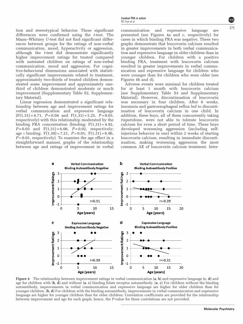
Linear regression demonstrated a significant rela-tionship between age and improvement ratings forverbal communication and expressive language(F(1,31) = 4.71, P = 0.04 and F(1,31) = 5.25, P = 0.03,respectively) with this relationship moderated by thebinding FRA concentration (binding: F(1,31) = 4.92,P = 0.03 and F(1,31) = 6.06, P = 0.02, respectively;age�binding: F(1,30) = 7.21, P = 0.01; F(1,31) = 6.96,P = 0.01, respectively). To examine the age effect in astraightforward manner, graphs of the relationshipbetween age and ratings of improvement in verbal
communication and expressive language arepresented (see Figures 4a and c, respectively) forcases in which binding FRA was negative. These twographs demonstrate that leucovorin calcium resultedin greater improvements in both verbal communica-tion and expressive language in older children than inyounger children. For children with a positivebinding FRA, treatment with leucovorin calciumresulted in greater improvements in verbal commu-nication and expressive language for children whowere younger than for children who were older (seeFigures 4b and d).
Adverse events were minimal for children treatedfor at least 1 month with leucovorin calcium(see Supplementary Table S3 and SupplementaryMaterial). However, discontinuation of leucovorinwas necessary in four children. After 6 weeks,insomnia and gastroesophageal reflux led to disconti-nuation of leucovorin calcium in one child. Inaddition, three boys, all of them concurrently takingrisperidone, were not able to tolerate leucovorincalcium for even a short period of time. These boysdeveloped worsening aggression (including self-injurious behavior in one) within 2 weeks of startingleucovorin calcium, resulting in immediate disconti-nuation, making worsening aggression the mostcommon AE of leucovorin calcium treatment. Inter-
Figure 4 The relationship between improvement ratings in verbal communication (a, b) and expressive language (c, d) andage for children with (b, d) and without (a, c) binding folate receptor autoantibody. (a, c) For children without the bindingautoantibody, improvements in verbal communication and expressive language are higher for older children than foryounger children. (b, d) For children with the binding autoantibody, improvements in verbal communication and expressivelanguage are higher for younger children than for older children. Correlation coefficients are provided for the relationshipbetween improvement and age for each graph; hence, the P-value for these correlations are not provided.
Cerebral FRA in autismRE Frye et al
375
Molecular Psychiatry

estingly, aggressive behavior was the indication forrisperidone, the medication that all three of thesechildren had in common. Of the 40 children whotolerated the leucovorin calcium well, only one wasconcurrently receiving risperidone (see Supplemen-tary Table S3 and Supplementary Material). Althoughsome parents rated mood (5%), stereotypical behavior(5%) and hyperactivity (17%) as mildly worse on theClinical Global Impression Scale in some children(Figure 3), they did not report these as AEs whenspecifically asked about AEs.
Discussion
This study is the first to examine FRA prevalence inchildren with ASD and to measure, in a controlledmanner, the response to leucovorin calcium in FRA-positive children. We also reviewed the clinicalcharacteristics of FRA-positive children and thecorrelation between FRA and CSF 5MTHF concentra-tions. Overall, this study demonstrates an unexpect-edly high FRA prevalence and a favorable response toleucovorin calcium in FRA-positive children withASD.
The prevalence of FRAs in ASDSeveral studies have reported the presence of FRAs inchildren with ASD.2–7 In these studies, children withASD had both CFD and significant neurologicalabnormalities. In this case series, a high FRAprevalence was found in a group of children withASD who did not have CFD or significant neurologi-cal abnormalities. The prevalence of blocking FRA(60%) was much higher than that reported in thecontrol population of women, estimated to be 4–7.2%in Spain38,39 and 9–13% in Ireland.23 To our knowl-edge, the prevalence of blocking FRA in the generalUS population has not been published, but has beenestimated to be 10–15% by Dr Quadros’ laboratory(unpublished data). The binding FRA prevalence of44% in this case series is also higher than thatreported in the general US population of 12%.23
Overall, our estimates suggest an elevated FRAprevalence in children with ASD; additional studiesshould be performed in ASD.
Characteristics of children with ASD and FRAsWe found few clinical differences between childrenwho were FRA positive compared with those whowere FRA negative. Thus, it seems to be difficult, atthis point, to distinguish children with ASD who areFRA positive from those who are FRA negative. Afamily history of autoimmune disorders was higher inchildren who were FRA negative compared withthose who were FRA positive. This does not supportthe idea that disorders associated with the FRA, suchas CFD, are autoimmune disorders. In fact, the lack ofany association between FRA-positive children andother autoimmune markers or autoantibodies pointsaway from the FRA being associated with classicalautoimmune disease. Given that a case of juvenile
rheumatoid arthritis was associated with CFD,40 it isclear that classical autoimmune disorders can resultin FRA elevations, but our data suggest that this maynot be common.
The blocking FRA concentration was found todecrease with increasing age. The reason for thisfinding is not clear. As a milk-free diet has beenreported to reduce blocking FRA concentrations,5 it ispossible that some children may have self-restrictedtheir milk intake as they became older. However, asmost of the children in this series, irrespective of age,were maintained on milk-free diets, this is unlikely. Itis also possible that this finding is associated with theunderlying pathophysiology that produces FRAs. Asthe onset of ASD is early in life (around toddler age)with improvement in some in early childhood, it ispossible that the peak of the pathophysiology thatcauses ASD (and potentially causes the FRA eleva-tions) could be most active early in life and decreasewith age.
Serum FRAs and CSF 5MTHF concentrationsIn this study, we found an inverse relationshipbetween the blocking FRA and CSF 5MTHF concen-trations, confirming previous findings.5 Given that thenormative mean CFD 5MTHF concentration is82±31 nmol l�1 (range: 44–181 nmol l�1)5, even thehighest CSF 5MTHF concentration in this study(81 nmol l�1 in a child with no blocking FRA) wasbelow the reported mean in normal children.
The estimated mean CSF 5MTHF with no blockingFRA (72.3 nmol l�1) was well below the mean con-centration reported for normal controls. Given thatthe CSF 5MTHF was estimated to be below normal,even in children without blocking FRAs, it is possiblethat children with ASD, in general, might have alower than normal CSF 5MTHF concentration. Thiswould be consistent with many studies that havereported folate-related abnormalities in children withASD.9,10,41–43 More importantly, children with ASDmay be predisposed to abnormalities in the transportof folates across the blood–brain barrier, irrespectiveof the presence of FRAs, as one study reported RFCgenetic variants associated with ASD9 and mitochon-drial disorders, the most common metabolic disorderin ASD,33 also may result in compromised FRafunction.26 Given that children with ASD alreadyhave abnormalities with folate metabolism, it is likelythat FRAs act synergistically with other abnormalitiesto reduce CSF 5MTHF concentration.
Treatment with leucovorinTreatment with leucovorin calcium in FRA-positivechildren with ASD was well tolerated and beneficialfor cognitive-behavioral functions, including verbalcommunication, receptive and expressive language,attention and stereotypical behavior. Approximatelytwo-thirds of the treated children were perceived asobtaining some benefit and about one-third had amoderate or much improvement in these cognitive-behavioral dimensions (Supplementary Table S2).
Cerebral FRA in autismRE Frye et al
376
Molecular Psychiatry

The mean length of treatment was 4 months. As somestudies have reported that 1–3 years of treatment withleucovorin calcium is required in children with CFDto obtain maximal improvements,4,25 it is possible thata longer treatment period would have resulted inadditional benefits.
The level of binding FRA and age seemed to affectthe degree of improvement for verbal communicationand expressive language. It has been suggested thatyounger children are relatively more responsive toleucovorin calcium than older children,2 but our dataindicate that this might depend on the binding FRAconcentration. For children negative for the bindingFRA, younger children improved less than olderchildren. However, these findings should be inter-preted with caution as it is very possible that thefollow-up period was not long enough to provide thenecessary time for optimal improvement.
As FRA concentrations were not significantlydifferent between children who did and did notundergo an LP, and all children who had an LP had aCSF 5MTHF concentration below the normativemean, it is likely that many FRA-positive childrenin this study also had a below-average CSF 5MTHFconcentration. Some investigators have reported sig-nificant clinical improvements with leucovorin cal-cium treatment in children with low normal CSF5MTHF concentrations.44 As the presence of even alow-blocking FRA concentration may be associatedwith a low CSF 5MTHF concentration, a low-blockingFRA titer may be clinically significant in somechildren with ASD, consistent with previous sugges-tions.5 The findings of this study suggest thatempirical treatment with leucovorin calcium withoutperforming an LP may be a reasonable and non-invasive approach in FRA-positive children withASD.
Leucovorin calcium was generally well-tolerated.The most common AE was worsening aggressivebehavior, which may have been associated withconcurrent treatment with risperidone. Although itis not clear why this side effect occurred, the rapiddevelopment of behavioral changes in these childrensuggests that it may have been due to an increase inCNS dopamine secondary to increased 5MTHF ortetrahydrobiopterin in the CNS, which then may havereduced the dopamine receptor-blocking effect ofrisperidone.45 Another possible cause of these behav-ioral changes may have been due to an inhibition ofmitochondrial function by risperidone.46,47 Animalmodels of mitochondrial dysfunction report similaraggressive behavior.48,49 However, for children whotolerated leucovorin calcium for at least 1 month,there was no indication of increased aggression. Thus,for children not concurrently receiving anti-psychoticmedications and without aggressive behavior, empiri-cal treatment of leucovorin calcium may be a reason-able course of treatment in FRA-positive childrenwith ASD.
Given that the RFC provides a pathway forleucovorin calcium to enter the CNS in children
who have reduced transport of folate across theblood–brain barrier due to the FRA, we assume thatthe effect of leucovorin calcium is through an increasein 5MTHF in the CNS. This is supported by studiesdemonstrating an increase in CNS 5MTHF concentra-tions after the use of leucovorin.2,4 However, the RFCis an important folate carrier in other tissues such asthe kidney and intestine. As gastrointestinal symp-toms have been associated with more aberrantbehavior in children with ASD,50 improvements ingastrointestinal function could result in improvedbehavior. Improvements in kidney function couldprovide better filtration of toxic metabolites asso-ciated with metabolic conditions that have beenassociated with ASD, thereby improving generalphysiology. In addition, leucovorin has also beendemonstrated to improve markers of oxidative stressin children with ASD.51 Thus, improvements noted asa result of leucovorin calcium in some children couldbe the result of an effect of leucovorin calcium onnon-CNS organs.
FRAs and ASDThe high prevalence of FRAs in this study isconsistent with the literature that documents eleva-tions in various autoantibodies in both children withASD and mothers of children with ASD.52,53 Althoughvarious autoantibodies have been reported in childrenwith ASD, the clinical significance of many of theseautoantibodies has not been fully documented, leav-ing open the possibility that some autoantibodies maybe an epiphenomenon of immune dysfunction.53 Inthis study, we have demonstrated that the serum-blocking FRA concentration can modulate the CSF5MTHF concentration and that leucovorin calciumtreatment results in cognitive-behavioral improve-ments. Thus, FRAs should be added to the list ofautoantibodies that may have clinical significance inthe cognitive-behavioral features of ASD, such asautoantibodies to GABAergic54 and cerebellar55 neu-rons in children with ASD and antibodies to fetalbrain proteins in mothers of children with ASD.56
It is likely that FRAs are not the only culprit inchildren with ASD, which result in depressed CSF5MTHF levels, but rather FRAs may work synergis-tically with other susceptibility factors, includingfactors that can depress systemic and central 5MTHFlevels,9,41–43,57 as well as factors that reduce thetransport of folates across the blood–brain bar-rier.9,26,33 For example, methylenetetrahydrofolatereductase gene variants reported in some childrenwith ASD41 can be associated with the decreasedproduction of 5MTHF. Interestingly, certain methylene-tetrahydrofolate reductase genotypes have been repor-ted to be associated with certain autistic behavior,including unusual body movements, hyperactivityand self-injurious behaviors.58 Furthermore, a geneticvariant in the dihydrofolate reductase enzyme, whichis involved in the process of producing 5MTHF43 andgenetic variants in the RFC9 have been reported insome children with ASD and could contribute to
Cerebral FRA in autismRE Frye et al
377
Molecular Psychiatry

impaired folate transport into the CNS. Finally,mitochondrial dysfunction, which has been associatedwith ASD,33 can contribute to reduced transport offolate across the blood–brain barrier. Gastrointestinalproblems reported in some children with ASD couldcompound this problem. For example, abnormal levelsof certain gut bacteria reported in some children withASD, including Clostridia species,59,60 can produceenteric short-chain fatty acids61 that have been shownto contribute to autistic-like behavior in animalmodels.62 These fatty acids can deplete carnitine storesand lead to secondary mitochondrial dysfunction.63
Thus, it is very possible that children who possess oneor more of these susceptibility factors could be atincreased risk for depressed CSF 5MTHF concentra-tions if FRAs develop during childhood. As FRa isessential for folate transport into the CNS whenextracellular folate concentrations are low,20 blockingof the FRa by FRA may be especially detrimental underconditions of low extracellular folate availability.
In addition, the timing of the development of FRAmay be a crucial factor in the phenotypic expressionof reduced CNS folate. As the FRA does have non-zero prevalence in non-ASD adult populations23,38,39
and is found in some siblings and parents of FRA-positive children with ASD, the mere presence ofFRAs does not necessarily cause ASD. Indeed, it islikely that FRAs appear during a period of rapiddevelopment, when folate is critically required forbrain growth. As human FRAs cross-react with folatereceptors in human, bovine and goat milk,5 it is verypossible that exposure to any of these milk products,as occurs during the first year of life, could triggerFRA production. Chronic elevations in FRA couldresult in a progressive depression in CNS folate overtime. Thus, symptoms of reduced CNS folate mayonly develop over a prolonged period of FRAelevations and reduced CNS folate.5 This would be atrue example of a genetic susceptibility interactingwith an environmental trigger. Unfortunately, wedid not have the opportunity to measure FRAconcentrations during the development of ASDsymptoms in these children, but it is possible thatFRA concentrations were higher during that timeperiod. Clearly, a longitudinal study would behelpful to determine whether there is a criticalwindow in which FRAs are most detrimental to braindevelopment.
Interestingly, these novel findings along with thehigh prevalence of FR autoantibodies may helpto integrate some apparently disparate findingsdescribed in some children with ASD. For example,folate deficiency in the CNS can lead to ASD in somechildren.4 Deficits in folate one-carbon metabolismhave been shown to promote methylation deficits andoxidative stress in some children with ASD.8–10 Thisin turn could be associated with oxidative stress inthe ASD brain.13,64 Moreover, chronic oxidative stresscan contribute to mitochondrial dysfunction,15 whichhas also been reported in the brains of some childrenwith ASD.16 Abnormal populations of gut bacteria
could contribute to increased production of entericshort-chain fatty acids, which could enhance theadverse effects of CNS folate deficiency and depletecarnitine levels, which has been described in somechildren with ASD.65 Furthermore, dietary cow’s milkexposure may increase the production of theseautoantibodies and help explain why some childrenwith ASD improve on a milk-free diet.66 Thesefindings point to metabolic-, environmental- andgastrointestinal-related factors in ASD, which mayhelp explain ASD behaviors and brain changes, andserve as a rationale for additional experimentationand study in these areas.
CFD: a disorder on a continuum?To our knowledge, this is the first study to report theprevalence of FRAs in a sample of children with ASDwho did not possess significant neurological abnorm-alities. The findings of this study suggest that CFDmay exist on a continuum, with less severely affectedchildren developing ASD, and more significantlyaffected children (that is, those with the lowest CSF5MTHF levels) possessing more severe neurologicalabnormalities including classical CFD syndrome.Additional studies examining children with ASDand children with CFD are required to explore thispossibility.
Study limitationsThis case series had several notable limitations,including a high male-to-female ratio (9:1), as com-pared with the more typical ratio for children withASD of 4.5:167 or CFD of 2.5–3:1.2,4,5 In addition,both clinics included in this report primarilyserve children with ASD who have persistentcognitive-behavioral deficits. This suggests that thesamples may be subject to referral bias. Future studiesare required to address whether a general ASDpopulation also has an elevated FRA prevalence.Even if the presence of FRA is limited to a smallersubset of the ASD population than reported here, thesignificant response to leucovorin calcium in somechildren suggests that this in an important abnorm-ality found in the ASD population that requiresfurther study.
This study was not a clinical trial and neither thephysicians nor the parents were blinded to treatment.In additional, our controls were not given a placebo;hence, the magnitude of the placebo effect was nottaken into account, and we used a subjective parentalreport rather than objective assessment measures.Double-blind, placebo-controlled studies are requiredto confirm and expand upon the findings of this caseseries.
Conclusions
Several studies have reported the presence of FRAs inchildren with ASD who had significant neurologicalabnormalities.2–7 In this study, an unexpectedly highprevalence (75.3%) of FRAs was found in 93 children
Cerebral FRA in autismRE Frye et al
378
Molecular Psychiatry

with ASD without significant neurological abnormal-ities. In 16 children, the blocking FRA concentrationwas correlated with the CSF 5MTHF concentration,which was below the normative mean in each case.FRA-positive children who were treated with leucov-orin calcium demonstrated significant improvementsin verbal communication, receptive and expressivelanguage, attention and stereotypical behavior com-pared with controls. The findings of this studysuggest that empirical treatment with leucovorincalcium without performing an LP may be a reason-able and non-invasive approach in FRA-positivechildren with ASD. This study also supports furtherinvestigations of folate receptor autoimmunity andleucovorin calcium treatment in ASD. Although thereis an animal model of hereditary folate malabsorp-tion,68 there is no animal model of CFD or the clinicaleffects of FRAs at the current time. Such a modelwould be very helpful is validating the clinicalsignificance of the FRA findings and leucovorintreatment. However, at this time, from the currentevidence, it appears that FRA testing can lead totreatment that may improve the lives of children withASD. As FRAs appears to be highly prevalent inchildren with ASD, we recommend that FRA testingshould be considered in all patients with ASD.
Conflict of interest
Daniel Rossignol has two children with ASD and is apracticing primary care physician who treats ASDchildren with standard and integrative treatments. Inthe past, Dr Frye has provided expert opinion forchild neurology cases for both the plaintiff anddefendant, including cases with children sufferingfrom mitochondrial disorders. All proceeds from suchwork are provided to the Department of Pediatrics atthe University of Texas with some of these fundssupporting Dr Frye’s research. Drs Quadros andSequeira are inventors on a US patent no. 7846672,issued to the Research Foundation of the StateUniversity of New York. Dr James has no conflict ofinterest to declare.
Acknowledgments
We thank Michelle R Christie and Jamie Chu for theirtechnical assistance. This study was supported, inpart, by funding from the Autism Research Instituteand the Jane Botsford Johnson Foundation.
References
1 Ramaekers VT, Hausler M, Opladen T, Heimann G, Blau N.Psychomotor retardation, spastic paraplegia, cerebellar ataxia anddyskinesia associated with low 5-methyltetrahydrofolate in cere-brospinal fluid: a novel neurometabolic condition responding tofolinic acid substitution. Neuropediatrics 2002; 33: 301–308.
2 Ramaekers VT, Blau N. Cerebral folate deficiency. Dev Med ChildNeurol 2004; 46: 843–851.
3 Moretti P, Peters SU, Del Gaudio D, Sahoo T, Hyland K, BottiglieriT et al. Brief report: autistic symptoms, developmental regression,
mental retardation, epilepsy, and dyskinesias in CNS folatedeficiency. J Autism Dev Disord 2008; 38: 1170–1177.
4 Ramaekers VT, Blau N, Sequeira JM, Nassogne MC, Quadros EV.Folate receptor autoimmunity and cerebral folate deficiency inlow-functioning autism with neurological deficits. Neuropedia-trics 2007; 38: 276–281.
5 Ramaekers VT, Sequeira JM, Blau N, Quadros EV. A milk-freediet downregulates folate receptor autoimmunity in cerebralfolate deficiency syndrome. Dev Med Child Neurol 2008; 50:346–352.
6 Ramaekers VT, Rothenberg SP, Sequeira JM, Opladen T, Blau N,Quadros EV et al. Autoantibodies to folate receptors in thecerebral folate deficiency syndrome. N Engl J Med 2005; 352:1985–1991.
7 Moretti P, Sahoo T, Hyland K, Bottiglieri T, Peters S, del Gaudio Det al. Cerebral folate deficiency with developmental delay, autism,and response to folinic acid. Neurology 2005; 64: 1088–1090.
8 James SJ, Cutler P, Melnyk S, Jernigan S, Janak L, Gaylor DW et al.Metabolic biomarkers of increased oxidative stress and impairedmethylation capacity in children with autism. Am J Clin Nutr2004; 80: 1611–1617.
9 James SJ, Melnyk S, Jernigan S, Cleves MA, Halsted CH, Wong DHet al. Metabolic endophenotype and related genotypes areassociated with oxidative stress in children with autism. Am JMed Genet B Neuropsychiatr Genet 2006; 141: 947–956.
10 Melnyk S, Fuchs GJ, Schulz E, Lopez M, Kahler SG, Fussell JJ et al.Metabolic imbalance associated with methylation dysregulationand oxidative damage in children with autism. J Autism DevDisord published online 26 April 2011 (e-pub ahead of print).
11 Pogribny IP, Karpf AR, James SR, Melnyk S, Han T, Tryndyak VP.Epigenetic alterations in the brains of Fisher 344 rats induced bylong-term administration of folate/methyl-deficient diet. Brain Res2008; 1237: 25–34.
12 Nagarajan RP, Hogart AR, Gwye Y, Martin MR, LaSalle JM.Reduced MeCP2 expression is frequent in autism frontal cortexand correlates with aberrant MECP2 promoter methylation.Epigenetics 2006; 1: e1–11.
13 Sajdel-Sulkowska EM, Xu M, McGinnis W, Koibuchi N. Brainregion-specific changes in oxidative stress and neurotrophinlevels in autism spectrum disorders (ASD). Cerebellum 2010; 10:43–48.
14 Ramaekers VT, Hansen SI, Holm J, Opladen T, Senderek J, HauslerM et al. Reduced folate transport to the CNS in female Rettpatients. Neurology 2003; 61: 506–515.
15 Chauhan A, Chauhan V. Oxidative stress in autism. Pathophysiol-ogy 2006; 13: 171–181.
16 Chauhan A, Gu F, Essa MM, Wegiel J, Kaur K, Ted Brown W et al.Brain region-specific deficit in mitochondrial electron transportchain complexes in children with autism. J Neurochem 2011; 117:209–220.
17 Duthie SJ, Hawdon A. DNA instability (strand breakage, uracilmisincorporation, and defective repair) is increased by folicacid depletion in human lymphocytes in vitro. FASEB J 1998;12: 1491–1497.
18 Crott JW, Mashiyama ST, Ames BN, Fenech M. The effect of folicacid deficiency and MTHFR C677T polymorphism on chromo-some damage in human lymphocytes in vitro. Cancer EpidemiolBiomarkers Prev 2001; 10: 1089–1096.
19 Iskandar BJ, Rizk E, Meier B, Hariharan N, Bottiglieri T, Finnell RHet al. Folate regulation of axonal regeneration in the rodent centralnervous system through DNA methylation. J Clin Invest 2010; 120:1603–1616.
20 Wollack JB, Makori B, Ahlawat S, Koneru R, Picinich SC, Smith Aet al. Characterization of folate uptake by choroid plexus epithelialcells in a rat primary culture model. J Neurochem 2008; 104:1494–1503.
21 Solanky N, Requena Jimenez A, D’Souza SW, Sibley CP, Glazier JD.Expression of folate transporters in human placenta andimplications for homocysteine metabolism. Placenta 2010; 31:134–143.
22 Weitman SD, Weinberg AG, Coney LR, Zurawski VR, Jennings DS,Kamen BA. Cellular localization of the folate receptor: potentialrole in drug toxicity and folate homeostasis. Cancer Res 1992; 52:6708–6711.
Cerebral FRA in autismRE Frye et al
379
Molecular Psychiatry

23 Molloy AM, Quadros EV, Sequeira JM, Troendle JF, Scott JM,Kirke PN et al. Lack of association between folate-receptorautoantibodies and neural-tube defects. N Engl J Med 2009; 361:152–160.
24 Berrocal-Zaragoza MI, Fernandez-Ballart JD, Murphy MM, Ca-valle-Busquets P, Sequeira JM, Quadros EV. Association betweenblocking folate receptor autoantibodies and subfertility. FertilSteril 2009; 91(4 Suppl): 1518–1521.
25 Ramaekers VT, Weis J, Sequeira JM, Quadros EV, Blau N.Mitochondrial complex I encephalomyopathy and cerebral5-methyltetrahydrofolate deficiency. Neuropediatrics 2007; 38:184–187.
26 Garcia-Cazorla A, Quadros EV, Nascimento A, Garcia-Silva MT,Briones P, Montoya J et al. Mitochondrial diseases associated withcerebral folate deficiency. Neurology 2008; 70: 1360–1362.
27 O’Byrne MR, Au KS, Morrison AC, Lin JI, Fletcher JM, OstermaierKK et al. Association of folate receptor (FOLR1, FOLR2, FOLR3)and reduced folate carrier (SLC19A1) genes with meningomyelo-cele. Birth Defects Res 2010; 88: 689–694.
28 Matherly LH, Hou Z, Deng Y. Human reduced folate carrier:translation of basic biology to cancer etiology and therapy. CancerMetastasis Rev 2007; 26: 111–128.
29 Yuasa H, Inoue K, Hayashi Y. Molecular and functional character-istics of proton-coupled folate transporter. J Pharma Sci 2009; 98:1608–1616.
30 Hou Z, Matherly LH. Oligomeric structure of the human reducedfolate carrier: identification of homo-oligomers and dominant-negative effects on carrier expression and function. J Biol Chem2009; 284: 3285–3293.
31 Hansen FJ, Blau N. Cerebral folate deficiency: life-changingsupplementation with folinic acid. Mol Genet Metab 2005; 84:371–373.
32 APA. Diagnostic and Statistical Manual of Mental Disorders, 4thedn American Psychiatric Association: Washington, DC, 1994.
33 Rossignol DA, Frye RE. Mitochondrial dysfunction in autismspectrum disorders: a systematic review and meta-analysis.Mol Psychiatry; published online 25 January 2011 (e-pub aheadof print).
34 Frye RE, Rossignol DA. Mitochondrial dysfunction can connectthe diverse medical symptoms associated with autism spectrumdisorders. Pediatric Res 2011; 69(5 Pt 2): 41R–47R.
35 Guy W: ECDEU Assessment Manual for Psychopharmacology -Revised (DHEW Publ No ADM 76-338). Rockville, MD, U.S.Department of Health, Education, and Welfare, Public HealthService, Alcohol, Drug Abuse, and Mental Health Administration,NIMH Psychopharmacology Research Branch, Division of Extra-mural Research Programs, 1976, pp 218–222.
36 Hulley SB, Cummings SR, Browner WS, Grady DG, Newman TB.Designing Clinical Research, 3rd edn Lippincott Williams &Wilkins: Philadephia, 2006.
37 Faul F, Erdfelder E, Lang AG, Buchner A. G*Power 3:a flexible statistical power analysis for the social, behavioral, andbiomedical sciences. Behavior Res Methods 2007; 39: 175–191.
38 Berrocal-Zaragoza MI, Fernandez-Ballart JD, Murphy MM,Cavalle-Busquets P, Sequeira JM, Quadros EV. Association betweenblocking folate receptor autoantibodies and subfertility. Fertil Steril2009; 91: 1518–1521.
39 Berrocal-Zaragoza MI, Murphy MM, Ceruelo S, Quadros EV,Sequeira JM, Fernandez-Ballart JD. High milk consumers have anincreased risk of folate receptor blocking autoantibody productionbut this does not affect folate status in Spanish men and women.J Nutr 2009; 139: 1037–1041.
40 Koenig MK, Perez M, Rothenberg S, Butler IJ. Juvenile onsetcentral nervous system folate deficiency and rheumatoid arthritis.J Child Neurol 2008; 23: 106–107.
41 Mohammad NS, Jain JM, Chintakindi KP, Singh RP, Naik U, AkellaRR. Aberrations in folate metabolic pathway and altered suscept-ibility to autism. Psychiatr Genet 2009; 19: 171–176.
42 Boris M, Goldblatt A, Galanko J, James SJ. Association of MTHFR genevariants with autism. J Am Physicians Surgeons 2004; 9: 106–108.
43 Adams M, Lucock M, Stuart J, Fardell S, Baker K, Ng X.Preliminary evidence for involvement of the folate gene poly-morphism 19bp deletion-DHFR in occurrence of autism. NeurosciLett 2007; 422: 24–29.
44 Hyland K, Shoffner J, Heales SJ. Cerebral folate deficiency.J Inherit Metab Dis 2010; 33: 563–570.
45 Miller AL. The methylation, neurotransmitter, and antioxidantconnections between folate and depression. Altern Med Rev 2008;13: 216–226.
46 Neustadt J, Pieczenik SR. Medication-induced mitochondrialdamage and disease. Mol Nutr Food Res 2008; 52: 780–788.
47 Ahn MS, Sims KB, Frazier JA. Risperidone-induced psychosis anddepression in a child with a mitochondrial disorder. J ChildAdolesc Psychopharmacol 2005; 15: 520–525.
48 Thomas RH, Foley KA, Mepham JR, Tichenoff LJ, Possmayer F,MacFabe DF. Altered brain phospholipid and acylcarnitine profilesin propionic acid infused rodents: further development of a potentialmodel of autism spectrum disorders. J Neurochem 2010; 113: 515–529.
49 MacFabe DF, Cain DP, Rodriguez-Capote K, Franklin AE, HoffmanJE, Boon F et al. Neurobiological effects of intraventricularpropionic acid in rats: possible role of short chain fatty acids onthe pathogenesis and characteristics of autism spectrum disorders.Behav Brain Res 2007; 176: 149–169.
50 Nikolov RN, Bearss KE, Lettinga J, Erickson C, Rodowski M, AmanMG et al. Gastrointestinal symptoms in a sample of children withpervasive developmental disorders. J Autism Dev Disord 2009; 39:405–413.
51 James SJ, Melnyk S, Fuchs G, Reid T, Jernigan S, Pavliv O et al.Efficacy of methylcobalamin and folinic acid treatment onglutathione redox status in children with autism. Am J Clin Nutr2009; 89: 425–430.
52 Croen LA, Braunschweig D, Haapanen L, Yoshida CK, Fireman B,Grether JK et al. Maternal mid-pregnancy autoantibodies to fetalbrain protein: the early markers for autism study. Biol Psychiatry2008; 64: 583–588.
53 Wills S, Cabanlit M, Bennett J, Ashwood P, Amaral D, Van deWater J. Autoantibodies in autism spectrum disorders (ASD). AnnNY Acad Sci 2007; 1107: 79–91.
54 Wills S, Rossi CC, Bennett J, Cerdeno VM, Ashwood P, Amaral DGet al. Further characterization of autoantibodies to GABAergicneurons in the central nervous system produced by a subset ofchildren with autism. Mol Autism 2011; 2: 5.
55 Wills S, Cabanlit M, Bennett J, Ashwood P, Amaral DG, Van deWater J. Detection of autoantibodies to neural cells of thecerebellum in the plasma of subjects with autism spectrumdisorders. Brain Behav Immun 2009; 23: 64–74.
56 Braunschweig D, Ashwood P, Krakowiak P, Hertz-Picciotto I,Hansen R, Croen LA et al. Autism: maternally derived antibodiesspecific for fetal brain proteins. Neurotoxicology 2008; 29: 226–231.
57 James SJ, Melnyk S, Jernigan S, Hubanks A, Rose S, Gaylor DW.Abnormal transmethylation/transsulfuration metabolism andDNA hypomethylation among parents of children with autism.J Autism Dev Disord 2008; 38: 1966–1975.
58 Goin-Kochel RP, Porter AE, Peters SU, Shinawi M, Sahoo T,Beaudet AL. The MTHFR 677C– > T polymorphism and behaviorsin children with autism: exploratory genotype-phenotype correla-tions. Autism Res 2009; 2: 98–108.
59 Finegold SM, Molitoris D, Song Y, Liu C, Vaisanen ML, Bolte Eet al. Gastrointestinal microflora studies in late-onset autism.Clin Infect Dis 2002; 35(Suppl 1): S6–S16.
60 Finegold SM. State of the art; microbiology in health and diseaseintestinal bacterial flora in autism. Anaerobe; published online16 April 2011 (e-pub ahead of print).
61 Williams BL, Hornig M, Buie T, Bauman ML, Cho Paik M,Wick I et al. Impaired carbohydrate digestion and transport andmucosal dysbiosis in the intestines of children with autism andgastrointestinal disturbances. PLoS One 2011; 6: e24585.
62 MacFabe DF, Thomas RH, Foley KA, Mepham JR, Tichenoff LJ,Possmayer F. Altered brain phospholipid and acylcarnitineprofiles in propionic acid infused rodents: further developmentof a potential model of autism spectrum disorders. J Neurochem2010; 113: 515–529.
63 MacFabe DF, Rodrıguez-Capote K, Hoffman JE, Franklin AE,Mohammad-Asef Y, Taylor AR et al. A novel rodent model ofautism: intraventricular infusions of propionic acid increaselocomotor activity and induce neuroinflammation and oxidativestress in discrete regions of adult rat brain. Am J Biochem Biotech2008; 4: 146–166.
Cerebral FRA in autismRE Frye et al
380
Molecular Psychiatry

64 Sajdel-Sulkowska EM, Xu M, Koibuchi N. Increase in cerebellarneurotrophin-3 and oxidative stress markers in autism. Cerebel-lum 2009; 8: 366–372.
65 Filipek PA, Juranek J, Nguyen MT, Cummings C, Gargus JJ. Relativecarnitine deficiency in autism. J Autism Dev Disord 2004; 34: 615–623.
66 Whiteley P, Haracopos D, Knivsberg AM, Reichelt KL, Parlar S,Jacobsen J et al. The ScanBrit randomised, controlled, single-blindstudy of a gluten- and casein-free dietary intervention for childrenwith autism spectrum disorders. Nutr Neurosci 2010; 13: 87–100.
67 Rice C. Prevalence of autism spectrum disorders –autism anddevelopmental disabilities monitoring network, United States,2006. MMWR Surveill Summ 2009; 58: 1–20.
68 Salojin KV, Cabrera RM, Sun W, Chang WC, Lin C, Duncan L et al.A mouse model of hereditary folate malabsorption: deletion ofthe PCFT gene leads to systemic folate deficiency. Blood 2011;117: 4895–4904.
This work is licensed under the CreativeCommons Attribution-NonCommercial-
No Derivative Works 3.0 Unported License. To viewa copy of this license, visit http://creativecommons.org/licenses/by-nc-nd/3.0/
Supplementary Information accompanies the paper on the Molecular Psychiatry website (http://www.nature.com/mp)
Cerebral FRA in autismRE Frye et al
381
Molecular Psychiatry
Related Documents