REVIEW ARTICLE published: 01 September 2011 doi: 10.3389/fneur.2011.00053 Cerebellum and ocular motor control Amir Kheradmand 1 * and David S. Zee 1,2,3,4 1 Department of Neurology,The Johns Hopkins University School of Medicine, Baltimore, MD, USA 2 Department of Ophthalmology,The Johns Hopkins University School of Medicine, Baltimore, MD, USA 3 Department of Otolaryngology–Head and Neck Surgery,The Johns Hopkins University School of Medicine, Baltimore, MD, USA 4 Department of Neuroscience,The Johns Hopkins University School of Medicine, Baltimore, MD, USA Edited by: Sergio Carmona, Instituto de Neurociencias de Buenos Aires INEBA, Argentina Reviewed by: Sergio Carmona, Instituto de Neurociencias de Buenos Aires INEBA, Argentina Matthew J. Thurtell, University of Iowa, USA Matt Stewart, Johns Hopkins Hospital, USA *Correspondence: Amir Kheradmand , Oculomotor Lab, Department of Neurology,The Johns Hopkins Hospital, Path 2-210, 600 North Wolfe Street, Baltimore, MD 21287, USA. e-mail: [email protected] An intact cerebellum is a prerequisite for optimal ocular motor performance. The cerebel- lum fine-tunes each of the subtypes of eye movements so they work together to bring and maintain images of objects of interest on the fovea. Here we review the major aspects of the contribution of the cerebellum to ocular motor control. The approach will be based on structural–functional correlation, combining the effects of lesions and the results from physiologic studies, with the emphasis on the cerebellar regions known to be most closely related to ocular motor function: (1) the flocculus/paraflocculus for high-frequency (brief) vestibular responses, sustained pursuit eye movements, and gaze holding, (2) the nodu- lus/ventral uvula for low-frequency (sustained) vestibular responses, and (3) the dorsal oculomotor vermis and its target in the posterior portion of the fastigial nucleus (the fastigial oculomotor region) for saccades and pursuit initiation. Keywords: saccade, vestibular, pursuit, flocculus, paraflocculus, nodulus, vermis, fastigial INTRODUCTION The cerebellum plays a pivotal role in the control of eye move- ments. Its core function is to optimize ocular motor performance so that images of objects of interest are promptly brought to the fovea – where visual acuity is best – and kept quietly there, so the brain has time to analyze and interpret the visual scene. The cerebellum has both immediate, on-line functions to make each individual movement accurate, and long-term, adaptive functions to keep ocular motor responses correctly calibrated to the stimuli that drive them. ANATOMICAL APPROACH TO THE CEREBELLUM AND OCULAR MOTOR CONTROL 1 The approach here will be based upon structural–functional cor- relations: cerebellar influences on eye movements will be discussed by anatomical regions (see Table 1 and Figure 1). Since the cere- bellum has an important role in motor learning that promotes adaptation and compensation for lesions, the functional effects of focal cerebellar lesions may not strictly represent the contribu- tion of the cerebellum to the control of eye movements in intact subjects. With this caveat, the ocular motor functions of the cere- bellum will be interpreted using the combination of the effects of acute and of chronic lesions, and the results from physiological studies. Here the emphasis will be on the areas within the cerebel- lum about which we know most: the flocculus/paraflocculus, the nodulus/ventral uvula (lobules IX and X of the cerebellar vermis), and the dorsal oculomotor vermis (OMV; lobules V–VII) and its 1 Some of this material is modified from Zee and Walker (2009). target in the posterior portion of the underlying fastigial nucleus called the fastigial oculomotor region (FOR). Recent studies also implicate more lateral regions of the cerebellar hemispheres in ocular motor control. The cerebellum has a distinctive anatomical organization. Purkinje cells of the cerebellar cortex primarily project to and inhibit cells within the underlying deep cerebellar nuclei; the vestibulocerebellum (flocculus, paraflocculus, nodulus, and uvula) also projects to the vestibular nuclei some of which can be thought of as “displaced” deep cerebellar nuclei. Axons arising from the fastigial nuclei decussate within the cerebellum and terminate pri- marily in the contralateral vestibular or premotor brain stem nuclei while Purkinje cells in the vestibulocerebellum project primarily to the ipsilateral brain stem nuclei. There is a rich innervation to the cerebellar cortex from the inferior olive, through climbing fibers directly to Purkinje cells, and from premotor brain stem nuclei (as well as the vestibular nerve), through mossy fibers to cerebellar granule cells – which in turn project to Purkinje cells via parallel fibers. There are also direct climbing fiber and mossy fiber inputs to the deep cerebellar nuclei, so that the cerebellar cortex can be thought of as a par- allel pathway that oversees and influences (via the projections of its Purkinje cells) the direct flow of information to and from the brain stem through the deep cerebellar nuclei. VESTIBULOCEREBELLUM: THE FLOCCULUS/PARAFLOCCULUS The flocculus and paraflocculus (or tonsil) together with the cau- dal portions of the cerebellar vermis (nodulus and uvula) are part of the oldest portion of the cerebellum or the archicerebellum, also called the vestibulocerebellum (Figure 1). Neurons within www.frontiersin.org September 2011 |Volume 2 | Article 53 | 1

Welcome message from author
This document is posted to help you gain knowledge. Please leave a comment to let me know what you think about it! Share it to your friends and learn new things together.
Transcript

REVIEW ARTICLEpublished: 01 September 2011doi: 10.3389/fneur.2011.00053
Cerebellum and ocular motor controlAmir Kheradmand 1* and David S. Zee1,2,3,4
1 Department of Neurology, The Johns Hopkins University School of Medicine, Baltimore, MD, USA2 Department of Ophthalmology, The Johns Hopkins University School of Medicine, Baltimore, MD, USA3 Department of Otolaryngology–Head and Neck Surgery, The Johns Hopkins University School of Medicine, Baltimore, MD, USA4 Department of Neuroscience, The Johns Hopkins University School of Medicine, Baltimore, MD, USA
Edited by:
Sergio Carmona, Instituto deNeurociencias de Buenos AiresINEBA, Argentina
Reviewed by:
Sergio Carmona, Instituto deNeurociencias de Buenos AiresINEBA, ArgentinaMatthew J. Thurtell, University ofIowa, USAMatt Stewart, Johns HopkinsHospital, USA
*Correspondence:
Amir Kheradmand, Oculomotor Lab,Department of Neurology, The JohnsHopkins Hospital, Path 2-210, 600North Wolfe Street, Baltimore, MD21287, USA.e-mail: [email protected]
An intact cerebellum is a prerequisite for optimal ocular motor performance. The cerebel-lum fine-tunes each of the subtypes of eye movements so they work together to bring andmaintain images of objects of interest on the fovea. Here we review the major aspectsof the contribution of the cerebellum to ocular motor control. The approach will be basedon structural–functional correlation, combining the effects of lesions and the results fromphysiologic studies, with the emphasis on the cerebellar regions known to be most closelyrelated to ocular motor function: (1) the flocculus/paraflocculus for high-frequency (brief)vestibular responses, sustained pursuit eye movements, and gaze holding, (2) the nodu-lus/ventral uvula for low-frequency (sustained) vestibular responses, and (3) the dorsaloculomotor vermis and its target in the posterior portion of the fastigial nucleus (the fastigialoculomotor region) for saccades and pursuit initiation.
Keywords: saccade, vestibular, pursuit, flocculus, paraflocculus, nodulus, vermis, fastigial
INTRODUCTIONThe cerebellum plays a pivotal role in the control of eye move-ments. Its core function is to optimize ocular motor performanceso that images of objects of interest are promptly brought to thefovea – where visual acuity is best – and kept quietly there, sothe brain has time to analyze and interpret the visual scene. Thecerebellum has both immediate, on-line functions to make eachindividual movement accurate, and long-term, adaptive functionsto keep ocular motor responses correctly calibrated to the stimulithat drive them.
ANATOMICAL APPROACH TO THE CEREBELLUM ANDOCULAR MOTOR CONTROL1
The approach here will be based upon structural–functional cor-relations: cerebellar influences on eye movements will be discussedby anatomical regions (see Table 1 and Figure 1). Since the cere-bellum has an important role in motor learning that promotesadaptation and compensation for lesions, the functional effectsof focal cerebellar lesions may not strictly represent the contribu-tion of the cerebellum to the control of eye movements in intactsubjects. With this caveat, the ocular motor functions of the cere-bellum will be interpreted using the combination of the effects ofacute and of chronic lesions, and the results from physiologicalstudies. Here the emphasis will be on the areas within the cerebel-lum about which we know most: the flocculus/paraflocculus, thenodulus/ventral uvula (lobules IX and X of the cerebellar vermis),and the dorsal oculomotor vermis (OMV; lobules V–VII) and its
1Some of this material is modified from Zee and Walker (2009).
target in the posterior portion of the underlying fastigial nucleuscalled the fastigial oculomotor region (FOR). Recent studies alsoimplicate more lateral regions of the cerebellar hemispheres inocular motor control.
The cerebellum has a distinctive anatomical organization.Purkinje cells of the cerebellar cortex primarily project to andinhibit cells within the underlying deep cerebellar nuclei; thevestibulocerebellum (flocculus,paraflocculus, nodulus, and uvula)also projects to the vestibular nuclei some of which can be thoughtof as “displaced” deep cerebellar nuclei. Axons arising from thefastigial nuclei decussate within the cerebellum and terminate pri-marily in the contralateral vestibular or premotor brain stem nucleiwhile Purkinje cells in the vestibulocerebellum project primarilyto the ipsilateral brain stem nuclei.
There is a rich innervation to the cerebellar cortex from theinferior olive, through climbing fibers directly to Purkinje cells,and from premotor brain stem nuclei (as well as the vestibularnerve), through mossy fibers to cerebellar granule cells – whichin turn project to Purkinje cells via parallel fibers. There are alsodirect climbing fiber and mossy fiber inputs to the deep cerebellarnuclei, so that the cerebellar cortex can be thought of as a par-allel pathway that oversees and influences (via the projections ofits Purkinje cells) the direct flow of information to and from thebrain stem through the deep cerebellar nuclei.
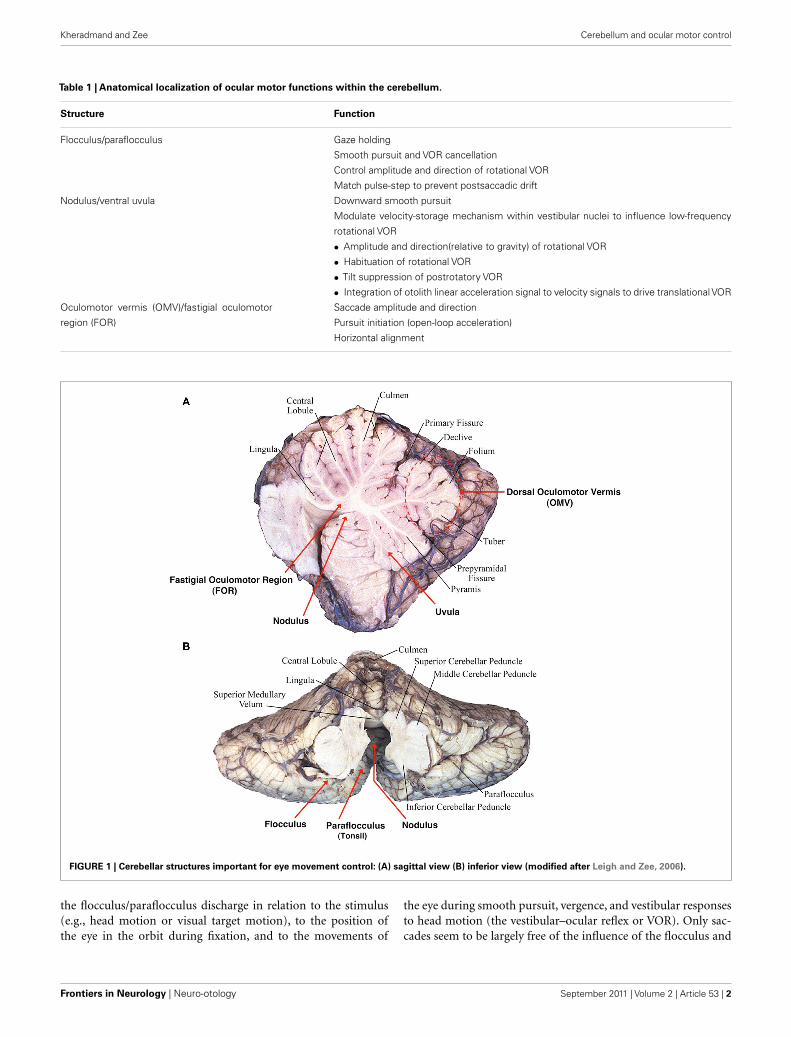
VESTIBULOCEREBELLUM: THE FLOCCULUS/PARAFLOCCULUSThe flocculus and paraflocculus (or tonsil) together with the cau-dal portions of the cerebellar vermis (nodulus and uvula) are partof the oldest portion of the cerebellum or the archicerebellum,also called the vestibulocerebellum (Figure 1). Neurons within
www.frontiersin.org September 2011 | Volume 2 | Article 53 | 1
Kheradmand and Zee Cerebellum and ocular motor control
Table 1 | Anatomical localization of ocular motor functions within the cerebellum.
Structure Function
Flocculus/paraflocculus Gaze holding
Smooth pursuit and VOR cancellation
Control amplitude and direction of rotational VOR
Match pulse-step to prevent postsaccadic drift
Nodulus/ventral uvula Downward smooth pursuit
Modulate velocity-storage mechanism within vestibular nuclei to influence low-frequency
rotational VOR
• Amplitude and direction(relative to gravity) of rotational VOR
• Habituation of rotational VOR
• Tilt suppression of postrotatory VOR
• Integration of otolith linear acceleration signal to velocity signals to drive translational VOR
Oculomotor vermis (OMV)/fastigial oculomotor
region (FOR)
Saccade amplitude and direction
Pursuit initiation (open-loop acceleration)
Horizontal alignment
FIGURE 1 | Cerebellar structures important for eye movement control: (A) sagittal view (B) inferior view (modified after Leigh and Zee, 2006).
the flocculus/paraflocculus discharge in relation to the stimulus(e.g., head motion or visual target motion), to the position ofthe eye in the orbit during fixation, and to the movements of
the eye during smooth pursuit, vergence, and vestibular responsesto head motion (the vestibular–ocular reflex or VOR). Only sac-cades seem to be largely free of the influence of the flocculus and
Frontiers in Neurology | Neuro-otology September 2011 | Volume 2 | Article 53 | 2
Kheradmand and Zee Cerebellum and ocular motor control
paraflocculus; they are the concern of the dorsal vermis (lobulesV–VII) and its lateral extension in the cerebellar hemispheres.Note, however, that brief, several hundred milliseconds drift ofthe eyes after each saccade (i.e., postsaccadic drift) is a character-istic abnormality following lesions in the flocculus/paraflocculus,arising from a mismatch between the innervation that drives theeye to its new position (the “pulse”) and the innervation that holdsit there (the “step”).
PURSUITLesions of the flocculus/paraflocculus impair smooth tracking ofa moving target, either when the head is still (smooth pursuit) orpassively moving (VOR cancellation; Zee et al., 1981; Belton andMcCrea, 2000, 2002; Rambold et al., 2002). Both the initiation ofpursuit and the response during sustained tracking can be affected.The floccular complex seems to play a lesser role with active headturns than passive head rotations during smooth tracking (Beltonand McCrea, 1999, 2004).
Complete lesions of the flocculus/paraflocculus lead to adecrease in gain (eye velocity/target velocity) of pursuit dur-ing steady-state (constant-velocity) tracking. Even with relativelycomplete floccular/parafloccular lesions pursuit function recoversconsiderably. This is likely mediated by other parts of the cere-bellum as studies in monkeys have shown that Purkinje cells inthe dorsal vermis and its lateral extension in the cerebellar hemi-sphere are also involved in the control of smooth pursuit, and theyshare some mossy and climbing fiber inputs with the paraflocculus(Ohki et al., 2009; Xiong et al., 2010). In humans, too, smooth pur-suit is affected by transcranial magnetic stimulation (TMS) of theskull over the dorsal vermis (Hashimoto and Ohtsuka, 1995; Oht-suka and Enoki, 1998). More caudal regions of the vermis (i.e., the
uvula and nodulus) are also implicated in the control of smoothtracking (Heinen and Keller, 1996; Walker et al., 2008b).
An inherent functional asymmetry for vertical pursuit eyemovements has been described within the flocculus. The majorityof Purkinje cells of the flocculus are active during downward asopposed to upward pursuit (Stone and Lisberger, 1990). This “up–down” asymmetry during vertical pursuit is also apparent duringfunctional imaging of the flocculus in healthy human subjects(Glasauer et al., 2009).
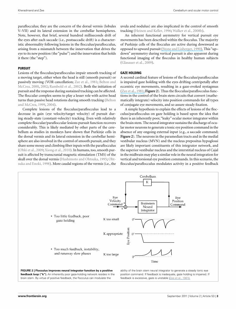
GAZE HOLDINGA second cardinal feature of lesions of the flocculus/paraflocculusis impaired gaze holding with the eyes drifting centripetally aftereccentric eye movements, resulting in a gaze-evoked nystagmus(Zee et al., 1981; Figure 2). Thus the flocculus/paraflocculus func-tions in the control of the brain stem circuits that convert (mathe-matically integrate) velocity into position commands for all typesof conjugate eye movements, and so assure steady fixation.
A simple hypothesis to explain the effects of lesions of the floc-culus/paraflocculus on gaze holding is based upon the idea thatthere is an inherently poor,“leaky” ocular motor integrator withinthe brain stem. The neural integrator sustains the discharge of ocu-lar motor neurons to generate a tonic eye position command in theabsence of any ongoing external input (e.g., a saccade command;Figure 2). The neurons in the paramedian tracts and in the medialvestibular nucleus (MVN) and the nucleus prepositus hypoglossiare likely important constituents of this integrator network, andthe superior vestibular nucleus and the interstitial nucleus of Cajalin the midbrain may play a similar role in the neural integration forvertical and torsional eye position commands. In this scenario, theflocculus/paraflocculus modulates activity in a positive feedback
FIGURE 2 | Flocculus improves neural integrator function by a positive
feedback loop (“k”). An inherently poor gaze-holding network resides in thebrain stem. By virtue of positive feedback, the flocculus can modulate the
ability of the brain stem neural integrator to generate a steady tonic eyeposition command. If feedback is inadequate, gaze holding is impaired. Iffeedback is excessive, gaze is unstable (Zee et al., 1981).
www.frontiersin.org September 2011 | Volume 2 | Article 53 | 3
Kheradmand and Zee Cerebellum and ocular motor control
loop around the brain stem neural integrator and improves itsperformance.
SPONTANEOUS NYSTAGMUSA third distinctive feature of lesions in the flocculus/paraflocculusis downbeat nystagmus, in which the eyes drift up (slow phase)and are brought back to the fixation target by a corrective down-ward saccade (quick phase). This form of nystagmus can belinked to the damage of the physiologic “up–down” asymmetryof floccular Purkinje cells (with predominant downward facili-tation) resulting in upward slow drift (Marti et al., 2005, 2008;Kalla et al., 2006; Hufner et al., 2007). An alternative hypoth-esis is based on the tonic inhibition by the flocculus upon theupward VOR (by inhibitory projections to the superior vestibularnucleus), and lack of corresponding projections from the flocculusto the brain stem structures that mediate downward vestibulo-ocular responses (reviewed in Leigh et al., 2002). According to thishypothesis, damage to the flocculus could release the tonic inhibi-tion upon the vestibular nuclei associated with the upward VORand therefore lead to slow, upward drifts of the eyes followed bycorrective, quick phases downwards (downbeat nystagmus).
A curious feature of the upward drift is the variability of itswaveform from subject to subject. For example, monkeys withexperimental flocculus/parafloccular lesions show downbeat nys-tagmus with either a decreasing- or an increasing-velocity wave-form (Zee et al., 1981). These different velocity profiles suggestdifferences in the effect of the lesion on the vertical neural integra-tor. If the integrator is sufficiently “leaky” (in the absence of a newexternal input its output decays rapidly with time), the velocity ofthe slow phase will be decreasing. If the integrator is “unstable”(in the absence of a new external input its output increases withtime), the velocity of the slow phase will be increasing. For thehorizontal system, monkeys with floccular/parafloccular lesionsalways develop a gaze-evoked nystagmus with a velocity-decayingwaveform, indicating that the flocculus/paraflocculus is criticalfor improving the performance of the inherently “leaky” horizon-tal brain stem integrator (Zee et al., 1981). The variable waveformof the upward slow phases of the downbeat nystagmus associ-ated with lesions of the flocculus/paraflocculus suggests that forthe vertical integrator the flocculus/paraflocculus has a more sub-tle, modulatory role, possibly related to the long-term adaptationcapability of an individual animal. In other words, based upon theanimal’s own ocular motor history (e.g., trauma or disease) andgenetic makeup, the inherent brain stem vertical neural integratorcould be relatively leaky or relatively unstable, and the cerebellarlesion then unmasks the “default” behavior (e.g., Zee et al., 1981).
Cerebellar patients with downbeat nystagmus may also showvelocity-increasing wave forms. In these patients the nystagmusbecomes more intense when the patient changes eye position tolook in the direction of the slow phase, the opposite of the com-mon pattern in which the nystagmus becomes more intense whenthe patient looks in the direction of the quick phase (Alexan-der’s law). 3,4-Diaminopyridine (3,4-DAP) and 4-Aminopyidine(4-AP), potassium channel blockers, can diminish downbeat nys-tagmus associated with cerebellar lesions (Strupp et al., 2003;Helmchen et al., 2007; Kalla et al., 2007). This may be by restoringthe precision of pacemaking in Purkinje cells through blocking
potassium channels and prolonging the action potential in thesecells (Alvina and Khodakhah, 2010).
Another form of nystagmus typically seen in patients with cere-bellar syndromes is rebound nystagmus (Lin and Young, 1999;Hashimoto et al., 2003). The nystagmus is short-lived and occurswhen the eyes are returned to the central position following sus-tained eccentric gaze. The rebound nystagmus beats oppositely tothe prior gaze-evoked nystagmus, i.e., the slow phase is toward theprior eccentric gaze position. Similar to gaze-evoked nystagmus,rebound nystagmus is linked to the gaze-holding neural integratorcontrolled by the vestibulocerebellum. Rebound nystagmus is alsopresent in normal subjects if fixation is removed when the eyesare returned to the straight-ahead position after sustained eccen-tric gaze. This phenomenon presumably reflects the action of anormal adaptive mechanism (which is also intact or even becomesexcessive in cerebellar syndromes) that changes the set point ornull position of the gaze-holding network toward the eye posi-tion that is most frequently used during fixation. In some patientsthe mechanism producing rebound nystagmus becomes unstableleading to a centripetal-beating nystagmus on eccentric gaze inwhich case slow phases are directed outwards.
POSTSACCADIC DRIFTA brief drift of the eyes lasting several hundred millisec-onds following each saccade, is another feature of the floccu-lar/parafloccular syndrome. The postsaccadic drift reflects a mis-match between the pulse (phasic) and the step (tonic) componentsof innervation that drives saccades and is due to the abnormalamplitude of the step output of the neural integrator relative to itsvelocity input, i.e., the pulse. Normally these two premotor signalsare matched precisely, so the eye abruptly stops at the end of thesaccade. In monkeys with lesions of the flocculus/paraflocculus,the direction of the postsaccadic drift, onward, or backward, isvariable from animal to animal, again in keeping with a modula-tory role of the cerebellum upon the inherent capabilities of thepremotor brain stem circuits generating eye movements (Zee et al.,1981).
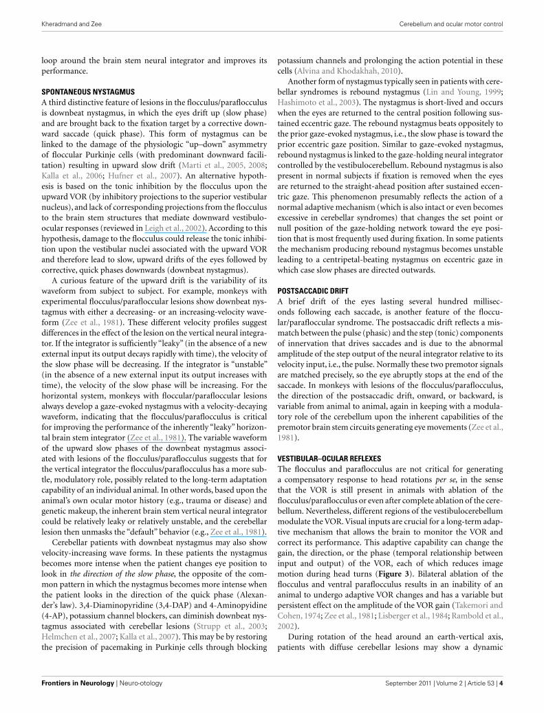
VESTIBULAR–OCULAR REFLEXESThe flocculus and paraflocculus are not critical for generatinga compensatory response to head rotations per se, in the sensethat the VOR is still present in animals with ablation of theflocculus/paraflocculus or even after complete ablation of the cere-bellum. Nevertheless, different regions of the vestibulocerebellummodulate the VOR. Visual inputs are crucial for a long-term adap-tive mechanism that allows the brain to monitor the VOR andcorrect its performance. This adaptive capability can change thegain, the direction, or the phase (temporal relationship betweeninput and output) of the VOR, each of which reduces imagemotion during head turns (Figure 3). Bilateral ablation of theflocculus and ventral paraflocculus results in an inability of ananimal to undergo adaptive VOR changes and has a variable butpersistent effect on the amplitude of the VOR gain (Takemori andCohen, 1974; Zee et al., 1981; Lisberger et al., 1984; Rambold et al.,2002).
During rotation of the head around an earth-vertical axis,patients with diffuse cerebellar lesions may show a dynamic
Frontiers in Neurology | Neuro-otology September 2011 | Volume 2 | Article 53 | 4
Kheradmand and Zee Cerebellum and ocular motor control
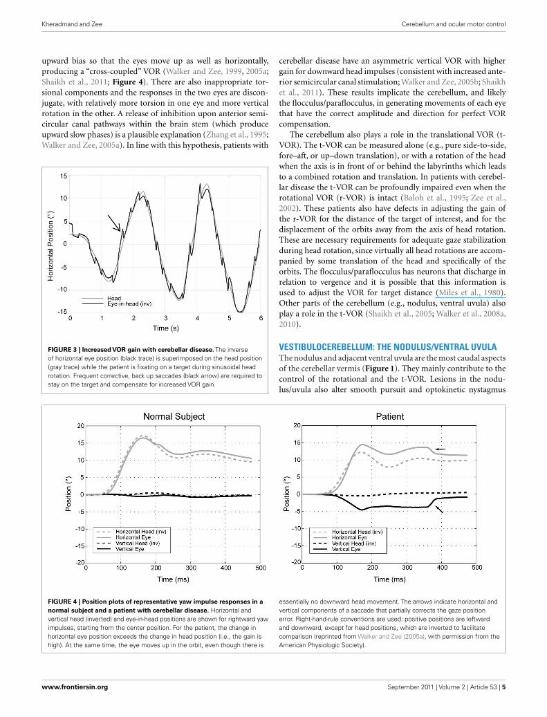
upward bias so that the eyes move up as well as horizontally,producing a “cross-coupled” VOR (Walker and Zee, 1999, 2005a;Shaikh et al., 2011; Figure 4). There are also inappropriate tor-sional components and the responses in the two eyes are discon-jugate, with relatively more torsion in one eye and more verticalrotation in the other. A release of inhibition upon anterior semi-circular canal pathways within the brain stem (which produceupward slow phases) is a plausible explanation (Zhang et al., 1995;Walker and Zee, 2005a). In line with this hypothesis, patients with
FIGURE 3 | Increased VOR gain with cerebellar disease. The inverseof horizontal eye position (black trace) is superimposed on the head position(gray trace) while the patient is fixating on a target during sinusoidal headrotation. Frequent corrective, back up saccades (black arrow) are required tostay on the target and compensate for increased VOR gain.
cerebellar disease have an asymmetric vertical VOR with highergain for downward head impulses (consistent with increased ante-rior semicircular canal stimulation; Walker and Zee, 2005b; Shaikhet al., 2011). These results implicate the cerebellum, and likelythe flocculus/paraflocculus, in generating movements of each eyethat have the correct amplitude and direction for perfect VORcompensation.
The cerebellum also plays a role in the translational VOR (t-VOR). The t-VOR can be measured alone (e.g., pure side-to-side,fore–aft, or up–down translation), or with a rotation of the headwhen the axis is in front of or behind the labyrinths which leadsto a combined rotation and translation. In patients with cerebel-lar disease the t-VOR can be profoundly impaired even when therotational VOR (r-VOR) is intact (Baloh et al., 1995; Zee et al.,2002). These patients also have defects in adjusting the gain ofthe r-VOR for the distance of the target of interest, and for thedisplacement of the orbits away from the axis of head rotation.These are necessary requirements for adequate gaze stabilizationduring head rotation, since virtually all head rotations are accom-panied by some translation of the head and specifically of theorbits. The flocculus/paraflocculus has neurons that discharge inrelation to vergence and it is possible that this information isused to adjust the VOR for target distance (Miles et al., 1980).Other parts of the cerebellum (e.g., nodulus, ventral uvula) alsoplay a role in the t-VOR (Shaikh et al., 2005; Walker et al., 2008a,2010).
VESTIBULOCEREBELLUM: THE NODULUS/VENTRAL UVULAThe nodulus and adjacent ventral uvula are the most caudal aspectsof the cerebellar vermis (Figure 1). They mainly contribute to thecontrol of the rotational and the t-VOR. Lesions in the nodu-lus/uvula also alter smooth pursuit and optokinetic nystagmus
FIGURE 4 | Position plots of representative yaw impulse responses in a
normal subject and a patient with cerebellar disease. Horizontal andvertical head (inverted) and eye-in-head positions are shown for rightward yawimpulses, starting from the center position. For the patient, the change inhorizontal eye position exceeds the change in head position (i.e., the gain ishigh). At the same time, the eye moves up in the orbit, even though there is
essentially no downward head movement. The arrows indicate horizontal andvertical components of a saccade that partially corrects the gaze positionerror. Right-hand-rule conventions are used: positive positions are leftwardand downward, except for head positions, which are inverted to facilitatecomparison (reprinted from Walker and Zee (2005a), with permission from theAmerican Physiologic Society).
www.frontiersin.org September 2011 | Volume 2 | Article 53 | 5
Kheradmand and Zee Cerebellum and ocular motor control
(OKN), but their exact contribution to these visual-followingreflexes is unclear.
VESTIBULAR–OCULAR REFLEXESThe nodulus/ventral uvula act upon the “low-frequency” (sus-tained) components of the VOR via projections to a “velocity-storage” mechanism within the vestibular nuclei. With a constant-velocity rotation of the head, the velocity-storage mechanismextends the duration of the VOR response beyond that expectedfrom the mechanical properties of the cupula–endolymph systemwithin the semicircular canals. This perseverating (integrating)action slows the decay of nystagmus that normally occurs duringa constant-velocity rotation in the dark. In other words, velocity-storage improves the performance of the horizontal VOR at lowfrequencies of rotation and so extends the“bandwidth”of frequen-cies over which the horizontal VOR can transduce the motion ofthe head into the correct compensatory eye movement. Secondly,during sustained“off-vertical axis”rotation of the head,when thereis an imposed changing linear acceleration due to the continuousreorientation of the head relative to the pull of gravity, the velocity-storage mechanism modulates the direction of compensatory slowphases, reorienting the axis of eye rotation toward earth-vertical. Itthus serves an orienting function so the brain can know the posi-tion of the head relative to the pull of gravity, as well as determinewhether a sensed linear acceleration of the head is from gravity oran imposed translation of the head (see below for a discussion ofthe tilt-translation ambiguity).
Lesions of the nodulus/uvula alter the velocity-storage mecha-nism for the horizontal VOR and increase the duration of vestibu-lar responses to a constant-velocity input around an earth-verticalaxis (i.e., the VOR time constant is increased; Waespe et al., 1985).The effect of nodulus/uvula lesions on the vertical and torsionalVOR, however, is the opposite; their time constants are short-ened (Waespe et al., 1985; Angelaki and Hess, 1994; Wearne et al.,1998). Lesions of the nodulus/uvula also disrupt the spatial ori-entation function of the velocity-storage mechanism; the VOR nolonger reorients the axis of eye rotation toward upright during off-vertical axis rotation (Wearne et al., 1998; Cohen et al., 2002). Withnodulus/uvula lesions, there is also a loss of the normal habitua-tion of the time constant of the VOR to repetitive stimulation aswell as loss of tilt suppression of postrotary nystagmus, the phe-nomenon by which the decay of postrotary nystagmus is hastenedwith pitching the head down immediately following the end of aconstant-velocity rotation (Hain et al., 1988; Cohen et al., 1992).
As mentioned earlier, patients with diffuse cerebellar diseasemay show abnormally directed slow phases, or “cross-coupling”of their VOR during high-frequency, high acceleration impulsetesting (Figure 4). This phenomenon also occurs during low-frequency head rotation around an earth-vertical axis, duringsustained optokinetic stimulation with the head upright, or afterhorizontal head shaking (Walker and Zee, 1999; Kim et al., 2005;Moon et al., 2009). The “cross-coupling” with low-frequencystimuli likely reflects dysfunction of the nodulus/uvula and theinability to control spatial orientation using the velocity-storagemechanism.
The nodulus/uvula also plays a role in other aspects of otolith–canal interaction including solving the tilt-translation ambiguity.
The otoliths signal linear acceleration regardless of its source, andcannot in themselves distinguish between a dynamic translationof the head and a static reorientation of the head with respect togravity (Yakusheva et al., 2008). It is the simultaneous presenceor absence of signals from the vertical semicircular canals thatresolves the issue of tilt in the roll plane (ear to shoulder) versustranslation.
The nodulus/uvula is also important for the processing ofotolith signals from both the utricle and saccule to drive the t-VORresponse. They are involved in the integration of linear head accel-eration signals to velocity signals derived from otolith afferents(Walker et al., 2010). Ablation of the nodulus/uvula in monkeysimpairs the ocular response to vertical translation and reduces eyevelocity during sustained horizontal translation along the interau-ral direction (Walker et al., 2008a). The reduction in the interauralt-VOR occurs only in the dark, whereas the vertical t-VOR is sub-stantially impaired even in the light. These finding suggest thenodulus/uvula might have different contribution to the horizontaland vertical t-VOR.
PURSUITAlthough the flocculus/paraflocculus is primarily implicated inthe control of pursuit, recent evidence also indicates a role forthe nodulus/uvula, particularly in vertical pursuit, as experimen-tal lesions of the nodulus/uvula impair downward pursuit withlittle effect on upward or horizontal pursuit (Walker et al., 2008b).A similar finding of impaired downward pursuit has been reportedin a patient with an isolated nodulus/uvula lesion (Helmchen et al.,2007). The uvula has been linked to horizontal smooth pursuitby lesion mapping in a group of patients with cerebellar stroke(Baier et al., 2009). In this study, however, the identified lesionswere not isolated to the uvula and involved other parts of thevermis.
OPTOKINETIC REFLEXThe optokinetic response is normally judged by both the nystag-mus during visual stimulation (OKN) and the slowly decayingafter-nystagmus (OKAN) in the dark that occurs after the stimu-lation has ceased. OKAN shares the same velocity-storage mecha-nism with the VOR. The smooth pursuit system also participatesat the onset of the response to an optokinetic (full-field) stimulusbringing the eyes quickly to the maximum velocity. Accordingly,the cerebellar regions involved in generating both pursuit and theVOR contribute to the tracking response to an optokinetic stim-ulus. For example, after flocculectomy in monkeys, due to thecoexisting pursuit deficit the initial eye velocity during constant-velocity optokinetic stimulation is diminished, and the time toincrease velocity from the onset of the optokinetic stimulus to asteady-state (matching the drum velocity) is prolonged (Zee et al.,1981). After removal of the nodulus/uvula, analogous to the VORresponses, horizontal OKAN is prolonged but the vertical and tor-sional OKAN response times are shortened (Angelaki and Hess,1994; Wearne et al., 1998). Similarly, spatial control of the eyevelocity vector during OKN or OKAN is impaired and the axisof eye rotation during imposed gravito-inertial acceleration doesnot reorient toward the upright (Cohen et al., 2002). There is alsoa change in the direction of the optokinetic response that results
Frontiers in Neurology | Neuro-otology September 2011 | Volume 2 | Article 53 | 6
Kheradmand and Zee Cerebellum and ocular motor control
in cross-coupling during OKN or OKAN (a dynamic torsional orupward bias with horizontal nystagmus; Walker and Zee, 1999).
SPONTANEOUS NYSTAGMUSPeriodic alternating nystagmus (PAN), a horizontal jerk nystag-mus that changes direction every few minutes, may appear fol-lowing lesions of the nodulus and its adjacent paravermal region(Waespe et al., 1985). PAN reflects the combined actions of a(1) disinhibited brain stem vestibular velocity-storage mechanism(due to loss of inhibition from Purkinje cells in the nodulusthat project to the vestibular nuclei) and (2) an intact adaptivemechanism that acts to null any sustained unidirectional nystag-mus, thus allowing PAN to change direction (Leigh et al., 1981).Because Purkinje cell inhibition is mediated through GABAB
receptors, treatment with baclofen (a GABAB agonist) disengagesthe velocity-storage mechanism and stops PAN (Halmagyi et al.,1980; Cohen et al., 1987). Note that like the mechanism produc-ing rebound nystagmus, the adaptive mechanism that leads to thereverse of the direction of PAN is intact or possibly increased afterlesions in the cerebellum.
Downbeat nystagmus is also reported with nodulus and uvulalesions (Walker et al., 2008b, 2009). The slow phase velocity ofthis nystagmus, unlike down beat nystagmus seen with flocculuslesions, is independent of orbital position (i.e., nystagmus did notdecrease with upward gaze or increase with lateral gaze) and canbe suppressed with visual fixation. Changing the orientation of thehead with respect to gravity may also alter the nystagmus (Walkeret al., 2009). Thus the downbeat nystagmus with nodulus/uvulalesions could be due to a bias in the vestibular system (either thet-VOR or r-VOR mechanisms) and need not reflect changes in thegaze-holding neural integrator.
FUNCTIONAL DIFFERENCES BETWEEN THEFLOCCULUS/PARAFLOCCULUS AND THE NODULUS/UVULAThe vestibulocerebellum has many functions and lesions producea variety of abnormalities. Are there overriding principles aboutthe functions of the two areas within the vestibulocerebellum? Onesimple hypothesis is that the flocculus and paraflocculus are moreconcerned with the relatively immediate and fast-acting aspectsof ocular motor function that relate to the needs of the fovea(e.g., holding images steady on the fovea during attempted fixa-tion, smooth pursuit, VOR, or immediately following saccades).On the other hand, the nodulus and ventral uvula are more con-cerned with the duration and axis of eye rotation in response tolow-frequency, sustained rotational stimuli, and so determine theorientation of images on the retina relative to upright. Recent evi-dence however indicates that an intact nodulus and uvula are alsonecessary for the proper function of the high-frequency t-VOR aswell as smooth pursuit (Walker et al., 2008b, 2010). Thus theredoes not appear to be an absolute compartmentalization of func-tions of the VOR and pursuit among the different parts of thevestibulocerebellum, and, as will be discussed below, the dorsalvermis and underlying fastigial nuclei also play a role in smoothpursuit. Such redundancy is certainly an advantage during loco-motion, considering the vital role vestibular and pursuit responsesplay in stabilizing gaze on stationary targets or in following targetsthat are also moving in the environment.
DORSAL CEREBELLAR VERMIS AND THE POSTERIORFASTIGIAL NUCLEUSThe dorsal vermis (lobules V–VII, also called the OMV) and theunderlying posterior fastigial nucleus (also called the FOR) areespecially important for the control of saccades, but also play arole in smooth pursuit (Figure 1).
SACCADESLesions in the OMV cause changes in the accuracy, latency, trajec-tory, and dynamic properties (speed and acceleration) of saccades(Takagi et al., 1998; Barash et al., 1999; Kojima et al., 2010a). Purk-inje cells in the OMV discharge before saccades, and stimulationof this same area can elicit saccades (Fujikado and Noda, 1987;Noda and Fujikado, 1987). In humans, too, artificial stimulationusing TMS, functional magnetic resonance imaging (fMRI), andmapping of lesions based on MRI in patients with cerebellar infarc-tions confirm the participation of the OMV in the generation ofsaccades (Hashimoto and Ohtsuka, 1995; Hayakawa et al., 2002;Ye et al., 2010). The OMV also plays an important role in saccadeadaptation, a mechanism that detects errors in motor performanceand updates saccade commands to accurately move the eye towarda target. OMV lesions impair adaptation of saccade amplitude(Takagi et al., 1998; Barash et al., 1999; Straube et al., 2001; Jenk-inson and Miall, 2010; Colnaghi et al., 2011). This defect in partmay be related to the increased variability of saccade amplitudeafter the lesion of the OMV, so that the adaptation mechanism nolonger receives a consistent error signal with which it can optimallyreprogram the correct response.
Neurons in the FOR also discharge in relation to saccades (Oht-suka and Noda, 1991; Fuchs et al., 1993). They supply a presaccadicburst for contraversive saccades (i.e., the right FOR bursts beforeleftward saccades) and a “braking” discharge, late during the sac-cade, for ipsiversive saccades (the right FOR bursts toward the endof rightward saccades). Thus, each FOR acts to facilitate contraver-sive saccades and contributes to the termination of ipsiversive sac-cades. Consequently, lesions in the FOR cause ipsiversive saccadichypermetria (overshoot) and contraversive hypometria (under-shoot; Robinson et al., 1993). A horizontal static fixation error(toward the side of the lesion) could also contribute to the sac-cadic dysmetria observed following FOR lesions (Robinson et al.,1993). Bilateral FOR lesions cause bilateral hypermetria.
The OMV receives information about performance during sac-cades and adjusts its inhibition upon the FOR to assure the saccadearrives on target (Goffart et al., 2003; Straube et al., 2009). Purk-inje cells in the OMV behave in a similar way to those of the FOR,though, as predicted from their inhibitory nature, their “sign” isopposite. Thus, each side of the vermis acts to facilitate ipsiver-sive saccades and contributes to the termination of contralateralsaccades. Accordingly, OMV lesions lead to hypometric ipsiversiveand hypermetric contraversive saccades (Takagi et al., 1998). Bilat-eral lesions in OMV cause hypometric saccades in both horizontaldirections.
The role of the cerebellum in the generation of vertical sac-cades is not as well understood. Vertical saccades show ipsipulsion(oblique trajectory toward the side of inactivation) with exper-imental lesions of the FOR (Straube et al., 2009). Ipsipulsion isalso a feature of Wallenberg’s syndrome, presumably due to a
www.frontiersin.org September 2011 | Volume 2 | Article 53 | 7
Kheradmand and Zee Cerebellum and ocular motor control
functional lesion of the FOR resulting from interruption of theclimbing fiber input (within the inferior cerebellar peduncle) tothe OMV and a consequent increased inhibition by Purkinje cellsupon the underlying FOR (Waespe and Wichmann, 1990). Otherareas, such as the interposed nucleus (IP; emboliform and glo-bose) and paraflocculus (tonsils), may also be important in thegeneration of vertical saccades (Robinson, 2000).
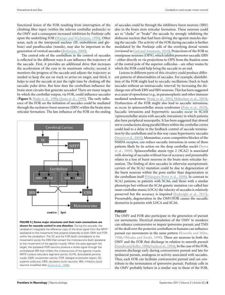
The central role of the cerebellum in the control of saccadesis reflected in the different ways it can influence the trajectory ofthe saccade. First, it provides an additional drive that increasesthe acceleration of the eyes to its maximum velocity; second, itmonitors the progress of the saccade and adjusts the trajectory asneeded to keep the eye on track to arrive on target; and third, ithelps to end the saccade at just the right time by choking off thesaccadic pulse drive. But how does the cerebellum influence thebrain stem circuits that generate saccades? There are many targetsby which the cerebellar output, via FOR, could influence saccades(Figure 5; Noda et al., 1990; Quaia et al., 1999). The early influ-ence of the FOR on the initiation of saccades could be mediatedthrough the excitatory burst neurons (EBN) within the brain stemreticular formation. The late influence of the FOR on the ending
FIGURE 5 | Some major structures and their main connections are
shown for saccade control in one direction. During the saccade, thecerebellum integrates the efference copy of the drive signal from the NRTP(ipsilateral to the movement) that projects bilaterally to both OMV and FORwithin the cerebellum. The SC and the FOR (both contralateral to themovement) excite the EBN that contact the motoneurons (both ipsilateralto the movement) of the agonist muscle. When the eyes approach thetarget, the ipsilateral FOR neurons produce a choke signal through thecontralateral IBN that inhibits the motoneurons of the agonist muscle.NRTP, nucleus reticularis tegmenti pontis; DLPN, dorsolateral pontinenuclei; OMV, oculomotor vermis; FOR, fastigial oculomotor region; SC,superior colliculus; EBN, excitatory burst neurons; IBN, inhibitory burstneurons (modified after Quaia et al., 1999).
of saccades could be through the inhibitory burst neurons (IBN)also in the brain stem reticular formation. These neurons couldact to “choke” or “brake” the saccade by strongly inhibiting theabducens nucleus that had been driving the agonist muscles dur-ing the saccade. The activity of the FOR during saccades is furthermodulated by the Purkinje cells of the overlying dorsal vermis(reviewed in Gad and Anastasio, 2010). Projections of the FOR toomnipause neurons (OPN), which inhibit premotor saccadic EBN– either directly or via projections to OPN from the fixation zoneof the rostral pole of the superior colliculus – are other routes bywhich the FOR could help bring the saccade to an end.
Lesions in different parts of this circuitry could produce differ-ent patterns of abnormalities of saccades. For example, disinhibi-tion of the FOR might lead to saccadic oscillations (back to backsaccades without an intersaccadic interval) by increasing the dis-charge rate of both EBN and IBN neurons. This has been suggestedas a cause of opsoclonus (e.g., in paraneoplastic and other immunemediated syndromes; Wong et al., 2001; Helmchen et al., 2003).Dysfunction of the FOR might also lead to saccadic intrusions,as occur in spinocerebellar ataxia syndromes (Pula et al., 2010).Saccadic intrusions and hypermetric saccades occur in SCASI(spinocerebellar ataxia with saccadic intrusions) in which patientsalso have peripheral neuropathy. It has been suggested that slowednerve conductions along parallel fibers within the cerebellar cortexcould lead to a delay in the feedback control of saccade termina-tion by the cerebellum and in this way cause hypermetric saccades(Swartz et al., 2003). Memantine, a non-competitive blocker of theNMDA receptor, can reduce saccadic intrusions in some of thesepatients likely by its action on the deep cerebellar nuclei (Serraet al., 2008). Spinocerebellar ataxia type 2 (SCA2) is associatedwith slowing of saccades without loss of accuracy and presumablyrelates to a loss of burst neurons in the brain stem reticular for-mation. The finding of slow saccades in otherwise asymptomaticcarriers of the SCA2 mutation could be due to degeneration ofthe burst neurons within the pons earlier than degeneration inthe cerebellum itself (Velazquez-Perez et al., 2009). In contrast toSCA2 patients, in patients with SCA6, and those with a similarphenotype but without the SCA6 genetic mutation (so-called lateonset cerebellar ataxia; LOCA) the velocity of saccades is relativelypreserved but the accuracy is impaired (Federighi et al., 2011).Presumably, degeneration in the OMV/FOR causes the saccadicdysmetria in patients with LOCA and SCA6.
PURSUITThe OMV and FOR also participate in the generation of pursuiteye movements. Electrical stimulation of the OMV in monkeyscan enhance contraversive or impair ipsiversive pursuit, and TMSof the skull over the posterior cerebellum in humans can influencepursuit eye movements in the same pattern (Krauzlis and Miles,1998; Ohtsuka and Enoki, 1998). There are neurons in both theOMV and the FOR that discharge in relation to smooth pursuit(Suzuki and Keller, 1988; Fuchs et al., 1994). In the case of the FOR,neurons discharge early during contraversive pursuit and late foripsilateral pursuit, analogous to activity associated with saccades.Thus, each FOR can facilitate contraversive pursuit and can con-tribute to the termination of ipsiversive pursuit. Purkinje cells inthe OMV probably behave in a similar way to those of the FOR,
Frontiers in Neurology | Neuro-otology September 2011 | Volume 2 | Article 53 | 8
Kheradmand and Zee Cerebellum and ocular motor control
though, as predicted from their inhibitory nature their “sign” isopposite. Each side of the vermis would act to facilitate ipsiversivepursuit and contribute to the termination of contralateral pursuit.Purkinje cells in the OMV encode the sum of eye velocity in theorbit and retinal slip velocity (the rate of image motion across theretina) during smooth pursuit that together represent target veloc-ity relative to the head (Suzuki et al., 1981). These Purkinje neuronsin the vermis differ from those in the flocculus/paraflocculus thatmodulate their discharge with eye velocity but not with the retinalslip velocity (Belton and McCrea, 2000).
The pursuit deficits reported after experimental lesions in theOMV and the FOR are largely in accord with the physiologicalfindings (Robinson et al., 1997; Takagi et al., 2000). With a lesionin the FOR contralateral pursuit is impaired, and with a lesionin the OMV ipsilateral pursuit is impaired. Vertical pursuit is lit-tle affected following OMV lesions whereas FOR lesions reducedownward pursuit more than upward pursuit. Bilateral lesionsof the OMV in monkeys produce horizontal pursuit deficits inboth directions (Takagi et al., 2000). On the other hand, bilat-eral FOR lesions leave pursuit relatively intact (Robinson et al.,1997). This also appears to be the case in patients with bilat-eral FOR lesions in whom pursuit is preserved (Buttner et al.,1994). These finding suggest the pursuit deficit is due to imbal-ance between opposing drives of the two FOR. Therefore, withbilateral FOR inactivation, and no FOR imbalance, the pursuitmovements remain intact.
Lesions of OMV and FOR mainly affect eye acceleration duringthe initial period of pursuit (the first 100 ms of tracking after a tar-get has started moving or has changed its speed) and have a smallereffect during the sustained tracking period (Robinson et al., 1997;Takagi et al., 2000). During this initial “open-loop” period, aneye acceleration command brings the eye toward a certain veloc-ity prior to the sustained tracking period. The inherent latency invisual processing requires the pursuit system to be able to calibratethe initial response adaptively, as opposed to the period of sus-tained tracking in which “on-line” visual feedback and predictivemechanisms can be used to keep pursuit accurate (Kettner et al.,2002). Lesions of the OMV also interfere with pursuit adaptation(Takagi et al., 2000).
As mentioned earlier, in addition to OMV/FOR, the floccu-lus/paraflocculus of the vestibulocerebellum has neurons thatdischarge with pursuit eye movements. Thus, both the vestibu-locerebellum and the OMV/FOR contribute to smooth pursuit.One possible division of labor between these two regions is thatthe OMV/FOR is more concerned with the initiation and termina-tion of the preprogrammed initial “open-loop” portion of pursuit(when retinal slip is high), and the vestibulocerebellum is moreconcerned with pursuit during sustained tracking.
A remarkable finding after OMV lesions in monkeys is thatthere are quantitative similarities in the effects of the lesions onsaccade amplitude, and on pursuit velocity during the“open-loop”initial period of pursuit tracking (Takagi et al., 1998, 2000). Thissimilarity is seen more clearly if one considers that the initialperiod of pursuit acceleration is an open-loop eye movement inthe same vein as a saccade is open-loop. With this hypothesis, forsaccades, premotor networks would be generating an eye velocitycommand to bring the eye to a certain position, and for pursuit,
an eye acceleration command to bring the eye toward a certainvelocity. Taking this analogy further, the FOR and the OMV couldcontrol the dynamic properties of the open-loop period of pur-suit (as reflected in eye acceleration) in the same way that theycontrol the dynamic properties of saccades (as reflected in eyevelocity). Compatible with this idea is the finding of neuronswithin the OMV that discharge for both pursuit and saccades,and that stimulation of the OMV can alter ongoing saccades orpursuit, depending upon the parameters of the stimulus (Sato andNoda, 1992).
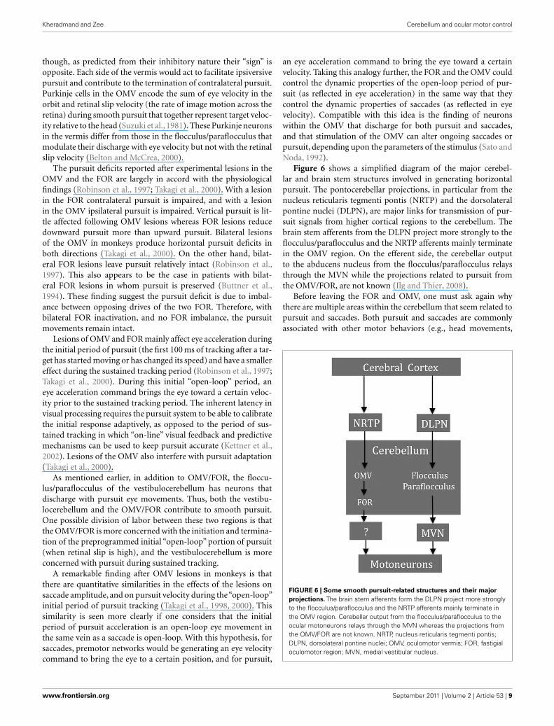
Figure 6 shows a simplified diagram of the major cerebel-lar and brain stem structures involved in generating horizontalpursuit. The pontocerebellar projections, in particular from thenucleus reticularis tegmenti pontis (NRTP) and the dorsolateralpontine nuclei (DLPN), are major links for transmission of pur-suit signals from higher cortical regions to the cerebellum. Thebrain stem afferents from the DLPN project more strongly to theflocculus/paraflocculus and the NRTP afferents mainly terminatein the OMV region. On the efferent side, the cerebellar outputto the abducens nucleus from the flocculus/paraflocculus relaysthrough the MVN while the projections related to pursuit fromthe OMV/FOR, are not known (Ilg and Thier, 2008).
Before leaving the FOR and OMV, one must ask again whythere are multiple areas within the cerebellum that seem related topursuit and saccades. Both pursuit and saccades are commonlyassociated with other motor behaviors (e.g., head movements,
FIGURE 6 | Some smooth pursuit-related structures and their major
projections. The brain stem afferents form the DLPN project more stronglyto the flocculus/paraflocculus and the NRTP afferents mainly terminate inthe OMV region. Cerebellar output from the flocculus/paraflocculus to theocular motoneurons relays through the MVN whereas the projections fromthe OMV/FOR are not known. NRTP, nucleus reticularis tegmenti pontis;DLPN, dorsolateral pontine nuclei; OMV, oculomotor vermis; FOR, fastigialoculomotor region; MVN, medial vestibular nucleus.
www.frontiersin.org September 2011 | Volume 2 | Article 53 | 9
Kheradmand and Zee Cerebellum and ocular motor control
arm movements, walking, or running) and also occur in differentcontexts (e.g., reflexive or self-generated). Thus, one can envi-sion a number of areas within the cerebellum associated withdifferent motor behaviors that might want to use informationabout associated eye movements to help optimize their ownperformance.
OTHER ABNORMALITIES OF OCULAR MOTOR CONTROLASSOCIATED WITH CEREBELLAR DYSFUNCTIONABNORMALITIES OF EYE TORSIONThe torsional orientation of the eye during fixation or movementis precisely controlled and is described by Donders’ and Listing’slaws. Donders’ law states that during steady fixation, the torsionalorientation of the eyes is fixed for a given eccentric (horizontal andvertical) eye position. In other words, the eye always assumes thesame unique spatial orientation at a final gaze position irrespec-tive of where it came from. Listing’s law describes the kinematicfeatures of eye rotations that fix the torsional orientation of theglobe for a given horizontal and vertical position of the eyes in theorbit. It states that all eye positions can be reached by a rotationfrom a given reference position (usually taken as straight-aheadposition) about axes that all lie in the same plane (Listing’s plane).
Listing’s law is not perfectly obeyed in patients with cerebellarlesions. This is partly caused by torsional drifts of the eye thatchange direction with horizontal gaze eccentricity (intorsional inabduction and extorsional in adduction; Straumann et al., 2000).This finding suggests that the cerebellum is involved in generat-ing a tonic torsional signal that maintains the eyes in Listing’splane against the mechanical forces of the globe. The eye position-dependent torsional drift would then be considered a “passive”violation of Listing’s law (determined by the elastic forces on theocular plant) in the absence of the mapped tonic signal. On theother hand, despite the evidence for abnormal torsion outside ofListing’s plane, such a violation did not occur during saccades inpatients with cerebellar degeneration (Thurtell et al., 2008).
Unlike saccades or pursuit, ocular torsion during the r-VORdepends considerably less on eye position and does not complywith Listing’s law (Misslisch et al., 1994). This torsional move-ment, however, evolves over time: at the onset of head rotation,the response is largely head-fixed (less dependent on eye posi-tion) but later torsion dependent on eye position slowly increases,reaching a plateau when steady-state head velocity is reached (Tianet al., 2006). Moreover, the acceleration of the stimulus affects thekinematics of eye motion; responses to high acceleration head(on body) impulses are more head-fixed than responses to low-acceleration en bloc chair rotations. The fact that the r-VOR ismore head-fixed has the theoretical advantage of adequately sta-bilizing the entire visual image on the retina when the head isrotating.
Patients with cerebellar degeneration show significantly moretorsion that is dependent on eye position (i.e., less head-fixed tor-sion) during the r-VOR (Ignashchenkova et al., 2009). This findingindicates that the cerebellum has a large effect on the evolutionover time of torsion that is dependent on eye position during theVOR. It may be that the brain monitors changes in torsional ori-entation of the eyes during the r-VOR, and depending on the eyeposition, imposes torsion to prevent the eyes from drifting too far
from Listing’s plane. The function of the cerebellum might thenbe to modulate this effect to achieve the compromise betweenthe competing goals of keeping eye positions in Listing’s planeand maintaining the stability of images on the retina during headrotation. The evolution of head-fixed to eye position-dependenttorsion could reflect the activity in the velocity-to-position ocularmotor neural integrator and points to the flocculus/paraflocculus,which is an integral part of the neural integrator circuit (Zee et al.,1981).
THE CEREBELLUM AND BINOCULAR OCULAR MOTOR CONTROLMost of the findings relating the cerebellum to ocular motor con-trol have dealt with conjugate eye movements. The cerebellum,however, is also involved in adjusting the relative position of theeyes during fixation (static alignment) and the yoking of the eyesduring movement (dynamic alignment). Patients with cerebellardamage sometimes show a skew deviation, a vertical misalignmentof the eyes that cannot be attributed to a simple ocular mus-cle weakness (Keane, 1985; Versino et al., 1996; Zee, 1996; Wiestet al., 2001; Wong and Sharpe, 2005; Brodsky et al., 2006; Colenet al., 2008). The misalignment in patients with cerebellar dam-age often changes its sense with horizontal eye position – mostcommonly the abducting eye is higher. The source of the skewmay be an imbalance in otolith–ocular reflexes. The otolith organs(utricle and saccule) sense acceleration during linear motion ofthe head for the t-VOR and during static lateral head tilt for ocularcounter roll. Patients with cerebellar skew deviation have reducedand disconjugate counter roll gains that depend on the directionof the head tilt (Wong and Sharpe, 2005). There is also a discon-jugate reduction in the gain of the t-VOR, but without differencesbetween directions of head translation (Schlenker et al., 2009).Since the vestibulocerebellum has an important role in modu-lation of the t-VOR using information from the otoliths, skewdeviation might be due to damage in this part of the cerebellum.The dentate nucleus has been also implicated in cerebellar skewdeviation based on MRI/CT lesion analysis (Baier et al., 2008).Patients with cerebellar lesions can also show misalignment of theeyes during the r-VOR and during saccades (Versino et al., 1996;Zee et al., 2002).
An esotropia (the eyes turn inward) also occurs in cerebellardisease and, since the esodeviation is greater at distance it has beenattributed to a divergence paralysis (Wiest et al., 2001). Analogousto conjugate smooth pursuit and saccade, vergence eye movementscan be classified by their response to disparities produced by stim-uli moving slowly (ramp or slow vergence) or abruptly (step or fastvergence). Patients with acute cerebellar lesions can show impairedslow but relatively intact fast vergence. Moreover, divergence, butnot convergence, can be affected particularly with the lesions inthe OMV (Sander et al., 2009). Patients with vestibulocerebellarlesions may also show a divergence-beating nystagmus (conver-gent slow phases with divergent quick phases; Yee et al., 1979).These abnormalities hint at an excess of convergence tone withsome cerebellar lesions.
In monkeys the majority of OMV Purkinje cells that dis-charge with pursuit also modulate their activity during ver-gence, and especially convergence (Nitta et al., 2008a). Temporaryinactivation of the OMV results in convergence failure (Nitta et al.,
Frontiers in Neurology | Neuro-otology September 2011 | Volume 2 | Article 53 | 10
Kheradmand and Zee Cerebellum and ocular motor control
2008b). A similar effect, however, has not been produced after per-manent ablation of the OMV in monkeys (Takagi et al., 2003).Rather there is an esodeviation that can also be seen in patientswith cerebellar degeneration, presumably reflecting an increasein convergence tone (Gamlin and Zhang, 1996; Ignashchenkovaet al., 2009). These findings might be related to sparing of theFOR as inactivation of these deep cerebellar nuclei causes con-vergence deficits with an exodeviation (Gamlin and Zhang, 1996;Scheurer et al., 2001). The posterior IP, flocculus, and cerebellarhemispheres are also implicated in the control of vergence eyemovement. The flocculus also has neurons that discharge in rela-tion to the vergence angle (Miles et al., 1980; Richter et al., 2004).The floccular/IP pathway may be involved in the control of diver-gence eye movements while the dorsal vermis/FOR pathway maybe more related to convergence, but this compartmentalization offunctions is hypothetical (Mays et al., 1986; Nitta et al., 2008b).Functional imaging studies in humans also show activation ofthe cerebellar hemispheres during convergence. Much like pursuit,multiple regions in the cerebellum may play a role in controllingthe conjugacy of eye movements and both dynamic and staticvergence.
OCULAR MOTOR LEARNING AND THE CEREBELLUMPerhaps the most intriguing aspect of cerebellar function is itsrole in motor learning. The flocculus/paraflocculus has a clearrole in ocular motor learning including the amplitude (gain) andthe direction (relative to head motion) of the VOR, the pulse-stepratio for saccades, and the gain of smooth pursuit (Takemori andCohen, 1974; Optican and Robinson, 1980; Zee et al., 1981; Lis-berger et al., 1984; Rambold et al., 2002; Medina and Lisberger,2009). The OMV/FOR, too, has a role in ocular motor learning,especially related to saccades (Golla et al., 2008; Prsa and Thier,2011). Transient bilateral inactivation of the FOR impairs the relayof adapted signals to the brain stem circuits that generate premo-tor commands for saccades, but it does not stop all adaptationfrom occurring (Robinson et al., 2002). This finding suggests theadaptation takes place upstream in the cerebellar cortex (i.e., inthe OMV). The OMV is especially involved in modification of theinitial acceleration of pursuit and the amplitude of saccades (Tak-agi et al., 1998, 2000). Temporary inactivation of the OMV withmuscimol injections, a GABA agonist, inhibits adaptive length-ening, but has relatively little effect on adaptive shortening ofsaccade amplitudes. In contrast, injections of bicuculline, a GABAantagonist, impairs adaptive shortening, and facilitates adaptivelengthening of saccade amplitudes. These findings suggest thatOMV activity is necessary for amplitude lengthening adaptationof saccades, whereas amplitude shortening adaptation may involvethe inhibitory circuits within the OMV (Kojima et al., 2011).
Recent studies have also implicated the cerebellar hemispheresin ocular motor learning. For example, fMRI studies show thatthe hemispheric lobules VIII–X are active in processing of sac-cadic errors (van Broekhoven et al., 2009), and TMS over thehemispheric lobule Crus I has a dual effect on saccadic plasticity;potentiating adaptive lengthening and depressing adaptive short-ening of saccade amplitudes (Panouilleres et al., 2011). There isalso evidence for involvement of the cerebellar hemispheres inadaptation for more voluntary, internally generated saccades such
as memory-guided saccades, as opposed to the OMV involvementin more reflexive, visually guided saccades (Nitschke et al., 2004;Alahyane et al., 2008; Kojima et al., 2010b). Damage to the cere-bellar cortex appears to affect a fast component of the adaptationprocess (seconds) more than a slower component that emergesover minutes (Xu-Wilson et al., 2009). There must be even longertimescales of learning that develop with practice over days andweeks. The inability to perform a sequence of movements whichdepends upon the cerebellum can be a factor in the defectiveadaptation process. Cerebellar patients have difficulty making asequence of visually guided saccades. When presented with ran-dom, two-step target jumps these patients failed to make a saccadeto the first target jump suggesting an inability to carry out parallelprocessing of saccades (King et al., 2011).
A memory trace for adaptation of the VOR appears to beacquired initially in the cerebellar cortex (i.e., flocculus) whereasthe memory for long-term VOR adaptation likely resides outsideof the cerebellum, perhaps in the vestibular nuclei (Galiana, 1986;Peterson et al., 1991; Raymond et al., 1996; Anzai et al., 2010).Recall, however, that the cerebellar recipient zones of the vestibu-lar nuclei can be considered a displaced deep cerebellar nuclei. TheVOR adaptation for both gain-up and gain-down learning is moreeffective at lower frequencies (below 4 Hz) than higher frequencies(Raymond and Lisberger, 1996; Broussard et al., 1999). When pas-sive rotation is used to induce changes in the VOR gain, the learnedchange in gain (whether an increase or a decrease) is greatest atthe frequency of head rotation and is more pronounced for gain-up than for gain-down learning (Robinson, 1976; Lisberger et al.,1983; Raymond and Lisberger, 1996; de Zeeuw et al., 1998; Kimpoet al., 2005; Titley et al., 2009). This selectivity of learning has led tothe idea that VOR signals are segregated into frequency “channels”or adaptive filters and the cerebellum may process these channelsselectively (Lisberger et al., 1983; Dean et al., 2010; Broussard et al.,2011).
The cerebellum also influences learning associated with dis-conjugate eye movements. Phoria adaptation to vertical and hor-izontal disparities can be impaired in patients with cerebellardysfunction (Milder and Reinecke, 1983; Kono et al., 2002). Infor-mation from animal studies is limited in this regard. Monkeyswith lesions of the dorsal vermis show a variable deficit in phoriaadaptation but those with flocculus/paraflocculus lesions can stilladapt to a horizontal disparity (Judge, 1987; Takagi et al., 2003).The deep cerebellar nuclei (FOR or interposed nuclei) are alsopossible candidates influencing phoria adaptation.
The effect of experimental lesions within the cerebellum onocular motor function often varies from animal to animal. Whiledifferences in the extent of lesions may explain some of thisvariability, another interpretation relates to the central role ofthe cerebellum in long-term ocular motor learning. In this for-mulation, the cerebellum acts to compensate for differences inmotor performance that are idiosyncratic from animal to ani-mal, depending upon the particular genetic makeup of each ani-mal, its “life history” of disease and trauma, and the interactionbetween inherent capabilities and environmental influences dur-ing development and aging. The cerebellar mechanisms involvedin different aspects of ocular motor learning are a major focus ofcontemporary neuroscience.
www.frontiersin.org September 2011 | Volume 2 | Article 53 | 11
Kheradmand and Zee Cerebellum and ocular motor control
REFERENCESAlahyane, N., Fonteille, V., Urquizar,
C., Salemme, R., Nighoghossian,N., Pelisson, D., and Tilikete, C.(2008). Separate neural substrates inthe human cerebellum for sensory-motor adaptation of reactive and ofscanning voluntary saccades. Cere-bellum 7, 595–601.
Alvina, K., and Khodakhah, K. (2010).The therapeutic mode of action of 4-aminopyridine in cerebellar ataxia. J.Neurosci. 30, 7258–7268.
Angelaki, D. E., and Hess, B. J. (1994).The cerebellar nodulus and ven-tral uvula control the torsionalvestibulo-ocular reflex. J. Neuro-physiol. 72, 1443–1447.
Anzai, M., Kitazawa, H., and Nagao,S. (2010). Effects of reversiblepharmacological shutdown of cere-bellar flocculus on the memoryof long-term horizontal vestibulo-ocular reflex adaptation in monkeys.Neurosci. Res. 68, 191–198.
Baier, B., Bense, S., and Dieterich,M. (2008). Are signs of ocular tiltreaction in patients with cerebel-lar lesions mediated by the dentatenucleus? Brain 131, 1445–1454.
Baier, B., Stoeter, P., and Dieterich,M. (2009). Anatomical correlates ofocular motor deficits in cerebellarlesions. Brain 132, 2114–2124.
Baloh, R. W., Yue, Q., and Demer,J. L. (1995). The linear vestibulo-ocular reflex in normal subjectsand patients with vestibular andcerebellar lesions. J. Vestib. Res. 5,349–361.
Barash, S., Melikyan, A., Sivakov, A.,Zhang, M., Glickstein, M., andThier, P. (1999). Saccadic dysme-tria and adaptation after lesions ofthe cerebellar cortex. J. Neurosci. 19,10931–10939.
Belton, T., and McCrea, R. A. (1999).Contribution of the cerebellar floc-culus to gaze control during activehead movements. J. Neurophysiol.81, 3105–3109.
Belton, T., and McCrea, R. A. (2000).Role of the cerebellar flocculusregion in the coordination ofeye and head movements duringgaze pursuit. J. Neurophysiol. 84,1614–1626.
Belton, T., and McCrea, R. A. (2002).Role of the cerebellar flocculusregion in cancellation of the VORduring passive whole body rotation.J. Neurophysiol. 84, 1599–1613.
Belton, T., and McCrea, R. A. (2004).Context contingent signal process-ing in the cerebellar flocculusand ventral paraflocculus duringgaze saccades. J. Neurophysiol. 92,797–807.
Brodsky, M. C., Donahue, S. P., Vaphi-ades, M., and Brandt, T. (2006). Skewdeviation revisited. Surv. Ophthal-mol. 51, 105–128.
Broussard, D. M., Bhatia, J. K.,and Hong, J. A. (1999). Thedynamics of the vestibulo-ocularreflex after peripheral vestibu-lar damage. II. Comparison withdynamics after optically-inducedlearning. Exp. Brain Res. 125,365–374.
Broussard, D. M., Titley, H. K., Antflick,J., and Hampson, D. R. (2011).Motor learning in the VOR: the cere-bellar component. Exp. Brain Res.210, 451–463.
Buttner, U., Straube, A., and Spuler,A. (1994). Saccadic dysmetria and“intact” smooth pursuit eye move-ments after bilateral deep cerebellarnuclei lesions. J. Neurol. Neurosurg.Psychiatr. 57, 832–834.
Cohen, B., Helwig, D., and Raphan, T.(1987). Baclofen and velocity stor-age: a model of the effects of thedrug on the vestibulo-ocular reflexin the rhesus monkey. J. Physiol. 393,703–725.
Cohen, B., John, P., Yakushin, S. B.,Buettner-Ennever, J., and Raphan,T. (2002). The nodulus and uvula:source of cerebellar control of spatialorientation of the angular vestibulo-ocular reflex. Ann. N. Y. Acad. Sci.978, 28–45.
Cohen, H., Cohen, B., Raphan, T., andWaespe, W. (1992). Habituation andadaptation of the vestibuloocularreflex: a model of differential con-trol by the vestibulocerebellum. Exp.Brain Res. 90, 526–538.
Colen, C., Ketko, A., George, E., and VanStavern, G. (2008). Periodic alternat-ing nystagmus and periodic alternat-ing skew deviation in spinocerebellarataxia type 6. J. Neuroophthalmol. 28,287–288.
Colnaghi, S., Ramat, S., D’Angelo, E.,Cortese, A., Beltrami, G., Moglia, A.,and Versino, M. (2011). Theta-burststimulation of the cerebellum inter-feres with internal representations ofsensory-motor information relatedto eye movements in humans. Cere-bellum doi: 10.1007/s12311-011-0282–281. [Epub ahead of print].
de Zeeuw, C. I., Hansel, C., Bian, F.,Koekkoek, S. K., van Alphen, A.M., Linden, D. J., and Oberdick,J. (1998). Expression of a proteinkinase C inhibitor in Purkinje cellsblocks cerebellar LTD and adapta-tion of the vestibulo-ocular reflex.Neuron 20, 495–508.
Dean, P., Porrill, J., Ekerot, C. F.,and Jorntell, H. (2010). The cere-bellar microcircuit as an adaptive
filter: experimental and computa-tional evidence. Nat. Rev. Neurosci.11, 30–43.
Federighi, P., Cevenini, G., Dotti, M.T., Rosini, F., Pretegiani, E., Fed-erico, A., and Rufa, A. (2011). Differ-ences in saccade dynamics betweenspinocerebellar ataxia 2 and late-onset cerebellar ataxias. Brain 134,879–891.
Fuchs, A. F., Robinson, F. R., andStraube, A. (1993). Role of thecaudal fastigial nucleus in sac-cade generation. I. Neuronal dis-charge pattern. J. Neurophysiol. 170,1723–1740.
Fuchs, A. F., Robinson, F. R., andStraube, A. (1994). Participationof the caudal fastigial nucleus insmooth-pursuit eye movements. I.Neuronal activity. J. Neurophysiol.72, 2714–2728.
Fujikado, T., and Noda, H. (1987).Saccadic eye movements evokedby micro-stimulation of lobuleVII of the cerebellar vermis ofmacaque monkeys. J. Physiol. 394,573–594.
Gad, Y. P., and Anastasio, T. J. (2010).Simulating the shaping of the fasti-gial deep nuclear saccade commandby cerebellar Purkinje cells. Neural.Netw. 23, 789–804.
Galiana, H. L. (1986). A new approachto understanding adaptive visual-vestibular interactions in the centralnervous system. J. Neurophysiol. 55,349–374.
Gamlin, P. D., and Zhang, H. Y. (1996).Effects of muscimol blockade of theposterior fastigial nucleus on ver-gence and ocular accommodation inthe primate. Abstr. Soc. Neurosci. 22,1034.
Glasauer, S., Stephan, T., Kalla, R.,Marti, S., and Straumann, D. (2009).Up-down asymmetry of cerebel-lar activation during vertical pur-suit eye movements. Cerebellum 8,385–388.
Goffart, L., Chen, L. L., and Sparks, D.L. (2003). Saccade dysmetria duringfunctional perturbation of the cau-dal fastigial nucleus in the monkey.Ann. N. Y. Acad. Sci. 1004, 220–228.
Golla, H., Tziridis, K., Haarmeier, T.,Catz, N., Barash, S., and Thier, P.(2008). Reduced saccadic resilienceand impaired saccadic adaptationdue to cerebellar disease. Eur. J.Neurosci. 27, 132–144.
Hain, T. C., Zee, D. S., and Maria,B. L. (1988). Tilt suppression ofvestibulo-ocular reflex in patientswith cerebellar lesions. Acta Oto-laryngol. 105, 13–20.
Halmagyi, G. M., Rudge, P., Gresty,M. A., Leigh, R. J., and Zee, D. S.
(1980). Treatment of periodic alter-nating nystagmus. Ann. Neurol. 8,609–611.
Hashimoto, M., and Ohtsuka, K. (1995).Transcranial magnetic stimulationover the posterior cerebellum dur-ing visually guided saccades in man.Brain 118, 1185–1193.
Hashimoto, T., Sasaki, O., Yoshida, K.,Takei, Y., and Ikeda, S. (2003).Periodic alternating nystagmus andrebound nystagmus in spinocerebel-lar ataxia type 6. Mov. Disord. 18,1201–1204.
Hayakawa, Y., Nakajima, T., Takagi,M., Fukuhara, N., and Abe, H.(2002). Human cerebellar activationin relation to saccadic eye move-ments: a functional magnetic reso-nance imaging study. Ophthalmolog-ica 216, 399–405.
Heinen, S. J., and Keller, E. L. (1996).The function of the cerebellar uvulain monkey during optokinetic andpursuit eye movements: single-unitresponses and lesion effects. Exp.Brain Res. 110, 1–14.
Helmchen, C., Gottschalk, S., Sander,T., Trillenberg, P., Rambold, H.,and Sprenger, A. (2007). Beneficialeffects of 3,4-diaminopyridine onpositioning down-beat nystagmusin a circumscribed uvulo-nodularlesion. J. Neurol. 254, 1126–1128.
Helmchen, C., Rambold, H., Sprenger,A., Erdmann, C., and Binkofski,F. (2003). Cerebellar activation inopsoclonus: an fMRI study. Neurol-ogy 61, 412–415.
Hufner, K., Stephan, T., Kalla, R.,Deutschlander, A., Wagner, J.,Holt-mannspotter, M., Schulte-Altedorneburg, G., Strupp, M.,Brandt, T., and Glasauers, S. (2007).Structural and functional MRIsdisclose cerebellar pathologies inidiopathic downbeat nystagmus.Neurology 69, 1128–1135.
Ignashchenkova, A., Dash, S., Dicke, P.W., Haarmeier, T., Glickstein, M.,and Thier, P. (2009). Normal spa-tial attention but impaired saccadesand visual motion perception afterlesions of the monkey cerebellum. J.Neurophysiol. 102, 3156–3168.
Ilg, U. J., and Thier, P. (2008). The neuralbasis of smooth pursuit eye move-ments in the rhesus monkey brain.Brain Cogn. 68, 229–240.
Jenkinson, N., and Miall, R. C. (2010).Disruption of saccadic adaptationwith repetitive transcranial mag-netic stimulation of the posteriorcerebellum in humans. Cerebellum 9,548–555.
Judge, S. J. (1987). Optically-inducedchanges in tonic vergence andAC/A ratio in normal monkeys and
Frontiers in Neurology | Neuro-otology September 2011 | Volume 2 | Article 53 | 12
Kheradmand and Zee Cerebellum and ocular motor control
monkeys with lesions of the floccu-lus and ventral paraflocculus. Exp.Brain Res. 66, 1–9.
Kalla, R., Deutschlander, A., Hufner, K.,Stephan, T., Jahn, K., Glasauer, S.,Brandt, T., and Strupp, M. (2006).Detection of floccular hypometab-olism in downbeat nystagmus byfMRI. Neurology 66, 281–283.
Kalla, R., Glasauer, S., Buttner, U.,Brandt, T., and Strupp, M. (2007). 4-aminopyridine restores vertical andhorizontal neural integrator func-tion in downbeat nystagmus. Brain130, 2441–2451.
Keane, J. R. (1985). Alternating skewdeviation: 47 patients. Neurology 35,725–728.
Kettner, R. E., Suh, M., Davis, D., andLeung, H. C. (2002). Complex pre-dictive eye pursuit in monkey: amodel system for cerebellar studiesof skilled movement. Arch. Ital. Biol.140, 331–340.
Kim, J. S., Ahn, K. W., Moon, S.Y., Choi, K. D., Park, S. H., andKoo, J. W. (2005). Isolated per-verted head-shaking nystagmus infocal cerebellar infarction. Neurology64, 575–576.
Kimpo, R., Boyden, E. S., Katoh, A., Ke,M. C., and Raymond, J. L. (2005).Distinct patterns of stimulus gener-alization of increases and decreasesin VOR gain. J. Neurophysiol. 94,3092–3100.
King, S., Chen, A. L., Joshi, A., Serra,A., and Leigh, R. J. (2011). Effectsof cerebellar disease on sequences ofrapid eye movements. Vision Res. 51,1064–1074.
Kojima, Y., Soetedjo, R., and Fuchs, A.F. (2010a). Effects of GABA ago-nist and antagonist injections intothe oculomotor vermis on hori-zontal saccades. Brain Res. 1366,93–100.
Kojima, Y., Soetedjo, R., and Fuchs, A.F. (2010b). Behavior of the ocu-lomotor vermis for five differenttypes of saccade. J. Neurophysiol.104, 3667–3676.
Kojima, Y., Soetedjo, R., and Fuchs, A.F. (2011). Effect of inactivation anddisinhibition of the oculomotor ver-mis on saccade adaptation. BrainRes. 1401, 30–39.
Kono, R., Hasebe, S., Ohtsuki, H.,Kashihara, K., and Shiro, Y. (2002).Impaired vertical phoria adaptationin patients with cerebellar dysfunc-tion. Invest. Ophthalmol. Vis. Sci. 43,673–678.
Krauzlis, R. J., and Miles, F. A. (1998).Role of the oculomotor vermis ingenerating pursuit and saccades:effects of microstimulation. J. Neu-rophysiol. 80, 2046–2062.
Leigh, R. J., Das, V. E., and Seidmann,S. H. (2002). A neurobiologicalapproach to acquired nystag-mus. Ann. N. Y. Acad. Sci. 956,380–390.
Leigh, R. J., Robinson, D. A., and Zee,D. S. (1981). A hypothetical explana-tion for periodic alternating nystag-mus: instability in the optokinetic-vestibular system. Ann. N. Y. Acad.Sci. 374, 619–635.
Leigh, R. J., and Zee, D. S. (2006). TheNeurology of Eye Movements. NewYork: Oxford University.
Lin, C. Y., and Young, Y. H. (1999). Clin-ical significance of rebound nystag-mus. Laryngoscope 109, 1803–1805.
Lisberger, S. G., Miles, F. A., andOptican, L. M. (1983). Frequency-selective adaptation: evidence forchannels in the vestibulo-ocularreflex? J. Neurosci. 3, 1234–1244.
Lisberger, S. G., Miles, F. A., and Zee,D. S. (1984). Signals used to com-pute errors in monkey vestibulooc-ular reflex: possible role of flocculus.J. Neurophysiol. 52, 1140–1153.
Marti, S., Straumann, D., Buttner, U.,and Glasauer, S. (2008). A model-based theory on the origin of down-beat nystagmus. Exp. Brain Res. 188,613–631.
Marti, S., Straumann, D., and Glasauer,S. (2005). The origin of downbeatnystagmus: an asymmetry in the dis-tribution of on-directions of verticalgaze-velocity Purkinje cells. Ann. N.Y. Acad. Sci. 1039, 548–553.
Mays, L. E., Porter, J. D., Gamlin, P.D., and Tello, C. A. (1986). Neuralcontrol of vergence eye movements:neurons encoding vergence velocity.J. Neurophysiol. 56, 1007–1021.
Medina, J. F., and Lisberger, S. G. (2009).Encoding and decoding of learnedsmooth-pursuit eye movements inthe floccular complex of the mon-key cerebellum. J. Neurophysiol. 102,2039–2054.
Milder, D. G., and Reinecke, R. D.(1983). Phoria adaptation to prisms:a cerebellar dependent response.Arch. Neurol. 40, 339–342.
Miles, F. A., Fuller, J. H., Braitman, D. J.,and Dow, B. M. (1980). Long-termadaptive changes in primate vestibu-loocular reflex. III. Electrophysio-logical observations in flocculus ofnormal monkeys. J. Neurophysiol.43, 1437–1476.
Misslisch, H., Tweed, D., Fetter, M., Siev-ering, D., and Koenig, E. (1994).Rotational kinematics of the humanvestibuloocular reflex. III. Listing’slaw. J. Neurophysiol. 72, 2490–2502.
Moon, I. S., Kim, J. S., Choi, K. D.,Kim, M. J., Oh, S. Y., Lee, H., Lee, H.S., and Park, S. H. (2009). Isolated
nodular infarction. Stroke 40,487–491.
Nitschke, M. F., Binkofski, F., Buc-cino, G., Posse, S., Erdmann, C.,Kompf, D., Seitz, R. J., and Heide,W. (2004). Activation of cerebellarhemispheres in spatial memoriza-tion of saccadic eye movements: anfMRI study. Hum. Brain Mapp. 22,155–164.
Nitta, T., Akao, T., Kurkin, S., andFukushima, K. (2008a). Vergence eyemovement signals in the cerebellardorsal vermis. Prog. Brain Res. 171,173–176.
Nitta, T., Akao, T., Kurkin, S., andFukushima, K. (2008b). Involve-ment of the cerebellar dorsal vermisin vergence eye movements in mon-keys. Cereb. Cortex 18, 1042–1057.
Noda, H., and Fujikado, T. (1987).Involvement of Purkinje cells inevoking saccadic eye movements bymicrostimulation of the posteriorcerebellar vermis of monkeys. J.Neurophysiol. 57, 1247–1261.
Noda, H., Sugita, S., and Ikeda, Y.(1990). Afferent and efferent con-nections of the oculomotor regionof the fastigial nucleus in themacaque monkey. J. Comp. Neurol.302, 330–348.
Ohki, M., Kitazawa, H., Hiramatsu,T., Kaga, K., Kitamura, T., Yamada,J., and Nagao, S. (2009). Role ofprimate cerebellar hemisphere involuntary eye movement controlrevealed by lesion effects. J. Neuro-physiol. 101, 934–937.
Ohtsuka, K., and Enoki, T. (1998).Transcranial magnetic stimulationover the posterior cerebellum dur-ing smooth pursuit eye movementsin man. Brain 121, 429–435.
Ohtsuka, K., and Noda, H. (1991). Sac-cadic burst neurons in the oculomo-tor region of the fastigial nucleus ofmacaque monkeys. J. Neurophysiol.65, 1422–1434.
Optican, L. M., and Robinson, D.A. (1980). Cerebellar-dependentadaptive control of primate sac-cadic system. J. Neurophysiol. 44,1058–1076.
Panouilleres, M., Neggers, S. F., Guttel-ing, T. P., Salemme, R., Stigchel, S.V., van der Geest, J. N., Frens, M.A., and Pelisson, D. (2011). Tran-scranial magnetic stimulation andmotor plasticity in human lateralcerebellum: dual effect on saccadicadaptation. Hum Brain Mapp. doi:10.1002/hbm.21301. [Epub ahead ofprint].
Peterson, B. W., Baker, J. F., and Houk,J. C. (1991). A model of adap-tive control of vestibuloocular reflexbased on properties of cross-axis
adaptation. Ann. N. Y. Acad. Sci. 627,319–337.
Prsa, M., and Thier, P. (2011). Therole of the cerebellum in saccadicadaptation as a window into neuralmechanisms of motor learning. Eur.J. Neurosci. 33, 2114–2128.
Pula, J. H., Gomez, C. M., and Kattah,J. C. (2010). Ophthalmologic fea-tures of the common spinocerebellarataxias. Curr. Opin. Ophthalmol. 21,447–453.
Quaia, C., Lefevre, P., and Optican, L.M. (1999). Model of the controlof saccades by superior colliculusand cerebellum. J. Neurophysiol. 82,999–1018.
Rambold, H., Churchland, A., Selig,Y., Jasmin, L., and Lisberger, S.G. (2002). Partial ablations ofthe flocculus and ventral parafloc-culus in monkeys cause linkeddeficits in smooth pursuit eye move-ments and adaptive modificationof the VOR. J. Neurophysiol. 87,912–924.
Raymond, J. L., and Lisberger, S. G.(1996). Behavioral analysis of sig-nals that guide learned changes inthe amplitude and dynamics of thevestibulo-ocular reflex. J. Neurosci.16, 7791–7802.
Raymond, J. L., Lisberger, S. G., andMauk, M. D. (1996). The cerebel-lum: a neuronal learning machine?Science 272, 1126–1131.
Richter, H. O., Costello, P., Sponheim,S. R., Lee, J. T., and Pardo, J. V.(2004). Functional neuroanatomyof the human near/far responseto blur cues: eye-lens accommoda-tion/vergence to point targets vary-ing in depth. Eur. J. Neurosci. 20,2722–2732.
Robinson, D. A. (1976). Adaptive gaincontrol of vestibuloocular reflex bythe cerebellum. J. Neurophysiol. 39,954–969.
Robinson, F. R. (2000). Role of the cere-bellar posterior interpositus nucleusin saccades I. Effect of tempo-rary lesions. J. Neurophysiol. 84,1289–1302.
Robinson, F. R., Fuchs, A., and Noto, C.(2002). Cerebellar influences on sac-cade plasticity. Ann. N. Y. Acad. Sci.956, 155–163.
Robinson, F. R., Straube, A., and Fuchs,A. F. (1993). Role of the caudal fasti-gial nucleus in saccade generation.II. Effects of muscimol inactivation.J. Neurophysiol. 70, 1741–1758.
Robinson, F. R., Straube, A., and Fuchs,A. F. (1997). Participation of caudalfastigial nucleus in smooth pursuiteye movements. II. Effects of musci-mol inactivation. J. Neurophysiol. 78,848–859.
www.frontiersin.org September 2011 | Volume 2 | Article 53 | 13
Kheradmand and Zee Cerebellum and ocular motor control
Sander, T., Sprenger, A., Neumann,G., Machner, B., Gottschalk, S.,Rambold, H., and Helmchen, C.(2009). Vergence deficits in patientswith cerebellar lesions. Brain 132,103–115.
Sato, H., and Noda, H. (1992). Posteriorvermal Purkinje cells in macaquesresponding during saccades, smoothpursuit, chair rotation and/or opto-kinetic stimulation. Neurosci. Res.12, 583–595.
Scheurer, W., Petz, T., Eggert, T., andStraube, A. (2001). Static alignmentafter unilateral and bilateral phar-macological inactivation of the cau-dal fastigial nucleus in the monkey.Abstr. Soc. Neurosci. 27.
Schlenker, M., Mirabella, G., Goltz, H.C., Kessler, P., Blakeman, A. W., andWong, A. M. (2009). The linearvestibulo-ocular reflex in patientswith skew deviation. Invest. Ophthal-mol. Vis. Sci. 50, 168–174.
Serra, A., Liao, K., Martinez-Conde,S., Optican, L. M., and Leigh, R.J. (2008). Suppression of saccadicintrusions in hereditary ataxiaby memantine. Neurology 70,810–812.
Shaikh, A. G., Ghasia, F. F., Dickman,J. D., and Angelaki, D. E. (2005).Properties of cerebellar fastigial neu-rons during translation, rotation,and eye movements. J. Neurophysiol.93, 853–863.
Shaikh, A. G., Marti, S., Tarnutzer,A. A., Palla, A., Crawford, T. O.,Straumann, D., Carey, J. P., Nguyen,K., and Zee, D. S. (2011). Ataxiatelangiectasia: a “disease model” tounderstand the cerebellar control ofvestibular reflexes. J. Neurophysiol.105, 3034–3041.
Stone, L. S., and Lisberger, S. G. (1990).Visual responses of Purkinje cellsin the cerebellar flocculus duringsmooth-pursuit eye movements inmonkeys. I. Simple spikes. J. Neuro-physiol. 63, 1241–1261.
Straube, A., Deubel, H., Ditterich, J.,and Eggert, T. (2001). Cerebel-lar lesions impair rapid saccadeamplitude adaptation. Neurology 57,2105–2108.
Straube, A., Scheuerer, W., Robinson,F. R., and Eggert, T. (2009). Tem-porary lesions of the caudal deepcerebellar nucleus in nonhuman pri-mates. Gain, offset, and ocular align-ment. Ann. N. Y. Acad. Sci. 1164,119–126.
Straumann, D., Zee, D. S., and Solomon,D. (2000). Three-dimensional kine-matics of ocular drift in humanswith cerebellar atrophy. J. Neuro-physiol. 83, 1125–1140.
Strupp, M., Schuler, O., Krafczyk,S., Jahn, K., Schautzer, F., But-tner, U., and Brandt, T. (2003).Treatment of downbeat nystag-mus with 3,4-diaminopyridine: aplacebo-controlled study. Neurology61, 165–170.
Suzuki, D., and Keller, E. L. (1988).The role of the posterior vermisof monkey cerebellum in smooth-pursuit eye movement control. I. Eyeand head movement-related activity.J. Neurophysiol. 59, 1–18.
Suzuki, D. A., Noda, H., and Kase,M. (1981). Visual and pursuit eyemovement-related activity in poste-rior vermis of monkey cerebellum. J.Neurophysiol. 46, 1120–1139.
Swartz, B. E., Li, S., Bespalova,I., Burmeister, M., Dulaney, E.,Robinson, F. R., and Leigh, R.J. (2003). Pathogenesis of clinicalsigns in recessive ataxia with sac-cadic intrusions. Ann. Neurol. 54,824–828.
Takagi, M., Tamargo, R., and Zee, D.S. (2003). Effects of lesions of thecerebellar oculomotor vermis on eyemovements in primate: binocularcontrol. Prog. Brain Res. 142, 19–33.
Takagi, M., Zee, D. S., and Tamargo, R. J.(1998). Effects of lesions of the ocu-lomotor vermis on eye movementsin primate: saccades. J. Neurophysiol.80, 1911–1931.
Takagi, M., Zee, D. S., and Tamargo, R.J. (2000). Effects of lesions of theoculomotor cerebellar vermis on eyemovements in primate: smooth pur-suit. J. Neurophysiol. 83, 2047–2062.
Takemori, S., and Cohen, B. (1974). Lossof visual suppression of vestibularnystagmus after flocculus lesions.Brain Res. 72, 213–224.
Thurtell, M. J., Raphan, T., Black, R.A., Todd, M. J., Leigh, R. J., andHalmagyi, G. M. (2008). Three-dimensional kinematics of saccadiceye movements in humans withcerebellar degeneration. Prog. BrainRes. 171, 215–218.
Tian, J., Zee, D. S., and Walker, M.F. (2006). Eye-position dependenceof torsional velocity during step-ramp pursuit and transient yaw rota-tion in humans. Exp. Brain Res. 171,225–230.
Titley, H. K., Heskin-Sweezie, R., andBroussard, D. M. (2009). Consolida-tion and disruption of motor mem-ory generalize across stimulus con-ditions in the vestibulo-ocular reflex.Brain Res. 1267, 37–43.
van Broekhoven, P. C., Schraa-Tam, C.K., van der Lugt, A., Smits, M., Frens,M. A., and van der Geest, J. N.(2009). Cerebellar contributions to
the processing of saccadic errors.Cerebellum 8, 403–415.
Velazquez-Perez, L., Seifried, C.,Abele, M., Rodriguez-Labrada, R.,Santos-Falcon, N., Sanchez-Cruz,G., Almaguer-Mederos, L., Tejeda,R., Canales-Ochoa, N., Fetter,M., Ziemann, U., Klockgether, T.,Medrano-Montero, J., Rodriguez-Diaz, J., Laffita-Mesa, J. M., andAuburger, G. (2009). Saccadevelocity is reduced in presymp-tomatic spinocerebellar ataxiatype 2. Clin. Neurophysiol. 120,632–635.
Versino, M., Hurko, O., and Zee, D. S.(1996). Disorders of binocular con-trol of eye movements in patientswith cerebellar dysfunction. Brain119, 1933–1950.
Waespe, W., Cohen, B., and Raphan,T. (1985). Dynamic modification ofthe vestibulo-ocular reflex by thenodulus and uvula. Science 228,199–202.
Waespe, W., and Wichmann, W. (1990).Oculomotor disturbances duringvisual-vestibular interaction in Wal-lenberg’s lateral medullary syn-drome. Brain 113, 821–846.
Walker,M. F.,Tian, J., Shan,X.,Tamargo,R. J.,Ying, H., and Zee, D. S. (2008a).Lesions of the cerebellar nodulusand uvula in monkeys: effect onotolith-ocular reflexes. Prog. BrainRes. 171, 167–172.
Walker,M. F.,Tian, J., Shan,X.,Tamargo,R. J.,Ying, H., and Zee, D. S. (2008b).Lesions of the cerebellar nodulusand uvula impair downward pursuit.J. Neurophysiol. 100, 1813–1823.
Walker,M. F.,Tian, J., Shan,X.,Tamargo,R. J., Ying, H., and Zee, D. S. (2010).The cerebellar nodulus/uvula inte-grates otolith signals for the transla-tional vestibulo-ocular reflex. PLoSONE 5, e13981. doi: 10.1371/jour-nal.pone.0013981
Walker, M. F., Tian, J., Shan, X., Ying, H.,Tamargo, R. J., and Zee, D. S. (2009).Enhancement of the bias componentof downbeat nystagmus after lesionsof the nodulus and uvula. Ann. N. Y.Acad. Sci. 1164, 482–485.
Walker, M. F., and Zee, D. S.(1999). Directional abnormalities ofvestibular and optokinetic responsesin cerebellar disease. Ann. N. Y. Acad.Sci. 871, 205–220.
Walker, M. F., and Zee, D. S. (2005a).Cerebellar disease alters the axisof the high-acceleration vestibu-loocular reflex. J. Neurophysiol. 94,3417–3429.
Walker, M. F., and Zee, D. S. (2005b).Asymmetry of the pitch vestibulo-ocular reflex in patients with
cerebellar disease. Ann. N. Y. Acad.Sci. 1039, 349–358.
Wearne, S., Raphan, T., and Cohen, B.(1998). Control of spatial orienta-tion of the angular vestibuloocularreflex by the nodulus and uvula. J.Neurophysiol. 79, 2690–2715.
Wiest, G., Tian, J. R., Baloh, R. W.,Crane, B. T., and Demer, J. L. (2001).Otolith function in cerebellar ataxiadue to mutations in the calciumchannel gene CACNA1A. Brain 124,2407–2416.
Wong, A. M., Musallam, S., Tom-linson, R. D., Shannon, P., andSharpe, J. A. (2001). Opsoclonusin three dimensions: oculographic,neuropathologic and modeling cor-relates. J. Neurol. Sci. 189, 71–81.
Wong, A. M., and Sharpe, J. A.(2005). Cerebellar skew deviationand the torsional vestibuloocularreflex. Neurology 65, 412–419.
Xiong, G., Nagao, S., and Kitazawa, H.(2010). Mossy and climbing fibercollateral inputs in monkey cerebel-lar paraflocculus lobulus petrosusand hemispheric lobule VII and theirrelevance to oculomotor functions.Neurosci. Lett. 468, 282–286.
Xu-Wilson, M., Chen-Harris, H., Zee,D. S., and Shadmehr, R. (2009).Cerebellar contributions to adaptivecontrol of saccades in humans. J.Neurosci. 29, 12930–12939.
Yakusheva, T., Blazquez, P. M., andAngelaki, D. E. (2008). Frequency-selective coding of translation andtilt in macaque cerebellar nodu-lus and uvula. J. Neurosci. 28,9997–10009.
Ye, B. S., Kim, Y. D., Nam, H. S., Lee, H.S., Nam, C. M., and Heo, J. H. (2010).Clinical manifestations of cerebel-lar infarction according to specificlobular involvement. Cerebellum 9,571–579.
Yee, R. D., Baloh, R. W., Honrubia, V.,Lau, C. G., and Jenkins, H. A. (1979).Slow build-up of optokinetic nystag-mus associated with downbeat nys-tagmus. Invest. Ophthalmol. Vis. Sci.18, 622–629.
Zee, D. S. (1996). Considerations onthe mechanisms of alternating skewdeviation in patients with cerebellarlesions. J. Vestib. Res. 6, 395–401.
Zee, D. S. and Walker, M. F. (2009).“Cerebellum and oculomotor con-trol,” in Encyclopedia of Neuroscience,ed. L. R. Squire (La Jolla, CA: ElsevierPress), 729–736.
Zee, D. S., Walker, M. F., andRamat, S. (2002). The cerebel-lar contribution to eye movementsbased upon lesions: binocular three-axis control and the translational
Frontiers in Neurology | Neuro-otology September 2011 | Volume 2 | Article 53 | 14
Kheradmand and Zee Cerebellum and ocular motor control
vestibulo-ocular reflex. Ann. N. Y.Acad. Sci. 956, 178–189.
Zee, D. S., Yamazaki, A., Butler,P. H., and Gucer, G. (1981).Effects of ablation of flocculus andparaflocculus on eye movementsin primate. J. Neurophysiol. 46,878–899.
Zhang, Y., Partsalis, A. M., and High-stein, S. M. (1995). Properties ofsuperior vestibular nucleus flocculus
target neurons in the squirrelmonkey. I. General properties incomparison with flocculus project-ing neurons. J. Neurophysiol. 73,2261–2278.
Conflict of Interest Statement: Theauthors declare that the research wasconducted in the absence of anycommercial or financial relationships
that could be construed as a potentialconflict of interest.
Received: 05 May 2011; paper pend-ing published: 27 June 2011; accepted:08 August 2011; published online: 01September 2011.Citation: Kheradmand A and ZeeDS (2011) Cerebellum and ocularmotor control. Front. Neur. 2:53. doi:10.3389/fneur.2011.00053
This article was submitted to Frontiers inNeuro-otology, a specialty of Frontiers inNeurology.Copyright © 2011 Kheradmand and Zee.This is an open-access article subjectto a non-exclusive license between theauthors and Frontiers Media SA, whichpermits use, distribution and reproduc-tion in other forums, provided the originalauthors and source are credited and otherFrontiers conditions are complied with.
www.frontiersin.org September 2011 | Volume 2 | Article 53 | 15
Related Documents


