Ceramide glycosylation potentiates cellular multidrug resistance YONG-YU LIU, TIE-YAN HAN, ARMANDO E. GIULIANO, AND MYLES C. CABOT 1 John Wayne Cancer Institute at Saint John’s Health Center, Santa Monica, CA 90404, USA ABSTRACT Ceramide glycosylation, through glu- cosylceramide synthase (GCS), allows cellular escape from ceramide-induced programmed cell death. This glycosylation event confers cancer cell resistance to cytotoxic anticancer agents [Liu, Y. Y., Han, T. Y., Giuliano, A. E., and M. C. Cabot. (1999) J. Biol. Chem. 274, 1140 –1146]. We previously found that glucosylce- ramide, the glycosylated form of ceramide, accumu- lates in adriamycin-resistant breast carcinoma cells, in vinblastine-resistant epithelioid carcinoma cells, and in tumor specimens from patients showing poor response to chemotherapy. Here we show that multidrug resis- tance can be increased over baseline and then totally reversed in human breast cancer cells by GCS gene targeting. In adriamycin-resistant MCF-7-AdrR cells, transfection of GCS upgraded multidrug resistance, whereas transfection of GCS antisense markedly re- stored cellular sensitivity to anthracyclines, Vinca alka- loids, taxanes, and other anticancer drugs. Sensitivity to the various drugs by GCS antisense transfection in- creased 7- to 240-fold and was consistent with the resumption of ceramide-caspase-apoptotic signaling. GCS targeting had little influence on cellular sensitivity to either 5-FU or cisplatin, nor did it modify P-glyco- protein expression or rhodamine-123 efflux. GCS anti- sense transfection did enhance rhodamine-123 uptake compared with parent MCF-7-AdrR cells. This study reveals that GCS is a novel mechanism of multidrug resistance and positions GCS antisense as an innovative force to overcome multidrug resistance in cancer che- motherapy.—Liu, Y.-Y., Han, T.-Y., Giuliano, A. E., and Cabot, M. C. Ceramide glycosylation potentiates cellu- lar multidrug resistance. FASEB J. 15, 719 –730 (2001) Key Words: glucosylceramide synthase z antisense z breast can- cer z apoptosis z chemotherapy Ceramide, a second messenger in apoptotic signaling, plays a principal role in the nature of cellular response to anticancer therapies, participating in reactions to both chemotherapy and radiation (1, 2). Accumulation of glucosylceramide (GC) is a characteristic of some multidrug-resistant (MDR) cancer cells of breast, ovar- ian, colon, and epithelioid carcinomas (3–5). Drug resistance induced by MDR1 transfection is accompa- nied by increased levels of GC (6). Further, preliminary clinical studies show that GC levels are elevated in tumor specimens from patients with breast cancer and melanoma who demonstrated poor response to chemo- therapy (7). The enzyme glucosylceramide synthase (GCS, ceramide glucosyltransferase, UDP-glucose:N- acylsphingosine D-glucosyltransferase, EC 2.4.1.80) transfers glucose from UDP-glucose to ceramide and produces GC. An increased capacity for ceramide gly- cosylation has been revealed in multidrug-resistant human breast carcinoma cells (8), and enhanced ex- pression of GCS by gene targeting confers resistance to adriamycin and to tumor necrosis factor a in breast cancer cells (9, 10). In an opposing scenario, GCS antisense transfection can reverse adriamycin resis- tance in drug-resistant breast cancer cells (11). As ceramide glycosylation emerges as a novel mechanism of adriamycin resistance (3–5, 8, 9, 11), we hypothe- sized that decreasing the potential for ceramide glyco- sylation would be one avenue to overcome cellular resistance to several classes of anticancer drugs. To identify the role of GCS in multidrug resistance, and to explore a novel approach to reverse resistance, we introduced GCS sense and GCS antisense cDNA into adriamycin-resistant human breast carcinoma cells and assessed the influence of GCS on the nature of multi- drug resistance, using classical antitumor agents be- longing to the anthracycline, Vinca alkaloid, and taxane classes. MATERIALS AND METHODS Materials [ 3 H]UDP-glucose (40 Ci/mmol) was purchased from Amer- ican Radiolabeled Chemicals (St. Louis, Mo.). [ 3 H]Palmitic acid (56.5 Ci/mmol) was from DuPont/NEN (Boston, Mass.), and [methyl- 3 H]thymidine (49.0 Ci/mmol) was from Amer- sham (Piscataway, N.J.). C 6 -Ceramide (N-hexanoylsphin- gosine) was purchased from LC Laboratories (Woburn, Mass.). Sulfatides (ceramide galactoside 3-sulfate) were from Matreya (Pleasant Gap, Pa.), and phosphatidylcholine (1,2- dioleoyl-sn-glycero-3-phosphocholine) was from Avanti Polar Lipids (Alabaster, Ala.). Adriamycin (doxorubicin hydrochlo- ride) and other chemicals were purchased from Sigma (St. Louis, Mo.). Silica Gel G thin-layer chromatography (TLC) plates were from Analtech (Newark, Del.). Fetal bovine serum (FBS) was purchased from HyClone (Logan, Utah). RPMI 1 Correspondence: John Wayne Cancer Institute, 2200 Santa Monica Blvd., Santa Monica, CA 90404, USA. E-mail: [email protected] 719 0892-6638/01/0015-0719 © FASEB

Welcome message from author
This document is posted to help you gain knowledge. Please leave a comment to let me know what you think about it! Share it to your friends and learn new things together.
Transcript

Ceramide glycosylation potentiates cellular multidrugresistance
YONG-YU LIU, TIE-YAN HAN, ARMANDO E. GIULIANO, AND MYLES C. CABOT1
John Wayne Cancer Institute at Saint John’s Health Center, Santa Monica, CA 90404, USA
ABSTRACT Ceramide glycosylation, through glu-cosylceramide synthase (GCS), allows cellular escapefrom ceramide-induced programmed cell death. Thisglycosylation event confers cancer cell resistance tocytotoxic anticancer agents [Liu, Y. Y., Han, T. Y.,Giuliano, A. E., and M. C. Cabot. (1999) J. Biol. Chem.274, 1140–1146]. We previously found that glucosylce-ramide, the glycosylated form of ceramide, accumu-lates in adriamycin-resistant breast carcinoma cells, invinblastine-resistant epithelioid carcinoma cells, and intumor specimens from patients showing poor responseto chemotherapy. Here we show that multidrug resis-tance can be increased over baseline and then totallyreversed in human breast cancer cells by GCS genetargeting. In adriamycin-resistant MCF-7-AdrR cells,transfection of GCS upgraded multidrug resistance,whereas transfection of GCS antisense markedly re-stored cellular sensitivity to anthracyclines, Vinca alka-loids, taxanes, and other anticancer drugs. Sensitivity tothe various drugs by GCS antisense transfection in-creased 7- to 240-fold and was consistent with theresumption of ceramide-caspase-apoptotic signaling.GCS targeting had little influence on cellular sensitivityto either 5-FU or cisplatin, nor did it modify P-glyco-protein expression or rhodamine-123 efflux. GCS anti-sense transfection did enhance rhodamine-123 uptakecompared with parent MCF-7-AdrR cells. This studyreveals that GCS is a novel mechanism of multidrugresistance and positions GCS antisense as an innovativeforce to overcome multidrug resistance in cancer che-motherapy.—Liu, Y.-Y., Han, T.-Y., Giuliano, A. E., andCabot, M. C. Ceramide glycosylation potentiates cellu-lar multidrug resistance. FASEB J. 15, 719–730 (2001)
Key Words: glucosylceramide synthase z antisense z breast can-cer z apoptosis z chemotherapy
Ceramide, a second messenger in apoptotic signaling,plays a principal role in the nature of cellular responseto anticancer therapies, participating in reactions toboth chemotherapy and radiation (1, 2). Accumulationof glucosylceramide (GC) is a characteristic of somemultidrug-resistant (MDR) cancer cells of breast, ovar-ian, colon, and epithelioid carcinomas (3–5). Drugresistance induced by MDR1 transfection is accompa-nied by increased levels of GC (6). Further, preliminaryclinical studies show that GC levels are elevated intumor specimens from patients with breast cancer and
melanoma who demonstrated poor response to chemo-therapy (7). The enzyme glucosylceramide synthase(GCS, ceramide glucosyltransferase, UDP-glucose:N-acylsphingosine D-glucosyltransferase, EC 2.4.1.80)transfers glucose from UDP-glucose to ceramide andproduces GC. An increased capacity for ceramide gly-cosylation has been revealed in multidrug-resistanthuman breast carcinoma cells (8), and enhanced ex-pression of GCS by gene targeting confers resistance toadriamycin and to tumor necrosis factor a in breastcancer cells (9, 10). In an opposing scenario, GCSantisense transfection can reverse adriamycin resis-tance in drug-resistant breast cancer cells (11). Asceramide glycosylation emerges as a novel mechanismof adriamycin resistance (3–5, 8, 9, 11), we hypothe-sized that decreasing the potential for ceramide glyco-sylation would be one avenue to overcome cellularresistance to several classes of anticancer drugs. Toidentify the role of GCS in multidrug resistance, and toexplore a novel approach to reverse resistance, weintroduced GCS sense and GCS antisense cDNA intoadriamycin-resistant human breast carcinoma cells andassessed the influence of GCS on the nature of multi-drug resistance, using classical antitumor agents be-longing to the anthracycline, Vinca alkaloid, and taxaneclasses.
MATERIALS AND METHODS
Materials
[3H]UDP-glucose (40 Ci/mmol) was purchased from Amer-ican Radiolabeled Chemicals (St. Louis, Mo.). [3H]Palmiticacid (56.5 Ci/mmol) was from DuPont/NEN (Boston, Mass.),and [methyl-3H]thymidine (49.0 Ci/mmol) was from Amer-sham (Piscataway, N.J.). C6-Ceramide (N-hexanoylsphin-gosine) was purchased from LC Laboratories (Woburn,Mass.). Sulfatides (ceramide galactoside 3-sulfate) were fromMatreya (Pleasant Gap, Pa.), and phosphatidylcholine (1,2-dioleoyl-sn-glycero-3-phosphocholine) was from Avanti PolarLipids (Alabaster, Ala.). Adriamycin (doxorubicin hydrochlo-ride) and other chemicals were purchased from Sigma (St.Louis, Mo.). Silica Gel G thin-layer chromatography (TLC)plates were from Analtech (Newark, Del.). Fetal bovine serum(FBS) was purchased from HyClone (Logan, Utah). RPMI
1 Correspondence: John Wayne Cancer Institute, 2200Santa Monica Blvd., Santa Monica, CA 90404, USA. E-mail:[email protected]
7190892-6638/01/0015-0719 © FASEB
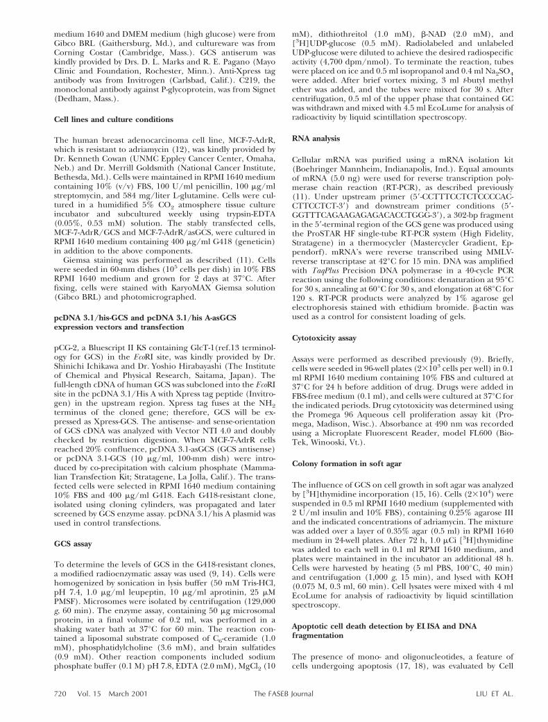
medium 1640 and DMEM medium (high glucose) were fromGibco BRL (Gaithersburg, Md.), and cultureware was fromCorning Costar (Cambridge, Mass.). GCS antiserum waskindly provided by Drs. D. L. Marks and R. E. Pagano (MayoClinic and Foundation, Rochester, Minn.). Anti-Xpress tagantibody was from Invitrogen (Carlsbad, Calif.). C219, themonoclonal antibody against P-glycoprotein, was from Signet(Dedham, Mass.).
Cell lines and culture conditions
The human breast adenocarcinoma cell line, MCF-7-AdrR,which is resistant to adriamycin (12), was kindly provided byDr. Kenneth Cowan (UNMC Eppley Cancer Center, Omaha,Neb.) and Dr. Merrill Goldsmith (National Cancer Institute,Bethesda, Md.). Cells were maintained in RPMI 1640 mediumcontaining 10% (v/v) FBS, 100 U/ml penicillin, 100 mg/mlstreptomycin, and 584 mg/liter L-glutamine. Cells were cul-tured in a humidified 5% CO2 atmosphere tissue cultureincubator and subcultured weekly using trypsin-EDTA(0.05%, 0.53 mM) solution. The stably transfected cells,MCF-7-AdrR/GCS and MCF-7-AdrR/asGCS, were cultured inRPMI 1640 medium containing 400 mg/ml G418 (geneticin)in addition to the above components.
Giemsa staining was performed as described (11). Cellswere seeded in 60-mm dishes (105 cells per dish) in 10% FBSRPMI 1640 medium and grown for 2 days at 37°C. Afterfixing, cells were stained with KaryoMAX Giemsa solution(Gibco BRL) and photomicrographed.
pcDNA 3.1/his-GCS and pcDNA 3.1/his A-asGCSexpression vectors and transfection
pCG-2, a Bluescript II KS containing GlcT-1(ref.13 terminol-ogy for GCS) in the EcoRI site, was kindly provided by Dr.Shinichi Ichikawa and Dr. Yoshio Hirabayashi (The Instituteof Chemical and Physical Research, Saitama, Japan). Thefull-length cDNA of human GCS was subcloned into the EcoRIsite in the pcDNA 3.1/His A with Xpress tag peptide (Invitro-gen) in the upstream region. Xpress tag fuses at the NH2terminus of the cloned gene; therefore, GCS will be ex-pressed as Xpress-GCS. The antisense- and sense-orientationof GCS cDNA was analyzed with Vector NTI 4.0 and doublychecked by restriction digestion. When MCF-7-AdrR cellsreached 20% confluence, pcDNA 3.1-asGCS (GCS antisense)or pcDNA 3.1-GCS (10 mg/ml, 100-mm dish) were intro-duced by co-precipitation with calcium phosphate (Mamma-lian Transfection Kit; Stratagene, La Jolla, Calif.). The trans-fected cells were selected in RPMI 1640 medium containing10% FBS and 400 mg/ml G418. Each G418-resistant clone,isolated using cloning cylinders, was propagated and laterscreened by GCS enzyme assay. pcDNA 3.1/his A plasmid wasused in control transfections.
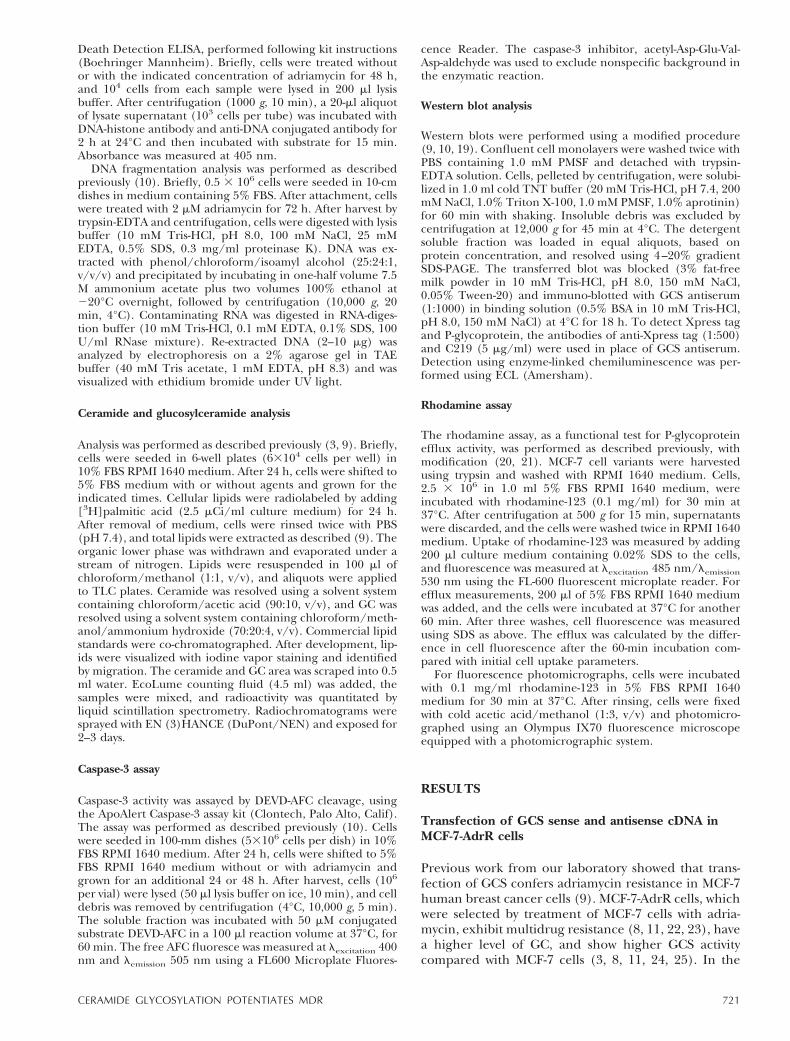
GCS assay
To determine the levels of GCS in the G418-resistant clones,a modified radioenzymatic assay was used (9, 14). Cells werehomogenized by sonication in lysis buffer (50 mM Tris-HCl,pH 7.4, 1.0 mg/ml leupeptin, 10 mg/ml aprotinin, 25 mMPMSF). Microsomes were isolated by centrifugation (129,000g, 60 min). The enzyme assay, containing 50 mg microsomalprotein, in a final volume of 0.2 ml, was performed in ashaking water bath at 37°C for 60 min. The reaction con-tained a liposomal substrate composed of C6-ceramide (1.0mM), phosphatidylcholine (3.6 mM), and brain sulfatides(0.9 mM). Other reaction components included sodiumphosphate buffer (0.1 M) pH 7.8, EDTA (2.0 mM), MgCl2 (10
mM), dithiothreitol (1.0 mM), b-NAD (2.0 mM), and[3H]UDP-glucose (0.5 mM). Radiolabeled and unlabeledUDP-glucose were diluted to achieve the desired radiospecificactivity (4,700 dpm/nmol). To terminate the reaction, tubeswere placed on ice and 0.5 ml isopropanol and 0.4 ml Na2SO4were added. After brief vortex mixing, 3 ml t-butyl methylether was added, and the tubes were mixed for 30 s. Aftercentrifugation, 0.5 ml of the upper phase that contained GCwas withdrawn and mixed with 4.5 ml EcoLume for analysis ofradioactivity by liquid scintillation spectroscopy.
RNA analysis
Cellular mRNA was purified using a mRNA isolation kit(Boehringer Mannheim, Indianapolis, Ind.). Equal amountsof mRNA (5.0 ng) were used for reverse transcription poly-merase chain reaction (RT-PCR), as described previously(11). Under upstream primer (59-CCTTTCCTCTCCCCAC-CTTCCTCT-39) and downstream primer conditions (59-GGTTTCAGAAGAGAGACACCTGGG-39), a 302-bp fragmentin the 59-terminal region of the GCS gene was produced usingthe ProSTAR HF single-tube RT-PCR system (High Fidelity,Stratagene) in a thermocycler (Mastercycler Gradient, Ep-pendorf). mRNA’s were reverse transcribed using MMLV-reverse transcriptase at 42°C for 15 min. DNA was amplifiedwith TaqPlus Precision DNA polymerase in a 40-cycle PCRreaction using the following conditions: denaturation at 95°Cfor 30 s, annealing at 60°C for 30 s, and elongation at 68°C for120 s. RT-PCR products were analyzed by 1% agarose gelelectrophoresis stained with ethidium bromide. b-actin wasused as a control for consistent loading of gels.
Cytotoxicity assay
Assays were performed as described previously (9). Briefly,cells were seeded in 96-well plates (23103 cells per well) in 0.1ml RPMI 1640 medium containing 10% FBS and cultured at37°C for 24 h before addition of drug. Drugs were added inFBS-free medium (0.1 ml), and cells were cultured at 37°C forthe indicated periods. Drug cytotoxicity was determined usingthe Promega 96 Aqueous cell proliferation assay kit (Pro-mega, Madison, Wisc.). Absorbance at 490 nm was recordedusing a Microplate Fluorescent Reader, model FL600 (Bio-Tek, Winooski, Vt.).
Colony formation in soft agar
The influence of GCS on cell growth in soft agar was analyzedby [3H]thymidine incorporation (15, 16). Cells (23104) weresuspended in 0.5 ml RPMI 1640 medium (supplemented with2 U/ml insulin and 10% FBS), containing 0.25% agarose IIIand the indicated concentrations of adriamycin. The mixturewas added over a layer of 0.35% agar (0.5 ml) in RPMI 1640medium in 24-well plates. After 72 h, 1.0 mCi [3H]thymidinewas added to each well in 0.1 ml RPMI 1640 medium, andplates were maintained in the incubator an additional 48 h.Cells were harvested by heating (5 ml PBS, 100°C, 40 min)and centrifugation (1,000 g, 15 min), and lysed with KOH(0.075 M, 0.3 ml, 60 min). Cell lysates were mixed with 4 mlEcoLume for analysis of radioactivity by liquid scintillationspectroscopy.
Apoptotic cell death detection by ELISA and DNAfragmentation
The presence of mono- and oligonucleotides, a feature ofcells undergoing apoptosis (17, 18), was evaluated by Cell
720 Vol. 15 March 2001 LIU ET AL.The FASEB Journal
Death Detection ELISA, performed following kit instructions(Boehringer Mannheim). Briefly, cells were treated withoutor with the indicated concentration of adriamycin for 48 h,and 104 cells from each sample were lysed in 200 ml lysisbuffer. After centrifugation (1000 g, 10 min), a 20-ml aliquotof lysate supernatant (103 cells per tube) was incubated withDNA-histone antibody and anti-DNA conjugated antibody for2 h at 24°C and then incubated with substrate for 15 min.Absorbance was measured at 405 nm.
DNA fragmentation analysis was performed as describedpreviously (10). Briefly, 0.5 3 106 cells were seeded in 10-cmdishes in medium containing 5% FBS. After attachment, cellswere treated with 2 mM adriamycin for 72 h. After harvest bytrypsin-EDTA and centrifugation, cells were digested with lysisbuffer (10 mM Tris-HCl, pH 8.0, 100 mM NaCl, 25 mMEDTA, 0.5% SDS, 0.3 mg/ml proteinase K). DNA was ex-tracted with phenol/chloroform/isoamyl alcohol (25:24:1,v/v/v) and precipitated by incubating in one-half volume 7.5M ammonium acetate plus two volumes 100% ethanol at220°C overnight, followed by centrifugation (10,000 g, 20min, 4°C). Contaminating RNA was digested in RNA-diges-tion buffer (10 mM Tris-HCl, 0.1 mM EDTA, 0.1% SDS, 100U/ml RNase mixture). Re-extracted DNA (2–10 mg) wasanalyzed by electrophoresis on a 2% agarose gel in TAEbuffer (40 mM Tris acetate, 1 mM EDTA, pH 8.3) and wasvisualized with ethidium bromide under UV light.
Ceramide and glucosylceramide analysis
Analysis was performed as described previously (3, 9). Briefly,cells were seeded in 6-well plates (63104 cells per well) in10% FBS RPMI 1640 medium. After 24 h, cells were shifted to5% FBS medium with or without agents and grown for theindicated times. Cellular lipids were radiolabeled by adding[3H]palmitic acid (2.5 mCi/ml culture medium) for 24 h.After removal of medium, cells were rinsed twice with PBS(pH 7.4), and total lipids were extracted as described (9). Theorganic lower phase was withdrawn and evaporated under astream of nitrogen. Lipids were resuspended in 100 ml ofchloroform/methanol (1:1, v/v), and aliquots were appliedto TLC plates. Ceramide was resolved using a solvent systemcontaining chloroform/acetic acid (90:10, v/v), and GC wasresolved using a solvent system containing chloroform/meth-anol/ammonium hydroxide (70:20:4, v/v). Commercial lipidstandards were co-chromatographed. After development, lip-ids were visualized with iodine vapor staining and identifiedby migration. The ceramide and GC area was scraped into 0.5ml water. EcoLume counting fluid (4.5 ml) was added, thesamples were mixed, and radioactivity was quantitated byliquid scintillation spectrometry. Radiochromatograms weresprayed with EN (3)HANCE (DuPont/NEN) and exposed for2–3 days.
Caspase-3 assay
Caspase-3 activity was assayed by DEVD-AFC cleavage, usingthe ApoAlert Caspase-3 assay kit (Clontech, Palo Alto, Calif).The assay was performed as described previously (10). Cellswere seeded in 100-mm dishes (53106 cells per dish) in 10%FBS RPMI 1640 medium. After 24 h, cells were shifted to 5%FBS RPMI 1640 medium without or with adriamycin andgrown for an additional 24 or 48 h. After harvest, cells (106
per vial) were lysed (50 ml lysis buffer on ice, 10 min), and celldebris was removed by centrifugation (4°C, 10,000 g, 5 min).The soluble fraction was incubated with 50 mM conjugatedsubstrate DEVD-AFC in a 100 ml reaction volume at 37°C, for60 min. The free AFC fluoresce was measured at lexcitation 400nm and lemission 505 nm using a FL600 Microplate Fluores-
cence Reader. The caspase-3 inhibitor, acetyl-Asp-Glu-Val-Asp-aldehyde was used to exclude nonspecific background inthe enzymatic reaction.
Western blot analysis
Western blots were performed using a modified procedure(9, 10, 19). Confluent cell monolayers were washed twice withPBS containing 1.0 mM PMSF and detached with trypsin-EDTA solution. Cells, pelleted by centrifugation, were solubi-lized in 1.0 ml cold TNT buffer (20 mM Tris-HCl, pH 7.4, 200mM NaCl, 1.0% Triton X-100, 1.0 mM PMSF, 1.0% aprotinin)for 60 min with shaking. Insoluble debris was excluded bycentrifugation at 12,000 g for 45 min at 4°C. The detergentsoluble fraction was loaded in equal aliquots, based onprotein concentration, and resolved using 4–20% gradientSDS-PAGE. The transferred blot was blocked (3% fat-freemilk powder in 10 mM Tris-HCl, pH 8.0, 150 mM NaCl,0.05% Tween-20) and immuno-blotted with GCS antiserum(1:1000) in binding solution (0.5% BSA in 10 mM Tris-HCl,pH 8.0, 150 mM NaCl) at 4°C for 18 h. To detect Xpress tagand P-glycoprotein, the antibodies of anti-Xpress tag (1:500)and C219 (5 mg/ml) were used in place of GCS antiserum.Detection using enzyme-linked chemiluminescence was per-formed using ECL (Amersham).
Rhodamine assay
The rhodamine assay, as a functional test for P-glycoproteinefflux activity, was performed as described previously, withmodification (20, 21). MCF-7 cell variants were harvestedusing trypsin and washed with RPMI 1640 medium. Cells,2.5 3 106 in 1.0 ml 5% FBS RPMI 1640 medium, wereincubated with rhodamine-123 (0.1 mg/ml) for 30 min at37°C. After centrifugation at 500 g for 15 min, supernatantswere discarded, and the cells were washed twice in RPMI 1640medium. Uptake of rhodamine-123 was measured by adding200 ml culture medium containing 0.02% SDS to the cells,and fluorescence was measured at lexcitation 485 nm/lemission530 nm using the FL-600 fluorescent microplate reader. Forefflux measurements, 200 ml of 5% FBS RPMI 1640 mediumwas added, and the cells were incubated at 37°C for another60 min. After three washes, cell fluorescence was measuredusing SDS as above. The efflux was calculated by the differ-ence in cell fluorescence after the 60-min incubation com-pared with initial cell uptake parameters.
For fluorescence photomicrographs, cells were incubatedwith 0.1 mg/ml rhodamine-123 in 5% FBS RPMI 1640medium for 30 min at 37°C. After rinsing, cells were fixedwith cold acetic acid/methanol (1:3, v/v) and photomicro-graphed using an Olympus IX70 fluorescence microscopeequipped with a photomicrographic system.
RESULTS
Transfection of GCS sense and antisense cDNA inMCF-7-AdrR cells
Previous work from our laboratory showed that trans-fection of GCS confers adriamycin resistance in MCF-7human breast cancer cells (9). MCF-7-AdrR cells, whichwere selected by treatment of MCF-7 cells with adria-mycin, exhibit multidrug resistance (8, 11, 22, 23), havea higher level of GC, and show higher GCS activitycompared with MCF-7 cells (3, 8, 11, 24, 25). In the
721CERAMIDE GLYCOSYLATION POTENTIATES MDR
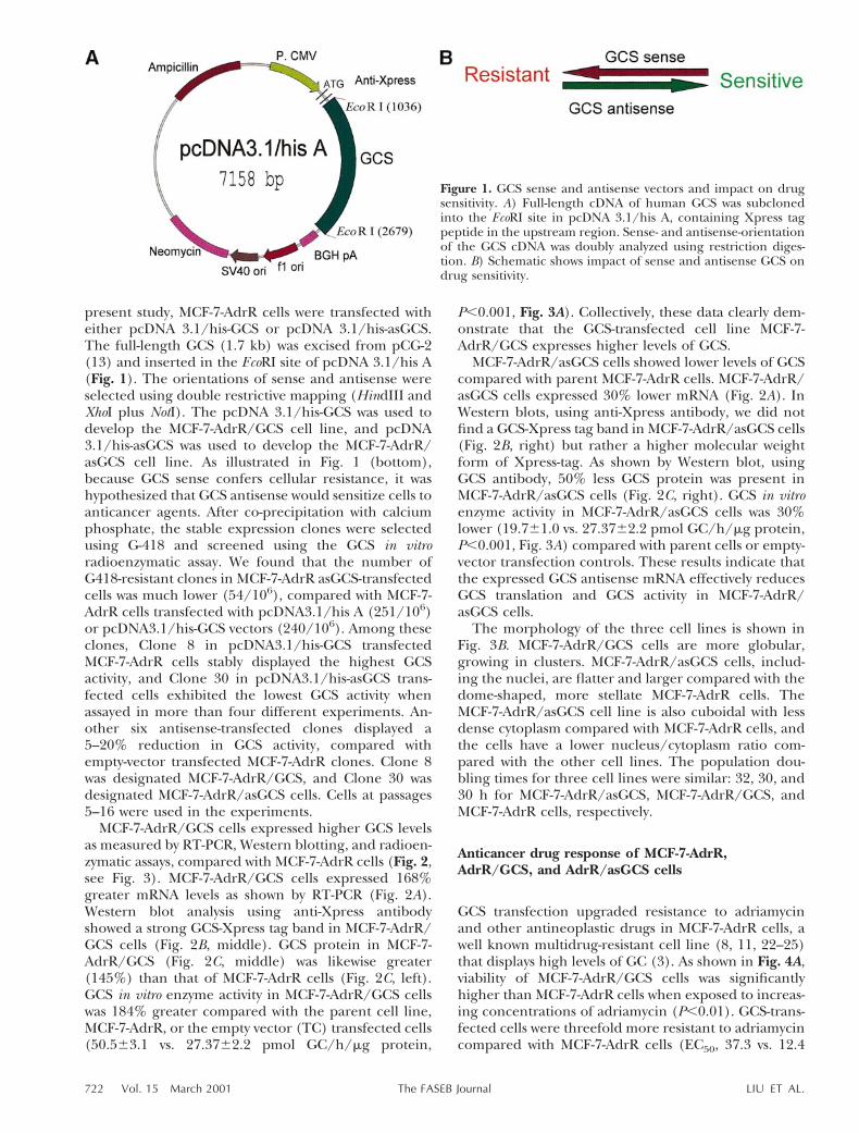
present study, MCF-7-AdrR cells were transfected witheither pcDNA 3.1/his-GCS or pcDNA 3.1/his-asGCS.The full-length GCS (1.7 kb) was excised from pCG-2(13) and inserted in the EcoRI site of pcDNA 3.1/his A(Fig. 1). The orientations of sense and antisense wereselected using double restrictive mapping (HindIII andXhoI plus NotI). The pcDNA 3.1/his-GCS was used todevelop the MCF-7-AdrR/GCS cell line, and pcDNA3.1/his-asGCS was used to develop the MCF-7-AdrR/asGCS cell line. As illustrated in Fig. 1 (bottom),because GCS sense confers cellular resistance, it washypothesized that GCS antisense would sensitize cells toanticancer agents. After co-precipitation with calciumphosphate, the stable expression clones were selectedusing G-418 and screened using the GCS in vitroradioenzymatic assay. We found that the number ofG418-resistant clones in MCF-7-AdrR asGCS-transfectedcells was much lower (54/106), compared with MCF-7-AdrR cells transfected with pcDNA3.1/his A (251/106)or pcDNA3.1/his-GCS vectors (240/106). Among theseclones, Clone 8 in pcDNA3.1/his-GCS transfectedMCF-7-AdrR cells stably displayed the highest GCSactivity, and Clone 30 in pcDNA3.1/his-asGCS trans-fected cells exhibited the lowest GCS activity whenassayed in more than four different experiments. An-other six antisense-transfected clones displayed a5–20% reduction in GCS activity, compared withempty-vector transfected MCF-7-AdrR clones. Clone 8was designated MCF-7-AdrR/GCS, and Clone 30 wasdesignated MCF-7-AdrR/asGCS cells. Cells at passages5–16 were used in the experiments.
MCF-7-AdrR/GCS cells expressed higher GCS levelsas measured by RT-PCR, Western blotting, and radioen-zymatic assays, compared with MCF-7-AdrR cells (Fig. 2,see Fig. 3). MCF-7-AdrR/GCS cells expressed 168%greater mRNA levels as shown by RT-PCR (Fig. 2A).Western blot analysis using anti-Xpress antibodyshowed a strong GCS-Xpress tag band in MCF-7-AdrR/GCS cells (Fig. 2B, middle). GCS protein in MCF-7-AdrR/GCS (Fig. 2C, middle) was likewise greater(145%) than that of MCF-7-AdrR cells (Fig. 2C, left).GCS in vitro enzyme activity in MCF-7-AdrR/GCS cellswas 184% greater compared with the parent cell line,MCF-7-AdrR, or the empty vector (TC) transfected cells(50.563.1 vs. 27.3762.2 pmol GC/h/mg protein,
P,0.001, Fig. 3A). Collectively, these data clearly dem-onstrate that the GCS-transfected cell line MCF-7-AdrR/GCS expresses higher levels of GCS.
MCF-7-AdrR/asGCS cells showed lower levels of GCScompared with parent MCF-7-AdrR cells. MCF-7-AdrR/asGCS cells expressed 30% lower mRNA (Fig. 2A). InWestern blots, using anti-Xpress antibody, we did notfind a GCS-Xpress tag band in MCF-7-AdrR/asGCS cells(Fig. 2B, right) but rather a higher molecular weightform of Xpress-tag. As shown by Western blot, usingGCS antibody, 50% less GCS protein was present inMCF-7-AdrR/asGCS cells (Fig. 2C, right). GCS in vitroenzyme activity in MCF-7-AdrR/asGCS cells was 30%lower (19.761.0 vs. 27.3762.2 pmol GC/h/mg protein,P,0.001, Fig. 3A) compared with parent cells or empty-vector transfection controls. These results indicate thatthe expressed GCS antisense mRNA effectively reducesGCS translation and GCS activity in MCF-7-AdrR/asGCS cells.
The morphology of the three cell lines is shown inFig. 3B. MCF-7-AdrR/GCS cells are more globular,growing in clusters. MCF-7-AdrR/asGCS cells, includ-ing the nuclei, are flatter and larger compared with thedome-shaped, more stellate MCF-7-AdrR cells. TheMCF-7-AdrR/asGCS cell line is also cuboidal with lessdense cytoplasm compared with MCF-7-AdrR cells, andthe cells have a lower nucleus/cytoplasm ratio com-pared with the other cell lines. The population dou-bling times for three cell lines were similar: 32, 30, and30 h for MCF-7-AdrR/asGCS, MCF-7-AdrR/GCS, andMCF-7-AdrR cells, respectively.
Anticancer drug response of MCF-7-AdrR,AdrR/GCS, and AdrR/asGCS cells
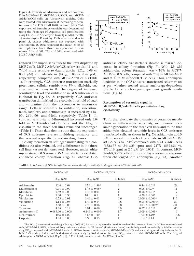
GCS transfection upgraded resistance to adriamycinand other antineoplastic drugs in MCF-7-AdrR cells, awell known multidrug-resistant cell line (8, 11, 22–25)that displays high levels of GC (3). As shown in Fig. 4A,viability of MCF-7-AdrR/GCS cells was significantlyhigher than MCF-7-AdrR cells when exposed to increas-ing concentrations of adriamycin (P,0.01). GCS-trans-fected cells were threefold more resistant to adriamycincompared with MCF-7-AdrR cells (EC50, 37.3 vs. 12.4
Figure 1. GCS sense and antisense vectors and impact on drugsensitivity. A) Full-length cDNA of human GCS was subclonedinto the EcoRI site in pcDNA 3.1/his A, containing Xpress tagpeptide in the upstream region. Sense- and antisense-orientationof the GCS cDNA was doubly analyzed using restriction diges-tion. B) Schematic shows impact of sense and antisense GCS ondrug sensitivity.
722 Vol. 15 March 2001 LIU ET AL.The FASEB Journal

mM, Table 1) and twofold more resistant to daunoru-bicin (EC50, 1.7 vs. 0.9 mM) and actinomycin D (EC50,0.15 vs. 0.08, Table 1, Fig. 4). However, an increase incellular resistance to other anthracyclines, Vinca alka-loids, taxanes, or cisplatin did not accompany GCStransfection (Table 1). As hypothesized, transfection ofGCS effectively upgraded MCF-7-AdrR cellular resis-tance to several natural product chemotherapeuticagents, including adriamycin, daunorubicin, and acti-nomycin D.
In contrast, the antisense-transfected model, MCF-7-AdrR/asGCS, displayed a marked increase in sensitivity
to several different classes of anticancer drugs. Asshown in Fig. 4, MCF-7-AdrR/asGCS cell viability inresponse to adriamycin treatment was significantly re-duced compared with MCF-7-AdrR cells, even at con-centrations as low as 0.5 mM (P,0.001), where survivaldropped below 50%. Adriamycin sensitivity in antisensetransfected cells increased 28-fold (Table 1). The EC50of adriamycin in MCF-7-AdrR/asGCS was 0.44 versus12.4 mM in MCF-7-AdrR and 37 mM in MCF-7-AdrR/GCS cells. For comparison from previous studies, theEC50 of adriamycin in MCF-7 cells was 0.37 mM (9).Thus, GCS antisense transfection of MCF-7-AdrR cells
Figure 2. Expression of GCS mRNA and protein. A) mRNA expression of GCS by RT-PCR. Isolated mRNA (5 ng) was amplifiedby high-fidelity RT-PCR and analyzed by 1% agarose gel electrophoresis, stained with ethidium bromide (top strip). b-actin wasused as control for even loading (bottom strip). The ODGCS/ODb-actin values are MCF-7-AdrR, 0.95; AdrR/GCS, 1.60;AdrR/asGCS, 0.56. Control lane, RT-PCR product without cellular mRNA. AdrR/GCS, MCF-7-AdrR/GCS cells; AdrR/as GCS,MCF-7-AdrR/asGCS cells. B) GCS-Xpress Western blot. The detergent soluble fraction (50 mg protein per lane) was resolvedusing 4–20% gradient SDS-PAGE. The transferred nitrocellulose blot was blocked (3% fat-free milk powder) and immuno-blotted with anti-Xpress antibody (1:1,000). Detection using enzyme-linked chemiluminescence was performed using ECL(Amersham). OD values (densitometry) of GCS-Xpress are MCF-7-AdrR, 7,861; AdrR/GCS, 122,407; AdrR/asGCS, 7,860. C)GCS protein in MCF-7-AdrR cells and transfectants. GCS antiserum was used at a 1:1000 dilution. OD values of GCSdensitometry for quantitative analysis yielded MCF-7-AdrR, 206,712; AdrR/GCS, 298,368; AdrR/asGCS, 110,808; AdrR/GCS,MCF-7-AdrR/GCS cells; AdrR/asGCS, MCF-7-AdrR/asGCS cells.
Figure 3. Glucosylceramide synthase activity and morphology of MCF-7-AdrR, MCF-7-AdrR/GCS, and MCF-7-AdrR/asGCS cells.A) GCS activity. Enzyme activity was measured as detailed in Materials and Methods. Data represent the mean 6 sd of threereplicates from three independent experiments. *P , 0.001 compared with MCF-7-AdrR cells. AdrR, MCF-7-AdrR cells; TC,transfection control (MCF-7-AdrR cells transfected with pcDNA 3.1 vector alone); GCS, MCF-7-AdrR/GCS cells; asGCS,MCF-7-AdrR/asGCS cells. B) Comparison of cell morphology in MCF-7-AdrR and GCS transfectants. After 48 h of subculture,cells were stained using the May-Grunwald-Giemsa method and photographed at 2003 magnification.
723CERAMIDE GLYCOSYLATION POTENTIATES MDR
restored adriamycin sensitivity to the level displayed byMCF-7 cells. MCF-7-AdrR/asGCS cells were also 11- and7-fold more sensitive to daunorubicin (EC50, 0.08 vs.0.91 mM) and idarubicin (EC50, 0.06 vs. 0.42 mM),respectively, compared with MCF-7-AdrR cells (Table1). Interestingly, GCS antisense transfection markedlypotentiated cellular sensitivity to Vinca alkaloids, tax-anes, and actinomycin D. The degree of increasedsensitivity to taxol and vinblastine in GCS antisense cellsis shown in Fig. 5A, B, respectively. GCS antisensetransfection diminished the cytotoxic threshold of taxoland vinblastine from the micromolar to nanomolarrange. Cellular sensitivity to vinblastine, vincristine,taxol, taxotere, and actinomycin D increased by 115-,50-, 241-, 66-, and 9-fold, respectively (Table 1). Incontrast, sensitivity to 5-flurouracil increased only 3.6-fold in MCF-7-AdrR/asGCS cells, and the EC50 ofcisplatin in the three cell lines remained unchanged(Table 1). These data demonstrate that the expressionof GCS antisense reverses multidrug resistance, andthat reversal is specific for certain drug classes.
Colony formation in soft agar under drug-free con-ditions was also evaluated, and a difference in the threecell lines was not demonstrated. However, under adria-mycin stress, GCS sense cDNA transfectants exhibitedenhanced colony formation (Fig. 6), whereas GCS
antisense cDNA transfectants showed a marked de-crease in colony formation (Fig. 6). With 2.5 mMadriamycin, colony formation was ,10% in MCF-7-AdrR/asGCS cells, compared with 70% in MCF-7-AdrRand 99% in MCF-7-AdrR/GCS cells. Thus, adriamycintoxicities in the GCS antisense-transfected cells were ona par, whether treated under anchorage-dependent(Table 1) or anchorage-independent growth condi-tions (Fig. 6).
Resumption of ceramide signal inMCF-7-AdrR/asGCS cells potentiates drugcytotoxicity
To further elucidate the dynamics of ceramide metab-olism in anthracycline sensitivity, we measured cer-amide generation in the three cell lines and found thatadriamycin elevated ceramide levels in GCS antisensetransfected cells. As shown in Fig. 7A, adriamycin at 0.5mM increased the levels of ceramide in MCF-7-AdrR/asGCS cells by 184% compared with MCF-7-AdrR cells(632667 vs. 344625 cpm) and 227% (857624 vs.376616 cpm) at 2.5 mM (P,0.001). In contrast, MCF-7-AdrR/GCS cells did not display a ceramide responsewhen challenged with adriamycin (Fig. 7A). Another
Figure 4. Toxicity of adriamycin and actinomycinD in MCF-7-AdrR, MCF-7-AdrR/GCS, and MCF-7-AdrR/asGCS cells. A) Adriamycin toxicity. Cellswere treated with adriamycin at increasing concen-trations in 5% FBS RPMI 1640 medium. After 72-hexposure, adriamycin cytotoxicity was determinedusing the Promega 96 Aqueous cell proliferationassay kit. {––{ Adriamycin toxicity in MCF-7 cells.B) Actinomycin D toxicity. Cells were treated as inpanel A, except adriamycin was replaced withactinomycin D. Data represent the mean 6 sd ofsix replicates from three independent experi-ments. *P , 0.001, **P , 0.0001 compared withMCF-7 AdrR cells.
TABLE 1. Influence of GCS transfection on chemotherapy sensitivity in drug-resistant MCF-7-AdrR cells
MCF-7-AdrR MCF-7-AdrR/GCS MCF-7-AdrR/asGCS
EC50 (mM) EC50 (mM) R. Index EC50 (mM) S. Index
Adriamycin 12.4 6 0.68 37.3 6 1.89* 3 0.44 6 0.01* 28Daunorubicin 0.91 6 0.09 1.73 6 0.06* 2 0.08 6 0.0* 11Idarubicin 0.42 6 0.0 0.45 6 0.01 1 0.06 6 0.01* 7Epirubicin 9.30 6 0.30 10.0 6 0.1 1 4.30 6 0.06* 2Vinblastine 0.73 6 0.01 0.46 6 0.01 0.6 0.006 6 0.000* 115Vincristine 2.14 6 0.03 1.40 6 0.14 0.6 0.05 6 0.0001* 50Taxol 3.38 6 0.06 2.75 6 0.06 0.8 0.014 6 0.0002* 241Taxotere 4.65 6 0.19 3.91 6 0.06 0.9 0.07 6 0.01* 66Actinomycin D 0.083.00 6 0.008 0.145 6 0.006* 1.7 0.009 6 0.001* 95-Flurouracil 49.5 6 1.29 54.5 6 1.29 1 13.5 6 1.29* 3.6Cisplatin 4.04 6 0.09 3.38 6 0.15 1 3.4 6 0.14 1
The EC50 (concentration of drug affecting a 50% kill) for each drug tested is listed for each of the three cell lines. In GCS-sense transfectedcells, MCF-7-AdrR/GCS, enhanced drug resistance is shown by “R. Index” (Resistance Index) and is designated numerically by fold increase indrug EC50 compared with MCF-7-AdrR cells. In GCS-antisense transfected cells, MCF-7-AdrR/asGCS, enhanced drug sensitivity is shown by “S.Index” (Sensitivity Index) and is designated numerically by fold decrease in drug EC50, compared with MCF-7-AdrR cells. The EC50 ofadriamycin in MCF-7 cells is 0.37 mM(10). * P , 0.001, compared with MCF-7-AdrR cells.
724 Vol. 15 March 2001 LIU ET AL.The FASEB Journal

agent, actinomycin D, also potentiated cellular cer-amide generation in GCS antisense transfected cells(Fig. 7B), with no significant changes occurring ineither MCF-7-AdrR or MCF-7-AdrR/GCS cells.
We next examined GC levels in the three cell linesunder basal conditions and under adriamycin expo-sure. As shown in Fig. 8A by TLC autoradiography, GCSantisense transfection reduced cellular levels of GC(lane 3), compared with MCF-7-AdrR cells (lane 1).When evaluated by scintillation spectroscopy, MCF-7-AdrR/asGCS cells exhibited an ;27% decrease in GCcompared with parent cells (1854654 vs. 25466133cpm, Fig. 8B, minus adriamycin). GC was slightly ele-vated in GCS sense transfectants (Fig. 8A, lane 2);however, the difference was not statistically significantcompared with MCF-7-AdrR cells (28196174 vs.25466133 cpm, Fig. 8B, minus adriamycin). Althoughadriamycin exposure elicits ceramide increases in MCF-7-AdrR/asGCS cells (Fig. 7A), exposure to this drug didnot alter GC metabolism in any of the cell lines (Fig.8B).
Increases in effector caspase activity in ceramide-governed apoptotic signaling are consistent withchanges in ceramide levels. With 10 mM adriamycin(EC50 in MCF-7-AdrR cells), caspase-3 activity in MCF-
7-AdrR/asGCS cells increased 3- (1.9960.005 vs.0.6360.005 nmole AFC/106 cells, P,0.001) and 3.6-fold (3.660.04 vs. 1.0260.03 nmole AFC/106 cells,P,0.001), at 24- and 48-h treatment, respectively, com-pared with MCF-7-AdrR cells (Fig. 9A). Under likeconditions, caspase-3 activity in MCF-7-AdrR/GCS cellshad no significant decrease, compared with activity in
Figure 5. Influence of GCS antisense transfection on taxoland vinblastine sensitivity. A) Taxol sensitivity. Cells weretreated with taxol at increasing concentrations in 5% FBSRPMI 1640 medium. After 72-h exposure, taxol cytotoxicitywas determined using the Promega 96 Aqueous cell prolifer-ation assay kit. B) Vinblastine sensitivity. Cells were treated asin panel A, except taxol was replaced with vinblastine. Datarepresent the mean 6 sd of six replicates from three inde-pendent experiments. *P , 0.0001 compared with MCF-7-AdrR cells under the same conditions.
Figure 6. Influence ofadriamycin on colony for-mation under anchorage-independent conditions inMCF-7-AdrR and GCStransfectants. Cells weretreated with the indicatedconcentration of adriamy-cin for 120 h, and colonyformation was analyzed by[3H]thymidine incorpora-tion. Data represent themean 6 sd of triplicatesfrom three independent
experiments. *P , 0.01, compared with MCF-7-AdrR cells.
Figure 7. Ceramide generation in response to adriamycin andactinomycin D. Cellular lipids were radiolabeled by incubat-ing cells with [3H]palmitic acid (2.5 mCi/ml culture medium)for 24 h, and total lipids were extracted for analysis. Ceramidewas resolved by TLC. Ceramide values are given as cpm per105 cpm total lipid. A) Influence of adriamycin dose oncellular ceramide metabolism. Cells were treated with theindicated concentrations of adriamycin for 48 h. B) Influenceof actinomycin D dose on cellular ceramide metabolism. Cellswere treated with the indicated concentrations of actinomy-cin D for 48 h. Data represent the mean 6 sd of triplicatesfrom three independent experiments. *P , 0.001 comparedwith MCF-7-AdrR cells under the same conditions.
Figure 8. Cellular glucosylceramide metabolism in responseto adriamycin treatment. Cellular lipids were radiolabeled byincubating cells with [3H]palmitic acid (2.5 mCi/ml culturemedium) for 24 h. Total lipids were extracted and GC wasresolved by TLC. GC values are given as cpm per 105 cpmtotal lipid. A) GC TLC autoradiograph. Lanes 1, 2, and 3 showtotal lipids from MCF-7-AdrR, MCF-7-AdrR/GCS and MCF-7-AdrR/asGCS cells, respectively. B) Influence of adriamycinon cellular GC metabolism. Cells were treated with adriamy-cin (2.0 mM) for 48 h. Data represent the mean 6 sd oftriplicates from two independent experiments. *P , 0.01compared with MCF-7-AdrR cells under the same conditions.
725CERAMIDE GLYCOSYLATION POTENTIATES MDR
MCF-7-AdrR cells at 48 h. Further characterizationrevealed that adriamycin elicited apoptosis only in theGCS antisense-transfected cells (Fig. 9B, C). The levelsof apoptosis in GCS antisense cells increased withincreasing concentration of adriamycin. The apoptoticindex was 370 (0.4460.01 vs. 0.1260.005 OD) and700% (0.8460.04 vs. 0.1260.005 OD) of untreatedMCF-7-AdrR/asGCS cells at 0.5 and 5.0 mM adriamycin,respectively (Fig. 9B). Using DNA fragmentation anal-ysis, adriamycin treatment (5.0 mM, 72 h) only inducedapoptosis in MCF-7-AdrR/asGCS cells (Fig. 9C, lane 6).Overall, these data show that reduced glycosylationpotential enhances cellular buildup of free ceramideunder adriamycin stress and potentiates the progres-sion of programmed cell death via caspase.
P-glycoprotein expression and efflux parameters
P-glycoprotein–modulated drug efflux is the mostwidely characterized drug resistance mechanism (26),and it is highly expressed in MCF-7-AdrR cells (9, 22).However, adriamycin resistance produced by enforcedexpression of GCS is not accompanied by increasedP-glycoprotein in MCF-7/GCS cells (9). To assess therole of the MDR1 phenotype in enhanced drug sensi-tivity expressed by GCS antisense transfectants, weanalyzed rhodamine-123 efflux and P-glycoprotein ex-pression in the MCF-7 cell variants (Fig. 10). Enhancedrhodamine-123 efflux was observed in all cell lines,except MCF-7 (Fig. 10A). The enhanced efflux corre-lated with overexpression of P-glycoprotein in the MCF-7-AdrR cell variants (Fig. 10B). Therefore, the up- ordown-regulation of GCS activity by transfection hadlittle influence on either rhodamine-123 efflux or P-glycoprotein expression (Fig. 10A, B). This suggests
that P-glycoprotein is not a major mechanism involvedin chemotherapy sensitization that accompanies GCSantisense transfection. Interestingly, we found en-hanced uptake of rhodamine-123 in MCF-7-AdrR/as-GCS cells, compared with MCF-7-AdrR and MCF-7-AdrR/GCS cells. The degree of enhancement wassimilar with the fluorescence intensity observed inwild-type MCF-7 cells (Fig. 11A). Quantitative fluores-cence measurement of intracellular rhodamine-123showed that MCF-7 cells took up 41% of the total(7,043657/1.73104 FU, [fluorescence units]) on aparallel with MCF-7-AdrR/asGCS (41%, 7,0776518/
Figure 9. Caspase-3 activity and apoptosis in response to adriamycin in MCF-7 GCS transfectants. A) Caspase-3 activity. Cells weretreated without or with adriamycin (10 mM) for 24 and 48 h. Data represent the mean 6 sd of triplicates from two independentexperiments. *P,0.0001 compared with MCF-7-AdrR cells exposed to adriamycin for each corresponding treatment period. B)Apoptosis. Cells were treated without or with the indicated concentrations of adriamycin for 48 h. Data represent the mean 6sd of triplicates from two independent experiments. *P,0.0001 compared with MCF-7-AdrR cells exposed to adriamycin foreach corresponding treatment period. C) DNA fragmentation. Cells were exposed to 5.0 mM adriamycin for 72 h, and cellularDNA was extracted and resolved by 2% agarose gel electrophoresis. Left lane, low DNA mass ladder standard. Lanes 1, 3, 5 showMCF-7-AdrR, MCF-7-AdrR/GCS and MCF-7-AdrR/asGCS cells, respectively. Lanes 2, 4, 6 show MCF-7-AdrR, MCF-7-AdrR/GCSand MCF-7-AdrR/asGCS cells treated with 5.0 mM adriamycin, respectively.
Figure 10. Rho-damine-123 effluxand P-glycoproteine x p r e s s i o n i nMCF-7 cell vari-ants. A) Rhoda-mine-123 efflux.Cells (2.5 3 106)were incubatedwith rhodamine-123 (0.1 mg/ml)for 30 min at 37°C.Efflux of rhoda-mine-123 was de-termined afteranother 60-min in-cubation and mea-sured at 485excita-tion and 530emission,as described in Ma-terials and Meth-ods. *P , 0.001 compared with the other three cell lines. AdrR,MCF-7-AdrR cells; GCS, MCF-7-AdrR/GCS; asGCS, MCF-7-AdrR/asGCS. B) P-glycoprotein expression by Western blot.C219 monoclonal antibody (5 mg/ml) was used to recognizeP-glycoprotein.
726 Vol. 15 March 2001 LIU ET AL.The FASEB Journal

1.73104 FU). Rhodamine-123 uptake in GCS antisensetransfectants was fivefold greater than uptake in MCF-7-AdrR cells (7,0776518 vs. 1,3316213 FU) and 10-foldgreater than uptake in MCF-7-AdrR/GCS cells (7,0776518 vs. 6696106 FU) (Fig. 11B).
DISCUSSION
MCF-7-AdrR cells exhibit cross-resistance to a widerange of antineoplastic agents including Vinca alka-loids, anthracyclines, and epipodophyllotoxins (12, 22,23). Although there is overexpression of P-glycoproteinand glutathione-S-transferase in MCF-7-AdrR cells (22,27), there have been no reports showing that antisensetransfection with either P-glycoprotein or glutathione-S-transferase reverses multidrug resistance in thesecells. Our study shows that drug resistance can bereversed by antisense manipulation of ceramide metab-olism, and the work reinforces the doctrine that drugresistance in cancer is multifactorial (28).
Through gene targeting, it has been shown that GCSsense cDNA transfection induces resistance to adriamy-cin by 11-fold in MCF-7 cells (9). Although MCF-7-AdrRcells constitutively display high GCS activity and ele-vated GC levels (3, 8), introducing GCS cDNA intothese multidrug-resistant cells upgraded, albeit onlyseveralfold, the resistance threshold for adriamycin(Fig. 4, Table 1), daunorubicin, and actinomycin D(Table 1). Compared with MCF-7 cells, the high basallevels of GC (sixfold more) and enhanced GCS enzymeactivity (8), which contribute to multidrug resistance,may interfere with the efficacy of GCS transfection forfurther enhancement of drug resistance in MCF-7-AdrRcells. On the other hand, GCS antisense, which effec-tively reintroduced ceramide/caspase signaling (Figs. 7and 8), substantially restored cellular sensitivity to manyanticancer drugs, including anthracyclines, taxanes,Vinca alkaloids, and actinomycin D (Table 1). All ofthese agents are substrates for P-glycoprotein (26),whereas 5-flurouracil and cisplatin, whose toxicities
were not greatly modified by GCS antisense, are notclassified as pump drugs. This raises the question ofselectivity of GCS antisense for reversal of drug resis-tance, a topic that is currently being pursued. GCSantisense also sensitized cells that were grown in softagar to adriamycin (Fig. 5) and sensitized cells grownunder hypoxic conditions (O2,5%, data not shown).These results suggest that GCS antisense is promisingfor restoring drug sensitivity under conditions thatmimic tumor in vivo environments. Overall, these dataindicate that GCS is a cause of multidrug resistance inMCF-7 breast cancer cells.
Overexpression of P-glycoprotein reduces intracellu-lar accumulation of chemotherapeutic agents and pro-duces multidrug resistance (26, 29). However, the rolethat P-glycoprotein exerts in breast cancer has beendifficult to define (30–32). Kim et al. reported thatP-glycoprotein (MDR-1) is a minor determinant of drugresistance in MCF-7-AdrR cells (32). We did not observeincreased P-glycoprotein expression in MCF-7/GCScells in which adriamycin resistance was induced byGCS overexpression (9). We also did not observedecreased expression of P-glycoprotein in MCF-7-AdrR/asGCS cells. When we analyzed rhodamine-123efflux and P-glycoprotein expression in MCF-7 cell lines(Fig. 8), our observations were much in line withprevious work showing that MCF-7-AdrR cells have anenhanced efflux capacity, complementing the en-hanced levels of P-glycoprotein (12, 22, 23). AlthoughGCS antisense transfection sensitized resistant cells toseveral different types of drugs, antisense did notmodulate either P-glycoprotein expression or effluxparameters. This suggests that P-glycoprotein is not themajor mechanism involved in chemotherapy sensitiza-tion that accompanies GCS antisense transfection.
Ceramide is a lipid second messenger in the apopto-tic pathway, participating in cell death initiated byanticancer drugs, cytokines, and ionizing radiation (33,34). The apoptotic impact of adriamycin, daunorubi-cin, and actinomycin D depends, in part, on cellularceramide generation (9, 24, 35, 36). Increased cer-
Figure 11. Rhodamine-123 uptake in MCF-7 cell variants. A) Fluorescence photomicrographs of MCF-7 cell variants afterincubation with rhodamine-123. Cells were incubated with rhodamine-123 (0.1 mg/ml) for 30 min at 37°C and fixed as detailedin Materials and Methods. B) Cellular uptake of Rhodamine-123. Cells (2.5 3 106) were incubated with rhodamine-123 (0.1mg/ml) for 30 min at 37°C. After washing, uptake of rhodamine-123 was determined in the SDS-lysate, by fluorescence, asdescribed in Materials and Methods. *P , 0.001 compared with MCF-7-AdrR cells.
727CERAMIDE GLYCOSYLATION POTENTIATES MDR
amide levels enhance the efficacy of vinblastine (37)and taxol (38). In this study, we show that impairmentof ceramide glycosylation increases intracellular cer-amide levels and propels apoptotic signaling in re-sponse to antineoplastic drugs. In addition to glycosyl-ation, ceramide can be converted to sphingomyelin (viasphingomyelin synthase), galactosylceramide (via galac-tosylceramide synthase), or undergo deacylation toform sphingosine (via ceramidase) (39). However, as aroute to remove ceramide, up-regulating the conver-sion of ceramide to sphingomyelin, through sphingo-myelin synthase, may be harmful to the cell, as thelevels of sphingomyelin are tightly regulated. There-fore, although sphingomyelin synthase regulates intra-cellular ceramide levels (40), GCS appears to be moreinfluential over cell growth and apoptosis. GCS con-verts cytotoxic ceramide to GC, an essential buildingblock of more complex cell membrane components.Various works show that GC stimulates cell prolifera-tion (41, 42) and tumor growth (43, 44), whereas GCSinhibitors arrest cell division (45, 46), exhibit anticar-cinogenic properties (46), and reduce metastasis (47).Complex derivatives of GC, such as gangliosides GM3,GM2, and GD2, are also involved in cell growth, tumormetastasis, and drug resistance (48–50). Therefore,lowering glycosphingolipid synthesis by modifying GCmetabolism appears to be a novel strategy in cancerchemotherapy (10, 51–53).
In the present study, GCS antisense markedly sensi-tized breast cancer cells to several well-known antitu-mor agents. Taking into account the 30% decrease inGCS activity in MCF-7-AdrR/asGCS cells (Fig. 3), mul-tifold increases in sensitivity to the various drugs (Table1) are striking. In addition to modulating cellularceramide and GC levels under stress, GCS catalyzes thefirst glycosylation step in the biosynthesis of glycosphin-golipids, lipids which are present on virtually all mam-malian cell plasma membranes (54, 55). Glycosphingo-lipids are integral components of plasma membranemicrodomains, such as rafts, caveolae, and GM3-en-riched microdomains (56, 57). Microdomains are in-volved in drug resistance (58, 59), mediating mem-brane trafficking and signal activity, and coupling celladhesion interactions with signaling (60, 61). A GCSknockout study in mice showed that the consequenceof homozygosity was embryonic lethality, revealing avital role for GCS during development and differentia-tion (62). Lowering GCS by 1-phenyl-2-decanoylamino-3-morpholino-propanol exposure brings about mor-phological change in Chinese hamster ovary cells (63),in cultured cortical neuron (64), and in PC12 cells(65). Ganglioside, GQ1b, is essential for synapse forma-tion and synaptic activity (64). We also found GCSantisense cDNA markedly altered MCF-7-AdrR cellmorphology (Fig. 2) and greatly increased the uptakeof rhodamine-123 (Fig. 10). This suggests that alter-ation of membrane glycosphingolipid composition, byenforced expression of GCS antisense, effects cell mor-phology and membrane functional activities, includingdrug transport. Enhanced drug import, which may
partially underlie the increased sensitivity to anticancerdrugs observed in MCF-7-AdrR/asGCS cells, is beingfurther investigated.
This work was supported in part by a PHS grant from theNational Cancer Institute (CA77632); The Streisand Founda-tion; the Strauss Foundation, Sandra Krause, Trustee; theFashion Footwear Association of New York, FFANY; theAssociates for Breast and Prostate Cancer Studies, Los Ange-les; the Leslie and Susan Gonda (Goldschmied) Foundation,and the Joseph B. Gould Foundation, Las Vegas, NV. Y. L. isa Joseph B. Gould Fellow in Breast Cancer Research.
We thank Dr. Shinichi Ichikawa and Dr. Yoshio Hiraba-yashi (Laboratory for Cellular Glycobiology, The Institute ofChemical and Physical Research, RIKEN, Saitama, Japan) forproviding the ceramide glucosyltransferase cDNA, pCG 2,and Dr. D. L. Marks and Dr. R. E. Pagano (Mayo Clinic andFoundation) for GCS antiserum. We appreciate the advice ofDr. James Hardin, John Wayne Cancer Institute, in the reviewof this work.
REFERENCES
1. Hannun, Y. A. (1996) Functions of ceramide in coordinatingcellular responses to stress. Science 274, 1855–1859
2. Kolesnick, R. N., and Kronke, M. (1998) Regulation of ceramideproduction and apoptosis. Annu. Rev. Physiol. 60, 643–665
3. Lavie, Y., Cao, H., Bursten, S. L., Giuliano, A. E., and Cabot,M. C. (1996) Accumulation of glucosylceramides in multidrug-resistant cancer cells. J. Biol. Chem. 271, 19530–19536
4. Nicholson, K. M., Quinn, D. M., Kellett, G. L., and Warr, J. R.(1999) Preferential killing of multidrug-resistant KB cells byinhibitors of glucosylceramide synthase. Br. J. Cancer 81, 423–430
5. Kok, J. W., Veldman, R. J., Klappe, K., Koning, H., Filipeanu,C. M., and Muller, M. (2000) Differential expression of sphin-golipids in MRP1 overexpressing HT29 cells. Int. J. Cancer 87,172–178
6. Lala, P., Ito, S., and Lingwood, C. A. (2000) Retroviral transfec-tion of Madin-Darby canine kidney cells with human MDR1results in a major increase in globotriaosylceramide and 105- to106-fold increased cell sensitivity to verocytotoxin: role of p-glycoprotein in glycolipid synthesis. J. Biol. Chem. 275, 6246–6251
7. Lucci, A., Cho, W. I., Han, T. Y., Giuliano, A. E., Morton, D. L.,and Cabot, M. C. (1998) Glucosylceramide: a marker formultiple-drug resistant cancers. Anticancer Res. 18, 475–480
8. Lavie, Y., Cao, H., Volner, A., Lucci, A., Han, T. Y., Geffen, V.,Giuliano, A. E., and Cabot, M. C. (1997) Agents that reversemultidrug resistance, tamoxifen, verapamil, and cyclosporin A,block glycosphingolipid metabolism by inhibiting ceramideglycosylation in human cancer cells. J. Biol. Chem. 272, 1682–1687
9. Liu, Y. Y., Han, T. Y., Giuliano, A. E., and Cabot, M. C. (1999)Expression of glucosylceramide synthase, converting ceramideto glucosylceramide, confers adriamycin resistance in humanbreast cancer cells. J. Biol. Chem. 274, 1140–1146
10. Liu, Y. Y., Han, T. Y., Giuliano, A. E., Ichikawa, S., Hirabayashi,Y., and Cabot, M. C. (1999) Glycosylation of ceramide potenti-ates cellular resistance to tumor necrosis factor-alpha-inducedapoptosis. Exp. Cell Res. 252, 464–470
11. Liu, Y. Y., Han, T. Y., Giuliano, A. E., Hansen, N., and Cabot,M. C. (2000) Uncoupling ceramide glycosylation by transfectionof glucosylceramide synthase antisense reverses adriamycin re-sistance. J. Biol. Chem. 275, 7138–7143
12. Cowan, K. H., Batist, G., Tulpule, A., Sinha, B. K., and Myers,C. E. (1986) Similar biochemical changes associated with mul-tidrug resistance in human breast cancer cells and carcinogen-induced resistance to xenobiotics in rats. Proc. Natl. Acad. Sci.USA 83, 9328–9332
13. Ichikawa, S., Sakiyama, H., Suzuki, G., Hidari, K. I., and Hiraba-yashi, Y. (1996) Expression cloning of a cDNA for human
728 Vol. 15 March 2001 LIU ET AL.The FASEB Journal
ceramide glucosyltransferase that catalyzes the first glycosylationstep of glycosphingolipid synthesis. Proc. Natl. Acad. Sci. USA 93,4638–4643
14. Shukla, G. S., and Radin, N. S. (1990) Glucosylceramide syn-thase of mouse kidney: further characterization with an im-proved assay method. Arch. Biochem. Biophys. 283, 372–378
15. Jones, C. A., Tsukamoto, T., O’Brien, P. C., Uhl, C. B., Alley,M. C., and Lieber, M. M. (1985) Soft agarose culture humantumour colony forming assay for drug sensitivity testing: [3H]-thymidine incorporation vs colony counting. Br. J. Cancer. 52,303–310
16. Chen, J., Bander, J. A., Santore, T. A., Chen, Y., Ram, P. T., Smit,M. J., and Iyengar, R. (1998) Expression of Q227L-galphas inMCF-7 human breast cancer cells inhibits tumorigenesis. Proc.Natl. Acad. Sci. USA 95, 2648–2652
17. Wyllie, A. H., Kerr, J. F., and Currie, A. R. (1980) Cell death: thesignificance of apoptosis. Int. Rev. Cytol. 68, 251–306
18. Bonfoco, E., Krainc, D., Ankarcrona, M., Nicotera, P., andLipton, S. A. (1995) Apoptosis and necrosis: two distinct eventsinduced, respectively, by mild and intense insults with N-methyl-D-aspartate or nitric oxide/superoxide in cortical cell cultures.Proc. Natl. Acad. Sci. USA 92, 7162–7166
19. Watanabe, R., Wu, K., Paul, P., Marks, D. L., Kobayashi, T.,Pittelkow, M. R., and Pagano, R. E. (1998) Up-regulation ofglucosylceramide synthase expression and activity during hu-man keratinocyte differentiation. J. Biol. Chem. 273, 9651–9655
20. Stein, U., Walther, W., Lemm, M., Naundorf, H., and Fichtner,I. (1997) Development and characterisation of novel humanmultidrug resistant mammary carcinoma lines in vitro and invivo. Int. J. Cancer 72, 885–891
21. Brouty-Boye, D., Kolonias, D., Wu, C. J., Savaraj, N., andLampidis, T. J. (1995) Relationship of multidrug resistance torhodamine-123 selectivity between carcinoma and normal epi-thelial cells: taxol and vinblastine modulate drug efflux. CancerRes. 55, 1633–1638
22. Fairchild, C. R., Ivy, S. P., Kao-Shan, C. S., Whang-Peng, J.,Rosen, N., Israel, M. A., Melera, P. W., Cowan, K. H., andGoldsmith, M. E. (1987) Isolation of amplified and overex-pressed DNA sequences from adriamycin-resistant humanbreast cancer cells. Cancer Res. 47, 5141–5148
23. Fairchild, C. R., Moscow, J. A., O’Brien, E. E., and Cowan, K. H.(1990) Multidrug resistance in cells transfected with humangenes encoding a variant P-glycoprotein and glutathione S-transferase-pi. Mol. Pharmacol. 37, 801–809
24. Cabot, M. C., and Giuliano, A. E. (1997) Apoptosis: a cellmechanism important for cytotoxic response to adriamycin anda lipid metabolic pathway that facilitates escape. Breast CancerRes. Treat. 46, 283
25. Cabot, M. C., Han, T. Y., and Giuliano, A. E. (1998) Themultidrug resistance modulator SDZ PSC 833 is a potentactivator of cellular ceramide formation. FEBS Lett. 431, 185–188
26. Gottesman, M. M., and Pastan, I. (1993) Biochemistry of multi-drug resistance mediated by the multidrug transporter. Annu.Rev. Biochem. 62, 385–427
27. Batist, G., Tulpule, A., Sinha, B. K., Katki, A. G., Myers, C. E.,and Cowan, K. H. (1986) Overexpression of a novel anionicglutathione transferase in multidrug-resistant human breastcancer cells. J. Biol. Chem. 261, 15544–15549
28. Lehnert, M. (1996) Clinical multidrug resistance in cancer: amultifactorial problem. Eur. J. Cancer 32A, 912–920
29. Riordan, J. R., and Ling, V. (1979) Purification of P-glycoproteinfrom plasma membrane vesicles of Chinese hamster ovary cellmutants with reduced colchicine permeability. J. Biol. Chem. 254,12701–12705
30. Decker, D. A., Morris, L. W., Levine, A. J., Pettinga, J. E.,Grudzien, J. L., and Farkas, D. H. (1995) Immunohistochemicalanalysis of P-glycoprotein expression in breast cancer: clinicalcorrelations. Ann. Clin. Lab. Sci. 25, 52–59
31. Schneider, J., and Romero, H. (1995) Correlation of P-glycop-rotein overexpression and cellular prognostic factors in forma-lin-fixed, paraffin-embedded tumor samples from breast cancerpatients. Anticancer Res. 15, 1117–1121
32. Kim, H. M., Oh, G. T., Hong, D. H., Kim, M. S., Kang, J. S., Park,S. M., and Han, S. B. (1997) MDR-1 gene expression is a minorfactor in determining the multidrug resistance phenotype ofMCF7/ADR and KB-V1 cells. FEBS Lett. 412, 201–206
33. Darvis, W. D., and Grant, S. (1998) The role of ceramide in thecellular response to cytotoxic agents. Curr. Opin. Oncol. 10,552–559
34. Radford, I. R. (1999) Initiation of ionizing radiation-inducedapoptosis: DNA damage-mediated or does ceramide have a role?Int. J. Radiat. Biol. 75, 521–528
35. Bose, R., Verheij, M., Haimovitz-Friedman, A., Scotto, K., Fuks,Z., and Kolesnick, R. (1995) Ceramide synthase mediatesdaunorubicin-induced apoptosis: an alternative mechanism forgenerating death signals. Cell 82, 405–414
36. Dbaibo, G. S., Pushkareva, M. Y., Rachid, R. A., Alter, N., Smyth,M. J., Obeid, L. M., and Hannun, Y. A. (1998) p53-dependentceramide response to genotoxic stress. J. Clin. Inv. 102, 329–339
37. Cabot, M. C., Giuliano, A. E., Han, T. Y., and Liu, Y. Y. (1999)SDZ PSC 933, the cyclosporine A analogue and multidrugresistance modulator, activates ceramide synthase and increasevinblastine sensitivity in drug-sensitive and drug-resistant cancercells. Cancer Res. 59, 880–885
38. Myrick, D., Blackinto, D., Klostergaard, J., Kouttab, N., Maizel,A., Wanebo, H., and Mehta, S. (1999) Paclitaxel-induced apo-ptosis in Jurkat, a leukemic T cell line, is enhanced by ceramide.Leuk. Res. 23, 569–578
39. Hannun, Y. A., and Luberto, C. (2000) Ceramide in theeukaryotic stress response. Trends Cell Biol. 10, 73–80
40. Luberto, C., and Hannun, Y. A. (1998) Sphingomyelin synthase,a potential regulator of intracellular levels of ceramide anddiacylglycerol during SV40 transformation: does sphingomyelinsynthase account for the putative phosphatidylcholine-specificphospholipase C? J. Biol. Chem. 273, 14550–14559
41. Marsh, N. L., Elias, P. M., and Holleran, W. M. (1995) Glucosyl-ceramides stimulate murine epidermal hyperproliferation.J. Clin. Invest. 95, 2903–2909
42. Datta, S. C., and Radin, N. S. (1988) Stimulation of liver growthand DNA synthesis by glucosylceramide. Lipids 23, 508–510
43. Perales, M., Cervantes, F., Cobo, F., and Montserrat, E. (1998)Non-Hodgkin’s lymphoma associated with Gaucher’s disease.Leuk. Lymphoma 31, 609–612
44. Yokoyama, K., Suzuki, M., Kawashima, I., Karasawa, K., Nojima,S., Enomoto, T., Tai, T., Suzuki, A., and Setaka, M. (1997)Changes in composition of newly synthesized sphingolipids ofHeLa cells during the cell cycle–suppression of sphingomyelinand higher-glycosphingolipid synthesis and accumulation ofceramide and glucosylceramide in mitotic cells. Eur. J. Biochem.249, 450–455
45. Rani, C. S., Abe, A., Chang, Y., Rosenzweig, N., Saltiel, A. R.,Radin, N. S., and Shayman, J. A. (1995) Cell cycle arrest inducedby an inhibitor of glucosylceramide synthase: correlation withcyclin-dependent kinases. J. Biol. Chem. 270, 2859–2867
46. Kyogashima, M., Inoue, M., Seto, A., and Inokuchi, J. (1996)Glucosylceramide synthetase inhibitor, D-threo-1-phenyl-2-de-canoylamino-3-morpholino-1-propanol exhibits a novel decarci-nogenic activity against Shope carcinoma cells. Cancer Lett. 101,25–30
47. Inokuchi, J., Jimbo, M., Momosaki, K., Shimeno, H., Nagamatsu,A., and Radin, N. S. (1990) Inhibition of experimental metas-tasis of murine Lewis lung carcinoma by an inhibitor of glucosyl-ceramide synthase and its possible mechanism of action. CancerRes. 50, 6731–6737
48. Fukumoto, H., Nishio, K., Ohta, S., Hanai, N., and Saijo, N.(1996) Reversal of adriamycin resistance with chimeric anti-ganglioside GM2 antibody. Int. J. Cancer 67, 676–680
49. Peterson, R. H., Meyers, M. B., Spengler, B. A., and Biedler, J. L.(1983) Alteration of plasma membrane glycopeptides and gan-gliosides of Chinese hamster cells accompanying developmentof resistance to daunorubicin and vincristine. Cancer Res. 43,222–228
50. Birkle, S., Gao, L., Zeng, G., and Yu, R. K. (2000) Down-regulation of GD3 ganglioside and its O-acetylated derivative bystable transfection with antisense vector against GD3-synthasegene expression in hamster melanoma cells: effects on cellulargrowth, melanogenesis, and dendricity. J. Neurochem. 74, 547–554
51. Lucci, A., Han, T. Y., Liu, Y. Y., Giuliano, A. E., and Cabot, M. C.(1999) Multidrug resistance modulators and doxorubicin syn-ergize to elevate ceramide levels and elicit apoptosis in drug-resistant cancer cells. Cancer 86, 300–311
729CERAMIDE GLYCOSYLATION POTENTIATES MDR

52. Lucci, A., Han, T. Y., Liu, Y. Y., Giuliano, A. E., and Cabot, M. C.(1999) Modification of ceramide metabolism increases cancercell sensitivity to cytotoxics. Int. J. Oncol. 15, 541–546
53. Radin, N. S. (1999) Chemotherapy by slowing glucosphingo-lipid synthesis. Biochem. Pharmacol. 57, 589–595
54. Basu, S. Kaufman, B., and Roseman, S. (1968) Enzymaticsynthesis of ceramide-glucose and ceramide-lactose by glycosyl-transferase from embryonic chicken brain. J. Biol. Chem. 243,5802–5804
55. Kolter, T., and Sandhoff, K. (1998) Recent advances in thebiochemistry of sphingolipidoses. Brain Pathol. 8, 79–100
56. Simons, K., and Ikonen, E. (1997) Functional rafts in cellmembranes. Nature (London) 387, 569–572
57. Harder, T., and Simons, K. (1997) Caveolae, DIGs, and thedynamics of sphingolipid-cholesterol microdomains. Curr. Opin.Cell Biol. 9, 534–542
58. Ferte, J. (2000) Analysis of the tangled relationships betweenP-glycoprotein-mediated multidrug resistance and the lipidphase of the cell membrane. Eur. J. Biochem. 267, 277–294
59. Lavie, Y., Fiucci, G., Czarny, M., and Liscovitch, M. (1999)Changes in membrane microdomains and caveolae constituentsin multidrug-resistant cancer cells. Lipids 34, S57–S63
60. Iwabuchi, K., Yamamura, S., Prinetti, A., Handa, K., and Ha-komori, S. (1998) GM3-enriched microdomain involved in cell
adhesion and signal transduction through carbohydrate-carbo-hydrate interaction in mouse melanoma B16 cells. J. Biol. Chem.273, 9130–9138
61. Hakomori, S. (1998) Cancer-associated glycosphingolipid anti-gens: their structure, organization, and function. Acta Anat. 161,79–90
62. Yamashita, T., Wada, R., Sasaki, T., Deng, C., Bierfreund, U.,Sandhoff, K., and Proia, R. L. (1999) A vital role for glycosphin-golipid synthesis during development and differentiation. Proc.Natl. Acad. Sci. USA 96, 9142–9147
63. Rosenwald, A. G., and Pagano, R. E. (1994) Effects of theglucosphingolipid synthesis inhibitor PDMP on lysosomes incultured cells. J. Lipid Res. 35, 1232–1240
64. Mizutani, A., Kuroda, Y., Muramoto, K., Kobayashi, K., Yamag-ishi, K., and Inokuchi, J. (1996) Effects of glucosylceramidesynthase inhibitor and ganglioside GQ1b on synchronous oscil-lations of intracellular Ca21 in cultured cortical neurons.Biochem. Biophys. Res. Commun. 222, 494–498
65. Mutoh, T., Tokuda, A., Inokuchi, J. I., and Kuriyama, M. (1998)glucosylceramide synthase inhibitor inhibits the action of nervegrowth factor in PC12 cells. J. Biol. Chem. 273, 1998
Received for publication April 27, 2000.Revised for publication August 7, 2000.
730 Vol. 15 March 2001 LIU ET AL.The FASEB Journal
Related Documents