Ceramic Inlays: Clinical Assessment and Survival Rate Massimo FuzziVGiorgio Rappellib Purpose: This study evaluates the olinical behavior of oeramic inlays piaced during the past decade. Materials and Methods: One hundred eighty-two inlays were examined in 66 patiehts. The interval be- tween placement and assessment was oh average 5.9 years ±2.7, ranging from 2 to 11.7 years. Restora- tions still present at the time of evaluation were clinically assessed according to modified USPHS criteria. Kaplan-Meier statistical analysis was used to assess the survival rate. Results: According to USPHS criteria, good results were obtained for color match, marginal discoloration, recurrent caries, contour, and marginal integrity. Six inlays failed: four for endodontic reasons, one due to recurrent caries, and the other due to fracture. The results indicate that a success rate of 95% could be predicted at 11.5 years. Conclusion: The lack of recurrent canes, the ohiy slight changes ih marginal discoloration and color match, combined with the excellent longevity prove that ceramic inlays are a valuable tool for the restora- tion of posterior teeth. J Adhesive Dent 1999; 1; 71-79. Submitted for publication: 06.10 98: accepted for publicai/on: 05.11,98. T he challenge of reproducing natural esthetics has been present for several decades, with a constant improvement of restorative materials and appiicatjon techniques. Since 1882, when Herbst introduced the first ce- ramic iniays,2^ great progress has been made. Today, thanks to improvements in adhesion tech- niques and material characteristics, indirect ce- ramic restorations are one of the aiternatives to amalgam, particularly in large cavities, enabling the esthetic demands of patients to be met for their posterior teeth.2.7.11.25.26 j^jg type of restoration aiso strengthens the tooth structure and preserves dentai tissue to a large extent.^ ^ Private practice. Bologna, Itaiy. ' Private practice, Osimo (An), itaiy Reprint requests: Dr Massimo Fuzzi, 7 Piazza P.ta Mascareila. 40128 Boiogna. Itaiy Fax: ++39 051 240513; E-mail: SAOS-FUZZim maii.asianet.it Porcelain remains the materiai of choice for nat- urai-looking, esthetic restorations, due to its excel- lent optical qualities, indirect fabrication process, and favorable bioiogical response. However, investi- gations on the longevity and ciinical behavior of ce- ramic inlays are insuffioient. in 1992, Mörmann and Krejct^^ estimated a suo- cess rate with the Cerec system of 75% after 5 years. That same year, Studer et ai^o reported a survival rate of 98% based on 130 iPS Empress in- lays, examined at 18±6 months. In 1994, Reiss^^ examined 1000 Cerec-type inlays and, using the Ka- pian-Meier survivai analysis, found a success rate of 91% after 6 years. In 1994, Moaci< and Rouieti^ examined porcelain inlays piaced by students and found a survivai rate of 75% after 4 years. In 1995, Rouletts assessed 123 iniays with the Kapian- iVleier analysis and found a success rate of 76% at 6 years. The failures were due to fractures and en- dodontic causes, partiy related to extended applica- tions. Studer and coworkers^^ published data in 1996 on 130 IPS-Empress iniays, reporting a sur- vivai rate of 97.5% after 2 years, in 1996, Qual- trough and Wilson^" obtained a success rate of Vol 1, No 1,1999,,, 71

Welcome message from author
This document is posted to help you gain knowledge. Please leave a comment to let me know what you think about it! Share it to your friends and learn new things together.
Transcript
Ceramic Inlays: Clinical Assessment andSurvival Rate
Massimo FuzziVGiorgio Rappellib
Purpose: This study evaluates the olinical behavior of oeramic inlays piaced during the past decade.
Materials and Methods: One hundred eighty-two inlays were examined in 66 patiehts. The interval be-
tween placement and assessment was oh average 5.9 years ±2.7, ranging from 2 to 11.7 years. Restora-
tions still present at the time of evaluation were clinically assessed according to modified USPHS criteria.
Kaplan-Meier statistical analysis was used to assess the survival rate.
Results: According to USPHS criteria, good results were obtained for color match, marginal discoloration,recurrent caries, contour, and marginal integrity. Six inlays failed: four for endodontic reasons, one due torecurrent caries, and the other due to fracture. The results indicate that a success rate of 95% could bepredicted at 11.5 years.
Conclusion: The lack of recurrent canes, the ohiy slight changes ih marginal discoloration and colormatch, combined with the excellent longevity prove that ceramic inlays are a valuable tool for the restora-tion of posterior teeth.
J Adhesive Dent 1999; 1; 71-79. Submitted for publication: 06.10 98: accepted for publicai/on: 05.11,98.
The challenge of reproducing natural estheticshas been present for several decades, with a
constant improvement of restorative materials andappiicatjon techniques.
Since 1882, when Herbst introduced the first ce-ramic iniays,2^ great progress has been made.Today, thanks to improvements in adhesion tech-niques and material characteristics, indirect ce-ramic restorations are one of the aiternatives toamalgam, particularly in large cavities, enabling theesthetic demands of patients to be met for theirposterior teeth.2.7.11.25.26 j ^ j g type of restorationaiso strengthens the tooth structure and preservesdentai tissue to a large extent.^
^ Private practice. Bologna, Itaiy.
' Private practice, Osimo (An), itaiy
Reprint requests: Dr Massimo Fuzzi, 7 Piazza P.ta Mascareila.40128 Boiogna. Itaiy Fax: ++39 051 240513; E-mail: SAOS-FUZZimmaii.asianet.it
Porcelain remains the materiai of choice for nat-urai-looking, esthetic restorations, due to its excel-lent optical qualities, indirect fabrication process,and favorable bioiogical response. However, investi-gations on the longevity and ciinical behavior of ce-ramic inlays are insuffioient.
in 1992, Mörmann and Krejct^^ estimated a suo-cess rate with the Cerec system of 75% after 5years. That same year, Studer et ai^o reported asurvival rate of 98% based on 130 iPS Empress in-lays, examined at 18±6 months. In 1994, Reiss^^examined 1000 Cerec-type inlays and, using the Ka-pian-Meier survivai analysis, found a success rateof 91% after 6 years. In 1994, Moaci< and Rouieti^examined porcelain inlays piaced by students andfound a survivai rate of 75% after 4 years. In 1995,Rouletts assessed 123 iniays with the Kapian-iVleier analysis and found a success rate of 76% at6 years. The failures were due to fractures and en-dodontic causes, partiy related to extended applica-tions. Studer and coworkers^^ published data in1996 on 130 IPS-Empress iniays, reporting a sur-vivai rate of 97.5% after 2 years, in 1996, Qual-trough and Wilson^" obtained a success rate of
Vol 1, No 1,1999,,, 71
Fuzzi/Rappeili
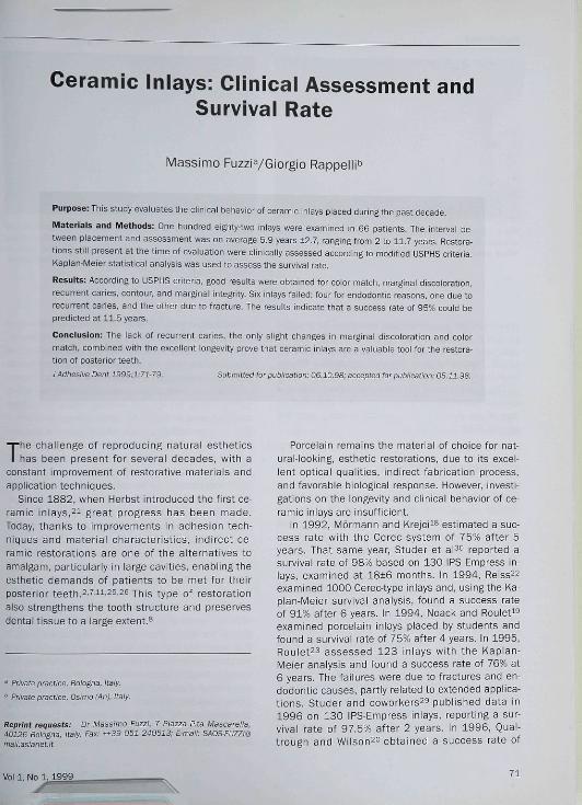
Fig 1 The margins of the preparation are placed on theenamel. The dentin side, when neoessary, is reoonstruotedwith a glass-ionomer photooured base.
Fig 2 The impression with poiyether materiai reproduces aiithe details of the preparation, A fluid msteriai (Permadynebiue, ESPE, Seefeid, Germany) is applied to the tooth, Athioi^er material [Impregum, ESPE, Seefeid, Germany) is ap-plied to the tray.
82% from 50 ceramic inlays examined at 3 years.In 1997, Fradeani et ai^ reported a 96% successrate over 125 iPS-Empress inlays examined at 4.5years. That same year, Roulet^* reported a 4% an-nual faiiure rate of Dicor porcelain inlays.
The present study assesses the survival rate andciinicai condition of porcelain inlays provided byone of the authors in his ciinicai practice over theperiod 1986 to 1996,
MATERIALS AND METHODS
One hundred eighty-two porcelain inlays were exam-ined in 66 patients, 41 female and 26 male. Pa-tient age ranged from 23 to 50 years. The iniaysincluded occiusai restorations (11), mesio-occlusal(35), occluso-distal (45), mesio-occlusal-distal (63)and cus pal-cove rage restorations (28), Cavity prepa-rations with aii margins iocated in enamei werecompleted in each tooth using Cerinlay diamondburs (Intensiv, Viganelio-Lugano, Switzeriand) (Fig1), Fuil-arch impressions including the preparationswere taken using a polyether impression material(Impregum, FSPF, Seefeid, Germany) (Fig 2), whilethe opposing arch was replicated with an algînateimpression. Fermit acrylic (Ivociar, Vivadent,Schaan, Liechtenstein) or a reiated material wasused to make temporary restorations.All restorations were made by the same technicianusing Microbond Natural fired ceramics (AustenalDental-Austenal Internationai Inc, Chicago, IL, USA)
in the period from 1986 to 1990 and Fortune firedceramics (Williams-lvociar, Amherst, New York, NY,USA) from 1991 to 1996. Luting was carried outwith a rubber dam in place, Cavex Protect Varnish(Cavex, Haarlem, Holland, by Kuraray Co Ltd, Japan)was used to protect the external part of the cavityduring luting; the varnish was applied with a pelletand removed with alcohol after luting the inlays^"(Fig 3). The fitting surface of the ceramic restora-tions was etched with hydrofiuoric acid (HF 10%) bythe technician after try-in. Siianization with 3MSilane (3IVI Dental Products Division, St Paul, MN,USA) was carried out by the dentist just before lut-ing. Enamel and dentin were etched and primed.The adhesive used in the period from 1986 tc
1992 was Scotchbond 2 (3M Dental Products Divi-sion, St Paul, MN, USA); enamel was etched with37% phosphoric acid for 30 s, rinsed, and dried, Abonding agent was used to achieve adhesion. From1993 to 1996, Scotchbond MP (3M Dental Prod-ucts Division, St Paui, MN, USA) was used as theadhesive. A 10% maieic acid or 37% phosphoricacid was applied to the enamei for 30 s and to thedentin for 15 s; after rinsing and gently drying, athin layer of primer was appiied to the dentin, and abonding agent was used to achieve adhesion. Theluting materials used were dual-cured composites:Ultra-Bond (Den Mat Corp, Santa Maria, CA, USA)from 1986 to 1990 and indirect Porceiain SystemDentist Bonding (3M Dental Products Division, StPaul, MN, USA) in the period from 1991 to 1996,Following restoration placement, excess luting ma-
72 The Journal of Adhesive Dentistry
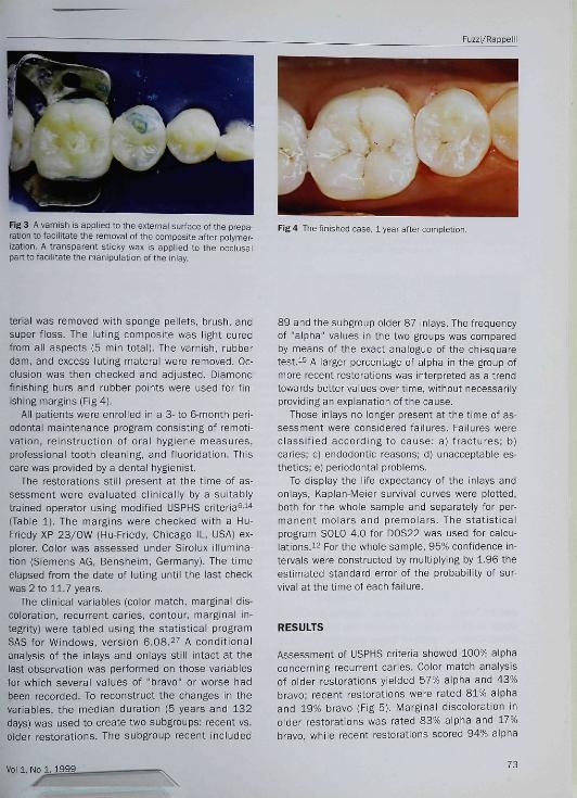
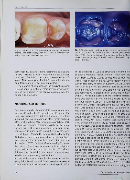
Fig 3 A varnish is applied to the external surface of the prepa-ration to faciiitate the removal of the composite after polymer-ization. A transparent sticky wax is applied to the occlusalpart to facilitate the manipulation of the inlay.
Fig 4 The finished case, 1 year after completion.
terial was removed with sponge pellets, brush, andsuper floss. The luting composite was light curedfrom all aspects (5 min totai). The varnish, rubberdam, and excess luting materai were removed. Oc-clusion was then checked and adjusted. Diamondfinishing burs and rubber points were used for fin-ishing margins (Fig 4).
Ali patients were enroiied in a 3-to 6-month peri-odontai maintenance program consisting of remoti-vation, reinstruction of oral hygiene measures,professional tooth cleaning, and fiuoridation. Thiscare was provided by a dental hygienist.
The restorations still present at the time of as-sessment were evaiuated clinically by a suitablytrained operator using modified USPHS criteria^i"(Table 1). The margins were checked with a Hu-Friedy XP 23/OW (Hu-Friedy, Chicago iL, USA] ex-plorer. Color was assessed under Siroiux iliumina-tion (Siemens AG, Bensheim, Germany). The timeelapsed from the date of luting until the iast checkwas 2 to 11.7 years.
The oiinicai variables (color match, marginal dis-coloration, recurrent caries, contour, marginal in-tegrity) were tabled using the statistical programSAS for Windows, version 6.08.2-' A conditionalanaiysis of the iniays and onlays still intact at thelast observation was performed on those variablesfor which several values of "bravo" or worse hadbeen recorded. To reconstruct the changes in thevariables, the median duration (5 years and 132days) was used to create two subgroups: recent vs.older restorations. The subgroup recent included
89 and the subgroup older 87 inlays. The frequencyof "alpha" vaiues in the two groups was comparedby means of the exact analogue of the chi-squaretest.i^ A iarger percentage of alpha in the group ofmore recent restorations was interpreted as a trendtowards better values over time, without necessarilyproviding an explanation ofthe cause.
Those inlays no longer present at the time of as-sessment were considered failures. Failures wereclassified according to cause: a) fractures; b)caries: c) endodontic reasons: d) unacceptable es-thetics; e] periodontal problems.
To display the life expectancy of the inlays andonlays, Kaplan-Meier survivai curves were piotted,both for the whole sample and separateiy for per-manent molars and premolars. The statisticalprogram SOLC 4,0 for D0S22 was used for calcu-lations.i^ For the whole sample, 95% confidence in-tervals were constructed by multiplying by 1.96 theestimated standard error of the probabiiity of sur-vival at the time of each failure.
RESULTS
Assessment of USPHS criteria showed 100% alphaconcerning recurrent caries. Color match analysisof older restorations yielded 57% alpha and 43%bravo: recent restorations were rated 81% alphaand 19% bravo (Fig 5), Marginal discoloration inolder restorations was rated 83% alpha and 17%bravo, whiie recent restorations scored 94% alpha
Vol 1, No 1, 1 9 9 9 . 73
Fuzzi/Rappeii
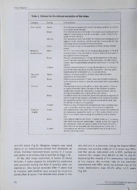
Table 1 Criteria for the clinical evaluation of the inlays
Category Rating Characteristic
Color matoh Alpha
Bravo
Charlie
Marginaldisoo i oration
Recurrentcaries
Marginalintegrity
Oscar
Aipha
Bravo
Alpha
Bravo
Alpha
Chariie
Alpha
The restoration appears to match the shade ahd transiucehcyof adjaoent tooth tissues.The restoration does hot match the shade and transiuoency ofadjacent tooth tissues, but the mismatch is within the normairange of tooth shades.The restoration does hot match the shade and transiucehcy ofadjacent tooth structure, ahd the mismatch is outside the hor-mal range of tooth shades and transiucency.The restoration cannot be examined without using a mouthmirror.There is no visual evidence of marginal discoloration differentfrom the color of the restorative matenai and from the coior ofthe adjacent tootli structure.There is visuai evidence of marginai discoloration at the junc-tion of the tooth structure and the restoration, but the disool-oration has not penetrated aiongthe restoration in a puipai di-rection.There is visual evidence of marginai discoioration at the junc-tion of the tooth structure and the restoration that has pene-trated aiongthe restoration in a puipai direction.There is no visual evidence of dark, deep discolorationadjacent to the restoration.There is visual evidenoe of dark, deep discoioration adjacentto the restoration (but not directly associated with cavosurfaoemargins).The restoration is a continuation of existing anatomic form oris siightiy fiattened. When the side of the explorer is piacedtangentiaiiy aoross the restoration, it does not touch two op-posing oavosurface line angles at the same time.Asurfaoeoonoavity is evident. When the side of an expiorer isplaced tangentiaiiy aoross a restoration, the explorer touchestwo opposing oavosurface iine angies at the same time, butthe dentin or base is not exposed.There is a ioss of restorative substance so that a surface con-cavity IS evident and the base and/or dentin is exposed.
The expiorer does not catch when drawh across the surface ofthe restoration toward the tooth, or, if the explorer doesoatoh,there is no visible crevice aiongthe periphery of the restora-tion.The expiorer catches and there is visibie evidence of a orevice,into whioli the explorer penetrates, indicating that the edge ofthe restoration does not adapt ciosely to the tooth structure.The dentin is not exposed, and the restoration is not mobiie,Tlie expiorer penetrates a orevice defect that extends to thedentin-enamei junction.
and 6% bravo (Fig 6¡, Marginal integrity was rated
alpha in all restorations except five assessed as
bravo. Contour received bravo scores in 2 cases
and alpha in all others (Tab 2 and Tab 3) (Figs 7, 8),
Of the 182 iniays examined, 6 failed. Of these
failures, 4 were caused by endodontic probiems
and occurred during the first 6 months after ce-
mentation. One failure occurred after 3 years due
to fracture, and another was caused by recurrent
caries after 8 years. Five failures took piace in mo-
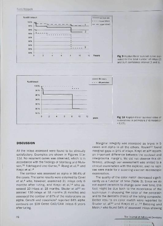
iars and one in a premolar. Using the Kaplan-Meier
analysis, the survivai index at 11.5 years was 95%,
and the values, calculated with a 95% confidence
interval (CI), ranged from 86.5% to 98,1% (Fig 9).
Separating the results of the premoiars from those
of the molars, the survivai rate of the premolar
restorations was 99%, while the survival rate of the
molar restorations was 90,2% after 11,5 years
(Fig 10),
74 The Journal of Adhesive Dentistry
Fuzzi/Rappelli
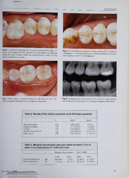
Fig 5 A ciinicai example of 3 ceramic restorations after 11years. The onlays on teeth 16 and 15 have been evaiuated asbravo for color match, while the restoration on tooth 14 hasbeen evaluated as aipha.
Fig 6 The marginal discoloration of two iniays after 4 years ina patient who smotes. Some points of the occlusal and proxi-mai margins of tooth 14 are stained.
Fig 7 MOD iniays on tooth 36 anO 37, OD inlay on tooth 35.Note the good ohromatic and marginai adaptation.
Fig 8 Radiographie verifications of the previous case. Noticethe absence of overhangs and the good marginal adaptation.
Table 2 Results of the clinical evaluation of 3ll 176 inlays examined
Recurrent canesMarginal integrityContourMarginal discolorationColor match
176176176176176
176 (100%) 0 (0%)171 [97.2%| 5 (2.8%)174 (98.4%) 2 (1.6%)156 ¡88.6%] 20 (11.4%]122(89.3%) 54(30.7%)
Table 3 Marginal discoioration and coior match of recent (< 5 y) vsolder {> 5 y) restorations; P < 0.05 (ChiMest)
Marginal discoloration Cclor match
Recent restorationsOlder restorations
Aipha Bravo Alpha Bravo84 (94%) 5 (6%) 72 (81%) 17 (19%)72(83%) 15(17%) 50(57%) 37(43%)
75
Fuzzi/Rap pe 11
Kstill intact
ss%
9S%
92%
90%
8fl%
86%
T- . , .,
1' 1 2
sjrviMlrBtt
: 3
0 2 4 S 8 10 12 Years Fig 9 Kaplan-Meier survival curve evai-uated for the totai number of inlays (2)and 95% confidence interval (1 and 3).
%slillinlacl
190% -|
98%
9B%
94%
92% .
90% .
r —
0 2 4 6 a 10 12
S4iii>lars
-38 premolars
Fig 10 Kapian-Meier survival rates ofrestorations in premolars and molars (P< 0.05).
DISCUSSION
All the inlays assessed were found to be clinicaiiysatisfactory. Exampies are shown in Figures 11a-l l d . No recurrent caries was observed, which is inaooordance with the findings of Stenberg and Mats-son,28 Tidehagand and Gunne,^! Âberg et al,25 andKrejci e t a l . "
The oontour was assessed as alpha in 98.4% ofthe oases. The same results were obtained by Cavelet al ," who, however, examined 31 inlays only 6months after luting, and Krejci et al.^'' who as-sessed 10 iniays at 18 months. Studer et aP*̂ ex-amined 130 inlays at 18 months (6 months) andassessed the contour of 87% of the restorations asalpha. Cerutti and ooworkers^ reported 84% alphacontours on 109 Cerec CAD/CAM inlays 6 yearsafter luting.
Marginai integrity was assessed as bravo in 5oases and alpha in ail the others. Rouletts foundmarginai gaps in 14% of inlays. Krejci et ai^''reportsan important difference between the occiusal andinterproximai margins. We did not observe this dif-ference, aithough our assessment was limited to aclinical examination with the expiorer, and no repli-cas were made for a scanning eieotron microscopicexamination.
The quality of the color matoh decreased signifi-cantly as a function of time (Tabie 3). Since we donot expect ceramics to change color over time, thisfact might be due both to the experience of thetechnician in choosing the coior of the porcelainand to the dentist's choice of the iuting composite.Better resuits on coior match were reported byStuder et al^f and Krejci et ai.l '^ Bessing andiVIolin,3 who found 54% of assessed inlays showing
76 The Journal of Adhesive Dentistry
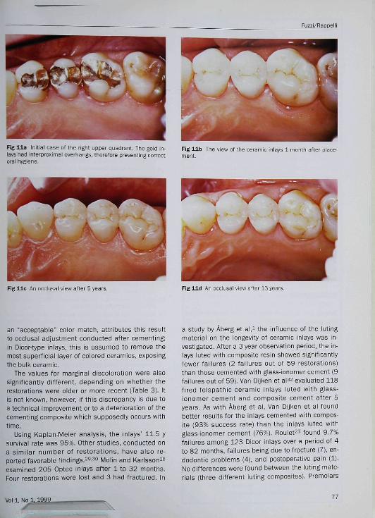
Fig l i a Initiai case of the right upper quadrant. The gold in-lays had interproximai overhangs, therefore preventing correotorai hygiene.
Fig l i b The view of the oeramic iniays 1 month after place-ment.
Fig l i e An ooclusai view after 5 years Fig l i d An occiusai view after 13 years.
an "acceptable" color match, attributes this resultto occiusal adjustment conducted after cementing;in Dicor-type inlays, this is assumed to remove themost superficial layer of colored ceramics, exposingthe bulk ceramic.
The values for marginal discoloration were alsosignificantiy different, depending on whether therestorations were oider or more recent (Tabie 3), Itis not known, however, if this discrepancy is due toa technical improvement or to a deterioration of thecementing oomposite which supposedly occurs withtime.
Using Kaplan-Meier analysis, the inlays' 11,5 ysurvivai rate was 95%, Other studies, conducted ona simiiar number of restorations, have aiso re-ported favorable findings.29-30 Molin and Karisson^^examined 205 Optec inlays after 1 to 32 months.Four restorations were lost and 3 had fractured. In
a study by Âberg et a\,^ the influence of the lutingmaterial on the longevity of ceramic inlays was in-vestigated. After a 3-year observation period, the in-lays luted with composite resin showed significantlyfewer failures (2 failures out of 59 restorations)than those cemented with glass-ionomer cement (9failures out of 59). Van Dijken et aP^ evaluated 118fired felspathic ceramic inlays luted with glass-ionomer cement and composite cement after 5years. As with Âberg et al. Van Dijken et al foundbetter results forthe inlays cemented with compos-ite (93% success rate) than the inlays luted withglass-ionomer cement (76%), Rouiet^s found 9,7%failures among 123 Dicor inlays over a period of 4to 82 months, failures being due to fracture (7), en-dodontic problems (4), and postoperative pain (1),No differences were found between the luting mate-rials (three different luting composites). Premolars
Vol 1, mi. 1999 77
Fuzzi/Rappeiii
tended to show a better success rate; however, the
difference was not statistically significant, Molin
and Karisson^'' reported a 90% success rate after 3
years: this was obtained by calculating the survival
rates of iniays performed by 10 dentists who pre-
sented very different results. This finding empha-
sizes the sensitivity of the ceramic iniay technique
and the dependence on personal operator skill and
clinical experience. Operator ability and care in ap-
plying the technique can be the main factors infiu-
encing the success rate.
In many studies, the main reason for failure
seems to be the fracture of inlays. Quaitrough and
Wiison,20 examining 50 ceramic iniays after 3
years, found 18% failures due to fracture. All fail-
ures reported by Fradeani et al^ were due to frac-
ture. Isidor and Brondum^s obtained even less
favorabie results after 57 months with 12 failures
out of 25 inlays, 10 of which were fractures. The se-
lection of cases, particulariy if bruxism patients are
invoived, must also be taken into account when
evaluating the mechanicai properties of ceramics.
It shouid also be noted that 4 of the 6 failures re-
ported in the present study occurred in the same
patient for endodontic reasons (puipitis), and that
these restorations were carried out 11.5 years ago,
when iess oiinicai information was avaiiabie about
the adhesive technique and microleakage controi.
CONCLUSIONS
Taking into account that this single-center study
was conducted without using a control group, the
following conclusions may be drawn:
1. Within the observation time (up to 11,5 years),
adhesively luted oeramic inlays showed no recur-
rent caries.
2. The slight occurrence of marginal discoloration
over time (17% bravo for inlays older than 5
years) is still clinically acceptable.
3. The color match of the ceramic inlays was found
to be better in the recent restorations subgroup
than in the older restorations subgroup.
4. After 11.5 years, a survival rate of 95% of all the
ceramic inlays must be considered as excellent.
5. The survival rate of premolar inlays (99%) was
superior to that of molar inlays (90%).
REFERENCES
1. Âberg CH, Van Dijken JWV. Olofsson AL. Three years compari-son of fired ceramic inlays cemented with composite resin crglass ionomer cement. Acta Odontol Scand 1994;52:140-149.
2. Banks RG. Conservative posterior ceramic restorations: a lit-erature review. J Prosthet Dent 1990;B3;619-626.
3. Bessihg C, Molin M. An in vivo study of glass ceramic ¡Dicor*]iniays. Acta Odontol Scand 1990:48:351-357.
4. Cavei WT, Keisey WP, Barkmeier WW, et al. A pilot study ofthe clinicai evaiuation of castabie ceramic inlays and a duai-cure resin cement. Quintessence Int 1988:19:257-252.
5. Gerutti A, Vehturi G. Putignano A, Prati C. Six-years clinicalevaiuation of 109 Cad/Cam inlays. Madrid: lADR/OED [ab-stract 295|, 1997.
6. Cvar JF and Ryge G. Criteria for the Ciihical Evaluation of Den-tai Restorative Materials, USPHS Publication no. 790-244,San Francisco: U.S. Government Printing Office 1971.
7. Dietschi D. Holz J, Restauration des dents postérieures. RevSuisse Odontostom 1990:100:1325-1332.
8. Douglas WH. Methods tc improve fracture resistance ofteeth, in: Vanherie G, Smith DC {eds). Posterior CompositeResm Dentai Restorative Materiais. Proc 3M Symposium. TheNetherlands: Peter Szulc Pubi Co, 1985: 433^42.
9. Fradeani M, Aquilano A, Bassein L. Longitudinal study ofpressed glass-ceramic inlays for four and half years, J Pros-thet Dent 1997;78:346-353.
10. Fuïii M. Porcelain bonded restoration. In: Dondi daii'OroiogioG. Fuizi M, Prati C (eds). Adhesion in Restorative Dentistry-Proceedings ofthe International Symposium. Bologna, 1995:87-97.
11. Garber DA, Goldstein RE. Porcelain and composite inlays andonlays. Chicago: Quintessence, 1994.
12. Hihtze JL. SOLO Statistical system BMDP Statistical Software,Cork, ireland, 1991 (Survivai Anaiysis Module).
13. Isidor F, Brondum K, A ciinjcal evaiuation of porceiain iniays.J Prosthet Dent 1995:74:140-144.
14. Krejci i, Krejci D, Lutz F. Ciinicai evaiuation of a new pressedgiassed ceramic iniay material over 1.5 years. Quintessenceint 1992:23:181-186.
15. Mehta C, Patel N. Statxact Turbo, Statisticai Software forExact Nonparametric inference. User's Manuai. Cytei Soft-ware Corporation, Cambridge, MA, 1992.
16. Molin M, Karisson S. A clinical evaluation of the Optec iniaysystem. Acta Odontol Scand 1992:50:227-233,
17. Molm M, Karisson S. A 3 year clinical follow-up study of a ce-ramic (Optec) inlay system. Acta Odontoi Scand 1996:54:145-149.
18. Mörmann W, Krejci i. Computer-designed inlays after 5 yearsin situ: clinical performance and scanning eiectron micro-scopic evaluation. Quintessence Int 1992:23:109-115.
19. Noack MJ, Rouiet JF, Survivai rates and mode of failure ofDicor iniays after 4 years |abstract759]. J Dent Res1994;73:196,
20. Quaitrough AJE, Wilson NHF, A 3-year ciinicsi evaluation of aporceiaih iniay system. J Dent 1996:24:317-323,
21. Quaitrough AJE, Wilson NHF, Smith GA. The porcelain inlay:An historical view. Oper Dent 1990:15:61-70.
22. Reiss B, Kiinische Langzeiterfahrungen mit Cerec-lnlays. DFZ1994:38:30-33.
78 The Journal of Adhesive Dentistry
\
Fuzzi/Rappelli
23, Rouiet JF. The longevity of giass ceramic iniays [abstract 36],J Dent Res 1995:74:405,
24, Roulet JF, Longevity of giass ceramic iniays and amaigam-re-sults up to 6 years. Clin Orai Invest 1997:1:40^6,
25, Rouiet JF, Degrange M, Ihlay restorations, J Caiif Dent Assoc1996:24:48-62,
26, Roulet JF, Herder S, Bonded ceramio inlays, Chicago: Quintes-sence, 1991.
27, SAS institute Inc., SAS Procedures Guide, Version 6, ThirdEdition, Second pnnting, Gary, NC, 1992.
28, Stenberg R, Matsson L Ciinical evaiuation of giass oeramicinlays (Dicor), Aota Odontoi Scand 1993:51:91-97.
29, Studer S, Lehner C, Brodbecii U, Scharer P, Short term re-sults of IPS-Empress ihlays and oniays, J Prosthodont1996:5:277-287,
30, Studer S, Lehner C, Schárer P. Glass-ceramic iniays and on-lays made by IPS empress: first clinicai resuits, J Dent Res1992:71:658,
31, Tidehag P, Gunne J, A 2-year ciinicai foilow-up study of iPSEmpress ceramic inlays, IntJ Prosthcdont 1995:8:456-460.
32, Van Dijken JWV, Aberg HC, et al. Five year evaluation of oe-ramic inlays [abstract 72]. J Dent Res 1996:75:1302,
Voi 1, NO 1, 1999_
New Frontiers inAdhesive Dentistry
HYBRIDIZATION OF
DENTAT, HARD TISSUES
HYBRIDIZATION OF DENTAL HARD TISSUESNobuo Nakabayashi and David H. Pashley
The hybridization of dentin—a process that cre-ates a molecular-level mixture of adhesive poly-mers and dental hard tissues—gives clinicians aversatile new material, useful in a wide array ofadvanced dental treatments. As the first in-depthexploration of the suhject, this book covers thedevelopment, present understanding, and futureresearch areas of this multifunctional dental mate-rial, A thorough review of the current literaturerounds out the text.
Valuable for students, researchers, and clini-cians seeking a greater understanding of resinhybridization of tooth structure.
CONTENTS
! Evolution of Dendn-Resin Bonding2 Properties of Dentin3 Acid Conditioning and Hybridization of
Substrates4 Characterization of the Hybrid Layer5 The Quality of the Hybndized Dentin6 Clinical Applications of Hybrid Layer
Formation
¡29 pp. 80 iHus ¡some in coior!:ISBN 0-87417-575-9 C3047; US S40
To ORDER
CallToll Free 1-800-621-0387or Fax 1-630-682-3288
book/
Visit our web site http://www.qijintpub.comQuintessence Publishing Co, Inc
Related Documents