© 2016 APOS Trends in Orthodontics | Published by Wolters Kluwer - Medknow 287 Address for Correspondence: Dr. Ashok Karad, 1/C 33, 2 Sujata Niwas, S V Road, Opp. Indus Ind Bank, Bandra (W), Mumbai ‑ 400 050, Maharashtra, India. E‑mail: [email protected] Cephalometric analysis for functional occlusion Abstract Background: Various elements contributing to good functional occlusion have not been clearly assessed with cephalometrics for the diagnosis of functional problems and their application in clinical practice. The aim of this study, therefore, was to analyze different components of functional occlusion to formulate concise functional cephalometric analysis. Materials and Methods: Eighty‑two cases (38 males and 44 females), with class I occlusion and balanced facial profile, were examined based on the selection criteria, and cephalograms were taken in natural head position. All the radiographs were then analyzed using various functional parameters. Results: The mean values of condylar path angle and incisal path angle were 55.83° and 65.67°, respectively, with large deviations. However, both showed positive correlation. The value of the angle of long axis of mandibular incisor with respect to the line joining center of condyle and lower incisor tip was 88.04°. Moreover, the angle between the occlusal plane and horizontal plane was 12.88°. In vertical plane, lower face height (LFH) was found to be slightly less than the upper face height. Maxilla contributed around 45% of the LFH while mandible formed about 60%. Furthermore, upper alveolar component (maxillary alveolar height) formed more than half of the maxilla (53.79%) whereas lower alveolar component (mandibular alveolar height) was 74.8% of the mandible. Conclusion: This study has analyzed various components of functional occlusion and formulated a concise functional cephalometric analysis for diagnosis, treatment planning, and assessment of treatment results. Key words: Cephalometrics, functional occlusion, natural head position INTRODUCTION An understanding of the form and function of the temporomandibular joint and occlusion is of significant importance to many disciplines of dentistry, including orthodontics, restorative dentistry, oral and maxillofacial surgery, and periodontics. The assessment of various elements contributing to good functional occlusion has not been clearly established with cephalometrics. If the function could be measured using cephalometric radiographs, orthodontists and expert clinicians from other disciplines of dentistry would more seriously consider the diagnosis of functional problems and their application in clinical practice. Aim of the study The purpose of this investigation was to cephalometrically study various components of functional occlusion and formulate functional cephalometric parameters, which will be of great importance to orthodontics and other disciplines of dentistry. The specific objectives were to analyze five key functional elements: (1) angle of articular eminence, (2) inclination of maxillary incisor functional Ashok Karad, Shruti Chhajed 1 Director and Orthodontist, Smile Care, 1 Associate Orthodontist, Smile Care, Mumbai, Maharashtra, India Original Article Access this article online Quick Response Code: Website: www.apospublications.com DOI: 10.4103/2321‑1407.194793 How to cite this article: Karad A, Chhajed S. Cephalometric analysis for functional occlusion. APOS Trends Orthod 2016;6:287‑94. This is an open access article distributed under the terms of the Creative Commons Attribution‑NonCommercial‑ShareAlike 3.0 License, which allows others to remix, tweak, and build upon the work non‑commercially, as long as the author is credited and the new creations are licensed under the identical terms. For reprints contact: [email protected]

Welcome message from author
This document is posted to help you gain knowledge. Please leave a comment to let me know what you think about it! Share it to your friends and learn new things together.
Transcript
© 2016 APOS Trends in Orthodontics | Published by Wolters Kluwer - Medknow 287
Address for Correspondence: Dr. Ashok Karad, 1/C 33, 2 Sujata Niwas, S V Road, Opp. Indus Ind Bank, Bandra (W), Mumbai ‑ 400 050, Maharashtra, India. E‑mail: [email protected]
Cephalometric analysis for functional occlusion
AbstractBackground: Various elements contributing to good functional occlusion have not been clearly assessed with cephalometrics for the diagnosis of functional problems and their application in clinical practice. The aim of this study, therefore, was to analyze different components of functional occlusion to formulate concise functional cephalometric analysis. Materials and Methods: Eighty‑two cases (38 males and 44 females), with class I occlusion and balanced facial profile, were examined based on the selection criteria, and cephalograms were taken in natural head position. All the radiographs were then analyzed using various functional parameters. Results: The mean values of condylar path angle and incisal path angle were 55.83° and 65.67°, respectively, with large deviations. However, both showed positive correlation. The value of the angle of long axis of mandibular incisor with respect to the line joining center of condyle and lower incisor tip was 88.04°. Moreover, the angle between the occlusal plane and horizontal plane was 12.88°. In vertical plane, lower face height (LFH) was found to be slightly less than the upper face height. Maxilla contributed around 45% of the LFH while mandible formed about 60%. Furthermore, upper alveolar component (maxillary alveolar height) formed more than half of the maxilla (53.79%) whereas lower alveolar component (mandibular alveolar height) was 74.8% of the mandible. Conclusion: This study has analyzed various components of functional occlusion and formulated a concise functional cephalometric analysis for diagnosis, treatment planning, and assessment of treatment results.
Key words: Cephalometrics, functional occlusion, natural head position
INTRODUCTION
An understanding of the form and function of the temporomandibular joint and occlusion is of significant importance to many disciplines of dentistry, including orthodontics, restorative dentistry, oral and maxillofacial surgery, and periodontics. The assessment of various elements contributing to good functional occlusion has not been clearly established with cephalometrics. If
the function could be measured using cephalometric radiographs, orthodontists and expert clinicians from other disciplines of dentistry would more seriously consider the diagnosis of functional problems and their application in clinical practice.
Aim of the studyThe purpose of this investigation was to cephalometrically study various components of functional occlusion and formulate functional cephalometric parameters, which will be of great importance to orthodontics and other disciplines of dentistry. The specific objectives were to analyze five key functional elements: (1) angle of articular eminence, (2) inclination of maxillary incisor functional
Ashok Karad, Shruti Chhajed1
Director and Orthodontist, Smile Care, 1Associate Orthodontist,
Smile Care, Mumbai, Maharashtra, India
Original Article
Access this article onlineQuick Response Code:
Website: www.apospublications.com
DOI: 10.4103/2321‑1407.194793
How to cite this article: Karad A, Chhajed S. Cephalometric analysis for functional occlusion. APOS Trends Orthod 2016;6:287‑94.
This is an open access article distributed under the terms of the Creative Commons Attribution‑NonCommercial‑ShareAlike 3.0 License, which allows others to remix, tweak, and build upon the work non‑commercially, as long as the author is credited and the new creations are licensed under the identical terms.
For reprints contact: [email protected]
Karad and Chhajed: Cephalometric analysis for functional occlusion
APOS Trends in Orthodontics | November 2016 | Vol 6 | Issue 6288
surface, (3) functional mandibular incisor position, (4) orientation of occlusal plane (OP), and (5) vertical dimension of occlusion (VDO).
MATERIALS AND METHODS
SampleFrom more than 300 patients examined from the author’s private practice, 100 adults (50 males and 50 females) of the age group between 17 and 25 years were selected based on the selection criteria, using simple random sampling method. All cases had natural Class I occlusions and were reasonably facially balanced. After thorough clinical examination, digital cephalograms were obtained in natural head position (NHP) with teeth in maximum intercuspation and passive lips. An ethical clearance had been taken by a local ethical committee to carry out this procedure. Of the 100 cephalograms, 18 were discarded because of lack of clarity of certain anatomic structures used in this study. Therefore, the final sample size of 82 (38 males and 44 females) comprised the cephalometric database for this analysis.
Inclusion criteria1. Angle’s class I molar relation2. Well‑aligned maxillary and mandibular dental arches3. Balanced, esthetically pleasing, straight profile4. Good facial symmetry.
Exclusion criteria1. Significant medical history2. Congenital facial deformity3. History of trauma4. Previous orthodontic treatment5. Maxillofacial or plastic surgery.
Cephalometric analysisAll cephalometric radiographs were traced and analyzed manually by a single examiner, on acetate tracing paper of 50 µ thickness, using 0.5 mm lead pencil under similar conditions of illumination. The important anatomic structures and hard tissue landmarks were marked on the headfilm.
Reference planesIn establishing proper diagnosis, and for assessing treatment progress, cephalometric analysis compares various elements of craniofacial morphology to specific reference planes. Various cephalometric analyses use Sella‑nasion (S‑N) or Frankfort horizontal (F‑H) planes as reference lines.[1‑4] A commonly used S‑N plane as a craniofacial reference line has been shown to have large interindividual standard deviations (SDs) when related to true vertical.[5‑7] Furthermore, measurements based on
another reference plane, F‑H plane, do not always match with the clinical findings.[8]
Lateral cephalograms based on NHP and the true horizontal have shown to have greater clinical application as the patient is presented as they appear in life. Analyses based on this, therefore, are more meaningful in depicting patient’s clinical situation and are considered reliable, less variable, realistic, standardized, and easily reproducible head position.[9] The rate of reproducibility of NHP has been found to be very high with a variance of about 2–4°.[10,11]
Considering this, all cephalograms in this study were obtained in NHP. It is the position that a person would assume when looking at distant object on the horizon. For this, a mirror at the eye level was used in the study. The case should look straight ahead into the mirror, with his pupil in the center of the eye. Bilateral ear rods and nosepiece were used to stabilize the head in the transverse and vertical plane (VP), respectively.
ParametersVarious planes and angles were drawn for the analysis and 13 parameters (4 angular and 9 linear) were measured.
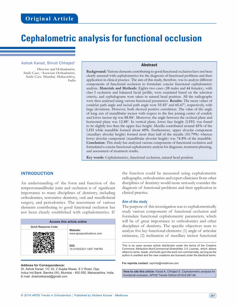
Various landmarks, true horizontal plane (HP), and true VP reference planes are shown in Figure 1.
Various parameters were grouped into five categories.
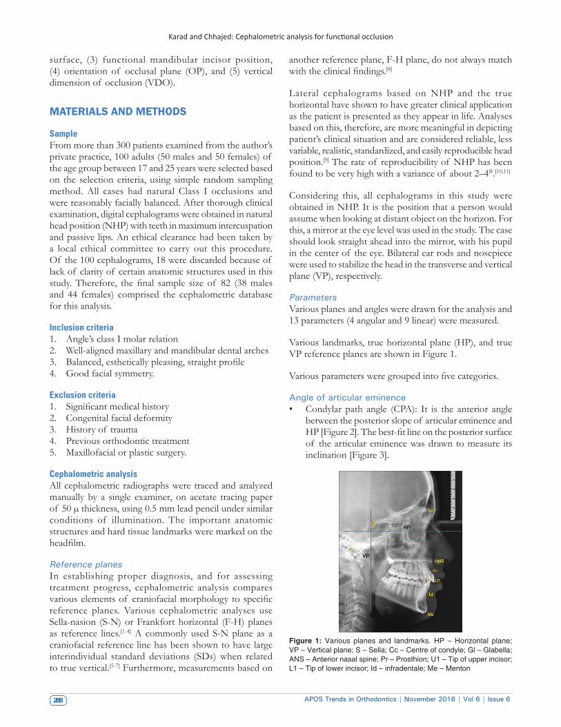
Angle of articular eminence• Condylar path angle (CPA): It is the anterior angle
between the posterior slope of articular eminence and HP [Figure 2]. The best‑fit line on the posterior surface of the articular eminence was drawn to measure its inclination [Figure 3].
Figure 1: Various planes and landmarks. HP – Horizontal plane; VP – Vertical plane; S – Sella; Cc – Centre of condyle; Gl – Glabella; ANS – Anterior nasal spine; Pr – Prosthion; U1 – Tip of upper incisor; L1 – Tip of lower incisor; Id – infradentale; Me – Menton
Karad and Chhajed: Cephalometric analysis for functional occlusion
APOS Trends in Orthodontics | November 2016 | Vol 6 | Issue 6 289
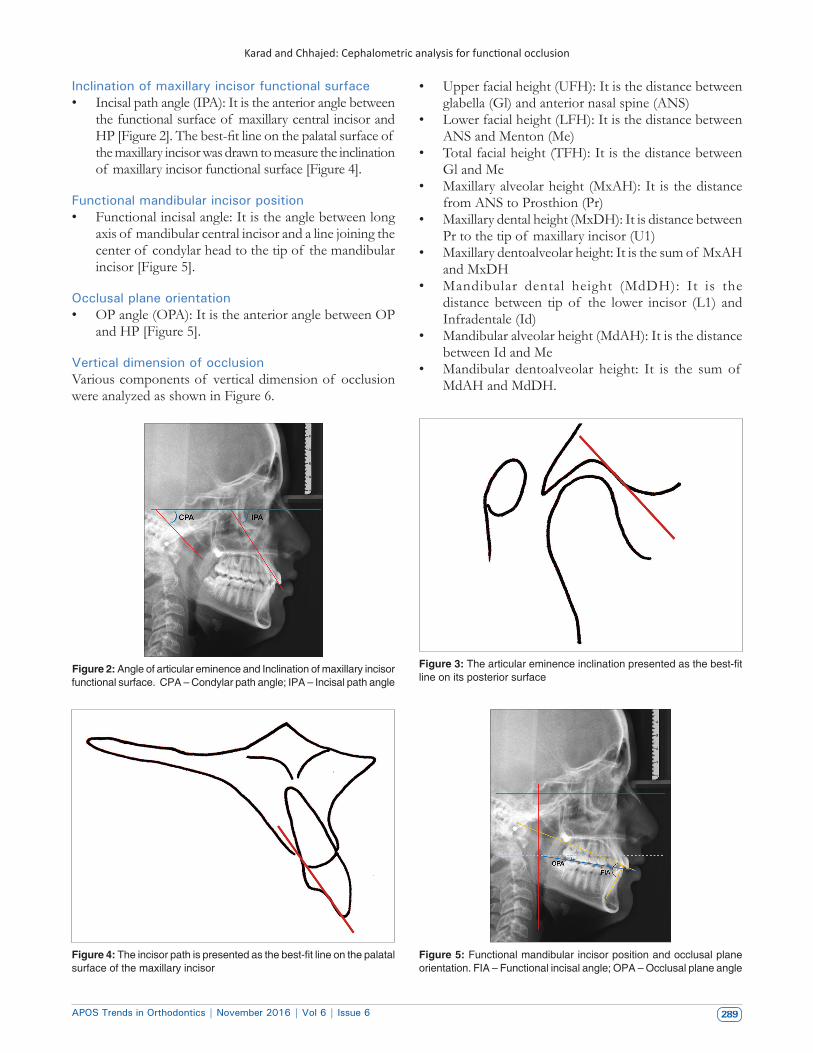
Inclination of maxillary incisor functional surface• Incisal path angle (IPA): It is the anterior angle between
the functional surface of maxillary central incisor and HP [Figure 2]. The best‑fit line on the palatal surface of the maxillary incisor was drawn to measure the inclination of maxillary incisor functional surface [Figure 4].
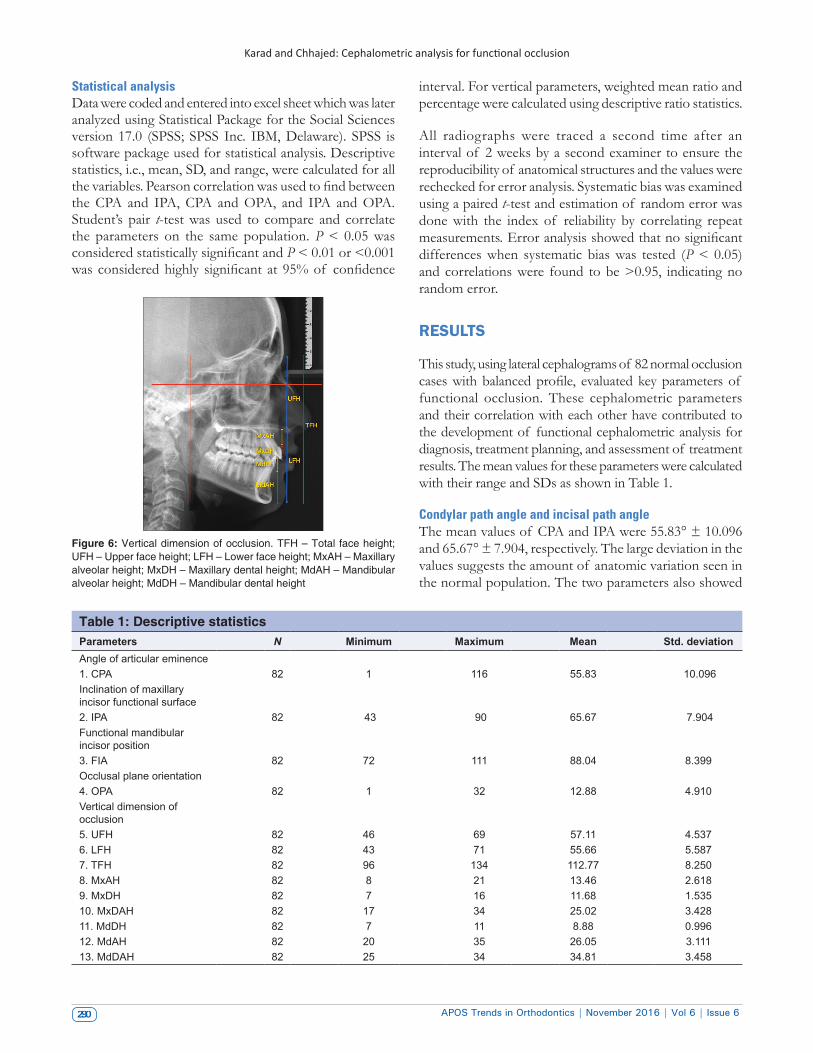
Functional mandibular incisor position• Functional incisal angle: It is the angle between long
axis of mandibular central incisor and a line joining the center of condylar head to the tip of the mandibular incisor [Figure 5].
Occlusal plane orientation• OP angle (OPA): It is the anterior angle between OP
and HP [Figure 5].
Vertical dimension of occlusionVarious components of vertical dimension of occlusion were analyzed as shown in Figure 6.
• Upper facial height (UFH): It is the distance between glabella (Gl) and anterior nasal spine (ANS)
• Lower facial height (LFH): It is the distance between ANS and Menton (Me)
• Total facial height (TFH): It is the distance between Gl and Me
• Maxillary alveolar height (MxAH): It is the distance from ANS to Prosthion (Pr)
• Maxillary dental height (MxDH): It is distance between Pr to the tip of maxillary incisor (U1)
• Maxillary dentoalveolar height: It is the sum of MxAH and MxDH
• Mandibular dental height (MdDH): It is the distance between tip of the lower incisor (L1) and Infradentale (Id)
• Mandibular alveolar height (MdAH): It is the distance between Id and Me
• Mandibular dentoalveolar height: It is the sum of MdAH and MdDH.
Figure 2: Angle of articular eminence and Inclination of maxillary incisor functional surface. CPA – Condylar path angle; IPA – Incisal path angle
Figure 3: The articular eminence inclination presented as the best‑fit line on its posterior surface
Figure 4: The incisor path is presented as the best‑fit line on the palatal surface of the maxillary incisor
Figure 5: Functional mandibular incisor position and occlusal plane orientation. FIA – Functional incisal angle; OPA – Occlusal plane angle
Karad and Chhajed: Cephalometric analysis for functional occlusion
APOS Trends in Orthodontics | November 2016 | Vol 6 | Issue 6290
Figure 6: Vertical dimension of occlusion. TFH – Total face height; UFH – Upper face height; LFH – Lower face height; MxAH – Maxillary alveolar height; MxDH – Maxillary dental height; MdAH – Mandibular alveolar height; MdDH – Mandibular dental height
Table 1: Descriptive statisticsParameters N Minimum Maximum Mean Std. deviationAngle of articular eminence1. CPA 82 1 116 55.83 10.096Inclination of maxillary incisor functional surface2. IPA 82 43 90 65.67 7.904Functional mandibular incisor position3. FIA 82 72 111 88.04 8.399Occlusal plane orientation4. OPA 82 1 32 12.88 4.910Vertical dimension of occlusion5. UFH 82 46 69 57.11 4.5376. LFH 82 43 71 55.66 5.5877. TFH 82 96 134 112.77 8.2508. MxAH 82 8 21 13.46 2.6189. MxDH 82 7 16 11.68 1.53510. MxDAH 82 17 34 25.02 3.42811. MdDH 82 7 11 8.88 0.99612. MdAH 82 20 35 26.05 3.11113. MdDAH 82 25 34 34.81 3.458
Statistical analysisData were coded and entered into excel sheet which was later analyzed using Statistical Package for the Social Sciences version 17.0 (SPSS; SPSS Inc. IBM, Delaware). SPSS is software package used for statistical analysis. Descriptive statistics, i.e., mean, SD, and range, were calculated for all the variables. Pearson correlation was used to find between the CPA and IPA, CPA and OPA, and IPA and OPA. Student’s pair t‑test was used to compare and correlate the parameters on the same population. P < 0.05 was considered statistically significant and P < 0.01 or <0.001 was considered highly significant at 95% of confidence
interval. For vertical parameters, weighted mean ratio and percentage were calculated using descriptive ratio statistics.
All radiographs were traced a second time after an interval of 2 weeks by a second examiner to ensure the reproducibility of anatomical structures and the values were rechecked for error analysis. Systematic bias was examined using a paired t‑test and estimation of random error was done with the index of reliability by correlating repeat measurements. Error analysis showed that no significant differences when systematic bias was tested (P < 0.05) and correlations were found to be >0.95, indicating no random error.
RESULTS
This study, using lateral cephalograms of 82 normal occlusion cases with balanced profile, evaluated key parameters of functional occlusion. These cephalometric parameters and their correlation with each other have contributed to the development of functional cephalometric analysis for diagnosis, treatment planning, and assessment of treatment results. The mean values for these parameters were calculated with their range and SDs as shown in Table 1.
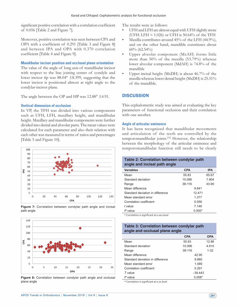
Condylar path angle and incisal path angleThe mean values of CPA and IPA were 55.83° ± 10.096 and 65.67° ± 7.904, respectively. The large deviation in the values suggests the amount of anatomic variation seen in the normal population. The two parameters also showed
Karad and Chhajed: Cephalometric analysis for functional occlusion
APOS Trends in Orthodontics | November 2016 | Vol 6 | Issue 6 291
Figure 7: Correlation between condylar path angle and incisal path angle
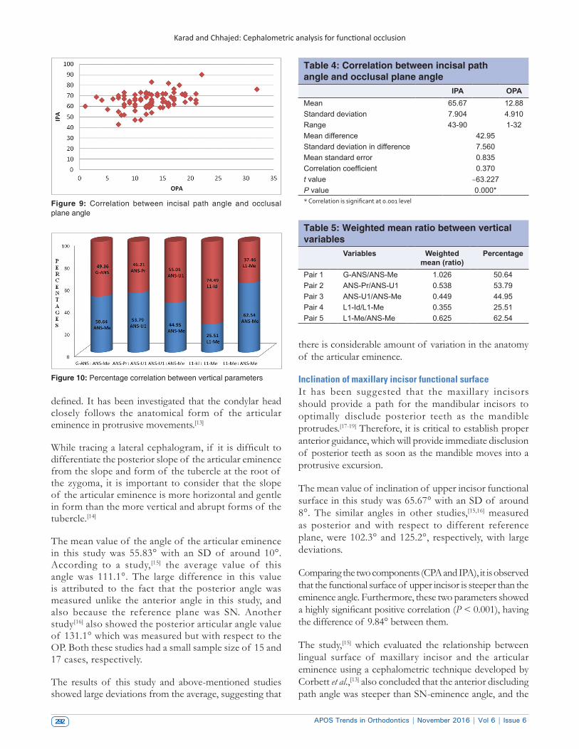
Figure 8: Correlation between condylar path angle and occlusal plane angle
significant positive correlation with a correlation coefficient of 0.056 [Table 2 and Figure 7].
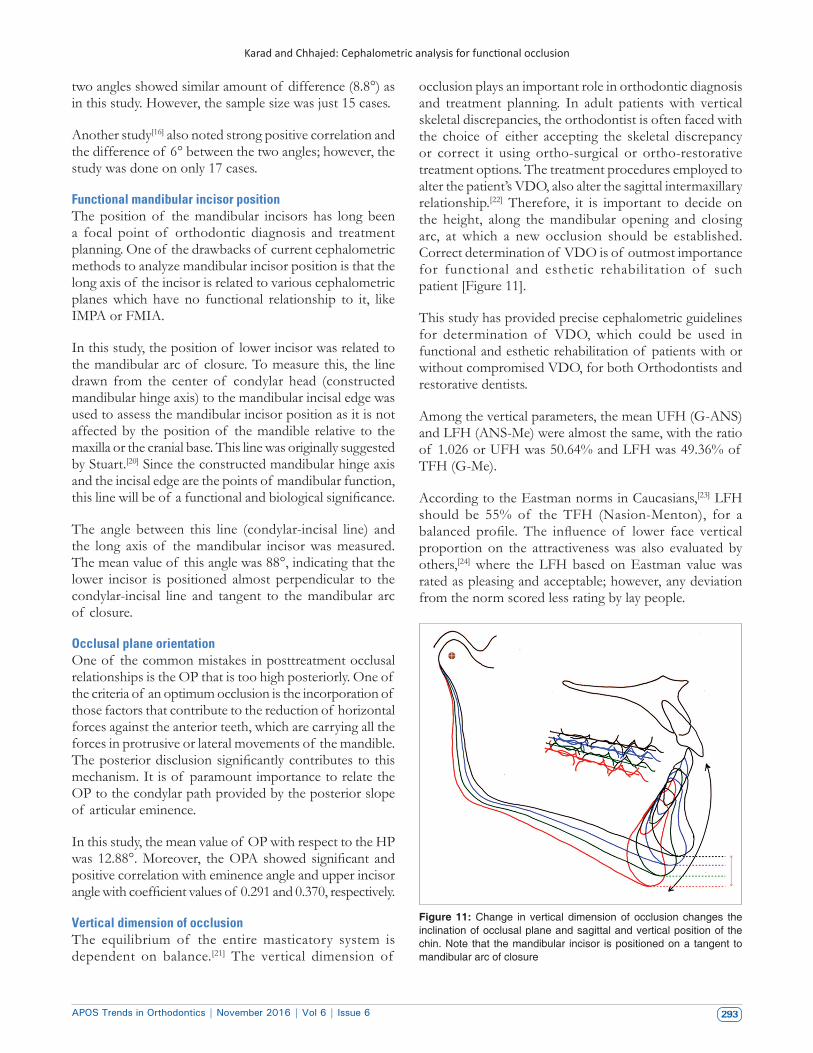
Moreover, positive correlation was seen between CPA and OPA with a coefficient of 0.291 [Table 3 and Figure 8] and between IPA and OPA with 0.370 correlation coefficient [Table 4 and Figure 9].
Mandibular incisor position and occlusal plane orientationThe value of the angle of long axis of mandibular incisor with respect to the line joining center of condyle and lower incisor tip was 88.04° ±8.399, suggesting that the lower incisor is positioned almost at right angle to the condylar‑incisor plane.
The angle between the OP and HP was 12.88° ±4.91.
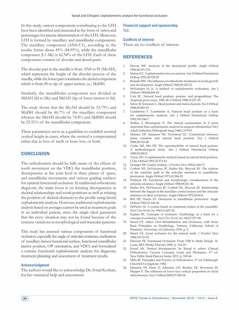
Vertical dimension of occlusionIn VP, the TFH was divided into various components such as UFH, LFH, maxillary height, and mandibular height. Maxillary and mandibular components were further divided into dental and alveolar parts. The mean values were calculated for each parameter and also their relation with each other was measured in terms of ratios and percentages [Table 5 and Figure 10].
The results were as follows:• UFH and LFH are almost equal with UFH slightly more
(UFH: LFH = 1.026) or UFH is 50.64% of the TFH• Maxilla contributes around 45% of the LFH (44.9%),
and on the other hand, mandible constitutes about 60% (62.54%)
• Upper alveolar component (MxAH) forms little more than 50% of the maxilla (53.79%) whereas lower alveolar component (MdAH) is 74.8% of the mandible
• Upper incisal height (MxDH) is about 46.7% of the maxilla whereas lower dental height (MxDH) is 25.51% of the mandible.
DISCUSSION
This cephalometric study was aimed at evaluating the key parameters of functional occlusion and their correlation with one another.
Angle of articular eminenceIt has been recognized that mandibular movements and articulation of the teeth are controlled by the temporomandibular joints.[12] However, the relationship between the morphology of the articular eminence and temporomandibular function still needs to be clearly
Table 2: Correlation between condylar path angle and incisal path angleVariables CPA IPAMean 55.83 65.67Standard deviation 10.096 7.904Range 38‑116 43‑90Mean difference 9.841Standard deviation in difference 12.471Mean standard error 1.377Correlation coefficient 0.056t value 7.146P value 0.000** Correlation is significant at 0.001 level
Table 3: Correlation between condylar path angle and occlusal plane angle
CPA OPAMean 55.83 12.88Standard deviation 10.096 4.910Range 38‑116 1‑32Mean difference 42.95Standard deviation in difference 9.860Mean standard error 1.089Correlation coefficient 0.291T value −39.443P value 0.008** Correlation is significant at 0.01 level
Karad and Chhajed: Cephalometric analysis for functional occlusion
APOS Trends in Orthodontics | November 2016 | Vol 6 | Issue 6292
there is considerable amount of variation in the anatomy of the articular eminence.
Inclination of maxillary incisor functional surfaceIt has been suggested that the maxillary incisors should provide a path for the mandibular incisors to optimally disclude posterior teeth as the mandible protrudes.[17‑19] Therefore, it is critical to establish proper anterior guidance, which will provide immediate disclusion of posterior teeth as soon as the mandible moves into a protrusive excursion.
The mean value of inclination of upper incisor functional surface in this study was 65.67° with an SD of around 8°. The similar angles in other studies,[15,16] measured as posterior and with respect to different reference plane, were 102.3° and 125.2°, respectively, with large deviations.
Comparing the two components (CPA and IPA), it is observed that the functional surface of upper incisor is steeper than the eminence angle. Furthermore, these two parameters showed a highly significant positive correlation (P < 0.001), having the difference of 9.84° between them.
The study,[15] which evaluated the relationship between lingual surface of maxillary incisor and the articular eminence using a cephalometric technique developed by Corbett et al.,[13] also concluded that the anterior discluding path angle was steeper than SN‑eminence angle, and the
defined. It has been investigated that the condylar head closely follows the anatomical form of the articular eminence in protrusive movements.[13]
While tracing a lateral cephalogram, if it is difficult to differentiate the posterior slope of the articular eminence from the slope and form of the tubercle at the root of the zygoma, it is important to consider that the slope of the articular eminence is more horizontal and gentle in form than the more vertical and abrupt forms of the tubercle.[14]
The mean value of the angle of the articular eminence in this study was 55.83° with an SD of around 10°. According to a study,[15] the average value of this angle was 111.1°. The large difference in this value is attributed to the fact that the posterior angle was measured unlike the anterior angle in this study, and also because the reference plane was SN. Another study[16] also showed the posterior articular angle value of 131.1° which was measured but with respect to the OP. Both these studies had a small sample size of 15 and 17 cases, respectively.
The results of this study and above‑mentioned studies showed large deviations from the average, suggesting that
Figure 9: Correlation between incisal path angle and occlusal plane angle
Figure 10: Percentage correlation between vertical parameters
Table 4: Correlation between incisal path angle and occlusal plane angle
IPA OPAMean 65.67 12.88Standard deviation 7.904 4.910Range 43‑90 1‑32Mean difference 42.95Standard deviation in difference 7.560Mean standard error 0.835Correlation coefficient 0.370t value −63.227P value 0.000** Correlation is significant at 0.001 level
Table 5: Weighted mean ratio between vertical variables
Variables Weighted mean (ratio)
Percentage
Pair 1 G‑ANS/ANS‑Me 1.026 50.64Pair 2 ANS‑Pr/ANS‑U1 0.538 53.79Pair 3 ANS‑U1/ANS‑Me 0.449 44.95Pair 4 L1‑Id/L1‑Me 0.355 25.51Pair 5 L1‑Me/ANS‑Me 0.625 62.54
Karad and Chhajed: Cephalometric analysis for functional occlusion
APOS Trends in Orthodontics | November 2016 | Vol 6 | Issue 6 293
two angles showed similar amount of difference (8.8°) as in this study. However, the sample size was just 15 cases.
Another study[16] also noted strong positive correlation and the difference of 6° between the two angles; however, the study was done on only 17 cases.
Functional mandibular incisor positionThe position of the mandibular incisors has long been a focal point of orthodontic diagnosis and treatment planning. One of the drawbacks of current cephalometric methods to analyze mandibular incisor position is that the long axis of the incisor is related to various cephalometric planes which have no functional relationship to it, like IMPA or FMIA.
In this study, the position of lower incisor was related to the mandibular arc of closure. To measure this, the line drawn from the center of condylar head (constructed mandibular hinge axis) to the mandibular incisal edge was used to assess the mandibular incisor position as it is not affected by the position of the mandible relative to the maxilla or the cranial base. This line was originally suggested by Stuart.[20] Since the constructed mandibular hinge axis and the incisal edge are the points of mandibular function, this line will be of a functional and biological significance.
The angle between this line (condylar‑incisal line) and the long axis of the mandibular incisor was measured. The mean value of this angle was 88°, indicating that the lower incisor is positioned almost perpendicular to the condylar‑incisal line and tangent to the mandibular arc of closure.
Occlusal plane orientationOne of the common mistakes in posttreatment occlusal relationships is the OP that is too high posteriorly. One of the criteria of an optimum occlusion is the incorporation of those factors that contribute to the reduction of horizontal forces against the anterior teeth, which are carrying all the forces in protrusive or lateral movements of the mandible. The posterior disclusion significantly contributes to this mechanism. It is of paramount importance to relate the OP to the condylar path provided by the posterior slope of articular eminence.
In this study, the mean value of OP with respect to the HP was 12.88°. Moreover, the OPA showed significant and positive correlation with eminence angle and upper incisor angle with coefficient values of 0.291 and 0.370, respectively.
Vertical dimension of occlusionThe equilibrium of the entire masticatory system is dependent on balance.[21] The vertical dimension of
occlusion plays an important role in orthodontic diagnosis and treatment planning. In adult patients with vertical skeletal discrepancies, the orthodontist is often faced with the choice of either accepting the skeletal discrepancy or correct it using ortho‑surgical or ortho‑restorative treatment options. The treatment procedures employed to alter the patient’s VDO, also alter the sagittal intermaxillary relationship.[22] Therefore, it is important to decide on the height, along the mandibular opening and closing arc, at which a new occlusion should be established. Correct determination of VDO is of outmost importance for functional and esthetic rehabilitation of such patient [Figure 11].
This study has provided precise cephalometric guidelines for determination of VDO, which could be used in functional and esthetic rehabilitation of patients with or without compromised VDO, for both Orthodontists and restorative dentists.
Among the vertical parameters, the mean UFH (G‑ANS) and LFH (ANS‑Me) were almost the same, with the ratio of 1.026 or UFH was 50.64% and LFH was 49.36% of TFH (G‑Me).
According to the Eastman norms in Caucasians,[23] LFH should be 55% of the TFH (Nasion‑Menton), for a balanced profile. The influence of lower face vertical proportion on the attractiveness was also evaluated by others,[24] where the LFH based on Eastman value was rated as pleasing and acceptable; however, any deviation from the norm scored less rating by lay people.
Figure 11: Change in vertical dimension of occlusion changes the inclination of occlusal plane and sagittal and vertical position of the chin. Note that the mandibular incisor is positioned on a tangent to mandibular arc of closure
Karad and Chhajed: Cephalometric analysis for functional occlusion
APOS Trends in Orthodontics | November 2016 | Vol 6 | Issue 6294
In this study, various components contributing to the LFH have been identified and measured in the form of ratios and percentages for precise determination of the LFH. Moreover, LFH is formed by maxillary and mandibular components. The maxillary component (ANS‑U1), according to the results, forms about 45% (44.95%), while the mandibular component (L1‑Me) is 62.54% of the LFH. Each of these components consists of alveolar and dental parts.
The alveolar part in the maxilla is from ANS to Pr (MxAH), which represents the height of the alveolar process of the maxilla, while the lower part constitutes the dental component which is from Pr to tip of upper incisor, i.e., MxDH.
Similarly, the mandibular component was divided as MdAH (Id to Me) and MdAH (tip of lower incisor to Id).
The study shows that the MxAH should be 53.79% and MxDH should be 46.7% of the maxillary component whereas the MdAH should be 74.8% and MdDH should be 25.51% of the mandibular component.
These parameters serve as a guideline to establish normal vertical height in cases, where the vertical is compromised either due to loss of teeth or bone loss, or both.
CONCLUSION
The orthodontists should be fully aware of the effects of tooth movement on the VDO, the mandibular position, discrepancies at the joint level in three planes of space, and mandibular movements and various guiding surfaces for optimal functional occlusion. In traditional orthodontic diagnosis, the main focus is on locating discrepancies in skeletal relationships and tooth positions as well as relating the position of skeletal elements to the profile using lateral cephalometric analysis. However, traditional cephalometric analysis based on averages cannot be used as treatment goals in an individual patient, since the single ideal parameter that fits every situation may not be found because of the extreme variations in morphological and muscular patterns.
This study has assessed various components of functional occlusion, especially the angle of articular eminence, inclination of maxillary incisor functional surface, functional mandibular incisor position, OP orientation, and VDO; and formulated a concise functional cephalometric analysis for diagnosis, treatment planning and assessment of treatment results.
AcknowledgmentThe authors would like to acknowledge Dr. Sonal Kothari, for her statistical help and assessment.
Financial support and sponsorshipNil.
Conflicts of interestThere are no conflicts of interest.
REFERENCES
1. Downs WF. Analysis of the dentofacial profile. Angle Orthod 1956;26:191‑212.
2. Steiner CC. Cephalometrics for you and me. Am J Orthod Dentofacial Orthop 1953;39:729‑55.
3. Ricketts RM. The influence of orthodontic treatment on facial growth and development. Angle Orthod 1960;30:103‑33.
4. McNamara JA Jr. A method of cephalometric evaluation. Am J Orthod 1984;86:449‑69.
5. Cole SC. Natural head position, posture, and prognathism: The chapman prize essay, 1986. Br J Orthod 1988;15:227‑39.
6. Solow B, Sonnesen L. Head posture and malocclusions. Eur J Orthod 1998;20:685‑93.
7. Lundström F, Lundström A. Natural head position as a basis for cephalometric analysis. Am J Orthod Dentofacial Orthop 1992;101:244‑7.
8. Wallen T, Bloomquist D. The clinical examination: Is it more important than cephalometric analysis in surgical orthodontics? Int J Adult Orthodon Orthognath Surg 1986;1:179‑91.
9. Madsen DP, Sampson WJ, Townsend GC. Craniofacial reference plane variation and natural head position. Eur J Orthod 2008;30:532‑40.
10. Cooke MS, Wei SH. The reproducibility of natural head posture: A methodological study. Am J Orthod Dentofacial Orthop 1988;93:280‑8.
11. Viazis AD. A cephalometric analysis based on natural head position. J Clin Orthod 1991;25:172‑81.
12. Granger ER. Centric relation. J Prosthet Dent 1952;2:160‑71.13. Corbett NE, DeVincenzo JP, Huffer RA, Shryock EF. The relation
of the condylar path to the articular eminence in mandibular protrusion. Angle Orthod 1971;41:286‑92.
14. Widman DJ. Functional and morphologic considerations of the articular eminence. Angle Orthod 1988;58:221‑36.
15. Huffer RA, DeVincenzo JP, Corbett NE, Shryock EF. Relationship between the lingual of the maxillary central incisor and the articular eminence in ideal occlusions. Angle Orthod 1972;42:44‑9.
16. Bell DE, Harris EF. Disclusion in mandibular protrusion. Angle Orthod 1983;53:146‑56.
17. DePietro AJ. A system based on rotational centers of the mandible. Dent Clin North Am 1963;11:607‑20.
18. Kaplan RL. Concepts of occlusion. Gnathology as a basis for a concept of occlusion. Dent Clin North Am 1963;7:577‑90.
19. Stuart CE, editor. Oral Rehabilitation and Occlusion, with Some Basic Principles on Gnathology. Ventura, California: School of Dentistry, University of California; 1976. p. 5.
20. Stuart CE. Good occlusion for the natural teeth. J Prosthet Dent 1964;14:716‑24.
21. Dawson PE. Functional Occlusion: From TMJ to Smile Design. St. Louis, MO: Mosby Elsevier; 2006. p. 114‑29.
22. Karad AK. Vertical discrepancies. In: Karad A, editor. Clinical Orthodontics: Current Concepts, Goals and Mechanics. 2nd ed. New Delhi: Reed Elsevier India; 2015. p. 109‑44.
23. Mills JR. Principles and Practice of Orthodontics. 2nd ed. Edinburgh: Churchill Livingstone; 1982.
24. Johnston DJ, Hunt O, Johnston CD, Burden DJ, Stevenson M, Hepper P. The influence of lower face vertical proportion on facial attractiveness. Eur J Orthod 2005;27:349‑54.
Related Documents