ORGANIZATION OF CENTRAL NERVOUS SYSTEM • Brain • Mind – set of operations carried by brain • Functional unit– Neurons • Contains almost 10 11 neurons, • 1000 different types but with same basic structure and characteristics

Welcome message from author
This document is posted to help you gain knowledge. Please leave a comment to let me know what you think about it! Share it to your friends and learn new things together.
Transcript
ORGANIZATION OF CENTRAL NERVOUS SYSTEM
• Brain
• Mind – set of operations carried by brain
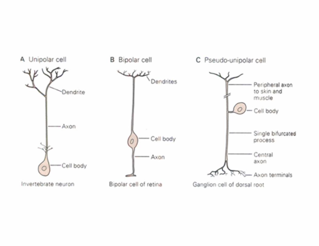
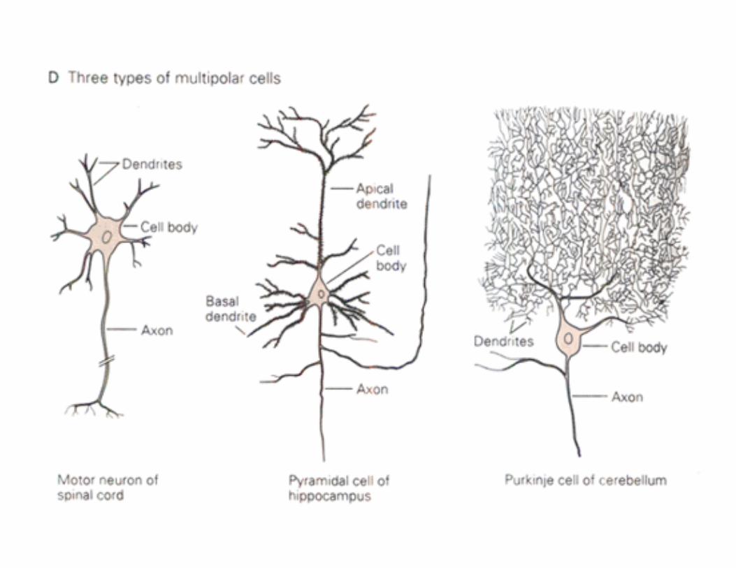
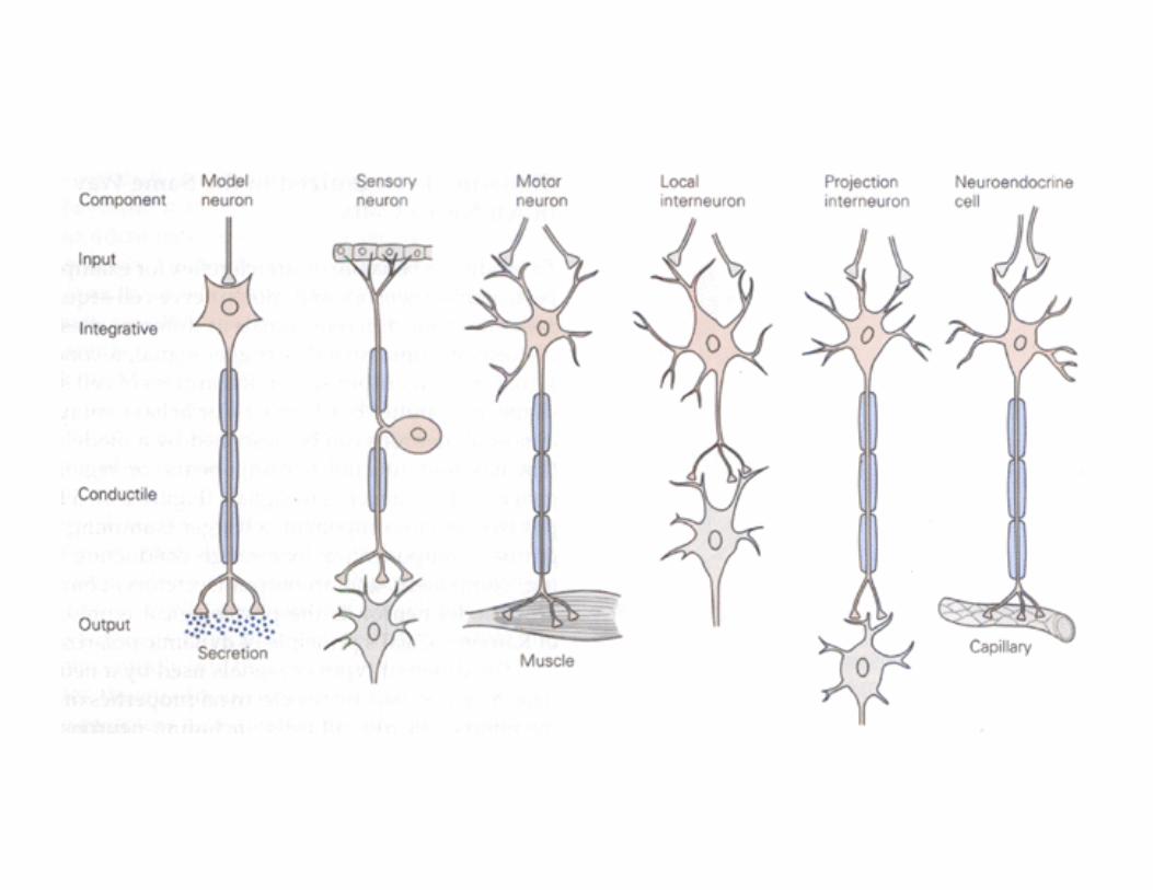
• Functional unit– Neurons • Contains almost 1011 neurons, • 1000 different types but with same basic structure and characteristics
• Human Behavior – Result of functioning of brain
– Depends more on the precise anatomical circuits formed by the neurons
– Less on the specialized nature of individual neurons
• Two classes of cells found in CNS
– Neurons – Glial cells
• Supporting cells • Some act as scavengers • Some take up chemicals released during synaptic transmission
• During brain development, they guide migrating neurons and direct the outgrowth of axons
– Micro and Macroglia
• Microglial Cells ‐ derived from phagocytes – Activated in Parkinsonism, Alzheimers, Multiple Sclerosis and AIDS related dementia
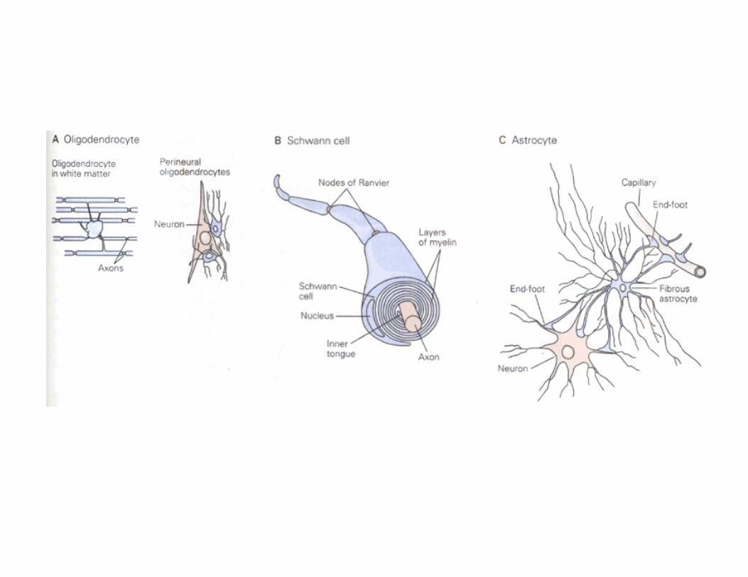
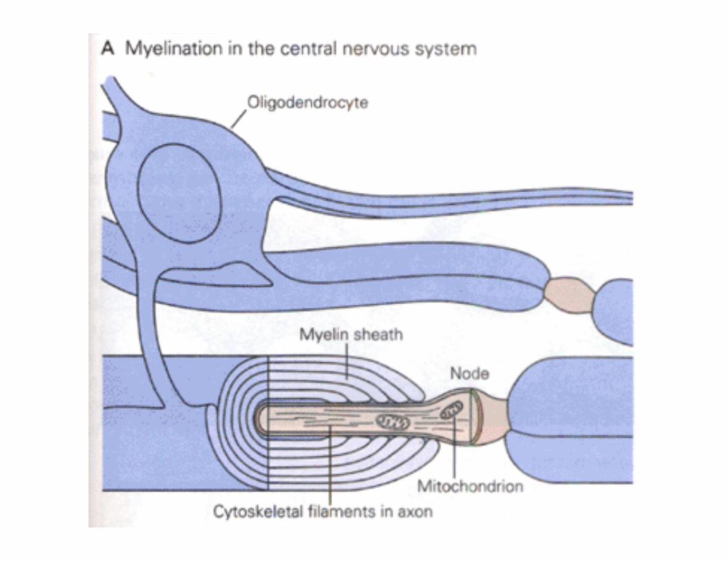
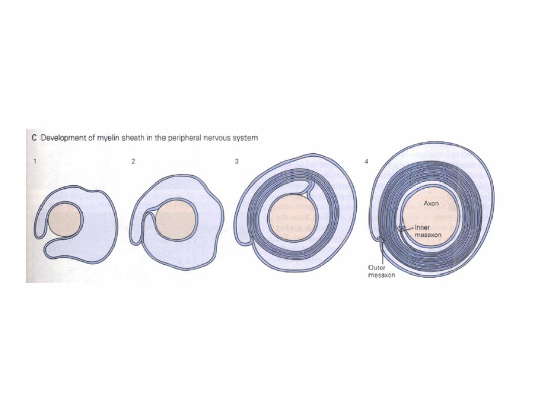
• Macroglia ‐ Oligodendrocytes & Schwann cells ‐ Astrocytes Oligodendrocytes – Found in CNS Insulates internodal axons, 15 internodal axons Schwann Cells – In periphery, one internode
• Astrocytes – Supply nutrition
– Forms Blood Brain Barrier – Maintains K+ level
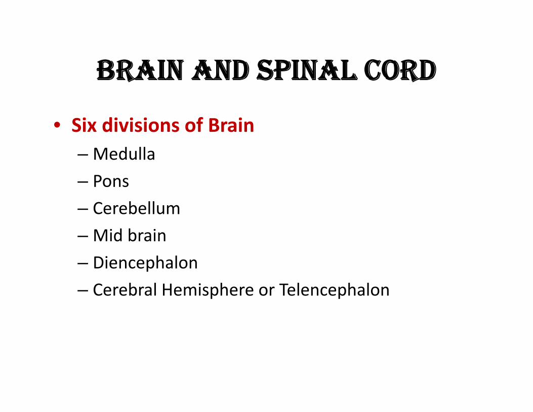
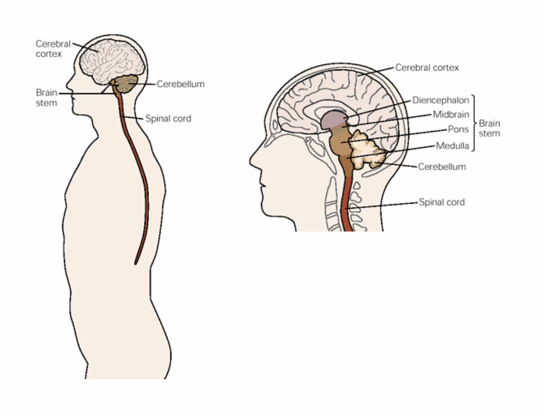
BRAIN AND SPINAL CORD
• Six divisions of Brain – Medulla
– Pons – Cerebellum
– Mid brain
– Diencephalon
– Cerebral Hemisphere or Telencephalon
THE SPINAL CORD • Most caudal part of the CNS
• From the base of the skull to the first lumbar vertebra.
• Receives sensory information from the skin, joints, and muscles of the trunk and limbs &
• Contains the motor neurons responsible for both voluntary and reflex movements.
• The gray matter‐ contains nerve cell bodies • Divided into dorsal and ventral horns
• The dorsal horn‐ contains sensory relay neurons that receive input from the periphery,
• The ventral horn‐ contains motor nuclei that innervate specific muscles.
• White matter ‐ longitudinal tracts of myelinated axons
• Ascending pathways carrying sensory information to brain
• Descending pathways that carry motor commands and modulatory influences from the brain
• 31 pairs of spinal nerves ‐nerve fibers linking the spinal cord with muscles and sensory receptors in the skin
THE BRAIN STEM • The medulla
– Direct rostral extension of the spinal cord
– Resembles the spinal cord both in organization and function
– Regulates blood pressure and respiration
– Also contains some of the early relay nuclei involved in taste, hearing, and maintenance of balance as well as the control of neck and facial muscles.
THE PONS • The ventral portion of the pons‐ contains the pontine nuclei – relay information about movement and sensation from the cerebral cortex to the cerebellum.
• The dorsal portion of the pons ‐contains structures involved in respiration, taste, and sleep.
THE MIDBRAIN • Provide important linkages between the components of motor systems – The cerebellum, the basal ganglia, and the cerebral hemispheres
• Also contains components of the auditory and visual systems.
• Several regions ‐ connected to the extraocular muscles of the eye
• The major pathway for controlling eye movements.
THE CEREBELLUM • Receives
– Somatosensory input from the spinal cord
– Motor information from the cerebral cortex – Input about balance from the vestibular organs of the inner ear
• Important for maintaining posture and for coordinating head and eye movements
• Also involved in fine tuning the movements of muscle and in learning motor skills
• Also involved in cognition
Diencephalon The thalamus and hypothalamus. The thalamus – Essential for the transfer of sensory information from periphery to cerebral hemispheres. – Determines whether sensory information reaches conscious awareness
– Participates in the integration of motor information from the cerebellum and the basal ganglia
– Transmits this information to the regions of the cerebral hemispheres concerned with movement.
– Also has reticular formation which influence levels of attenti and consciousness.
THE HYPOTHALAMUS • Ventral to the thalamus • Regulates several behaviors that are essential for homeostasis and reproduction – Growth, eating, drinking, and maternal behavior, by regulating the hormonal secretions of the pituitary gland.
– Also influences behavior through its extensive afferent and efferent connections with practically every region of the central nervous system
– An essential component of the motivational system of the brain
– Regulates circadian rhythms, cyclical behaviors entrained to the daily light‐dark cycle
THE CEREBRAL HEMISPHERE • The largest region of the human brain consists of
‐‐The cerebral cortex – The underlying white matter – Three deep‐lying structures – The basal ganglia, the amygdala, and the hippocampas
• Concerned with perceptual, motor, and cognitivefunctions, including memory and emotion.
• The two hemispheres‐ interconnected by the corpus callosum
• The amygdala ‐ concerned with social behavior and emotion,
• The hippocampus‐memory • The basal ganglia‐ with the control of fine movement.
Five Principles Governing the Organization of the Major Functional Systems
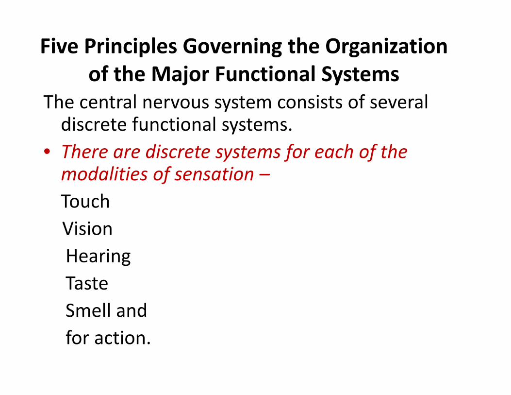
The central nervous system consists of several discrete functional systems.
• There are discrete systems for each of the modalities of sensation – Touch Vision Hearing Taste Smell and for action.
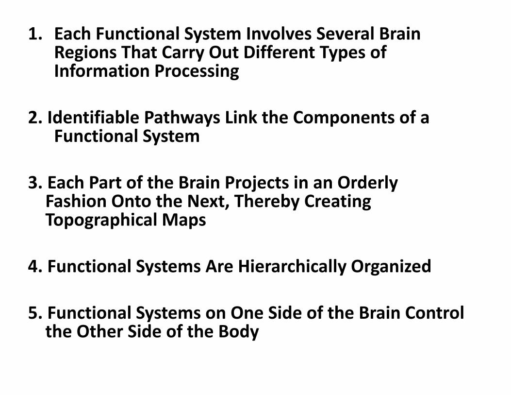
1. Each Functional System Involves Several BrainRegions That Carry Out Different Types ofInformation Processing
2. Identifiable Pathways Link the Components of aFunctional System
3. Each Part of the Brain Projects in an OrderlyFashion Onto the Next, Thereby CreatingTopographical Maps
4. Functional Systems Are Hierarchically Organized
5. Functional Systems on One Side of the Brain Controlthe Other Side of the Body
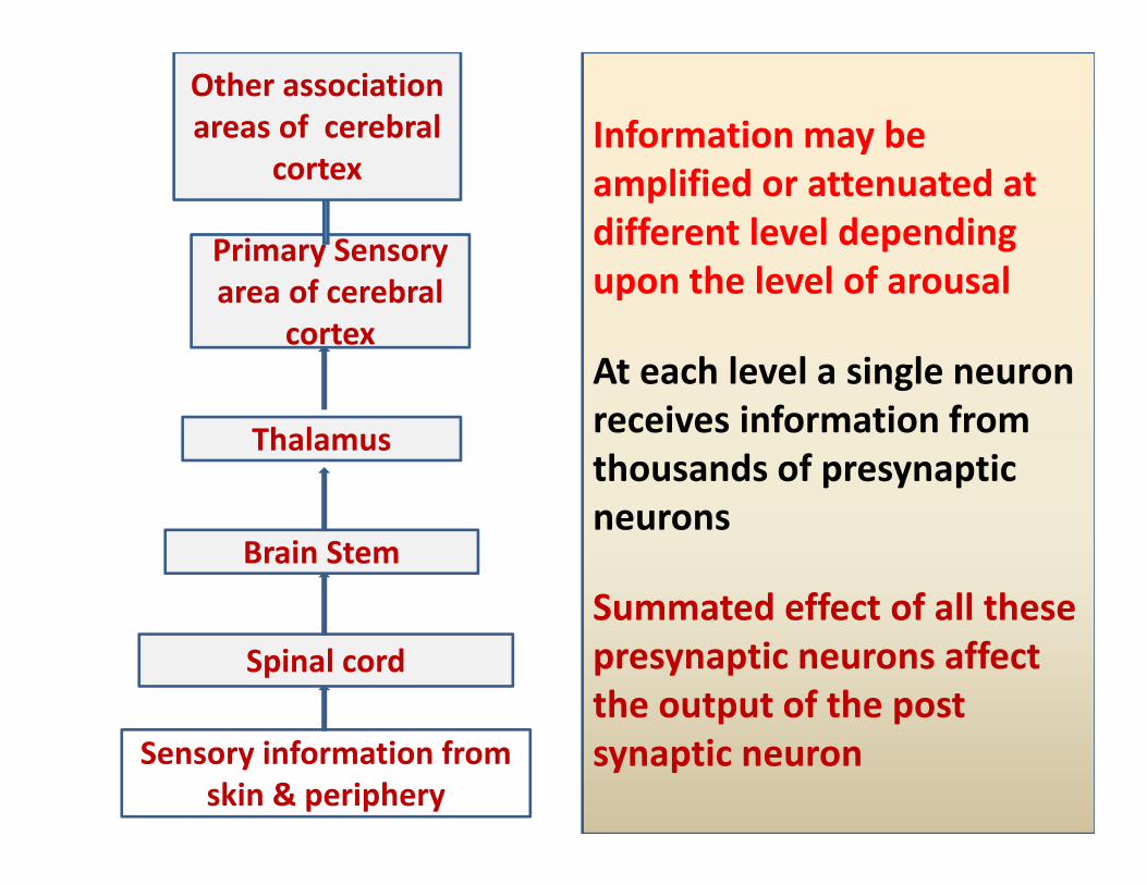
Primary Sensory area of cerebral
cortex
Other association areas of cerebral
cortex
Sensory information from skin & periphery
Spinal cord
Brain Stem
Thalamus
Information may be amplified or attenuated at different level depending upon the level of arousal
At each level a single neuron receives information from thousands of presynaptic neurons
Summated effect of all these presynaptic neurons affect the output of the post synaptic neuron
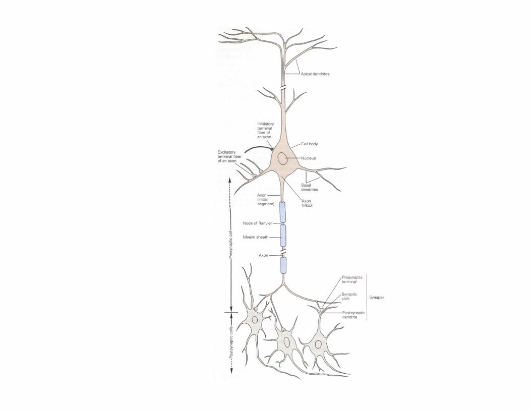
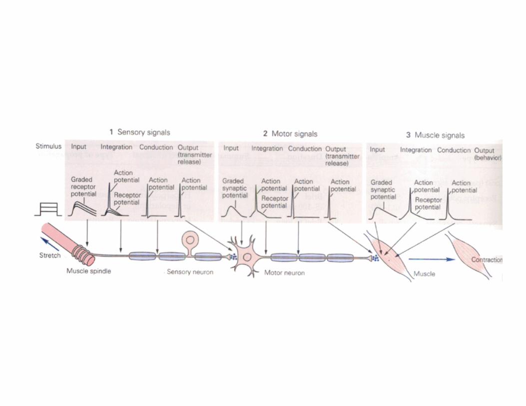
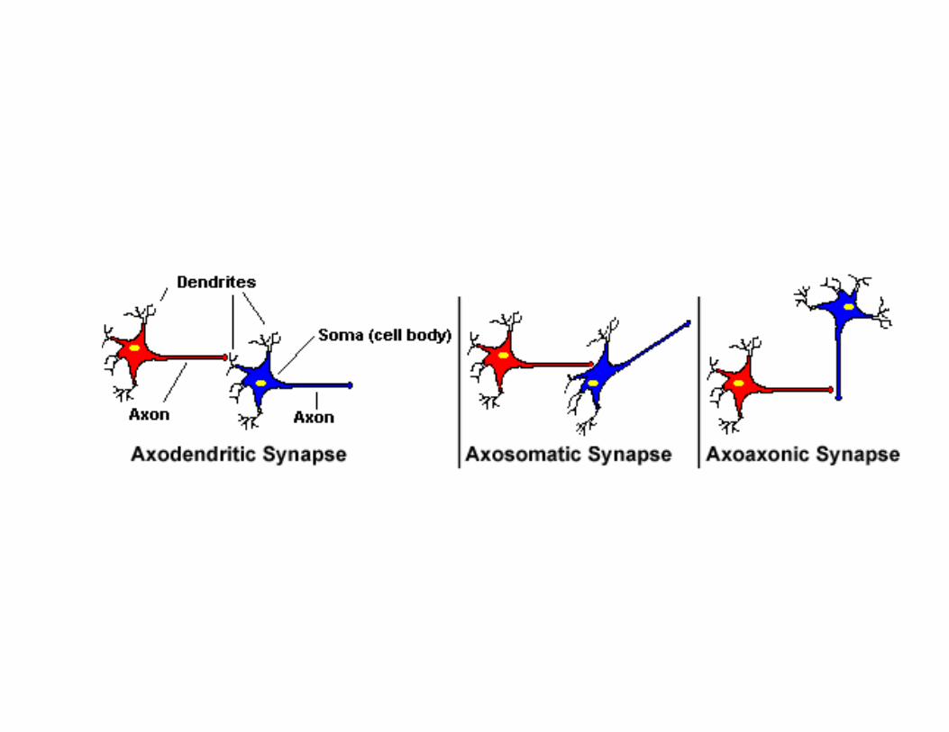
Difference between motor and sensory neurons‐ the location of their synaptic inputs
The sensory neuron ‐ few synapse on its cell body or along the peripheral branch of its axon
Primary input ‐ from sensory receptors at the terminal of the peripheral axon.
The motor neuron receives primary and modifying inputs throughout its dendrites and cell body. ‐Almost all presynaptic boutons on motor neurons are located on the dendritic branches; ‐only 5% are located on the cell body
Functional Pattern distribution Most inhibitory synapses ‐ on the cell body or close to it Excitatory‐ located farther out along the dendrites
Inhibitory inputs are strategically placed close to the trigger zone to have maximal influence on the final tally of inputs to the neuron

The information flow from sensory neurons to motor neurons is both divergent and convergent
Each sensory neuron contacts 500–1000 motor neurons and typically forms two to six synapses on a single motor neuron (divergence of information)
At the same time each motor neuron receives input from many sensory neurons (convergence of information)
Inputs from more than 100 sensory neurons are needed for a motor neuron to reach the threshold for firing.
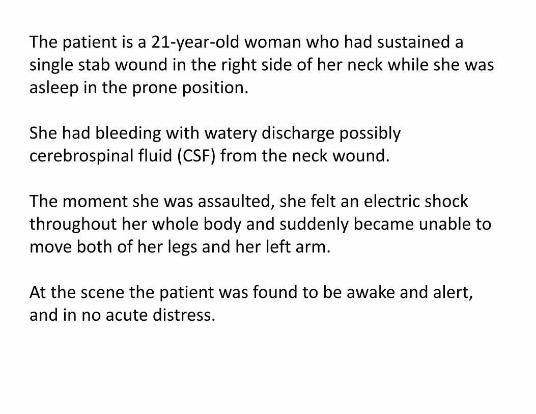
The patient is a 21‐year‐old woman who had sustained a single stab wound in the right side of her neck while she was asleep in the prone position.
She had bleeding with watery discharge possibly cerebrospinal fluid (CSF) from the neck wound.
The moment she was assaulted, she felt an electric shock throughout her whole body and suddenly became unable to move both of her legs and her left arm.
At the scene the patient was found to be awake and alert, and in no acute distress.
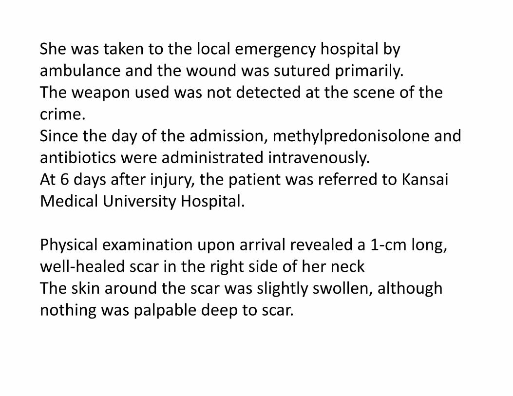
She was taken to the local emergency hospital by ambulance and the wound was sutured primarily. The weapon used was not detected at the scene of the crime. Since the day of the admission, methylpredonisolone and antibiotics were administrated intravenously. At 6 days after injury, the patient was referred to Kansai Medical University Hospital.
Physical examination upon arrival revealed a 1‐cm long, well‐healed scar in the right side of her neck The skin around the scar was slightly swollen, although nothing was palpable deep to scar.
Neurological examination revealed Impairement of motor function in both of legs and in her left arm Her left leg ‐ completely flaccid, Right leg could be moved slightly Joint position and vibration modalities ‐ diminished up to the level of Th2 on both sides. Decrease in pain and temperature sensation below the C8 level on right side Paring of sacral sensation She developed urinary retention The deep tendon reflexes ‐ absent in the left leg with positive Babinski tests in the feet bilaterally. No significant past medical or surgical history
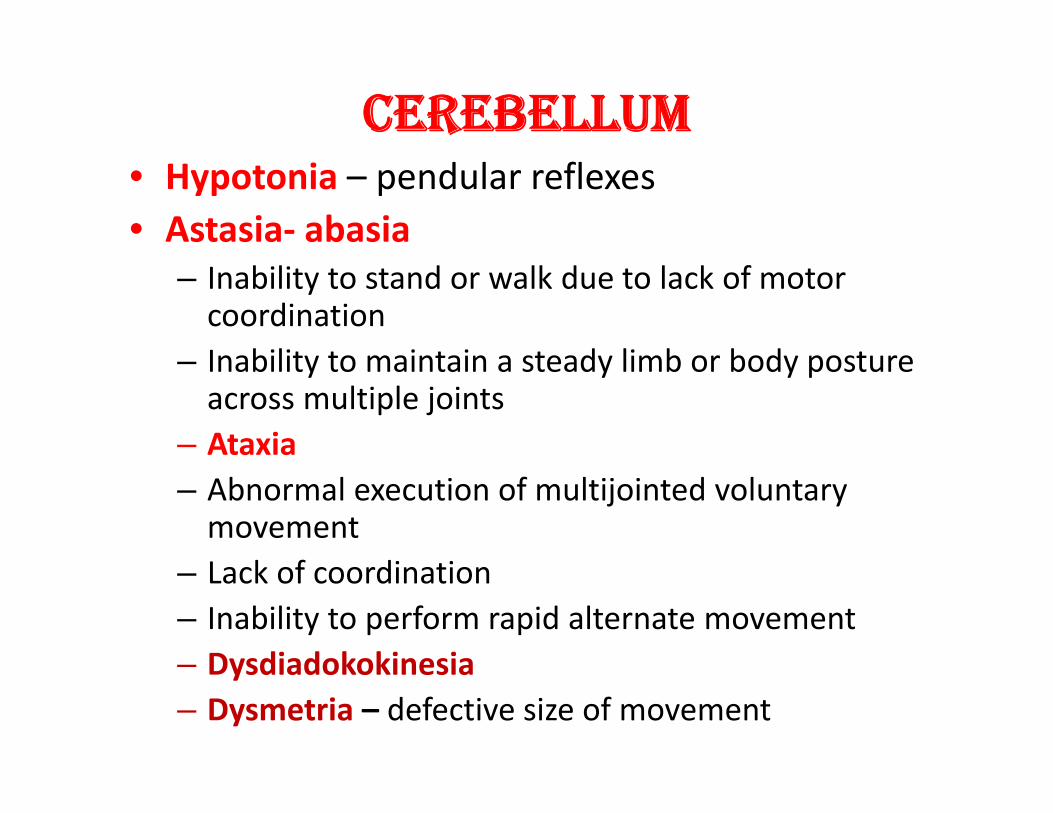
CEREBELLUM • Hypotonia – pendular reflexes • Astasia‐ abasia
– Inability to stand or walk due to lack of motor coordination
– Inability to maintain a steady limb or body posture across multiple joints
– Ataxia – Abnormal execution of multijointed voluntary movement
– Lack of coordination – Inability to perform rapid alternate movement – Dysdiadokokinesia – Dysmetria – defective size of movement
• Intention Tremor – Inability ot switch on and off the agonists and antagonists
– Incoordinated movement during execution of movement especially towards the end of movement
– Erroneous correction in the range of movement
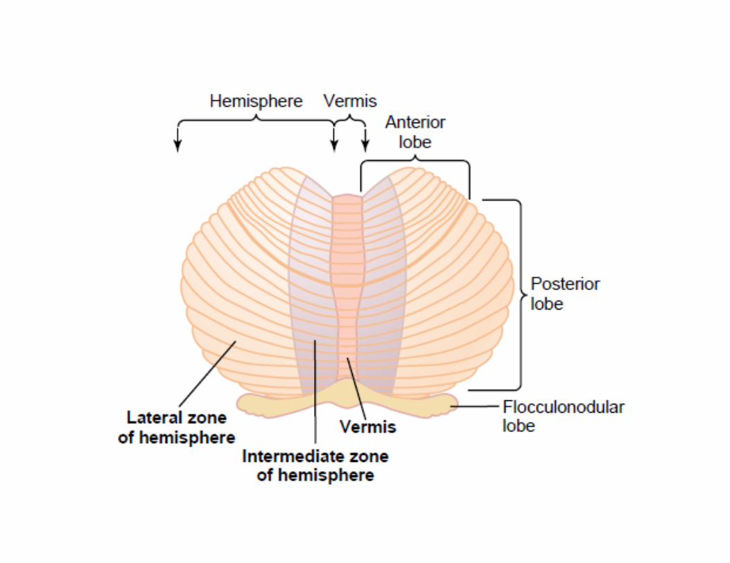
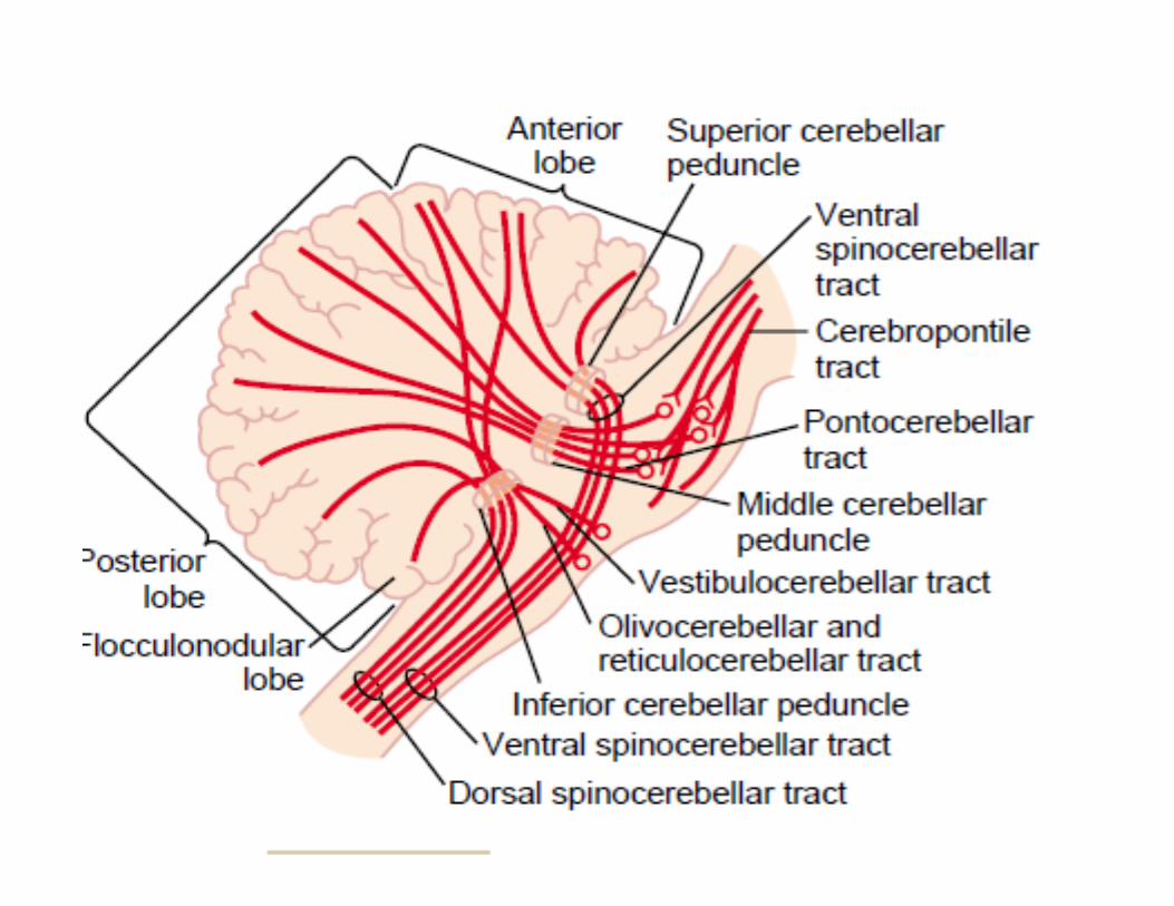
• THE FLOCCULONODULAR LOBE – Most primitive part – Its cortex receives input directly from primary vestibular afferents and somatosensory input from neck, labyrinth and visual information
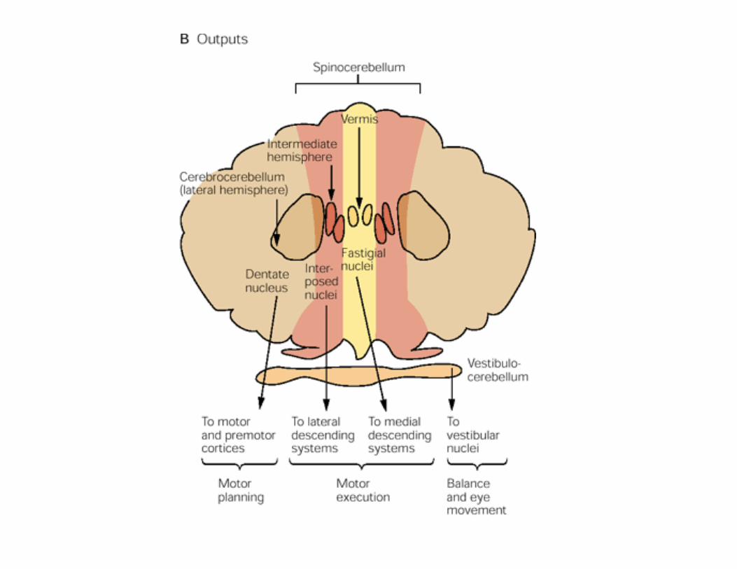
– Projects to the vestibular nuclei – Control balance, vestibular reflexes and eye movement – Vestibulocerebellum
• The vermis and intermediate hemispheres • Vermis receives • visual, auditory, and vestibular as well as somatic sensory input from the head and proximal parts of the body.
• Vermis fastigial nucleus cortex and brain stem medial descending systems that control proximal muscles of the body and limbs.
• Vermis governs posture, locomotion and gaze.
• The adjacent intermediate zone receive somatosensory input from the limbs and information from motor cortex
• Intermediate zone interposed nucleus lateral corticospinal and rubrospinal and reticular systems – Controls the more distal muscles of the limbs and digits
• Because the vermis and intermediate hemispheres are the only regions to receive somatosensory inputs from the spinal cord, they are known as spinocerebellum.
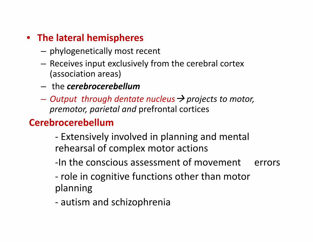
• The lateral hemispheres – phylogenetically most recent – Receives input exclusively from the cerebral cortex (association areas)
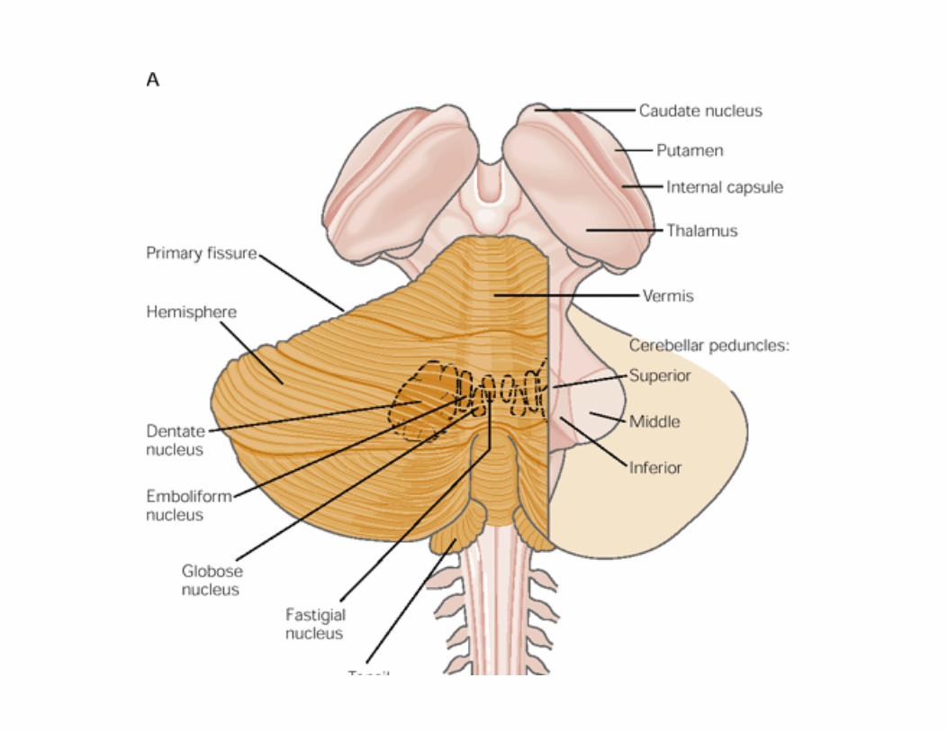
– the cerebrocerebellum – Output through dentate nucleus projects to motor, premotor, parietal and prefrontal cortices
Cerebrocerebellum ‐ Extensively involved in planning and mental rehearsal of complex motor actions ‐In the conscious assessment of movement errors ‐ role in cognitive functions other than motor planning ‐ autism and schizophrenia
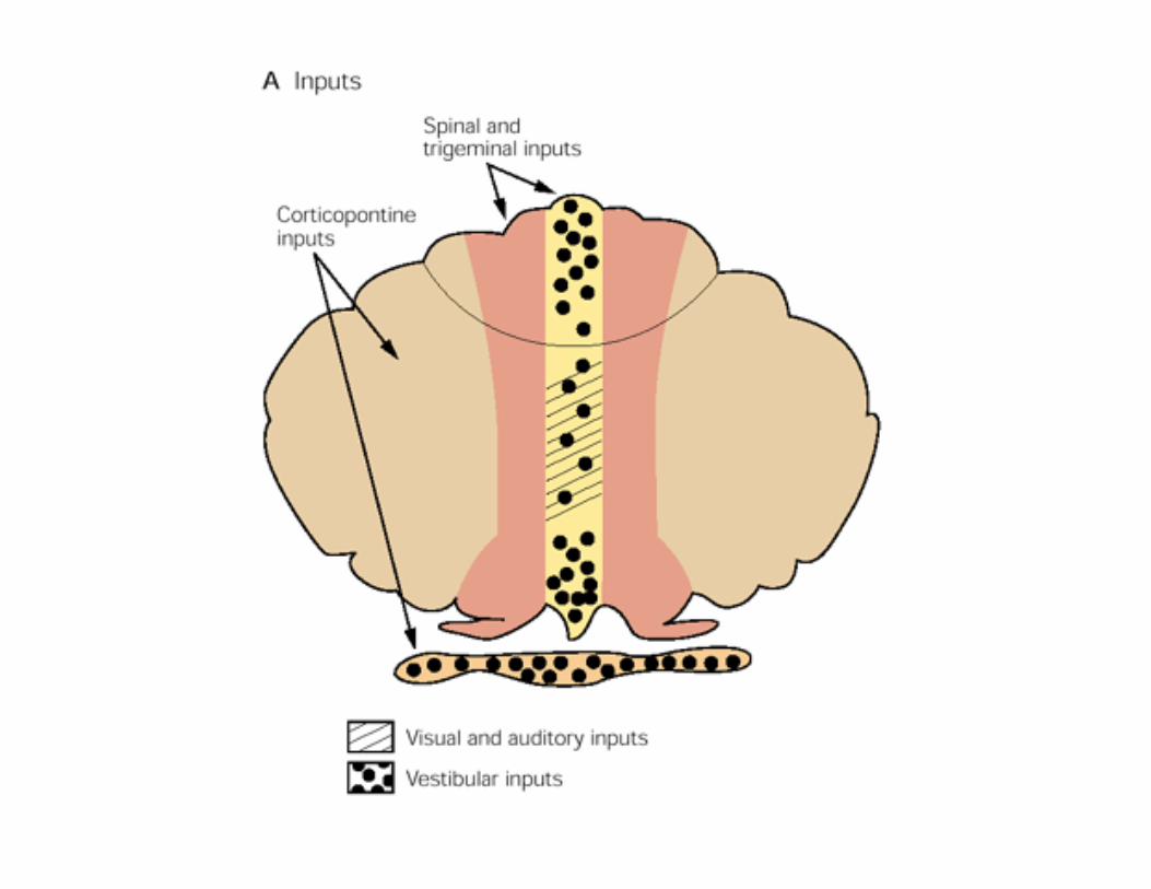
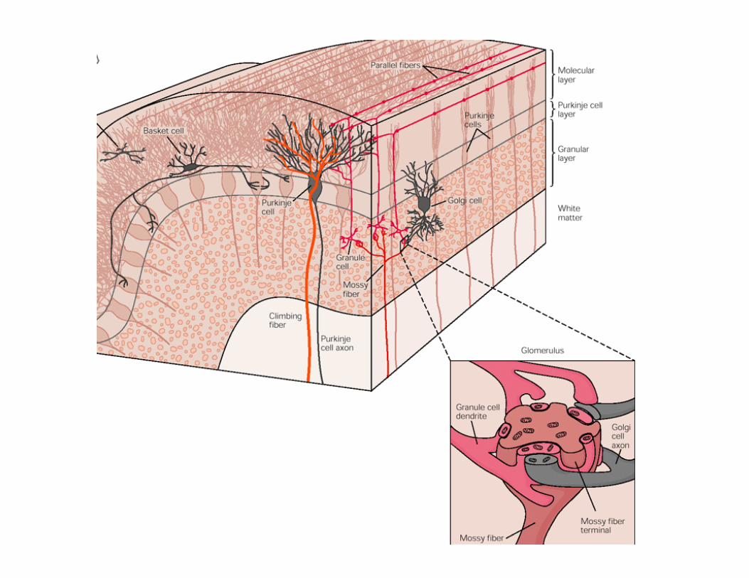
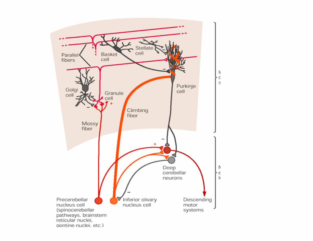
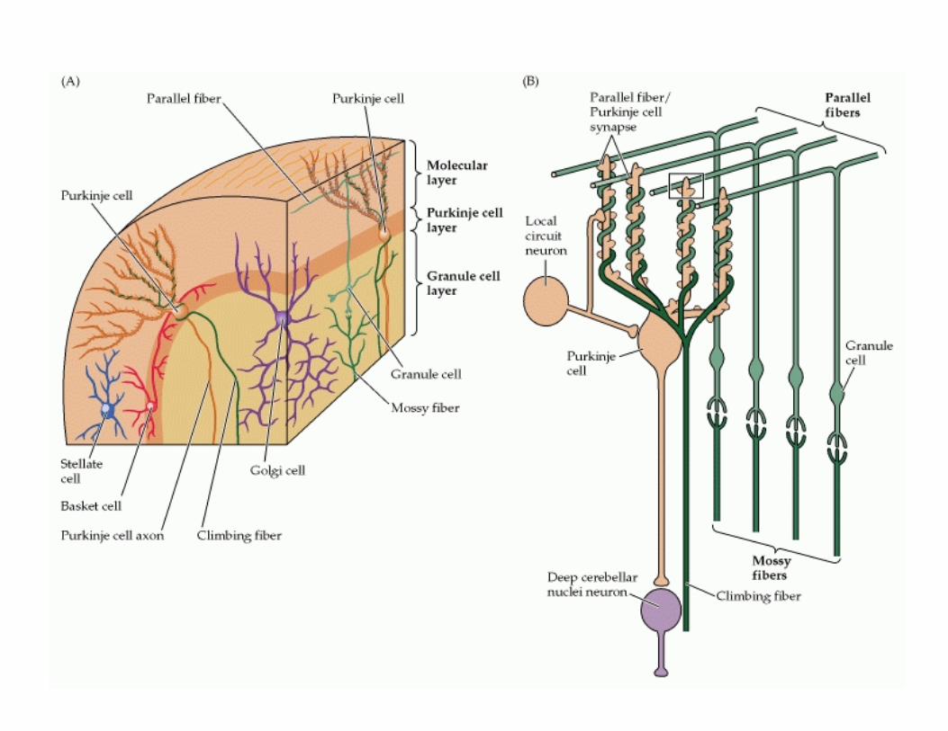
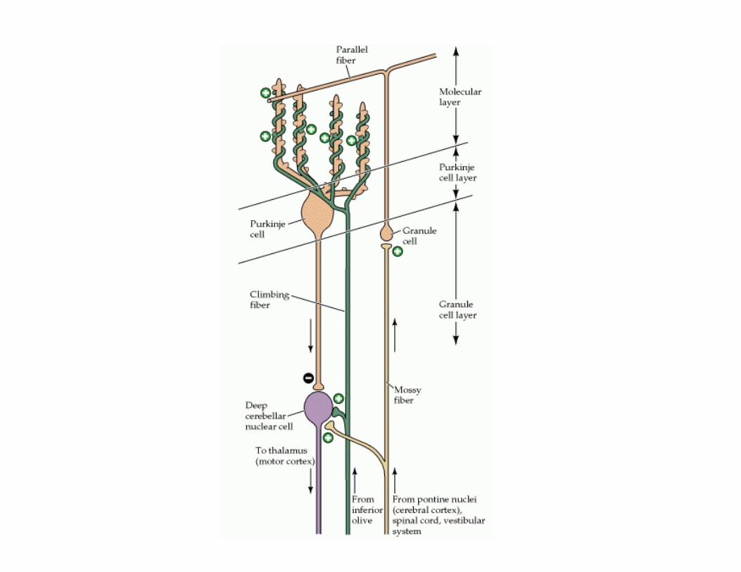
• Mossy fibres‐ sensory information from periphery, brainstem and information from cerebral cortex
• Simple spike in granule cells • Highly convergent ‐ 200,000 to 1million granule cells for 1 purkinje cells .
• Parallel fibres –divergent to purkinje cells
• Climbing fibres –sensory information from periphery and information from cerebrum
• Divergent ‐1 to 10 purkinje cells • One purkinje cells receive input from one climbing fibre only.
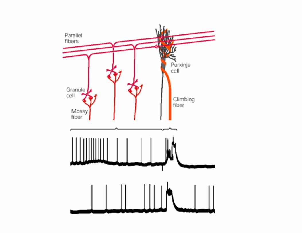
• Produces Complex spike • Somatic and axonal spikes travel down axon but dendritic spikes do not travel down axon
• Increases Ca++ conductance in dendrites Ca++ mediated K+ channel opening hyperpolarization Post complex spike pausemodulation of PC response to parallel fibers
• Alter cerebellar output by modulating the synaptic effect of parallel fiber input to Purkinje cells in two ways AP in climbing fiber slightly reduce the strength of the parallel fiber input to the Purkinje cell
– Activity in climbing fibers can induce selective long‐term depression in the synaptic strength of parallel fibers that are active concurrently
• PC cell plasticity
• Reciprocity of complex and simple spike

CF • Lazy but strong • Powerful effect on PC – Increases Ca++ in soma and dendrites for prolonged period prolonged depolarization complex spike initial largeamplitude AP followed by small amplitude AP
• In awake person ‐ Low rate of complex spike ‐1‐3/sec • During activity – single action potential in relation tospecefic sensory event
• Climbing fibres do not fire frequently • Synchronus firing of few climbing fibers signalimportant event
• CF in inf. Olivary nuclei are connected electrotonically
Mossy fibers • Purkinje cells – tonically discharging—simple spikes with spontaneous firing rate ‐100/sec even during rest
• During activity –several hundred spikes /sec • Due to activity in mossy fibre –granule cell system • Parallel fibers to PC – brief,small excitatory potential in PC neuron spread to initial segment propagate down the axon
• Large no. of parallel fibers discharge simultaneously to stimulate PC – Encodes the magnitude and duration of stimuli –by controlling firing rate of purkinje cells
• Dual effect of mossy fibers on Deep Nuclei – Direct collateral stimulatory – Through parallel fibers via PC – Inhibitory
• In cerebellar cortex also both excitatory and inhibitory control end on PC
• Excitatory‐ Through parallel fibers • Parallel fibers excite th inhibitory interneurons inhibit PC
• Same parallel fibers excite stellate cell and PC and the SCinhibit PC
• Different parallel fibers excite PC and basket cell and BC inhibit PC– a type of lateral inhibition
Comparison of excitatory and inhibitory inputs both in deep nuclei and cerebellar cortex
• Deep nuclei ‐Mossy fibres direct stimulation
‐Via purkinje cells inhibition
• Cerebellar cortex – Purkinje cells –directly excited by parellel fibres – Inhibted by disynaptic connection through basket and stellate cells
RECURRENT LOOPS
• With Inferior Olive – Cerebellum Inhibitory output from deep nuclei (GABAergic) to inferior olive modulation of climbing fibers from inferior olive
Regulation on climbing fibers excitatory input to deep nuclei and Purkinje cells in cerebellum
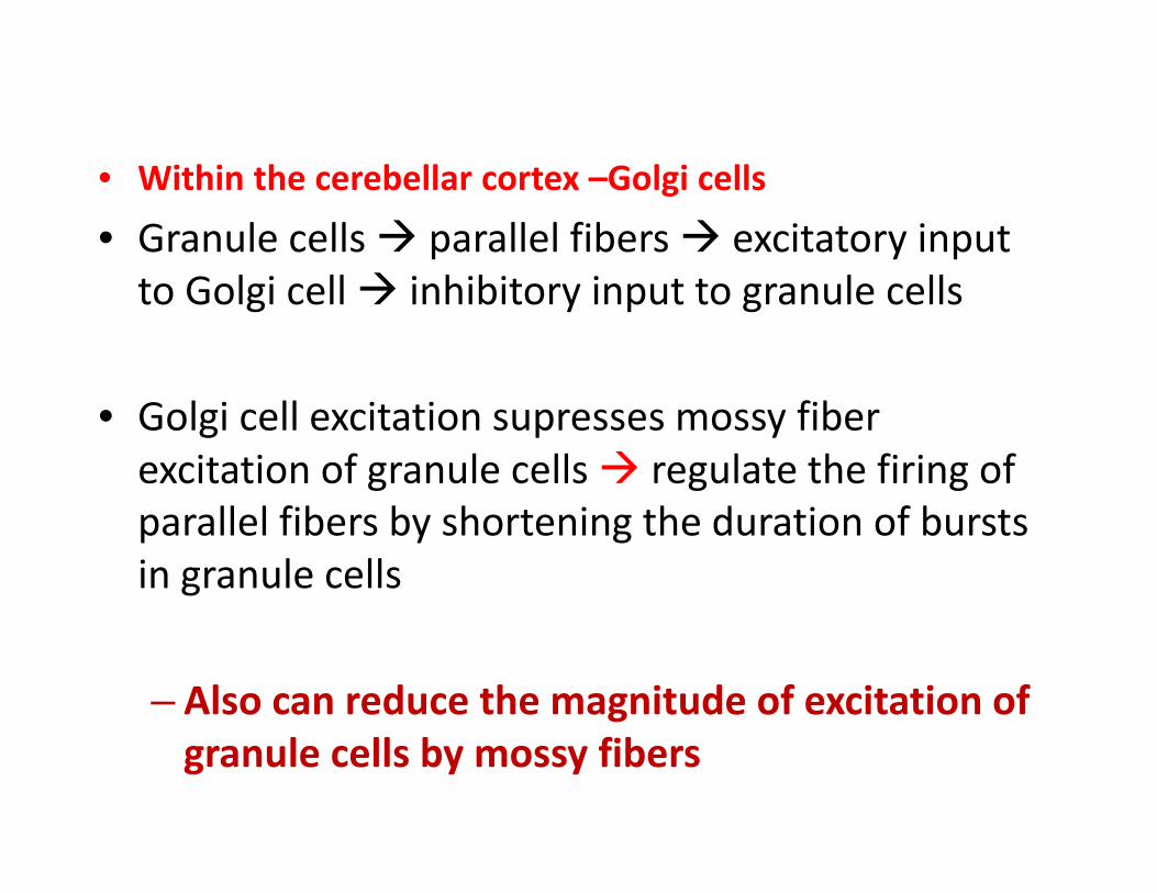
• Within the cerebellar cortex –Golgi cells
• Granule cells parallel fibers excitatory input to Golgi cell inhibitory input to granule cells
• Golgi cell excitation supresses mossy fiber excitation of granule cells regulate the firing of parallel fibers by shortening the duration of bursts in granule cells
– Also can reduce the magnitude of excitation of granule cells by mossy fibers
Cerebellum and cerebral cortex
• Cerebral cortex pontine nuclei Lateral cerebellum Thalamus premotor and motor cortex
• Involvement of cerebellum in the motor circuit from cerebral cortex to spinal cord
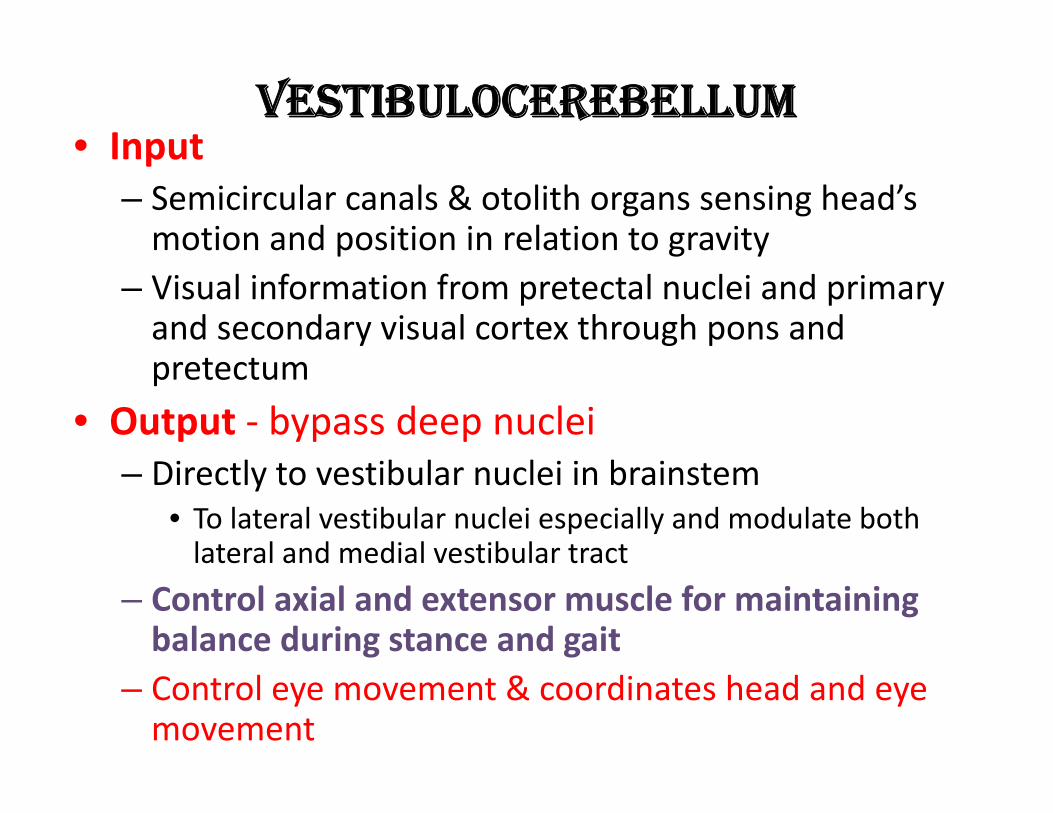
VESTIBULOCEREBELLUM • Input
– Semicircular canals & otolith organs sensing head’s motion and position in relation to gravity
– Visual information from pretectal nuclei and primary and secondary visual cortex through pons and pretectum
• Output ‐ bypass deep nuclei – Directly to vestibular nuclei in brainstem
• To lateral vestibular nuclei especially and modulate both lateral and medial vestibular tract
– Control axial and extensor muscle for maintaining balance during stance and gait
– Control eye movement & coordinates head and eye movement
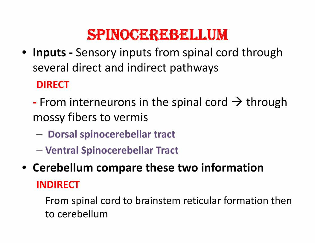
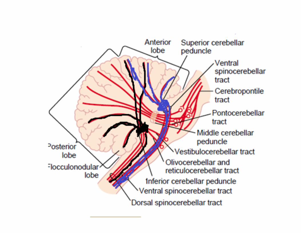
SPINOCEREBELLUM • Inputs ‐ Sensory inputs from spinal cord through several direct and indirect pathways DIRECT
‐ From interneurons in the spinal cord through mossy fibers to vermis – Dorsal spinocerebellar tract – Ventral Spinocerebellar Tract
• Cerebellum compare these two information INDIRECT
From spinal cord to brainstem reticular formation then to cerebellum
• Dorsal spinocebellar tract (Inferior cerebellar Peduncle) – Somatosensory information from muscle and joint receptors
– Convey sensory feedback about the consequences of movement
– Input flows whether the movement is passive or active
Ventral Spinocerebellar tract ( superior cerebellar peduncle bilateral)
– impulses reaching to motor neuron via Corticospinal tract and Rubrospinal tract and local pattern generators of spinal cord .
– Efference copy of the total command received at the level of spinal cord
• Activated only during active movement
• Cuneocerebellum and rostral cerebellar tract carry sensory information from upper part of the body
OUTPUT/EFFERENT PATHWAY • Spinocerebellum
From vermis( PC) of both anterior and posterior lobe Fastigial nuclei bilateral projection to brainstem reticular formation & lateral vestibular nuclei spinal cord through Reticulospinal and Vestibulospinal Tracts • Mainly to medial descending system Muscles of neck, trunk and proximal parts of arms
• These brainstem areas also receive inputs from descending tracts (cortical) and sensory inputs from spinal cord output from these brainstem area is modulated by cerebellum
Intermediate part Interposed nuclei sup cerebellar peduncle Contralateral red nucleus(magnocellular ) axons cross the midline rubrospinal tract
• Interposed nuclei Ventrolateral nuclei of thalamus limb control areas of primary moter cortex
• Act on limb and axial muscles both • Somatotopic organization of deep nuclei neurons • Cerebellar neurons fire in synchrony with motor cortex neurons during movement
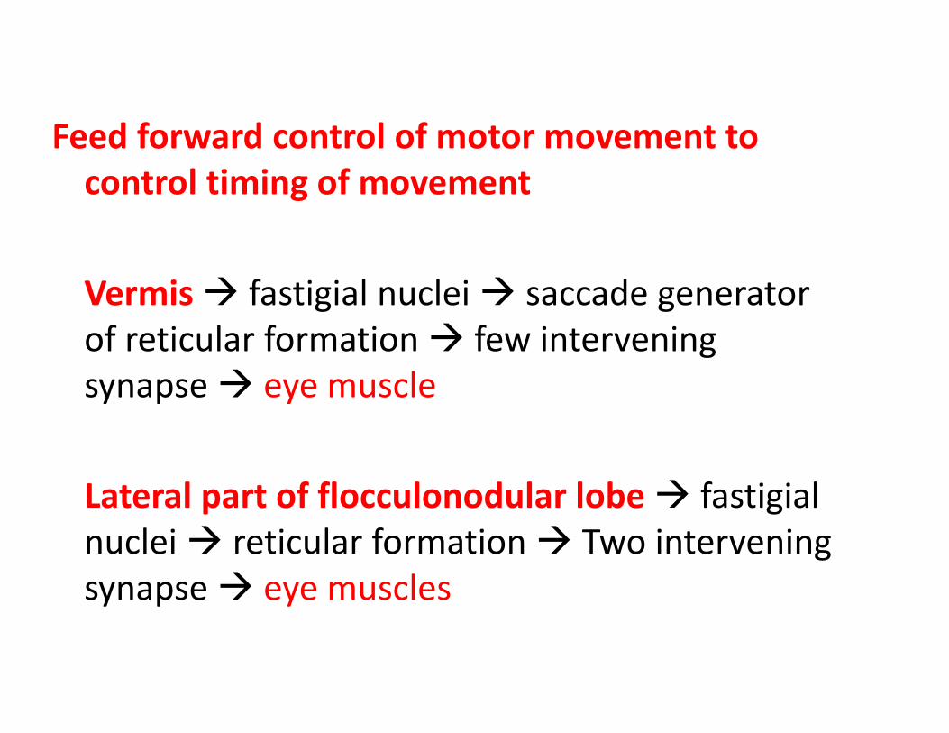
Feed forward control of motor movement to control timing of movement
Vermis fastigial nuclei saccade generator of reticular formation few intervening synapse eye muscle
Lateral part of flocculonodular lobe fastigial nuclei reticular formation Two intervening synapse eye muscles
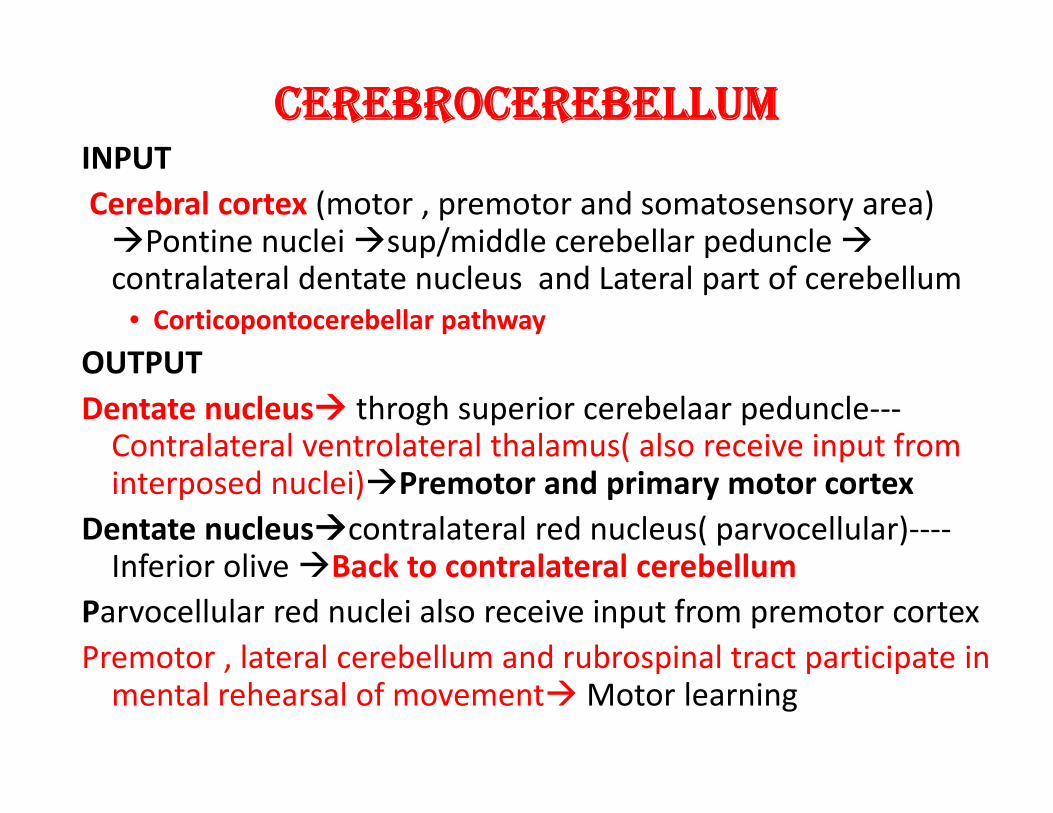
CEREBROCEREBELLUM INPUT Cerebral cortex (motor , premotor and somatosensory area) Pontine nuclei sup/middle cerebellar peduncle contralateral dentate nucleus and Lateral part of cerebellum
• Corticopontocerebellar pathway
OUTPUT Dentate nucleus throgh superior cerebelaar peduncle‐‐‐Contralateral ventrolateral thalamus( also receive input from interposed nuclei)Premotor and primary motor cortex
Dentate nucleuscontralateral red nucleus( parvocellular)‐‐‐‐Inferior olive Back to contralateral cerebellum
Parvocellular red nuclei also receive input from premotor cortex Premotor , lateral cerebellum and rubrospinal tract participate in mental rehearsal of movementMotor learning

Olivocerebellar tract
• From – Cerebral cortex – Basal ganglia
– Reticular formation
– Spinal cord
• Vestibulocerebellar tract – Vestibular apparatus ,vestibular nuclei – flocculonodular lobe and fastigial nuclei
• Compares sensory input from head velocity with eye velocity
• Reticulocerebellar tract – Brainstem reticular formation Vermis
Cerebellar Disorder • Hypotonia
– Pendular reflexes
• Astasia‐Abasia
• Ataxia – Dysmetria, dysdiadochokinesis
• Action /Intentional tremor • Loss of automatic, unconscious nature of movement
• Cerebrocerebellum
• Input –association cortex – Pontine nuclei—dentate nucleus ‐‐‐motor ,premotor, prefrontal and parietal cortex
•
LANGUAGE
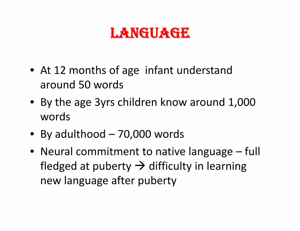
• At 12 months of age infant understand around 50 words
• By the age 3yrs children know around 1,000 words
• By adulthood – 70,000 words • Neural commitment to native language – full fledged at puberty difficulty in learning new language after puberty
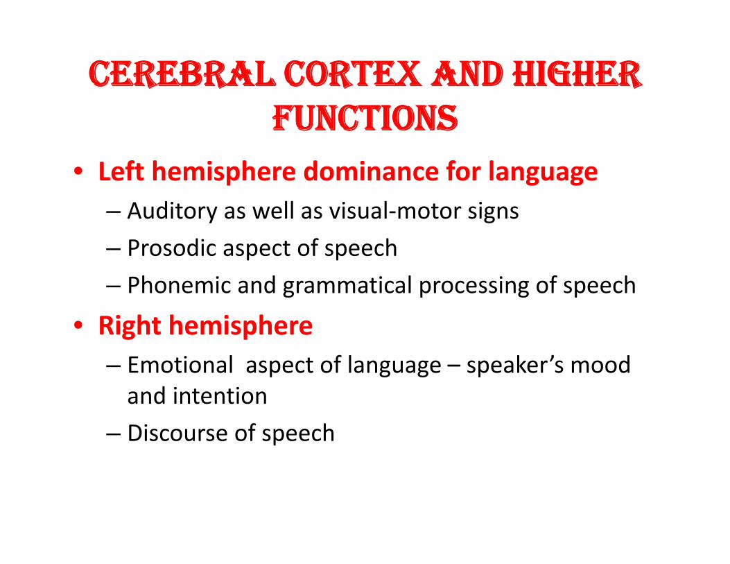
CEREBRAL CORTEX AND HIGHER FUNCTIONS
• Left hemisphere dominance for language – Auditory as well as visual‐motor signs – Prosodic aspect of speech
– Phonemic and grammatical processing of speech
• Right hemisphere – Emotional aspect of language – speaker’s mood and intention
– Discourse of speech
• Bilinguals – If the second language acquired early age – two languages are processed in the same area
– Late bilinguals – second and native language processed in spatially seperated area of the language processing cortex
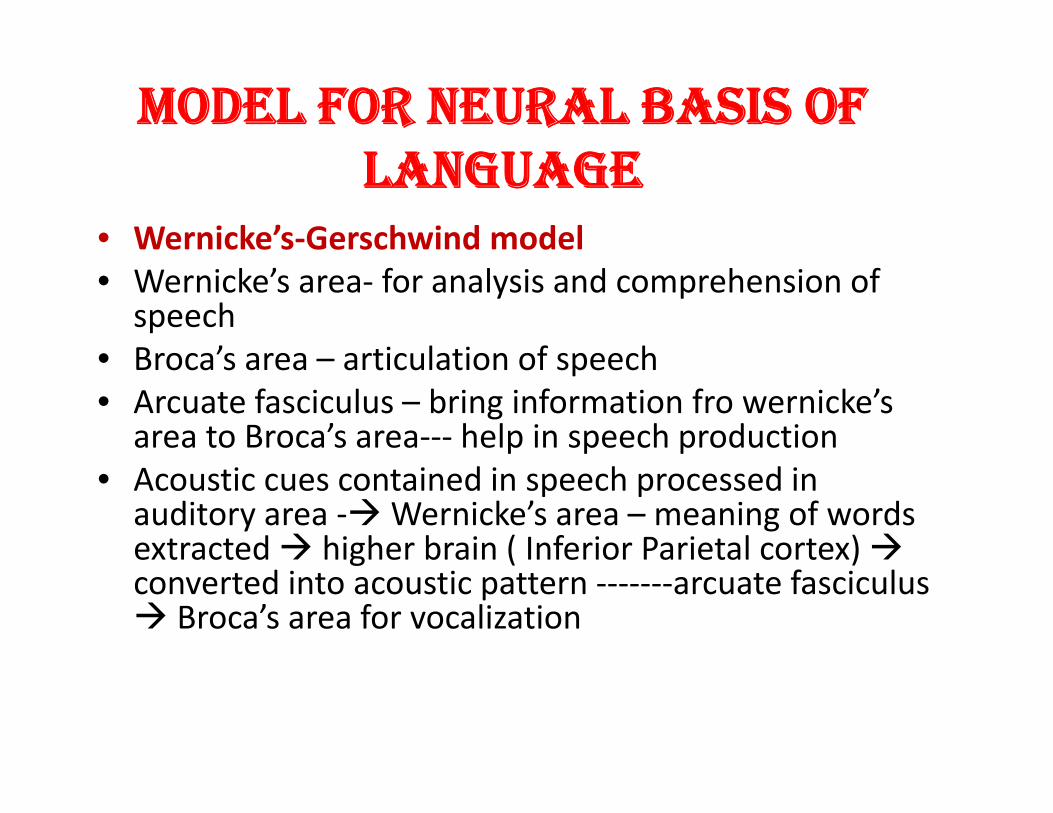
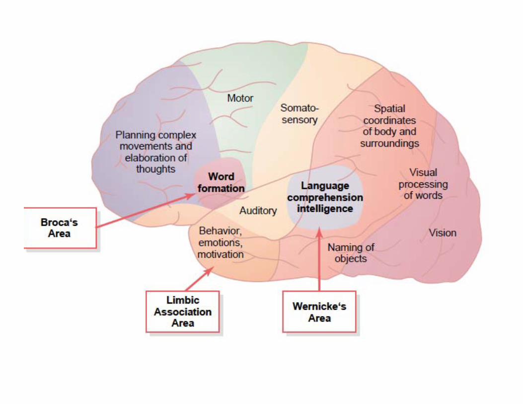
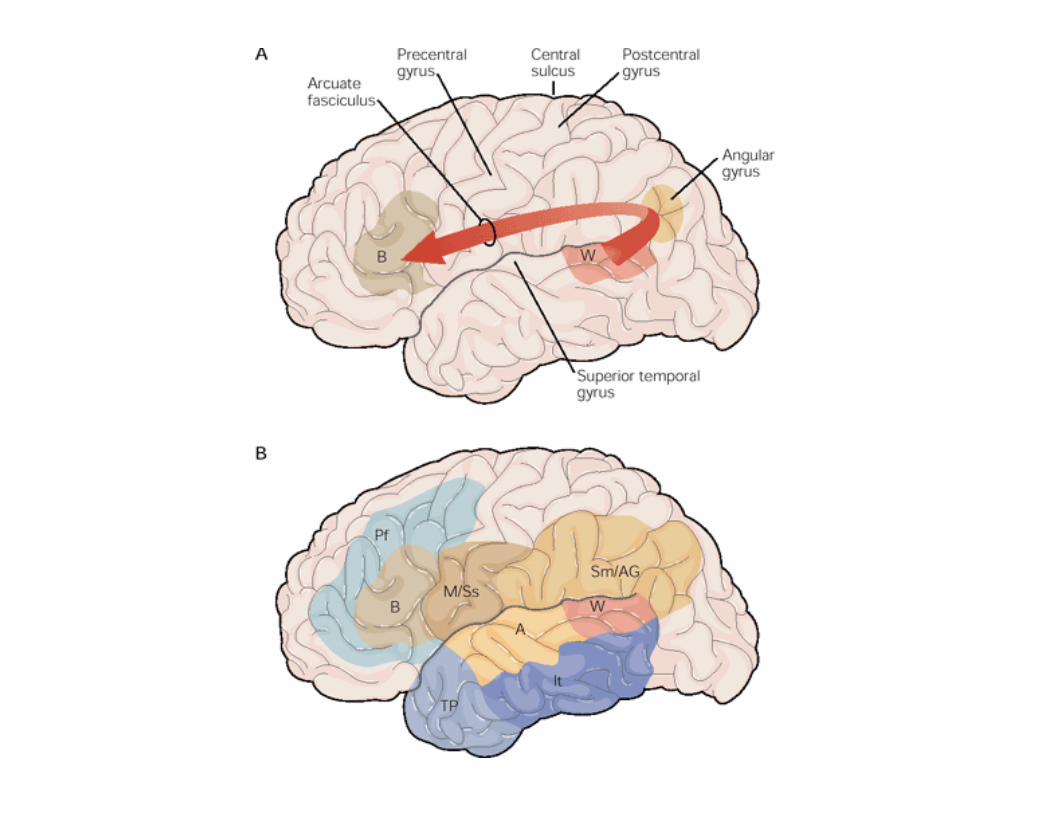
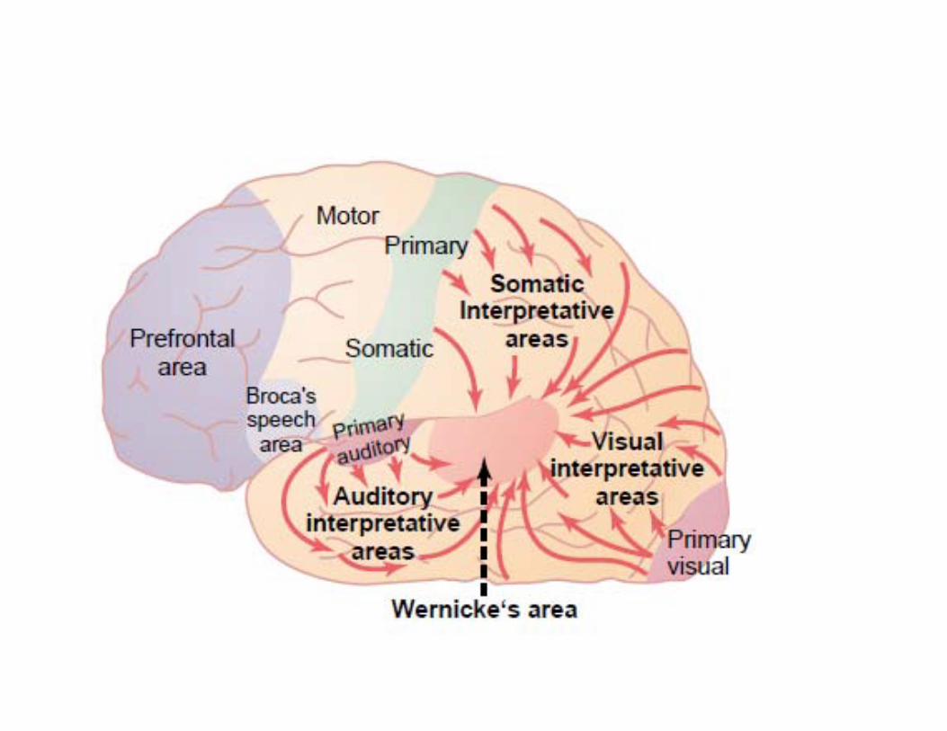
MODEL FOR NEURAL BASIS OF LANGUAGE
• Wernicke’s‐Gerschwind model • Wernicke’s area‐ for analysis and comprehension of speech
• Broca’s area – articulation of speech • Arcuate fasciculus – bring information fro wernicke’sarea to Broca’s area‐‐‐ help in speech production
• Acoustic cues contained in speech processed inauditory area ‐Wernicke’s area – meaning of words extracted higher brain ( Inferior Parietal cortex) converted into acoustic pattern ‐‐‐‐‐‐‐arcuate fasciculus Broca’s area for vocalization
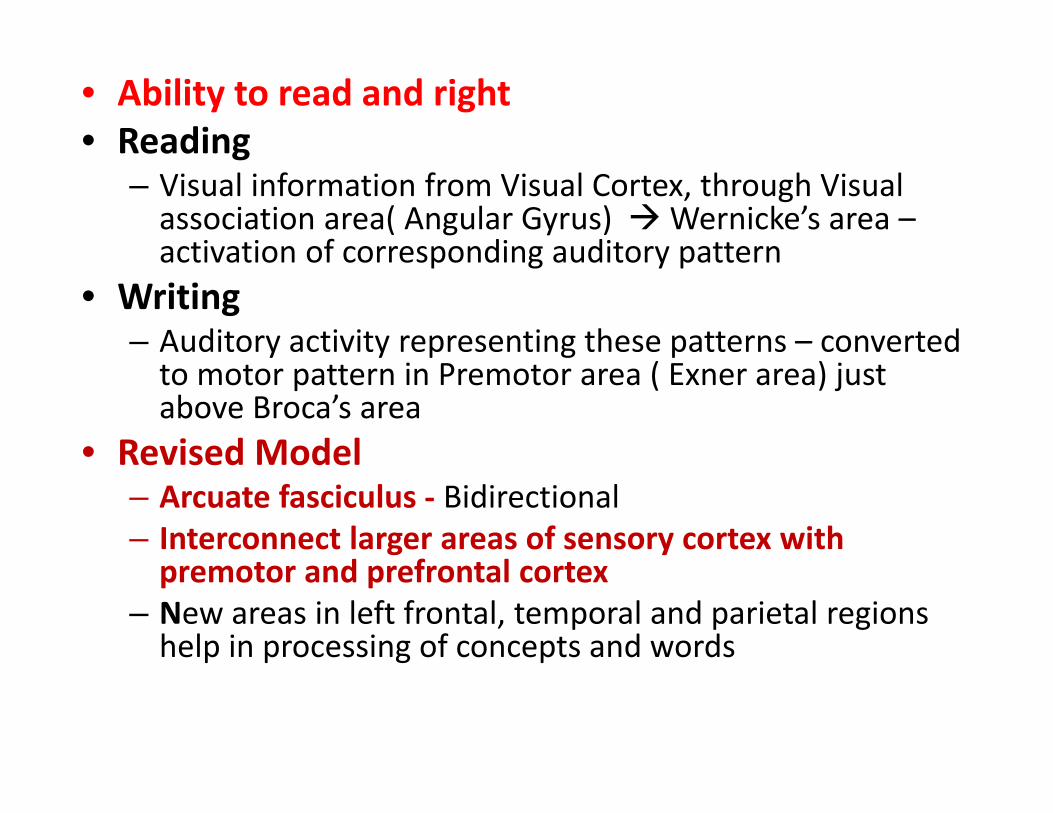
• Ability to read and right • Reading
– Visual information from Visual Cortex, through Visualassociation area( Angular Gyrus) Wernicke’s area – activation of corresponding auditory pattern
• Writing – Auditory activity representing these patterns – convertedto motor pattern in Premotor area ( Exner area) justabove Broca’s area
• Revised Model – Arcuate fasciculus ‐ Bidirectional – Interconnect larger areas of sensory cortex withpremotor and prefrontal cortex
– New areas in left frontal, temporal and parietal regionshelp in processing of concepts and words
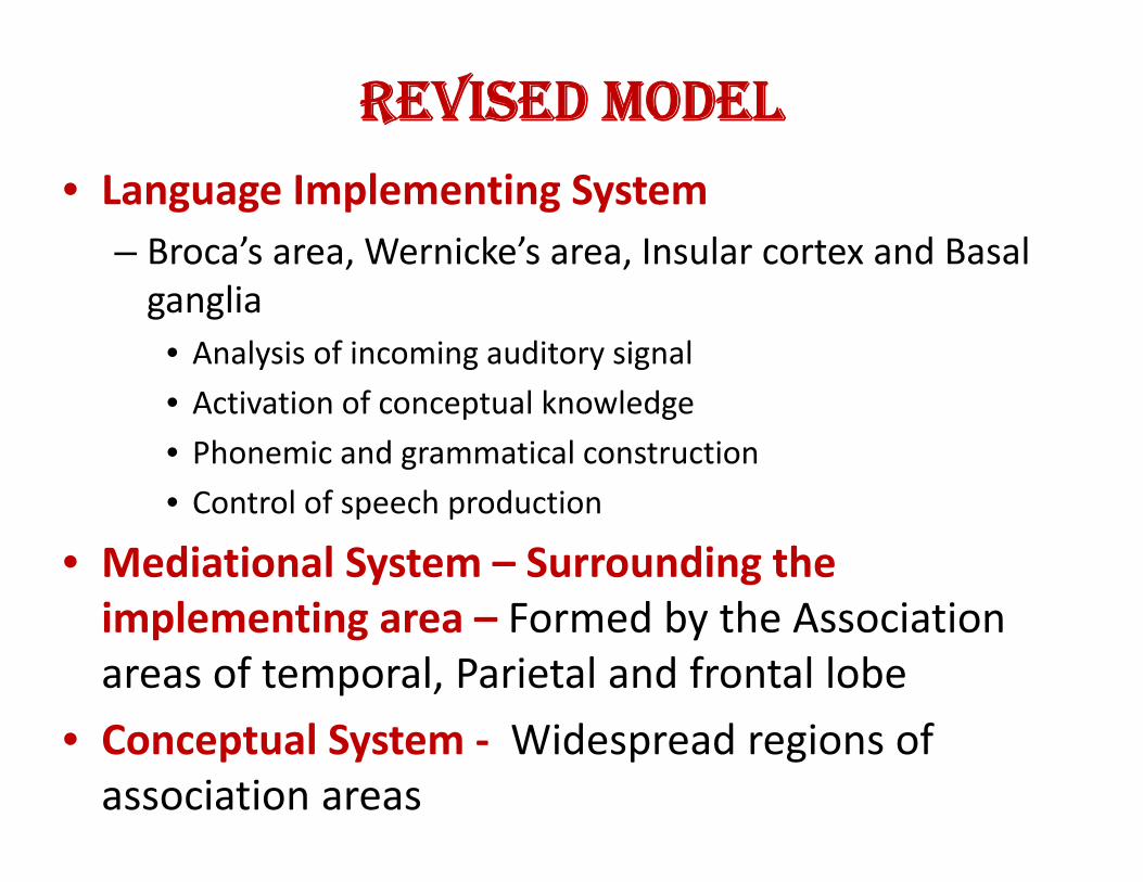
REVISED MODEL
• Language Implementing System – Broca’s area, Wernicke’s area, Insular cortex and Basal ganglia
• Analysis of incoming auditory signal • Activation of conceptual knowledge
• Phonemic and grammatical construction
• Control of speech production
• Mediational System – Surrounding the implementing area – Formed by the Association areas of temporal, Parietal and frontal lobe
• Conceptual System ‐ Widespread regions of association areas
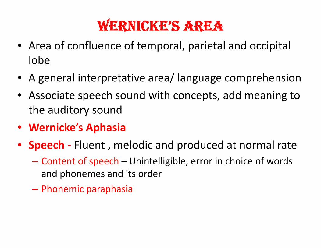
WERNICKE’S AREA • Area of confluence of temporal, parietal and occipital lobe
• A general interpretative area/ language comprehension
• Associate speech sound with concepts, add meaning to the auditory sound
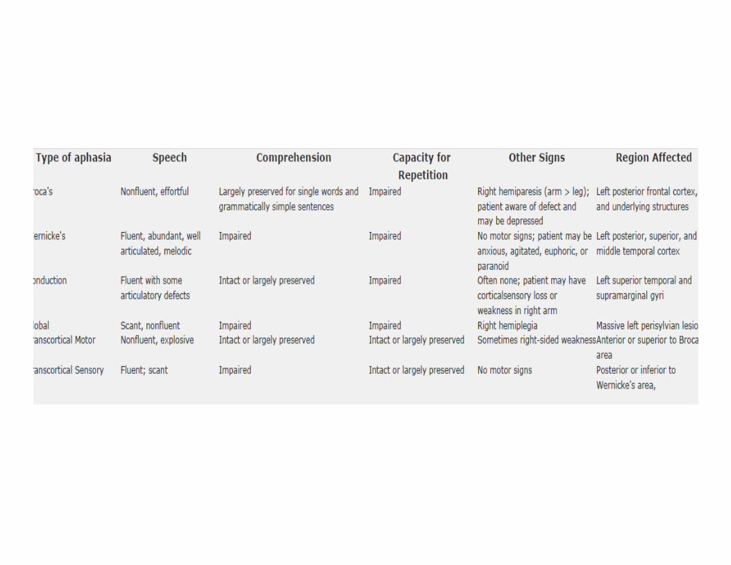
• Wernicke’s Aphasia
• Speech ‐ Fluent , melodic and produced at normal rate – Content of speech – Unintelligible, error in choice of words and phonemes and its order
– Phonemic paraphasia
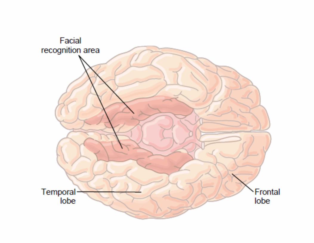

• Prosopagnosia
• Lesion in the inferior temporal lobe
NEUROTRANSMITTERS
• John Newport Langley – 1905
• Thomas Renton Elliot • Otto loewi ‐ 1921 – Acetylcholine from frog’s vagus
• A substance released by a neuron and affects a specific target in a specific manner
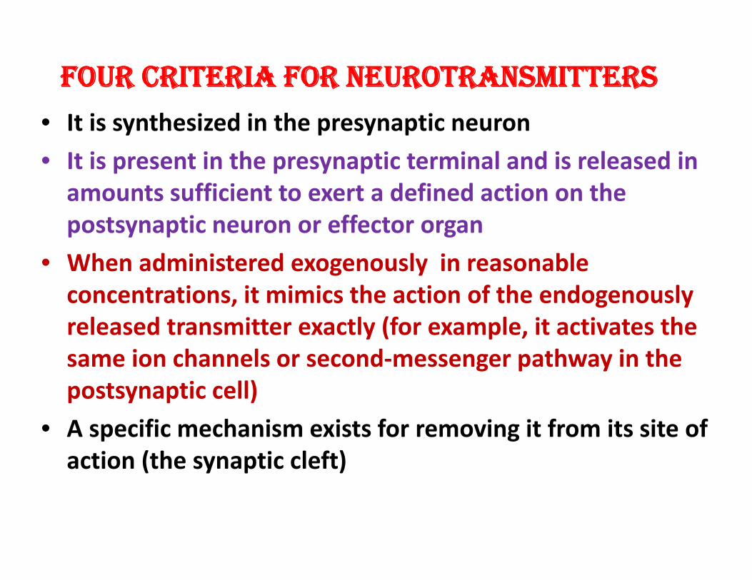
FOUR CRITERIA FOR NEUROTRANSMITTERS
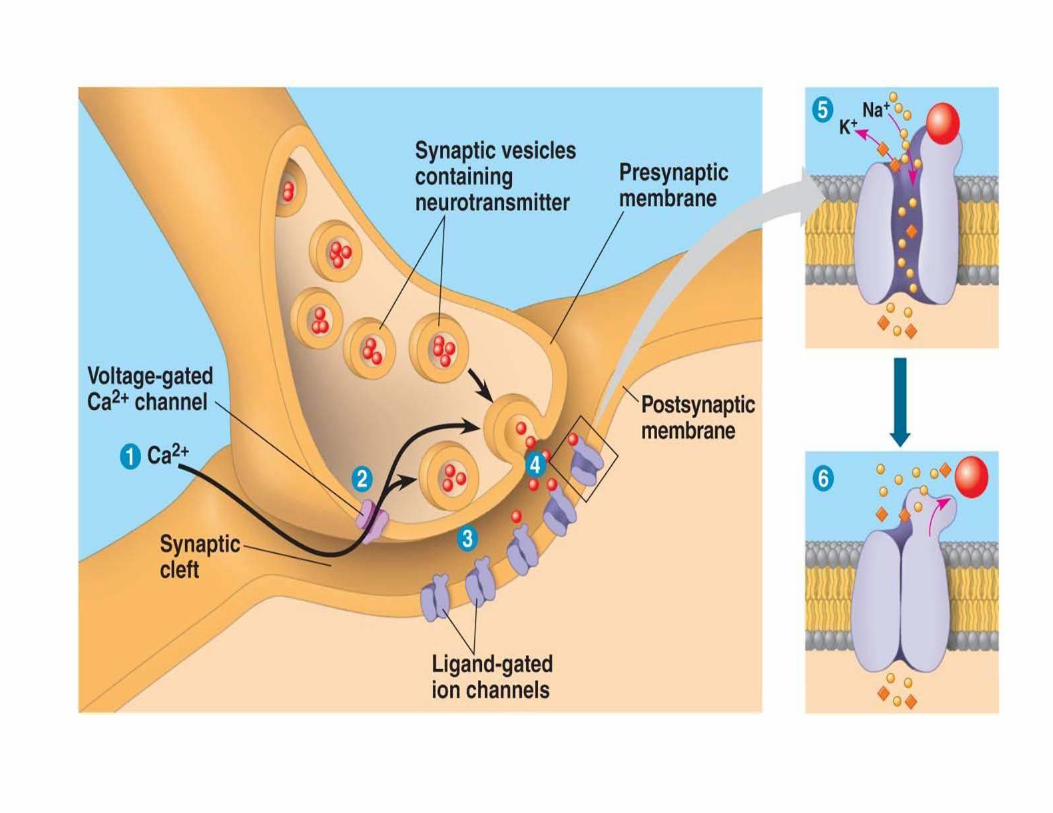
• It is synthesized in the presynaptic neuron
• It is present in the presynaptic terminal and is released in amounts sufficient to exert a defined action on the postsynaptic neuron or effector organ
• When administered exogenously in reasonable concentrations, it mimics the action of the endogenously released transmitter exactly (for example, it activates the same ion channels or second‐messenger pathway in the postsynaptic cell)
• A specific mechanism exists for removing it from its site of action (the synaptic cleft)
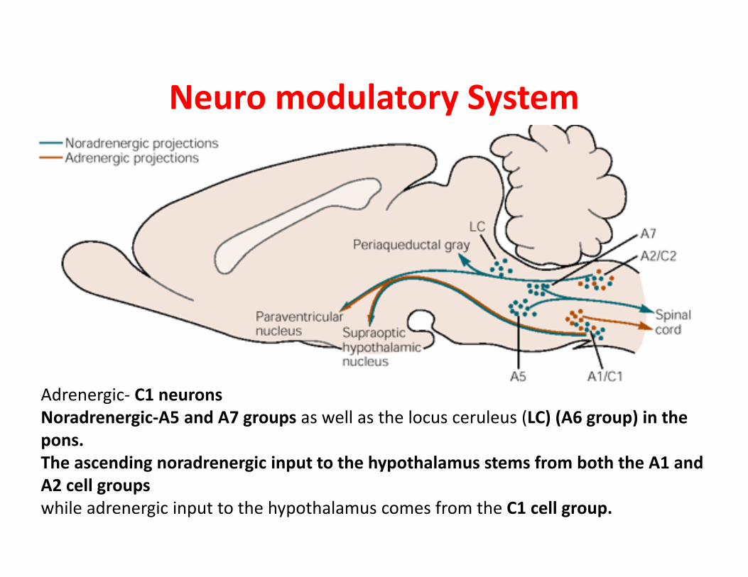
Neuro modulatory System
Adrenergic‐ C1 neurons Noradrenergic‐A5 and A7 groups as well as the locus ceruleus (LC) (A6 group) in the pons. The ascending noradrenergic input to the hypothalamus stems from both the A1 and A2 cell groups while adrenergic input to the hypothalamus comes from the C1 cell group.
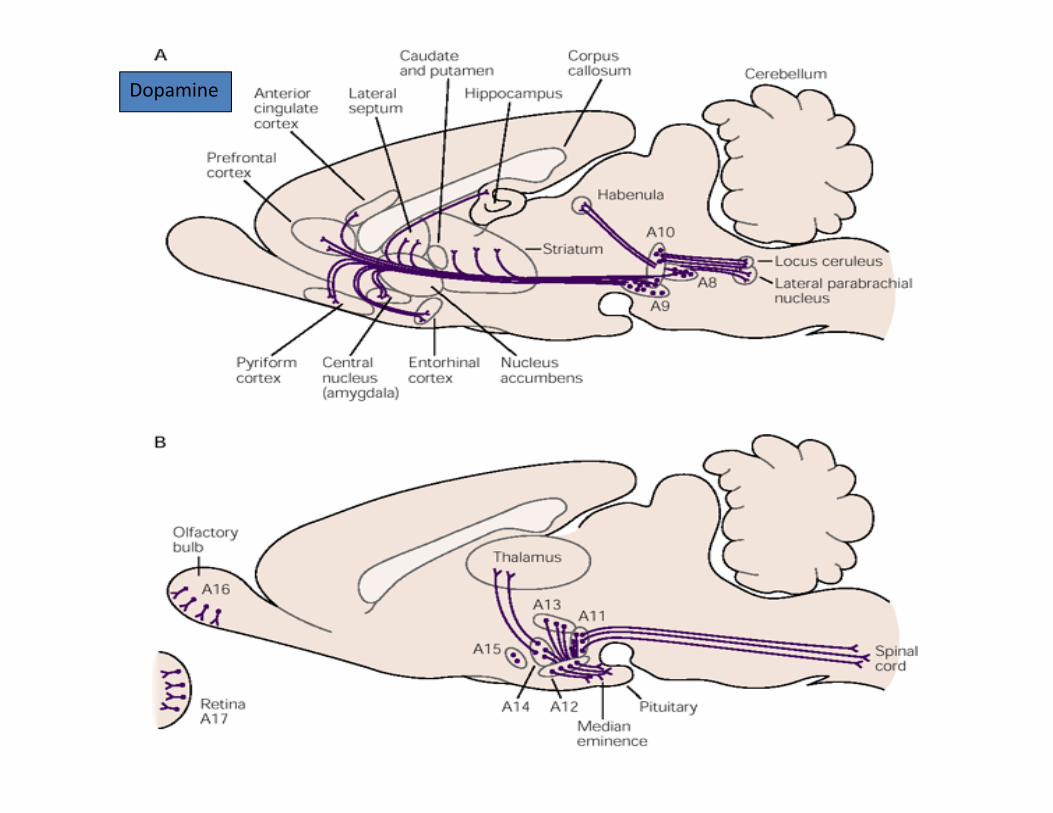
DopDopamine
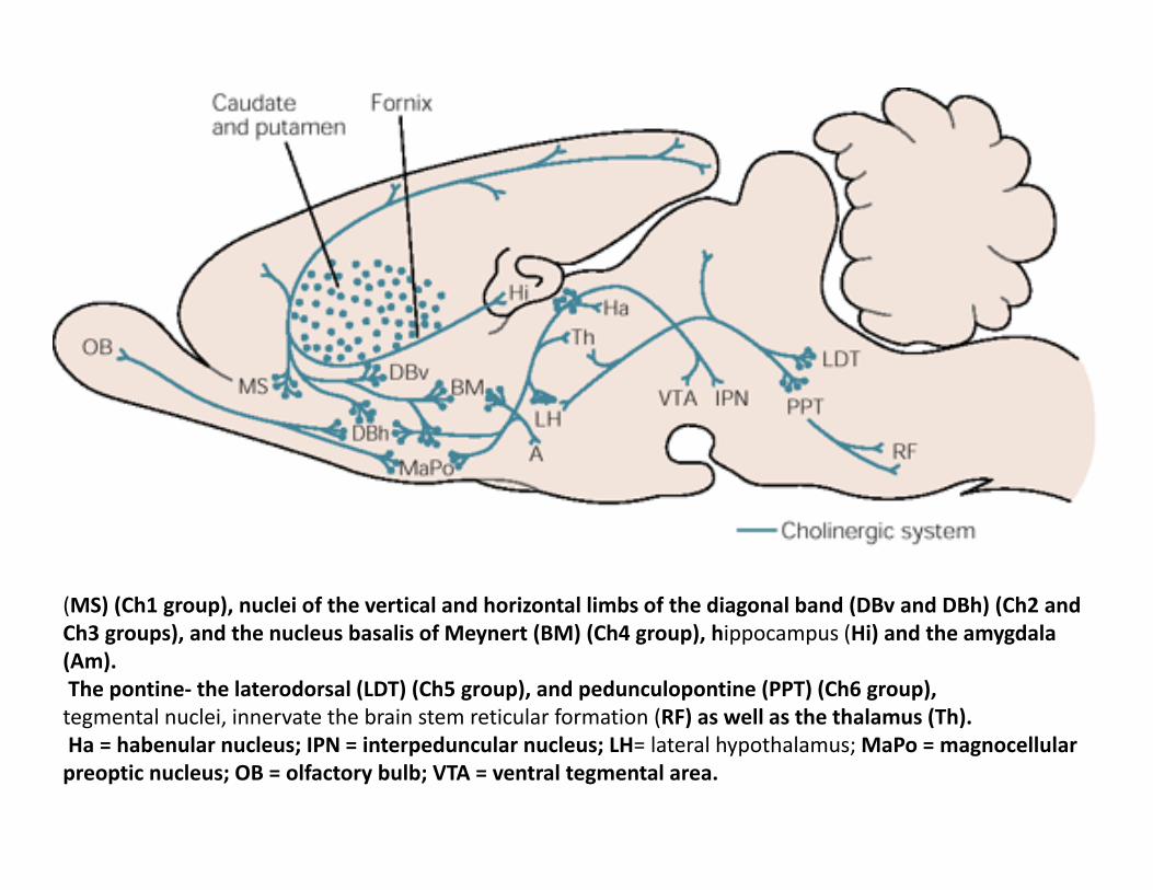
(MS) (Ch1 group), nuclei of the vertical and horizontal limbs of the diagonal band (DBv and DBh) (Ch2 and Ch3 groups), and the nucleus basalis of Meynert (BM) (Ch4 group), hippocampus (Hi) and the amygdala (Am). The pontine‐ the laterodorsal (LDT) (Ch5 group), and pedunculopontine (PPT) (Ch6 group), tegmental nuclei, innervate the brain stem reticular formation (RF) as well as the thalamus (Th). Ha = habenular nucleus; IPN = interpeduncular nucleus; LH= lateral hypothalamus; MaPo = magnocellular preoptic nucleus; OB = olfactory bulb; VTA = ventral tegmental area.
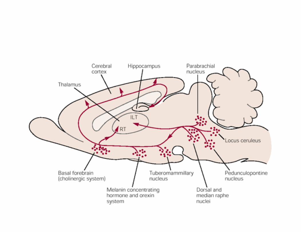
• Ascending monoaminergic and cholinergic projection – Ascending arousal system/ Reticular activating system
– Damage to this system or its projection in the thalamus and hypothalamus coma
• 1. The locus ceruleus – Noradrenergic system
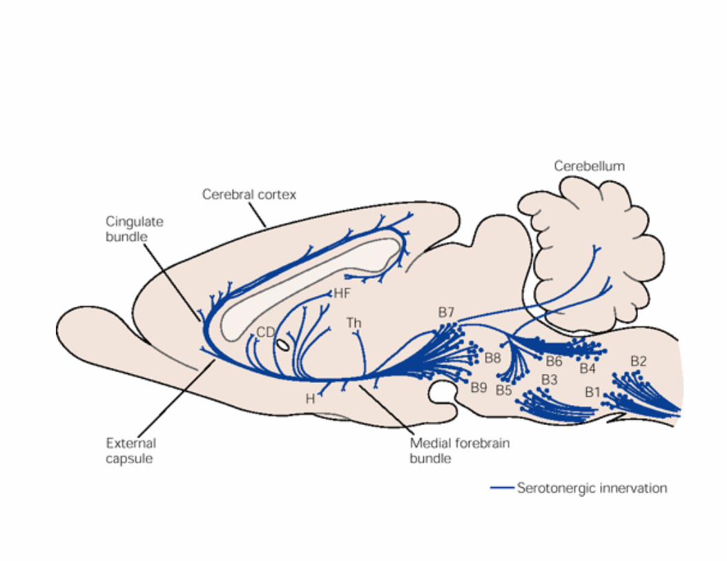
• 2. The dorsal and Median Raphe nuclei‐Primarily serotonergic, few dopaminergic
• 3. The Pedunculopontine and laterodorsal tegmental nuclei‐ cholinergic
• 4. Tuberomamillary nucleus ‐ histaminergic
• The locus ceruleus and tuberomamillary group‐Increase in EEG arousal
• At the junction of midbrain and diencephalon – ascending arousal system ventral and dorsal branches
– Dorsal Thalamus – Ventral Lateral hypothalamus—joined by ascending projections from hypothalamus and basal forebrain distributed throughout the cortex
– Lesion to either of two – Impaired consciousness.
MONOAMINERGIC SYSTEM
• Properties – Spontaneous action potential( tonic ) – Action potential followed by a slow depolarization
– Regulated by intrinsic pacemaker current – Some of the terminals do not form conventional synaptic connections– release neurotransmitters diffusely to many target neurons.
– Most act through metabotropic G protein receptors – Few also corelease neuropeptides
• Noradrenergic, serotonergic and histaminergic neurons –maximally active during waking, firing rate decreases during NREM sleep and completely stops during REM sleep.
• Dopaminergic‐ also active during waking, not distinguishable during REM and NREM sleep
• Monoaminergic neurons activate the cortical arousal by modulating the activity in the hypothalamus, basal forebrain and thalamus which in turn activate cortex.
• Pharmacological agents producing sleep – Antihistamines, serotonin reuptake blockers (reduces REM sleep)
• Patients of parkinsonism also loose noradrenergic neurons in the ascending arousal system Abnormally sleepy during day
Regulation of Cognitive functions
• Locus Ceruleus – attention
• Monoaminergic input to dorsolateral prefrontal cortex ‐‐‐Improve working memory
• D1 agonists improve memory
• Activity of dopaminergic neurons increases during reward based learning
• In trained animals‐when reward follows a conditioned response – Increase in dopaminergic discharge after CS not after reward – Reward predicting error signal—reinforcement learning
• Regulation of autonomic behaviors‐ breathing, vascular tone ( rostral ventrolateral medulla)
• Serotonin‐ GIT peristalsis, thermoregulation, cvs control and breathing
• Medullary raphe nuclei‐ increase in HR & BP • Some medullary serotonergic neuron act as central chemoreceptor‐ sense arterial CO2
• Induce arousal, anxiety and changes in cerebral blood flow in response to increased CO2
• Modulation of pain
• Facilitation of motor activity‐Most critical dopaminergic
• Serotonergic‐ important for generation of motor program
• Serotonin syndrome – hyperactivity, myoclonus tremors and rigidity
• Noradrenergic‐ Pontine fibres facilitate motor neurons in stereotypic rhythmic activity
Cholinergic System • In brain stem – Pedunculopontine and laterodorsal tegmental nuclei‐ send widespread projections to thalamus and reticular nuclei‐mediated by G protein coupled muscarinic receptors
• Decresed firing during NREM sleep
• Increased firing –REM sleep
• Important for arousal and attention

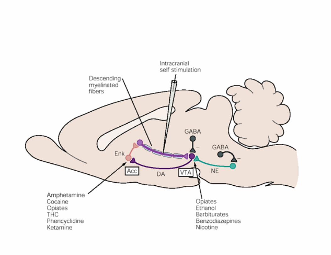
Drug Addiction • Dopaminergic projections from ventral tegmental area to Nucleus accumbens, medial prefrontal cortex and other forebrain structures – central components of reward circuit– receive excitatory signals from cholinergic cells in the laterodorsal tegmental and pedunculopontine nuclei
• Reward centres – Medial forebrain bundle, lateral hypothalamus, Olfactory bulb, Nucleus of the solitary tract, the ventromedial portion of head of caudate nucleus
Related Documents