MOLECULAR AND CELLULAR BIOLOGY, 0270-7306/99/$04.0010 May 1999, p. 3423–3434 Vol. 19, No. 5 Copyright © 1999, American Society for Microbiology. All Rights Reserved. Cellular Activation Triggered by the Autosomal Dominant Polycystic Kidney Disease Gene Product PKD2 THIERRY ARNOULD, 1 LORENZ SELLIN, 1 THOMAS BENZING, 1 LEONIDAS TSIOKAS, 1 HERBERT T. COHEN, 2 EMILY KIM, 3 AND GERD WALZ 1 * Department of Medicine, Renal Division Beth Israel Deaconess Medical Center, Boston, Massachusetts 02215 1 ; Renal Section, Department of Medicine, Boston University Medical Center, Boston, Massachusetts 02118 2 ; and Laboratory of Molecular and Developmental Neuroscience, Massachusetts General Hospital, Harvard Medical School, Boston, Massachusetts 02114 3 Received 13 November 1998/Accepted 19 January 1999 Autosomal dominant polycystic kidney disease (ADPKD) is caused by germ line mutations in at least three ADPKD genes. Two recently isolated ADPKD genes, PKD1 and PKD2, encode integral membrane proteins of unknown function. We found that PKD2 upregulated AP-1-dependent transcription in human embryonic kidney 293T cells. The PKD2-mediated AP-1 activity was dependent upon activation of the mitogen-activated protein kinases p38 and JNK1 and protein kinase C (PKC) «, a calcium-independent PKC isozyme. Stauro- sporine, but not the calcium chelator BAPTA [1,2-bis(o-aminophenoxy)ethane-N, N, N*, N*-tetraacetate], inhib- ited PKD2-mediated signaling, consistent with the involvement of a calcium-independent PKC isozyme. Co- expression of PKD2 with the interacting C terminus of PKD1 dramatically augmented PKD2-mediated AP-1 activation. The synergistic signaling between PKD1 and PKD2 involved the activation of two distinct PKC iso- zymes, PKC a and PKC «, respectively. Our findings are consistent with others that support a functional connection between PKD1 and PKD2 involving multiple signaling pathways that converge to induce AP-1 activ- ity, a transcription factor that regulates different cellular programs such as proliferation, differentiation, and apoptosis. Activation of these signaling cascades may promote the full maturation of developing tubular ep- ithelial cells, while inactivation of these signaling cascades may impair terminal differentiation and facilitate the development of renal tubular cysts. Human autosomal dominant polycystic kidney disease (ADPKD), one of the most prevalent inherited disorders with an incidence of 1 in 500 to 1 in 1,000 individuals, is charac- terized by the development of gradually enlarging renal epithelial cysts that progressively impair renal function (16, 17). The vast majority of patients are affected by mutations in one of three ADPKD genes (7, 27, 43). PKD1 is mutated in more than 85% of ADPKD patients, and it encodes a large integral glycoprotein with multiple transmembrane do- mains and a large extracellular domain with significant ho- mology to membrane proteins involved in cell-cell and/or cell-matrix interactions (1, 2, 20, 21, 45). PKD2 encodes a 968-amino-acid integral membrane protein with six trans- membrane domains, and it is mutated in approximately 10 to 15% of all patients (35). Despite homologies to the family of voltage-gated calcium channel a 1 subunits, the function of PKD2 remains elusive. It has been hypothesized that PKD1 and PKD2 function in a common signaling pathway. Patients with PKD1 and those with PKD2 have a similar clinical phenotype, while both PKD1 2/2 and PKD2 2/2 mice develop kidney and liver cysts resembling the human phenotype (30, 54). Recent studies have shown that PKD1 and PKD2 interact via their C-terminal cytoplasmic domains (41, 52). Thus, PKD1 and PKD2 appear to work in conjunction with each other, but it remains unclear by which mechanism they control tubular proliferation and differentiation and thereby prevent cyst formation. Cystic epi- thelial cells are thought to be incompletely differentiated and persistently proliferative. Several proto-oncogenes, including c-erbB-2, c-Fos, c-Ki-ras, and c-myc, are abnormally regulated in cyst cells derived from ADPKD patients and animal models of cystic renal disease (6, 17, 28, 50, 51). It has been proposed that ADPKD gene products control proto-oncogenes and cel- lular programs directing cell cycle progression and cellular differentiation. Consistent with this hypothesis, we recently demonstrated that the C-terminal cytoplasmic domain of PKD1 stimulates protein kinase C (PKC) a-dependent and c-Jun N- terminal protein kinase (JNK)-dependent activation of AP-1 (1). We now report that PKD2 stimulates the phosphorylation of c-Jun and the induction of AP-1 activity through signaling molecules partially distinct from those involved in PKD1-me- diated activation. Furthermore, a transcriptionally inactive PKD1 truncation greatly enhanced the PKD2-mediated activation of AP-1. MATERIALS AND METHODS Reagents and plasmids. Genistein, staurosporine, wortmannin (Calbiochem, La Jolla, Calif.), and BAPTA-AM [1,2-bis(o-aminophenoxy)ethane-N, N, N9, N9- tetraacetate–acetoxymethyl ester] (Molecular Probe, Eugene, Oreg.) were used at various concentrations. A PKD2 expression vector was constructed in CDM8 by assembling the entire coding region of PKD2 from the clone K1-1 (kindly provided by S. Somlo) and the EST clone yj63h09 (Genome Systems, St. Louis, Mo.). The luciferase constructs, the CD16.7-PKD1 fusion protein, and the Cdc42, Rac1, RhoA, HA-p38, and PKC a constructs were recently described (1, 52). The hemagglutinin (HA)-tagged p42 was kindly provided by J. S. Gutkind, and a dominant-negative form of PKC ε was kindly provided by G. M. Cooper. Dominant-negative mutants of p42 and p44 were kindly provided by M. H. Cobb. Dominant-negative mutants of MKK3 and MKK6 were kindly provided by R. J. Davis. * Corresponding author. Mailing address: Renal Division, Depart- ment of Medicine, Beth Israel Deaconess Medical Center, 330 Brook- line Ave., Boston, MA 02215. Phone: (617) 667-5918. Fax: (617) 667- 1610. E-mail: [email protected]. 3423 on January 21, 2015 by guest http://mcb.asm.org/ Downloaded from

Welcome message from author
This document is posted to help you gain knowledge. Please leave a comment to let me know what you think about it! Share it to your friends and learn new things together.
Transcript
MOLECULAR AND CELLULAR BIOLOGY,0270-7306/99/$04.0010
May 1999, p. 3423–3434 Vol. 19, No. 5
Copyright © 1999, American Society for Microbiology. All Rights Reserved.
Cellular Activation Triggered by the Autosomal DominantPolycystic Kidney Disease Gene Product PKD2
THIERRY ARNOULD,1 LORENZ SELLIN,1 THOMAS BENZING,1 LEONIDAS TSIOKAS,1
HERBERT T. COHEN,2 EMILY KIM,3 AND GERD WALZ1*
Department of Medicine, Renal Division Beth Israel Deaconess Medical Center, Boston, Massachusetts 022151;Renal Section, Department of Medicine, Boston University Medical Center, Boston, Massachusetts 021182;
and Laboratory of Molecular and Developmental Neuroscience, Massachusetts General Hospital,Harvard Medical School, Boston, Massachusetts 021143
Received 13 November 1998/Accepted 19 January 1999
Autosomal dominant polycystic kidney disease (ADPKD) is caused by germ line mutations in at least threeADPKD genes. Two recently isolated ADPKD genes, PKD1 and PKD2, encode integral membrane proteins ofunknown function. We found that PKD2 upregulated AP-1-dependent transcription in human embryonickidney 293T cells. The PKD2-mediated AP-1 activity was dependent upon activation of the mitogen-activatedprotein kinases p38 and JNK1 and protein kinase C (PKC) «, a calcium-independent PKC isozyme. Stauro-sporine, but not the calcium chelator BAPTA [1,2-bis(o-aminophenoxy)ethane-N,N,N*,N*-tetraacetate], inhib-ited PKD2-mediated signaling, consistent with the involvement of a calcium-independent PKC isozyme. Co-expression of PKD2 with the interacting C terminus of PKD1 dramatically augmented PKD2-mediated AP-1activation. The synergistic signaling between PKD1 and PKD2 involved the activation of two distinct PKC iso-zymes, PKC a and PKC «, respectively. Our findings are consistent with others that support a functionalconnection between PKD1 and PKD2 involving multiple signaling pathways that converge to induce AP-1 activ-ity, a transcription factor that regulates different cellular programs such as proliferation, differentiation, andapoptosis. Activation of these signaling cascades may promote the full maturation of developing tubular ep-ithelial cells, while inactivation of these signaling cascades may impair terminal differentiation and facilitatethe development of renal tubular cysts.
Human autosomal dominant polycystic kidney disease(ADPKD), one of the most prevalent inherited disorders withan incidence of 1 in 500 to 1 in 1,000 individuals, is charac-terized by the development of gradually enlarging renalepithelial cysts that progressively impair renal function (16,17). The vast majority of patients are affected by mutationsin one of three ADPKD genes (7, 27, 43). PKD1 is mutatedin more than 85% of ADPKD patients, and it encodes alarge integral glycoprotein with multiple transmembrane do-mains and a large extracellular domain with significant ho-mology to membrane proteins involved in cell-cell and/orcell-matrix interactions (1, 2, 20, 21, 45). PKD2 encodes a968-amino-acid integral membrane protein with six trans-membrane domains, and it is mutated in approximately 10 to15% of all patients (35). Despite homologies to the family ofvoltage-gated calcium channel a1 subunits, the function ofPKD2 remains elusive.
It has been hypothesized that PKD1 and PKD2 function ina common signaling pathway. Patients with PKD1 and thosewith PKD2 have a similar clinical phenotype, while bothPKD12/2 and PKD22/2 mice develop kidney and liver cystsresembling the human phenotype (30, 54). Recent studies haveshown that PKD1 and PKD2 interact via their C-terminalcytoplasmic domains (41, 52). Thus, PKD1 and PKD2 appearto work in conjunction with each other, but it remains unclearby which mechanism they control tubular proliferation and
differentiation and thereby prevent cyst formation. Cystic epi-thelial cells are thought to be incompletely differentiated andpersistently proliferative. Several proto-oncogenes, includingc-erbB-2, c-Fos, c-Ki-ras, and c-myc, are abnormally regulatedin cyst cells derived from ADPKD patients and animal modelsof cystic renal disease (6, 17, 28, 50, 51). It has been proposedthat ADPKD gene products control proto-oncogenes and cel-lular programs directing cell cycle progression and cellulardifferentiation. Consistent with this hypothesis, we recentlydemonstrated that the C-terminal cytoplasmic domain of PKD1stimulates protein kinase C (PKC) a-dependent and c-Jun N-terminal protein kinase (JNK)-dependent activation of AP-1(1). We now report that PKD2 stimulates the phosphorylationof c-Jun and the induction of AP-1 activity through signalingmolecules partially distinct from those involved in PKD1-me-diated activation. Furthermore, a transcriptionally inactive PKD1truncation greatly enhanced the PKD2-mediated activation ofAP-1.
MATERIALS AND METHODS
Reagents and plasmids. Genistein, staurosporine, wortmannin (Calbiochem,La Jolla, Calif.), and BAPTA-AM [1,2-bis(o-aminophenoxy)ethane-N, N, N9, N9-tetraacetate–acetoxymethyl ester] (Molecular Probe, Eugene, Oreg.) were usedat various concentrations. A PKD2 expression vector was constructed in CDM8by assembling the entire coding region of PKD2 from the clone K1-1 (kindlyprovided by S. Somlo) and the EST clone yj63h09 (Genome Systems, St. Louis,Mo.). The luciferase constructs, the CD16.7-PKD1 fusion protein, and theCdc42, Rac1, RhoA, HA-p38, and PKC a constructs were recently described (1,52). The hemagglutinin (HA)-tagged p42 was kindly provided by J. S. Gutkind,and a dominant-negative form of PKC ε was kindly provided by G. M. Cooper.Dominant-negative mutants of p42 and p44 were kindly provided by M. H. Cobb.Dominant-negative mutants of MKK3 and MKK6 were kindly provided by R. J.Davis.
* Corresponding author. Mailing address: Renal Division, Depart-ment of Medicine, Beth Israel Deaconess Medical Center, 330 Brook-line Ave., Boston, MA 02215. Phone: (617) 667-5918. Fax: (617) 667-1610. E-mail: [email protected].
3423
on January 21, 2015 by guesthttp://m
cb.asm.org/
Dow
nloaded from
Luciferase assay. Human embryonic kidney (HEK) 293T cells seeded in 12-well plates were transiently transfected by the calcium phosphate method with aluciferase reporter construct, a b-galactosidase expression vector (kindly pro-vided by C. Cepko), and a PKD2 expression vector. The total DNA amount was1.0 or 1.5 mg/well. Cells were serum starved for 24 h, harvested in cold phos-phate-buffered saline, and lysed in 100 ml of reporter lysis buffer (Promega,Madison, Wis.) for 15 min at room temperature. Lysates were centrifuged at18,000 3 g for 3 min to remove insoluble material. Luciferase activity wasdetermined with a commercial assay system (Promega) following the manufac-turer’s instructions and normalized for b-galactosidase activity to correct for thetransfection efficiency. Pharmacological inhibitors at the indicated concentrationwere added for 6 h before the assay.
Western blot analysis. Cells from one 10-cm dish were lysed 24 h after trans-fection in 1 ml of cold lysis buffer containing 1% Triton X-100, 150 mM NaCl,10 mM Tris-HCl (pH 7.5), 1 mM EDTA, 1 mM EGTA, 1 mM Na3VO4, and aprotease inhibitor cocktail (Boehringer Mannheim, Indianapolis, Ind.), fraction-ated by sodium dodecyl sulfate-polyacrylamide gel electrophoresis (SDS-PAGE),and transferred to a polyvinylidene difluoride membrane. Western blot analysiswas performed with an anti-PKD2 polyclonal antiserum, a polyclonal antibodyagainst Jun family members (Santa Cruz Biotechnology, Santa Cruz, Calif.), orwith a specific anti-phospho-c-Jun (Ser 63) (New England Biolabs, Beverly,Mass.) antibody, followed by incubation with the appropriate secondary antibod-ies. Immobilized antibodies were detected by enhanced chemiluminescence(Pierce, Rockford, Ill.). An MBP-PKD2 fusion protein containing amino acids742 to 871 of PKD2 was utilized to generate a polyclonal rabbit antiserum. ForWestern blot analysis, the antiserum was protein A purified and used at a con-centration of 1:2,000. This antiserum detects a specific band at approximately 110kDa, the predicted size of PKD2. A comparison of HA-tagged PKD2 versionsdetected by the polyclonal rabbit antiserum and by a monoclonal antibodydirected against the HA tag (Boehringer Mannheim) revealed that both versionshave the same protein species.
AP-1 gel shift assay. Electromobility shift assays were performed as previouslydescribed (4). Cells were transfected with plasmids encoding PKD2 and a vectorcontrol (CDM8), or they were stimulated with 100 nM phorbol myristate acetate(PMA) for 60 min. Cells were lysed in 500 ml of hypotonic buffer (HB) (20 mMHEPES [pH 7.9], 5 mM NaF, 1 mM Na2MoO4, 0.1 mM EDTA, 1 mM dithio-threitol [DTT], 0.5 mM phenylmethylsulfonyl fluoride, and a protease inhibitorcocktail) containing 0.2% Nonidet P-40. Nuclei were recovered through centrif-ugation and resuspended in 100 ml of HB containing 20% glycerol. Nuclearproteins were extracted for 30 min at 4°C in 200 ml of HB, 20% glycerol, and 0.8M NaCl. Nuclear debris was removed by centrifugation; supernatants werealiquoted and stored at 280°C. Binding reactions were performed for 20 minwith 5 mg of nuclear proteins in a final volume of 25 ml of binding buffer (2 mMHEPES [pH 7.9], 8 mM NaCl, 0.2 mM EDTA, 12% [vol/vol] glycerol, 5 mMDTT, 0.5 mM phenylmethylsulfonyl fluoride), 1 mg of poly(dI-dC), and 2 3 104
cpm of 32P-labeled oligonucleotide containing the AP-1/tetradecanoyl phorbolacetate-responsive element (TRE) consensus sequence TGACTCA (Promega).For the supershift experiment, a monoclonal antibody that specifically recog-nized c-Jun (or polyclonal antisera that specifically recognized JunB) and JunD(Santa Cruz Biotechnology) were added. The mutant AP-1 oligonucleotide uti-lized in the gel shift assays contained a CA-to-TG substitution in the AP-1-binding motif (Santa Cruz Biotechnology). DNA-protein complexes wereseparated on a native 6% polyacrylamide gel and detected by autoradiogra-phy.
In vitro kinase assays. Immune complex kinase assays were carried out aspreviously described (18). Briefly, 293T cells were cotransfected with HA-taggedmitogen-activated protein kinases (MAPKs) and the PKD2 construct at a 1:1
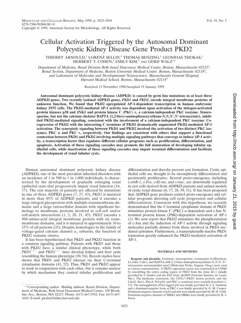
FIG. 1. PKD2 activates AP-1- and c-Jun (TRE)-dependent transcription. (A)PKD2 triggers activation of an AP-1 reporter construct containing four tandemAP-1-binding sites and a c-Jun reporter construct containing three repeats of thesecond TRE of the c-Jun promoter (Jun2TRE). HEK 293T cells were trans-fected with 1 mg of vector (CDM8) (white bars) or PKD2 (black bars), as well asluciferase reporter constructs for AP-1, c-Jun, c-myc, CREB, NF-kB, or c-Fos.Transactivation was determined after 36 h of incubation, and luciferase activitywas expressed after normalization for b-galactosidase activity as fold increaseover the vector control. The results represent the means 6 SD of transfectionsperformed in triplicate. (B) PKD2 activates the AP-1 reporter in a dose-depen-dent fashion. Increasing amounts of PKD2 vector were transfected to trans-activate the AP-1 reporter construct. The total amount of plasmid DNA (2mg/transfection) was balanced with vector (CDM8). (C) PKD2 activates theJun2TRE reporter in a dose-dependent fashion. Increasing amounts of PKD2vector were transfected to transactivate the Jun2TRE reporter. The total amountof plasmid DNA (2 mg/transfection) was balanced with vector (CDM8). (D)PKD2 activates the collagenase promoter. HEK 293T cells were cotransfectedwith 1 mg of PKD2 or vector (CDM8) and a collagenase promoter constructcontaining only a single AP-1-binding site. Cells were stimulated with 10% serumfor 8 h. The results represent the means 6 SD of transfections performed intriplicate. p, P , 0.05; pp, P , 0.01; ppp, P , 0.001.
3424 ARNOULD ET AL. MOL. CELL. BIOL.
on January 21, 2015 by guesthttp://m
cb.asm.org/
Dow
nloaded from
ratio. Cells from one 10-cm dish were lysed 24 h after transfection in 1 ml of coldlysis buffer containing 1% Triton X-100, 150 mM NaCl, 10 mM Tris-HCl (pH7.5), 1 mM EDTA, 1 mM EGTA, 1 mM Na3VO4, and a protease inhibitor cock-tail (Boehringer Mannheim). After centrifugation for 15 min at 4°C, the HA-tagged kinases were immunoprecipitated from the cleared lysate with 1 mg ofanti-HA monoclonal antibody (Boehringer Mannheim) for 2 h at 4°C. Immunecomplexes were immobilized by adding 30 ml of Gamma-Bind Sepharose (Phar-macia, Piscataway, N.J.) and washed three times with lysis buffer and twice withkinase reaction buffer (25 mM HEPES, 20 mM MgCl2, 2 mM DTT, 0.1 mMNa3VO4 [pH 7.6]). The immunoprecipitates were resuspended in 30 ml of kinasereaction buffer containing 3 mg of substrates, PHAS-1 (Stratagene, La Jolla,Calif.) for HA-p38 and HA-p42 or GST-c-Jun(1–79) (Stratagene) for HA-JNK1.The assay was carried out in the presence of 20 mM unlabeled ATP and 10 mCiof [g-32P]ATP for 30 min at 37°C and stopped by the addition of 30 ml of SDSsample buffer. The reaction mixture was fractionated by SDS–12% PAGE. Phos-phorylated substrates were visualized by autoradiography. HA-tagged kinaseimmunoprecipitates were analyzed by SDS-PAGE and Western blot analysis byusing anti-HA polyclonal antiserum (Santa Cruz Biotechnology), a horseradishperoxidase-conjugated goat anti-rabbit immunoglobulin antibody (Amersham,Arlington Heights, Ill.), and an enhanced chemiluminescence kit (Pierce).
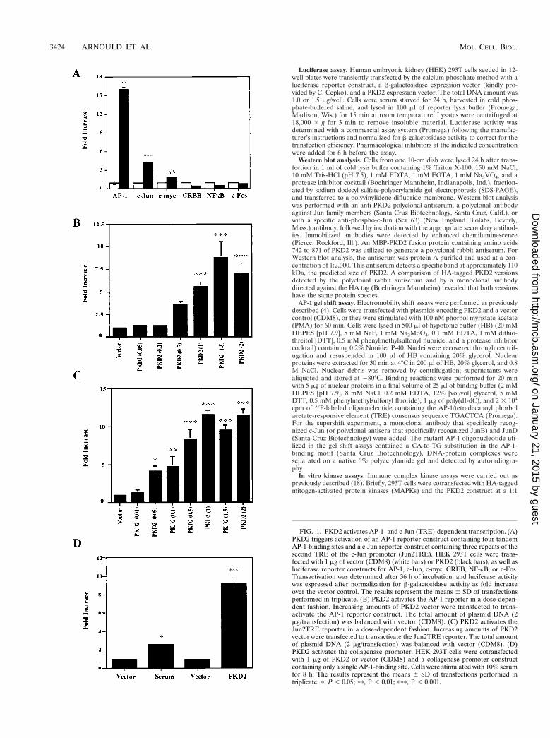
FIG. 2. PKD2 triggers AP-1-binding activity. (A) PKD2 induces binding ofnuclear proteins to the AP-1/TRE DNA-binding site. Nuclear extracts were iso-lated from HEK 293T cells transfected with either vector or PKD2 or from cellsstimulated with 100 nM PMA for 60 min and analyzed by gel retardation assayfor AP-1 binding. No nuclear extract was added to the radiolabeled AP-1 oligo-nucleotide in lane 1 (Free Probe). Addition of a 10- or 100-fold excess of unla-beled probe prevented the formation of a DNA-protein complex, while additionof a 10- or 100-fold excess of mutated AP-1 oligonucleotide had little effect. Asignificant supershift of the DNA-protein complex was observed after the addi-tion of a monoclonal antibody to c-Jun but not an antibody to JunB or JunD ora nonspecific antibody. A mutated AP-1 oligonucleotide failed to bind nuclearproteins isolated from PKD2-transfected HEK 293T cells. The positions of theAP-1 complex (A), supershift (S), and free probe (F) are indicated. (B) Westernblot analysis showing the phosphorylation of c-Jun after cotransfection of a His-tagged c-Jun plasmid with a construct encoding PKD2 or a vector control (CDM8).c-Jun phosphorylated on serine 63 was detected with a specific polyclonal anti-serum. To demonstrate equal amounts of total c-Jun, the membrane was re-probed with an anti-Jun antiserum. FIG. 3. PKD2 activates the JNK1 but not the MAPK p42. (A) HEK 293T
cells were cotransfected with HA-tagged JNK1 and vector control (CDM8),PKD2, or PKD2 in combination with dominant-negative (DN) mutants of thesmall G proteins Cdc42, Rac1, and RhoA at equal ratios. Cells were harvestedafter 24 h, and immunoprecipitated JNK1 was incubated with GST-c-Jun(1–79)in the presence of [g-32P]ATP. Incorporated radioactivity was visualized by SDS–10% PAGE and autoradiography. PKD2 increases JNK1 activity (top panel); thisactivation was inhibited by the DN Cdc42, Rac1, and RhoA. Western blotanalysis revealed equal amounts of precipitated kinases (middle panel); theexpression of PKD2 was not affected by the presence of the DN Cdc42, Rac1,and RhoA (bottom panel). (B) HEK 293T cells were cotransfected with HA-tagged p42 in combination with a vector control (CDM8) or PKD2 at equalratios. Cells were harvested after 24 h, and immunoprecipitated p42 was incu-bated with PHAS-1 in the presence of [g-32P]ATP. Incorporated radioactivitywas visualized by SDS–12% PAGE and autoradiography. Only stimulation with50 nM PMA for 30 min resulted in activation of p42. The lower panel demon-strates equal amounts of p42 kinase in each condition as determined by Westernblot analysis. (C) PKD2-mediated AP-1 activation is unaffected by DN mutantsof the MAPK p42 or p44. HEK 293T cells were transfected with the AP-1reporter construct, vector control, PKD2, or PKD2 in combination with DNmutants of p42 and p44. Transactivation was determined after 36 h of incubation,and luciferase activity was expressed after normalization for b-galactosidaseactivity as fold increase over the vector control. The results represent themeans 6 SD of transfections performed in triplicate.
VOL. 19, 1999 SIGNALING BY PKD2 3425
on January 21, 2015 by guesthttp://m
cb.asm.org/
Dow
nloaded from
Measurement of PKC activity. Total PKC activity was determined as previ-ously described (1), using a colorimetric PKC assay with neurogranin as a dye-labeled synthetic peptide substrate (Pierce). Cells were harvested and lysed onice for 10 min in 45 ml of cold hypotonic buffer (1 mM HEPES, 5 mM MgCl2, 25mg of leupeptin per ml, 25 mg of pepstatin per ml). Isotonicity was reestablishedby adding 5 ml of HEPES (200 mM, pH 7.4) and 25 ml of an equilibrium buffer(20 mM HEPES, 5 mM MgCl2, 1 mM NaF, 0.1 mM Na3VO4). The PKC reactionwas performed at 30°C for 30 min with 10 ml of cleared lysates, following theinstructions of the manufacturer. The absorbance of the phosphorylated sub-strates was spectrophotometrically determined at 570 nm, and a microproteinassay (Bio-Rad, Hercules, Calif.) was used to normalize the PKC activities forthe protein content.
In vitro kinase assay for immunoprecipitated PKC isozymes. To determinethe activity of individual PKC isozymes, in vitro kinase assays were performedessentially as previously described (38). HEK 293T cells were lysed 24 h after
transfection with cold lysis buffer containing 1% Triton X-100, 150 mM NaCl,10 mM Tris-HCl (pH 7.5), 1 mM EDTA, 1 mM EGTA, 1 mM Na3VO4, and aprotease inhibitor cocktail (Boehringer Mannheim). PKC isoforms were isolatedfrom the cleared lysates by using a 1-mg/ml concentration of monoclonal anti-bodies against the different PKC isoforms as indicated (Transduction Laborato-ries, Lexington, Ky.). The immune complexes were immobilized with 30 ml ofGamma-Bind Sepharose (Pharmacia), followed by three washes in lysis bufferand two washes in a kinase reaction buffer containing 50 mM HEPES (pH 7.6),75 mM KCl, 1 mM Na3VO4, 10 mM MgCl2, and 0.1 mM CaCl2. The kinasereaction was performed for 20 min at 30°C in 50 ml of kinase buffer in thepresence of 5 mCi of [g-32P]ATP (10 mCi/mmol), 20 mM unlabeled ATP, and 5mg of neurogranin as an exogenous substrate. Adding 5% acetic acid terminatedthe reactions. The incorporated radioactivity was determined after two washeswith 75 mM phosphoric acid by liquid scintillation spectrophotometry with aphosphocellulose membrane (Pierce). PKC immunoprecipitates were analyzed
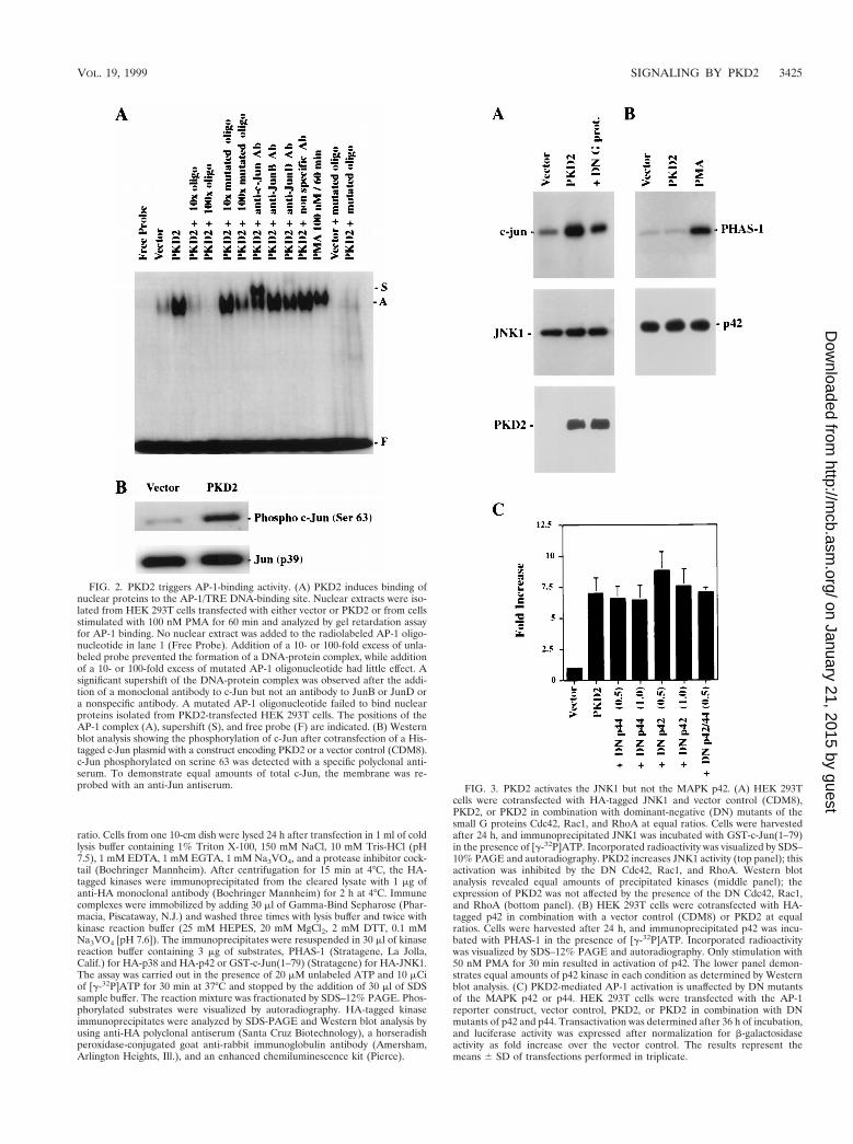
FIG. 4. PKD2 activates p38 MAPK. (A) Dominant-negative (DN) mutants of MKK3 and MKK6 significantly inhibit PKD2-mediated p38 activation. In vitro kinaseassays were performed after transfection of HEK 293T cells with HA-tagged p38 and PKD2 or a vector control (CDM8). Immunoprecipitated kinases were incubatedwith PHAS-1 as an exogenous substrate in the presence of [g-32P]ATP. Incorporated radioactivity was visualized by SDS–12% PAGE and autoradiography. Theamounts of immunoprecipitated p38, the expression of PKD2, and the expression of the DN MKK3 and MKK6 were monitored by Western blot analysis. PKD2activates p38 in an MKK3- and MKK6-dependent fashion; coexpression of C-terminal PKD1 does not contribute to the PKD2-mediated p38 activation. (B) A DNmutant of MKK3 significantly inhibits PKD2-mediated AP-1 activation in a dose-dependent fashion. AP-1 luciferase assays were performed after transfection of HEK293T cells with 1 mg of PKD2 and increasing amounts of a DN mutant of MKK3. The results represent the means 6 SD of transfections performed in triplicate. (C)A DN mutant of MKK6 significantly inhibits PKD2-mediated AP-1 activation in a dose-dependent fashion. AP-1 luciferase assays were performed after transfectionof HEK 293T cells with 1 mg of PKD2 and increasing amounts of a DN mutant of MKK6. The results represent the means 6 SD of transfections performed in triplicate.(D) Wild-type (WT) MKK3 and MKK6 enhance PKD2-mediated AP-1 activation. AP-1 luciferase assays were performed after transfection of HEK 293T cells with1 mg of PKD2 in combination with WT MKK3 or MKK6. The results represent the means 6 SD of transfections performed in triplicate. ppp, significantly differentfrom control vector (P , 0.001); '''', significantly different from PKD2-transfected cells (P , 0.001).
3426 ARNOULD ET AL. MOL. CELL. BIOL.
on January 21, 2015 by guesthttp://m
cb.asm.org/
Dow
nloaded from
by SDS-PAGE and Western blot analysis by using specific anti-PKC monoclonaland polyclonal antibodies (anti-PKC d, l/t, and ε from Transduction Laborato-ries; anti-PKC z from Upstate Biotechnology; and anti-PKC m from Santa CruzBiotechnology) in combination with a horseradish peroxidase-conjugated goatanti-mouse immunoglobulin antibody (Dako, Carpinteria, Calif.) and enhancedchemiluminescence (Pierce).
Statistical analysis. Results were expressed as means 6 standard deviations(SD). Analysis of variance with subsequent Scheffe’s test was used to determinesignificant difference in multiple comparisons. Values of P less than 0.05 wereconsidered to be significant.
RESULTS
PKD2 induces AP-1 activity and triggers phosphorylation ofc-Jun. To identify potential signaling pathways of PKD2, wetransiently coexpressed PKD2 in HEK 293T cells with the lu-ciferase reporter constructs indicated in Fig. 1. PKD2 specifi-cally induced AP-1 activity, activating AP-1 and the Jun2TREreporter constructs (Fig. 1A). PKD2 transactivated both pro-moter constructs in a dose-dependent fashion (Fig. 1B and C).However, PKD2 had no effect on CREB, NF-kB, or c-Fos lu-ciferase reporter constructs, while a weak but not significantactivation was detectable for the c-myc promoter (,2-fold in-crease) (Fig. 1A). To demonstrate the physiological signif-icance of the PKD2-mediated AP-1 activation, we tested theeffect of PKD2 on a collagenase promoter construct contain-ing only one AP-1 binding site. While serum induced a two- tothreefold activation of this promoter, PKD2 induced a ninefoldactivation of the single AP-1-binding site (Fig. 1D). PKD2protein levels were unaffected by the coexpression of differentluciferase constructs; thus, variations in PKD2 levels could notaccount for the differences in transcriptional activities (datanot shown).
Since PKD2 activated the Jun2TRE but not the c-Fos pro-moter, the PKD2-induced AP-1 activity appeared to involvemainly Jun family members. Electromobility shift assays witha double-stranded AP-1/TRE oligonucleotide confirmed thatnuclear proteins from HEK 293T cells transfected with PKD2bind to this motif (Fig. 2A); a similar activity was observedafter stimulation with PMA, a well-known AP-1 activator (15,19). Preincubation of nuclear extract with specific antisera in-duced a significant supershift of the AP-1 band for c-Jun butnot for JunB or JunD. The specificity of the AP-1/TRE DNA-binding protein was confirmed by the addition of increasingamounts of unlabeled AP-1 oligonucleotide that almost com-pletely inhibited the formation of the radiolabeled DNA-pro-tein complex at a 10-fold excess of unlabeled oligonucleotide.Conversely, the mutant AP-1 oligonucleotide failed to form aDNA-protein complex and had little effect on the AP-1 bindingto the authentic AP-1 oligonucleotide. PKD2-dependent phos-phorylation of c-Jun was demonstrated by using a specific an-tiserum against phosphorylated serine 63. As shown in Fig. 2B,PKD2, but not the control, induced c-Jun protein phosphory-lation. Collectively, these data demonstrate that PKD2 stimu-lates the phosphorylation of c-Jun and the formation of tran-scriptionally active AP-1.
PKD2 activates JNK1 and p38 but not p42. Several kinasecascades have been demonstrated to regulate AP-1 activity(31), including the Hog1p homologue p38, the MAPKs p42and p44, and members of the JNK family (reviewed in refer-ence 47). To further delineate the signaling pathway throughwhich PKD2 triggers AP-1 activation, we examined the activityof these kinases in HEK 293T cells expressing PKD2. HA-tagged p38 and p42 and JNK1 were coexpressed with PKD2 orthe vector control CDM8. After serum starvation for 16 h, HA-tagged kinases were immunoprecipitated, and the activity ofeach kinase was assessed by in vitro kinase assays. PKD2 acti-vated JNK1 but not p42 (Fig. 3A and B). In addition, domi-
nant-negative mutants of p42 and p44 had no effect on PKD2-mediated AP-1 activation (Fig. 3C), providing further evidencethat PKD2-mediated AP-1 activation does not involve p42 orp44 MAPKs. In contrast, PKD2 induced MKK3- and MKK6-dependent activation of p38 (Fig. 4A). MKK3 and MKK6 are
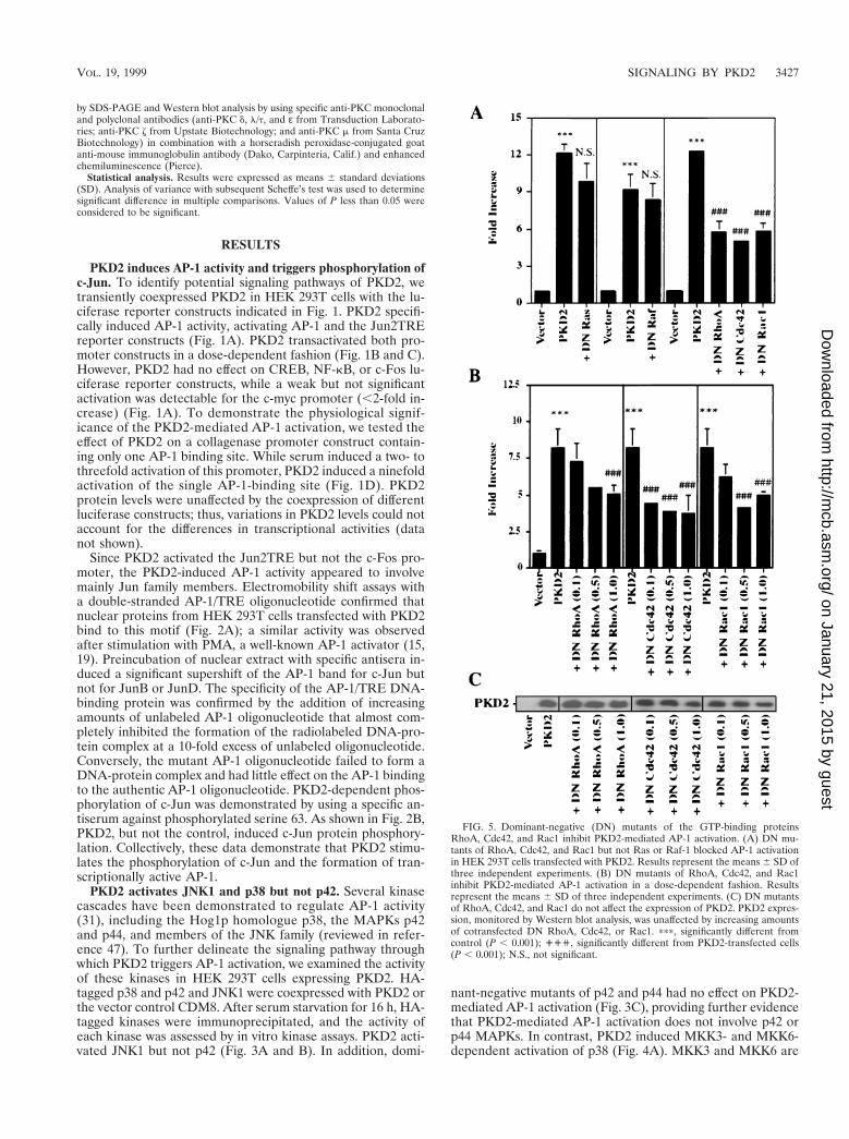
FIG. 5. Dominant-negative (DN) mutants of the GTP-binding proteinsRhoA, Cdc42, and Rac1 inhibit PKD2-mediated AP-1 activation. (A) DN mu-tants of RhoA, Cdc42, and Rac1 but not Ras or Raf-1 blocked AP-1 activationin HEK 293T cells transfected with PKD2. Results represent the means 6 SD ofthree independent experiments. (B) DN mutants of RhoA, Cdc42, and Rac1inhibit PKD2-mediated AP-1 activation in a dose-dependent fashion. Resultsrepresent the means 6 SD of three independent experiments. (C) DN mutantsof RhoA, Cdc42, and Rac1 do not affect the expression of PKD2. PKD2 expres-sion, monitored by Western blot analysis, was unaffected by increasing amountsof cotransfected DN RhoA, Cdc42, or Rac1. ppp, significantly different fromcontrol (P , 0.001); ''', significantly different from PKD2-transfected cells(P , 0.001); N.S., not significant.
VOL. 19, 1999 SIGNALING BY PKD2 3427
on January 21, 2015 by guesthttp://m
cb.asm.org/
Dow
nloaded from
two MAPKs that are reported to selectively phosphorylate andactivate p38 (10, 22, 23). Dominant-negative mutants of MKK3and MKK6 significantly blocked the PKD2-mediated p38activation as determined by in vitro kinase assays. To dem-onstrate the critical involvement of p38 in PKD2 signaling,we tested the effects of dominant-negative versions of MKK3and MKK6 on PKD2-mediated AP-1 activation. As shown inFig. 4B and C, coexpression of dominant-negative MKK3 orMKK6 resulted in a dose-dependent inhibition of PKD2-mediated AP-1 activation. Conversely, wild-type MKK3 andMKK6 augmented the PKD2-mediated AP-1 activation (Fig.4D).
The small GTPases Rac and Cdc42 are important mediatorsof JNK and p38 activation (5, 33, 36). To determine the role ofsmall GTP-binding proteins in the PKD2-mediated activationof AP-1, we coexpressed PKD2 with dominant-negative mu-tants of RhoA (N19), Cdc42 (N17), and Rac1 (N17). Dom-inant-negative RhoA, Cdc42, and Rac1 abrogated the PKD2-mediated JNK activation (Fig. 3A) and the PKD2-mediatedAP-1 activation (Fig. 5A). The dominant-negative mutants in-
hibited the PKD2-mediated AP-1 activation in a dose-depen-dent fashion that was not related to a change in PKD2 expres-sion (Fig. 5B and C). In contrast, dominant-negative forms ofp21-Ras and Raf-1 had no effect on AP-1 activation (Fig. 5A).Thus, small GTP-binding proteins of the Rho family appearto play a central role in the signaling pathway triggered byPKD2.
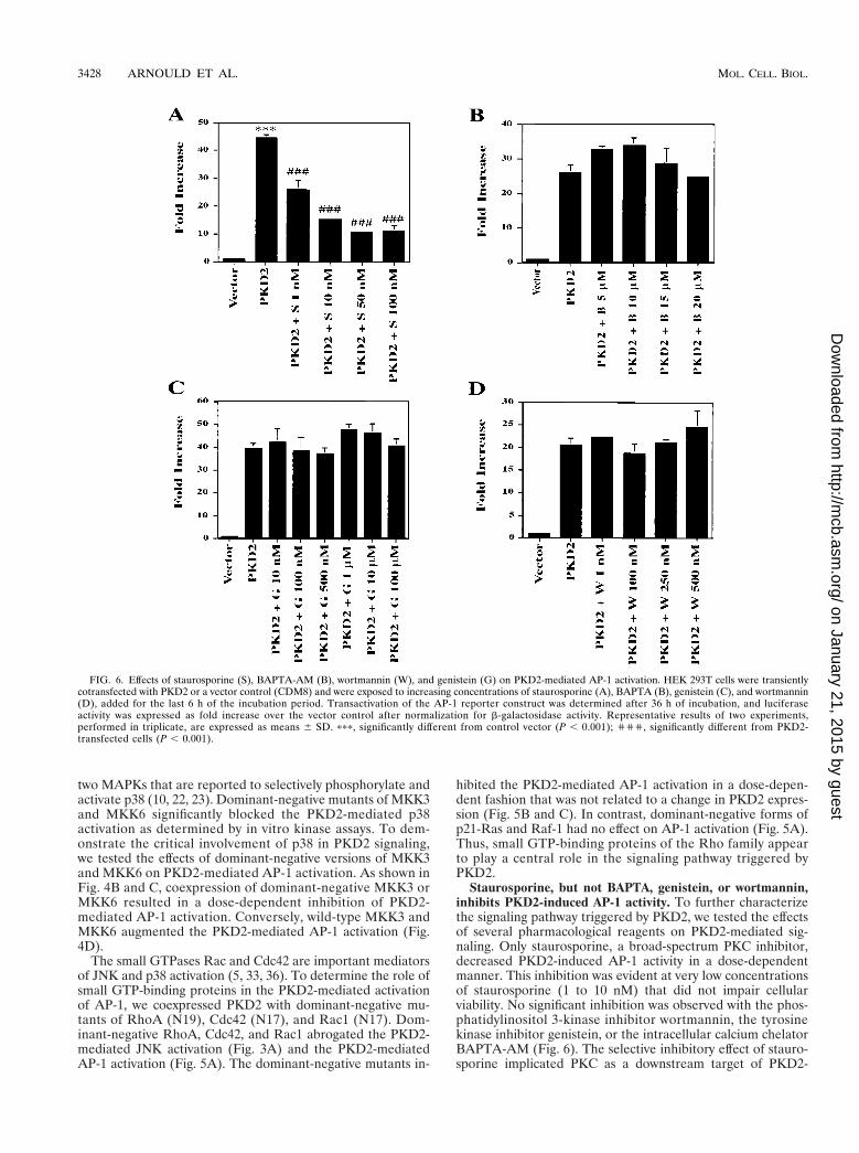
Staurosporine, but not BAPTA, genistein, or wortmannin,inhibits PKD2-induced AP-1 activity. To further characterizethe signaling pathway triggered by PKD2, we tested the effectsof several pharmacological reagents on PKD2-mediated sig-naling. Only staurosporine, a broad-spectrum PKC inhibitor,decreased PKD2-induced AP-1 activity in a dose-dependentmanner. This inhibition was evident at very low concentrationsof staurosporine (1 to 10 nM) that did not impair cellularviability. No significant inhibition was observed with the phos-phatidylinositol 3-kinase inhibitor wortmannin, the tyrosinekinase inhibitor genistein, or the intracellular calcium chelatorBAPTA-AM (Fig. 6). The selective inhibitory effect of stauro-sporine implicated PKC as a downstream target of PKD2-
FIG. 6. Effects of staurosporine (S), BAPTA-AM (B), wortmannin (W), and genistein (G) on PKD2-mediated AP-1 activation. HEK 293T cells were transientlycotransfected with PKD2 or a vector control (CDM8) and were exposed to increasing concentrations of staurosporine (A), BAPTA (B), genistein (C), and wortmannin(D), added for the last 6 h of the incubation period. Transactivation of the AP-1 reporter construct was determined after 36 h of incubation, and luciferaseactivity was expressed as fold increase over the vector control after normalization for b-galactosidase activity. Representative results of two experiments,performed in triplicate, are expressed as means 6 SD. ppp, significantly different from control vector (P , 0.001); ''', significantly different from PKD2-transfected cells (P , 0.001).
3428 ARNOULD ET AL. MOL. CELL. BIOL.
on January 21, 2015 by guesthttp://m
cb.asm.org/
Dow
nloaded from
mediated signaling, while the failure of BAPTA-AM to interferewith PKD2 signaling suggested that a calcium-independentPKC isozyme was involved in the AP-1 activity induced byPKD2.
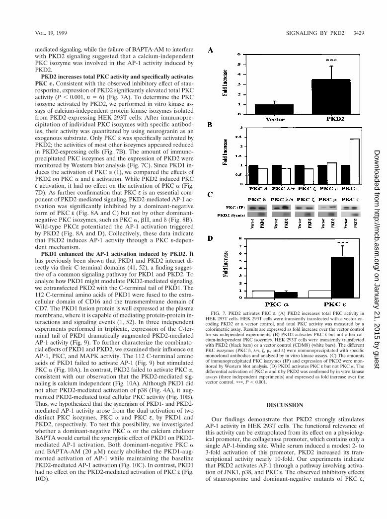
PKD2 increases total PKC activity and specifically activatesPKC «. Consistent with the observed inhibitory effect of stau-rosporine, expression of PKD2 significantly elevated total PKCactivity (P , 0.001, n 5 6) (Fig. 7A). To determine the PKCisozyme activated by PKD2, we performed in vitro kinase as-says of calcium-independent protein kinase isozymes isolatedfrom PKD2-expressing HEK 293T cells. After immunopre-cipitation of individual PKC isozymes with specific antibod-ies, their activity was quantitated by using neurogranin as anexogenous substrate. Only PKC ε was specifically activated byPKD2; the activities of most other isozymes appeared reducedin PKD2-expressing cells (Fig. 7B). The amount of immuno-precipitated PKC isozymes and the expression of PKD2 weremonitored by Western blot analysis (Fig. 7C). Since PKD1 in-duces the activation of PKC a (1), we compared the effects ofPKD2 on PKC a and ε activation. While PKD2 induced PKCε activation, it had no effect on the activation of PKC a (Fig.7D). As further confirmation that PKC ε is an essential com-ponent of PKD2-mediated signaling, PKD2-mediated AP-1 ac-tivation was significantly inhibited by a dominant-negativeform of PKC ε (Fig. 8A and C) but not by other dominant-negative PKC isozymes, such as PKC a, bII, and d (Fig. 8B).Wild-type PKCε potentiated the AP-1 activation triggeredby PKD2 (Fig. 8A and D). Collectively, these data indicatethat PKD2 induces AP-1 activity through a PKC ε-depen-dent mechanism.
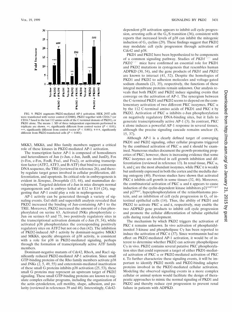
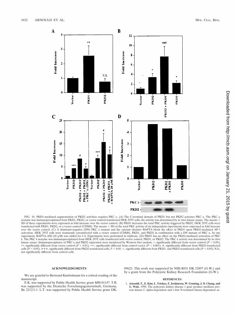
PKD1 enhanced the AP-1 activation induced by PKD2. Ithas previously been shown that PKD1 and PKD2 interact di-rectly via their C-terminal domains (41, 52), a finding sugges-tive of a common signaling pathway for PKD1 and PKD2. Toanalyze how PKD1 might modulate PKD2-mediated signaling,we cotransfected PKD2 with the C-terminal tail of PKD1. The112 C-terminal amino acids of PKD1 were fused to the extra-cellular domain of CD16 and the transmembrane domain ofCD7. The PKD1 fusion protein is well expressed at the plasmamembrane, where it is capable of mediating protein-protein in-teractions and signaling events (1, 52). In three independentexperiments performed in triplicate, expression of the C-ter-minal tail of PKD1 dramatically augmented PKD2-mediatedAP-1 activity (Fig. 9). To further characterize the combinato-rial effects of PKD1 and PKD2, we examined their influence onAP-1, PKC, and MAPK activity. The 112 C-terminal aminoacids of PKD1 failed to activate AP-1 (Fig. 9) but stimulatedPKC a (Fig. 10A). In contrast, PKD2 failed to activate PKC a,consistent with our observation that the PKD2-mediated sig-naling is calcium independent (Fig. 10A). Although PKD1 didnot alter PKD2-mediated activation of p38 (Fig. 4A), it aug-mented PKD2-mediated total cellular PKC activity (Fig. 10B).Thus, we hypothesized that the synergism of PKD1- and PKD2-mediated AP-1 activity arose from the dual activation of twodistinct PKC isozymes, PKC a and PKC ε, by PKD1 andPKD2, respectively. To test this possibility, we investigatedwhether a dominant-negative PKC a or the calcium chelatorBAPTA would curtail the synergistic effect of PKD1 on PKD2-mediated AP-1 activation. Both dominant-negative PKC aand BAPTA-AM (20 mM) nearly abolished the PKD1-aug-mented activation of AP-1 while maintaining the baselinePKD2-mediated AP-1 activation (Fig. 10C). In contrast, PKD1had no effect on the PKD2-mediated activation of PKC ε (Fig.10D).
DISCUSSION
Our findings demonstrate that PKD2 strongly stimulatesAP-1 activity in HEK 293T cells. The functional relevance ofthis activity can be extrapolated from its effect on a physiolog-ical promoter, the collagenase promoter, which contains only asingle AP-1-binding site. While serum induced a modest 2- to3-fold activation of this promoter, PKD2 increased its tran-scriptional activity nearly 10-fold. Our experiments indicatethat PKD2 activates AP-1 through a pathway involving activa-tion of JNK1, p38, and PKC ε. The observed inhibitory effectsof staurosporine and dominant-negative mutants of PKC ε,
FIG. 7. PKD2 activates PKC ε. (A) PKD2 increases total PKC activity inHEK 293T cells. HEK 293T cells were transiently transfected with a vector en-coding PKD2 or a vector control, and total PKC activity was measured by acolorimetric assay. Results are expressed as fold increase over the vector controlfor six independent experiments. (B) PKD2 activates PKC ε but not other cal-cium-independent PKC isozymes. HEK 293T cells were transiently transfectedwith PKD2 (black bars) or a vector control (CDM8) (white bars). The differentPKC isozymes (PKC d, l/t, z, m, and ε) were immunoprecipitated with specificmonoclonal antibodies and analyzed by in vitro kinase assays. (C) The amountsof immunoprecipitated PKC isozymes (IP) and expression of PKD2 were mon-itored by Western blot analysis. (D) PKD2 activates PKC ε but not PKC a. Thedifferential activation of PKC a and ε by PKD2 was confirmed by in vitro kinaseassays (three independent experiments) and expressed as fold increase over thevector control. ppp, P , 0.001.
VOL. 19, 1999 SIGNALING BY PKD2 3429
on January 21, 2015 by guesthttp://m
cb.asm.org/
Dow
nloaded from
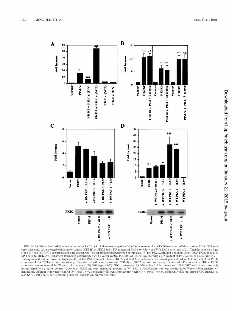
FIG. 8. PKD2-mediated AP-1 activation requires PKC ε. (A) A dominant-negative (DN) PKC ε mutant blocks PKD2-mediated AP-1 activation. HEK 293T cellswere transiently cotransfected with a vector control (CDM8) or PKD2 and a DN mutant of PKC ε or wild-type (WT) PKC ε at a ratio of 2:1. Transfections with 1 mgof the WT and DN PKC ε constructs alone are also shown. The experiment was performed in triplicate. (B) DN PKC a, bII, and d mutants do not affect PKD2-mediatedAP-1 activity. HEK 293T cells were transiently cotransfected with a vector control (CDM8) or PKD2, together with a DN mutant of PKC a, bII, or d at a ratio of 2:1.The experiment was performed in triplicate. (C) A DN PKC ε mutant inhibits PKD2-mediated AP-1 activation in a dose-dependent fashion but does not affect PKD2expression. HEK 293T cells were transiently cotransfected with a vector control (CDM8) or PKD2 and with increasing amounts of a DN mutant of PKC ε. PKD2expression was monitored by Western blot analysis. (D) Wild-type (WT) PKC ε augments PKD2-mediated AP-1 activation. HEK 293T cells were transientlycotransfected with a vector control (CDM8) or PKD2 and with increasing amounts of WT PKC ε. PKD2 expression was monitored by Western blot analysis. pp,significantly different from vector control (P , 0.01); ppp, significantly different from control vector (P , 0.001); ''', significantly different from PKD2-transfectedcells (P , 0.001); N.S., not significantly different from PKD2-transfected cells.
3430 ARNOULD ET AL. MOL. CELL. BIOL.
on January 21, 2015 by guesthttp://m
cb.asm.org/
Dow
nloaded from
MKK3, MKK6, and Rho family members support a criticalrole of these kinases in PKD2-mediated AP-1 activation.
The transcription factor AP-1 is composed of homodimersand heterodimers of Jun (v-Jun, c-Jun, JunB, and JunD), Fos(v-Fos, c-Fos, FosB, Fra1, and Fra2), or activating transcrip-tion factor (ATF2, ATF3, and B-ATF) that bind to a consensusDNA sequence, the TRE (reviewed in reference 26), and there-by regulate target genes involved in cellular proliferation, dif-ferentiation, and apoptosis. Its critical role in embryogenesis isevident in Xenopus, Drosophila (13, 44), and mammalian de-velopment. Targeted deletion of c-Jun in mice disrupts normalorganogenesis and is embryo lethal at E12 to E14 (24), sug-gesting that AP-1 could play a role in nephrogenesis.
AP-1 activity can be generated through a variety of sig-naling events. Gel shift and supershift analysis revealed thatPKD2 increased the binding of Jun-containing AP-1 to theTRE. Moreover, PKD2 increased the amount of c-Jun phos-phorylated on serine 63. Activated JNKs phosphorylate c-Jun on serines 63 and 73, two positively regulatory sites inthe transcriptional activation domain of c-Jun (9, 34), whileactivated p38 phosphorylates the corresponding positivelyregulatory sites on ATF2 but not on c-Jun (42). The inhibitionof PKD2-induced AP-1 activity by dominant-negative MKK3and MKK6, specific abrogators of p38 activity, is consistentwith a role for p38 in PKD2-mediated signaling, perhapsthrough the formation of transcriptionally active ATF familymembers.
Dominant-negative mutants of Cdc42, RhoA, and Rac1 sig-nificantly reduced PKD2-mediated AP-1 activation. Since smallGTP-binding proteins of the Rho family members activate p38and JNKs (2, 5, 49, 55) and enterotoxin-mediated inactivationof these small G proteins inhibits p38 activation (25, 53), thesesmall G proteins may represent an upstream target of PKD2signaling. These small GTP-binding proteins are known to reg-ulate complex cellular programs, including the organization ofthe actin cytoskeleton, cell motility, shape, adhesion, and po-larity (reviewed in references 39 and 48). Interestingly, Cdc42-
dependent p38 activation appears to inhibit cell cycle progres-sion, arresting cells at the G1/S transition (36), consistent withreports that increased levels of p38 can inhibit the mitogenicinduction of G1 cyclins (29). These findings suggest that PKD2may modulate cell cycle progression through activation ofCdc42 and p38.
PKD1 and PKD2 have been hypothesized to be componentsof a common signaling pathway. Studies of PKD12/2 andPKD22/2 mice have confirmed an essential role for PKD1and PKD2 mutations in cystogenesis that resembles humanADPKD (30, 54), and the gene products of PKD1 and PKD2are known to interact (41, 52). Despite the homologies ofPKD1 and PKD2 to adhesion molecules and voltage-gatedsodium channels (21, 35), respectively, the functions of theseintegral membrane proteins remain unknown. Our analysis re-veals that both PKD1 and PKD2 induce signaling events thatconverge on the activation of AP-1. The synergism betweenthe C-terminal PKD1 and PKD2 seems to depend on the com-lementary activation of two different PKC isozymes, PKC aby the 112 C-terminal amino acids of PKD1 and PKC ε byPKD2. Activation of PKC a inhibits c-Jun phosphorylationon negatively regulatory DNA-binding sites, but it fails togenerate transcriptionally active AP-1 (3). In contrast, PKCε alone induces a powerful AP-1 response in several tissues,although the precise signaling cascade remains unclear (8,15, 37).
Although AP-1 is a clearly defined target of convergingPKD1 and PKD2 signaling, other cellular programs triggeredby the combined activation of PKC a and ε should be exam-ined. Numerous studies document the growth-promoting prop-erties of PKC; however, there is increasing evidence that somePKC isozymes are involved in cell growth inhibition and dif-ferentiation (reviewed in reference 13). In renal tissue, PKC a,d, and z are the most abundant isozymes, while PKC ε is weaklybut uniformly expressed in both the cortex and the medulla dur-ing ontogeny (40). Previous studies have shown that activatedPKC ε inhibits cell growth in some tissues (32, 46). Moreover,the combinatorial activation of PKC a and ε appears to causeinduction of the cyclin-dependent kinase inhibitors p21waf1/cip1
and p27kip1, hyperphosphorylation of the retinoblastoma pro-tein, and an inhibition of cell cycle progression in G1 of in-testinal epithelial cells (14). Thus, the ability of PKD1 andPKD2 to activate PKC a and ε, respectively, may enable thetwo ADPKD gene products to inhibit cell cycle progressionand promote the cellular differentiation of tubular epithelialcells during renal development.
The mechanism by which PKD2 triggers the activation ofPKC ε remains unknown. In vivo activation of phosphatidyl-inositol 3-kinase and phospholipase Cg has been reported toinduce the activation of PKC ε (37). Since wortmannin had noeffect on PKD2-mediated AP-1 activation, it would be of in-terest to determine whether PKD2 can activate phospholipaseCg in vivo. PKD2 contains several putative PKC phosphoryla-tion sites that could represent a target of either PKD1-mediat-ed activation of PKC a or PKD2-mediated activation of PKCε. To further characterize these signaling events, it will be im-portant to identify PKD2 motifs and PKD2-binding adapterproteins involved in the PKD2-mediated cellular activation.Modeling the observed signaling events in a more complexcellular or animal system would facilitate the design of thera-peutic approaches to mimic the normal signaling of PKD1 andPKD2 and thereby reduce cyst progression to prevent renalfailure in patients with ADPKD.
FIG. 9. PKD1 augments PKD2-mediated AP-1 activation. HEK 293T cellswere transfected with vector control (CDM8), PKD2 together with CD16.7 (orCD16.7 fused to the last 112 amino acids of the C-terminal domain of PKD1), orPKD1 alone. The means 6 SD of three independent experiments performed intriplicate are shown. pp, significantly different from control vector (P , 0.01);ppp, significantly different from control vector (P , 0.001); ''', significantlydifferent from PKD2-transfected cells (P , 0.001).
VOL. 19, 1999 SIGNALING BY PKD2 3431
on January 21, 2015 by guesthttp://m
cb.asm.org/
Dow
nloaded from
ACKNOWLEDGMENTS
We are grateful to Bertrand Knebelmann for a critical reading of themanuscript.
E.K. was supported by Public Health Service grant MH-01147. T.B.was supported by the Deutsche Forschungsgemeinshaft, Germany,Be 2212/1-1. L.T. was supported by Public Health Service grant DK-
09625. This work was supported by NIH-RO1 DK 52897 (G.W.) andby a grant from the Polycystic Kidney Research Foundation (G.W.).
REFERENCES
1. Arnould, T., E. Kim, L. Tsiokas, F. Jochimsen, W. Gruning, J. D. Chang, andG. Walz. 1998. The polycystic kidney disease 1 gene product mediates pro-tein kinase C alpha-dependent and c-Jun N-terminal kinase-dependent ac-
FIG. 10. PKD1-mediated augmentation of PKD2 activities requires PKC a. (A) The C-terminal domain of PKD1 but not PKD2 activates PKC a. The PKC aisozyme was immunoprecipitated from PKD1, PKD2, or vector control-transfected HEK 293T cells; the activity was determined by in vitro kinase assays. The means 6SD of three experiments were expressed as fold increase over the vector control. (B) PKD1 increases the total PKC activity triggered by PKD2. HEK 293T cells weretransfected with PKD1, PKD2, or a vector control (CDM8). The means 6 SD of the total PKC activity of six independent experiments were expressed as fold increaseover the vector control. (C) A dominant-negative (DN) PKC a mutant and the calcium chelator BAPTA block the effect of PKD1 upon PKD2-mediated AP-1activation. HEK 293T cells were transiently cotransfected with a vector control (CDM8), PKD1, and PKD2 in combination with a DN mutant of PKC a. In oneexperiment, BAPTA-AM (20 mM) was added for 6 h. Experiments were performed in triplicate. (D) PKD1 has no effect on the PKD2-mediated activation of PKCε. The PKC ε isozyme was immunoprecipitated from HEK 293T cells transfected with vector control, PKD1, or PKD2. The PKC ε activity was determined by in vitrokinase assays. Immunoprecipitates of PKC ε and PKD2 expression were monitored by Western blot analysis. p, significantly different from vector control (P , 0.05);pp, significantly different from vector control (P , 0.01); ppp, significantly different from control vector (P , 0.001); ', significantly different from PKD2-transfectedcells (P , 0.05); ''', significantly different from PKD2-transfected cells, P , 0.05; 1, significantly different from PKD1- and PKD2-transfected cells (P , 0.05); N.S.,not significantly different from control cells.
3432 ARNOULD ET AL. MOL. CELL. BIOL.
on January 21, 2015 by guesthttp://m
cb.asm.org/
Dow
nloaded from
tivation of the transcription factor AP-1. J. Biol. Chem. 273:6013–6018.2. Bagrodia, S., B. Derijard, R. J. Davis, and R. A. Cerione. 1995. Cdc42 and
PAK-mediated signaling leads to Jun kinase and p38 mitogen-activatedprotein kinase activation. J. Biol. Chem. 270:27995–27998.
3. Boyle, W. J., T. Smeal, L. H. Defize, P. Angel, J. R. Woodgett, M. Karin, andT. Hunter. 1991. Activation of protein kinase C decreases phosphorylation ofc-Jun at sites that negatively regulate its DNA-binding activity. Cell 64:573–584.
4. Chen, Y., A. Takeshita, K. Ozaki, S. Kitano, and S. Hanazawa. 1996. Tran-scriptional regulation by transforming growth factor beta of the expression ofretinoic acid and retinoid X receptor genes in osteoblastic cells is mediatedthrough AP-1. J. Biol. Chem. 271:31602–31606.
5. Coso, O. A., M. Chiariello, J. C. Yu, H. Teramoto, P. Crespo, N. Xu, T. Miki,and J. S. Gutkind. 1995. The small GTP-binding proteins Rac1 and Cdc42regulate the activity of the JNK/SAPK signaling pathway. Cell 81:1137–1146.
6. Cowley, B. D., Jr., L. J. Chadwick, J. J. Grantham, and J. P. Calvet. 1991.Elevated proto-oncogene expression in polycystic kidneys of the C57BL/6J(cpk) mouse. J. Am. Soc. Nephrol. 1:1048–1053.
7. Daoust, M. C., D. M. Reynolds, D. G. Bichet, and S. Somlo. 1995. Evidencefor a third genetic locus for autosomal dominant polycystic kidney disease.Genomics 2535:733–736.
8. Decock, J. B., J. Gillespie-Brown, P. J. Parker, P. H. Sugden, and S. J. Fuller.1994. Classical, novel and atypical isoforms of PKC stimulate ANF- andTRE/AP-1-regulated-promoter activity in ventricular cardiomyocytes. FEBSLett. 356:275–278.
9. Derijard, B., M. Hibi, I. H. Wu, T. Barrett, B. Su, T. Deng, M. Karin, andR. J. Davis. 1994. JNK1: a protein kinase stimulated by UV light and Ha-Rasthat binds and phosphorylates the c-Jun activation domain. Cell 76:1025–1037.
10. Derijard, B., J. Raingeaud, T. Barrett, I. H. Wu, J. Han, R. J. Ulevitch, andR. J. Davis. 1995. Independent human MAP-kinase signal transduction path-ways defined by MEK and MKK isoforms. Science 267:682–685.
11. Dong, Z., R. H. Xu, J. Kim, S. N. Zhan, W. Y. Ma, N. H. Colburn, and H.Kung. 1996. AP-1/jun is required for early Xenopus development and me-diates mesoderm induction by fibroblast growth factor but not by activin.J. Biol. Chem. 271:9942–9946.
12. The European Polycystic Kidney Disease Consortium. 1994. The polycystickidney disease 1 gene encodes a 14 kb transcript and lies within a duplicatedregion on chromosome 16. Cell 77:881–894.
13. Fishman, D. D., S. Segal, and E. Livneh. 1998. The role of protein kinase Cin G1 and G2/M phases of the cell cycle. Int. J. Oncol. 12:181–186.
14. Frey, M. R., M. L. Saxon, X. Zhao, A. Rollins, S. S. Evans, and J. D. Black.1997. Protein kinase C isozyme-mediated cell cycle arrest involves inductionof p21(waf1/cip1) and p27(kip1) and hypophosphorylation of the retino-blastoma protein in intestinal epithelial cells. J. Biol. Chem. 272:9424–9435.
15. Genot, E. M., P. J. Parker, and D. A. Cantrell. 1995. Analysis of the role ofprotein kinase C-alpha, -epsilon, and -zeta in T cell activation. J. Biol. Chem.270:9833–9839.
16. Grantham, J. J. 1983. Polycystic kidney disease: a predominance of giantnephrons. Am. J. Physiol. 244:F3–F10.
17. Harding, M. A., V. H. Gattone II, J. J. Grantham, and J. P. Calvet. 1992.Localization of overexpressed c-myc mRNA in polycystic kidneys of the cpkmouse. Kidney Int. 41:317–325.
18. Hibi, M., A. Lin, T. Smeal, A. Minden, and M. Karin. 1993. Identification ofan oncoprotein- and UV-responsive protein kinase that binds and potenti-ates the c-Jun activation domain. Genes Dev. 7:2135–2148.
19. Huang, T. S., S. C. Lee, and J. K. Lin. 1991. Suppression of c-Jun/AP-1activation by an inhibitor of tumor promotion in mouse fibroblast cells. Proc.Natl. Acad. Sci. USA 88:5292–5296.
20. Hughes, J., C. J. Ward, B. Peral, R. Aspinwall, K. Clark, J. L. San Millan,V. Gamble, and P. C. Harris. 1995. The polycystic kidney disease 1 (PKD1)gene encodes a novel protein with multiple cell recognition domains. Nat.Genet. 10:151–160.
21. The International Polycystic Kidney Disease Consortium. 1995. Polycystickidney disease: the complete structure of the PKD1 gene and its protein. Cell81:289–298.
22. Jiang, Y., C. Chen, Z. Li, W. Guo, J. A. Gegner, S. Lin, and J. Han. 1996.Characterization of the structure and function of a new mitogen-activatedprotein kinase (p38beta). J. Biol. Chem. 271:17920–17926.
23. Jiang, Y., H. Gram, M. Zhao, L. New, J. Gu, L. Feng, F. Di Padova, R. J.Ulevitch, and J. Han. 1997. Characterization of the structure and function ofthe fourth member of p38 group mitogen-activated protein kinases,p38delta. J. Biol. Chem. 272:30122–30128.
24. Johnson, R. S., B. van Lingen, V. E. Papaioannou, and B. M. Spiegelman.1993. A null mutation at the c-jun locus causes embryonic lethality andretarded cell growth in culture. Genes Dev. 7:1309–1317.
25. Just, I., M. Wilm, J. Selzer, G. Rex, C. von Eichel-Streiber, M. Mann, and K.Aktories. 1995. The enterotoxin from Clostridium difficile (ToxA) monoglu-cosylates the Rho proteins. J. Biol. Chem. 270:13932–13936.
26. Karin, M., Z. Liu, and E. Zandi. 1997. AP-1 function and regulation. Curr.Opin. Cell Biol. 9:240–246.
27. Kimberling, W. J., S. Kumar, P. A. Gabow, J. B. Kenyon, C. J. Connolly, andS. Somlo. 1993. Autosomal dominant polycystic kidney disease: localizationof the second gene to chromosome 4q13-q23. Genomics 18:467–472.
28. Lanoix, J., V. D’Agati, M. Szabolcs, and M. Trudel. 1996. Dysregulation ofcellular proliferation and apoptosis mediates human autosomal dominantpolycystic kidney disease (ADPKD). Oncogene 13:1153–1160.
29. Lavoie, J. N., G. L’Allemain, A. Brunet, R. Muller, and J. Pouyssegur. 1996.Cyclin D1 expression is regulated positively by the p42/p44MAPK and neg-atively by the p38/HOGMAPK pathway. J. Biol. Chem. 271:20608–20616.
30. Lu, W., and J. Zhou. 1997. Perinatal lethality with kidney and pancreasdefects in mice with a targeted Pkd1 mutation. Nat. Genet. 17:179–181.
31. Marshall, C. J. 1995. Specificity of receptor tyrosine kinase signaling: tran-sient versus sustained extracellular signal-regulated kinase activation. Cell80:179–185.
32. Mihalik, R., G. Farkas, L. Kopper, M. Benczur, and A. Farago. 1996. Pos-sible involvement of protein kinase C-epsilon in phorbol ester-inducedgrowth inhibition of human lymphoblastic cells. Int. J. Biochem. Cell Biol.28:925–933.
33. Minden, A., A. Lin, F. X. Claret, A. Abo, and M. Karin. 1995. Selectiveactivation of the JNK signaling cascade and c-Jun transcriptional activity bythe small GTPases Rac and Cdc42Hs. Cell 81:1147–1157.
34. Minden, A., A. Lin, T. Smeal, B. Derijard, M. Cobb, R. Davis, and M. Karin.1994. c-Jun N-terminal phosphorylation correlates with activation of theJNK subgroup but not the ERK subgroup of mitogen-activated proteinkinases. Mol. Cell. Biol. 14:6683–6688.
35. Mochizuki, T., G. Wu, T. Hayashi, S. L. Xenophontos, B. Veldhuisen, J. J.Saris, D. M. Reynolds, Y. Cai, P. A. Gavow, A. Pierides, W. J. Kimberling,M. H. Breuning, C. C. Deltas, D. J. Peters, and S. Somlo. 1996. PKD2, a genefor polycystic kidney disease that encodes an integral membrane protein.Science 272:1339–1342.
36. Molnar, A., A. M. Theodoras, L. I. Zon, and J. M. Kyriakis. 1997. Cdc42Hs,but not Rac1, inhibits serum-stimulated cell cycle progression at G1/Sthrough a mechanism requiring p38/RK. J. Biol. Chem. 272:13229–13235.
37. Moriya, S., A. Kazlauskas, K. Akimoto, S. Hirai, K. Mizuno, T. Takenawa,Y. Fukui, Y. Watanabe, S. Ozaki, and S. Ohno. 1996. Platelet-derived growthfactor activates protein kinase C epsilon through redundant and independentsignaling pathways involving phospholipase C gamma or phosphatidylinosi-tol 3-kinase. Proc. Natl. Acad. Sci. USA 93:151–155.
38. Musial, A., A. Mandal, E. Coroneos, and M. Kester. 1995. Interleukin-1 andendothelin stimulate distinct species of diglycerides that differentially regu-late protein kinase C in mesangial cells. J. Biol. Chem. 270:21632–21638.
39. Narumiya, S. 1996. The small GTPase Rho: cellular functions and signaltransduction. J. Biochem. 120:215–228.
40. Ostlund, E., C. F. Mendez, G. Jacobsson, J. Fryckstedt, B. Meister, and A.Aperia. 1995. Expression of protein kinase C isoforms in renal tissue. KidneyInt. 47:766–773.
41. Qian, F., F. J. Germino, Y. Cai, X. Zhang, S. Somlo, and G. G. Germino.1997. PKD1 interacts with PKD2 through a probable coiled-coil domain.Nat. Genet. 16:179–183.
42. Raingeaud, J., S. Gupta, J. S. Rogers, M. Dickens, J. Han, R. J. Ulevitch, andR. J. Davis. 1995. Pro-inflammatory cytokines and environmental stresscause p38 mitogen-activated protein kinase activation by dual phosphoryla-tion on tyrosine and threonine. J. Biol. Chem. 270:7420–7426.
43. Reeders, S. T., M. H. Breuning, K. E. Davies, R. D. Nicholls, A. P. Jarman,D. R. Higgs, P. L. Pearson, and D. J. Weatherall. 1985. A highly polymorphicDNA marker linked to adult polycystic kidney disease on chromosome 16.Nature 317:542–544.
44. Riesgo-Escovar, J. R., and E. Hafen. 1997. Common and distinct roles ofDFos and DJun during Drosophila development. Science 278:669–672.
45. Sandford, R., B. Sgotto, S. Aparicio, S. Brenner, M. Vaudin, R. K. Wilson, S.Chissoe, K. Pepin, A. Bateman, C. Chothia, J. Hughes, and P. Harris. 1997.Comparative analysis of the polycystic kidney disease 1 (PKD1) gene revealsan integral membrane glycoprotein with multiple evolutionary conserveddomains. Hum. Mol. Genet. 6:1483–1489.
46. Sasaguri, T., C. Kosaka, M. Hirata, J. Masuda, K. Shimokado, M. Fuji-shima, and J. Ogata. 1993. Protein kinase C-mediated inhibition of vascularsmooth muscle cell proliferation: the isoforms that may mediate G1/S inhi-bition. Exp. Cell Res. 208:311–320.
47. Seger, R., and E. G. Krebs. 1995. The MAPK signaling cascade. FASEB J.9:726–735.
48. Tapon, N., and A. Hall. 1997. Rho, Rac and Cdc42 GTPases regulate theorganization of the actin cytoskeleton. Curr. Opin. Cell Biol. 9:86–92.
49. Teramoto, H., P. Crespo, O. A. Coso, T. Igishi, N. Xu, and J. S. Gutkind.1996. The small GTP-binding protein rho activates c-Jun N-terminal kinases/stress-activated protein kinases in human kidney 293T cells. Evidence for aPak-independent signaling pathway. J. Biol. Chem. 271:25731–25734.
50. Trudel, M., V. D’Agati, and F. Constantini. 1991. C-myc as an inducer ofpolycystic kidney disease in transgenic mice. Kidney Int. 39:665–671.
51. Trudel, M., J. Lanoix, L. Barisoni, M. J. Blouin, M. Desforges, C. L’Italien,and V. D’Agati. 1997. C-myc-induced apoptosis in polycystic kidney diseaseis Bcl-2 and p53 independent. J. Exp. Med. 186:1873–1884.
52. Tsiokas, L., E. Kim, T. Arnould, V. P. Sukhatme, and G. Walz. 1997. Homo-
VOL. 19, 1999 SIGNALING BY PKD2 3433
on January 21, 2015 by guesthttp://m
cb.asm.org/
Dow
nloaded from
and heterodimeric interactions between the gene products of PKD1 andPKD2. Proc. Natl. Acad. Sci. USA 94:6965–6970.
53. Wesselborg, S., M. K. A. Bauer, M. Vogt, M. L. Schmitz, and K. Schulze-Osthoff. 1997. Activation of transcription factor NF-kappaB and p38 mito-gen-activated protein kinase is mediated by distinct and separate stresseffector pathways. J. Biol. Chem. 272:12422–12429.
54. Wu, G., V. D’Agati, Y. Cai, G. Markowitz, J. H. Park, D. M. Reynolds, Y.
Maeda, T. C. Le, H. Hou, Jr., R. Kucherlapati, W. Edelmann, and S. Somlo.1998. Somatic inactivation of Pkd2 results in polycystic kidney disease. Cell93:177–188.
55. Zhang, S., J. Han, M. A. Sells, J. Chernoff, U. G. Knaus, R. J. Ulevitch, andG. M. Bokoch. 1995. Rho family GTPases regulate p38 mitogen-activatedprotein kinase through the downstream mediator Pak1. J. Biol. Chem. 270:23934–23936.
3434 ARNOULD ET AL. MOL. CELL. BIOL.
on January 21, 2015 by guesthttp://m
cb.asm.org/
Dow
nloaded from
Related Documents