Cells and Tissues Part 2

Cells and Tissues Part 2. Stages of Mitosis Figure 3.15.
Dec 27, 2015
Welcome message from author
This document is posted to help you gain knowledge. Please leave a comment to let me know what you think about it! Share it to your friends and learn new things together.
Transcript
Cells and Tissues Part 2
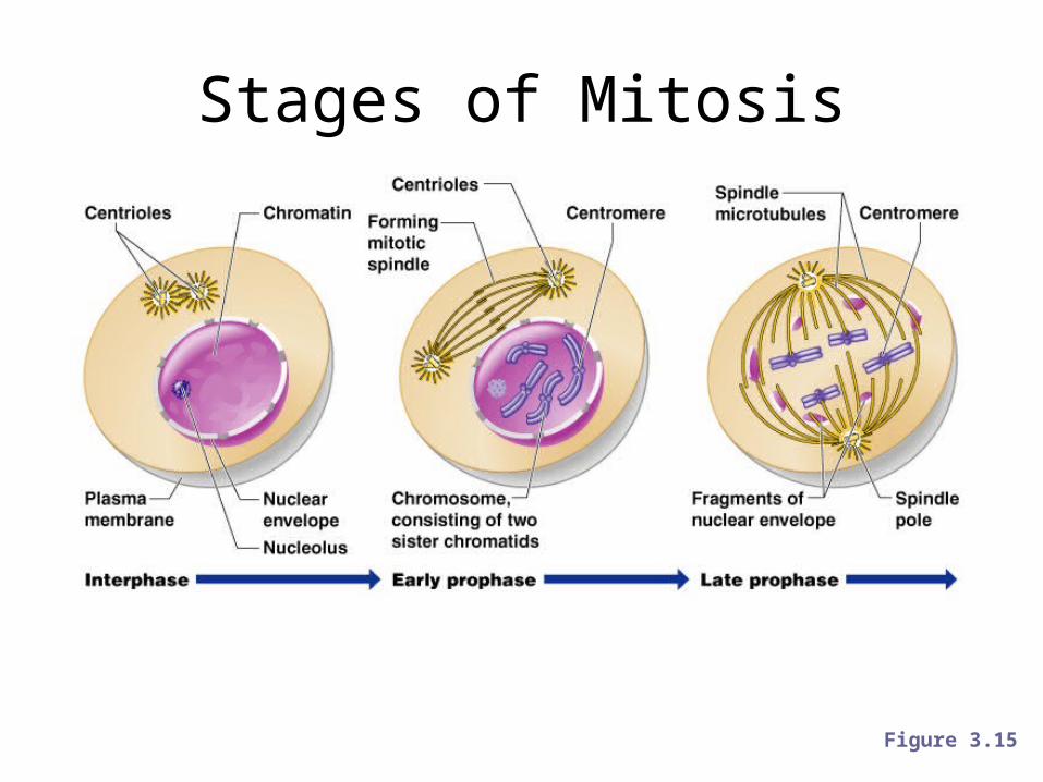
Stages of Mitosis
Figure 3.15
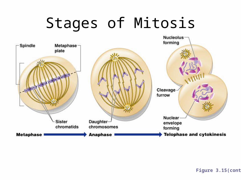
Stages of Mitosis
Figure 3.15(cont)
Protein Synthesis
• Gene – DNA segment that carries a blueprint for building one protein
• Proteins have many functions– Building materials for cells– Act as enzymes (biological catalysts)
• RNA is essential for protein synthesis
Body Tissues
• Cells are specialized for particular functions• Tissues– Groups of cells with similar structure and function– Four primary types• Epithelium• Connective tissue• Nervous tissue• Muscle
Epithelial Tissues
• Found in different areas– Body coverings– Body linings– Glandular tissue
• Functions– Protection– Absorption– Filtration– Secretion
Epithelium Characteristics
• Cells fit closely together• Tissue layer always has one free surface• The lower surface is bound by a basement
membrane• Avascular (have no blood supply)• Regenerate easily if well nourished
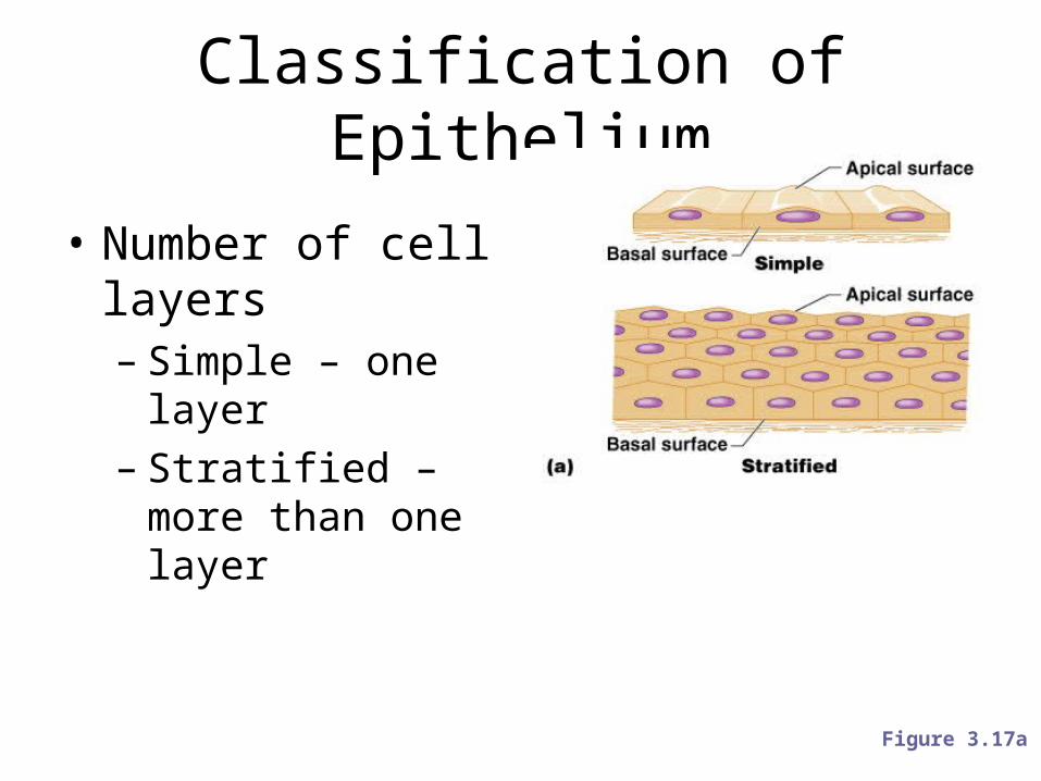
Classification of Epithelium
• Number of cell layers– Simple – one layer– Stratified – more than
one layer
Figure 3.17a
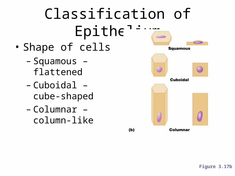
Classification of Epithelium
• Shape of cells– Squamous – flattened– Cuboidal – cube-
shaped– Columnar – column-
like
Figure 3.17b
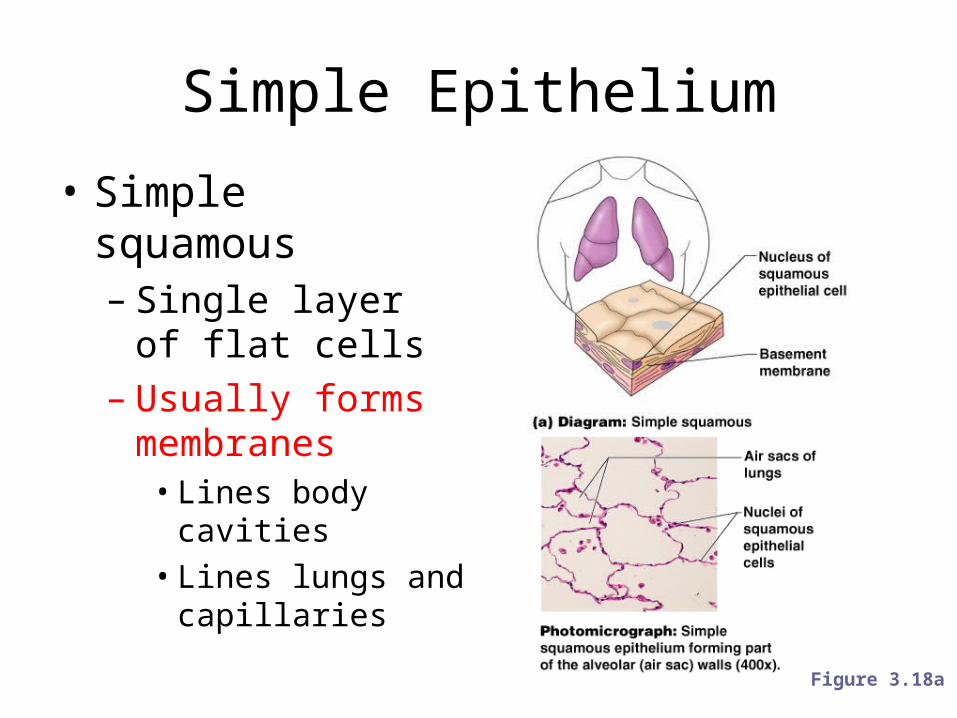
Simple Epithelium
• Simple squamous– Single layer of flat
cells– Usually forms
membranes• Lines body cavities• Lines lungs and
capillaries
Figure 3.18a
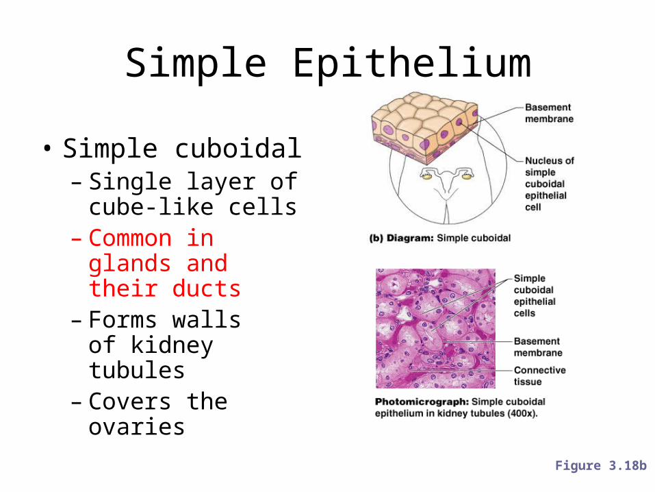
Simple Epithelium
• Simple cuboidal– Single layer of cube-
like cells– Common in glands
and their ducts– Forms walls
of kidney tubules– Covers the ovaries
Figure 3.18b
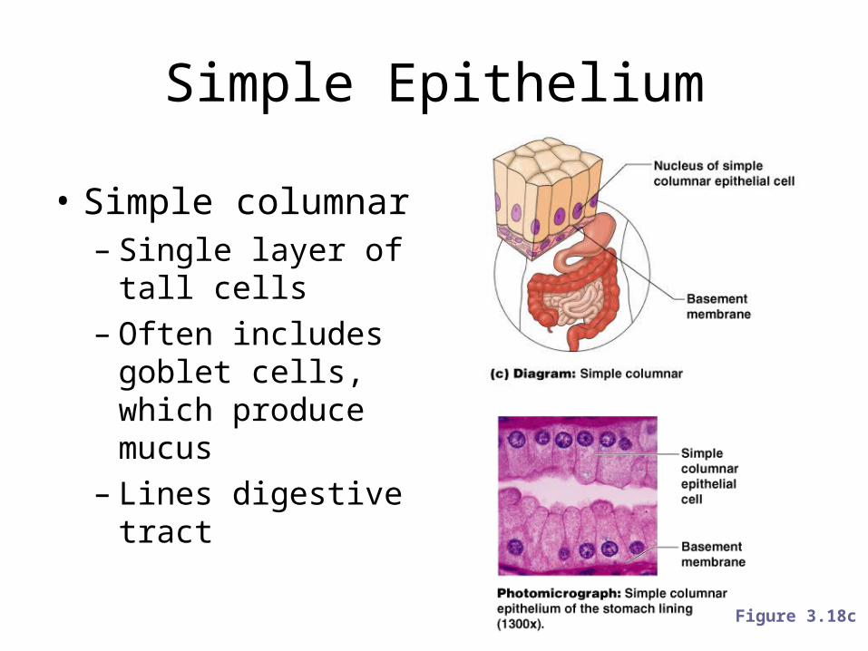
Simple Epithelium
• Simple columnar– Single layer of tall
cells– Often includes goblet
cells, which produce mucus
– Lines digestive tract
Figure 3.18c
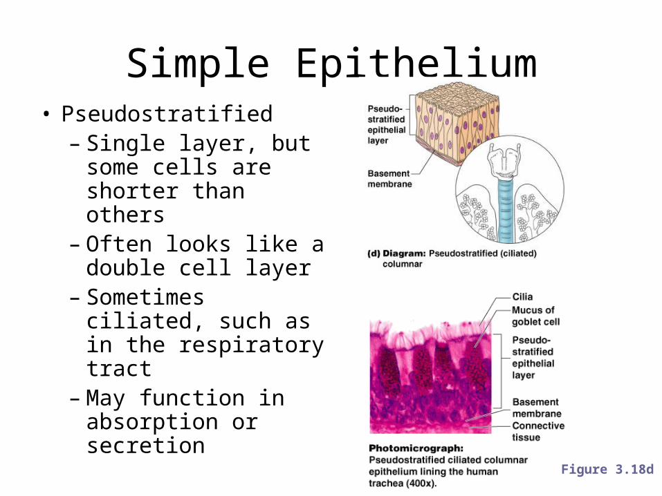
Simple Epithelium• Pseudostratified– Single layer, but some
cells are shorter than others
– Often looks like a double cell layer
– Sometimes ciliated, such as in the respiratory tract
– May function in absorption or secretion
Figure 3.18d
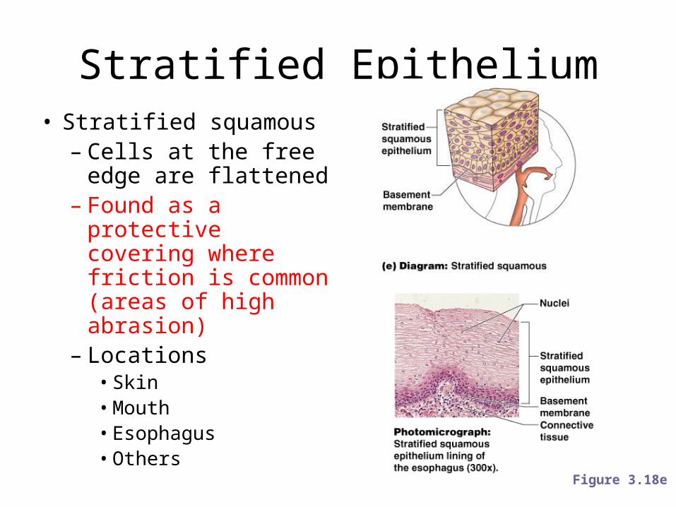
Stratified Epithelium• Stratified squamous– Cells at the free edge
are flattened– Found as a protective
covering where friction is common (areas of high abrasion)
– Locations• Skin• Mouth• Esophagus• Others
Figure 3.18e
Stratified Epithelium
• Stratified cuboidal– Two layers of cuboidal cells
• Stratified columnar– Surface cells are columnar, cells underneath vary
in size and shape
• Stratified cuboidal and columnar– Rare in human body– Found mainly in ducts of large glands
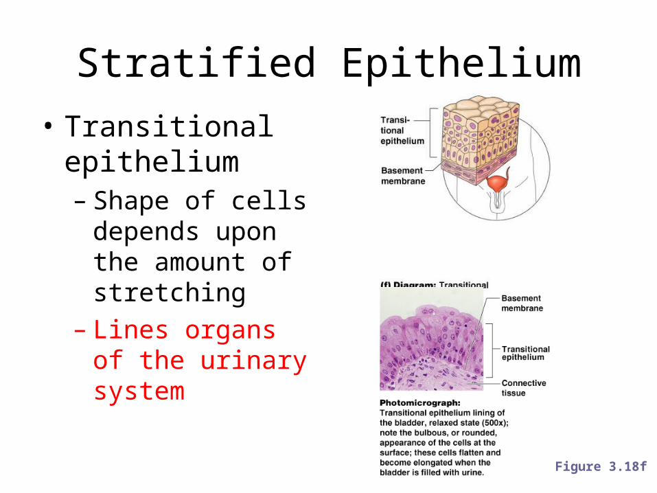
Stratified Epithelium• Transitional
epithelium– Shape of cells
depends upon the amount of stretching
– Lines organs of the urinary system
Figure 3.18f
Glandular Epithelium
• Gland – one or more cells that secretes a particular product
• Two major gland types– Endocrine gland• Ductless• Secretions are hormones
– Exocrine gland• Empty through ducts to the epithelial surface• Include sweat and oil glands
Connective Tissue
• Found everywhere in the body• Includes the most abundant and widely
distributed tissues• Functions– Binds body tissues together– Supports the body– Provides protection
Connective Tissue Characteristics
• Variations in blood supply– Some tissue types are well vascularized– Some have poor blood supply or are avascular
• Made of living cells and an extracellular matrix– Extracellular matrix: Non-living material that
surrounds living cells
Extracellular Matrix
• Two main elements– Ground substance – mostly water along with
adhesion proteins and polysaccharide molecules– Fibers• Produced by the cells• Three types– Collagen fibers– Elastic fibers– Reticular fibers
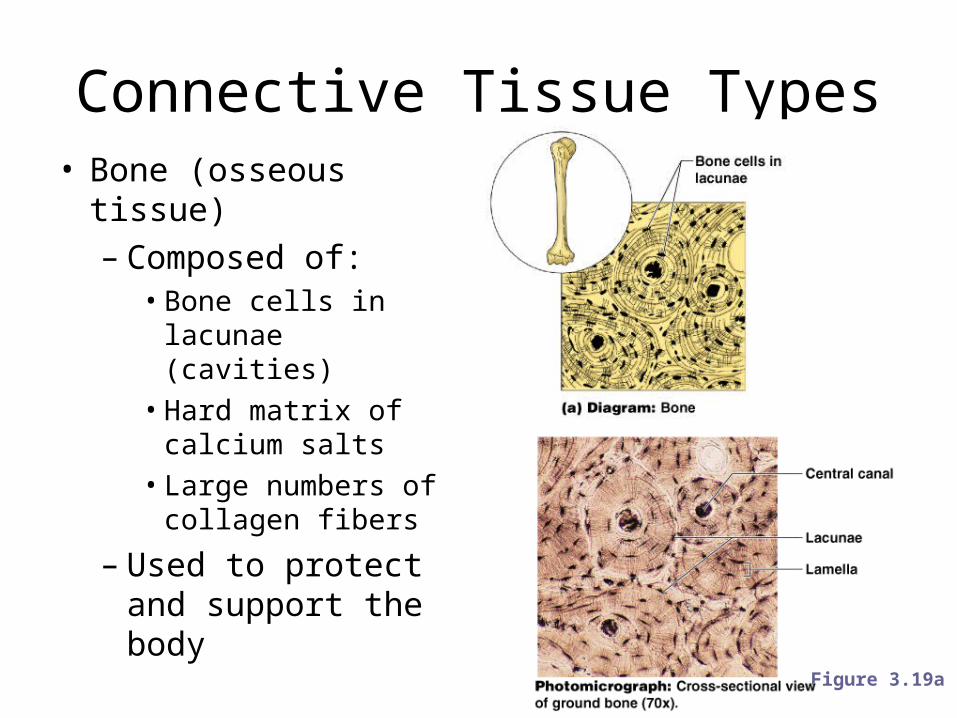
Connective Tissue Types• Bone (osseous tissue)– Composed of:• Bone cells in lacunae
(cavities)• Hard matrix of
calcium salts• Large numbers of
collagen fibers
– Used to protect and support the body
Figure 3.19a
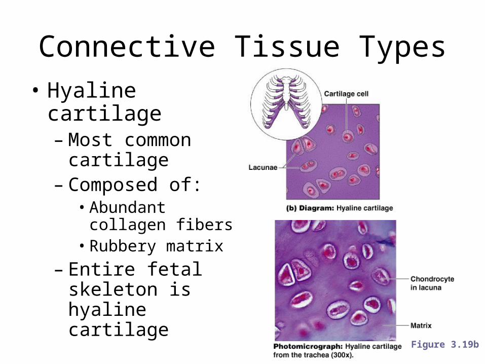
Connective Tissue Types• Hyaline cartilage– Most common
cartilage– Composed of:• Abundant collagen
fibers• Rubbery matrix
– Entire fetal skeleton is hyaline cartilage
Figure 3.19b
Connective Tissue Types
• Elastic cartilage– Provides elasticity– Example: supports the external ear
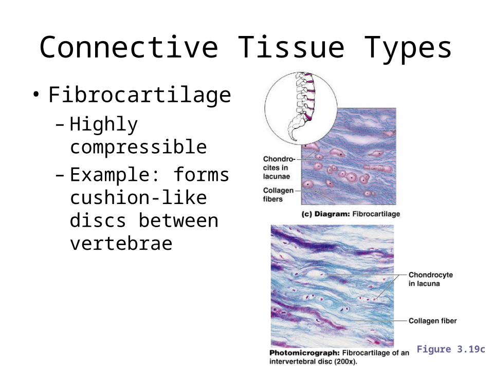
Connective Tissue Types• Fibrocartilage– Highly compressible– Example: forms
cushion-like discs between vertebrae
Figure 3.19c
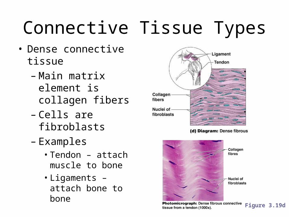
Connective Tissue Types• Dense connective tissue– Main matrix element
is collagen fibers– Cells are fibroblasts– Examples• Tendon – attach
muscle to bone• Ligaments – attach
bone to bone
Figure 3.19d
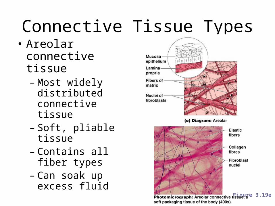
Connective Tissue Types• Areolar connective
tissue– Most widely
distributed connective tissue
– Soft, pliable tissue– Contains all fiber
types– Can soak up excess
fluid
Figure 3.19e
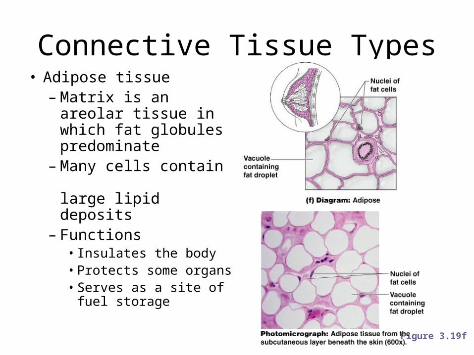
Figure 3.19f
Connective Tissue Types• Adipose tissue– Matrix is an areolar
tissue in which fat globules predominate
– Many cells contain large lipid deposits
– Functions• Insulates the body• Protects some organs• Serves as a site of
fuel storage
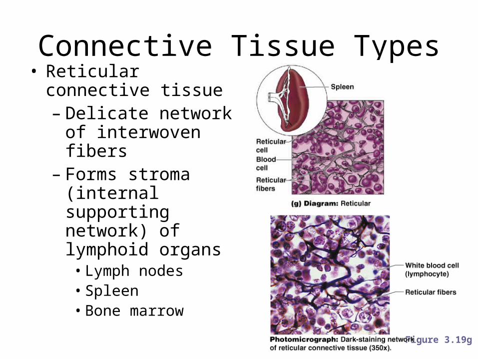
Connective Tissue Types• Reticular connective
tissue– Delicate network of
interwoven fibers– Forms stroma
(internal supporting network) of lymphoid organs• Lymph nodes• Spleen• Bone marrow
Figure 3.19g
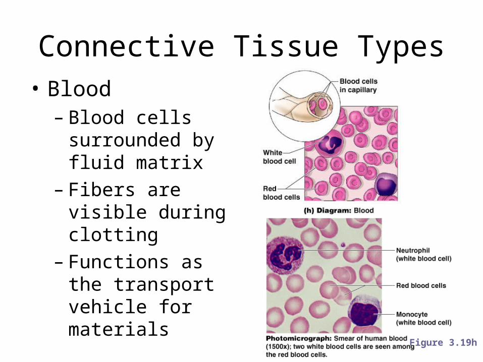
Connective Tissue Types• Blood– Blood cells
surrounded by fluid matrix
– Fibers are visible during clotting
– Functions as the transport vehicle for materials
Figure 3.19h
Muscle Tissue
• Function is to produce movement by contracting.
• Three types– Skeletal muscle– Cardiac muscle– Smooth muscle
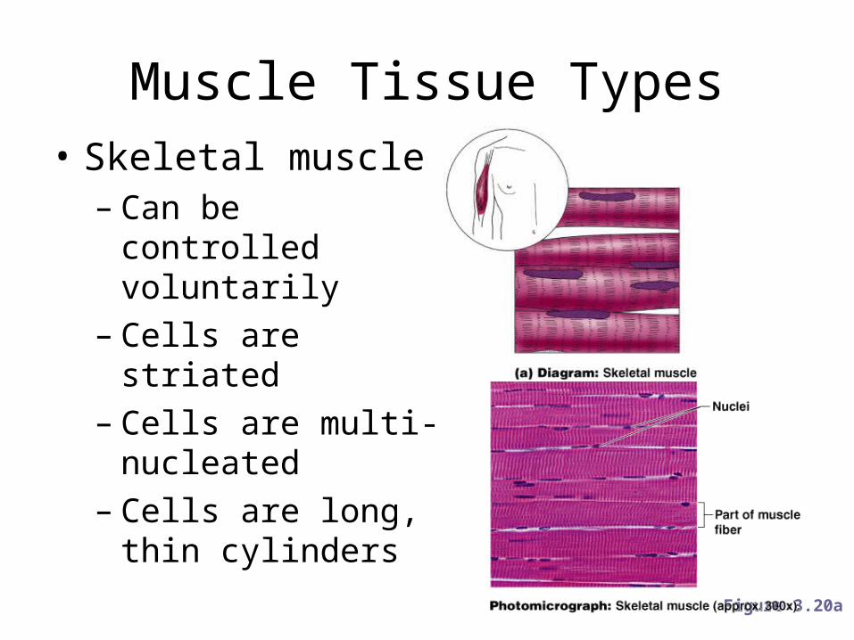
Muscle Tissue Types• Skeletal muscle– Can be controlled
voluntarily– Cells are striated– Cells are multi-
nucleated– Cells are long, thin
cylinders
Figure 3.20a
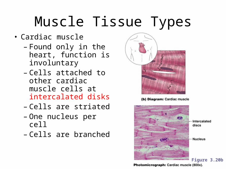
Muscle Tissue Types• Cardiac muscle– Found only in the
heart, function is involuntary
– Cells attached to other cardiac muscle cells at intercalated disks
– Cells are striated– One nucleus per cell– Cells are branched
Figure 3.20b
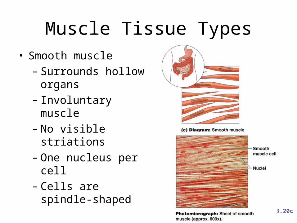
Muscle Tissue Types• Smooth muscle– Surrounds hollow
organs– Involuntary muscle– No visible striations– One nucleus per cell– Cells are spindle-
shaped
Figure 3.20c
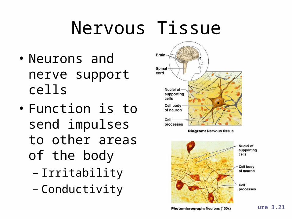
Nervous Tissue
• Neurons and nerve support cells
• Function is to send impulses to other areas of the body– Irritability– Conductivity
Figure 3.21
Regeneration of Tissues
• Tissues that regenerate easily– Epithelial tissue– Fibrous connective tissue and bone
• Tissues that regenerate poorly– Skeletal muscle
• Tissues that are replaced largely with scar tissue– Cardiac muscle– Nervous tissue within the brain and spinal cord
Developmental Aspects of Tissue
• Epithelial tissue arises from all three primary germ layers– Muscle and connective tissue arise from the
mesoderm– Nervous tissue and skin arise from the ectoderm– Most internal organs arise from the endoderm
• With old age there is a decrease in mass and viabililty in most tissues
Related Documents