Cell, Vol. 64, 201-208, January 11, 1991, Copyright 0 1991 by Cell Press Selective Expression of RAG-2 in Chicken B Cells Undergoing lmmunoglobulin Gene Conversion Louise M. Carlson;t* Marjorie A. Oettinger,§II David G. Schatz,§lI Emma L. Masteller,* Elizabeth A. Hurley,” Wayne T. McCormack;? David Baltimore,5 II l * and Craig B. Thompson’ tt l Howard Hughes Medical Institute t Department of Internal Medicine *Department of Microbiology and Immunology University of Michigan Medical Center Ann Arbor, Michigan 48109 §Whitehead Institute for Biomedical Research 9 Cambridge Center Cambridge, Massachusetts 02142 11 Department of Biology Massachusetts Institute of Technology Cambridge, Massachusetts 02139 * *The Rockefeller University New York, New York 10021 Summary Chickens create their immunoglobulin (Ig) repertoires during B cell development in the bursa of Fabrlcius by intrachromosomal gene conversion. Recent evidence has suggested that Ig gene conversion may involve cis-acting DNA elements related to those involved in V(D)J recombination. Therefore, we have examined the potential role of the V(D)J recombination activat- ing genes, RAG-1 and RAG-2, in regulating chicken lg gene conversion. In contrast to the coexpression of RAG-1 and RAG-2 observed in mammalian B cells that undergo V(D)J recombination, chicken B cells isolated from the bursa of Fabricius express high levels of the RAG-2 mRNA but do not express RAG-1 mRNA. The developmental and phenotyplc characteristics of the bursal lymphocytes and chicken B cell lines that ex- press RAG-2 mRNA demonstrate that selective RAG-2 expression occurs specifically in B cells undergoing lg diversification by gene conversion. These data sug- gest that RAG-2 plays a fundamental role in Ig-specific gene conversion. Introduction The primary repertoire of mammalian immunoglobulin (lg) and T ceil receptor (TCR) genes is generated by a se- ries of genomic rearrangements that occur during lym- phoid development (reviewed by Alt et al., 1988; Hunkapiller and Hood, 1989). In both sets of genes, re- arrangement is mediated by recombination signal se- quences that flank variable (V), diversity (D), and joining (J) gene segments (Tonegawa, 1983). These signal se- quences have been highly conserved in species that carry out V(D)J rearrangement and are functionally inter- changeable (Yancopoulos et al., 1986; Bucchini et al., 1987; Goodhardt et al., 1987; Lieber et al., 1987). V(D)J recombination requires the expression of two unrelated but tightly linked genes, RAG-l and RAG-2 (Schatz et al., 1989; Oettinger et al., 1990). Upon transfection, neither RAG-1 nor RAG-2 alone is sufficient to induce V(D)J rear- rangement in fibroblasts. However, cotransfection of the RAG-1 and RAG-2 cDNAs induces a high frequency of V(D)J-specific recombination without inducing the expres- sion of other pre-B or pre-T cell markers (Schatz et al., 1989; Oettinger et al., 1990). A survey of mammalian pre-B and preT cell lines that undergo lg or TCR gene rearrangement demonstrated that all of these cell lines coexpress RAG-1 and RAG-2. Thus, the data suggest that together RAG-l and RAG-2 encode the lymphoid-specific component of the V(D)J recombination system. In contrast to mammals, lg gene rearrangement in chickens does not lead to the creation of an immunoglob- ulin repertoire. Instead, chickens use rearrangement of the lg heavy(H) and light (L) chain loci only to activate the expression of surface lg. During chicken B cell develop- ment, 6 cell progenitors rearrange their IgH and IgL genes for only a brief period between days 10 and 15 of embryogenesis (Weill et al., 1986; McCormack et al., 1989a). The cells that undergo lg gene rearrangement are present primarily in the spleen, which is a major site of hematopoeisis during this embryonic period (reviewed by Ratcliffe, 1985; Pink, 1986; McCormack and Thompson, 1990a). Following lg gene rearrangement the S cell pro- genitors migrate to the bursa of Fabricius, where those progenitors that express surface lg undergo rapid clonal expansion (McCormacket al., 1989a). Beginning between days 15 and 18 of embryogenesis, this proliferating lg+ population of cells undergoes repetitive gene conversion of the single rearranged VH1 and Vu segments using V segment pseudogenes as sequence donors (Reynaud et al., 1987, 1989; McCormack et al., 1989b; Carlson et al., 1990). After accumulating four to ten gene conversion events within their VHt and Vu gene segments, individual bursal lymphocytes cease to proliferate and begin to mi- grate to the peripheral lymphoid organs (Thompson and Neiman, 1987). lg gene conversion is an ongoing process in the proliferating population of bursal lymphocytes until the bursa involutes at 4-6 months of age. Previous studies have demonstrated that the gene con- version process in bursal lymphocytes is restricted to the lg loci. The lymphoid-specific CaS-tubulin gene has a genomic organization similar to the IgL gene. It is com- posed of three exons, is actively transcribed in bursal lym- phocytes, and is preceded by a 5’ pseudogene segment that has an extensive open reading frame (Pratt and Cleveland, 1988). Despite this, the CaS-tubulin gene fails to undergo gene conversion in developing bursal lym- phocytes (Thompson, 1989). Similarly, the histone H2b genes, which are organized in two tightly linked clusters, fail to display evidence of gene conversion in developing bursal lymphocytes (Thompson, 1989). The lg specificity of bursal-dependent gene conversion led us to investigate whether RAG-1 and/or RAG-2 might also play a role in the regulation of lg diversification by

Welcome message from author
This document is posted to help you gain knowledge. Please leave a comment to let me know what you think about it! Share it to your friends and learn new things together.
Transcript
Cell, Vol. 64, 201-208, January 11, 1991, Copyright 0 1991 by Cell Press
Selective Expression of RAG-2 in Chicken B Cells Undergoing lmmunoglobulin Gene Conversion
Louise M. Carlson;t* Marjorie A. Oettinger,§II David G. Schatz,§lI Emma L. Masteller,* Elizabeth A. Hurley,” Wayne T. McCormack;? David Baltimore,5 II l * and Craig B. Thompson’tt l Howard Hughes Medical Institute t Department of Internal Medicine *Department of Microbiology and Immunology University of Michigan Medical Center Ann Arbor, Michigan 48109 §Whitehead Institute for Biomedical Research 9 Cambridge Center Cambridge, Massachusetts 02142 11 Department of Biology Massachusetts Institute of Technology Cambridge, Massachusetts 02139 * *The Rockefeller University New York, New York 10021
Summary
Chickens create their immunoglobulin (Ig) repertoires during B cell development in the bursa of Fabrlcius by intrachromosomal gene conversion. Recent evidence has suggested that Ig gene conversion may involve cis-acting DNA elements related to those involved in V(D)J recombination. Therefore, we have examined the potential role of the V(D)J recombination activat- ing genes, RAG-1 and RAG-2, in regulating chicken lg gene conversion. In contrast to the coexpression of RAG-1 and RAG-2 observed in mammalian B cells that undergo V(D)J recombination, chicken B cells isolated from the bursa of Fabricius express high levels of the RAG-2 mRNA but do not express RAG-1 mRNA. The developmental and phenotyplc characteristics of the bursal lymphocytes and chicken B cell lines that ex- press RAG-2 mRNA demonstrate that selective RAG-2 expression occurs specifically in B cells undergoing lg diversification by gene conversion. These data sug- gest that RAG-2 plays a fundamental role in Ig-specific gene conversion.
Introduction
The primary repertoire of mammalian immunoglobulin (lg) and T ceil receptor (TCR) genes is generated by a se- ries of genomic rearrangements that occur during lym- phoid development (reviewed by Alt et al., 1988; Hunkapiller and Hood, 1989). In both sets of genes, re- arrangement is mediated by recombination signal se- quences that flank variable (V), diversity (D), and joining (J) gene segments (Tonegawa, 1983). These signal se- quences have been highly conserved in species that carry out V(D)J rearrangement and are functionally inter- changeable (Yancopoulos et al., 1986; Bucchini et al., 1987; Goodhardt et al., 1987; Lieber et al., 1987). V(D)J recombination requires the expression of two unrelated
but tightly linked genes, RAG-l and RAG-2 (Schatz et al., 1989; Oettinger et al., 1990). Upon transfection, neither RAG-1 nor RAG-2 alone is sufficient to induce V(D)J rear- rangement in fibroblasts. However, cotransfection of the RAG-1 and RAG-2 cDNAs induces a high frequency of V(D)J-specific recombination without inducing the expres- sion of other pre-B or pre-T cell markers (Schatz et al., 1989; Oettinger et al., 1990). A survey of mammalian pre-B and preT cell lines that undergo lg or TCR gene rearrangement demonstrated that all of these cell lines coexpress RAG-1 and RAG-2. Thus, the data suggest that together RAG-l and RAG-2 encode the lymphoid-specific component of the V(D)J recombination system.
In contrast to mammals, lg gene rearrangement in chickens does not lead to the creation of an immunoglob- ulin repertoire. Instead, chickens use rearrangement of the lg heavy(H) and light (L) chain loci only to activate the expression of surface lg. During chicken B cell develop- ment, 6 cell progenitors rearrange their IgH and IgL genes for only a brief period between days 10 and 15 of embryogenesis (Weill et al., 1986; McCormack et al., 1989a). The cells that undergo lg gene rearrangement are present primarily in the spleen, which is a major site of hematopoeisis during this embryonic period (reviewed by Ratcliffe, 1985; Pink, 1986; McCormack and Thompson, 1990a). Following lg gene rearrangement the S cell pro- genitors migrate to the bursa of Fabricius, where those progenitors that express surface lg undergo rapid clonal expansion (McCormacket al., 1989a). Beginning between days 15 and 18 of embryogenesis, this proliferating lg+ population of cells undergoes repetitive gene conversion of the single rearranged VH1 and Vu segments using V segment pseudogenes as sequence donors (Reynaud et al., 1987, 1989; McCormack et al., 1989b; Carlson et al., 1990). After accumulating four to ten gene conversion events within their VHt and Vu gene segments, individual bursal lymphocytes cease to proliferate and begin to mi- grate to the peripheral lymphoid organs (Thompson and Neiman, 1987). lg gene conversion is an ongoing process in the proliferating population of bursal lymphocytes until the bursa involutes at 4-6 months of age.
Previous studies have demonstrated that the gene con- version process in bursal lymphocytes is restricted to the lg loci. The lymphoid-specific CaS-tubulin gene has a genomic organization similar to the IgL gene. It is com- posed of three exons, is actively transcribed in bursal lym- phocytes, and is preceded by a 5’ pseudogene segment that has an extensive open reading frame (Pratt and Cleveland, 1988). Despite this, the CaS-tubulin gene fails to undergo gene conversion in developing bursal lym- phocytes (Thompson, 1989). Similarly, the histone H2b genes, which are organized in two tightly linked clusters, fail to display evidence of gene conversion in developing bursal lymphocytes (Thompson, 1989).
The lg specificity of bursal-dependent gene conversion led us to investigate whether RAG-1 and/or RAG-2 might also play a role in the regulation of lg diversification by
Cell 202
gene conversion. In this report, we demonstrate that RAG-2, but not RAG-l, is expressed during B cell develop- ment in the bursa of Fabricius. These results suggest that a specific mechanism has evolved for the differential reg- ulation of RAG-1 and RAG-2 during B cell development. Bursal RAG-2 expression was first detected between days 15 and 16 of embryogenesis, the same time period during which developing bursal lymphocytes begin to diversify their lg genes by gene conversion. Selective RAG-2 ex- pression within the bursa continued until bursal involution at 4-6 months of age. Since gene conversion has been associated with cells undergoing proliferative expansion within the bursa of Fabricius, we examined RAG-2 gene expression with respect to the proliferative state of the bur- sal lymphocyte. Quiescent bursal lymphocytes destined to migrate into the periphery failed to express RAG-P, whereas B cells undergoing proliferative expansion in the bursa of Fabricius expressed high levels of RAG-2 mRNA.
Finally, in contrast to other non-gene-convert ing avian B and T cell lines, the bursal lymphoma cell line DT40, which undergoes constitutive lg-specific gene conversion in cul- ture, was found to express RAG-2 but not RAG-1 mRNA. Together these data suggest that RAG-2 plays a role in the lg-specific gene conversion process that occurs during B cell development in the bursa of Fabricius.
Results
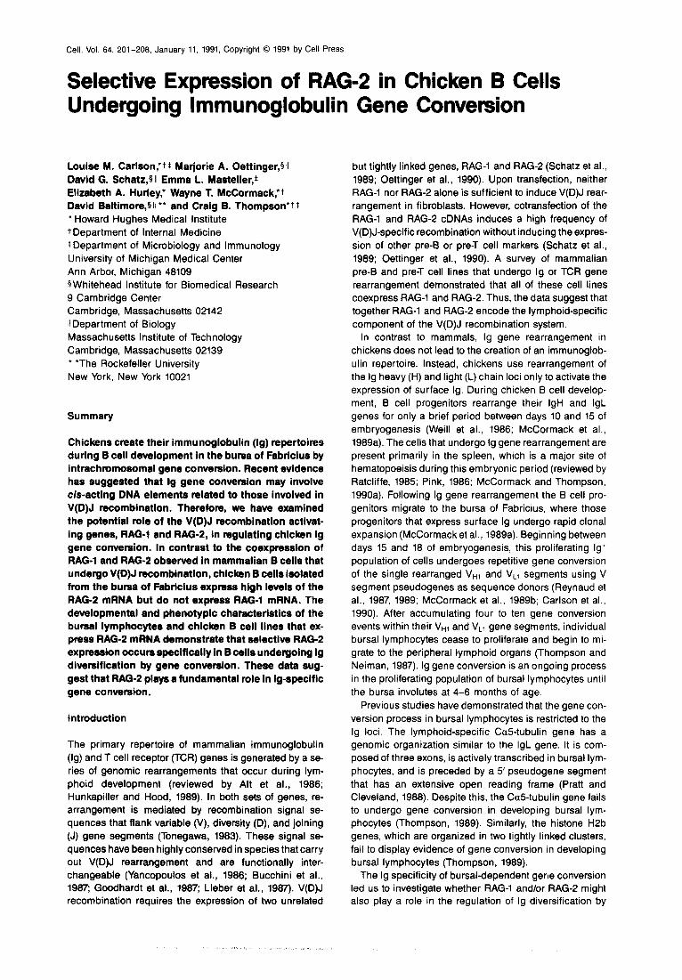
The Genomic Organization of RAG-1 and RAG-2 Has Been Highly Conserved during Vertebrate Evolution To characterize the expression of RAG-1 and RAG-2 dur- ing avian B and T cell development, it was first necessary to clone the chicken RAG-1 and RAG-2 genes. Hybridiza- tion of chicken DNA with a human RAG-l-specific probe detected a single-copy gene in the chicken genome (data
x x S s s xx xs x XL s x s s x L 1; ; N
B. RAG-l 200 400 600 coo 1000
I I I I I
C. RAG-2 loo 200 300 400 600
I I I I I
I I I I I 100 200 so0 400 600
Figure 1. Genomic Organization of the Chicken Recombination Activating Locus (A) The genomic organization of the chicken RAG-1 and RAG-2 genes has been deduced by characterizing two genomic clones (21469 and 11591) containing overlapping fragments of chicken genomic DNA. The positions of the RAG-1 and RAG-2 open reading frames are indicated. Arrows indi- cate the transcriptional orientation (S, Sack X. Xbal; L, Sall). Schematic comparisons of the amino acid sequences of chicken and mouse RAG-1 (B) and RAG-2 (C)were generated using the GAP and GAPSHOW programs of the Genetics Computer Group software package (University of Wis- consin). Amino acid sequences are shown as horizontal lines, with gaps denoted by openings in the horizontal lines and by small vertical lines. Identity between the chicken (top line) and mouse (bottom line) sequences is denoted at each position by a large vertical line.

Seective RAG-Z Expression in Bursal Lymphocytes
not shown), and several genomic clones were isolated from a chicken genomic library. One genomic clone, 11469, was found to hybridize at low stringency to a mu- rine RAG-2 probe, and this clone was used in the initial characterization of the chicken RAG locus (Figure 1).
The organization of the RAG locus has been highly con- served during vertebrate evolution. The coding regions of both chicken and murine RAG-1 and RAG-2 are located on single large exons that are separated by approximately 9 kb of genomic DNA and are convergently transcribed. Sequence analysis of chicken RAG-l demonstrated that the amino acid sequences of the chicken and mouse pro- teins are 75% identical. Conservation is most striking within the carboxy-terminal half of the molecule (66% amino acid identity), the same region in which homology was recently reported between RAG-1 and the yeast gene HPR7 (Wang et al., 1990). The HPR7 gene itself shares ho- mologies with yeast DNA topoisomerase I, and mutations in HPR7 increase intrachromosomal recombination and, to a lesser extent, gene conversion (Aguilera and Klein, 1966, 1990). Alignment of the chicken and murine RAG-2 amino acid sequences revealed 70% amino acid identity between the two sequences. Small blocks of higher con- servation were identified throughout the RAG-2 sequence. These regions were used to search the GenBank and NBRF data bases, but no significant homologies to known proteins were identified. DNA fragments that were specific for the coding regions of chicken RAG-1 and RAG-2 were isolated from genomic clone X1469. When hybridized to Southern blots containing chicken genomic DNA, each of these fragments hybridized only to single-copy genes (data not shown).
RAG-2 but Not RAG-l Is Expressed in Lymphocytes Isolated from the Buraa of Fabricius Unlike mammals, chickens contain two organs for lym- phoid development. As described above, the lg repertoire of chicken B cells is generated by lg-specific gene conver- sion events during B cell development in the bursa of Fabricius. The chicken T cell repertoire is generated by recombination of TCR V, D, and J elements during T cell development within the thymus (Tjoelker et al., 1990). TCR gene rearrangement is an ongoing process within the thymus during development, as T cell precursors con- tinue to seed the thymus during the first several months of life (Coltey et al., 1969; W. T M. and C. B. T., unpub- lished data). Lymphocytes isolated from the chicken thy- mus were found to express high levels of both RAG-1 and RAG-2 mRNA (Figure 2). In contrast, we found that bursal lymphocytes isolated from a 4-week-old chick expressed only RAG-e-specif ic mRNA. RNA isolated from bursal lymphocytes consistently failed to hybridize to RAG-l- specific probes. Lymphocytes isolated from the spleen failed to express either RAG-1 or RAG-2 mRNA.
Selective expression of RAG-2 mRNA has not been ob- sewed previously. It is unlikely to result from ongoing lg gene rearrangement, because V(D)J recombination re- quires the expression of both RAG-1 and RAG-2 (Cet- tinger et al., 1990) and because we have been unable to detect the circular episome that results from IgL rear-
TBS
RAG-1
RAG-2
ACTIN
Figure 2. Northern Slot of Bursal, Thymic, and Splenic Lymphocyte RNA Isolated from a 4-Week-Old Chicken
A Northern blot was prepared using lymphocyte RNA isolated from the thymus (T), bursa (S), and spleen (S) of a 4-week-old chick, as de- scribed in Experimental Procedures. The Northern blot was hybridized sequentially with 32P-labeled probes specific for RAG-l, RAG-P, and p-actin, and the resulting autoradiograms are shown.
rangement during this period of development (McCor- mack et al., 1969a, 1969b). Furthermore, we found that at 4 weeks of age, bursal lymphocytes uniformly express sur- face lg (Figure 3) and therefore no longer require lg recombinase activity for V(D)J rearrangement.
These data argue that a specific mechanism has evolved in the regulation of the recombination activating locus that results in the selective expression of RAG-2. The bursal lymphocyte has been shown to be the site of a novel lg gene recombination process that results in lg diversification by intrachromosomal gene conversion (Rey- naud et al., 1967, 1969; Carlson et al., 1990). To establish whether there is a correlation between selective RAG-2 mRNA expression and bursalspecific gene conversion, a series of experiments was carried out to correlate the known phenotypic characteristics of B cells undergoing gene conversion in the bursa of Fabricius with the expres- sion of RAG-P.
Both Bursal Lymphocyte RAG2 Expression and Ig Gene Conversion Begin between Days 15 and 18 of Embryogenesis Rearranged V gene segments isolated from bursal lym- phocytes on or before day 15 of embryogenesis fail to dis- play evidence of lg gene conversion (Thompson and Nei- man, 1987; Reynaud et al., 1987; McCormack et al., 1989b). In contrast, bursal lymphocytes isolated from day 16 of embryogenesis display between one and four in- dividual gene conversion events within their rearranged V gene segments (Reynaud et al., 1987; McCormack and Thompson, 199Ob). These data suggest that the bursal-
Cell 204
Log Fluorescence Intensity ---+
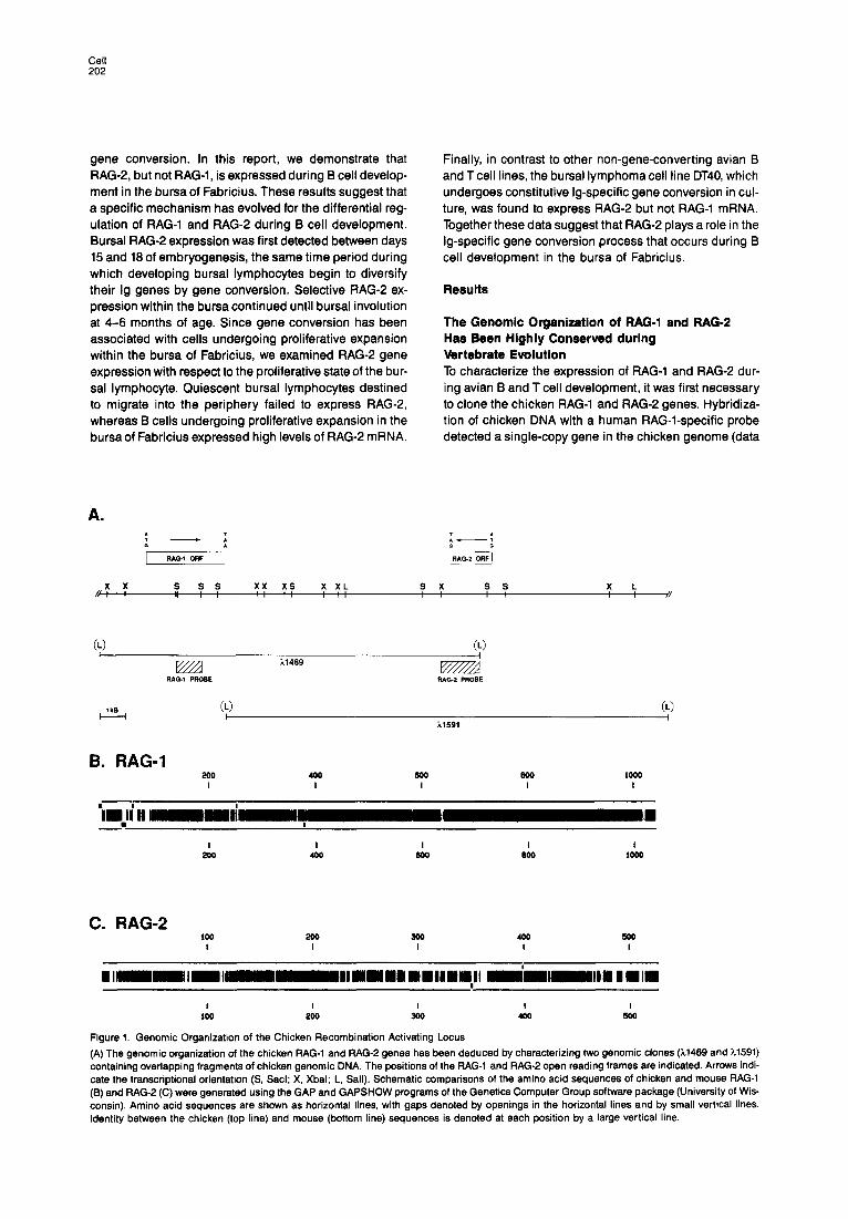
Figure 3. Expression of Surface lmmunoglobulin in Bursal Lympho- cytes isolated from a 4-Week-Old Chicken
Chicken bursal lymphocytes isolated from a 4-week-old chick were in- cubated with rabbit anti-chicken IgG antibody. Binding was assayed by indirect immunofluorescence following staining with a FITC-con- jugated donkey anti-rabbit IgG secondary antibody (solid line) using a fluorescence-activated cell sorter. Background staining was assayed by direct immunofluorescence of cells stained with the FITC-conju- gated donkey anti-rabbit IgG antibody alone (dashed line).
specific process of lg gene conversion begins in most bur- sal lymphocytes between days 15 and 18 of embryogene- sis. After that time, B cells proliferating within the bursal environment continue to accumulate lg gene conversion events until the bursa involutes at sexual maturity at 4-6 months of age.
To determine the temporal pattern of RAG-2 gene ex- pression during 6 cell development in the bursa of Fab- ricius, RNA was isolated from the bursal anlage or from
Day of Embryogenesis Post-hatch
I 7 9 11 13 15 18 II ++?w 14w
RAG-1
ACTIN (, I)
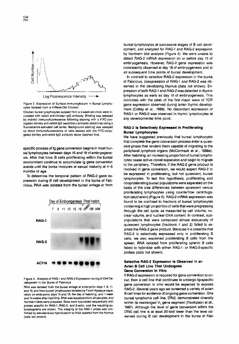
Figure 4. Analysis of RAG-1 and RAG-2 Expression during B Cell De- velopment in the Bursa of Fabricius
RNA was isolated from the bursal anlage at embryonic days 7, 9, 11, and 13, and from bursal lymphocytes isolated by Ficoll-Hypaque sepa- ration on embryonic days 15 and 19, the day of hatching, and 1 week and 14 weeks after hatching. RNA was equalized from all samples, and Northern blots were prepared. Blots were hybridized sequentially with probes specific for RAG-l, RAG-P, and 9-actin, and the resulting au- toradiograms are shown. The integrity of the RAG-l probe was con- firmed by simultaneous hybridization to RNA isolated from the thymus (data not shown).
bursal lymphocytes at successive stages of B cell devel- opment, and analyzed for RAG-l and RAG-2 expression by Northern blot analysis (Figure 4). We were unable to detect RAG-2 mRNA expression on or before day 15 of embryogenesis. However, RAG-2 gene expression was consistently observed at day 18 of embryogenesis and at all subsequent time points of bursal development.
In contrast to selective RAG-2 expression in the bursa of Fabricius, coexpression of RAG-1 and RAG-2 was ob- served in the developing thymus (data not shown). Ex- pression of both RAG-1 and RAG-2 was detected in thymic lymphocytes as early as day 15 of embryogenesis. This coincides with the peak of the first major wave of TCR gene expression observed during avian thymic develop- ment (Coltey et al., 1989). No discordant expression of RAG-1 or RAG-2 was observed in thymic lymphocytes at any developmental time point.
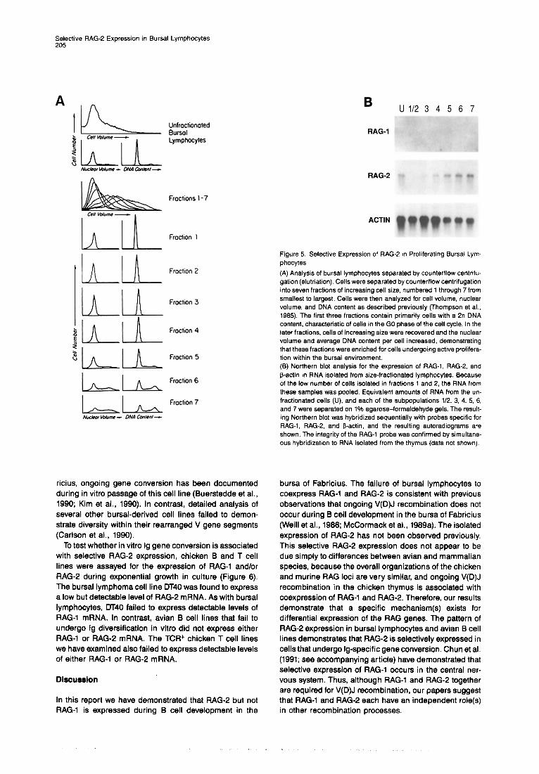
RAG-2 Is Selectively Expressed in Proliferating Bursal Lymphocytes We have suggested previously that bursal lymphocytes that complete the gene conversion process enter a quies- cent phase that renders them capable of migrating to the peripheral lymphoid organs (McCormack et al., 1989a). After hatching, an increasing proportion of bursal lympho- cytes cease active clonal expansion and begin to migrate to the periphery. Therefore, if the RAG-2 gene product is involved in gene conversion, we would expect RAG-2 to be expressed in proliferating, but not quiescent, bursal lymphocytes. To test this hypothesis, proliferating and nonproliferating bursal populations were separated on the basis of the size differences between quiescent versus proliferating lymphocytes using counterflow centrifuga- tion (elutriation) (Figure 5). RAG-2 mRNA expression was found to be confined to fractions of bursal lymphocytes containing a high proportion of cells that were progressing through the cell cycle, as measured by cell volume, nu- clear volume, and nuclear DNA content. In contrast, sub- populations that were composed almost exclusively of quiescent lymphocytes (fractions 1 and 2) failed to ex- press the RAG-2 gene product. Because it is possible that RAG-2 is selectively expressed only in proliferating B cells, we also examined proliferating B cells from the spleen. RNA isolated from proliferating splenic B cells failed to hybridize with either RAG-l- or RAG-2-specif ic probes (data not shown).
Selective RAG-2 Expression Is Observed in an Avlan B Cell Line That Undergoes Gene Conversion In Vltn, If RAG-2 expression is required for gene conversion to oc- cur, then a cell line that continues to undergo Ig-specific gene conversion in vitro would be expected to express RAG-2. Several years ago we screened a variety of avian B cell lines for evidence of ongoing gene conversion. One bursal lymphoma cell line, DT40, demonstrated diversity within its rearranged VL gene segment (Thompson et al., 1987). Although the level of gene conversion within the DT40 cell line is at least 20-fold lower than the level ob- served during B cell development in the bursa of Fab-
.SSective RAG-2 Expression in Bursal Lymphocytes
A I n U112 3 4 5 6 7
Froctrons l-7
Cc// Vakne - ,
h 1 1 Fro&on 1
1 n Frochon 2 1 A
1 A / A Froctron 3
1 1 n 1 h Fraction 4
$ k 1 k Froctron 5
1 - 1 L Froctron 6
1 - / - Froctron 7
Nucbw Wum - DNA Cmtent -
ricius, ongoing gene conversion has been documented during in vitro passage of this cell line (Buerstedde et al., 1990; Kim et al., 1990). In contrast, detailed analysis of several other bursal-derived cell lines failed to demon- strate diversity within their rearranged V gene segments (Carlson et al., 1990).
To test whether in vitro lg gene conversion is associated with selective RAG-2 expression, chicken B and T cell lines were assayed for the expression of RAG-1 and/or RAG-2 during exponential growth in culture (Figure 6). The bursal lymphoma cell line DT40 was found to express a low but detectable level of RAG-2 mRNA. As with bursal lymphocytes, DT40 failed to express detectable levels of RAG-1 mRNA. In contrast, avian B cell lines that fail to undergo lg diversification in vitro did not express either RAG-1 or RAG-2 mRNA. The TCR+ chicken T cell lines we have examined also failed to express detectable levels of either RAG-l or RAG-2 mRNA.
Discussion
In this report we have demonstrated that RAG-2 but not RAG-1 is expressed during B cell development in the
RAG-2 i.1
AC-RN
Figure 5 Selective Expression of RAG-2 in Proliferating Bursat Lym- phocytes
(A) Analysis of bursal lymphocytes separated by counterflow centrifu- gation (elutriation). Cells were separated by counterflow centrifugation into seven fractions of increasing cell size, numbered 1 through 7 from smallest to largest. Cells were then analyzed for cell volume, nuclear volume, and DNA content as described previously (Thompson et al., 1985). The first three fractions contain primarily cells with a 2n DNA content, characteristic of cells in the GO phase of the cell cycle. In the later fractions, cells of increasing size were recovered and the nuclear volume and average DNA content per cell increased, demonstrating that these fractions were enriched for cells undergoing active prolifera- tion within the bursal environment. (6) Northern blot analysis for the expression of RAG-l, RAG-P, and f3-actin in RNA isolated from size-fractionated lymphocytes. Because of the low number of cells isolated in fractions 1 and 2, the RNA from these samples was pooled. Equivalent amounts of RNA from the un- fractionated cells (U), and each of the subpopulations 112. 3, 4. 5, 6, and 7 were separated on 1% agarose-formaldehyde gels. The result- ing Northern blot was hybridized sequentially with probes specific for RAG-I, RAG-P, and B-act& and the resulting autoradiograms are shown. The integrity of the RAG-1 probe was confirmed by simultane- ous hybridization to RNA isolated from the thymus (data not shown).
bursa of Fabricius. The failure of bursal lymphocytes to coexpress RAG-1 and RAG-2 is consistent with previous observations that ongoing V(D)J recombination does not occur during B cell development in the bursa of Fabricius (Weill et al., 1966; McCormack et al., 1969a). The isolated expression of RAG-2 has not been observed previously. This selective RAG-2 expression does not appear to be due simply to differences between avian and mammalian species, because the overall organizations of the chicken and murine RAG loci are very similar, and ongoing V(D)J recombination in the chicken thymus is associated with coexpression of RAG-1 and RAG-P. Therefore, our results demonstrate that a specific mechanism(s) exists for differential expression of the RAG genes. The pattern of RAG-2 expression in bursal lymphocytes and avian B cell lines demonstrates that RAG-2 is selectively expressed in cells that undergo Ig-specific gene conversion. Chun et al. (1991; see accompanying article) have demonstrated that selective expression of RAG-1 occurs in the central ner- vous system. Thus, although RAG-1 and RAG-2 together are required for V(D)J recombination, our papers suggest that RAG-1 and RAG-2 each have an independent role(s) in other recombination processes.

Cell 206
TB123456 T
RAG-1
Figure 6. Expression of RAG-1 and RAG-2 in Bursal and Thymic- Derived Cell Lines
RNA samples (10 ug) from thymus (T), bursa (B), chicken bursal- derived cell lines 30LI (lanes 1 and 6) and DT40 (lanes 2 and 5), and chicken thymus-derived cell lines MSB-1 (lane 3) and UG-9 (lane 4) were separated on a 1% agarose-formaldehyde gel and transferred to nitrocellulose. The resulting Northern blot was hybridized sequentially with probes specific for RAG-l, RAG-P, and b-actin. The two bursal- derived cell lines have been extensively characterized, and DT40 (lanes 2 and 5) has been shown to undergo constitutive gene conver- sion in culture, whereas 30LI (lanes 1 and 6) does not display this abil- ity (Buerstedde et al., 1990; Kim et al., 1990; Carlson et al., 1990). None of the cell lines expressed RNA that hybridized with the RAG-l-specific probe, but the DT40 cell line mRNA consistently hybridized at a low level with the RAG-P-specific probe (lane 2, passage 56; lane 5, pas- sage 150). Analysis of four additional bursal lymphoma cell lines failed to demonstrate expression of either RAG-1 or RAG-2 (data not shown).
Our data suggest that the RAG gene products may play a wider role in the creation of lg diversity than previously considered. Diversity within the primary immunologic repertoire has been shown to result from three processes: V(D)J recombination, V region replacement, and gene conversion. Based on our current and past results, it seems possible that the RAG locus is involved in the generation of lg diversity by each of these processes. We consider each in turn.
V(D)J recombination is a complex process (for review see Lewis and Gellert, 1989) that normally requires the presence of two pairs of recombination signal sequences that are comprised of conserved heptamer and nonamer elements. A complete V(D)J recombination requires the activity of multiple gene products. It appears that RAG-1 and RAG-2 encode proteins that are the lymphoid-specific components of the V(D)J recombinase (Oettinger et al., 1990). In addition, other, ubiquitously expressed factors are involved. For example, the scid mutation, which results in the failure to form a coding joint during V(D)J recombination, also results in the failure to repair radi- ation-induced DNA damage (Fulop and Phillips, 1990). Together, the components of the V(D)J recombinase serve to create diversity as a result of combinatorial and junc- tional heterogeneity.
Another way in which B cells create lg diversity is V re-
gion replacement by V(D)J recombination between an ex- pressed lg gene and a germline V gene segment (Reth et al., 1986; Kleinfield et al., 1986). Such recombination ap- pears to result from the recognition of a conserved hep- tamer element present in the rearranged V gene segment immediately 5’ of CDRS. Thus the minimal sequence ele- ment required to target the V(D)J recombinase to a rear- ranged V segment is the presence of a heptamer se- quence at the site of recombination.
lmmunoglobulin diversification by gene conversion dis- plays several features that suggest that gene conversion may also involve components of the Ig/TCR recombinase. Like V region replacement, gene conversion is highly re- stricted to an expressed V gene segment (Thompson and Neiman, 1987). Other genes that undergo equivalent rates of lymphoid-specific transcription and that are lo- cated in close proximity to homologous donor templates fail to undergo gene conversion in developing bursal lym- phocytes (Thompson, 1989). We have found that three heptamer sequences similar to those present in lg and TCR recombination signal sequences have been con- served in the avian VL gene segment (McCormack et al., 1989c). One of these is immediately 5’of CDRl, a second is at the FR2-CDR2 boundary, and the third is located im- mediately 5’ of the CDR3 domain. Similar heptamer se- quences are located 5’ of CDR3 in the functional VH ele- ment and within the coding region of the D elements (Reynaud et al., 1989).
Reynaud et al. (1987) have speculated that the hep- tamer sequence 5’ of CDR3 might play a role in the high frequency of gene conversion observed in that domain. In support of this hypothesis, recent data have shown that nucleotides that encompass the three conserved hep- tamer sites in the chicken VL region are involved in gene conversion tracts at a statistically higher frequency than other sequences within the V region (McCormack and Thompson, 1990b). It has been postulated that heptamer- specific strand nicking or cleavage might be involved in the initiation of a gene conversion event, and a molecular model based on these observations has recently been proposed (McCormack and Thompson, 1990b). The ob- servation that RAG-2 is selectively expressed in bursal lymphocytes suggests that RAG-2 may be the V(D)J recombinase component involved in the gene conversion process. Based on the existing data, it is tempting to spec- ulate that RAG-2 may encode or regulate a heptamer- nicking activity.
As suggested above, the recombination events that serve to create primary immunologic repertoires in differ- ent species appear to share some common &-regulatory elements and transacting mediators. In particular, the RAG locus seems to be involved not only in V(D)J recom- bination, but also in lg gene conversion during avian B cell development. The conservation of the RAG locus during evolution suggests that the multiple roles of RAG genes in lg diversification may have evolved early during ver- tebrate evolution. Consistent with this hypothesis, some avian species have been shown to use combinatorial diversity to generate a primary immunologic repertoire
Selective RAG-2 Expression in Bursal Lymphocytes 207
(McCormack et al., 1989c), and rabbits appear to create an IgH repertoire primarily by gene conversion (Becker and Knight, 1990; Roux et al., 1991).
In summary, our data suggest that the ability of avian B ceils to generate an lg repertoire by gene conversion results from an alteration in the expression of genes in- volved in lg gene rearrangement. A simple model can be proposed to account for the relationship of lg gene re- arrangement and gene conversion. The expression of RAG-2 in cells undergoing either V(D)J recombination or lg gene conversion suggests that RAG-2 is involved in ini- tiating the assembly of a recombination intermediate. It is possible that both lg-specific recombination events in- volve a common intermediate. In the presence of RAG-l, such an intermediate would result in the production of a V(D)J rearrangement. In the absence of RAG-l, V(D)J rearrangement does not occur and the recombinational intermediate is resolved to leave a gene conversion tract.
Experimental Pmcedums
Cells and Cell Lines Bursal and thymic lymphocytes were isolated as single-cell suspen- sions by gently teasing apart the bursa of Fabricius or thymus. and filtering the cells through nylon mesh to remove clumps. Contaminat- ing nonlymphoid cells were removed by centrifugation of the cell sus- pension over Ficoll-Hypaque. Greater than 95% of the cells prepared from either organ in this manner stained positively with monoclonal an- tibodies specific for cells of the respective organ (data not shown). For embryonic samples isolated prior to day 15 of embryogenesis, it was not possible to efficiently separate the lymphoid component of the bursa; therefore, RNA was prepared from the total bursa or the posterior cloaca. To obtain size-dependent subpopulations of cells, single-cell suspensions were loaded into a counterflow centrifuge (elutriator), and cell subpopulations of increasing mean cell volume were sequentially isolated as previously described (Thompson et al., 1985). Cell volume, nuclear volume; and average DNA content of cells from each fraction were determined as previously described (Thomp- son et al., 1985). Avian B cell lines derived from the bursa of Fabricius following retroviral infection on the day of hatching were isolated and propagated as previously described (Thompson et al., 1987). The avian Tcell lines were grown and passaged using standard conditions (Chan et al., 1988).
FACS Analysis One million bursal lymphocytes isolated from a 4-week-old chicken were stained with 2 ul of rabbit anti-chicken IgG (Cappel-Worthington, 2 mglml) and incubated on ice for 30 min. After two washes with 1 ml of PBS, 0.1% azide, the cells were stained with 5 PI of FITC-donkey anti-rabbit IgG (Jackson lmmunoresearch Laboratories, Inc., 1.4 mglml) for 30 min on ice. The cells were washed twice with 1 ml PBS, 0.1% azide, and analyzed immediately after staining using a Becton- Dickinson FACScan.
Avlan RAG-l/RAG-O Cloning The 0.9 kb human RAG-1 fragment was labeled by nick translation and used to probe nitrocellulose lifts of a primary plating of a chicken lFlX genomic library (the generous gift of K. Conklin, University of Min- nesota). Blots were prehybridized at 42oC in 50% (v/v) formamide and 5x SSC. lx Denhardt’s solution, 25 mM sodium phosphate (pH 8.5) 250 @ml torula RNA. Hybridizations were carried out for 16 to 20 hr under identical conditions except for the addition of 10% (w/v) dextran sulfate and labeled probe at lo6 dpmlml. After hybridization, blots were rinsed twice at room temperature in 2x SSC. 0.1% SDS and then washed for 20 min in 0.2x SSC, 0.1% SDS at 56OC. Blots were then air dried and exposed to X-ray film for 8 to 20 hr at -70°C with intensify- ing screens. Following initial isolation, RAG-l+ genomic clones were rescreened with the 2.1 kb mouse RAG-2 cDNA probe. A resulting
RAG-l+/RAG-2+ clone (11489) was subcloned in two Sall fragments into pBluescript SK(-), from which RAG-l- and RAG-P-specific probes were isolated.
Probes The following probes were utilized: a 0.9 kb Xhol-Hindlll human RAG-1 probe derived from the 3’end of the coding region (Schatz et al., 1989) and a 2.1 kb murine RAG-2 cDNA probe containing the entire coding region and flanking 5’ and 3’ untranslated regions (Oettinger et al., 1990). Chicken-derived probes included: a 1.1 kb Sac1 fragment of RAG-l, isolated from the middle of the coding region; a 1.8 kb Xbal-Sall fragment, which contains two-thirds of the RAG-2 coding re- gion at the 3’end and the 3’untranslated region; and a 0.59 kb Hindlll fragment from the J’untranslated region of the 8-actin gene (Cleveland et al., 1980).
Northern Blots For analysis of gene-specific mRNA levels, RNA was extracted using guanidinium isothiocyanate (Chirgwin et al., 1979). Samples were equalized for ribosomal RNA and the equalization confirmed by ethidium bromide staining of RNA samples separated on a non- denaturing 1% agarose gel. Equalized RNA samples (2-10 ug) were separated on 1% agarose-formaldehyde gels and transferred to nitrocelluose. Filters were then hybridized sequentially with gene- specific probes for RAG-l, RAG-2 and 8-actin. Resulting autoradio- grams were exposed for 2 to 48 hr. The relative molecular masses of the mRNAs that hybridized to each of the probes agreed with previ- ously published values (Schatz et al., 1989; Oettinger et al., 1990; Cleveland et al., 1980).
Acknowledgments
We thank our colleagues Jeff Leiden, Eric Humphries, and John Lowe for helpful discussions and review of the manuscript, and Beverly Burck for preparation of figures. This work was supported by Public Service Award CA48023 to C. B. T., and CA51466 to D. 8. from the Na- tional Institutes of Health, and by fellowships from the Whitaker Health Sciences Fund (M. A. 0.) and the Life and Health Insurance Medical Research Fund (D. G. S.).
The costs of publication of this article were defrayed in part by the payment of page charges. This article must therefore be hereby marked “advertisement” in accordance with 18 USC Section 1734 solely to indicate this fact.
Received November 2, 1990; revised November 20, 1990.
References
Aguilera, A., and Klein, H. L. (1988). Genetic control of intrachromo- somal recombination in Sacchammyzes cerevisiae. I. Isolation and genetic characterization of hyper-recombination mutations. Genetics 179, 779-790.
Aguilera, A., and Klein, H. L. (1990). HPRI, a novel yeast gene that pre- vents intrachromosomal excision recombination, shows carboxy terminal homology to the Sacchammyces cerevisiae TOP7 gene. Mol. Cell. Biol. 10, 1439-1451.
Alt, F. W., Blackwell, T. K., DePinho, R. A., and Yancopoulos, G. D. (1986). Regulation of genome rearrangement events during lympho- cyte differentiation. Immunol. Rev. 89, 5-30.
Becker, R. S.. and Knight, K. L. (1990). Somatic divprsification of im- munoglobulin heavy chain VDJ genes: evidence for somatic gene con- version in rabbits. Cell 63, 987-997.
Bucchini. D., Reynaud, C.-A., Ripoche, M.. Grimal, H., Jami, J., and Weill, J.-C. (1987). Rearrangement of a chicken immunoglobulin gene occurs in the lymphoid lineage of transgenic mice. Nature 326, 409-411.
Buerstedde, J.-M., Reynaud, CA., Humphries, E. H., Olson, W., Ewert, D. L., and Weill, J.-C. (1990). Light chain gene conversion con- tinues at high rate in an ALV-induced cell line. EMBO J. 9, 921-927.
Carlson, L. M., McCormack. W. T., Postema, C. E., Humphries, E. H., and Thompson, C. B. (1990). Templated insertions in the rearranged

Cell 208
chicken lg, V gene segment arise by intrachromosomal gene conver- sion. Genes Dev. 4, 536-547.
Chan, M. M.. Chen, C. H., Ager, L. L., and Cooper, M. D. (1988). Iden- tification of the avian homologues of mammalian CD4 and CD8 anti- gens. J. Immunol. 740, 2133-2138.
Chirgwin, J. M., Przybyla, A. E., MacDonald, Ft. J., and Flutter, W. J. (1979). Isolation of biologically active ribonucleic acid from sources en- riched in ribonuclease. Biochemistry 18, 5294-5299.
Chun, J. J. M., Schatz, D. G., Oettinger, M. A., Jaenisch, Ft., and Balti- more, D. (1991). The recombination activating gene-l (RAG-l) transcript is present in the murine central nervous system. Cell 64, this issue.
Cleveland, D. W., Lopata, M. A., MacDonald, Ft. J., Cowan, N. J., Rut- ter, W. J., and Kirschner, M. W. (1980). Number and evolutionary con- servation of a- and 8-tubulin and cytoplasmic 8- and y-actin genes using specific cloned cDNA probes. Cell 20, 95-105.
Coltey, M., Bucy, R. P, Chen, C. H., Cihak, J.. Losch, Lt., Char, D., LeDouarin, N. M., and Cooper, M. D. (1989). Analysis of the first two waves of thymus homing stem cells and their progeny in chick-quail chimeras. J. Exp. Med. 770, 543-557.
Fulop, G. M., and Phillips, R. A. (1990). The scid mutation in mice causes a general defect in DNA repair. Nature 347, 479-482.
Goodhardt, M., Cavelier, P., Akimenko, M. A., Lutfalla, G., Babinet, C., and Rougeon, F. (1987). Rearrangement and expression of rabbit im- munoglobulin K light chain gene in transgenic mice. Proc. Natl. Acad. Sci. USA 84, 4229-4233.
Hunkapiller, T., and Hood, L. E. (1989). Diversityof the immunoglobulin gene superfamily. Adv. Immunol. 44, I-63.
Kim, S., Humphries, E. H., Tjoelker, L., Carlson, L., and Thompson, C. B. (1990). Ongoing diversification of the rearranged immunoglobulin light-chain gene in a bursal lymphoma cell line. Mol. Cell. Biol. IO. 3224-3231.
Kleinfield, R., Hardy, R. R., Tarlinton. D., Dangl, J., Herzenberg, L. A., and Weigert, M. (1986). Recombination between an expressed immu- noglobulin heavy-chain gene and a germline variable gene segment in a Lyl+ B cell lymphoma. Nature 322, 843-846.
Lewis, S., and Gellert, M. (1989). The mechanism of antigen receptor gene assembly. Cell 59, 585-588.
Lieber, M. R., Hesse, J. E., Mizuuchi, K., and Gellert, M. (1987). De- velopmental stage specificity of the V(D)J recombination activity. Genes Dev. 1. 751-761.
McCormack, W. T., and Thompson, C. 8. (1990a). Somatic diversifica- tion of the chicken immunoglobulin light-chain gene. Adv. Immunol. 48, 41-67.
McCormack, W. T., andThompson, C. B. (1990b). Chicken Iglvariable region gene conversions display pseudogene donor preference and 5’ to 3’ polarity. Genes Dev. 4, 548-558.
McCormack, W. T., Tjoelker, L. W., Barth, C. F., Carlson, L. M., Petry niak, B., Humphries, E. H., and Thompson, C. B. (1989a). Selection for B cells with productive IgL gene rearrangements occurs in the bursa of Fabricius during chicken embryonic development. Genes Dev. 3, 838-847.
McCormack, W. T., Tjoelker, L. W., Carlson, L. M.. Petryniak, B., Barth, C. F., Humphries, E. H., and Thompson, C. 8. (1989b). Chicken lg, gene rearrangement involves deletion of a circular episome and addition of single nonrandom nucleotides to both coding segments. Cell 56, 785-791. McCormack, W. T., Carlson, L. M., Tjoelker, L. W., and Thompson, C. B. (1989c). Evolutionary comparison of the avian lg, locus: combi- natorial diversity plays a role in the generation of the antibody reper- toire in some avian species. Int. Immunol. 7, 332-341.
Oettinger, M. A., Schatz, D. G., Gorka, A., and Baltimore, D. (1990). RAG-1 and RAG-P, adjacent genes that synergistically activate V(D)J recombination. Science 248, 1517-1522.
Pink, J. R. L. (1986). Counting components of the chicken’s B cell sys- tem. Immunol. Rev. 97, 115-128. Pratt, L. F., and Cleveland, D. W. (1988). A survey of the a-tubulin gene family in chicken: unexpected sequence heterogeneity in the polypep- tides encoded by five expressed genes. EMBO J. 7, 931-940.
Ratcliffe, M. J. H. (1985). The ontogeny and cloning of B cells In the bursa of Fabricius. Immunol. Today. 6, 223-227.
Reth. M., Gehrmann, P, Petrac, E., and Wiese, P (1986). A novel Vn to VnDJn joining mechanism in heavy-chain-negative (null) pre-B cells results in heavy-chain production. Nature 322, 840-842.
Reynaud, C.-A., Anquez, V., Grimal, H., and Weill, J.-C. (1987). A hyper- conversion mechanism generates the chicken light chain preimmune repertoire. Cell 48, 379-388.
Reynaud, C.-A., Dahan, A., Anquez, V., and Weill, J.-C. (1989). Somatic hyperconversion diversifies the single Vn gene of the chicken with a high incidence in the D region. Cell 59, 171-183.
Roux, K. H., Dhanarajan, P, Gottschalk, V., McCormack, W. T., and Renshaw, R. W. (1991). Latent a1 VH germline genes in an a2a2 rabbit: Evidence for gene conversion at both the germline and somatic levels. J. Immunol., in press. Schatz, D. G., Oettinger, M. A., and Baltimore, D. (1989). The V(D)J recombination activating gene, RAG-l. Cell 59. 1035-1048.
Thompson, C. B. (1989). Avian bursal lymphomas. In Mechanisms of B Cell Neoplasia, 1989 (Basel: Editiones Roche), pp. 46-54.
Thompson, C. B., and Neiman, P E. (1987). Somatic diversification of the chicken immunoglobulin light chain gene is limited to the rear- ranged variable gene segment. Cell 48, 369-378.
Thompson, C. B., Challoner, P. B., Neiman, P E., and Groudine, M. (1985). Levels of c-myc oncogene mRNA are invariant throughout the cell cycle. Nature 314, 363-366.
Thompson, C. B., Humphries, E. H., Carlson, L. M., Chen. C-L. H., and Neiman, P E. (1987). The effect of alterations in rnyc gene expres- sion on B cell development in the bursa of Fabricius. Cell 57, 371-381.
Tjoelker, L. W., Carlson, L. C., Lee, K., Lahti, J., McCormack, W. T., Leiden, J. M., Chen, C. H., Cooper, M. D., and Thompson, C. B. (1990). Evolutionary conservation of antigen recognition: the chicken T-cell receptor 8 chain. Proc. Natl. Acad. Sci. USA 87; 7856-7860.
Tonegawa, S. (1983). Somaticgeneration of antibody diversity. Science 241, 577-580.
Wang, J. C., Caron, P. R., and Kim, Ft. A. (1990). The role of DNA topoisomerases in recombination and genome stability: a double- edged sword? Cell 62, 403-406.
Weill, J.-C.. Reynaud, C-A.. Lassila, 0.. and Pink, J. R. L. (1986). Rear- rangement of chicken immunoglobulin genes is not an ongoing pro- cess in the embryonic bursa of Fabricius. Proc. Natl. Acad. Sci. USA 83, 3336-3340.
Yancopoulos, G., Blackwell, T. K., Suh, H., Hood, L., and Alt, F. W. (1986). Introduced T cell receptor variable region gene segments recombine in pre-B cells: evidence that B and T cells use a common recombinase. Cell 44, 251-259.
GenBank Accession Numbers
The accession numbers for the sequences reported in this paper are M58530 (chicken RAG-l) and M58531 (chicken RAG-P).
Related Documents